Embed Size (px)
Citation preview
1
Immunohistochemical study and mRNA cytokine profile of the local immune response in
cattle naturally infected with Calicophoron daubneyi.
Miguel Fuertesa, Yolanda Manga-Gonzáleza, Julio Benavidesa, M. Camino González-Lanzaa,
Francisco Javier Giráldeza, Mercedes Mezob, Marta González-Warletab, Miguel Fernándeza,
Javier Regidor-Cerrilloc, Pablo Castañoa, Marcos Royoa, Luis M. Ortega-Morac, Valentín Péreza,
M. Carmen Ferrerasa*
(a) Departamento de Sanidad Animal, Instituto de Ganadería de Montaña CSIC-ULE. Facultad de
Veterinaria, Universidad de León, Campus de Vegazana s/n, 24071 León, Spain.
(b) Centro de Investigaciones Agrarias de Mabegondo-INGACAL, Xunta de Galicia. Carretera
Betanzos-Mesón do Vento, km 7. 15318 Abegondo, A Coruña, Spain.
(c) SALUVET, Departamento de Sanidad Animal, Facultad de Veterinaria. Universidad
Complutense de Madrid, Ciudad Universitaria s/n, 28040 Madrid, Spain.
*Corresponding author: [email protected]
Abstract
In order to recognize the local immune response of the definitive host to Calicophoron daubneyi
natural infection, an immunohistochemical study was carried out in the reticulum and rumen in 49
naturally infected cattle. The role of cytokines (IL-4 and IL-10 interleukins and IFN-γ) in the
activation of specific defence mechanisms was evaluated by reverse transcriptase quantitative
polymerase chain reaction (RT-qPCR) assays to study cytokine mRNA expression. In all infected
animals, CD3+ T lymphocytes seemed to be the main element of the inflammatory infiltrate in the
reticular and ruminal lamina propria at the point of the parasite adhesion. Intraepithelial globule
leukocytes also showed immunolabelling for CD3. Most CD3+ cells also expressed CD4 (T cell
helper) antigen although sporadic CD8+-cytotoxic lymphocytes were observed. Local expression
of IFN-γ was observed in damaged papillae at the site of parasite attachment and in scattered
2
cells in the lamina propria. B cells (CD79αcy+, CD45+ and IgG+) were found constantly in relation
to lymphoid aggregates. MAC387 was expressed in squamous epithelium and in macrophages
of the lamina propria of affected papillae. Macrophages in this location also stained positively for
CD163 and CD68. Intraepithelial Langerhans cells and macrophages located in the lamina
propria showed immunopositivity for MHCII in the affected areas. RT-qPCR analysis confirmed a
statistical significant increase of IFN-γ, and IL-10 expression (p<0.01) in the rumen associated
with the presence of flukes. These findings suggest a predominant Th1 polarized local immune
response with the probable involvement of Th regulatory cells in cattle C. daubneyi natural
infection.
Keywords
Calicophoron daubneyi, local immune response, cytokines, cattle.
Introduction
Paramphistomosis is a ruminant digestive parasitosis caused by trematodes of the family
Paramphistomidae (Trematoda, Digenea), belonging to different genera (Paramphistomum ,
Calicophoron and Cotylophoron), which is considered, at present, as an important emerging
disease of livestock still highly underestimated (Tandon et al, 2014). In two recent studies, the
importance of this infection in Spain, where Calicophoron daubneyi was the only agent found, has
been reported (González-Warleta et al., 2013; Ferreras et al., 2014).
There is a scarcity of information on the immunophenotypical characterization of the inflammatory
cells present in the lesions or the local immune responses associated with trematodes infections
in the large animal definitive hosts. The majority of these studies have been carried out in
Schistosoma spp, Fasciola spp and Dicrocoelium spp infections. Broadly, they have shown that
inflammatory cells are composed of a mixed population of B and T lymphocytes, mainly CD4+
and CD8+, together with macrophages, globular leukocytes and IgG+ plasma cells (Ferreras et
al., 2000, 2007; Pérez et al., 2002; Molina and Skerrat, 2005; Zafra et al., 2009). None of these
studies have evaluated the local expression of cytokines.Concerning paramphistomes, these
trematodes were confirmed as the cause of inflammatory lesions in the rumen and reticulum
whose severity was directly related to the parasite burden, and characterized morphologically by
lymphocytes together with some macrophages and eosinophils (Fuertes et al., 2015). However,
3
the phenotypical characteristics of the inflammatory infiltrates against this trematode or the
cytokine genic expression in infected tissues have not been documented to date.
In general, the adaptive immune response of the host to extracellular parasites, such as
helminths, involves the development of T helper cells with characteristic T-helper type 2 (Th2)
anti-inflammatory cytokine profiles (Interleukins 4, 5 and 10: IL-4, IL-5, IL-10). In contrast,
intracellular parasites induce a T-helper type 1 (Th1) response characterized by production of
pro-inflammatory mediators such as IL-12 and Interferon-gamma (IFN-γ). However, certain
extracellular helminths (Trichuris muris) and intracellular protozoan (Leishmania major) parasites
are capable to induce both Th1 and Th2 components (Jankovic et al, 2001). In Fasciola hepatica
infections, it has been observed that infected cattle show an early Th1 type response which may
be polarized in chronic infection to that of a Th2 type (Clery et al, 1996).
The main objective of this study is to immunohistochemically investigate the host local cellular
immune response in natural infections of cattle with C. daubneyi. Moreover, the mRNA expression
of three cytokines (IFN-γ, IL-4 and IL-10), representative of the Th1 and Th2 T cell responses,
were evaluated with the aim of understanding their possible role in this parasitosis. The
knowledge of the host local immune response against C. daubneyi would be of interest to
understand the pathogenesis of cattle paramphistomosis.
Materials and methods
Sampling
This study has been carried out in 49 cattle naturally infected by C. daubneyi and 15 healthy
animals as controls, all of them slaughtered in an abattoir (Ferreras et al., 2014). The
characteristics of the animals and the inflammatory lesions present in these animals were
described in detail in previous works (Ferreras et al., 2014; Fuertes et al., 2015).
4
For each animal, different tissue samples of the reticulum and different anatomical parts of the
rumen (at least 12 per animal) were fixed in 10% neutral-buffered formalin and were dehydrated
through graded alcohols before being embedded in paraffin wax for histopathological and
immunohistochemical studies. Besides, tissue samples of the ruminal atrium of infected and
uninfected cattle were snap frozen and stored at -80 ⁰C until used.
In 11 parasitized cattle and 4 uninfected control cattle (without gross or microscopical ruminal
lesions) two samples of the ruminal atrium and other two from the ruminal dorsal sac,
approximately 5 mm3 each, were obtained for each animal, placed in RNAlater (Sigma-Aldrich,
Saint Louis, MO, USA) and stored at -20 ⁰C until they were used for cytokine mRNA expression
analysis. No flukes were detected in the ruminal dorsal sac in the 11 parasitized animals.
Immunohistochemistry
Selected sections from the rumen and reticulum were immunohistochemically labelled with a
panel of antibodies. Table 1 lists the primary antibodies and immunostaining protocols used. In
all the cases, a polymer-based detection system (EnVision® System Labelled Polymer-HRP;
Dako, Glostrup, Denmark) was employed and immunolabelling was developed with a solution of
3,3´diaminobenzidine (DAB) (Vector Laboratories, Burlingame, CA, USA). The slides were
counterstained with Harris’ haematoxylin and mounted in hydrophobic medium. Technique
specificity and sensitivity were controlled by omitting of the primary antibody or Envision® and
using tissue samples of bovine lymph node as positive control for all the primary antibodies.
Quantification of cytok ine mRNA expression levels in the rumen
Total RNA for cytokine expression analysis was extracted from the above mentioned ruminal
tissues by a combined method based on the TRIzol Reagent (Life Technologies, Pasley, UK) and
Qiagen RNeasy Mini Kit (Qiagen, Hilden, Germany). Reverse transcription was carried out using
SuperScript® VILO™ cDNA Synthesis Kit (Life Technologies, Pasley, UK). Primers used for
bovine IFN-γ, IL-4 and IL-10 cytokines and the housekeeping gene β-actin were designed using
the Primer-3Plus software (Untergasser et al, 2007) and are described in Regidor-Cerrillo et al.,
(2014). Real-time PCR reactions were performed in 20 μl using Power SYBR®PCR Master Mix
(Applied Biosystems, Foster City¸ CA, USA), 10 pmol of each primer and 5 μl of diluted cDNA
5
samples in an ABI 7300 Real Time PCR System (Applied Biosystems). The cytokine mRNA level
was expressed in fg of cytokine mRNA/mg of tissue. The relative quantification of cytokine mRNA
expression levels (x-fold change in expression) was carried out by the comparative 2–ΔΔCt method
(Schmittgen and Livak, 2008).
Statistical analysis
Statistical analysis was conducted with the aid of SAS package (SAS Institute, 2008). All data
were analysed for normality and homogeneity of variances using the Saphiro-Wilk and Levene
tests, respectively. Within each anatomical area (atrium and dorsal sac), comparison of IFN-γ,
IL10- and IL4-citokyne mRNA expression between infected and non-infected animals were
performed using the Mann-Whitney U-test.
Results
Immunohistochemical findings
In all infected animals, CD3+ T lymphocytes, diffusely scattered or forming lymphoid aggregates,
were seen as the principal element of the inflammatory infiltrate in the reticular and ruminal lamina
propria mainly at the point where the parasite was attached to conical papillae. Furthermore,
labelling of CD3 epitopes revealed grouped cells in the periphery of the lymphoid follicles and few
cells inside them (Fig. 1a). Intraepithelial CD3+ lymphocytes (IELs) were observed in suprabasal
location. Cytoplasmatic granules of the intraepithelial globule leukocytes (GLs) also stained
positively for CD3 (Fig. 1b). Most of CD3+ cells also expressed CD4+ T-helper cells forming
mantles around the lymphoid follicles and within the epithelium (Fig. 1c). Sporadic CD8+ cytotoxic
T cells were observed surrounding lymphoid follicles. CD79 αcy B lymphocytes were observed
constantly scattered in small groups but were also demonstrated in relation to lymphoid
aggregates and follicles as well as the IgG+ plasma cells. The CD45 mAb stained groups of
lymphocytes located in the lamina propria, in subepithelial location, only in the papillae on which
the parasites were attached.
Immunolabelling for the anti-INF-γ mAb was observed in the lining epithelial cells, scattered or
forming clusters, at the site of parasite attachment in the base of the affected papillae (Fig. 1e).
6
Sparse IFN-γ+ cells, consistent with lymphocytes, were also located in the lamina propria of the
rumen and reticulum in relation to damaged conical papillae.
MAC387 mAb was expressed in squamous epithelia and in the macrophages located in the
lamina propria of the rumen and reticulum in the areas of parasite attachment (Fig. 1d). Within
the epithelium of the affected papillae, epithelial cells showed diffuse cytoplasmic
immunoreactivity for this mAb which was more intense in epithelial cells located in the base of the
papillae encircled by the parasite ventral sucker. In the lamina propria, positively stained MAC387
macrophages, scattered or in groups, were located in the inflammatory infiltrate. The distribution
of CD68 and CD163 positive macrophages was similar. These cells were found scattered in the
lamina propria between lymphoid aggregates and follicles at the site of parasite attachment.
MHCII expression was detected in dendritic shaped Langerhans cells located within the
squamous cell layer of ruminal epithelium, mainly in the conical papillae in which C. daubneyi
parasites were attached (Fig. 1g). Numerous MHCII reactive cells with abundant cytoplasm and
pale ovoid nuclei compatible with macrophages were also observed intermingled with lymphoid
cells in the lamina propria in affected areas (Fig. 1f).
Cytok ine mRNA expression levels in the ruminal tissues.
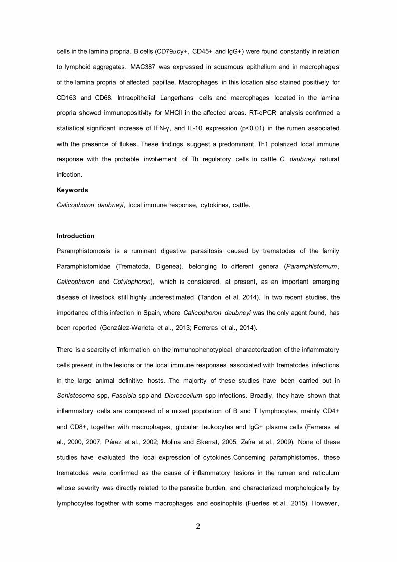
Within the group of parasitized animals, the gene expression levels of IFN-γ, IL-4 and IL-10
showed a significant increase (p<0.01) in ruminal atrium samples when they are compared with
those in the dorsal sac, an area free of parasites. In the control, unparasitized cattle, the gene
expression of IL-4 and IL-10 was also significantly higher (p<0.05) in the ruminal atrium than in
the dorsal sac. However, no differences were observed for IFN- γ levels in this group.
When comparing parasitized and control cattle, significant increases in the IL-10 and IFN-γ gene
expression levels (p<0,01 and p<0.05 respectively) were found in the ruminal atrium of the
parasitized animals. In the dorsal sac, all three cytokine levels were raised in the infected group
but no statistical differences could be established with the unparasitized animals (Figure 2).
Discussion
In this study we have used the same tissue samples to immunohistochemically characterize the
cellular inflammatory infiltrates and the cytokine mRNA expression levels in selected ruminal
7
tissues, where lesions associated with natural C. daubneyi infection in cattle were previous ly
evaluated (Fuertes et al., 2015). Phenotypic analysis of the inflammatory cellular infiltrates in the
reticular and ruminal mucosa in C. daubneyi naturally infected cattle has shown that they are
formed by both T and B lymphocytes. The T cell response was composed predominantly of CD4+
T helper cells, distributed as lymphoid aggregates and follicles in the lamina propria and
epithelium (Intraepithelial lymphocytes or IELs) of damaged conical ruminal and reticular papillae.
An intense infiltration of T and B lymphocytes in hepatic lesions has also been reported also in
other trematodoses like experimental fasciolosis and dicrocoeliosis in ruminants (Pérez et al,
2002; Ferreras et al., 2007; Zafra et al, 2009). These data demonstate that C. daubneyi is also
able to trigger a local immune response against flukes and/or their secretion products. The scant
amount of CD8+ cells in the present study can be linked to the cytotoxic activity of these cells
(Tizard, 2009), that would not be needed in extracellular parasitosis such as C. daubneyi infection.
A remarkable finding was the presence of CD3+ intraepithelial globule leukocytes (GLs) in high
amount in some parasitized cattle. This result supports a lymphocytic origin for GLs in cattle in
agreement with previous studies in cats (Konno et al, 1994; Roccabianca et al, 2006). The
presence of CD79 αcy+ B lymphocytes and IgG plasma cells intermingled with T cells in the
ruminal and reticular lamina propria may be indicative of a local humoral immune response
against C. daubneyi that could be linked with the serum antibody response in natural
paramphistome infections (Díaz et al, 2006).
The presence of strong immunoreactivity for the cytosolic protein complex L1 or calprotectin
(MAC387+) in the epithelium and in the macrophages within the lamina propria of altered papillae
was another noteworthy finding. It is known that the L1 antigen is a calcium binding protein
expressed by squamous epithelium of mucous membranes and injured epidermis, but not by
normal epidermis and other cells harboured in the skin (Brandtzaeg et al, 1988; Paquet and
Piérard, 1999). Besides expression of the MAC387 antigen in the inflamed epidermis is directly
associated with cell-mediated activity in the papillary dermis (Kirkham et al, 1990). In the present
study modifications of MAC387 labelling in the rumen and reticulum mucosa were observed at
the point of fluke attachment in parasitized cattle in comparison to uninfected animals. Rumen
and reticulum mucosa are then considered as reactive pointing out squamous cell damage due
to this parasitosis.
8
The MAC387+ monocytes/macrophages constitutes as a recently infiltrated cell subpopulation in
local inflammatory response so they are considered active inflammation markers (Soulas et al.,
2011). The presence of these cells in the inflammatory infiltrate of ruminal and reticular lamina
propria in paramphistomosis could be associated to a continuous antigenic stimulus by fluke
excretion products or tegumental molecules.
In this study intraepithelial dendritic cells, morphologically compatible with Langerhans cells, as
well as macrophages within the lamina propria in lesion sites showed immunolabelling for MHCII
antigens. The role of Langerhans cells in the presentation of antigens to T cells through dendritic
cell MHCII antigen complex during inflammation and the induction of a Th response is well known
(Tizard, 2009) and these results emphasize that C. daubneyi infection is able to mount a local
immune response triggered by MHCII expression in ruminal antigen presenting cells. .
In other trematodoses, an initial proinflammatory reaction mediated by Th1 cytokines, related to
the presence of immature flukes, is followed by a Th2 response (Flynn et al., 2010; Chuah et al.,
2014). In the present study the local expression of IFN-γ (main Th1 proinflammatory cytokine
associated with classical macrophage activation) was detected in infected cattle using
immunohistochemistry and qPCR techniques. This result would indicate that C. daubneyi
infection is able to trigger a cell mediated immune response at the local level, as it has been
pointed out in other trematodosis such as fasciolosis in the hepatic lymph nodes (Zafra et al,
2009).
Besides the immunolabelling associated to T cells, in the present study the presence of IFN-γ
was observed focally in the epithelial lining cells of those papillae encircled by the parasite ventral
sucker. Although IFN-γ is produced mainly by lymphocytes, recently the ability of respiratory
epithelial cells to produce IFN-γ after being infected by human parainfluenza virus type 3 has
been described (Lewandowska-Polak et al., 2015). This finding would indicate that epithelial cells
in areas in contact with the parasite could contribute to the cellular response to trematode
antigens or the mechanical damage due to fluke attachment.
Concerning mRNA cytokine level results, our findings showed that IFN-γ and IL-10 mRNA
expression was significantly increased (upregulated) in ruminal atrium mucosa of parasitized
animals in comparison to the control group and with the ruminal dorsal sac levels in parasitized
9
cattle, leading to the conclusion that their expression is directly related to the presence of
parasites. In contrast, no significant differences were found concerning the IL-4 mRNA levels in
the same locations, although comparatively high amount of this type 2 cytokine were produced in
the ruminal atrium of infected cattle. The significant local production of IFN-γ by lymphocytes
related to the presence of parasites, may indicate the establishment of a cell-mediated immunity
which, however, was ineffective in the elimination of the parasite, as already stated for
extracellular parasites (Jankovic et al., 2001).
The simultaneous and constant high gene expression levels of for IL-10 found in this study would
indicate that in C. daubneyi naturally infected cattle this cytokine probably downregulates or
represses the expression of proinflammatory cytokines such as IFN-γ and protects against the
tissular epithelial damage caused by inflammation as it has been suggested (Ouyang et al, 2011;
Chuah et al., 2014). The finding that IL-10 regulates both Th1 and Th2 responses at mucosal
surfaces has previously been reported in infected mice with Trichinella spiralis (Helmby and
Grencis, 2003).
Finally, in unparasitized animals both immunomodulating IL4 and IL-10 cytokine gene expression
was upregulated in the ruminal atrium when compared with the dorsal sac. This could be due to
a necessary immunomodulation on ruminal mucosa consequence of constant antigenic
stimulation by exogenous and endogenous microbiota of ruminal content (Koboziev et al., 2014).
This condition is not present in dorsal sac where ruminal content in direct contact with mucosa is
composed by gases and flora is scant or absent.
Our results suggest that a regulatory parasite antigen-specific mechanism exists in natural C.
daubneyi infection in cattle at the local site of attachment of this trematode towards the production
of a polarized Th1 immune response, with the involvement of Th regulatory cells, which may be
associated with a mixed cell population activity in this infection. The differences in the cytokine
responses may be due to the antigen molecules actively secreted by the surface tegument of the
adult rumen fluke in direct contact with the host tissues. Further studies are required to understand
host defence against these parasites.

10
Competing interests
The authors declare that they have no competing interests.
Acknowledgements
This work was supported by grant LE023A10-2 from Junta de Castilla y León. The authors wish
to thank Dr. Natalia Elguezabal for her critical review of the final manuscript.
11
References
Brandtzaeg, P., Jones, D.B., Flavell, D.J., Fagerhol, M.K., 1988. Mac 387 antibody and
detection of formalin resistant myelomonocytic L1 antigen. J. Clin. Pathol. 41, 963-970.
Chuah, C., Jones, M.K., Burke, M.L., McManus, D.P., Gobert, G.N., 2014. Cellular and
chemokine-mediated regulation in schistosome-induced hepatic pathology. Trends Parasitol.
30, 141-150.
Clery, D., Torgerson, P., Mulcahy, G., 1996. Immune reponses of chronically infected adult
cattle to Fasciola hepatica. Vet. Parasitol. 62, 71-82.
Díaz, P., Lomba, C., Pedreira, J., Arias, M., Sánchez.Andrade, R., Suárez, J.L., Díez-Baños P.,
Morrondo, P., Paz-Silva, A., 2006. Analysis of the IgG antibody response against
Paramphistomidae trematoda in naturally infected cattle. Application to serological surveys. Vet.
Parasitol. 140, 281-288.
Ferreras, M.C., García-Iglesias, M.J., Manga-González, M.Y., Pérez-Martínez, C., Mizinska, Y.,
Ramajo, V., González-Lanza, M.C., Escudero, A., García-Marín, J.F., 2000. Histopathological
and immunohistochemical study of lambs experimentally infected with Fasciola hepatica and
Schistosoma bovis. J. Vet. Med. B Infect. Dis. Vet. Public Health. 47, 763-73.
Ferreras, M.C., Campo, R., González-Lanza, C., Pérez, V., García-Marín, J.F., Manga-
González, M.Y., 2007. Immunohistochemical study of the local immune response in lambs
experimentally infected with Dicrocoelium dendriticum (Digenea). Parasitol. Res. 101, 547-555.
Ferreras, M.C, González-Lanza, C., Pérez, V., Fuertes, M., Benavides, J., Mezo, M., González-
Warleta M., Giráldez, J., Martínez-Ibeas, A.M., Delgado, L., Fernández, M., Manga-González,
M.Y., 2014. Calicophoron daubneyi (Paramphistomidae) in slaughtered cattle in Castilla y León
(Spain). Vet. Parasitol. 199, 268-271.
12
Flynn, R.J., Mulcahy, G., Elsheika, H.M., 2010. Coordinating innate and adaptive immunity in
Fasciola hepatica infection: implications for control. Vet. Parasitol. 169, 235-240.
Fuertes, M., Pérez, V., Benavides, J., González-Lanza, M.C., Mezo, M., González-Warleta, M.,
Giráldez, F.J., Fernández, M., Manga-González, M.Y., Ferreras, M.C., 2015. Pathological
changes in cattle naturally infected by Calicophoron daubneyi adult flukes. Vet. Parasitol. 209,
188-196.
González-Warleta, M., Lladosa, S., Castro-Hermida, J.A., Martínez-Ibeas, A.M., Conesa, D.,
Muñoz, F., López-Quílez, A., Manga-González, Y., Mezo, M., 2013. Bovine paramphistomosis in
Galicia (Spain): Prevalence, intensity, aetiology and geospatial distribution of the infection. Vet.
Parasitol. 191, 252-263.
Helmby, H., Grencis, K., 2003. Contrasting roles for IL-10 in protective immunity to different life
cycle stages of intestinal nematode parasites. Eur. J. Immunol. 33, 2382-2390.
Jankovic, D., Liu, Z., Gause, W.C., 2001. Th1- and Th2-cell commitment during infectious
disease: asymmetry in divergent pathways. Trends Immunol. 22, 450-457.
Kirkham, N., Peacock, S.J., Jones, D.B., 1990. Monoclonal antibody MAC 387 recognizes a
myelomonocytic antigen shared by epithelial cells in inflammatory skin diseases. Br. J.
Dermatol. 122, 61-69.
Koboziev, I., Reinoso-Webb, C., Furr, K.L., Grisham, M.B., 2014. Role of the enteric microbiota
in intestinal homeostasis and inflammation. Free Radic. Biol. Med. 0, 122-133.
Konno, A., Hashimoto, Y., Kon, Y., Sugimura, M., 1994. Perforin-like immunoreactivity in feline
globule leukocytes and their distribution. J. Vet. Med. Sci. 56, 1101-1105.
13
Lewandowska-Polak A., Brauncajs, M., Paradowska, E., Jarzebska, M., Kurowski, M., Moskwa,
S., Lesnikowski, Z.J., Kowalski, M.L., 2015. Human parainfluenza virus type III (HPIV3) induces
production of IFNγ and RANTES in human nasal epithelial cells (HNECs). J. Inflamm. 12, 16.
Molina, E.C., Skerrat, L.F., 2005. Cellular and humoral responses in liver of cattle and buffaloes
infected with a single dose of Fasciola gigantica. Vet. Parasitol. 131, 157-163.
Ouyang, W., Rutz, S., Crellin, N.K., Valdez, P.A., Hymowitz, S.G., 2011. Regulation and
functions of the IL-10 family of cytokines in inflammation and disease. Annu. Rev. Immunol. 29,
71-109.
Paquet, P., Piérard, G.E., 1999. Epidermal calprotectin in drug-induced toxic epidermal
necrolysis. J. Cutan. Pathol. 26, 301-305
Pérez, J., Ortega, J., Moreno, T., Morrondo, P., López-Sánchez, C., Martínez-Moreno, A., 2002.
Pathological and immunohistochemical study of the liver and hepatic lymph nodes of sheep
chronically reinfected with Fasciola hepatica, with or without triclabendazole treatment. J.
Comp. Pathol. 127, 30-36.
Regidor-Cerrillo, J., Arranz-Solís, D., Benavides, J., Gómez-Bautista, M., Castro-Hermida, J.A.,
Mezo, M., Pérez, V., Ortega-Mora, L.M., González-Warleta, M., 2014. Neospora caninum
infection during early pregnancy in cattle: how the isolate influences infection dynamics, clinical
outcome and peripheral and local immune responses. Vet. Res. 45, 10.
Roccabianca, P., Vernau, W., Caniatti, M., Moore, P.F., 2006. Feline large granular lymphocyte
(LGL) lymphoma with secondary leukaemia: primary intestinal origin with predominance of a
CD3/CD8αα phenotype. Vet. Pathol. 43, 15-28
SAS Inst. Inc., 2008. SAS/STAT® 9.2. User`s guide. SAS Inst. Inc., Cary, NC, USA.
14
Schmittgen, T.D., Livak, K.J., 2008. Analyzing real-time PCR data by the comparative C(T)
method. Nat. Protoc. 3, 1101-1108.
Soulas, C., Conerly, C., Kim, W-K., Burdo, T.H., Alvarez, X., Lackner, A.A., Williams, K.C.,
2011. Recently infiltrating MAC+ Monocytes/Macrophages. A third macrophage population
involved in SIV and HIV encephalitic lesion formation. Immun. Infect. Dis. 178, 2121-2135.
Tandon, V., Roy, B., Shylla, J.A., Ghatani, S., 2014. Amphistomes. In : Toledo R, Fried B (Eds.),
Digenetic Trematodes. Advances in Experimental Medicine and Biology. Vol. 766. Springer,
New York, pp. 365-392.
Tizard, I., 2009. Veterinary Immunology. Elsevier, Amstedam, 592 pp.
Untergasser, A., Nijveen, H., Rao, X., Bisseling, T., Geurts, R., Leunissen, J.A., 2007.
Primer3Plus, an enhanced web interface to Primer3. Nucleic Acids Res. 35, 71-74.
Zafra, R., Buffoni, L., Pérez-Écija, R.A., Mendes, R.E., Martínez-Moreno, A., Martínez-Moreno,
F.J., Pérez, J., 2009. Study of the local immune response to Fasciola hepatica in the liver and
hepatic lymph nodes of goats immunised with a peptide of the Sm14 antigen. Res. Vet. Sci. 87,
226-232.
15
Figures.
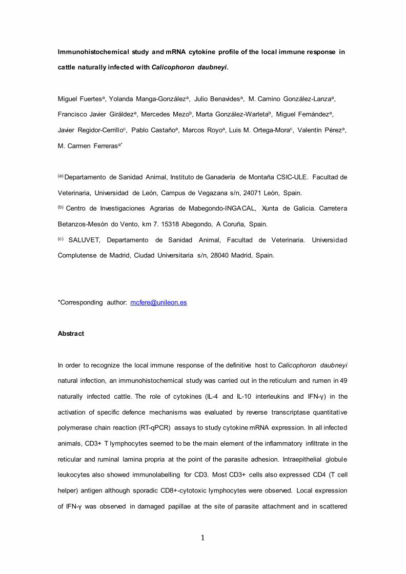
Figure 1. a) Rumino-reticular orifice. Conical papilla encircled by fluke ventral sucker showing
inflammatory infiltrate composed by CD3+ T cells. Parasite burden= 93 (12 worms recovered
from the rumino-reticular orifice). Envision® System. Haematoxylin counterstain. Bar= 200µm.
b) Reticulum. Cytoplasmatic granules of Globular Leukocytes (GLs) intensely immunostained by
pAb CD3 (arrows). Parasite burden= 908 (400 worms recovered from the reticulum). Envision®
System. Haematoxylin counterstain. Bar= 30µm. c) Ruminal atrium. Inflammatory infiltrate in the
lamina propria of a mushroom-shaped affected papilla composed mainly by CD4+ T helper
cells. Parasite burden= 253 (82 worms recovered from the ruminal atrium). Envision® System.
Haematoxylin counterstain. Bar= 200µm. d) Rumino-reticular fold. Papillar epithelium
expressing mAb MAC387 especially in areas in close contact with the tegument of fluke ventral
sucker. Sparse immunolabelled macrophages in lamina propria. Parasite burden= 8005 (571
worms recovered from the rumino-reticular fold). Envision® System. Haematoxylin counterstain.
Bar= 200µm. e) Rumino-retilcular fold. IFN-γ Immunolabeling of intraepithelial lining cells in the
papillar areas in close contact with the parasite. Parasite burden= 215 (324 worms recovered
from the rumino-reticular fold). Envision® System. Haematoxylin counterstain. Bar=100µm. f)
Reticulum. MHC II expression in dendritic cells in the organized inflammatory infiltrate of the
lamina propria. Parasite burden= 908 (400 worms recovered from the reticulum). Envision®
System. Haematoxylin counterstain. Bar= 50µm. g) Reticulum. Detail of f). Expression of MHC II
in Langerhans cells within the lining epithelium. Envision® System. Haematoxylin counterstain.
Bar= 10µm.
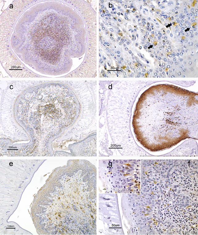
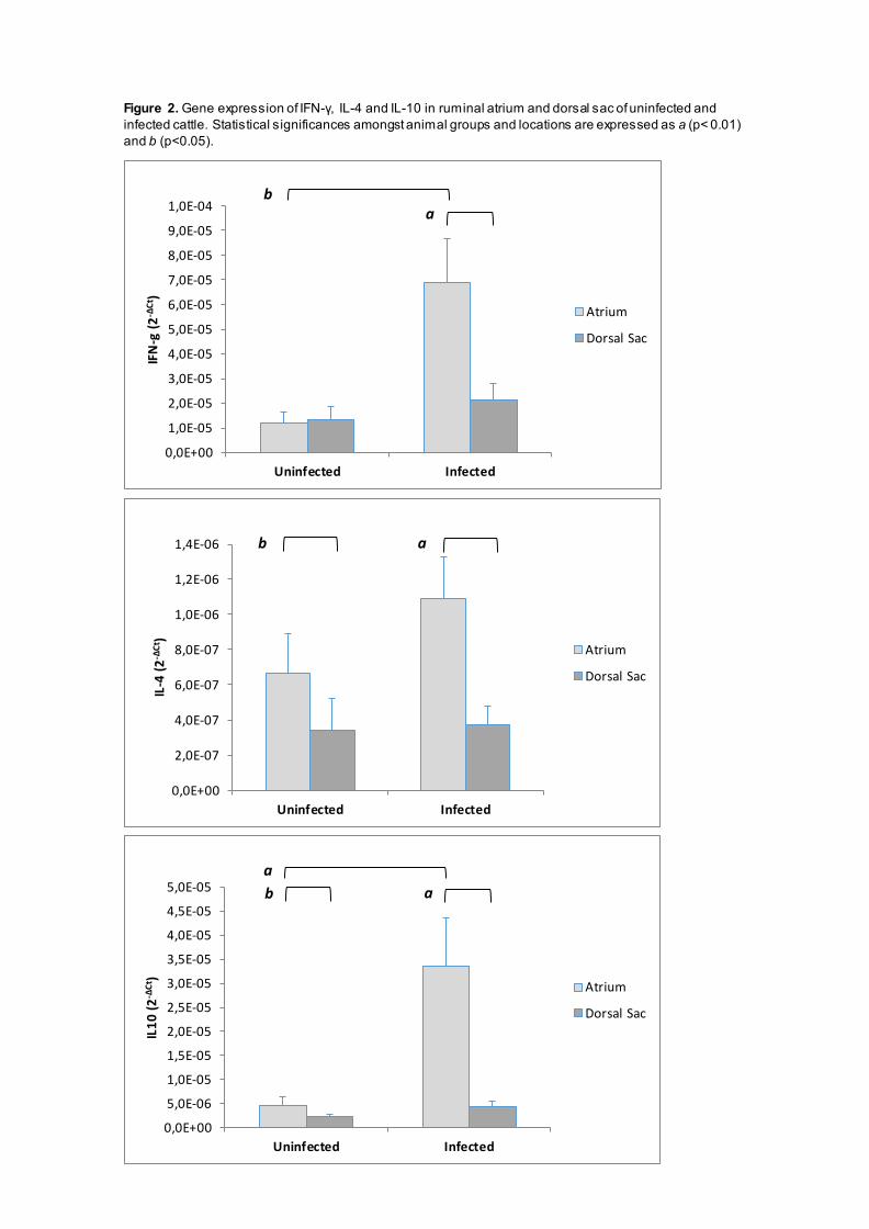
Figure 2. Gene expression of IFN-γ, IL-4 and IL-10 in ruminal atrium and dorsal sac of uninfected and infected cattle. Statistical significances amongst animal groups and locations are expressed as a (p< 0.01) and b (p<0.05).
0,0E+00
1,0E-05
2,0E-05
3,0E-05
4,0E-05
5,0E-05
6,0E-05
7,0E-05
8,0E-05
9,0E-05
1,0E-04
Uninfected Infected
IFN
-g (2
-ΔCt
)
Atrium
Dorsal Sac
ab
0,0E+00
2,0E-07
4,0E-07
6,0E-07
8,0E-07
1,0E-06
1,2E-06
1,4E-06
Uninfected Infected
IL-4
(2-Δ
Ct)
Atrium
Dorsal Sac
ab
0,0E+00
5,0E-06
1,0E-05
1,5E-05
2,0E-05
2,5E-05
3,0E-05
3,5E-05
4,0E-05
4,5E-05
5,0E-05
Uninfected Infected
IL10
(2-Δ
Ct)
Atrium
Dorsal Sac
aba
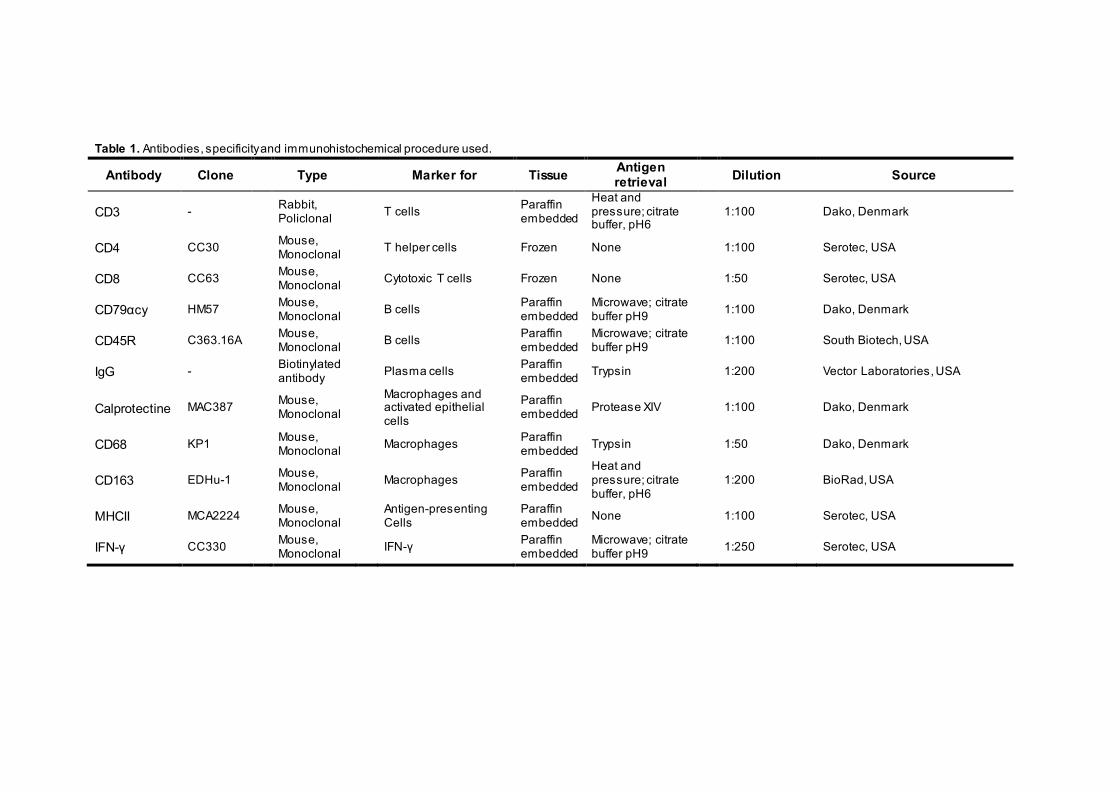
Table 1. Antibodies, specificity and immunohistochemical procedure used.
Antibody Clone Type Marker for Tissue Antigen retrieval
Dilution Source
CD3 - Rabbit,
Policlonal
T cells Paraffin
embedded
Heat and pressure; citrate buffer, pH6
1:100
Dako, Denmark
CD4 CC30 Mouse,
Monoclonal
T helper cells Frozen None
1:100
Serotec, USA
CD8 CC63 Mouse,
Monoclonal
Cytotoxic T cells Frozen None
1:50
Serotec, USA
CD79αcy HM57 Mouse,
Monoclonal
B cells Paraffin embedded
Microwave; citrate buffer pH9
1:100
Dako, Denmark
CD45R C363.16A Mouse,
Monoclonal
B cells Paraffin embedded
Microwave; citrate buffer pH9
1:100
South Biotech, USA
IgG - Biotinylated antibody
Plasma cells Paraffin embedded Trypsin 1:200 Vector Laboratories, USA
Calprotectine MAC387 Mouse,
Monoclonal Macrophages and
activated epithelial cells
Paraffin embedded Protease XIV
1:100
Dako, Denmark
CD68 KP1 Mouse,
Monoclonal
Macrophages Paraffin embedded Trypsin
1:50
Dako, Denmark
CD163 EDHu-1 Mouse,
Monoclonal
Macrophages Paraffin
embedded
Heat and pressure; citrate buffer, pH6
1:200
BioRad, USA
MHCII MCA2224 Mouse,
Monoclonal Antigen-presenting
Cells Paraffin embedded None
1:100
Serotec, USA
IFN-γ CC330 Mouse, Monoclonal
IFN-γ Paraffin embedded
Microwave; citrate buffer pH9
1:250 Serotec, USA