Embed Size (px)
Citation preview
Accepted in AUTISM Nov 1 2013
Improving emotional face perception in autism with diuretic
bumetanide: a proof-‐of-‐concept behavioral and functional brain
imaging pilot study
Authors: Nouchine Hadjikhani1,2,3*, Nicole R Zürcher1,2, Ophelie Rogier2, Torsten Ruest2, Loyse Hippolyte2, Yehezkel Ben-‐Ari4 and Eric Lemonnier5.
Affiliations: 1MGH/HST/HMS Athinoula A. Martinos Center for Biomedical Imaging, Boston, USA 2Brain Mind Institute, EPFL, Lausanne, Switzerland 3 Gillberg Neuropsychiatric Center, University of Gothenburg, Sweden 4 INMED, Marseilles, France 5 Laboratoire de Neurosciences, Université de Brest EA4645, France Short title: Bumetanide improves emotional perception in autism Corresponding author: Nouchine Hadjikhani A. A. Martinos Center for Biomedical Imaging 149 13th Street Charlestown, MA 02129 Tel: +1 617 724 5625 Fax: +1 530 309 4976 Email: [email protected]
Abstract
Clinical observations have shown that GABA-‐acting benzodiazepines exert
paradoxical excitatory effects in autism, suggesting elevated intracellular chloride (Cl-‐
)i and excitatory action of GABA. In a previous double blind randomized study, we
have shown that the diuretic NKCC1 chloride importer antagonist bumetanide, that
decreases (Cl-‐)i and reinforces GABAergic inhibition, reduces the severity of autism
symptoms. Here, we report results from an open-‐label trial pilot study in which we
used functional magnetic resonance imaging and neuropsychological testing to
determine the effects of 10 months bumetanide treatment in adolescents and young
adults with autism. We show that bumetanide treatment improves emotion
recognition and enhances the activation of brain regions involved in social and
emotional perception during the perception of emotional faces. The improvement of
emotion processing by bumetanide reinforces the usefulness of bumetanide as a
promising treatment to improve social interactions in autism.
Keywords
Autism spectrum disorders, GABA, bumetanide, fMRI, emotion, face perception,
treatment.
Introduction
Autism spectrum disorder (ASD) is a neurodevelopmental, life-‐long condition
characterized by deficits in social interactions and communication, and by the
presence of repetitive behaviors, that affects approximately 1% of the population
((CDC) and Baio, 2012: l; American_Psychiatric_Association, 2013). Genetic mutations
that impact synapse operation (Jamain et al., 2003; Bourgeron, 2009; Giannandrea et
al., 2010; Weiss, 2009; Tabuchi et al., 2007) as well as environmental factors during
pregnancy (Croen et al., 2011b; Croen et al., 2011a; Patterson, 2009; Kemper and
Bauman, 1998; Dossche, 2005) contribute to the emergence of ASD. Research on the
genetic basis of ASD has identified hundreds of possible genetic mutations, but how
brain malformations are induced and how they lead to neurological sequelae is still
not understood. The development of autism seems to already start in utero
(Courchesne et al., 2011; Bauman and Kemper, 1985; Ploeger et al., 2010).
GABAergic signaling is affected in ASD, resulting in an imbalance between
excitation and inhibition (Chao et al., 2010; Pizzarelli and Cherubini, 2011; Gogolla et
al., 2009; Dossche, 2005). ASD patients have reduced gamma oscillations (Grice et al.,
2001; Brown et al., 2005; Wilson et al., 2007), which are generated by GABAergic
neurons (Lewis et al., 2005; Lisman and Buzsaki, 2008; Pizzarelli and Cherubini,
2011) and are instrumental in sensory binding and higher cognitive functions
(Singer, 1993; Lisman and Idiart, 1995; Murthy and Fetz, 1992). Interestingly, the
GABA acting benzodiazepines that enhance GABAergic inhibition exert paradoxical
actions on autistic children augmenting agitation and other symptoms (Marrosu et al.,
1987). This paradoxical reaction has been shown to result from elevated intracellular
chloride ([Cl-‐]i) that shifts the polarity of GABA from excitation to inhibition (Nardou
et al., 2011b). Indeed, in epilepsies, but also spinal cord insults, the levels of [Cl-‐]i are
elevated leading to excitatory GABA actions that are further enhanced by GABA-‐
acting benzodiazepines or phenobarbital (Nardou et al., 2011b; Nardou et al., 2011a).
This observation has raised considerable interest for the use of diuretics in order to
reestablish the hyperpolarization of GABAergic signals and hence to reinforce its
inhibitory potency, and has led to therapeutic assays in epilepsy treatment.
The increase of [Cl-‐]i in pathology has a dual origin: an internalisation of the
chloride exporter KCC2 – leading to a failure of neurons to export excessive chloride -‐
and a persistent or enhanced activity of the chloride importer NKCC1 leading to
exacerbated accumulation of chloride (Dzhala et al., 2005; Nardou et al., 2011b).
KCC2 is at present an unlikely target for drug treatments because it is labile, readily
internalized, highly activity-‐dependent, and because there are currently no selective
agonists available. In contrast, NKCC1 is stable and antagonists have been identified,
notably the diuretic and highly specific NKCC1 antagonist bumetanide. Bumetanide
has been extensively utilized since 1975 in adults and since 1986 in children to treat
acute and long-‐term conditions including hypertension, broncho-‐pulmonary
dysplasia, nephritic syndrome or congestive heart failure (Sullivan et al., 1996).
Bumetanide has a short half-‐life (between 1h30 and 3h) and (poorly) crosses the
blood-‐brain barrier (Li et al., 2011) thanks to an active transporter (SLC16A50)
(Murakami et al., 2005). The use of bumetanide is safe provided that it is
accompanied with regular controls of kaliemia and kidney functions in patients to
determine possible adverse effects.
Relying on the observation of paradoxical actions of benzodiazepines on ASD
patients (Marrosu et al., 1987), the effects of chronic bumetanide treatment were
recently tested in a double blind randomized study (Lemonnier et al., 2012). In this
trial, conventional measures of behavioral and clinical evaluation of autism in
children were used, including Childhood Autism Rating Scale (CARS), Clinical Global
Impression (CGI) and Autism Diagnostic Observation Schedule (ADOS), and a
significant amelioration of clinical symptoms was found. However, it remained
important to determine the effects of this treatment on some core symptoms of
autism, notably on the processing of facial expressions and recognition of emotions.
The goal of the present study was therefore to examine potential brain mechanisms
underlying the action of bumetanide.
Here, using quantitative behavioral testing (experiment 1) and functional MRI
(experiment 2) in an open-‐label trial design, we tested the effect of bumetanide
treatment on performance in emotion recognition. Using fMRI, we assessed changes
in brain activation in response to the perception of dynamic movies of facial
expressions in two separate sessions before and after treatment. We tested the
hypothesis that bumetanide treatment would improve performance for emotion
recognition, and lead to increased activation of brain areas involved in emotion
processing.
Materials and Methods
Ethics Statement: The study was approved by the Committee of Persons
Protections CPP west 6-‐570-‐6/4/2009, and by the French Health Products Safety
Agency (AFSAPS -‐A90936-‐66 4/12/2009, NCT01078714). The behavioral and fMRI
protocols were approved by Hospital Ethical Committee. All adult participants gave
written consent before the start of the study. Minor participants gave their assent and
one of their parents gave written consent. All procedures followed the Declaration of
Helsinki.
Seven high-‐functioning males with ASD took part in the study. All participants
underwent repeated scanning after 10 months of bumetanide treatment (1mg/day).
They were 19.3±4.6 (mean±SD) years old at the first testing session (range: 14.8-‐
28.5).
Participants were diagnosed by an experienced clinician according to DSM IV-‐
TR (APA, 2000) criteria and the Autism Diagnosis Observation Schedule (ADOS)(Lord
et al., 2000) and the Autism Diagnostic Interview-‐Revised (ADI-‐R)(Lord et al., 1994).
All participants met criteria for ASD and were diagnosed with Autism (n=2) and
Asperger Syndrome (N=5) based on their language development history. They were
also asked to complete the Autism Quotient (AQ) (Baron-‐Cohen et al., 2006) and
Empathy Quotient (EQ) (Baron-‐Cohen and Wheelwright, 2004) self report
questionnaires. The performance IQ (PIQ) was assessed using Wechsler non-‐verbal
scales (WASI, 1999; Wechsler and Naglieri, 2006). In addition, the Toronto
Alexithymia Scale (TAS-‐20) was assessed before and after treatment in seven of the
nine participants (Bagby et al., 1994).
Participants’ characteristics are given in Table 1.
During the treatment period, patients underwent clinical controls as well as
monitoring of electrolytes (including potassium and sodium), kidney and liver
functions and blood sugar at days 7, 14, 30, 60 and then every six months. We
frequently observed increased urinary output, but this was never accompanied by
signs of dehydration (no weight reduction, no increase in Na+ levels). No side effects
such as orthostatic hypotension, cramps, weakness, diarrhea, myalgia, arthralgia,
dizziness or nausea were observed. In one of the patients, we observed hypokaliemia
after one month of treatment that was readily corrected by oral potassium
supplement.
Experiment 1: Behavioral testing for emotional labeling
Stimuli and task: In this task, participants had to recognize a low-‐intensity
and therefore ambiguous facial expression. Four expressions were used: happy,
fearful, angry and neutral. Dynamic morphs were created from the NimStim
Emotional Face Stimuli database (http://www.macbrain.org/faces/index.htm#faces)
between NEUTRAL and each EMOTIONAL expression using Morph Age Pro
(http://www.creaceed.com/morphage/), and still images were created at 40%
intensity level between neutral and the full emotional expressions. Participants were
then presented with one still image depicting happy, fearful or angry at 40% intensity
or NEUTRAL on the left side of the computer screen, while on the right still Ekman
stimuli (Ekman and Friesen, 1976) representing each of the four full emotional
expressions were shown. The location of a particular facial expression presented on
the right was counterbalanced across trials to avoid habituation and control for
location. For each type of emotion, four trials were delivered, totaling to 16 stimuli
shown in pseudorandom order. The test was non-‐verbal: participants had to indicate
using a button box which of the four facial expressions presented on the right
matched the best with the expression seen on the left. The images remained on
screen until a response was given. Performance measures consisting of reaction time
(RT) and accuracy were recorded. To control that the concept of classification was
understood and that effects were specific to faces, we designed stimuli showing 4
OBJECT categories: instruments, fruits, clothes, and animals. In total, 16 different
exemplars were shown. As for the experiment above, participants had to indicate on a
button box which image on the right was the best match for the picture on the left. No
feedback on accuracy was given in any of these non-‐verbal tests, so that participants
could not learn the task during the session. A pairwise Wilcoxon rank test was used to
compare emotion/object category recognition performance before and after
treatment.
Experiment 2: Functional brain imaging
fMRI data acquisition
Anatomical and functional MR images of brain activity were collected in a 3T
high-‐speed echoplanar-‐imaging device (Tim Trio, Siemens, Erlangen) using a 12-‐
channel matrix coil. Participants lay on a padded scanner couch and wore foam
earplugs. Foam padding stabilized the head. High-‐resolution (1.0 x 1.0 x 1.0mm)
structural images were obtained with a multi-‐echo magnetization-‐prepared rapid
acquisition gradient echo (ME-‐MPRAGE) sequence (176 slices, 256x256 matrix, echo
time (TE1)=1.64 ms, (TE2)=3.5 ms, (TE3)=5.36 (TE4)=7.22 ms; repetition time
(TR)=2530 ms; flip=7°). MR images of brain activity were then collected. Functional
sessions began with an initial sagittal localizer scan. Slices were automatically
positioned using AutoAlign Head LS (Landmark Survey) from Siemens. The co-‐
registered functional acquisition (TR=3,000 ms, 46 AC-‐PC 3mm thick slices,
TE=30ms, flip angle 90°, matrix=64x64) lasted 417 seconds. Other anatomical and
functional sequences were also acquired during this session but are not described in
the present report.
Stimuli and task for the fMRI experiment
During the functional scan, dynamic face stimuli were presented. We used a
series of 24 short movies created from the NimStim database, representing morphs of
facial expressions from neutral to fearful, happy or angry. In order to control for
emotional expression and movements, morphs were also created for the NEUTRAL
condition, using NEUTRAL faces and their mirror images. Each movie lasted for 5
seconds, with a dynamic morph starting from NEUTRAL and going to full emotional
expression, lasting 3 seconds, followed by 2 seconds of the final full emotional
expression. Stimuli were presented in a block design. There were 8 blocks in total, 2
for each facial expression. Eight stimuli were presented per block. So a total of 16
morphs were presented for each facial expression. A red fixation cross was presented
for 1 second between movies and for an additional 3 seconds between blocks. Four
times in the run a blue fixation cross was presented. To ensure that participants were
paying attention to the stimuli, a button box was used to record participants’
responses to the presence of the blue cross between stimuli presentation.
Participants were instructed to look attentively at the faces, and to press a button
every time they saw a blue cross. Functional data from one participant for this
paradigm could not be acquired during the first session due to technical problems
with the scanner, and therefore his data were only included for the behavioral
experiment.
fMRI data Analysis
FSL (FMRIB Software Library) package and techniques were used in data
preparation and processing. Brain extraction of high-‐resolution anatomical images
was carried out using Christian Gaser's VBM8 toolbox for SPM8 (Ashburner et al.,
2000) and fed into FEAT. fMRI data processing was performed using FEAT (FMRI
Expert Analysis Tool) version 5.98 (Smith et al., 2004; Woolrich et al., 2009; Worsley,
2001). Each functional run was first motion-‐corrected with MCFLIRT (Cox, 1996) and
spatially smoothed with full width at half maximum of 8 mm. First level analyses
were carried out for each subject to compute the contrast of interest, i.e. [EMOTION >
NEUTRAL]. Subsequently, treatment effects [after > before] were assessed by
submitting these contrasts to a paired higher-‐level mixed effects GLM analysis using
FLAME 1+2 (FMRIB’s Local Analysis of Mixed Effects).
Mixed effect variance is the sum of fixed-‐effects variance (the within-‐session
across-‐time variances estimated in the first-‐level analyses) and "random-‐effects" (RE)
variance (the "true" cross-‐session variances of first-‐level parameter estimates). Mixed
effect analysis was chosen because it models the session and subject variability and
therefore allows inferences to be made to a wider population from which the subjects
were drawn (http://fsl.fmrib.ox.ac.uk/fsl/fsl4.0/feat5/detail.html).
Clusters were formed using FSL's cluster tool, and data are reported with a
threshold of z > 2.3 and a minimum cluster size of 300 voxels.
Results
Effects of bumetanide treatment on behavior: Emotional face recognition
As shown in Fig.1, bumetanide treatment significantly improved overall
accuracy in emotion matching of faces with 40% intensity to their 100% intensity
counterpart (overall mean accuracy [% correct] ± SD before: 62.5 ± 21.0; after: 75.0 ±
13.5; p=0.04). Bumetanide treatment also significantly improved overall reaction
time for face emotion matching (overall mean reaction time [seconds] ±SD before:
8.18 ± 3.42; after: 5.82 ± 1.54; p=0.04). This effect was only seen for emotional
matching of faces. (Fig. 1). Object matching was not significantly different between
sessions (overall mean accuracy [% correct] ± SD before: 100 ± 0; after: 98.2 ± 3.0;
p=0.16, overall mean reaction time [seconds] ±SD before: 2.25 ± 0.61; after: 2.02 ±
0.62; p=0.24).
Effects of bumetanide treatment on behavior: Alexithymia
Alexithymia was assessed with the TAS-‐20. The TAS-‐20 uses a cut-‐off scoring:
Scores equal or less than 51 are considered as non-‐alexithymia, and scores equal or
greater than 61 are considered as alexithymia, with a grey zone between 52 to 60.
Mean score before treatment was 60.8±10.9, and significantly improved to a score of
55.4±12.0 after treatment (p=0.03). These results indicate that bumetanide treatment
may improve the ability to identify and describe one’s own emotions.
Effects of bumetanide treatment on brain activation
The results presented here show the comparison between EMOTIONAL and
NEUTRAL faces, and the modulatory effect of emotion on brain activation PRE and
POST treatment with bumetanide.
After 10 months of treatment, significantly increased activation was observed
for EMOTIONAL compared to NEUTRAL faces (see Table 2, Figure 2). Bumetanide
treatment increased brain activation for EMOTIONAL faces in early visual areas, as
well as in face processing areas including the inferior occipital cortex and the
fusiform cortex. In addition, increased activation was seen in cortical and subcortical
areas involved in emotional processing, including the nucleus accumbens and the
amygdala, as well as the orbitofrontal cortex and the temporal pole; in areas involved
in social processing, including the superior temporal cortex; in areas involved in
attentional processing (intraparietal sulcus, superior parietal lobule); in the lobule
and vermis VI of the cerebellum, as well as in crus I, involved in emotional and
cognitive processing.
Discussion
Our study is an open-‐label trial with only 7 treated patients, necessarily
limiting the scope of the conclusions. However, this is the first such study comparing
behavioral performance for emotion recognition and brain activation in response to
dynamic emotional faces before and after bumetanide treatment.
The imaging data showed a striking increase of brain activation in the face and
social/emotional processing network between sessions. EMOTIONAL faces elicited
significantly more activation in face encoding areas, including the inferior occipital
cortex, and the fusiform cortex, key face processing regions. Increased activation after
treatment was also observed bilaterally in the superior temporal cortex, involved in
the processing of dynamic, expressive aspects of faces (Allison et al., 2000). The right
superior temporal cortex region plays a key role in facial emotion recognition and is
involved in social perception. Previous studies have demonstrated that selective
attention to facial emotion specifically enhances activity of the right superior
temporal cortex compared with attention to the face per se (Narumoto et al., 2001);
increased activation was also present in the parietal cortex, involved in attentional
aspects of emotion processing (Narumoto et al., 2001).
Increased activation was also observed in areas involved in reward,
motivation and emotion, including the nucleus accumbens, the amygdala, and the
orbitofrontal cortex, possibly indicating increased interest for emotional faces after
treatment, and increased emotional processing. Finally, increased activation was
observed in cerebellum lobule VI and crus I, both involved in emotional processing
(Schmahmann and Sherman, 1998; Buckner et al., 2011). In a recent study, we
showed that neurotypical controls showed more activation than high-‐functioning
individuals with ASD during thatcherized face processing in the cerebellum (Zurcher
et al., 2013). The present data, showing increased cerebellar and cortical activation,
suggest normalization in brain activation during face perception after bumetanide
treatment.
Social interaction impairments are at the core of difficulties encountered by
individuals with ASD, and a previous study has shown that oxytocin, given
intranasally, can improve social behavior and increase gazing time in the eye-‐region
of faces in a group of 13 participants with autism (Andari et al., 2010). However this
effect is punctual and is limited to the window of action of oxytocin, which is about 1-‐
2 hours. Interestingly, recent studies performed by Ben-‐Ari and colleagues have
shown that delivery in rodents is associated with an abrupt and dramatic reduction of
[Cl-‐]i that exerts a neuro-‐protective role and analgesic action on the newborn’s brain
(Tyzio et al., 2006; Mazzuca et al., 2011). The analgesic actions are mimicked by
bumetanide that, like oxytocin, reduces activity in pain pathways by reducing
intracellular chloride (Mazzuca et al., 2011). Collectively, these observations suggest
that the behavioral improvement observed with bumetanide and oxytocin may share
common mechanisms.
Of further interest, NKCC1 is up-‐regulated in epilepsy and other disorders in
which [Cl-‐]i are elevated and GABA excitatory (Zhu et al., 2008; Cohen et al., 2002).
These alterations therefore appear to be common responses of neurons to insults and
suggest that elevated activity of the co-‐transporter occurs in adult neurons in a
variety of pathological conditions.
The present study contains several limitations, as it is an open label trial, with
a limited number of participants, scanned in two sessions that were separated by long
time interval (~ 10 months) and in which all the participants had normal intelligence
(so we do not know whether the same behavioral and brain activation would also be
observed in patients with intellectual deficiencies). In addition, the data of this open-‐
label trial could be interpreted as the result of placebo and repetition effects as the
patients were tested on the same tests twice, although the fact that emotional faces
but not neutral ones were more readily identified would tend to speak against this
interpretation. Future large, double blind randomized control trials will need to
address these issues.
In conclusion, bumetanide treatment appears to enhance pro-‐social behavior
by improving emotion processing. It bears stressing that to the best of our
knowledge, fMRI has not been used in earlier studies to compare the effects of drug
treatment in ASD, and that the only fMRI study published on the effect of behavioral
therapy only reported an N=2 (Voos et al., 2013). In conclusion, in spite of their
intrinsic limitations, our proof-‐of-‐concept results combined with the highly promising
results of the double blind randomized trial with bumetanide (Lemonnier et al., 2012)
converge to call for larger cohorts of participants, of different ages and with different
symptom severity to confirm the effect of bumetanide on social processing in autism.
Acknowledgments: We want to warmly thank all the participants and families who
took part to this study. Our thanks also go to Carole Burget, Karine Metrailler,
Anthony Lissot for their precious logistics help and Juliana Iranpour for the
behavioral study stimuli preparation.
Funding: This work was supported by Swiss National Science Foundation PP00P3-‐
130191 to NH; by the Centre d’Imagerie BioMédicale (CIBM) of the University of
Lausanne (UNIL), the Swiss Federal Institute of Technology Lausanne (EPFL), as well
as the Foundation Rossi Di Montalera. The funders had no role in study design, data
collection and analysis, decision to publish, or preparation of the manuscript.
Competing interests: All the authors have no conflict of interest, no financial and
personal relationships with other people or organizations including employment,
consultancies, stock ownership, honoraria, paid expert testimony, and travel grants
all during the conduction and termination of the work submitted. An application
patent on the use of diuretics has been deposited by the French medical research
council on the basis of the clinical results published by our team (Lemonnier et al.,
2012). Subsequent to completion of this study, three authors (NH EL and YBA) have
become shareholder of a company (Neurochlore, created early 2012) seeking to
develop and commercialize bumetanide as a treatment for autism.
References and Notes:
(CDC) CfDCaP and Baio J. (2012) Prevalence of Autism Spectrum Disorders — Autism and Developmental Disabilities Monitoring Network, 14 Sites, United States, 2008. Morbidity and Mortality Weekly Report (MMWR) 61: 1-19.
Allison T, Puce A and McCarthy G. (2000) Social perception from visual cues: role of the STS region. Trends Cogn Sci 4: 267-278.
American_Psychiatric_Association T. (2013) Diagnostic and statistical manual of mental disorders (5th ed), Arlington, VA: American Psychiatric Publishing.
Andari E, Duhamel JR, Zalla T, et al. (2010) Promoting social behavior with oxytocin in high-functioning autism spectrum disorders. Proceedings of the National Academy of Sciences of the United States of America 107: 4389-4394.
APA. (2000) Diagnostic and Statistical Manual of Mental Disorders, DSM-IV-TR.
Ashburner J, Andersson JL and Friston KJ. (2000) Image registration using a symmetric prior--in three dimensions. Human brain mapping 9: 212-225.
Bagby RM, Parker JDA and Taylor GJ. (1994) The twenty-item Toronto Alexithymia Scale-I. Item selection and cross-validation of the factor structure. Journal of Psychosomatic Research 38: 23-32.
Baron-Cohen S, Hoekstra RA, Knickmeyer R, et al. (2006) The Autism-Spectrum Quotient (AQ)--adolescent version. Journal of autism and developmental disorders 36: 343-350.
Baron-Cohen S and Wheelwright S. (2004) The empathy quotient: an investigation of adults with Asperger syndrome or high functioning autism, and normal sex differences. J Autism Dev Disord 34: 163-175.
Bauman M and Kemper TL. (1985) Histoanatomic observations of the brain in early infantile autism. Neurology 35: 866-874.
Bourgeron T. (2009) A synaptic trek to autism. Curr Opin Neurobiol 19: 231-234.
Brown C, Gruber T, Boucher J, et al. (2005) Gamma abnormalities during perception of illusory figures in autism. Cortex; a journal devoted to the study of the nervous system and behavior 41: 364-376.
Buckner RL, Krienen FM, Castellanos A, et al. (2011) The organization of the human cerebellum estimated by intrinsic functional connectivity. J Neurophysiol 106: 2322-2345.
Chao HT, Chen H, Samaco RC, et al. (2010) Dysfunction in GABA signalling mediates autism-like stereotypies and Rett syndrome phenotypes. Nature 468: 263-269.
Cohen I, Navarro V, Clemenceau S, et al. (2002) On the origin of interictal activity in human temporal lobe epilepsy in vitro. Science 298: 1418-1421.
Courchesne E, Mouton PR, Calhoun ME, et al. (2011) Neuron number and size in prefrontal cortex of children with autism. JAMA : the journal of the American Medical Association 306: 2001-2010.
Cox RW. (1996) AFNI: software for analysis and visualization of functional magnetic resonance neuroimages. Comput Biomed Res 29: 162-173.
Croen LA, Connors SL, Matevia M, et al. (2011a) Prenatal exposure to beta2-adrenergic receptor agonists and risk of autism spectrum disorders. Journal of neurodevelopmental disorders 3: 307-315.
Croen LA, Grether JK, Yoshida CK, et al. (2011b) Antidepressant use during pregnancy and childhood autism spectrum disorders. Arch Gen Psychiatry 68: 1104-1112.
Dossche D. (2005) GABA in autism. International Review of Neurobiology 71: 1-481.
Dzhala VI, Talos DM, Sdrulla DA, et al. (2005) NKCC1 transporter facilitates seizures in the developing brain. Nature medicine 11: 1205-1213.
Ekman P and Friesen WV. (1976) Pictures of facial affects, Palo Alto: Consulting Psychologists Press.
Giannandrea M, Bianchi V, Mignogna ML, et al. (2010) Mutations in the small GTPase gene RAB39B are responsible for X-linked mental retardation associated with autism, epilepsy, and macrocephaly. American journal of human genetics 86: 185-195.
Gogolla N, Leblanc JJ, Quast KB, et al. (2009) Common circuit defect of excitatory-inhibitory balance in mouse models of autism. Journal of neurodevelopmental disorders 1: 172-181.
Grice SJ, Spratling MW, Karmiloff-Smith A, et al. (2001) Disordered visual processing and oscillatory brain activity in autism and Williams syndrome. Neuroreport 12: 2697-2700.
Jamain S, Quach H, Betancur C, et al. (2003) Mutations of the X-linked genes encoding neuroligins NLGN3 and NLGN4 are associated with autism. Nature genetics 34: 27-29.
Kemper TL and Bauman M. (1998) Neuropathology of infantile autism. J Neuropathol Exp Neurol 57: 645-652.
Lemonnier E, Degrez C, Phelep M, et al. (2012) A randomised controlled trial of bumetanide in the treatment of autism in children. Transl Psychiatry 2: e202.
Lewis DA, Hashimoto T and Volk DW. (2005) Cortical inhibitory neurons and schizophrenia. Nature reviews. Neuroscience 6: 312-324.
Li Y, Cleary R, Kellogg M, et al. (2011) Sensitive isotope dilution liquid chromatography/tandem mass spectrometry method for quantitative analysis of bumetanide in serum and brain tissue. J Chromatogr B Analyt Technol Biomed Life Sci 879: 998-1002.
Lisman J and Buzsaki G. (2008) A neural coding scheme formed by the combined function of gamma and theta oscillations. Schizophrenia bulletin 34: 974-980.
Lisman JE and Idiart MA. (1995) Storage of 7 +/- 2 short-term memories in oscillatory subcycles. Science 267: 1512-1515.
Lord C, Risi S, Lambrecht L, et al. (2000) The autism diagnostic observation schedule-generic: a standard measure of social and communication deficits associated with the spectrum of autism. J Autism Dev Disord 30: 205-223.
Lord C, Rutter M and Le Couteur A. (1994) Autism Diagnostic Interview-Revised: a revised version of a diagnostic interview for caregivers of individuals with possible pervasive developmental disorders. J Autism Dev Disord 24: 659-685.
Marrosu F, Marrosu G, Rachel MG, et al. (1987) Paradoxical reactions elicited by diazepam in children with classic autism. Funct Neurol 2: 355-361.
Mazzuca M, Minlebaev M, Shakirzyanova A, et al. (2011) Newborn Analgesia Mediated by Oxytocin during Delivery. Frontiers in cellular neuroscience 5: 3.
Murakami Y, Kohyama N, Kobayashi Y, et al. (2005) Functional characterization of human monocarboxylate transporter 6 (SLC16A5). Drug Metab Dispos 33: 1845-1851.
Murthy VN and Fetz EE. (1992) Coherent 25- to 35-Hz oscillations in the sensorimotor cortex of awake behaving monkeys. Proceedings of the National Academy of Sciences of the United States of America 89: 5670-5674.
Nardou R, Yamamoto S, Bhar A, et al. (2011a) Phenobarbital but Not Diazepam Reduces AMPA/kainate Receptor Mediated Currents and Exerts Opposite Actions on Initial Seizures in the Neonatal Rat Hippocampus. Frontiers in cellular neuroscience 5: 16.
Nardou R, Yamamoto S, Chazal G, et al. (2011b) Neuronal chloride accumulation and excitatory GABA underlie aggravation of neonatal epileptiform activities by phenobarbital. Brain 134: 987-1002.
Narumoto J, Okada T, Sadato N, et al. (2001) Attention to emotion modulates fMRI activity in human right superior temporal sulcus. Brain Res Cogn Brain Res 12: 225-231.
Patterson PH. (2009) Immune involvement in schizophrenia and autism: etiology, pathology and animal models. Behavioural Brain Research 204: 313-321.
Pizzarelli R and Cherubini E. (2011) Alterations of GABAergic signaling in autism spectrum disorders. Neural plasticity 2011: 297153.
Ploeger A, Raijmakers ME, van der Maas HL, et al. (2010) The association between autism and errors in early embryogenesis: what is the causal mechanism? Biol Psychiatry 67: 602-607.
Schmahmann JD and Sherman JC. (1998) The cerebellar cognitive affective syndrome. Brain 121: 561-579.
Singer W. (1993) Synchronization of cortical activity and its putative role in information processing and learning. Annual review of physiology 55: 349-374.
Smith SM, Jenkinson M, Woolrich MW, et al. (2004) Advances in functional and structural MR image analysis and implementation as FSL. Neuroimage 23 Suppl 1: S208-219.
Sullivan JE, Witte MK, Yamashita TS, et al. (1996) Analysis of the variability in the pharmacokinetics and pharmacodynamics of bumetanide in critically ill infants. Clinical pharmacology and therapeutics 60: 414-423.
Tabuchi K, Blundell J, Etherton MR, et al. (2007) A neuroligin-3 mutation implicated in autism increases inhibitory synaptic transmission in mice. Science 318: 71-76.
Tyzio R, Cossart R, Khalilov I, et al. (2006) Maternal oxytocin triggers a transient inhibitory switch in GABA signaling in the fetal brain during delivery. Science 314: 1788-1792.
Voos AC, Pelphrey KA, Tirrell J, et al. (2013) Neural mechanisms of improvements in social motivation after pivotal response treatment: two case studies. J Autism Dev Disord 43: 1-10.
WASI. (1999) Wechsler Abbreviated Scale of Intelligence, San Antonio, TX: The Psychological Corporation.
Wechsler D and Naglieri J. (2006) Wechsler Nonverbal Scale of Ability, San Antonio, TX: PsychCorp Edition, A Brand of Harcourt Assessment.
Weiss LA. (2009) Autism genetics: emerging data from genome-wide copy-number and single nucleotide polymorphism scans. Expert review of molecular diagnostics 9: 795-803.
Wilson TW, Rojas DC, Reite ML, et al. (2007) Children and adolescents with autism exhibit reduced MEG steady-state gamma responses. Biological psychiatry 62: 192-197.
Woolrich MW, Jbabdi S, Patenaude B, et al. (2009) Bayesian analysis of neuroimaging data in FSL. Neuroimage 45: S173-186.
Worsley K. (2001) Statistical analysis of activation images. In: Jezzard P, Matthews PM and Smith SM (eds) Functional MRI: An Introduction to Methods. Oxford: OUP.
Zhu L, Polley N, Mathews GC, et al. (2008) NKCC1 and KCC2 prevent hyperexcitability in the mouse hippocampus. Epilepsy Res 79: 201-212.
Zurcher N, Donnelly N, Rogier O, et al. (2013) It’s All in the Eyes: Subcortical and Cortical Activation during Grotesqueness Perception in Autism. PLoS One 8: e54313.
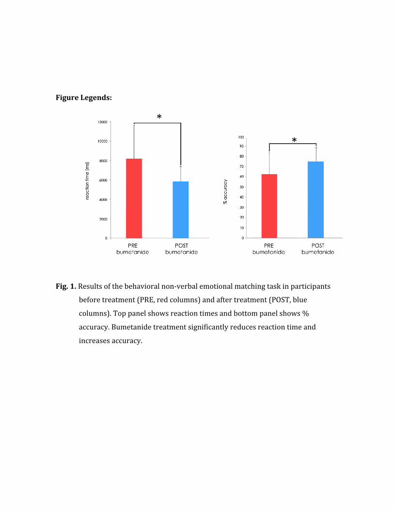
Figure Legends:
Fig. 1. Results of the behavioral non-‐verbal emotional matching task in participants
before treatment (PRE, red columns) and after treatment (POST, blue
columns). Top panel shows reaction times and bottom panel shows %
accuracy. Bumetanide treatment significantly reduces reaction time and
increases accuracy.
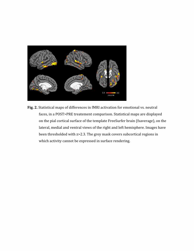
Fig. 2. Statistical maps of differences in fMRI activation for emotional vs. neutral
faces, in a POST>PRE treatement comparison. Statistical maps are displayed
on the pial cortical surface of the template FreeSurfer brain (fsaverage), on the
lateral, medial and ventral views of the right and left hemisphere. Images have
been thresholded with z>2.3. The grey mask covers subcortical regions in
which activity cannot be expressed in surface rendering.
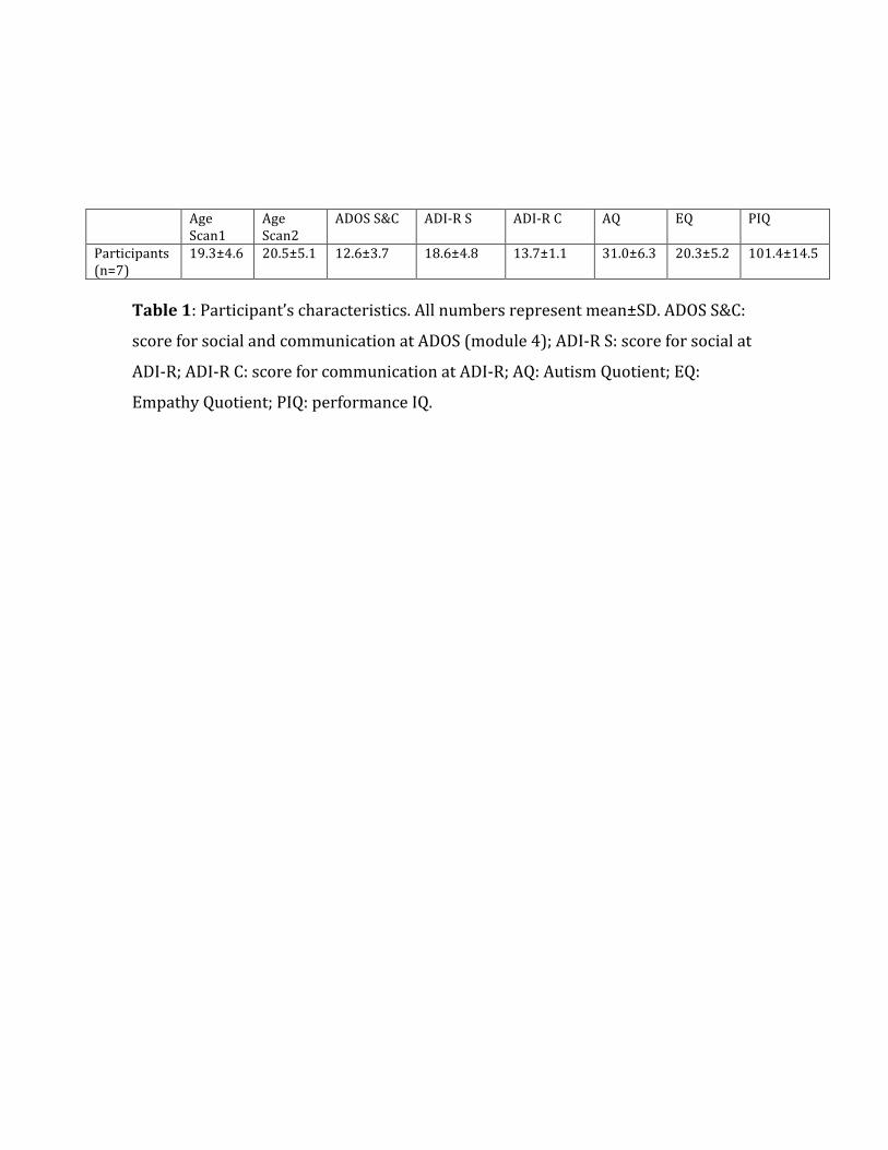
Age Scan1
Age Scan2
ADOS S&C ADI-‐R S ADI-‐R C AQ EQ PIQ
Participants (n=7)
19.3±4.6
20.5±5.1
12.6±3.7
18.6±4.8
13.7±1.1 31.0±6.3 20.3±5.2 101.4±14.5
Table 1: Participant’s characteristics. All numbers represent mean±SD. ADOS S&C:
score for social and communication at ADOS (module 4); ADI-‐R S: score for social at
ADI-‐R; ADI-‐R C: score for communication at ADI-‐R; AQ: Autism Quotient; EQ:
Empathy Quotient; PIQ: performance IQ.
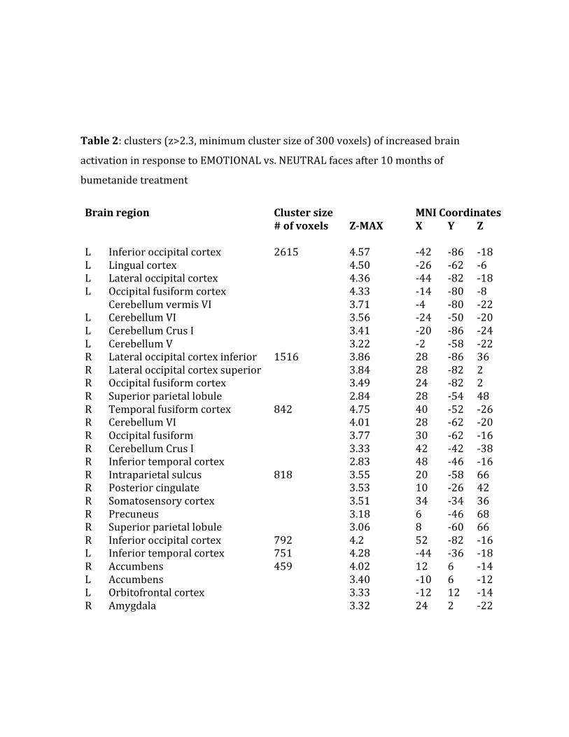
Table 2: clusters (z>2.3, minimum cluster size of 300 voxels) of increased brain
activation in response to EMOTIONAL vs. NEUTRAL faces after 10 months of
bumetanide treatment
Brain region Cluster size MNI Coordinates # of voxels Z-‐MAX X Y Z L Inferior occipital cortex 2615 4.57 -‐42 -‐86 -‐18 L Lingual cortex 4.50 -‐26 -‐62 -‐6 L Lateral occipital cortex 4.36 -‐44 -‐82 -‐18 L Occipital fusiform cortex 4.33 -‐14 -‐80 -‐8 Cerebellum vermis VI 3.71 -‐4 -‐80 -‐22 L Cerebellum VI 3.56 -‐24 -‐50 -‐20 L Cerebellum Crus I 3.41 -‐20 -‐86 -‐24 L Cerebellum V 3.22 -‐2 -‐58 -‐22 R Lateral occipital cortex inferior 1516 3.86 28 -‐86 36 R Lateral occipital cortex superior 3.84 28 -‐82 2 R Occipital fusiform cortex 3.49 24 -‐82 2 R Superior parietal lobule 2.84 28 -‐54 48 R Temporal fusiform cortex 842 4.75 40 -‐52 -‐26 R Cerebellum VI 4.01 28 -‐62 -‐20 R Occipital fusiform 3.77 30 -‐62 -‐16 R Cerebellum Crus I 3.33 42 -‐42 -‐38 R Inferior temporal cortex 2.83 48 -‐46 -‐16 R Intraparietal sulcus 818 3.55 20 -‐58 66 R Posterior cingulate 3.53 10 -‐26 42 R Somatosensory cortex 3.51 34 -‐34 36 R Precuneus 3.18 6 -‐46 68 R Superior parietal lobule 3.06 8 -‐60 66 R Inferior occipital cortex 792 4.2 52 -‐82 -‐16 L Inferior temporal cortex 751 4.28 -‐44 -‐36 -‐18 R Accumbens 459 4.02 12 6 -‐14 L Accumbens 3.40 -‐10 6 -‐12 L Orbitofrontal cortex 3.33 -‐12 12 -‐14 R Amygdala 3.32 24 2 -‐22
R Temporal pole 3.21 24 -‐6 -‐28 R Superior Temporal cortex 444 4.13 68 -‐34 8 R Parietal operculum 2.46 54 -‐28 14 L Superior Temporal cortex 400 4.14 -‐62 -‐4 -‐2 L Central operculum 3.26 -‐54 -‐10 -‐6 L Precentral cortex 2.56 -‐62 2 6 L Supplementary motor area 345 3.48 2 6 66 L Superior frontal gyrus 3.40 14 8 66 L Somatosensory cortex 323 4.21 14 -‐40 74


![2016 HF essential for cardio fellow CRS and diuretic ......Microsoft PowerPoint - 2016 HF essential for cardio fellow_CRS and diuretic resistance.pptx [Last saved by user] Author Administrator](https://img.pdfslide.tips/doc/110x75/5f6cd0edc842c945cb4e50a9/2016-hf-essential-for-cardio-fellow-crs-and-diuretic-microsoft-powerpoint.jpg)