Embed Size (px)
Citation preview
Title: In situ evaluation of fluoride-, stannous- and polyphosphate-containing
solutions against enamel erosion
Short title: fluoride, stannous and polyphosphates against erosion
Authors: Samira Helena João-Souzaa, Sávio José C. Bezerrab, Patrícia Moreira de Freitasc,
Nelson B de Limad, Ana Cecília Corrêa Aranhae, Anderson T. Haraf, Taís Scaramuccig,*
Affiliations
aDepartment of Restorative Dentistry, University of São Paulo, School of Dentistry. Av. Prof.
Lineu Prestes 2227, Cidade Universitária, São Paulo, SP, Brazil. Zip code: 05508-000. E-
mail: [email protected]
bDepartment of Restorative Dentistry, University of São Paulo, School of Dentistry. Av. Prof.
Lineu Prestes 2227, Cidade Universitária, São Paulo, SP, Brazil. Zip code: 05508-000. E-
mail: [email protected]
cDepartment of Restorative Dentistry, University of São Paulo, School of Dentistry. Av. Prof.
Lineu Prestes 2227, Cidade Universitária, São Paulo, SP, Brazil. Zip code: 05508-000. E-
mail: [email protected]
dCentro de Ciência e Tecnologia dos Materiais, Instituto de Pesquisas Energéticas e
Nucleares, IPEN-CNEN/SP, Butantã, P.O. Box 11049, São Paulo, SP 05422-970, Brazil. E-
mail: [email protected]
eDepartment of Restorative Dentistry, University of São Paulo, School of Dentistry. Av. Prof.
Lineu Prestes 2227, Cidade Universitária, São Paulo, SP, Brazil. Zip code: 05508-000. E-
mail: [email protected]
fDepartment of Cariology, Operative Dentistry and Dental Public Health, Indiana University,
School of Dentistry, 415 Lansing street, 46202-2855, Indianapolis, IN, USA. E-mail:
gDepartment of Restorative Dentistry, University of São Paulo, School of Dentistry. Av. Prof.
Lineu Prestes 2227, Cidade Universitária, São Paulo, SP, Brazil. Zip code: 05508-000. E-
mail: [email protected]
*Corresponding author; Taís Scaramucci. Department of Restorative Dentistry, University
of São Paulo, School of Dentistry. Av. Prof. Lineu Prestes 2227, Cidade Universitária,
Butantã, São Paulo, SP, Brazil. Zip code: 05508-000. Phone: +55112648-8021. E-mail:
_________________________________________________________________________________ This is the author's manuscript of the article published in final edited form as:
João-Souza, S. H., Bezerra, S. J. C., de Freitas, P. M., de Lima, N. B., Aranha, A. C. C., Hara, A. T., & Scaramucci, T. (2017). In situ evaluation of fluoride-, stannous- and polyphosphate-containing solutions against enamel erosion. Journal of Dentistry. https://doi.org/10.1016/j.jdent.2017.05.014
2
Abstract
Objective: To evaluate the anti-erosive effect of solutions containing sodium fluoride (F: 225
ppm of fluoride), sodium fluoride + stannous chloride (F+Sn: 225 ppm of fluoride + 800 ppm
of stannous), sodium fluoride + stannous chloride + sodium linear polyphosphate
(F+Sn+LPP: 225 ppm of fluoride + 800 ppm of stannous + 2% of sodium linear
polyphosphate), and deionized water (C: control), using a four-phase, single-blind, crossover
in situ clinical trial.
Methods: In each phase, 12 volunteers wore appliances containing 4 enamel specimens,
which were submitted to a 5-day erosion-remineralization phase that consisted of 2h of
salivary pellicle formation with the appliance in situ, followed by 2min extra-oral immersion in
1% citric acid (pH 2.4), 6x/day, with 90min of exposure to saliva in situ between the
challenges. Treatment with the test solutions was performed extra-orally for 2min, 2x/day. At
the end of the experiment, surface loss (SL, in µm) was evaluated by optical profilometry.
Data were analyzed using ANOVA and Tukey tests (α=0.05). The surface of additional
specimens was evaluated by x-ray diffraction after treatments (n=3).
Results: C (mean SL ± standard-deviation: 5.97±1.70) and F (5.36±1.59) showed the highest
SL, with no significant difference between them (p>0.05). F+Sn (2.68±1.62) and F+Sn+LPP
(2.10±0.95) did not differ from each other (p>0.05), but presented lower SL than the other
groups (P<0.05). Apatite and stannous deposits on specimen surfaces were identified in the
x-ray analysis for F+Sn and F+Sn+LPP.
Conclusions: Sodium fluoride solution exhibited no significant anti-erosive effect. The
combination between sodium fluoride and stannous chloride reduced enamel erosion,
irrespective of the presence of linear sodium polyphosphate.
Clinical significance: Under highly erosive conditions, sodium fluoride rinse may not be a
suitable alternative to prevent enamel erosion. A rinse containing sodium fluoride and
stannous chloride was shown to be a better treatment option, which was not further improved
by addition of the sodium linear polyphosphate.
Keywords: enamel; erosion; fluoride; stannous chloride; phosphate polymer
Introduction
Dental erosion is a complex condition that affects different age groups in populations
worldwide [1]. The overall increase in its presence could be related to changes in lifestyles
and nutritional habits, with a higher consumption of acidic foods and beverages [1,2]. In
addition to avoiding exposure to erosive sources, the use of fluoridated products is highly
3
recommended for patients with erosion [3]. However, their effectiveness against erosion
seems to be dependent on the type of fluoridated compound, F concentration and pH. Many
studies have tested the anti-erosive ability of F solutions containing metal cations, such as
stannous (Sn), with promising outcomes [4–7]. Sn can incorporate into enamel through a
complex process of demineralization and reprecipitation; it can also induce the surface
deposition of acid-resistant precipitates [8]. In situ investigations have shown that a solution
containing 500 ppm F and 800 ppm Sn was able to reduce enamel and dentin loss in the
range of 45-67% and 47-68%, respectively [4,5].
Despite these positive results, studies have demonstrated that the protection offered
by F and Sn can be increased by combining them with some polymers. A dentifrice
containing F, Sn and the biopolymer chitosan showed improved enamel erosion protection
compared with a dentifrice containing F+Sn alone [9]. A previous in vitro investigation by our
group demonstrated that the addition of a phosphate polymer (sodium linear polyphosphate -
LPP) could increase the protection of a solution containing 225 ppm F and 800 ppm Sn by
11% under highly erosive conditions [6], irrespective of the presence of simulated salivary
pellicle [7].
The salivary pellicle is important when evaluating film-forming agents such as LPP,
due to the possibility of competition for binding sites on the enamel surface [10]. The salivary
pellicle formed in vitro is known to differ from the in situ because, among other changes, the
proteins of the saliva collected can undergo alteration or degradation [11,12]. Considering
this fact, this study sought to evaluate the protective effect of the combination of F+Sn+LPP
against erosion under more clinically relevant conditions, such as those achieved in in situ
models. Our hypothesis was that LPP would improve the protective effects of F+Sn against
enamel erosion.
Materials and methods
Experimental design
This study consisted of a four-phase, single-blind crossover in situ clinical trial,
involving 12 volunteers who met the inclusion/exclusion criteria described in detail elsewhere
[13]. Briefly, the volunteers were at least 18 years old, with good general and oral health,
without any allergy or any other condition that could compromise their safety. Their
unstimulated and stimulated salivary flow rate had to be ≥ 0.5 ml/min and ≥ 1 ml/min,
respectively. The exclusion criteria were: pregnancy (or intention to become pregnant) during
the study period, nursing, concomitant participation in another research study, and inability to
comply with study procedures. In each study phase, the volunteers used removable
mandibular devices containing 4 specimens of bovine enamel. The study followed a
completely randomized experimental design, with test solution as the single experimental
4
factor, at 4 levels: C: Control (deionized water); F: Sodium fluoride solution (11.83 mM of
NaF; 225 ppm of fluoride; pH 4.5); F+Sn: Sodium fluoride plus stannous chloride solution
(11.83 mM of NaF + 10.75 mM of SnCl2; 225 ppm F, 800 ppm Sn; pH 4.5); F+Sn+LPP:
Sodium fluoride, stannous chloride and sodium linear polyphosphate solutions (11.83 mM of
NaF + 10.75 mM of SnCl2 + 2% of LPP; 225 ppm F, 800 ppm Sn; pH 4.5). The response
variable was tooth surface loss, in µm, determined by optical profilometry at the end of the
clinical phase. As an additional test, the surface of extra enamel specimens was evaluated
by x-ray diffraction after treatments (n=3).
Ethical Aspects
This study was conducted in the Restorative Dentistry Department of School of
Dentistry, University of São Paulo, São Paulo, SP, Brazil. The study protocol was reviewed
and approved by the local Ethics Committee on Research with Humans (CAAE:
27621214.9.0000.0075). To participate in the study, all subjects had to sign a term of free
and informed consent.
Sample size
For this in situ study, 12 subjects were recruited. This sample size was chosen based
on a previous study [14] with a similar design, which showed significant difference between
experimental groups using a sample size of 10 individuals.
Study population
The recruitment of the subjects was carried out in the Restorative Dentistry
Department of School of Dentistry, University of São Paulo. First, the subjects were informed
about the nature of the study, its possible risks and data confidentiality. After agreeing to
participate, their medical and dental history was evaluated. Unstimulated and stimulated
salivary flow rates were measured using established procedures, as previously described
[13].
The subjects who met the inclusion/exclusion criteria received an oral hygiene kit
containing a toothpaste (Colgate Cavity Protection, 1500 ppm F, Colgate-Palmolive, Osasco,
SP, Brazil), a regular toothbrush (Colgate Twister Fresh, Colgate-Palmolive, Osasco, SP,
Brazil) and dental floss, to be used on the 7 days before the study began (lead-in phase) and
throughout the entire study period. They were not allowed to use any other oral hygiene
products. Subjects were instructed to perform oral hygiene twice a day, with the oral
appliance removed from the mouth. They were also advised not to brush their teeth with
toothpaste in the 2 h prior the beginning of the experimental procedures, and also in the 30
min after eating.
5
All the eligible subjects were identified by a unique study number. In each week, they
were randomly assigned to the treatment solutions according to a standard randomization
table. Before the study began, the subjects were thoroughly trained in all experimental
procedures, and they received a written protocol containing all the instructions. In each study
phase, they also received a schedule and a digital timer to guide their conduct and recording
of the experimental procedures.
Intraoral device
An impression of each subject’s mandibular arch was taken with heavy consistency
condensation silicone (Clonage®, DFL, Jacarepagua, RJ, Brazil). From the impressions, bi-
lateral mandibular intraoral appliances were prepared with acrylic resin [15]. In these
devices, four niches of approximately 4 mm x 4 mm x 2 mm were made on the buccal
surfaces of the premolars and molars.
The intraoral devices were disinfected with 2% chlorhexidine solution for 10 min
before and after each study phase, and rinsed with tap water. Before mounting the
specimens in the appliances, they were sterilized with gamma radiation (Experimental
irradiator Cobalt-60, Gamacell 220, IPEN, São Paulo, SP, Brazil). The day before each
phase began, the sterilized specimens received adhesive unplasticized polyvinyl chloride
(UPVC) tapes on their polished surface, leaving a central area of 3 mm x 1 mm exposed.
The specimens were fixed in the 4 niches with sticky wax, so that their surfaces remained 1
mm below the appliance surface, to avoid abrasion of the buccal soft tissues.
Specimen preparation
Enamel specimens were prepared from bovine incisors that were firstly cleaned with
periodontal curettes (Hu-Friedy, Chicago, USA), and subjected to prophylaxis with a mixture
of pumice and water applied with rubber cup at low speed. The teeth were kept in 0.1%
thymol solution (Sigma-Aldrich Co.), at 4°C, until the experiment began. The crowns were
separated from the roots. Then enamel specimens measuring 3 mm x 3 mm x 1.5 mm were
sectioned from the buccal sides of the crowns, by using a precision cutting machine (Isomet
1000, Buehler Ltd, Lake Buff, Illinois, USA). The pulp surfaces of the specimens were
flattened with a polishing machine (Buehler Ltd, Lake Buff, Illinois, USA), fitted with a #600
grit abrasive disc (Buehler Ltd), under constant water cooling. Subsequently, the buccal
surfaces were ground flat and polished using a sequence of abrasive discs with decreasing
granulations: #600, #1200, #2400 and #4000 (Buehler Ltd), and polishing cloth sprayed with
diamond suspension (1 μm, Buehler Ltd) for 3 min. At the end of the polishing procedures,
the specimens were sonicated with distilled water for 3 min. Specimens without any cracks or
structural defects were selected.
6
The surface microhardness (SMH) of the specimens was analyzed. Three
indentations were made in the central area of the specimens with a Knoop indenter
(Shimadzu Co, Tokyo, Japan), using a load of 50 g for 15 s, with a distance of 100 μm
between each indentation [16]. The mean value of the 3 indentations was calculated, and
specimens with similar SMH were then selected. These specimens were further analyzed
with an optical profilometer (Proscan 2100, Scantron, Tauton, UK) to identify those with
surface curvature below 0.3 μm, which were finally included in this study.
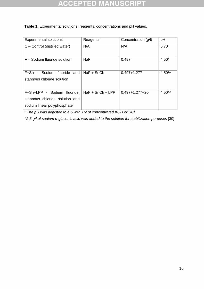
Experimental solutions
The experimental solutions are described in Table 1. The NaF (Sodium fluoride,
Sigma Aldrich, St Louis MO, USA), LPP (Sodium polyphosphate, Merck Milipore, Darmstadt,
Germany) and Sn (Stannous chloride, Sigma Aldrich Co.) concentrations were in accordance
with those of previous studies [6,7,17]. Deionized water was used as control. All the solutions
(except the water) had the pH adjusted to 4.5, with KOH solution or concentrated HCl. For
the erosive challenge 1% citric acid (Sigma-Aldrich, St. Louis, MO, USA) was used, with
natural pH of 2.4 [6].
Erosive challenge
At the beginning of each day, subjects wore the intraoral devices for 2 h to allow
salivary pellicle formation. After this, the specimens were immersed in 20 ml of the acid
solution extra-orally, for 2 min, 6x/day, with 90-min intervals between them, during which
specimens were exposed to saliva in situ.
After each erosion challenge, the excess acid was removed with absorbent paper and
the intraoral devices were returned to the mouth. For the treatments, the specimens were
immersed in 10 ml of their respective experimental solution for 2 min, 2x/day, 45 min after
the first and the last erosive challenges. The excess solution was also removed with
absorbent paper after treatments. To avoid contact of the individual´s teeth with the acidic
solution and with the experimental solutions, the intraoral devices containing the specimens
were immersed in the solutions extra-orally. All solutions were renewed for each exposure.
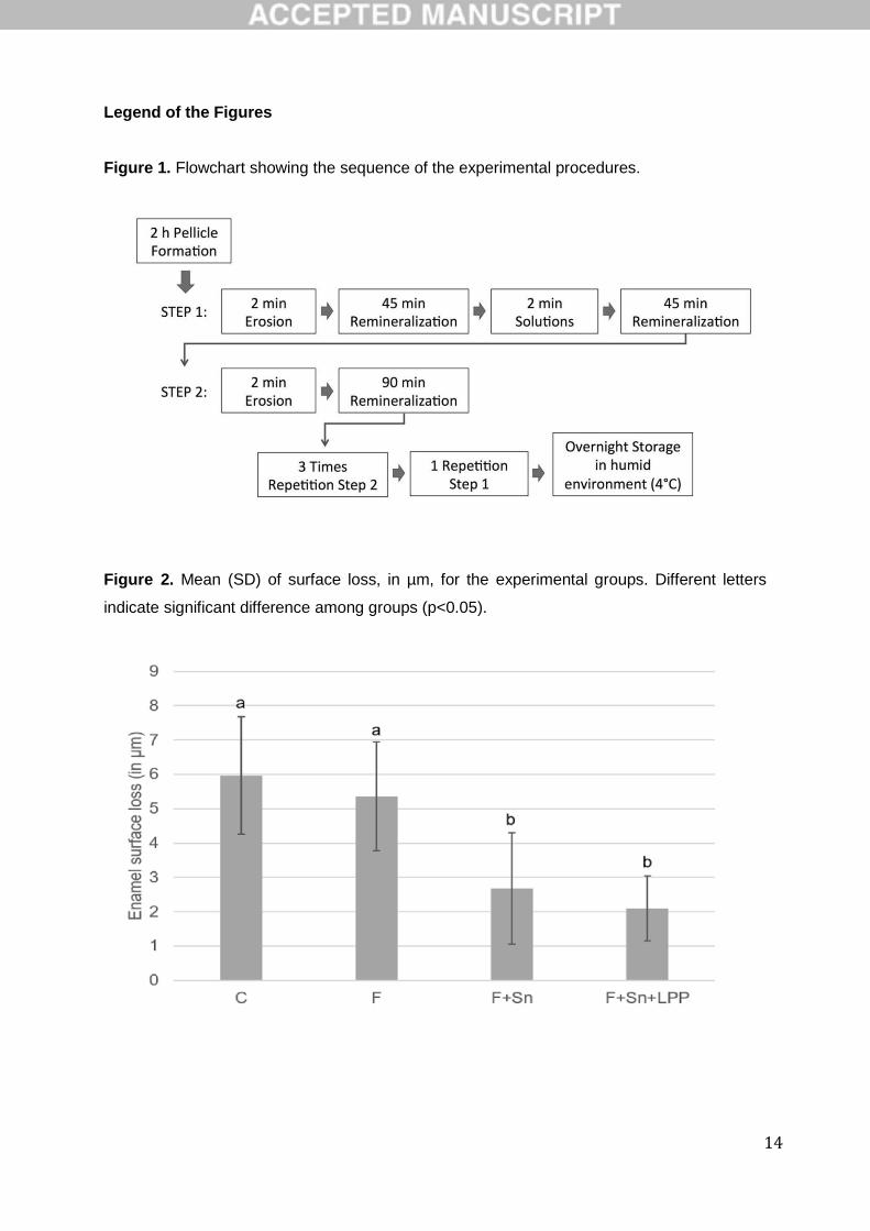
Figure. 1 shows a flowchart of the experimental procedures.
The intraoral devices were used during the day, except during the meals and oral
hygiene procedures, when they were stored in containers with gauze moistened with distilled
water. The intraoral devices were also stored in these containers during the overnight period,
under refrigeration. Between the study phases, a wash out period of 7 days was incorporated
into the study design.
Surface loss
7
At the end of the experimental procedures, the specimens were removed from the
intraoral devices and had the adhesive tapes removed from their surfaces. Subsequently, a
central area of 2 mm x 1 mm of the specimen surface was scanned with an optical
profilometer, according to the following parameters: 200 steps of 0.01 mm in the X axis and
20 steps of 0.05 mm in the Y axis. This analysis covered both reference surfaces and treated
surface. For the surface loss assessment, specific software was used (Proscan Application
Software version 2.0.1.7).
Additional test
As an additional test, 12 extra specimens (3 for each group) were prepared as
described before, and treated with the test solutions for 2 min. Afterwards, they were rinsed
in distilled water and evaluated by x-ray diffraction, using a Rigaku diffractometer (Rigaku
Americas, The Woodlands, Texas, USA), model DMAX-2000, equipped with a chrome tube
and a vanadium filter. A Grazing incidence angle of 2.5°, 2θ varying from 380 a 1300 was
used. All measurements were performed on the specimen surface plane.
Data analysis
Solutions were compared for differences in mean surface loss using an ANOVA
suitable for a crossover study, which included a random effect for subject and fixed effects
for treatment sequence, study phase and solution. Pair-wise comparisons were made using
the Tukey multiple comparisons procedure. The level of significance was set at 5%. The
diffraction patterns obtained were compared with the data from the International Centre for
Diffraction Data (ICDD).
Results
The selected specimens presented a mean SMH value (standard-deviation) of 335
(25) and a mean (SD) surface curvature of 0.21 (0.07). All volunteers completed the study.
ANOVA showed a significant difference in surface loss among the solutions
(p<0.001). Figure 2 shows the means and standard deviations (SD) of the profilometry
analysis for each experimental group.
Control and F showed the highest surface loss, with no significant difference between
them. F+Sn and F+Sn+LPP did not differ significantly and presented significantly lower
surface loss than C and F.
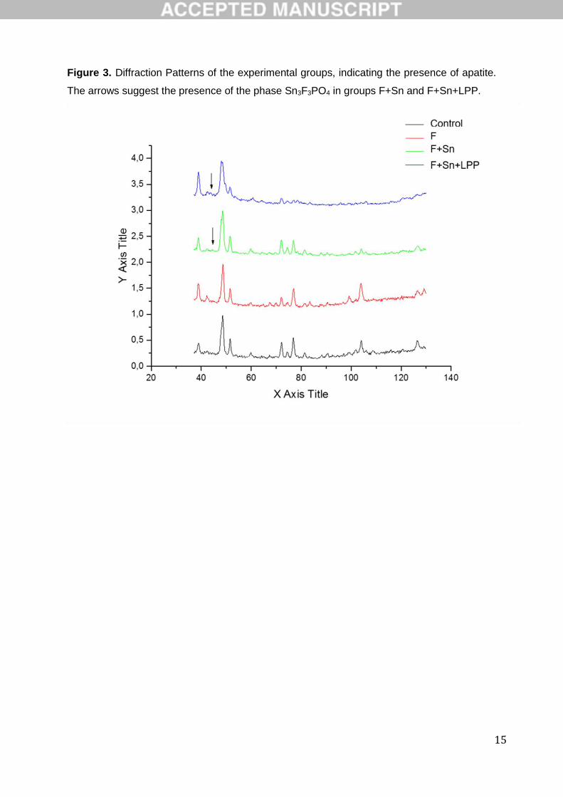
The patterns obtained in the x-ray diffraction analysis for the 4 experimental groups
are shown in Figure 3. The matrix was identified as potassium calcium hydrogen carbonate
phosphate hydrate as ICDD file No. 47-260. The arrows in the patterns of groups F+Sn and
8
F+Sn+LPP suggested the appearance of the phase of Sn3F3PO4, as ICDD file No. 76-2280,
in very low concentration and only at the surface.
Discussion
In this study, LPP was unable to improve the protection of F+Sn against enamel
erosion, leading to rejection of our hypothesis. This result was unexpected and contrasts with
our previous laboratory findings [6,7]. We speculated that the lack of additional protection by
the LPP may be explained by the presence of the naturally-formed acquired salivary pellicle
in the present study. The salivary pellicle is an organic layer, mainly composed of adsorbed
salivary proteins, which covers the dental structures in the natural oral environment [18,19].
Many of the pellicle proteins contain calcium-binding domains [20], presenting high affinity to
the enamel surface, which could have occupied the potential sites for LPP interaction,
thereby reducing its effectiveness. Although the salivary pellicle was simulated with clarified
human saliva in our previous in vitro study [7], we speculated that the naturally formed
pellicle presented different structural and maturation levels, having a different influence on
the LPP binding to enamel and subsequent protective effect. To allow the pellicle formation
and relative maturation in the present in situ study, the volunteers wore the oral devices for
2h before the experimental procedures [21].
Another possible explanation for the lack of LPP effect would be the reduction in the
frequency of application, which was previously used 3 times a day in vitro [6,7] versus the 2
times used in the present study. The frequency of application was reduced to resemble the
clinical scenario more faithfully, as mouth rinses are frequently used only once or twice a day
[15]. It could be suggested that higher frequency would allow prolonged protection, which
would be translated into lower enamel surface loss. However, in a preliminary in vitro test
(unpublished data) we observed no additional protection when F+Sn+LPP solution was
applied three times a day. Therefore, it is unlikely that exposure to additional LPP rinses
would lead to increased protection.
Although extensive in vitro investigations were performed to determine the optimal
concentration of LPP used in this study, we suggest that values higher than 2% may be
needed to lead to enamel protection in situ. Condensed inorganic phosphates, such as LPP,
also have the ability to complex with polymers, especially proteins [22]. Therefore, in the right
concentration, it could adsorb to the salivary pellicle potentially increasing its anti-
demineralization ability. Corroborating this idea, a previous in situ study revealed that the
addition of 9% sodium hexametaphosphate to a 1% NaF gel reduced enamel surface loss
more than the 1% NaF gel did without it [23]. Another investigation reported that the addition
of 5% trimetaphosphate to a 2.5% NaF varnish increased its protection against erosion and
erosion-abrasion [24]. Nevertheless, it has to be born in mind that higher concentrations of
9
phosphate polymers influence the properties of the solution, such as viscosity, and may
interfere in the interaction between F/Sn and the enamel surface. This may be a concern
especially when dealing with long chain length polymers. Further studies are needed to verify
the feasibility of using higher LPP concentrations.
In line with previous reports, the combination between F and Sn reduced enamel
erosion [4,6,17]. This could be attributed to the formation of less soluble precipitates, such as
Sn2OHPO4, Sn3F3PO4 and Ca(SnF3)2, as described in a study using x-ray diffraction to
identify these crystalline compounds [25]. In the cited study, a SnF2 solution with a higher
concentration was used, and the pattern of diffraction was obtained by analyzing
hydroxyapatite powder and not the enamel surface per se. In the present investigation, the x-
ray diffraction analysis of enamel samples treated once with the Sn-containing solutions
yielded the presence of Sn3F3PO4, at very low concentration. It can be suggested that the
low concentrations of F and Sn present at the solutions did not allow for a detectable amount
of precipitates by this method, which is 2% of the component in the sample. In addition, the
evaluation was performed in enamel specimens and not in hydroxyapatite powder, which has
a higher surface area for the precipitation of deposits. This result may strengthen the theory
that in the context of erosion, Sn incorporation into enamel would be more relevant for
surface protection than the formation of less soluble precipitates [8].
In situ studies have shown positive results with sodium fluoride rinses in the
prevention of enamel erosion, with fluoride concentrations ranging from 500 to 950 ppm F
[4,26,27]. However, the present study failed to show a difference between the control and the
fluoride (F) groups. This could be related to the low concentration of F used (225 ppm F),
which was chosen in attempt to simulate the concentration usually found in commercially
available oral rinse products. Probably, at the low pH (4.5) and concentration used, only little
CaF2-like deposits were formed. The pH of 4.5 was chosen for the F solution to avoid having
a confounding factor, because the Sn-containing solutions are not stable at higher pH levels
[7]. It has to be considered that the protocol of some in situ studies required the erosive
challenge and treatments to be performed intra-orally, in contrast to the present study, in
which they were used extra-orally, because of the experimental nature of the testing
solutions. Intraoral exposure to the fluoridated rinses may allow more sites to be found for F
retention, such as the soft oral tissues, which could potentially increase F availability [28].
Moreover, when rinsing is performed intra-orally, it may lead to F interaction with the calcium
from the saliva, allowing the formation of more CaF2-like material. Finally, the in situ model
used simulated highly erosive conditions, with successive episodes of exposure to acid and
no exposure to saliva overnight. This experimental condition could have minimized the
remineralization enhancing action of the test solutions, which could also explain the reduced
protection observed by the F solution [29].
10
Conclusions
In conclusion, sodium fluoride associated with stannous chloride was capable of
reducing enamel erosion; however, this effect was not improved by the presence of the
sodium linear polyphosphate. Further studies are needed to verify the effectiveness of LPP in
improving F+Sn protection by using a higher LPP concentration.
Acknowledgements
The authors would like to thank São Paulo Research Foundation (FAPESP) for the
grant received (#2014/14055-4) and George J. Eckert for conducting the statistical analyses.
11
References
[1] T. Jaeggi, A. Lussi, Prevalence, incidence and distribution of erosion., Monogr. Oral
Sci. 25 (2014) 55–73. doi:10.1159/000360973.
[2] A. Lussi, T.S. Carvalho, Erosive tooth wear: a multifactorial condition of growing
concern and increasing knowledge., Monogr. Oral Sci. 25 (2014) 1–15.
doi:10.1159/000360380.
[3] M.C. Huysmans, A. Young, C. Ganss, The role of fluoride in erosion therapy., Monogr.
Oral Sci. 25 (2014) 230–43. doi:10.1159/000360555.
[4] C. Ganss, L. Neutard, J. von Hinckeldey, J. Klimek, N. Schlueter, Efficacy of a
Tin/Fluoride Rinse: a Randomized in situ Trial on Erosion, J. Dent. Res. 89 (2010)
1214–1218. doi:10.1177/0022034510375291.
[5] N. Schlueter, J. Klimek, C. Ganss, Efficacy of tin-containing solutions on erosive
mineral loss in enamel and dentine in situ, Clin. Oral Investig. 15 (2011) 361–367.
doi:10.1007/s00784-010-0386-x.
[6] T. Scaramucci, A.B. Borges, F. Lippert, D.T. Zero, I.V Aoki, A.T Hara, Anti-erosive
properties of solutions containing fluoride and different film-forming agents, J. Dent. 43
(2015) 458–465. doi:10.1016/j.jdent.2015.01.007.
[7] T. Scaramucci, S.H. João-Souza, F. Lippert, G.J. Eckert, I. V Aoki, A.T. Hara,
Influence of Toothbrushing on the Antierosive Effect of Film-Forming Agents., Caries
Res. 50 (2016) 104–10. doi:10.1159/000443619.
[8] N. Schlueter, M. Hardt, A. Lussi, F. Engelmann, J. Klimek, C. Ganss, Tin-containing
fluoride solutions as anti-erosive agents in enamel: an in vitro tin-uptake, tissue-loss,
and scanning electron micrograph study., Eur. J. Oral Sci. 117 (2009) 427–34.
doi:10.1111/j.1600-0722.2009.00647.x.
[9] C. Ganss, J. von Hinckeldey, A. Tolle, K. Schulze, J. Klimek, N. Schlueter, Efficacy of
the stannous ion and a biopolymer in toothpastes on enamel erosion/abrasion, J.
Dent. 40 (2012) 1036–1043. doi:10.1016/j.jdent.2012.08.005.
[10] M.E. Barbour, R.P. Shellis, D.M. Parker, G.C. Allen, M. Addy, An investigation of
some food-approved polymers as agents to inhibit hydroxyapatite dissolution, Eur. J.
Oral Sci. 113 (2005) 457–461. doi:10.1111/j.1600-0722.2005.00248.x.
[11] G.R. Batista, C.R.G. Torres, B. Sener, T. Attin, A. Wiegand, Artificial Saliva
Formulations versus Human Saliva Pretreatment in Dental Erosion Experiments,
Caries Res. 50 (2016) 78–86. doi:10.1159/000443188.
[12] A.F. Hall, C.A. Buchanan, D.T. Millett, S.L. Creanor, R. Strang, R.H. Foye, The effect
of saliva on enamel and dentine erosion, J. Dent. 27 (1999) 333–339.
doi:10.1016/S0300-5712(98)00067-0.

12
[13] T. Scaramucci, M.A.P. Sobral, G.J. Eckert, D.T. Zero, A.T. Hara, In situ evaluation of
the erosive potential of orange juice modified by food additives, Caries Res. 46 (2012).
doi:10.1159/000335572.
[14] N. Schlueter, J. Klimek, C. Ganss, Effect of a chitosan additive to a Sn2+-containing
toothpaste on its anti-erosive/anti-abrasive efficacy—a controlled randomised in situ
trial, Clin. Oral Investig. 18 (2014) 107–115. doi:10.1007/s00784-013-0941-3.
[15] C.V. da Silva, T.M. Ramos-Oliveira, T.F. Mantilla, P.M. de Freitas, Frequency of
Application of AmF/NaF/SnCl2; Solution and Its Potential in Controlling Human Enamel
Erosion Progression: An in situ Study, Caries Res. 51 (2017) 141–148.
doi:10.1159/000455051.
[16] T. Scaramucci, A.T. Hara, D.T. Zero, S.S. Ferreira, I.V. Aoki, M.A.P. Sobral, In vitro
evaluation of the erosive potential of orange juice modified by food additives in enamel
and dentine, J. Dent. 39 (2011). doi:10.1016/j.jdent.2011.09.004.
[17] N. Schlueter, L. Neutard, J. von Hinckeldey, J. Klimek, C. Ganss, Tin and fluoride as
anti-erosive agents in enamel and dentine in vitro., Acta Odontol. Scand. 68 (2010)
180–4. doi:10.3109/00016350903555395.
[18] U. Lendenmann, J. Grogan, F.G. Oppenheim, Saliva and dental pellicle--a review.,
Adv. Dent. Res. 14 (2000) 22–28. doi:10.1177/08959374000140010301.
[19] J.L. Jensen, M.S. Lamkin, F.G. Oppenheim, Adsorption of Human Salivary Proteins to
Hydroxyapatite: A Comparison Between Whole Saliva and Glandular Salivary
Secretions, J. Dent. Res. 71 (1992) 1569–1576.
doi:10.1177/00220345920710090501.
[20] M. Hannig, C. Hannig, The Pellicle and Erosion, in: 2014: pp. 206–214.
doi:10.1159/000360376.
[21] M. Hannig, N.J. Hess, W. Hoth-Hannig, M. De Vrese, Influence of salivary pellicle
formation time on enamel demineralization--an in situ pilot study., Clin. Oral Investig. 7
(2003) 158–161. doi:10.1007/s00784-003-0219-2.
[22] I.S. Kulaev, V.M. Vagabov, T. V. Kulakovskaya, The Chemical Structures and
Properties of Condensed Inorganic Phosphates, in: Biochem. Inorg. Polyphosphates,
John Wiley & Sons, Ltd, Chichester, UK, 2005: pp. 3–13.
doi:10.1002/0470858192.ch1.
[23] J.M. Conceição, A.C.B. Delbem, M. Danelon, D.M. da Camara, A. Wiegand, J.P.
Pessan, Fluoride gel supplemented with sodium hexametaphosphate reduces enamel
erosive wear in situ., J. Dent. 43 (2015) 1255–60. doi:10.1016/j.jdent.2015.08.006.
[24] M.J. Moretto, A.C.B. Delbem, M.M. Manarelli, J.P. Pessan, C.C.R. Martinhon, Effect of
fluoride varnish supplemented with sodium trimetaphosphate on enamel erosion and
abrasion: An in situ/ex vivo study, J. Dent. 41 (2013) 1302–1306.
13
doi:10.1016/j.jdent.2013.09.008.
[25] F.D. Babcock, J.C. King, T.H. Jordan, The reaction of stannous fluoride and
hydroxyapatite., J. Dent. Res. 57 (1978) 933–8. doi:10.1177/00220345780570092301.
[26] K.R. Stenhagen, L.H. Hove, B. Holme, A.B. Tveit, The effect of daily fluoride mouth
rinsing on enamel erosive/abrasive wear in situ, Caries Res. 47 (2013) 2–8.
doi:10.1159/000342619.
[27] N. Schlueter, J. Klimek, C. Ganss, Efficacy of an experimental tin-f-containing solution
in erosive tissue loss in enamel and dentine in situ, Caries Res. 43 (2009) 415–421.
doi:10.1159/000252974.
[28] D.T. Zero, R.F. Raubertas, A.M. Pedersen, J. Fu, A.L. Hayes, J.D. Featherstone,
Studies of fluoride retention by oral soft tissues after the application of home-use
topical fluorides., J. Dent. Res. 71 (1992) 1546–52.
http://www.ncbi.nlm.nih.gov/pubmed/1522285 (accessed February 7, 2017).
[29] A.T. Hara, S.A. Kelly, C. Gonzalez-Cabezas, G.J. Eckert, A.P. Barlow, S.C. Mason,
D.T. Zero, Influence of fluoride availability of dentifrices on eroded enamel
remineralization in situ, Caries Res. 43 (2009) 57–63. doi:10.1159/000201591.
[30] A.T. Hara, F. Lippert, D.T. Zero, Interplay between experimental dental pellicles and
stannous-containing toothpaste on dental erosion-abrasion, Caries Res. 47 (2013)
325–329. doi:10.1159/000347051.
14
Legend of the Figures
Figure 1. Flowchart showing the sequence of the experimental procedures.
Figure 2. Mean (SD) of surface loss, in µm, for the experimental groups. Different letters
indicate significant difference among groups (p<0.05).
15
Figure 3. Diffraction Patterns of the experimental groups, indicating the presence of apatite.
The arrows suggest the presence of the phase Sn3F3PO4 in groups F+Sn and F+Sn+LPP.
16
Table 1. Experimental solutions, reagents, concentrations and pH values.
Experimental solutions Reagents Concentration (g/l) pH
C – Control (distilled water)
N/A N/A 5.70
F – Sodium fluoride solution
NaF 0.497 4.501
F+Sn - Sodium fluoride and
stannous chloride solution
NaF + SnCl2 0.497+1.277 4.501,2
F+Sn+LPP - Sodium fluoride,
stannous chloride solution and
sodium linear polyphosphate
NaF + SnCl2 + LPP 0.497+1.277+20 4.501,2
1 The pH was adjusted to 4.5 with 1M of concentrated KOH or HCl
2 2,3 g/l of sodium d-gluconic acid was added to the solution for stabilization purposes [30]