Embed Size (px)
Citation preview
Vision :IMC aspires to be a leader in applied medical sciences, health care education and research.
Dr. Mohammed EMAMPh.D., Paris-Sud 11 University
RAD 364
Nuclear Medicine Technique II
Lecture’s Title: Quality Control in Gamma Camera
Radiological Sciences Department
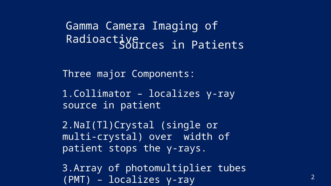
Gamma Camera Imaging of Radioactive
2
Sources in Patients
Three major Components:1.Collimator – localizes γ-ray source in patient2.NaI(Tl)Crystal (single or multi-crystal) over width of patient stops the γ-rays.3.Array of photomultiplier tubes (PMT) – localizes γ-ray interaction in crystal
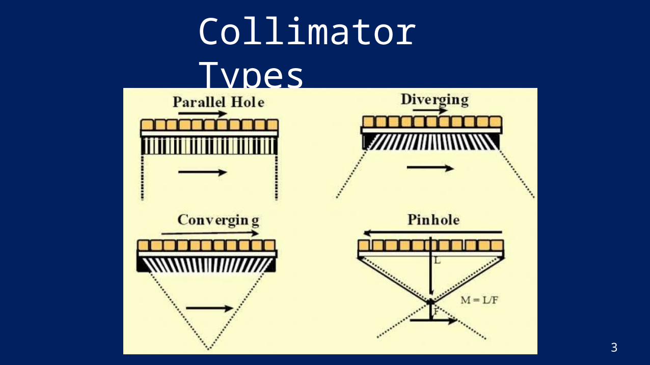
Collimator Types
3
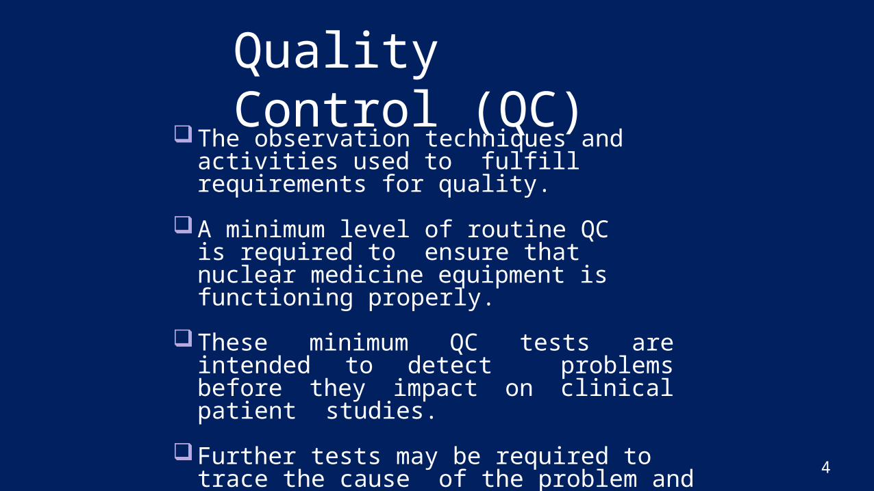
Quality Control (QC)
4
The observation techniques and activities used to fulfill requirements for quality.
A minimum level of routine QC is required to ensure that nuclear medicine equipment is functioning properly.
These minimum QC tests are intended to detect problems before they impact on clinical patient studies.
Further tests may be required to trace the cause of the problem and to ensure that the equipment is performing properly after service or adjustment
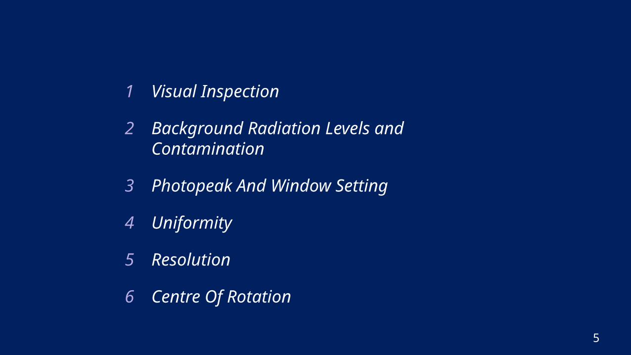
1 Visual Inspection
2 Background Radiation Levels and Contamination
3 Photopeak And Window Setting
4 Uniformity
5 Resolution
6 Centre Of Rotation
5
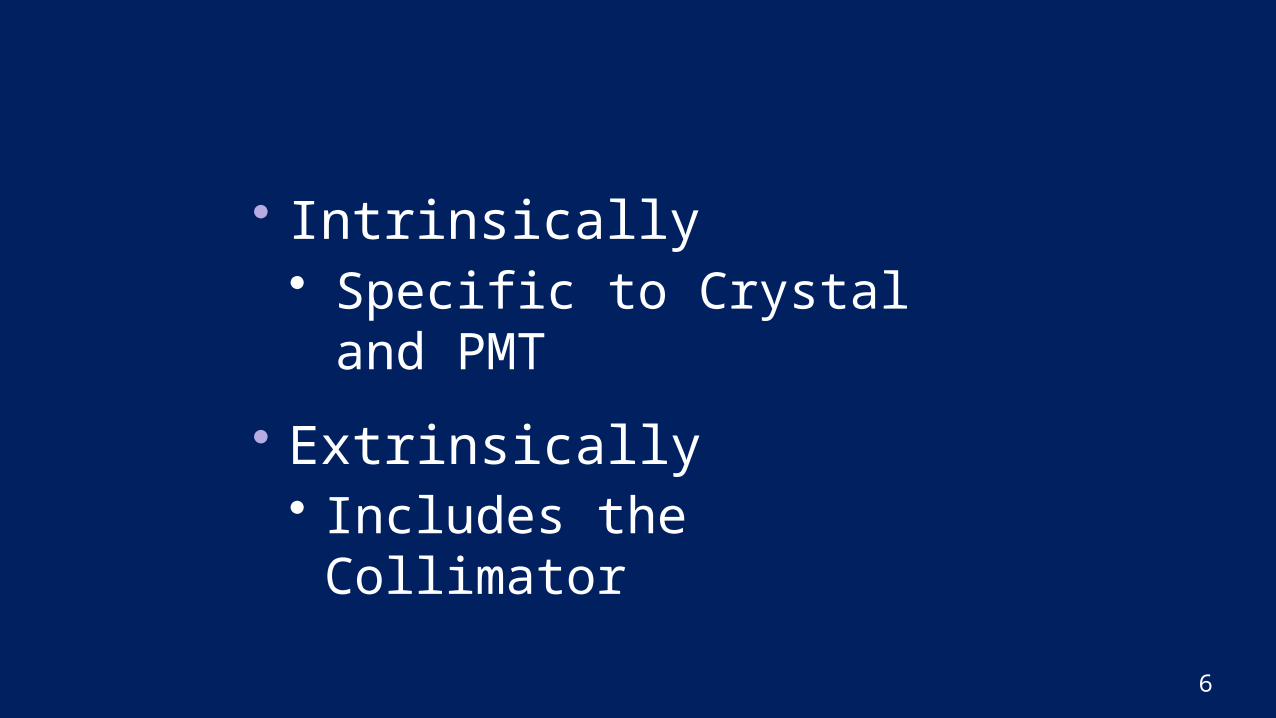
• Intrinsically• Specific to Crystal and
PMT• Extrinsically• Includes the Collimator
6
Visual Inspection
7
• A visual inspection may reveal obvious defects which may compromise the safety or the imaging efficacy of the system (e.g. frayed or damaged electrical cables).• Of particular importance is the visual inspection of
collimators on a regular basis and whenever collimators are changed.
• Signs of dents and scratches may indicate mechanical damage to the collimator, and stains may be a sign of possible contamination.• Both of these may produce artefacts such as cold or hot spots on planar images and rings on SPECT images.
Background Radiation Levels and Contamination
8
• High levels of background radiation may arise from “hot” patients in the proximity of the imaging system or other sources of unshielded radiation.
• If high energy imaging agents are being used, the potential also exists for penetration through the back of the gamma camera, where shielding is thinner.
• Other sources of background radiation may include radioactive contamination on the floor, walls or even the detector itself.
Background Radiation Levels and Contamination
9
• Background radiation, if it is of sufficient intensity, has the potential to seriously distort any type of imaging.
• Even moderately elevated background levels have the potential to seriously degrade intrinsic uniformity or other intrinsic measurements.
Photopeak And Window Setting
10
• Incorrect photopeak energy window setting(s) can degrade uniformity, reduce sensitivity or can increase the scatter contribution to the image.
• Peak settings should be checked and adjusted in a consistent manner and the settings should be recorded to detect long term drift in the settings.
• Sudden changes in peak setting indicate a possible fault in the camera and should be fully investigated and rectified if necessary before the camera is again used for clinical studies.
Uniformity
11
• The uniformity or "flood" QC procedure checks that the response of the detector to a uniform irradiation is uniform within defined limits.• It is one of the most basic QC tests of the gamma camera.• Interpretation of clinical images taken with the
gamma camera rely on the assumption that differences seen are due to differences in tracer distribution in the patient only and not differences introduced by the gamma camera.
Resolution
• The purpose of resolution checks is to detect gradual, long term deterioration/ drop of resolution, rather than detecting abrupt/ acute changes.
• Inappropriate adjustments carried out during service may affect the resolution, without necessarily being apparent in the uniformity or other checks.
12
Centre Of Rotation
13
• The rotation axis (or centre of rotation) assumed by the reconstruction program has to accurately coincide with the mechanical axis of rotation to avoid loss of resolution and distortion in the reconstructed slices.
• Centre of rotation offset can vary with collimator and as a function of detector rotation and radius of rotation.
• It is important to establish which factors affect COR offset on each particular camera and then make appropriate allowances for it.
Recommended Frequency of QC Tests
14
DAILYGamma Camera• Visual Inspection• Background/Contamination Check• Photopeak check and adjustment, if
necessary. Depending on the camera, this may require check and adjustment of photopeaks for all radionuclides used with the particular camera.
• Uniformity check on gamma camera
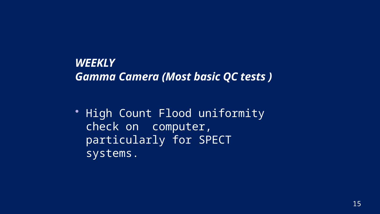
WEEKLY Gamma Camera (Most basic QC tests )
• High Count Flood uniformity check on computer, particularly for SPECT systems.
15
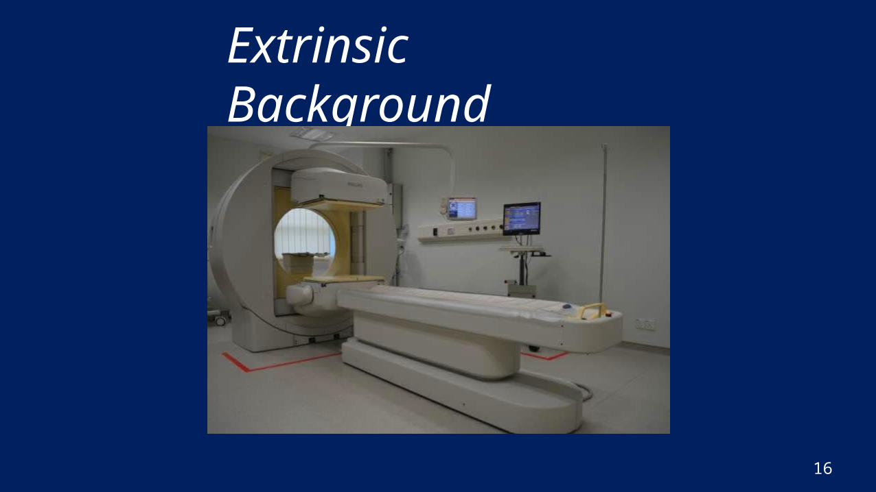
Extrinsic Background
16
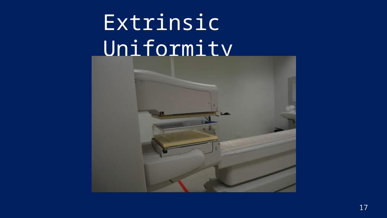
Extrinsic Uniformity
17
Result
18
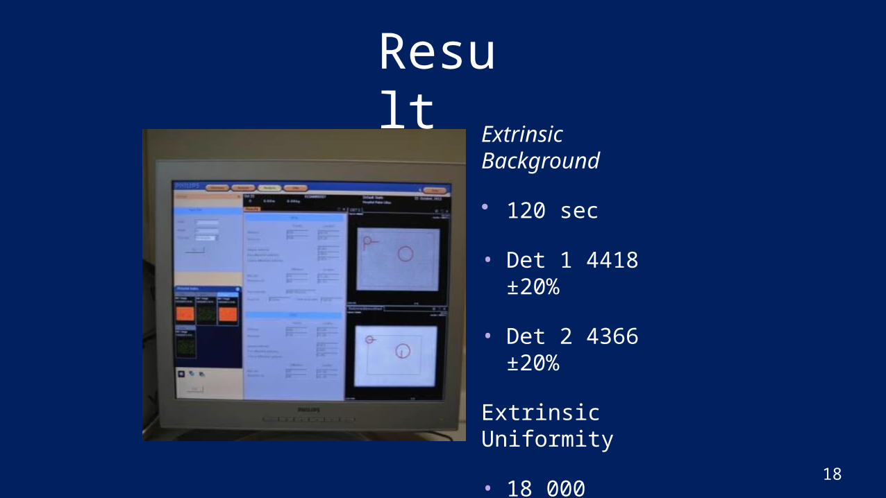
Extrinsic Background
• 120 sec
• Det 1 4418 ±20%
• Det 2 4366 ±20%
Extrinsic Uniformity
• 18 000 counts
• ≤6.0% UFOV
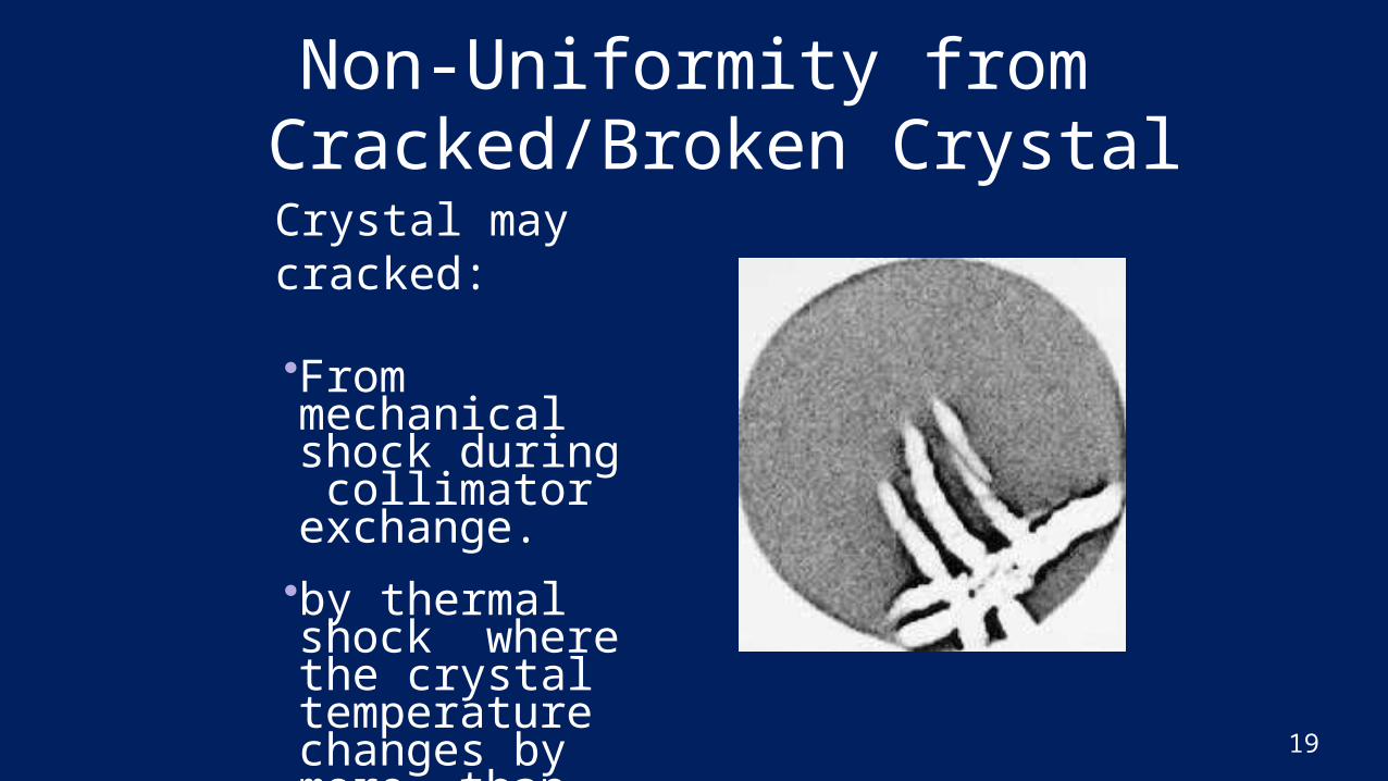
Non-Uniformity from Cracked/Broken Crystal
19
Crystal may cracked:•From mechanical shock during collimator exchange.•by thermal shock where the crystal temperature changes by more than 10 deg./hour.
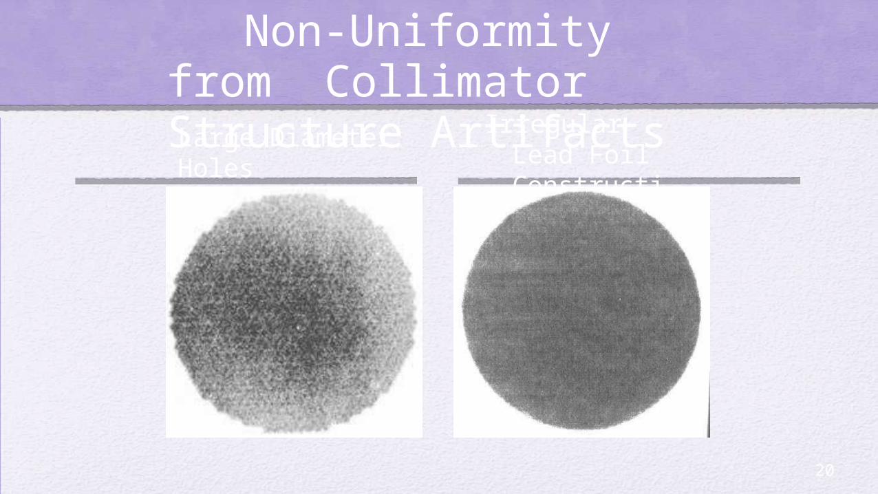
Non-Uniformity from Collimator Structure Artifacts
20
Large Diameter Holes
Irregular Lead Foil Construction
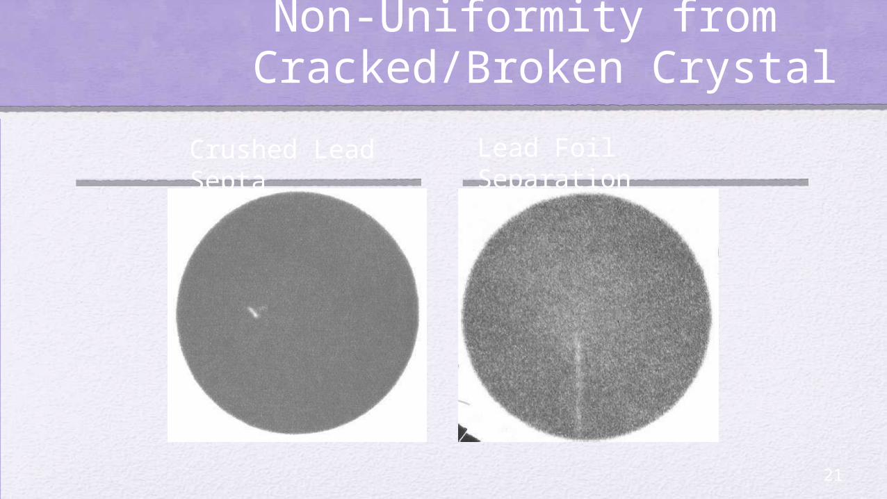
Non-Uniformity from Cracked/Broken Crystal
21
Crushed Lead Septa
Lead Foil Separation
References
22
• Minimum QC for Nuclear Medicine – ANZSNM Technical Standards Subcommittee
• http://www.who.int/medicines/areas/quality_s afety/quality_assurance/en/
Thank you
Floor is open for Questions and Discussion
R D