Embed Size (px)
Citation preview
台大農藝系 遺傳學 601
20000Chapter 1 slide 1
Inborn Error Of
Metabolism
MGL-9
July 17th 2013
Mohammed El-Khateeb
2
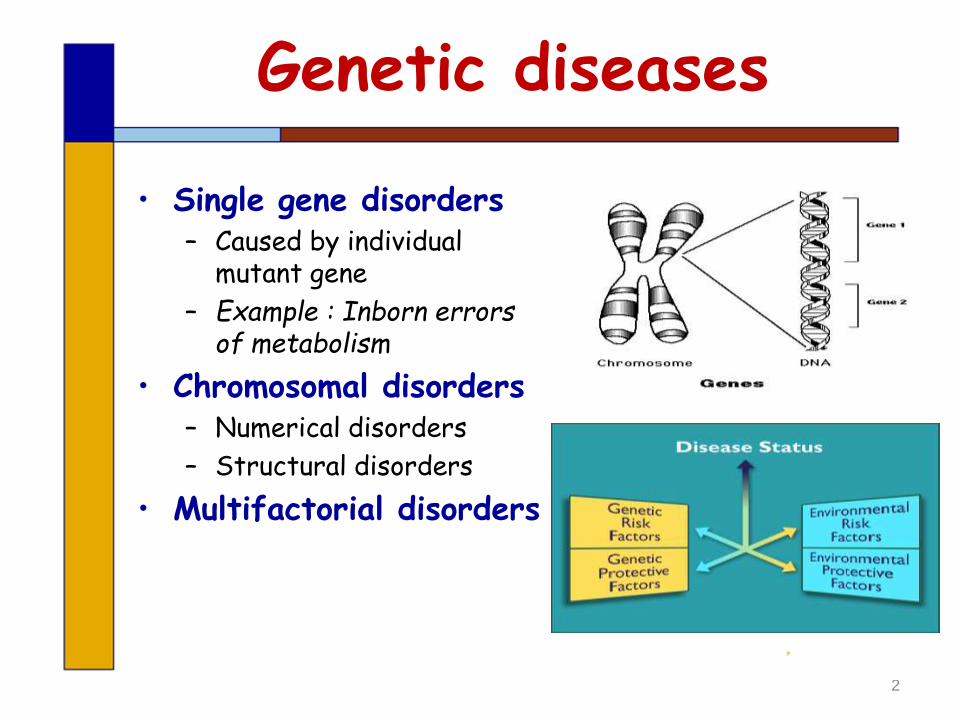
Genetic diseases
• Single gene disorders– Caused by individual
mutant gene
– Example : Inborn errors of metabolism
• Chromosomal disorders– Numerical disorders
– Structural disorders
• Multifactorial disorders
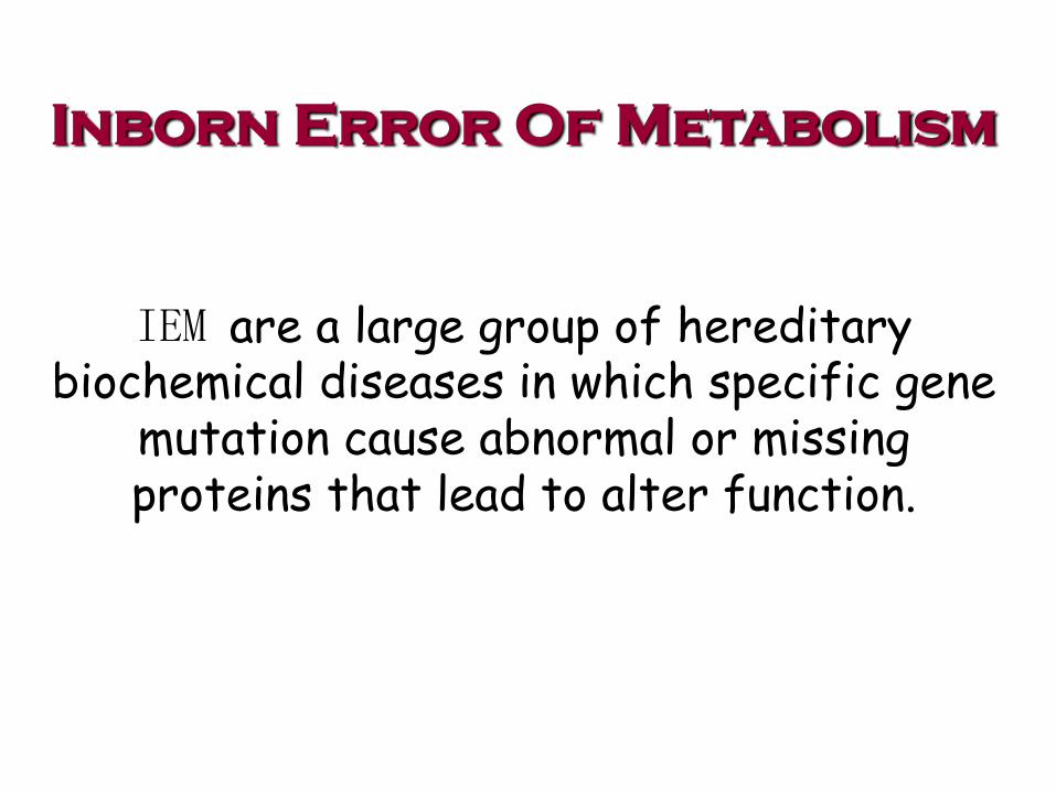
IEM are a large group of hereditary biochemical diseases in which specific gene
mutation cause abnormal or missing proteins that lead to alter function.
Inborn Error Of Metabolism
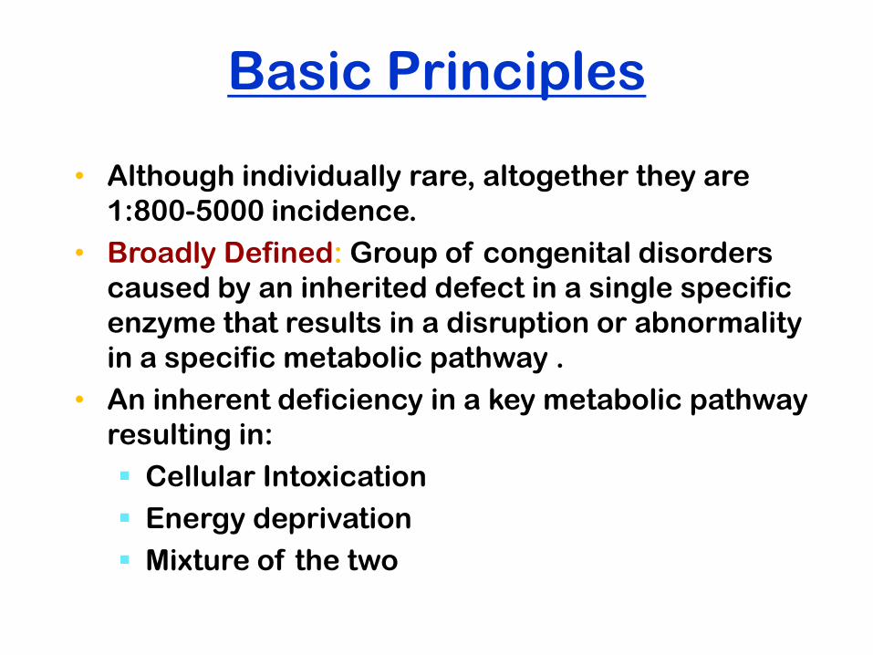
Basic Principles
• Although individually rare, altogether they are
1:800-5000 incidence.
• Broadly Defined: Group of congenital disorders
caused by an inherited defect in a single specific
enzyme that results in a disruption or abnormality
in a specific metabolic pathway .
• An inherent deficiency in a key metabolic pathway
resulting in:
Cellular Intoxication
Energy deprivation
Mixture of the two
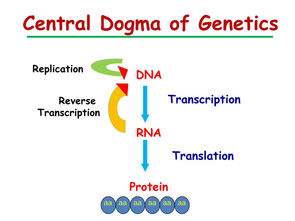
Central Dogma of Genetics
DNA
RNA
Protein
Replication
Transcription
Translation
Reverse Transcription
aa aa aa aa aa aa
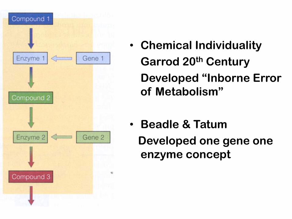
• Chemical Individuality
Garrod 20th Century
Developed “Inborne Error
of Metabolism”
• Beadle & Tatum
Developed one gene one
enzyme concept.
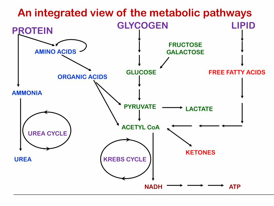
PROTEINGLYCOGEN LIPID
AMINO ACIDSFRUCTOSE
GALACTOSE
FREE FATTY ACIDS
AMMONIA
UREA
UREA CYCLE
ORGANIC ACIDSGLUCOSE
PYRUVATE
ACETYL CoA
KREBS CYCLE
NADH
KETONES
ATP
LACTATE
An integrated view of the metabolic pathways
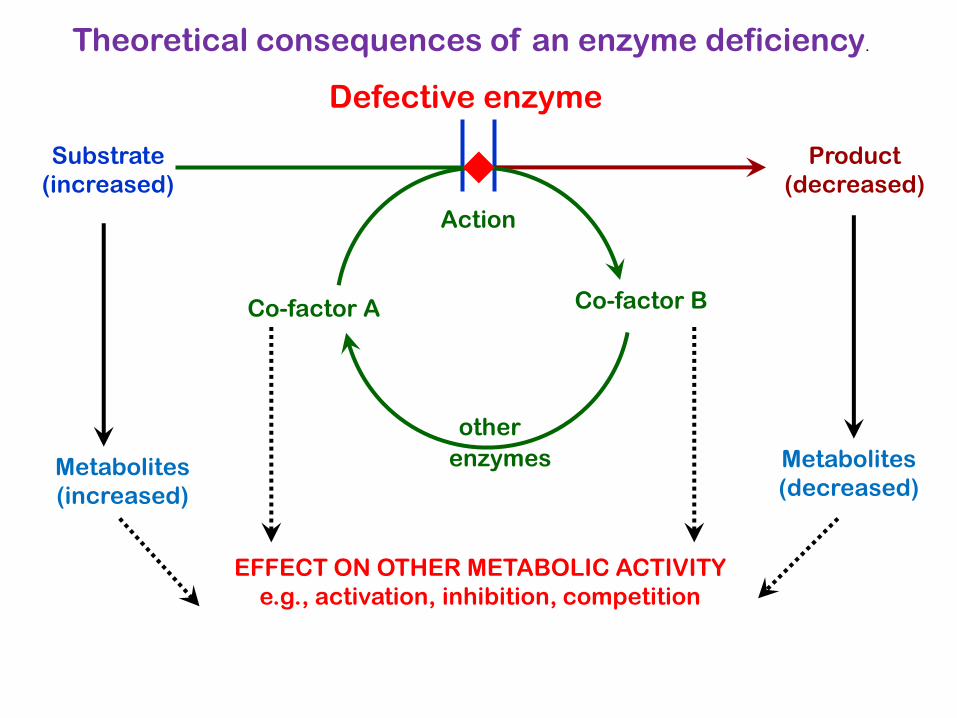
Defective enzyme
Substrate
(increased)
Product
(decreased)
Action
Metabolites
(increased)
Co-factor A Co-factor B
other
enzymes Metabolites
(decreased)
EFFECT ON OTHER METABOLIC ACTIVITY
e.g., activation, inhibition, competition
Theoretical consequences of an enzyme deficiency.
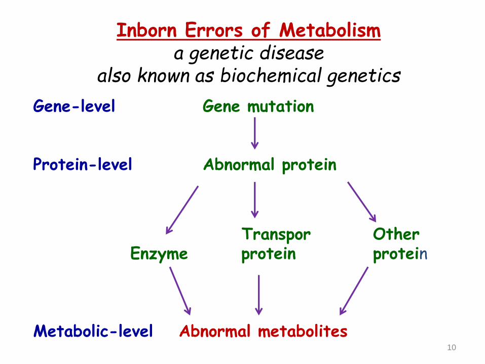
10
Inborn Errors of Metabolisma genetic disease
also known as biochemical genetics
Gene-level Gene mutation
Protein-level Abnormal protein
Transpor OtherEnzyme protein protein
Metabolic-level Abnormal metabolites
Inborn Errors Overview
• General mechanism of problems
Substrate accumulates to toxic levels
Toxic byproducts produced from shunting of accumulated substrate
Deficiency of end product
Poor regulation results in overproduction of intermediates to toxic level
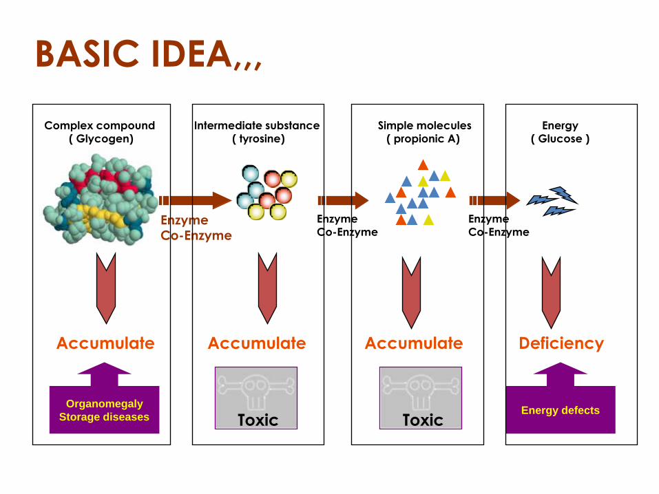
Complex compound
( Glycogen)
Intermediate substance
( tyrosine)
Simple molecules
( propionic A)
Energy
( Glucose )
Accumulate Accumulate Accumulate Deficiency
EnzymeCo-Enzyme
Enzyme
Co-Enzyme
Enzyme
Co-Enzyme
Organomegaly
Storage diseasesEnergy defects
BASIC IDEA,,,
Toxic Toxic
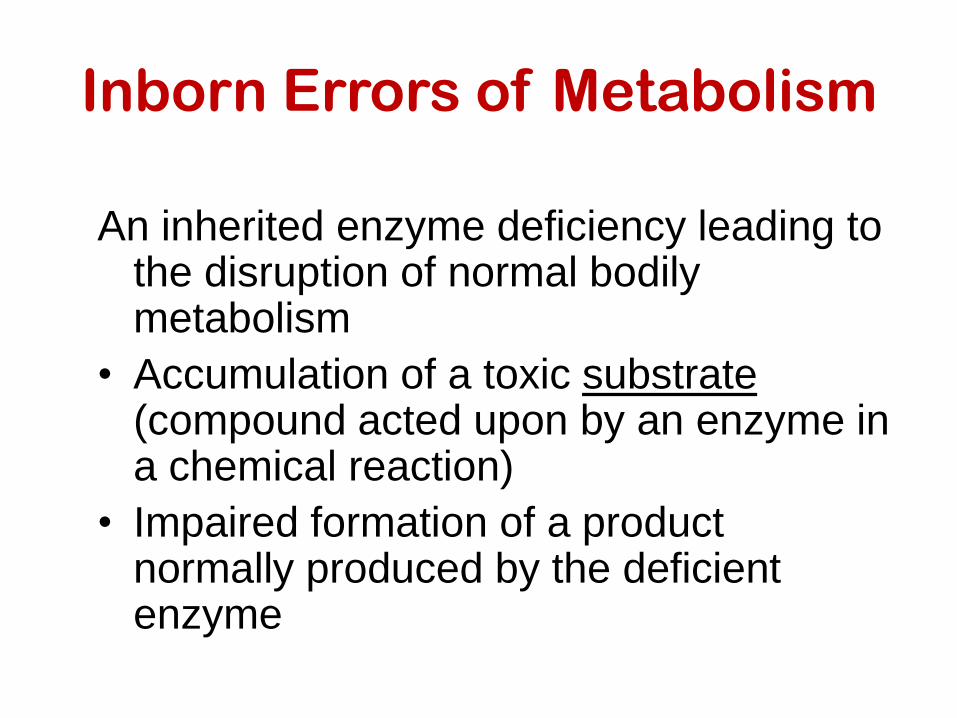
Inborn Errors of Metabolism
An inherited enzyme deficiency leading to the disruption of normal bodily metabolism
• Accumulation of a toxic substrate(compound acted upon by an enzyme in a chemical reaction)
• Impaired formation of a product normally produced by the deficient enzyme
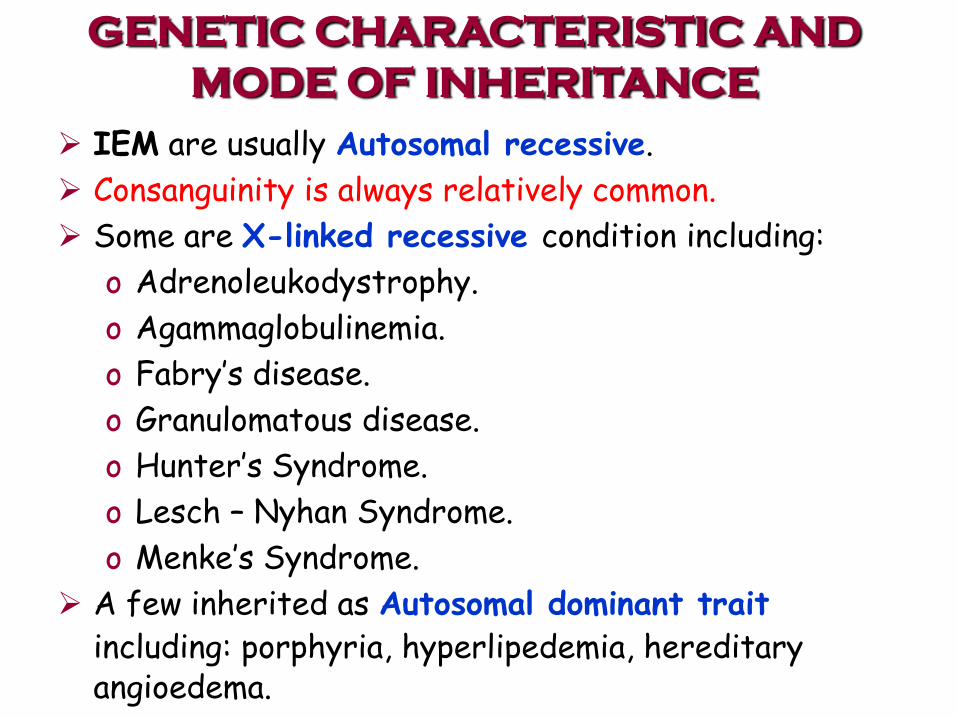
IEM are usually Autosomal recessive.
Consanguinity is always relatively common.
Some are X-linked recessive condition including:
o Adrenoleukodystrophy.
o Agammaglobulinemia.
o Fabry’s disease.
o Granulomatous disease.
o Hunter’s Syndrome.
o Lesch – Nyhan Syndrome.
o Menke’s Syndrome.
A few inherited as Autosomal dominant trait
including: porphyria, hyperlipedemia, hereditaryangioedema.
GENETIC CHARACTERISTIC AND
MODE OF INHERITANCE
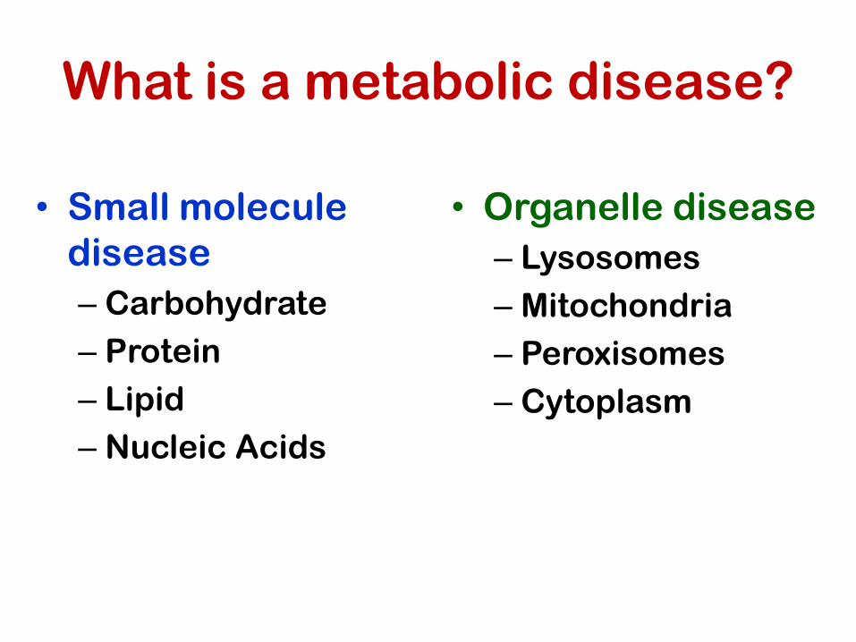
What is a metabolic disease?
• Small molecule
disease
– Carbohydrate
– Protein
– Lipid
– Nucleic Acids
• Organelle disease
– Lysosomes
– Mitochondria
– Peroxisomes
– Cytoplasm
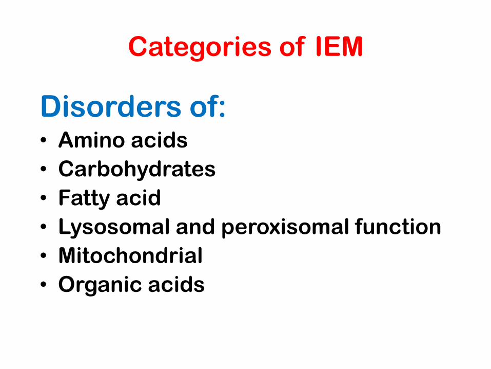
Categories of IEM
Disorders of:• Amino acids
• Carbohydrates
• Fatty acid
• Lysosomal and peroxisomal function
• Mitochondrial
• Organic acids
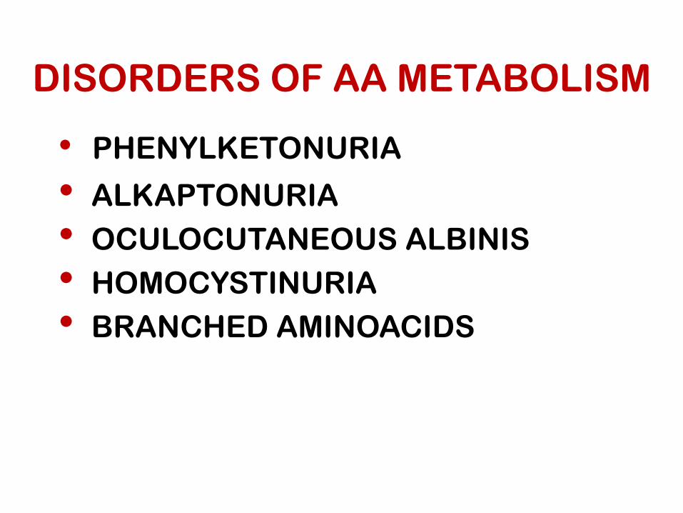
DISORDERS OF AA METABOLISM
• PHENYLKETONURIA
• ALKAPTONURIA
• OCULOCUTANEOUS ALBINIS
• HOMOCYSTINURIA
• BRANCHED AMINOACIDS
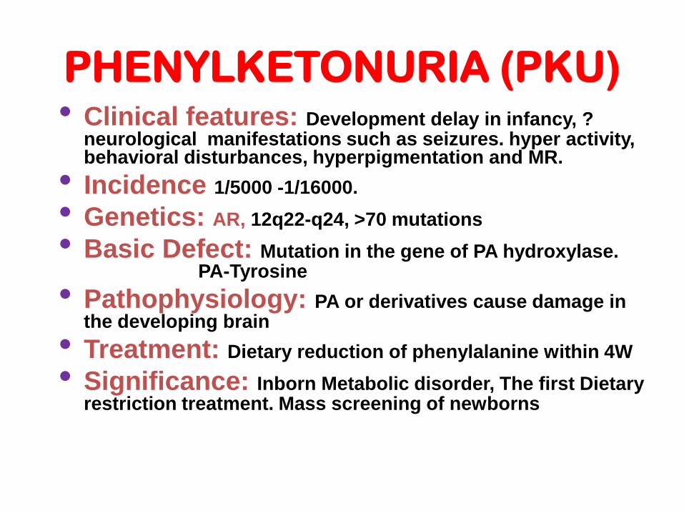
PHENYLKETONURIA (PKU)• Clinical features: Development delay in infancy, ?
neurological manifestations such as seizures. hyper activity, behavioral disturbances, hyperpigmentation and MR.
• Incidence 1/5000 -1/16000.
• Genetics: AR, 12q22-q24, >70 mutations
• Basic Defect: Mutation in the gene of PA hydroxylase. PA-Tyrosine
• Pathophysiology: PA or derivatives cause damage in the developing brain
• Treatment: Dietary reduction of phenylalanine within 4W
• Significance: Inborn Metabolic disorder, The first Dietary restriction treatment. Mass screening of newborns
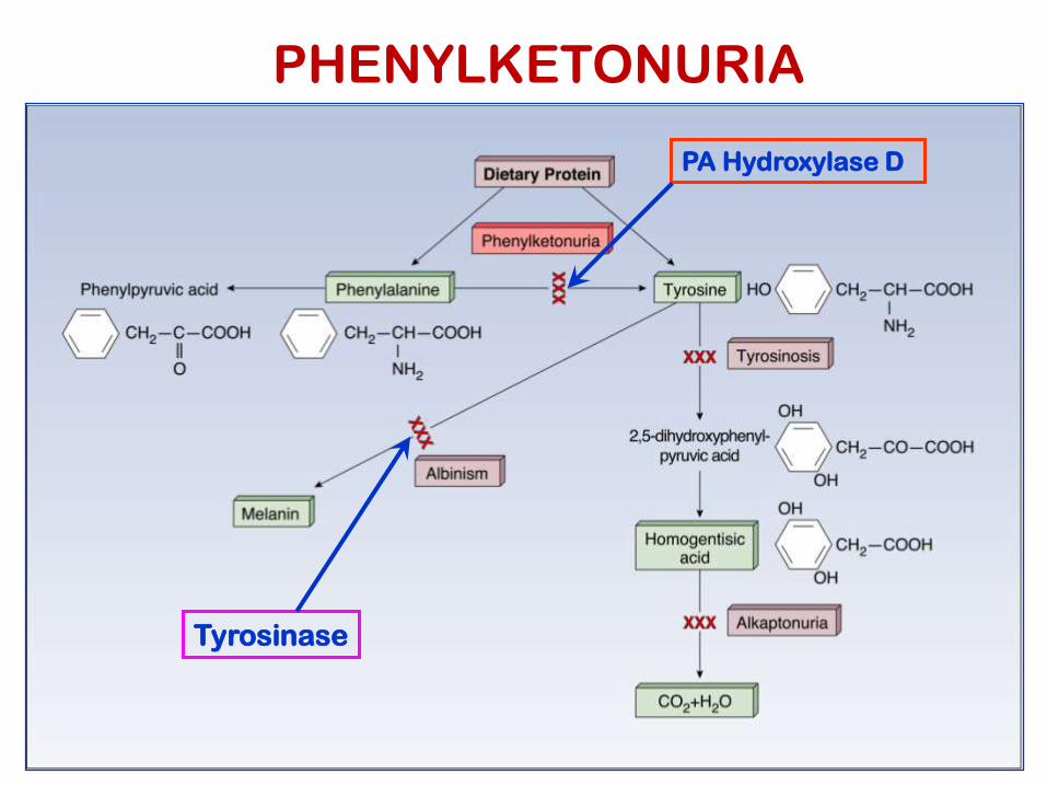
PA Hydroxylase D
Tyrosinase
PHENYLKETONURIA
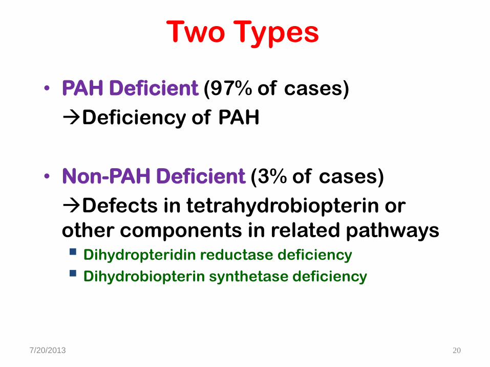
Two Types
• PAH Deficient (97% of cases)
Deficiency of PAH
• Non-PAH Deficient (3% of cases)
Defects in tetrahydrobiopterin or
other components in related pathways Dihydropteridin reductase deficiency
Dihydrobiopterin synthetase deficiency
7/20/2013 20
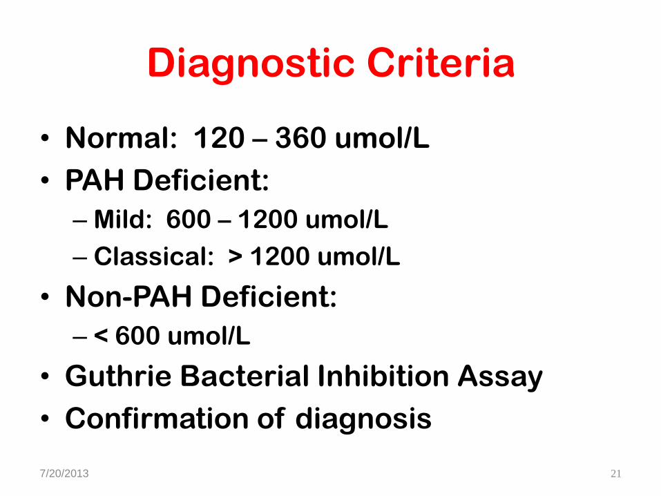
Diagnostic Criteria
• Normal: 120 – 360 umol/L
• PAH Deficient:
– Mild: 600 – 1200 umol/L
– Classical: > 1200 umol/L
• Non-PAH Deficient:
– < 600 umol/L
• Guthrie Bacterial Inhibition Assay
• Confirmation of diagnosis
7/20/2013 21
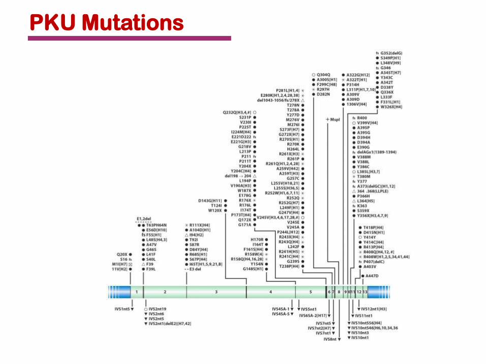
PKU Mutations
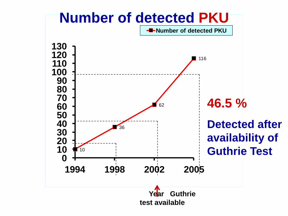
10
36
62
116
0102030405060708090
100110120130
1994 1998 2002 2005
Number of detected PKU
Number of detected PKU
Year Guthrie
test available
46.5 %
Detected after
availability of
Guthrie Test
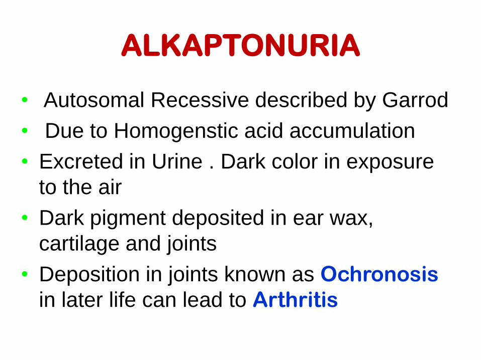
ALKAPTONURIA
• Autosomal Recessive described by Garrod
• Due to Homogenstic acid accumulation
• Excreted in Urine . Dark color in exposure
to the air
• Dark pigment deposited in ear wax,
cartilage and joints
• Deposition in joints known as Ochronosis
in later life can lead to Arthritis
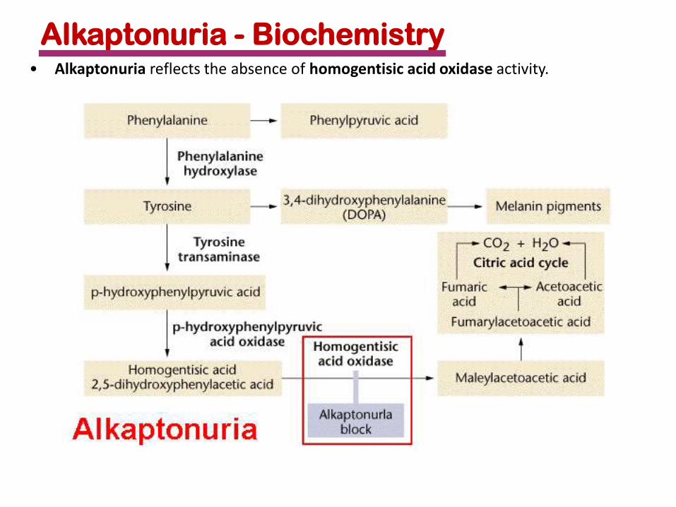
Alkaptonuria - Biochemistry• Alkaptonuria reflects the absence of homogentisic acid oxidase activity.
Normal urine
Urine from patients with
alkaptonuria
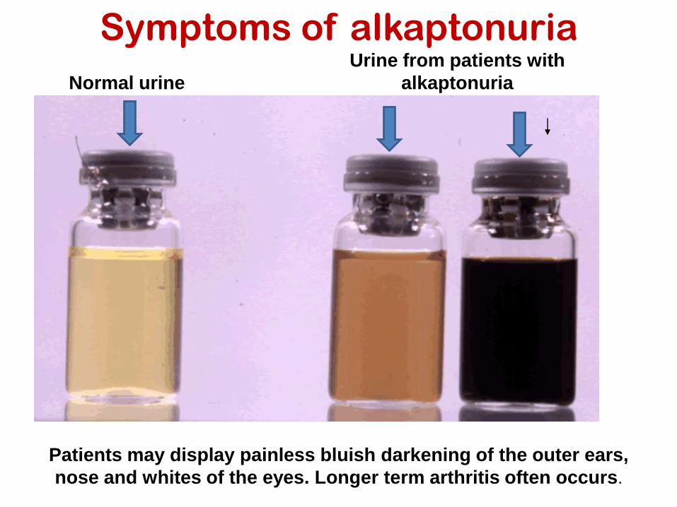
Symptoms of alkaptonuria
Patients may display painless bluish darkening of the outer ears,
nose and whites of the eyes. Longer term arthritis often occurs.
OCULOCUTANEOUS ALBINISM
• OCA is AR due to tyrosinase deficiency no
melanine formation
• No pigment in skin, hair, iris and ocular fundus
• Nystagmus
• Genetically and bichemically heterogeneous
Classical tyrosinase negative
Tyrosinase positive, reduced enzyme level (type 1) OCA 1
located on chromosome11q.
OCA 2 on chromosome 15q (pink-eye)
Third loci OCA-3 not related to above mentioned
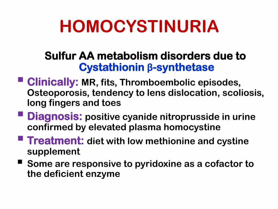
HOMOCYSTINURIA
Sulfur AA metabolism disorders due toCystathionin β-synthetase
Clinically: MR, fits, Thromboembolic episodes, Osteoporosis, tendency to lens dislocation, scoliosis, long fingers and toes
Diagnosis: positive cyanide nitroprusside in urine confirmed by elevated plasma homocystine
Treatment: diet with low methionine and cystine supplement
Some are responsive to pyridoxine as a cofactor to the deficient enzyme
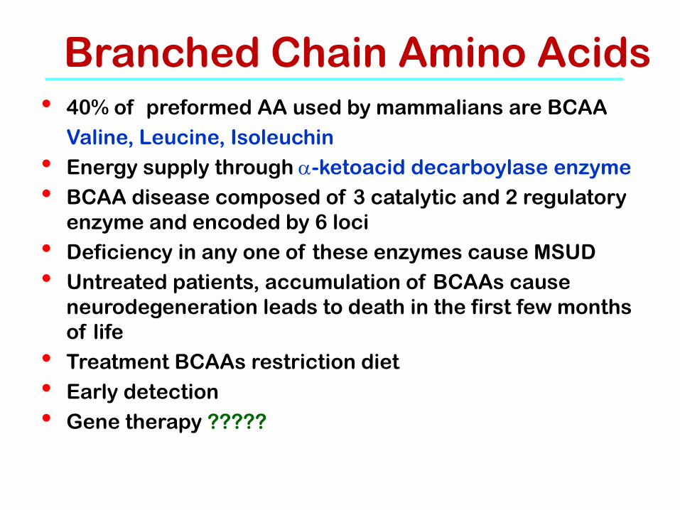
Branched Chain Amino Acids• 40% of preformed AA used by mammalians are BCAA
Valine, Leucine, Isoleuchin
• Energy supply through -ketoacid decarboylase enzyme
• BCAA disease composed of 3 catalytic and 2 regulatory
enzyme and encoded by 6 loci
• Deficiency in any one of these enzymes cause MSUD
• Untreated patients, accumulation of BCAAs cause
neurodegeneration leads to death in the first few months
of life
• Treatment BCAAs restriction diet
• Early detection
• Gene therapy ?????
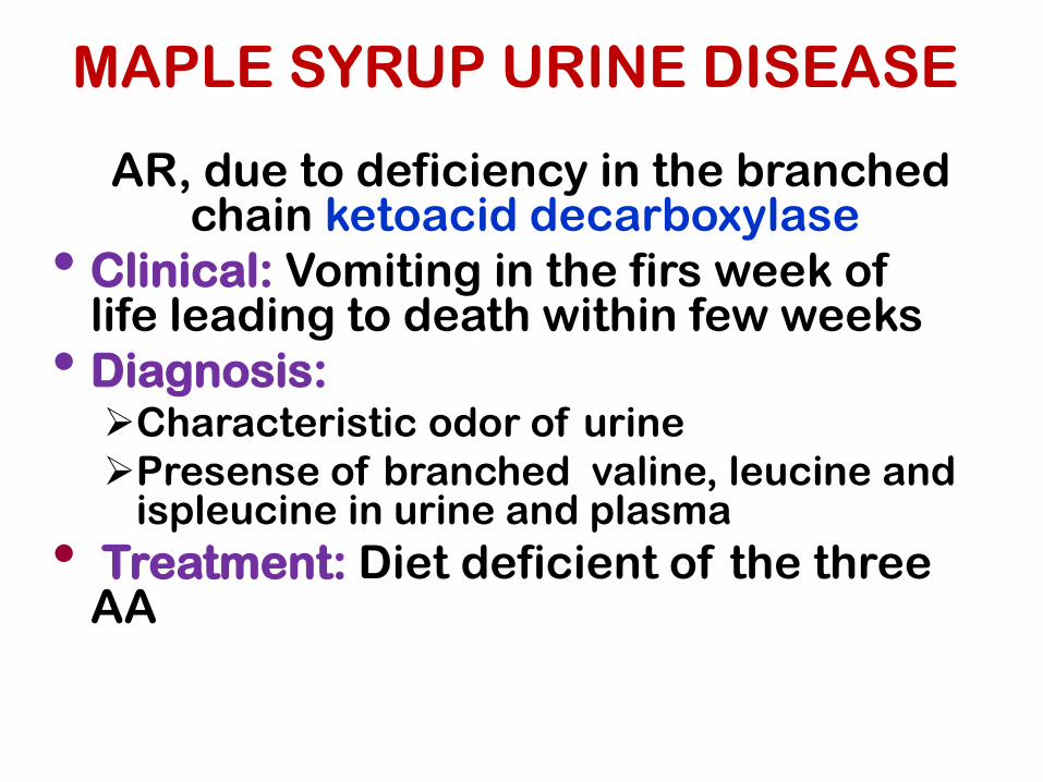
MAPLE SYRUP URINE DISEASE
• AR, due to deficiency in the branched chain ketoacid decarboxylase
• Clinical: Vomiting in the firs week of life leading to death within few weeks
• Diagnosis:Characteristic odor of urine
Presense of branched valine, leucine and ispleucine in urine and plasma
• Treatment: Diet deficient of the three AA
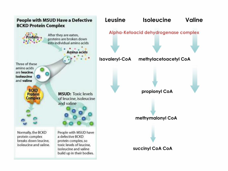
Leusine Isoleucine Valine
Isovaleryl-CoA methylacetoacetyl CoA
propionyl CoA
methymalonyl CoA
succinyl CoA CoA
Alpha-Ketoacid dehydrogenase complex
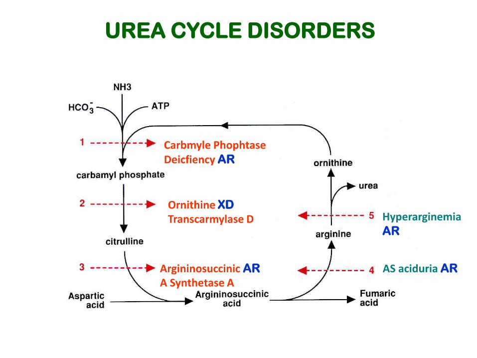
UREA CYCLE DISORDERS
Carbmyle Phophtase Deicfiency AR
Ornithine XD
Transcarmylase D
Argininosuccinic AR
A Synthetase AAS aciduria AR
HyperarginemiaAR
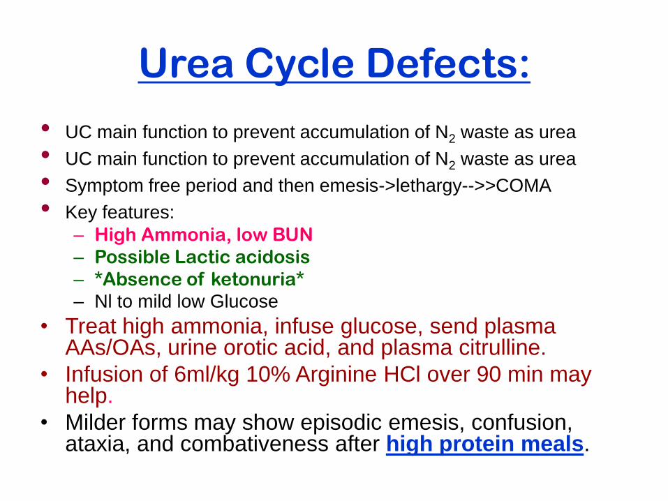
Urea Cycle Defects:
• UC main function to prevent accumulation of N2 waste as urea
• UC main function to prevent accumulation of N2 waste as urea
• Symptom free period and then emesis->lethargy-->>COMA
• Key features:
– High Ammonia, low BUN
– Possible Lactic acidosis
– *Absence of ketonuria*
– Nl to mild low Glucose
• Treat high ammonia, infuse glucose, send plasma AAs/OAs, urine orotic acid, and plasma citrulline.
• Infusion of 6ml/kg 10% Arginine HCl over 90 min may help.
• Milder forms may show episodic emesis, confusion, ataxia, and combativeness after high protein meals.
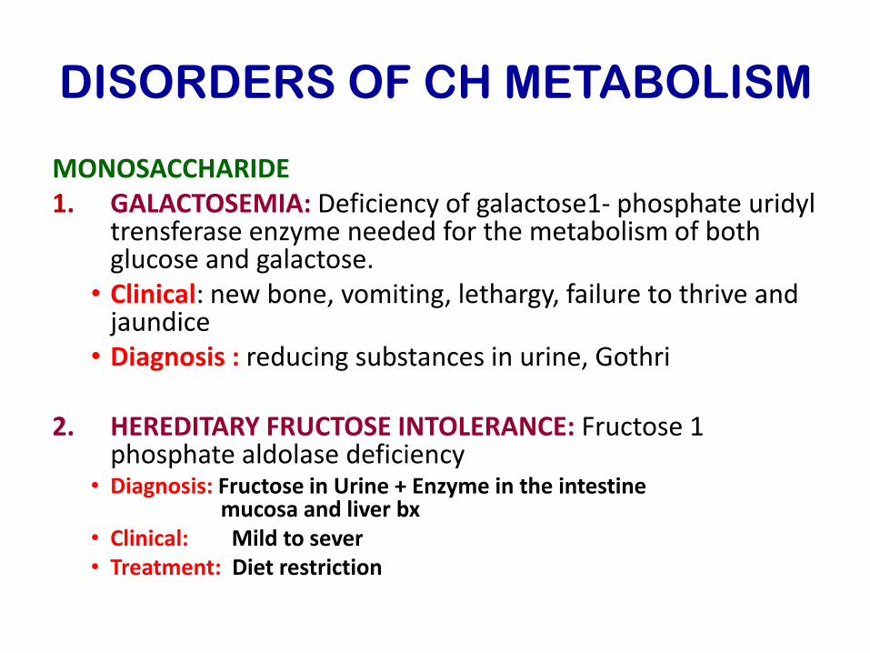
DISORDERS OF CH METABOLISM
MONOSACCHARIDE1. GALACTOSEMIA: Deficiency of galactose1- phosphate uridyl
trensferase enzyme needed for the metabolism of both glucose and galactose.
• Clinical: new bone, vomiting, lethargy, failure to thrive and jaundice
• Diagnosis : reducing substances in urine, Gothri t• est.2. HEREDITARY FRUCTOSE INTOLERANCE: Fructose 1
phosphate aldolase deficiency• Diagnosis: Fructose in Urine + Enzyme in the intestine
mucosa and liver bx• Clinical: Mild to sever• Treatment: Diet restriction
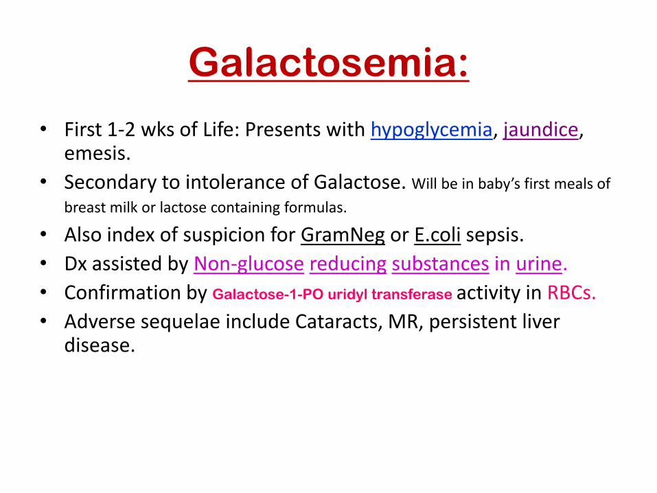
Galactosemia:
• First 1-2 wks of Life: Presents with hypoglycemia, jaundice, emesis.
• Secondary to intolerance of Galactose. Will be in baby’s first meals of
breast milk or lactose containing formulas.
• Also index of suspicion for GramNeg or E.coli sepsis.
• Dx assisted by Non-glucose reducing substances in urine.
• Confirmation by Galactose-1-PO uridyl transferase activity in RBCs.
• Adverse sequelae include Cataracts, MR, persistent liver disease.

Galactosemia
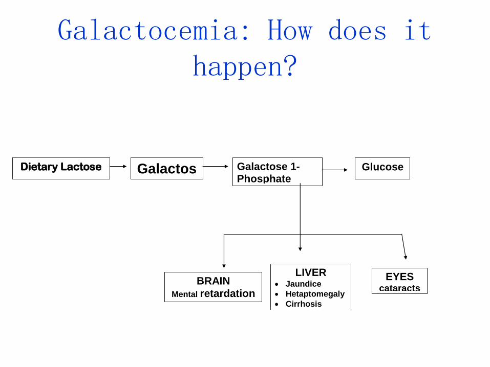
Galactocemia: How does it happen?
Dietary Lactose Galactose
Galactose 1-Phosphate
Glucose
BRAIN
Mental retardation
LIVER Jaundice
Hetaptomegaly
Cirrhosis
EYES cataracts
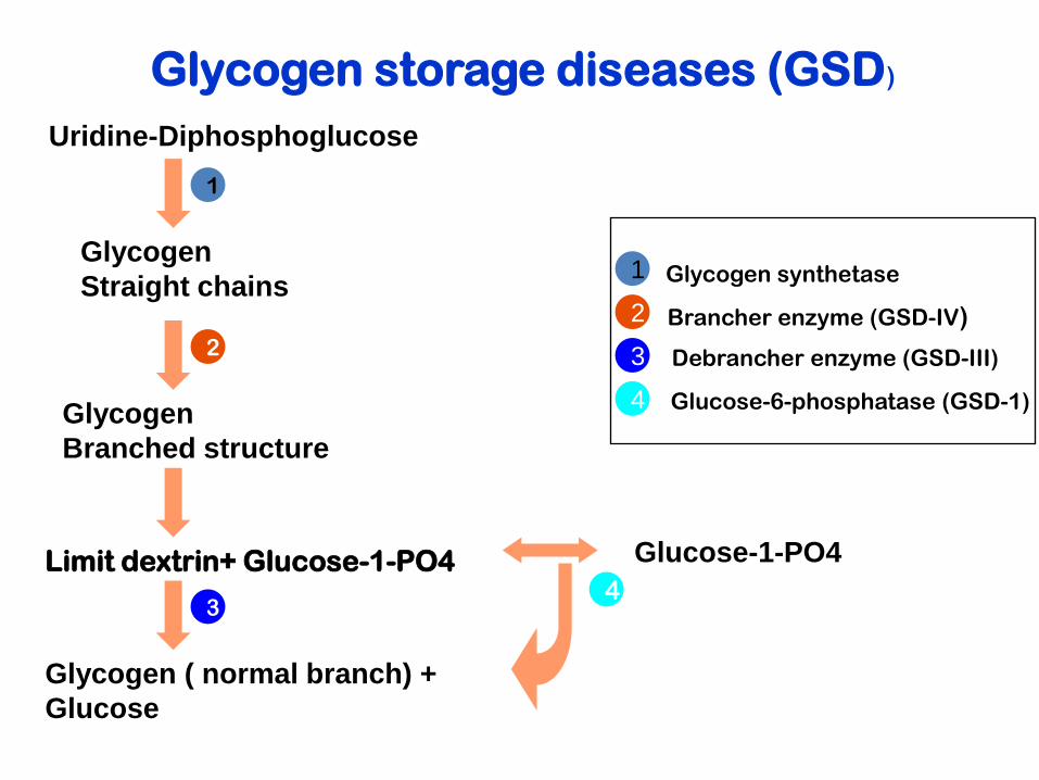
Uridine-Diphosphoglucose
Glycogen
Straight chains
Glycogen
Branched structure
Limit dextrin+ Glucose-1-PO4 Glucose-1-PO4
Glycogen ( normal branch) +
Glucose
3
1
1
2
Glycogen synthetase
Brancher enzyme (GSD-IV)
Debrancher enzyme (GSD-III)
Glucose-6-phosphatase (GSD-1)
2
3
4
4
Glycogen storage diseases (GSD)
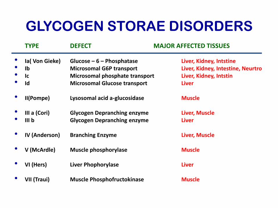
GLYCOGEN STORAE DISORDERS
TYPE DEFECT MAJOR AFFECTED TISSUES
• Ia( Von Gieke) Glucose – 6 – Phosphatase Liver, Kidney, Intstine• Ib Microsomal G6P transport Liver, Kidney, Intestine, Neurtro• Ic Microsomal phosphate transport Liver, Kidney, Intstin• Id Microsomal Glucose transport Liver
• II(Pompe) Lysosomal acid a-glucosidase Muscle
• III a (Cori) Glycogen Depranching enzyme Liver, Muscle• III b Glycogen Depranching enzyme Liver
• IV (Anderson) Branching Enzyme Liver, Muscle
• V (McArdle) Muscle phosphorylase Muscle
• VI (Hers) Liver Phophorylase Liver
• VII (Traui) Muscle Phosphofructokinase Muscle
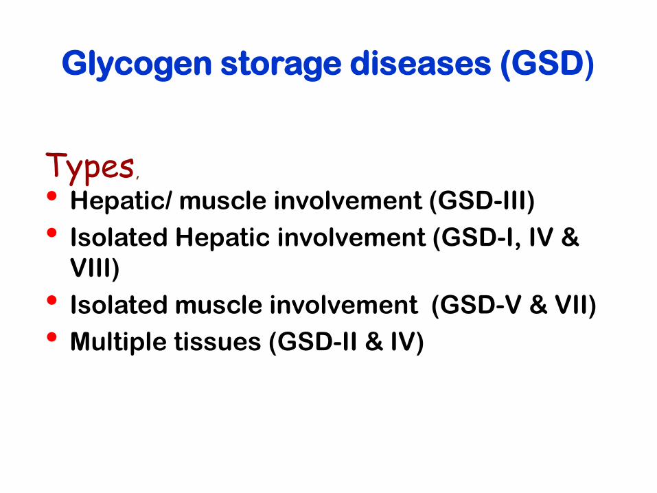
Glycogen storage diseases (GSD)
• Hepatic/ muscle involvement (GSD-III)
• Isolated Hepatic involvement (GSD-I, IV &
VIII)
• Isolated muscle involvement (GSD-V & VII)
• Multiple tissues (GSD-II & IV)
Types,
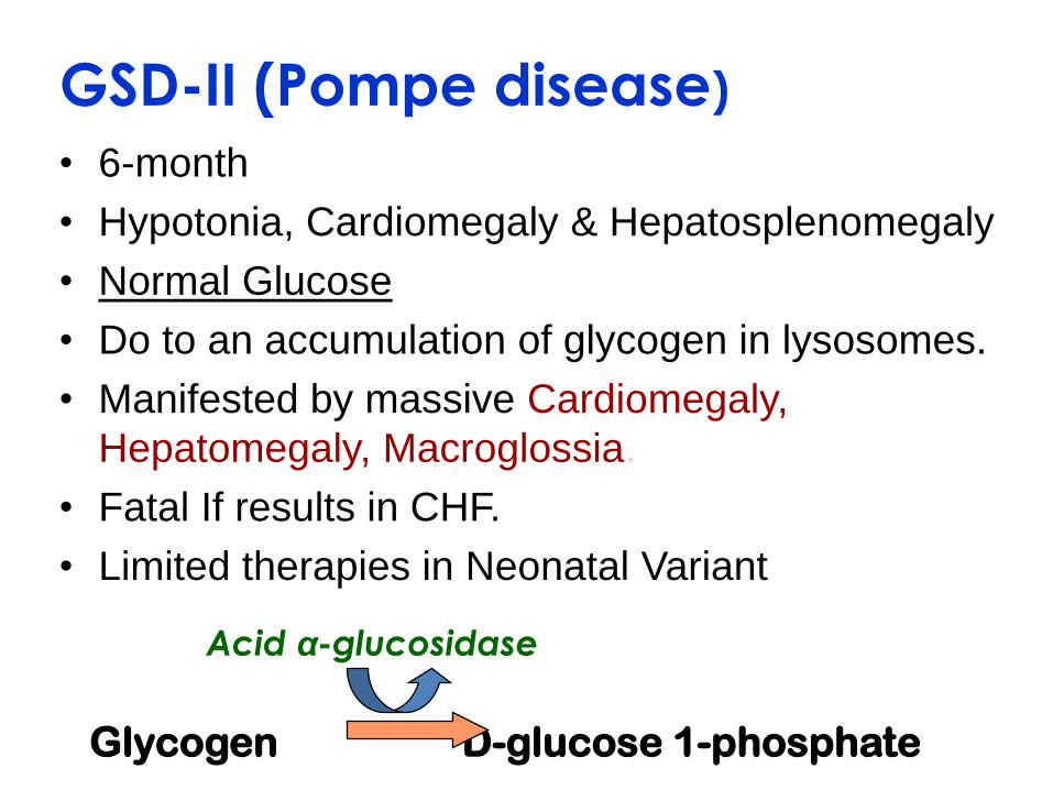
GSD-II (Pompe disease)
• 6-month
• Hypotonia, Cardiomegaly & Hepatosplenomegaly
• Normal Glucose
• Do to an accumulation of glycogen in lysosomes.
• Manifested by massive Cardiomegaly,
Hepatomegaly, Macroglossia.
• Fatal If results in CHF.
• Limited therapies in Neonatal Variant
Acid α-glucosidase
Glycogen D-glucose 1-phosphate
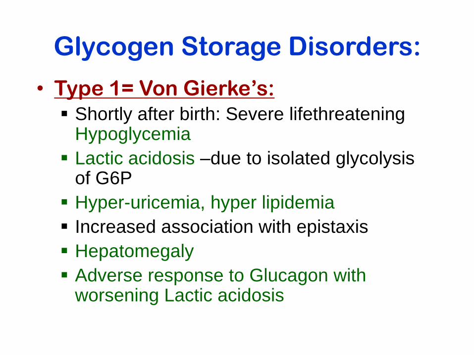
Glycogen Storage Disorders:
• Type 1= Von Gierke’s: Shortly after birth: Severe lifethreatening
Hypoglycemia
Lactic acidosis –due to isolated glycolysis of G6P
Hyper-uricemia, hyper lipidemia
Increased association with epistaxis
Hepatomegaly
Adverse response to Glucagon with worsening Lactic acidosis
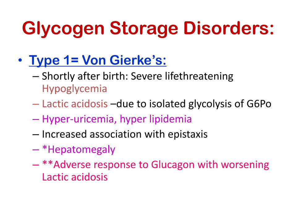
Glycogen Storage Disorders:
• Type 1= Von Gierke’s:– Shortly after birth: Severe lifethreatening
Hypoglycemia
– Lactic acidosis –due to isolated glycolysis of G6Po
– Hyper-uricemia, hyper lipidemia
– Increased association with epistaxis
– *Hepatomegaly
– **Adverse response to Glucagon with worsening Lactic acidosis
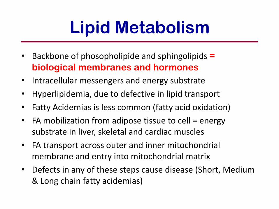
Lipid Metabolism
• Backbone of phosopholipide and sphingolipids =
biological membranes and hormones
• Intracellular messengers and energy substrate
• Hyperlipidemia, due to defective in lipid transport
• Fatty Acidemias is less common (fatty acid oxidation)
• FA mobilization from adipose tissue to cell = energy substrate in liver, skeletal and cardiac muscles
• FA transport across outer and inner mitochondrial membrane and entry into mitochondrial matrix
• Defects in any of these steps cause disease (Short, Medium & Long chain fatty acidemias)
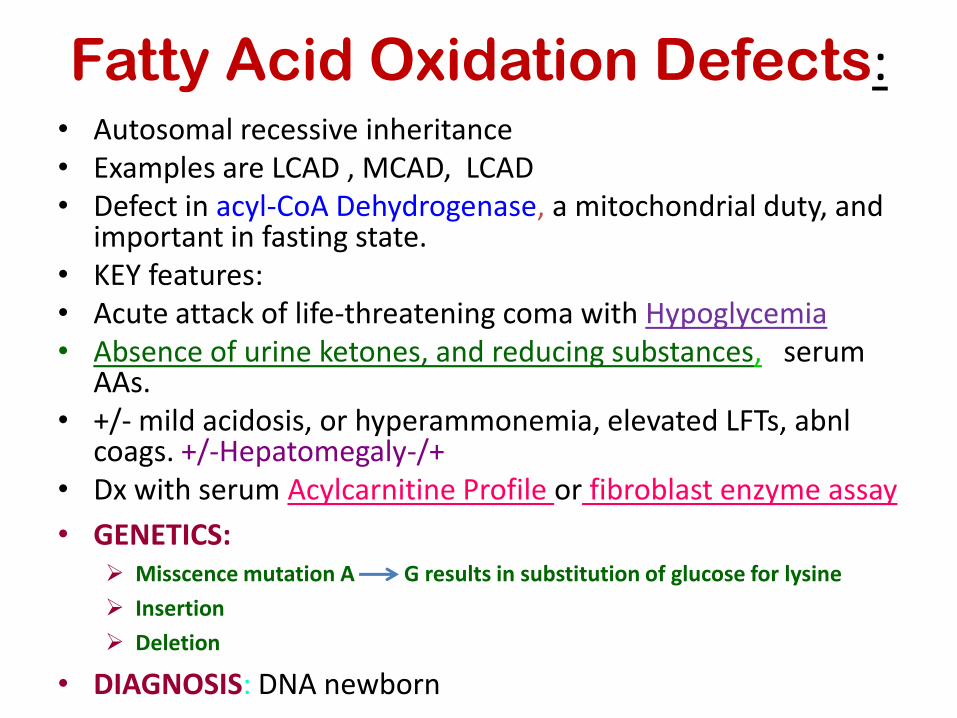
Fatty Acid Oxidation Defects:• Autosomal recessive inheritance • Examples are LCAD , MCAD, LCAD• Defect in acyl-CoA Dehydrogenase, a mitochondrial duty, and
important in fasting state. • KEY features:• Acute attack of life-threatening coma with Hypoglycemia• Absence of urine ketones, and reducing substances, serum
AAs. • +/- mild acidosis, or hyperammonemia, elevated LFTs, abnl
coags. +/-Hepatomegaly-/+• Dx with serum Acylcarnitine Profile or fibroblast enzyme assay
• GENETICS: Misscence mutation A G results in substitution of glucose for lysine
Insertion
Deletion
• DIAGNOSIS: DNA newborn
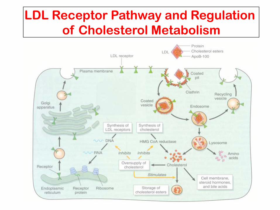
LDL Receptor Pathway and Regulation
of Cholesterol Metabolism
Mucoploysaccharides
(glycosaminoglycans
Bone, connective tissue,
skin, cornea,joints etc
Cell membranes,
organelles
Bacteria,
viruses
Lysosome
Sphingolipids,
glycolipids etc
Food
particles
Glycoproteins
Acid hydrolases
“The cells
wrecking crew”
Glycogen
Abnormal
lysosomal
storage leads to
developmental regression
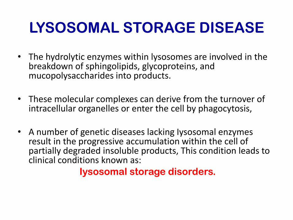
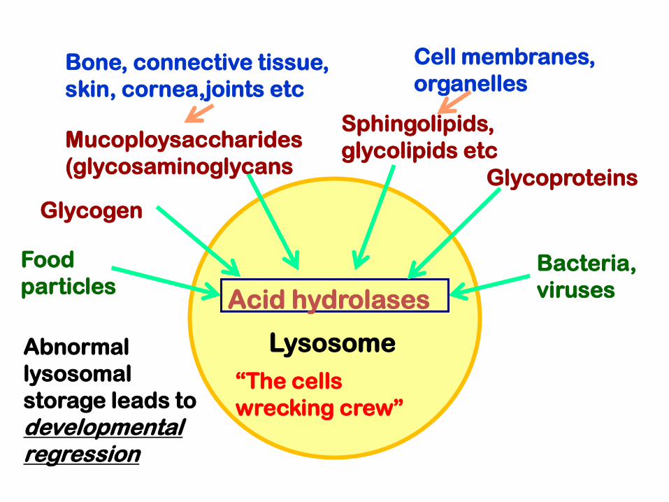
LYSOSOMAL STORAGE DISEASE
• The hydrolytic enzymes within lysosomes are involved in the breakdown of sphingolipids, glycoproteins, and mucopolysaccharides into products.
• These molecular complexes can derive from the turnover of intracellular organelles or enter the cell by phagocytosis,
• A number of genetic diseases lacking Iysosomal enzymes result in the progressive accumulation within the cell of partially degraded insoluble products, This condition leads to clinical conditions known as:
Iysosomal storage disorders.
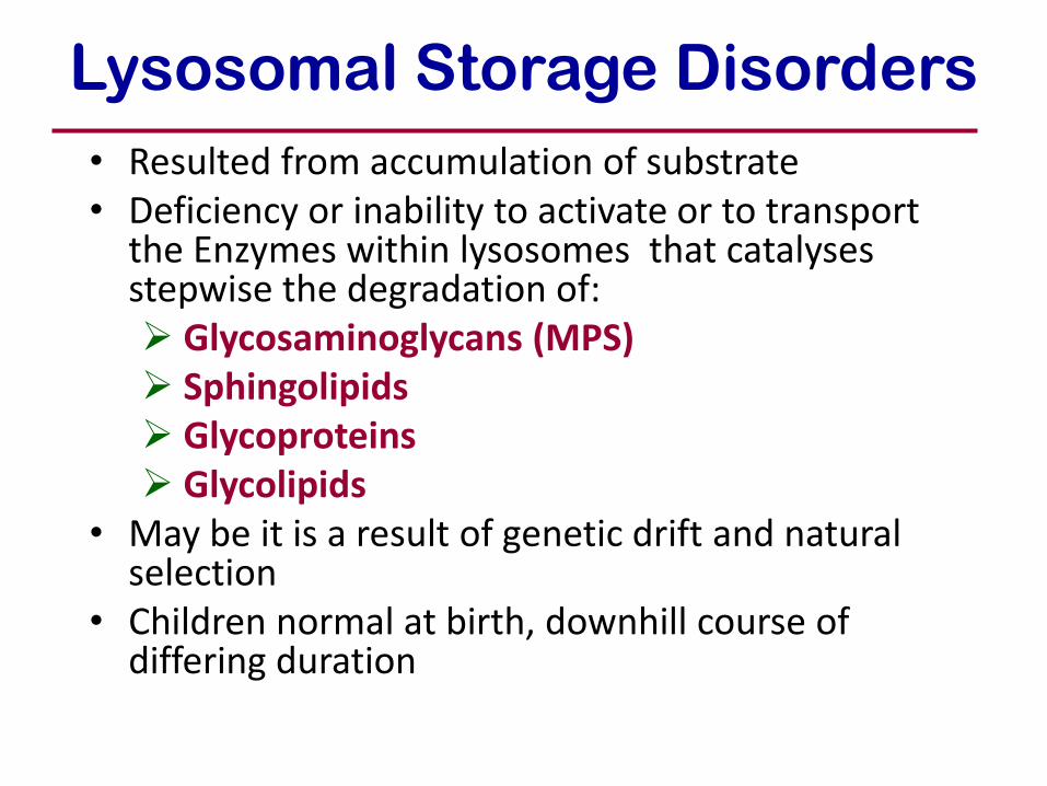
Lysosomal Storage Disorders
• Resulted from accumulation of substrate• Deficiency or inability to activate or to transport
the Enzymes within lysosomes that catalyses stepwise the degradation of: Glycosaminoglycans (MPS) Sphingolipids Glycoproteins Glycolipids
• May be it is a result of genetic drift and natural selection
• Children normal at birth, downhill course of differing duration
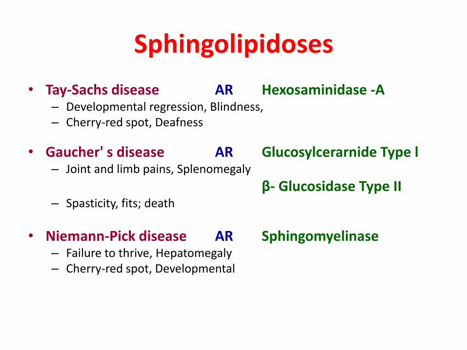
Sphingolipidoses
• Tay-Sachs disease AR Hexosaminidase -A– Developmental regression, Blindness, – Cherry-red spot, Deafness
• Gaucher' s disease AR Glucosylcerarnide Type l– Joint and limb pains, Splenomegaly
β- Glucosidase Type II– Spasticity, fits; death
• Niemann-Pick disease AR Sphingomyelinase– Failure to thrive, Hepatomegaly– Cherry-red spot, Developmental regression
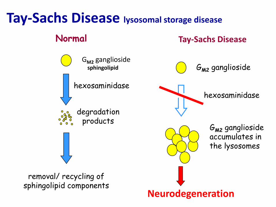
Tay-Sachs Disease lysosomal storage disease
Normal Tay-Sachs Disease
GM2 gangliosidesphingolipid
hexosaminidase A
GM2 ganglioside
hexosaminidase A
removal/ recycling ofsphingolipid components
GM2 ganglioside accumulates in the lysosomes
degradationproducts
Neurodegeneration
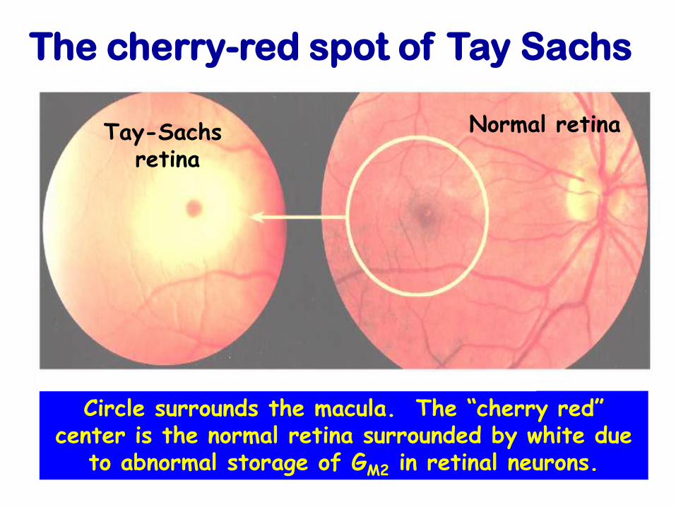
The cherry-red spot of Tay Sachs
Circle surrounds the macula. The “cherry red” center is the normal retina surrounded by white due
to abnormal storage of GM2 in retinal neurons.
Tay-Sachs retina
Normal retina
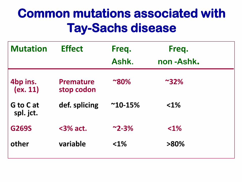
Common mutations associated with
Tay-Sachs disease
Mutation Effect Freq. Freq.
Ashk. non -Ashk.
4bp ins. Premature ~80% ~32%(ex. 11) stop codon
G to C at def. splicing ~10-15% <1%spl. jct.
G269S <3% act. ~2-3% <1%
other variable <1% >80%
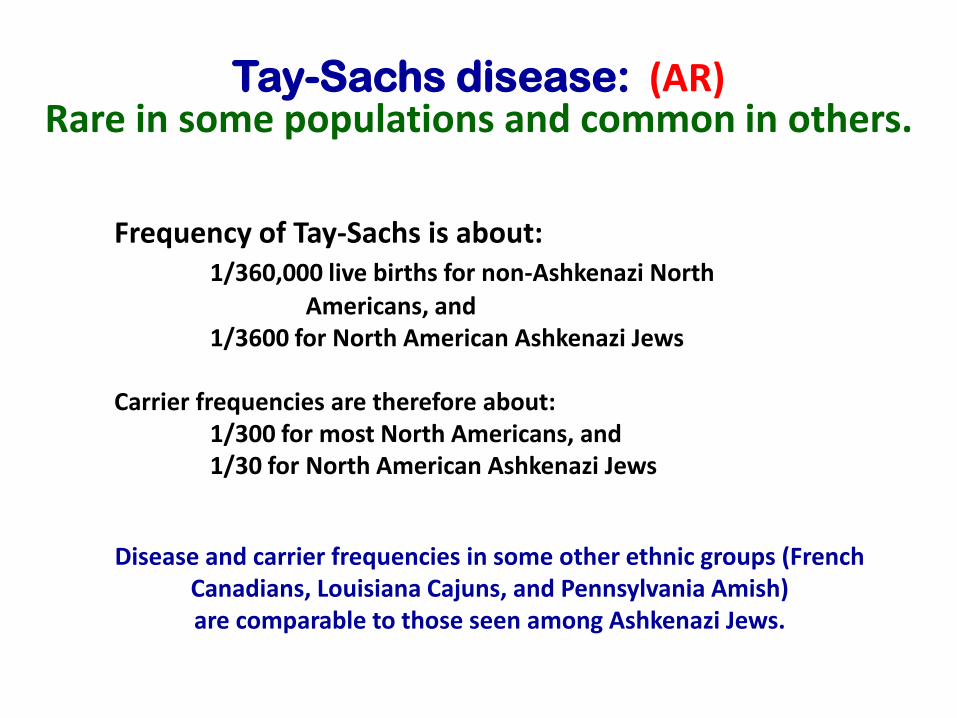
Tay-Sachs disease: (AR)Rare in some populations and common in others.
Disease and carrier frequencies in some other ethnic groups (French Canadians, Louisiana Cajuns, and Pennsylvania Amish) are comparable to those seen among Ashkenazi Jews.
Frequency of Tay-Sachs is about:1/360,000 live births for non-Ashkenazi North
Americans, and 1/3600 for North American Ashkenazi Jews
Carrier frequencies are therefore about: 1/300 for most North Americans, and1/30 for North American Ashkenazi Jews

Peroxisomal Disorders
• Zellweger Syndrome• aka: Cerebro-hepato-
renal syndrome
• Typical and easily recognized dysmorphic facies.
• Progressive degeneration of Brain/Liver/Kidney, with death ~6 mo after onset.
• When screening for PDs. obtain serum Very Long Chain Fatty Acids-VLCFAs
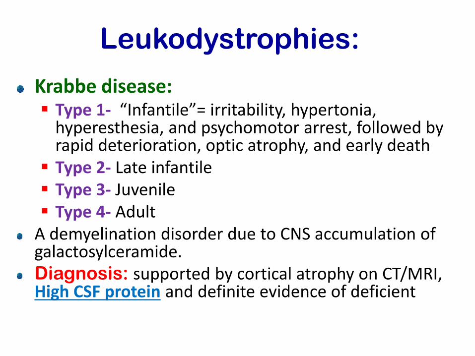
Leukodystrophies:
Krabbe disease: Type 1- “Infantile”= irritability, hypertonia,
hyperesthesia, and psychomotor arrest, followed by rapid deterioration, optic atrophy, and early death Type 2- Late infantile Type 3- Juvenile Type 4- Adult
A demyelination disorder due to CNS accumulation of galactosylceramide. Diagnosis: supported by cortical atrophy on CT/MRI, High CSF protein and definite evidence of deficient
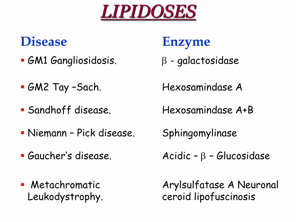
LIPIDOSES
Enzyme Disease
- galactosidase GM1 Gangliosidosis.
Hexosamindase A GM2 Tay –Sach.
Hexosamindase A+B Sandhoff disease.
Sphingomylinase Niemann – Pick disease.
Acidic – – Glucosidase Gaucher’s disease.
Arylsulfatase A Neuronal ceroid lipofuscinosis
MetachromaticLeukodystrophy.
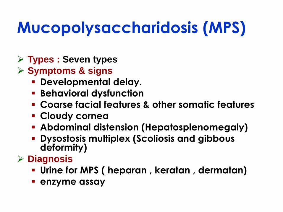
Types : Seven types
Symptoms & signs
Developmental delay.
Behavioral dysfunction
Coarse facial features & other somatic features
Cloudy cornea
Abdominal distension (Hepatosplenomegaly)
Dysostosis multiplex (Scoliosis and gibbous deformity)
Diagnosis
Urine for MPS ( heparan , keratan , dermatan)
enzyme assay
Mucopolysaccharidosis (MPS)
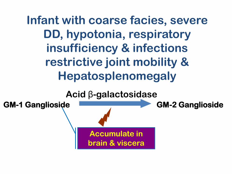
Infant with coarse facies, severe
DD, hypotonia, respiratory
insufficiency & infections
restrictive joint mobility &
Hepatosplenomegaly
GM-1 Ganglioside GM-2 Ganglioside
Acid β-galactosidase
Accumulate in
brain & viscera
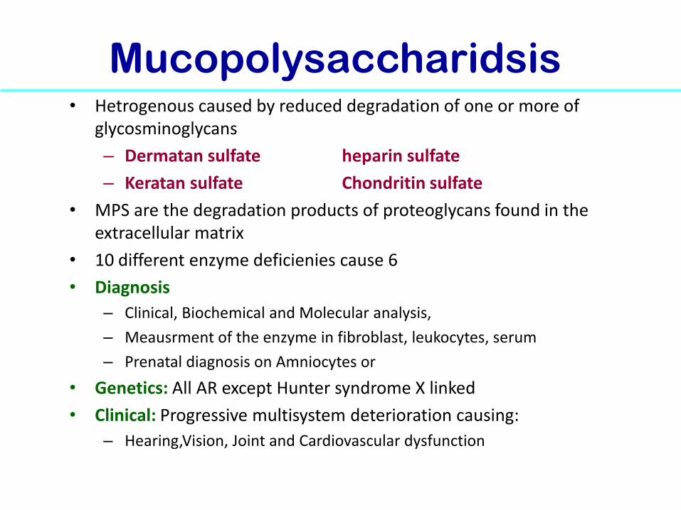
Mucopolysaccharidsis• Hetrogenous caused by reduced degradation of one or more of
glycosminoglycans
– Dermatan sulfate heparin sulfate
– Keratan sulfate Chondritin sulfate
• MPS are the degradation products of proteoglycans found in the extracellular matrix
• 10 different enzyme deficienies cause 6 MPS disorders
• Diagnosis
– Clinical, Biochemical and Molecular analysis,
– Meausrment of the enzyme in fibroblast, leukocytes, serum
– Prenatal diagnosis on Amniocytes or CVS
• Genetics: All AR except Hunter syndrome X linked
• Clinical: Progressive multisystem deterioration causing:
– Hearing,Vision, Joint and Cardiovascular dysfunction
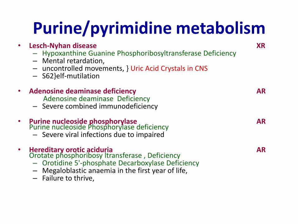
Purine/pyrimidine metabolism• Lesch-Nyhan disease XR
– Hypoxanthine Guanine Phosphoribosyltransferase Deficiency– Mental retardation, – uncontrolled movements, } Uric Acid Crystals in CNS– S62}elf-mutilation
• Adenosine deaminase deficiency ARAdenosine deaminase Deficiency
– Severe combined immunodeficiency
• Purine nucleoside phosphorylase ARPurine nucleoside Phosphorylase deficiency– Severe viral infections due to impaired– T-cell function
• Hereditary orotic aciduria AROrotate phosphoribosy ltransferase , Deficiency– Orotidine 5'-phosphate Decarboxylase Deficiency– Megaloblastic anaemia in the first year of life, – Failure to thrive, – Developmental delay
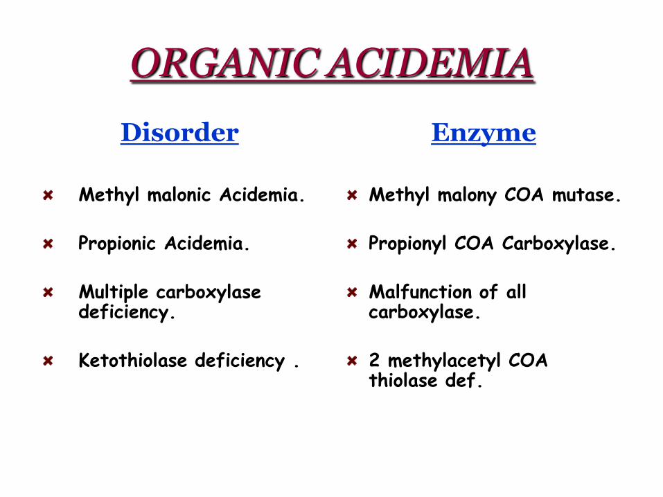
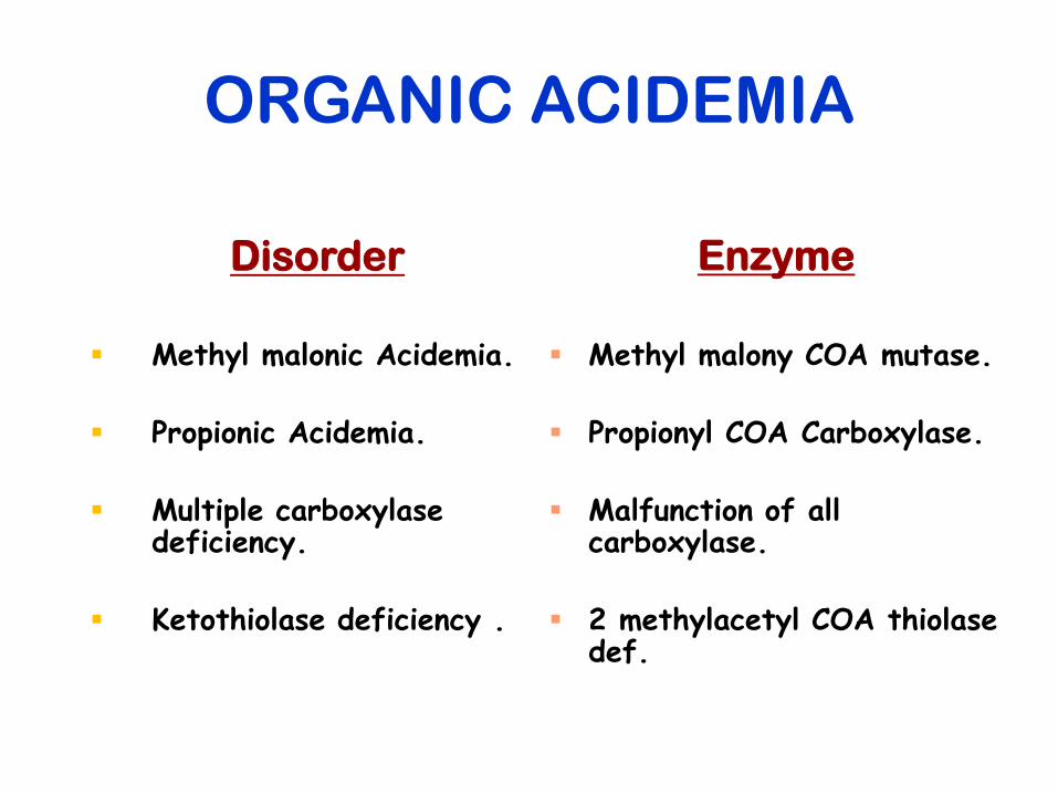
ORGANIC ACIDEMIA
Disorder
Methyl malonic Acidemia.
Propionic Acidemia.
Multiple carboxylase deficiency.
Ketothiolase deficiency .
Enzyme
Methyl malony COA mutase.
Propionyl COA Carboxylase.
Malfunction of all carboxylase.
2 methylacetyl COA thiolase def.
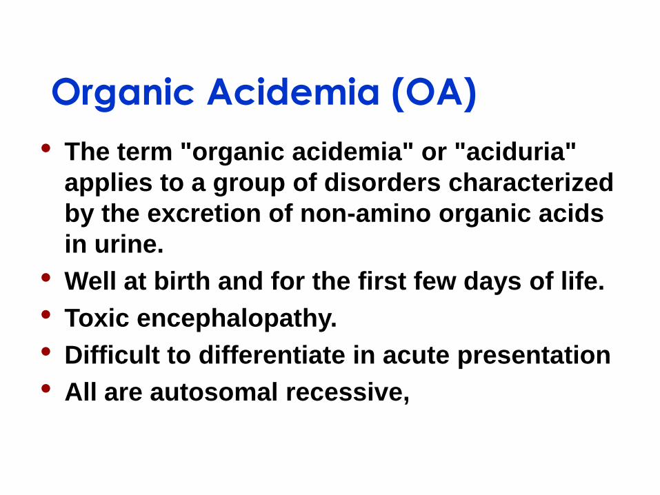
Organic Acidemia (OA)
• The term "organic acidemia" or "aciduria"
applies to a group of disorders characterized
by the excretion of non-amino organic acids
in urine.
• Well at birth and for the first few days of life.
• Toxic encephalopathy.
• Difficult to differentiate in acute presentation
• All are autosomal recessive,
ORGANIC ACIDEMIA
Disorder
Methyl malonic Acidemia.
Propionic Acidemia.
Multiple carboxylase deficiency.
Ketothiolase deficiency .
Enzyme
Methyl malony COA mutase.
Propionyl COA Carboxylase.
Malfunction of all carboxylase.
2 methylacetyl COA thiolase def.
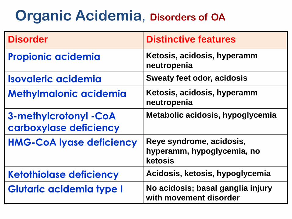
Disorder Distinctive features
Propionic acidemia Ketosis, acidosis, hyperamm
neutropenia
Isovaleric acidemia Sweaty feet odor, acidosis
Methylmalonic acidemia Ketosis, acidosis, hyperamm
neutropenia
3-methylcrotonyl -CoA
carboxylase deficiency
Metabolic acidosis, hypoglycemia
HMG-CoA lyase deficiency Reye syndrome, acidosis,
hyperamm, hypoglycemia, no
ketosis
Ketothiolase deficiency Acidosis, ketosis, hypoglycemia
Glutaric acidemia type I No acidosis; basal ganglia injury
with movement disorder
Organic Acidemia, Disorders of OA
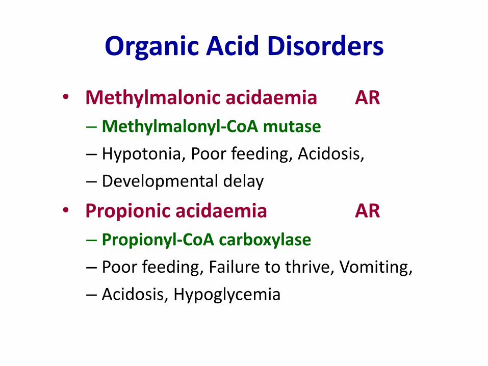
Organic Acid Disorders
• Methylmalonic acidaemia AR
– Methylmalonyl-CoA mutase
– Hypotonia, Poor feeding, Acidosis,
– Developmental delay
• Propionic acidaemia AR
– Propionyl-CoA carboxylase
– Poor feeding, Failure to thrive, Vomiting,
– Acidosis, Hypoglycemia
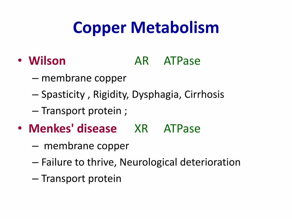
Copper Metabolism
• Wilson AR ATPase
– membrane copper
– Spasticity , Rigidity, Dysphagia, Cirrhosis
– Transport protein ;
• Menkes' disease XR ATPase
– membrane copper
– Failure to thrive, Neurological deterioration
– Transport protein
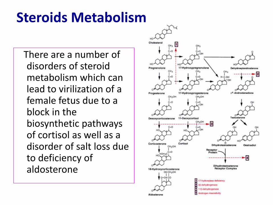
Steroids Metabolism
There are a number of disorders of steroid metabolism which can lead to virilization of a female fetus due to a block in the biosynthetic pathways of cortisol as well as a disorder of salt loss due to deficiency of aldosterone
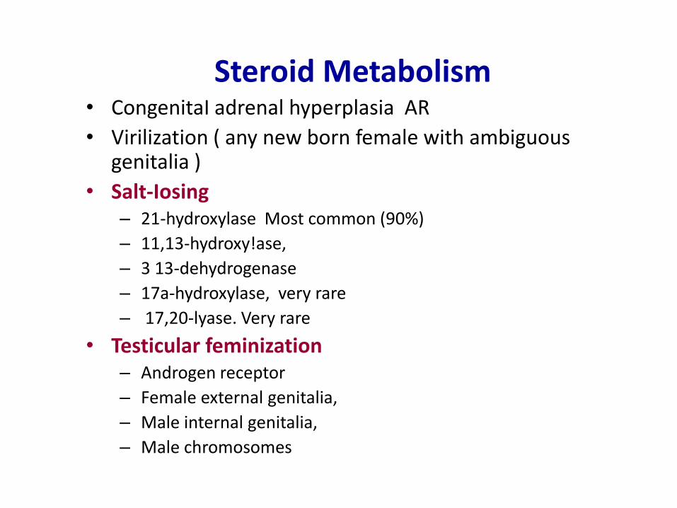
Steroid Metabolism• CongenitaI adrenal hyperplasia AR
• Virilization ( any new born female with ambiguous genitalia )
• Salt-Iosing – 21-hydroxylase Most common (90%)
– 11,13-hydroxy!ase,
– 3 13-dehydrogenase
– 17a-hydroxylase, very rare
– 17,20-lyase. Very rare
• Testicular feminization XR– Androgen receptor
– Female external genitalia,
– Male internal genitalia,
– Male chromosomes
71
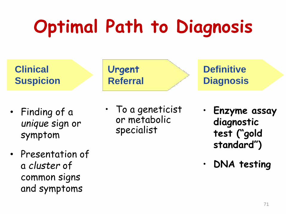
Optimal Path to Diagnosis
• Finding of aunique sign or symptom
• Presentation of a cluster of common signs and symptoms
• Enzyme assay diagnostic test (“gold standard”)
• DNA testing
Clinical
Suspicion
UrgentReferral
Definitive
Diagnosis
• To a geneticist or metabolic specialist
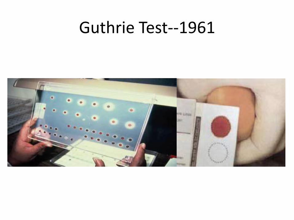
Guthrie Test--1961
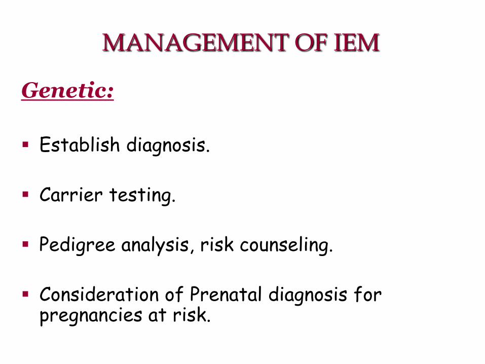
Genetic:
Establish diagnosis.
Carrier testing.
Pedigree analysis, risk counseling.
Consideration of Prenatal diagnosis for pregnancies at risk.
MANAGEMENT OF IEM
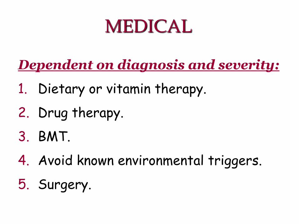
Dependent on diagnosis and severity:
1. Dietary or vitamin therapy.
2. Drug therapy.
3. BMT.
4. Avoid known environmental triggers.
5. Surgery.
MEDICAL

Family counseling and support.
Education to promote increased compliancewith special form of therapy such asProtein – restricted diet.
Assessment of community resources andsupport groups.
PSYCHOSOCIAL, EDUCATIONAL, FAMILIAL
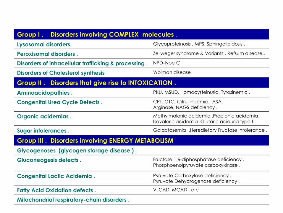
Group I . Disorders involving COMPLEX molecules .
Glycoproteinosis , MPS, Sphingolipidosis .Lysosomal disorders.
Zellweger syndrome & Variants , Refsum disease,.Peroxisomal disorders .
NPD-type CDisorders of intracellular trafficking & processing .
Wolman diseaseDisorders of Cholesterol synthesis
Group II . Disorders that give rise to INTOXICATION .
PKU, MSUD. Homocysteinuria, Tyrosinemia .Aminoacidopathies .
CPT, OTC, Citrullinaemia, ASA.
Arginase, NAGS deficiency .Congenital Urea Cycle Defects .
Methylmalonic acidemia .Propionic acidemia .
Isovaleric acidemia .Glutaric aciduria type I .Organic acidemias .
Galactosemia .Heredietary Fructose intolerance .Sugar intolerances .
Group III . Disorders involving ENERGY METABOLISM
Glycogenoses (glycogen storage disease ) .
Fructose 1,6-diphosphatase deficiency .
Phosphoenolpyruvate carboxykinase .Gluconeogesis defects .
Pyruvate Carboxylase deficiency .
Pyruvate Dehydrogenase deficiency .Congenital Lactic Acidemia .
VLCAD, MCAD , etc Fatty Acid Oxidation defects .
Mitochondrial respiratory-chain disorders .
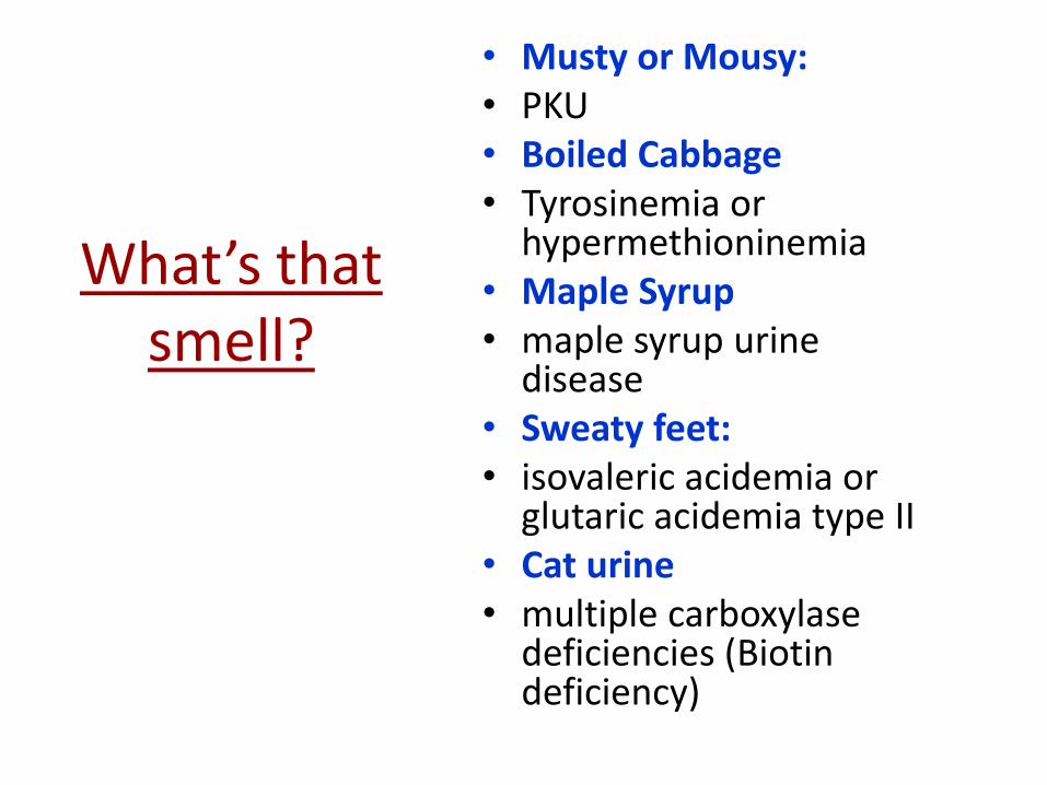
What’s that smell?
• Musty or Mousy:• PKU• Boiled Cabbage• Tyrosinemia or
hypermethioninemia• Maple Syrup• maple syrup urine
disease• Sweaty feet:• isovaleric acidemia or
glutaric acidemia type II • Cat urine• multiple carboxylase
deficiencies (Biotin deficiency)
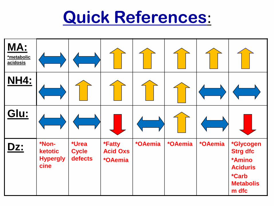
Quick References:
MA:*metabolic
acidosis
NH4:
Glu:
Dz: *Non-
ketotic
Hypergly
cine
*Urea
Cycle
defects
*Fatty
Acid Oxs
*OAemia
*OAemia *OAemia *OAemia *Glycogen
Strg dfc
*Amino
Aciduris
*Carb
Metabolis
m dfc
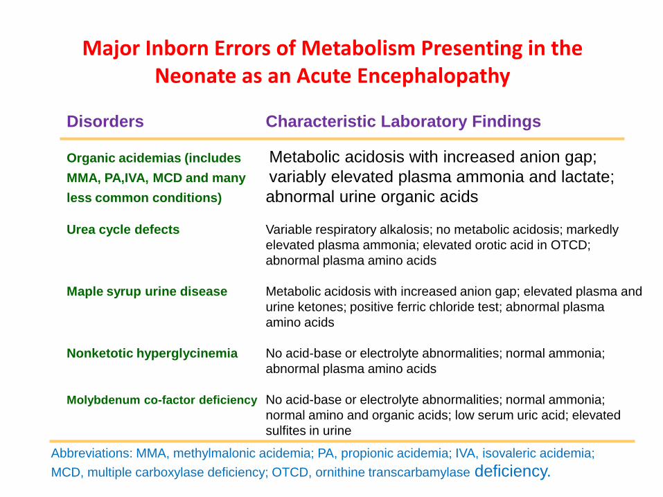
Major Inborn Errors of Metabolism Presenting in the Neonate as an Acute Encephalopathy
Disorders Characteristic Laboratory Findings
Organic acidemias (includes Metabolic acidosis with increased anion gap;
MMA, PA,IVA, MCD and many variably elevated plasma ammonia and lactate;
less common conditions) abnormal urine organic acids
Urea cycle defects Variable respiratory alkalosis; no metabolic acidosis; markedly
elevated plasma ammonia; elevated orotic acid in OTCD;
abnormal plasma amino acids
Maple syrup urine disease Metabolic acidosis with increased anion gap; elevated plasma and
urine ketones; positive ferric chloride test; abnormal plasma
amino acids
Nonketotic hyperglycinemia No acid-base or electrolyte abnormalities; normal ammonia;
abnormal plasma amino acids
Molybdenum co-factor deficiency No acid-base or electrolyte abnormalities; normal ammonia;
normal amino and organic acids; low serum uric acid; elevated
sulfites in urine
Abbreviations: MMA, methylmalonic acidemia; PA, propionic acidemia; IVA, isovaleric acidemia;
MCD, multiple carboxylase deficiency; OTCD, ornithine transcarbamylase deficiency.
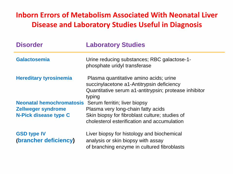
Inborn Errors of Metabolism Associated With Neonatal Liver Disease and Laboratory Studies Useful in Diagnosis
Disorder Laboratory Studies
Galactosemia Urine reducing substances; RBC galactose-1-
phosphate uridyl transferase
Hereditary tyrosinemia Plasma quantitative amino acids; urine
succinylacetone a1-Antitrypsin deficiency
Quantitative serum a1-antitrypsin; protease inhibitor
typing
Neonatal hemochromatosis Serum ferritin; liver biopsy
Zellweger syndrome Plasma very long-chain fatty acids
N-Pick disease type C Skin biopsy for fibroblast culture; studies of
cholesterol esterification and accumulation
GSD type IV Liver biopsy for histology and biochemical
(brancher deficiency) analysis or skin biopsy with assay
of branching enzyme in cultured fibroblasts