Embed Size (px)
Citation preview
Indoxyl Sulfate Induces Leukocyte-Endothelial Interactionsthrough Up-regulation of E-selectin*□S
Received for publication, July 30, 2010, and in revised form, September 28, 2010 Published, JBC Papers in Press, October 11, 2010, DOI 10.1074/jbc.M110.166686
Shunsuke Ito‡§, Mizuko Osaka‡, Yusuke Higuchi§, Fuyuhiko Nishijima§, Hideto Ishii‡, and Masayuki Yoshida‡1
From the ‡Department of Life Science and Medical Ethics, Graduate School of Medicine, Tokyo Medical and Dental University,Tokyo 113-8510 and the §Biomedical Research Laboratories, Kureha Corporation, Tokyo 169-8503, Japan
Despite a positive correlation between chronic kidney dis-ease and atherosclerosis, the causative role of uremic toxins inleukocyte-endothelial interactions has not been reported. Wethus examined the effects of indoxyl sulfate, a uremic toxin, onleukocyte adhesion to activated endothelial cells and the un-derlying mechanisms. Pretreatment of human umbilical veinendothelial cells (HUVEC) with indoxyl sulfate significantlyenhanced the adhesion of human monocytic cells (THP-1 cellline) to TNF-�-activated HUVEC under physiological flowconditions. Treatment with indoxyl sulfate enhanced the ex-pression level of E-selectin, but not that of ICAM-1 orVCAM-1, in HUVEC. Indoxyl sulfate treatment enhanced theactivation of JNK, p38 MAPK, and NF-�B in TNF-�-activatedHUVEC. Inhibitors of JNK and NF-�B attenuated indoxyl sul-fate-induced E-selectin expression in HUVEC and subsequentTHP-1 adhesion. Furthermore, treatment with the NAD(P)Hoxidase inhibitor apocynin and the glutathione donor N-ace-tylcysteine inhibited indoxyl sulfate-induced enhancement ofTHP-1 adhesion to HUVEC. Next, we examined the in vivoeffect of indoxyl sulfate in nephrectomized chronic kidney dis-ease model mice. Indoxyl sulfate-induced leukocyte adhesionto the femoral artery was significantly reduced by anti-E-selec-tin antibody treatment. These findings suggest that indoxylsulfate enhances leukocyte-endothelial interactions throughup-regulation of E-selectin, presumably via the JNK- and NF-�B-dependent pathway.
Cardiovascular disease is a major cause of death in chronickidney disease (CKD)2 (1). Atherosclerosis is highly prevalentin patients with severe renal failure and advances more rap-idly in individuals with renal dysfunction compared with thegeneral population (2). Reduced kidney function is associated
with the risk of cardiovascular events, even when the dysfunc-tion is mild (3).Leukocyte-endothelial interactions play an important role
in the development of atherosclerosis (4). Cell adhesion mole-cules belonging to the immunoglobulin superfamily, such asICAM-1 (intercellular cell adhesion molecule-1) andVCAM-1 (vascular cell adhesion molecule-1), together withmembers of the selectin family, including E-selectin, are up-regulated to mediate monocyte/macrophage infiltration intoatherosclerotic lesions (4, 5).Indoxyl sulfate is a uremic toxin synthesized in the liver
from indole, a metabolite of tryptophan produced by the in-testinal flora (6). In CKD patients, the serum levels of indoxylsulfate are increased significantly compared with those inhealthy individuals (7), and a number of studies have indi-cated that indoxyl sulfate accelerates glomerular sclerosis,whereas its accumulation promotes renal failure (8–10).Other studies also showed that indoxyl sulfate induces endo-thelial dysfunction by releasing endothelial microparticles(11) and producing reactive oxygen species (ROS) (12). How-ever, its effect on endothelial inflammatory processes such asleukocyte recruitment to vascular endothelium has not beenreported.We report for the first time that indoxyl sulfate enhances
monocyte adhesion to vascular endothelium through up-reg-ulation of E-selectin and augmentation of oxidative stress inboth in vitro and in vivomodels. The underlying mechanismsseem to involve activation of JNK and NF-�B. Our findingsreveal a previously unrecognized molecular link between ure-mic toxins and cardiovascular diseases.
EXPERIMENTAL PROCEDURES
Reagents—Indoxyl sulfate, N-acetylcysteine, probenecid,RPMI 1640 medium, and Dulbecco’s PBS were obtained fromSigma. The JNK phosphorylation inhibitor SP600125, the p38MAPK phosphorylation inhibitor SB203580, the ERK1/2 in-hibitor U0126, and the I�B phosphorylation inhibitor BAY11-7082 were purchased from Calbiochem. Recombinant humanTNF-� was obtained from R&D Systems (Minneapolis, MN).A monoclonal antibody against E-selectin (clone 7A9) wasobtained from American Type Culture Collection (Manassas,VA) (13). Antibodies against ICAM-1, VCAM-1, the NF-�Bp65 subunit, and the phospho-NF-�B p65 subunit and amonoclonal blocking antibody against mouse E-selectin(clone UZ4) were obtained from Santa Cruz Biotechnology,Inc. (Santa Cruz, CA). Anti-ERK, anti-phospho-ERK, anti-p38MAPK, anti-phospho-p38 MAPK, anti-JNK, and anti-phos-
* This work was supported in part by Ministry of Education, Science, andTechnology Grant-in-aid for Scientific Research 10178102 and specialcoordination funds, a grant-in-aid from the Ministry of Culture of Japan, agrant from the Ministry of Health, Labor, and Welfare of Japan, and agrant-in-aid from the Ono Medical Research Foundation.
□S The on-line version of this article (available at http://www.jbc.org) con-tains supplemental Figs. S1–S6 and Table S1.
1 To whom correspondence should be addressed: Dept. of Life Science andMedical Ethics, Graduate School of Medicine, Tokyo Medical and DentalUniversity, 1-5-45 Yushima, M and D Tower, 9th Floor, Bunkyo-ku, Tokyo113-8510, Japan. Tel.: 81-3-5803-4724; Fax: 81-3-5803-4725; E-mail: [email protected].
2 The abbreviations used are: CKD, chronic kidney disease; ROS, reactiveoxygen species; HUVEC, human umbilical vein endothelial cell(s); IVM,intravital microscopy; OAT, organic anion transporter.
THE JOURNAL OF BIOLOGICAL CHEMISTRY VOL. 285, NO. 50, pp. 38869 –38875, December 10, 2010© 2010 by The American Society for Biochemistry and Molecular Biology, Inc. Printed in the U.S.A.
DECEMBER 10, 2010 • VOLUME 285 • NUMBER 50 JOURNAL OF BIOLOGICAL CHEMISTRY 38869
by guest on June 25, 2018http://w
ww
.jbc.org/D
ownloaded from
pho-JNK antibodies were purchased from Cell SignalingTechnology (Beverly, MA). Control IgM was from eBio-science (San Diego, CA). Western blotting was performedusing standard protocols with ECL reagents (AmershamBiosciences).Cell Cultures—Human umbilical vein endothelial cells
(HUVEC) were purchased from Sanko Junyaku (Tokyo, Ja-pan) and cultured in endothelial growth medium-2 (Lonza,Walkersville, MD) at 37 °C in a humidified atmosphere of 5%carbon dioxide. Plastic culture dishes were precoated with 1%(w/v) collagen, and HUVEC were used between passages 1and 3. For use in a flow chamber apparatus, HUVEC wereplaced onto 22-mm fibronectin-coated glass coverslips.THP-1, a human monocytic cell line, was obtained fromAmerican Type Culture Collection, and the cells were main-tained in RPMI 1640 medium supplemented with 10% FCS,100 IU/ml penicillin, 100 �g/ml streptomycin, and 2 mmol/liter L-glutamine.Monocyte Adhesion Assay—HUVEC monolayers on cover-
slips were treated with various concentrations of indoxyl sul-fate for 20 h and then stimulated by the addition of TNF-� for4 h. The parallel plate flow chamber and the protocol for theadhesion assay under physiological flow conditions have beendescribed in detail previously (14). In brief, HUVEC monolay-ers were positioned in a flow chamber mounted on a Nikoninverted microscope. THP-1 cells (1 � 106/ml) were perfusedthrough the chamber with a syringe pump (PHD2000, Har-vard Apparatus Inc., Holliston, MA) for 10 min at a controlledflow rate to generate a shear stress of 1.0 dyne/cm2. The en-tire period of perfusion was recorded by videotape and thentransferred to a personal computer for image analysis to de-termine the number of adherent cells on HUVEC monolayersin 10 randomly selected �15 microscope fields.Luciferase Reporter Gene Assay—An NF-�B-firefly lucifer-
ase cDNA construct (pNF-�B-Luc) and a thymidine kinase-Renilla luciferase construct (pRL-TK) were obtained fromClontech. HUVEC were cultured in 24-well plates and tran-siently transfected using Lipofectamine LTX transfection re-agents (Invitrogen). Briefly, 500 ng of the pNF-�B-Luc vectorand 10 ng of the internal control pRL-TK were cotransfectedinto HUVEC. The culture medium was changed 4 h aftertransfection, and the HUVEC were incubated for another 18 hbefore use. The transfected HUVEC were incubated with in-doxyl sulfate for 20 h and then stimulated with TNF-� (100pg/ml) for 4 h. The firefly luciferase activity of the whole celllysate was measured with the Dual-Luciferase reporter assaysystem (Promega, Madison, WI), according to the manufac-turer’s protocol, using a luminometer. Renilla luciferase activ-ity was used to normalize the activity of firefly luciferase.Surgical Procedure—Renal failure was induced in 9-week-
old male C57BL/6J mice (Oriental Yeast, Tokyo) or BALB/cmice (Japan Crea Laboratory, Tokyo) using two-step surgicalnephrectomy as reported previously (15). Briefly, under intra-peritoneal anesthesia with sodium pentobarbital (Schering-Plough Corp., Kenilworth, NJ) at 65 mg/kg, two of the threebranches of the left renal artery were ligated through a lateralincision. One week after the first operation, the right kidney
was removed after ligation of the renal blood vessels and ure-ter under anesthesia as described above.Four weeks after the procedure, blood and blood pressure
were measured. Mice with blood urea nitrogen between 53and 90 mg/dl and systolic blood pressure between 118 and154 mm Hg were allocated to experimental groups. Sevenweeks after the procedure, half of the mice were administered0.065% indoxyl sulfate (200 mg/kg/day) in drinking water (re-ferred to as Nx�IS (nephrectomized with indoxyl sulfatetreatment); n � 5), whereas the other half were given onlywater (Nx; n � 5). Ten days later, leukocyte adhesion to thefemoral artery was assessed by intravital microscopy (IVM)and image analyses as described previously (16). In brief, micewere injected via the left femoral vein with rhodamine 6Gchloride (0.3 mg/kg in 200–300 �l of PBS; Molecular Probes)to label leukocytes in vivo. The femoral artery was foundwithin 30 min after injection of rhodamine 6G chloride andvisualized with an Olympus microscope (Model BX51WI)equipped with a water immersion objective. Adhesion of la-beled leukocytes was clearly visualized on the anterior half ofthe vessels facing the objective. All images were recorded us-ing a computer-assisted image analysis program (Meta-Morph). The number of adherent leukocytes (i.e. those thatdid not move for �3 s during the 1-min recording period) wascounted along a region of interest.The mice were killed, and whole blood samples were col-
lected from the heart using heparinized syringes. After perfu-sion via the left ventricle with ice-cold PBS, the aorta was dis-sected, snap-frozen in liquid nitrogen, and stored at �80 °Cuntil RNA isolation. For E-selectin blocking study, 1 mg/kganti-E-selectin monoclonal antibody (n � 9) or isotype con-trol antibody (control IgM; n � 9) was injected into the tailveins of BALB/c mice once daily for 9 consecutive days priorto IVM analysis on day 10.Plasma Biochemistry—Plasma urea, total cholesterol, HDL
cholesterol, and triglyceride measurements were performedusing an automated biochemical analyzer (SpotchemTM SP-4410, Arkray Co., Kyoto, Japan). Plasma indoxyl sulfate wasdetermined by HPLC.Complementary DNA Preparation and Real-time Quantita-
tive PCR—Individual mouse aortas were homogenized, andtotal RNA was isolated with an RNeasy mini column kit (Qia-gen, Hilden, Germany). RNA purity and concentration weredetermined by measuring absorbance at 260 and 280 nm, re-spectively. cDNA was produced from 0.5 �g of RNA using aPrimeScript RT-PCR reagent kit (TAKARA BIO Inc., Kyoto)with random hexamers in 10 �l of reaction solution at 37 °Cfor 15 min.Real-time quantitative RT-PCR was performed with a
LightCycler (Roche Applied Science) to quantitate the mRNAexpression of ICAM-1, VCAM-1, E-selectin, p47phox, p22phox,and GAPDH in mouse aortas. All primers were obtainedfrom Qiagen. Quantitative RT-PCR was carried out using aQuantiTectTM SYBR� Green RT-PCR kit (Qiagen) withGAPDH as an internal control.Statistical Analysis—Data are expressed as means � S.E.
One-way analysis of variance with Tukey’s post hoc test ortwo-tailed unpaired t test was used to estimate statistical sig-
Indoxyl Sulfate Induces Leukocyte-Endothelial Interactions
38870 JOURNAL OF BIOLOGICAL CHEMISTRY VOLUME 285 • NUMBER 50 • DECEMBER 10, 2010
by guest on June 25, 2018http://w
ww
.jbc.org/D
ownloaded from
nificance. A p value �0.05 was considered to be statisticallysignificant.
RESULTS
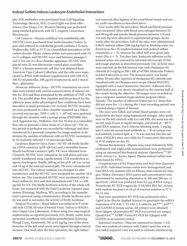
Indoxyl Sulfate Enhances Monocyte Adhesion to TNF-�-activated Vascular Endothelium—First, we examined the ef-fects of indoxyl sulfate on leukocyte-endothelial interactionsunder physiological flow conditions (shear stress of 1.0 dyne/cm2). Although indoxyl sulfate did not induce significantmonocytic THP-1 cell adhesion to non-stimulated HUVEC, itsignificantly enhanced THP-1 cell adhesion to TNF-�-stimu-lated HUVEC (Fig. 1A). The effect of indoxyl sulfate becamesignificant when HUVEC were treated with 100 pg/ml TNF-�(Fig. 1B). Considering the relatively low blood levels of TNF-�even in atherogenic conditions, our finding may indicatepathophysiologically relevant inflammatory situations in vivo.Indoxyl sulfate enhanced monocytic THP-1 cell adhesion toTNF-�-activated HUVEC in a dose-dependent manner (Fig.1C). Furthermore, indoxyl sulfate also enhanced the adhesion
of freshly isolated human peripheral blood monocytes toHUVEC (supplemental Fig. S1).To identify the adhesion molecules responsible for this ef-
fect of indoxyl sulfate, Western blot analysis was carried out.As shown in Fig. 1D, indoxyl sulfate enhanced TNF-�-in-duced E-selectin expression in HUVEC. The effect of indoxylsulfate (2.0 mmol/liter) was observed at a TNF-� concentra-tion as low as 25 pg/ml and became saturated at 250 pg/ml(Fig. 1D), whereas as little as 0.2 mmol/liter indoxyl sulfatesignificantly increased TNF-�-induced E-selectin expression(Fig. 1E). The expression of ICAM-1 and VCAM-1 was notsignificantly enhanced by indoxyl sulfate treatment (Fig. 1, Dand E).On the basis of these results, we incubated HUVEC with
indoxyl sulfate (0.2 mmol/liter) for 20 h, followed by TNF-�(100 pg/ml) for 4 h in subsequent cell adhesion assays. In-doxyl sulfate also enhanced THP-1 cell adhesion to IL-1�-stimulated HUVEC (supplemental Fig. S2).To confirm the importance of indoxyl sulfate-enhanced
E-selectin expression, we examined adhesion assays usingfunctional blocking antibodies against E-selectin, ICAM-1,and VCAM-1. As shown in supplemental Fig. S3, anti-E-se-lectin antibody, but not antibodies to ICAM-1 and VCAM-1,inhibited indoxyl sulfate-enhanced leukocyte adhesion,whereas all antibodies inhibited base-line leukocyte adhesioninduced by TNF-�.JNK Signaling Pathway Is Involved in Indoxyl Sulfate-en-
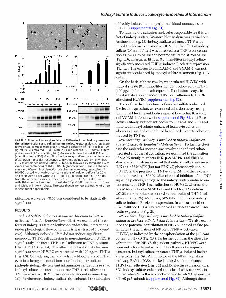
hanced Leukocyte-Endothelial Interactions—To further eluci-date the molecular mechanisms involved in indoxyl sulfate-mediated endothelial activation, we examined the activationof MAPK family members JNK, p38 MAPK, and ERK1/2.Western blot analyses revealed that indoxyl sulfate enhancedJNK and p38 MAPK (but not ERK1/2) phosphorylation inHUVEC in the presence of TNF-� (Fig. 2A). Further experi-ments showed that SP600125, a chemical inhibitor of the JNKsignaling pathway, abrogated the indoxyl sulfate-induced en-hancement of THP-1 cell adhesion to HUVEC, whereas thep38 MAPK inhibitor SB203580 and the ERK1/2 inhibitorU0126 did not influence indoxyl sulfate-induced THP-1 celladhesion (Fig. 2B). Moreover, SP600125 suppressed indoxylsulfate-induced E-selectin expression. In contrast, neitherSB203580 nor U0126 altered indoxyl sulfate-enhanced E-se-lectin expression (Fig. 2C).NF-�B Signaling Pathway Is Involved in Indoxyl Sulfate-
enhanced Leukocyte-Endothelial Interactions—We also exam-ined the potential contribution of NF-�B. Indoxyl sulfate po-tentiated the activation of NF-�B in TNF-�-activatedHUVEC, as indicated by the phosphorylation of the p65 com-ponent of NF-�B (Fig. 3A). To further confirm the direct in-volvement of an NF-�B-dependent pathway, HUVEC weretransiently transfected with an NF-�B promoter-reporterconstruct. Indoxyl sulfate enhanced TNF-�-induced lucifer-ase activity (Fig. 3B). An inhibitor of the NF-�B signalingpathway, BAY11-7082, blocked indoxyl sulfate-enhancedTHP-1 cell adhesion (Fig. 3C) and E-selectin expression (Fig.3D). Indoxyl sulfate-enhanced endothelial activation was in-hibited when NF-�B was knocked down by siRNA against theNF-�B p65 subunit (supplemental Fig. S4). Although these
FIGURE 1. Effects of indoxyl sulfate on TNF-�-induced leukocyte-endo-thelial interactions and cell adhesion molecule expression. A, represen-tative phase-contrast micrographs showing adhesion of THP-1 cells to 100pg/ml TNF-�-activated HUVEC with (IS�) and without (IS�) indoxyl sulfatepretreatment (2.0 mmol/liter, 20 h). Arrows indicate adherent THP-1 cells(magnification � 200). B and D, adhesion assay and Western blot detectionof adhesion molecules, respectively, in HUVEC treated with (�) or without(�) 2.0 mmol/liter indoxyl sulfate (IS) for 20 h, followed by stimulation withvarious concentrations of TNF-�. HPF, high-power field. C and E, adhesionassay and Western blot detection of adhesion molecules, respectively, inHUVEC treated with various concentrations of indoxyl sulfate for 20 hand then with (�) or without (�) TNF-� (100 pg/ml) for 4 h. The datafrom the adhesion assay are means � S.E. (n � 10). *, p � 0.01 versuswith TNF-� and without indoxyl sulfate; **, p � 0.001 versus with TNF-�and without indoxyl sulfate. The data shown are representative of threeindependent experiments.
Indoxyl Sulfate Induces Leukocyte-Endothelial Interactions
DECEMBER 10, 2010 • VOLUME 285 • NUMBER 50 JOURNAL OF BIOLOGICAL CHEMISTRY 38871
by guest on June 25, 2018http://w
ww
.jbc.org/D
ownloaded from
inhibitors reduced base-line THP-1 adhesion to activatedHUVEC, they significantly inhibited indoxyl sulfate-enhancedadhesion (supplemental Fig. S5).We also examined whether indoxyl sulfate modifies TNF-�
receptor expression in HUVEC. We documented that indoxylsulfate did not change the mRNA expression levels of TNFR1
and TNFR2 even after treatment with TNF-� (supplementalFig. S6).Role of Oxidative Stress in Monocytic Cell Adhesion and
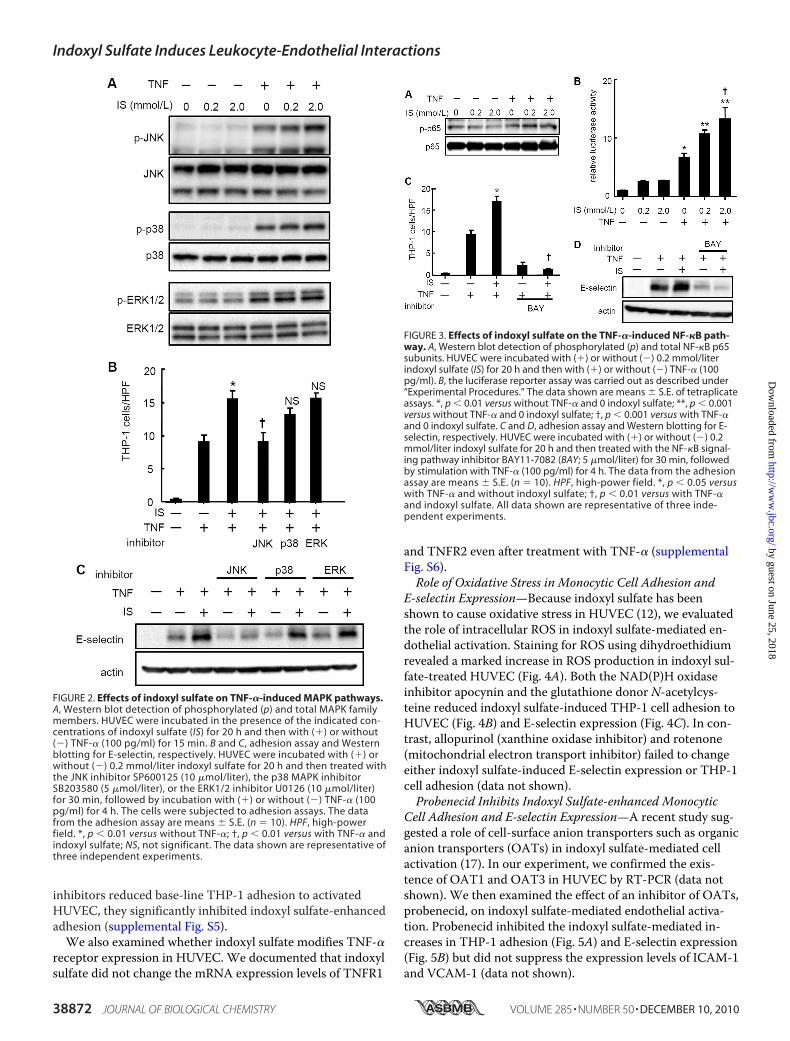
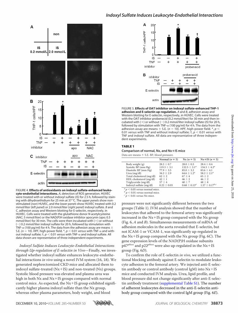
E-selectin Expression—Because indoxyl sulfate has beenshown to cause oxidative stress in HUVEC (12), we evaluatedthe role of intracellular ROS in indoxyl sulfate-mediated en-dothelial activation. Staining for ROS using dihydroethidiumrevealed a marked increase in ROS production in indoxyl sul-fate-treated HUVEC (Fig. 4A). Both the NAD(P)H oxidaseinhibitor apocynin and the glutathione donor N-acetylcys-teine reduced indoxyl sulfate-induced THP-1 cell adhesion toHUVEC (Fig. 4B) and E-selectin expression (Fig. 4C). In con-trast, allopurinol (xanthine oxidase inhibitor) and rotenone(mitochondrial electron transport inhibitor) failed to changeeither indoxyl sulfate-induced E-selectin expression or THP-1cell adhesion (data not shown).Probenecid Inhibits Indoxyl Sulfate-enhanced Monocytic
Cell Adhesion and E-selectin Expression—A recent study sug-gested a role of cell-surface anion transporters such as organicanion transporters (OATs) in indoxyl sulfate-mediated cellactivation (17). In our experiment, we confirmed the exis-tence of OAT1 and OAT3 in HUVEC by RT-PCR (data notshown). We then examined the effect of an inhibitor of OATs,probenecid, on indoxyl sulfate-mediated endothelial activa-tion. Probenecid inhibited the indoxyl sulfate-mediated in-creases in THP-1 adhesion (Fig. 5A) and E-selectin expression(Fig. 5B) but did not suppress the expression levels of ICAM-1and VCAM-1 (data not shown).
FIGURE 2. Effects of indoxyl sulfate on TNF-�-induced MAPK pathways.A, Western blot detection of phosphorylated (p) and total MAPK familymembers. HUVEC were incubated in the presence of the indicated con-centrations of indoxyl sulfate (IS) for 20 h and then with (�) or without(�) TNF-� (100 pg/ml) for 15 min. B and C, adhesion assay and Westernblotting for E-selectin, respectively. HUVEC were incubated with (�) orwithout (�) 0.2 mmol/liter indoxyl sulfate for 20 h and then treated withthe JNK inhibitor SP600125 (10 �mol/liter), the p38 MAPK inhibitorSB203580 (5 �mol/liter), or the ERK1/2 inhibitor U0126 (10 �mol/liter)for 30 min, followed by incubation with (�) or without (�) TNF-� (100pg/ml) for 4 h. The cells were subjected to adhesion assays. The datafrom the adhesion assay are means � S.E. (n � 10). HPF, high-powerfield. *, p � 0.01 versus without TNF-�; †, p � 0.01 versus with TNF-� andindoxyl sulfate; NS, not significant. The data shown are representative ofthree independent experiments.
FIGURE 3. Effects of indoxyl sulfate on the TNF-�-induced NF-�B path-way. A, Western blot detection of phosphorylated (p) and total NF-�B p65subunits. HUVEC were incubated with (�) or without (�) 0.2 mmol/literindoxyl sulfate (IS) for 20 h and then with (�) or without (�) TNF-� (100pg/ml). B, the luciferase reporter assay was carried out as described under“Experimental Procedures.” The data shown are means � S.E. of tetraplicateassays. *, p � 0.01 versus without TNF-� and 0 indoxyl sulfate; **, p � 0.001versus without TNF-� and 0 indoxyl sulfate; †, p � 0.001 versus with TNF-�and 0 indoxyl sulfate. C and D, adhesion assay and Western blotting for E-selectin, respectively. HUVEC were incubated with (�) or without (�) 0.2mmol/liter indoxyl sulfate for 20 h and then treated with the NF-�B signal-ing pathway inhibitor BAY11-7082 (BAY; 5 �mol/liter) for 30 min, followedby stimulation with TNF-� (100 pg/ml) for 4 h. The data from the adhesionassay are means � S.E. (n � 10). HPF, high-power field. *, p � 0.05 versuswith TNF-� and without indoxyl sulfate; †, p � 0.01 versus with TNF-�and indoxyl sulfate. All data shown are representative of three inde-pendent experiments.
Indoxyl Sulfate Induces Leukocyte-Endothelial Interactions
38872 JOURNAL OF BIOLOGICAL CHEMISTRY VOLUME 285 • NUMBER 50 • DECEMBER 10, 2010
by guest on June 25, 2018http://w
ww
.jbc.org/D
ownloaded from
Indoxyl Sulfate Induces Leukocyte-Endothelial Interactionsthrough Up-regulation of E-selectin in Vivo—Finally, we inves-tigated whether indoxyl sulfate enhances leukocyte-endothe-lial interactions in vivo using a novel IVM system (16, 18). Wegenerated nephrectomized CKD mice and allocated them toindoxyl sulfate-treated (Nx�IS) and non-treated (Nx) groups.Systolic blood pressure was elevated and plasma urea washigh in both Nx and Nx�IS groups compared with normalcontrol mice. As expected, the Nx�IS group exhibited signifi-cantly higher plasma indoxyl sulfate than the Nx group,whereas other plasma parameters, body weight, and blood
pressure were not significantly different between the twogroups (Table 1). IVM analysis showed that the number ofleukocytes that adhered to the femoral artery was significantlyincreased in the Nx�IS group compared with the Nx group(Fig. 6, A and B). Simultaneous measurements of mRNA ofadhesion molecules in the aorta revealed that E-selectin, butnot ICAM-1 or VCAM-1, was significantly up-regulated inthe Nx�IS group compared with the Nx group (Fig. 6C). Thegene expression levels of the NAD(P)H oxidase subunitsp47phox and p22phox were also up-regulated in the Nx�ISgroup (Fig. 6D).To confirm the role of E-selectin in vivo, we utilized a func-
tional blocking antibody against E-selectin to modulate leuko-cyte adhesion to the femoral artery. We injected anti-E-selec-tin antibody or control antibody (control IgM) into Nx�ISmice and conducted IVM analysis. Urea, lipid profile, andblood pressure did not change significantly after anti-E-selec-tin antibody treatment (supplemental Table S1). The numberof adherent leukocytes decreased in the anti-E-selectin anti-body group compared with the control IgM group (Fig. 6E).
FIGURE 5. Effects of OAT inhibitor on indoxyl sulfate-enhanced THP-1adhesion and E-selectin up-regulation. A and B, adhesion assay andWestern blotting for E-selectin, respectively, in HUVEC. Cells were treatedwith the OAT inhibitor probenecid (0.2 mmol/liter) for 30 min and then in-cubated with (�) or without (�) 0.2 mmol/liter indoxyl sulfate (IS) for 20 h,followed by stimulation with TNF-� (100 pg/ml) for 4 h. The data from theadhesion assay are means � S.E. (n � 10). HPF, high-power field. *, p �0.01 versus with TNF and without indoxyl sulfate; †, p � 0.01 versus withTNF and indoxyl sulfate. All data are representative of three indepen-dent experiments.
TABLE 1Comparison of normal, Nx, and Nx�IS miceData are means � S.E. BP, blood pressure.
Normal (n � 5) Nx (n � 5) Nx�IS (n � 5)
Body weight (g) 28.2 � 0.7 28.0 � 0.3 28.4 � 0.4Systolic BP (mm Hg) 119.3 � 3.1 132.5 � 3.1a 134.3 � 4.2aDiastolic BP (mm Hg) 77.9 � 3.5 85.0 � 3.2 83.6 � 4.1Urea (mg/dl) 34.2 � 2.9 64.6 � 1.2a 58.2 � 2.1aTotal cholesterol (mg/dl) 62 � 2 67 � 4 65 � 2HDL cholesterol (mg/dl) 42 � 1 46 � 2 46 � 2Triglyceride (mg/dl) 57 � 4 40 � 7 48 � 8Indoxyl sulfate (mg/dl) 0.22 � 0.03 0.68 � 0.13a 1.57 � 0.17b,c
a p � 0.05 versus normal mice.b p � 0.01 versus normal mice.c p � 0.01 versus Nx mice.
FIGURE 4. Effects of antioxidants on indoxyl sulfate-enhanced leuko-cyte-endothelial interactions. A, detection of ROS generation. HUVECwere treated with or without indoxyl sulfate (IS) for 2.5 h, followed by stain-ing with dihydroethidium for 25 min at 37 °C. The upper panels show non-stimulated (non) HUVEC, and the lower panels show HUVEC treated with 0.2mmol/liter (left panel) or 2.0 mmol/liter (right panel) indoxyl sulfate. B andC, adhesion assay and Western blotting for E-selectin, respectively, inHUVEC. Cells were treated with the glutathione donor N-acetylcysteine(NAC; 5 mmol/liter) or the NAD(P)H oxidase inhibitor apocynin (apo; 0.5mmol/liter) for 30 min. The cells were then incubated with (�) or without(�) 0.2 mmol/liter indoxyl sulfate for 20 h, followed by stimulation withTNF-� (100 pg/ml) for 4 h. The data from the adhesion assay are means �S.E. (n � 10). HPF, high-power field. *, p � 0.01 versus with TNF-� and with-out indoxyl sulfate; †, p � 0.01 versus with TNF-� and indoxyl sulfate. Alldata shown are representative of three independent experiments.
Indoxyl Sulfate Induces Leukocyte-Endothelial Interactions
DECEMBER 10, 2010 • VOLUME 285 • NUMBER 50 JOURNAL OF BIOLOGICAL CHEMISTRY 38873
by guest on June 25, 2018http://w
ww
.jbc.org/D
ownloaded from

DISCUSSION
In this study, we found that pretreatment of HUVEC withindoxyl sulfate prior to incubation with TNF-� or IL-1� in-creased the adhesion of monocytic THP-1 cells or human pe-ripheral blood monocytes to HUVEC in a dose-dependentmanner (Fig. 1 and supplemental Fig. S1). Considering thatthe concentrations of indoxyl sulfate (0.2 mmol/liter) (7, 8)and TNF-� and IL-1� (19) were comparable with their bloodlevels observed in CKD patients, our findings indicate an im-portant molecular background underlying the inflammatoryprocess in CKD patients with high circulating uremic toxins.Our data may explain a recent observation by Barreto et al.(20) that serum indoxyl sulfate is associated with the inci-dence of vascular diseases and total mortality in CKDpatients.
Indoxyl Sulfate and Endothelial Activation—We also foundthat indoxyl sulfate enhanced TNF-�- and IL-1�-activatedE-selectin expression in HUVEC, which plays a dominant rolein monocyte adhesion during inflammation (21, 22). Further-more, anti-E-selectin antibody significantly inhibited leuko-cyte adhesion enhanced by indoxyl sulfate (supplemental Fig.S3). Thus, the present results raise the possibility that indoxylsulfate exacerbates CKD-related vascular endothelial inflam-mation through up-regulation of E-selectin.In CKD patients, monocyte/macrophage accumulation has
been observed in the extracapillary areas of glomeruli (23, 24).Intense expression of E-selectin (24, 25) and activated glomer-ular endothelial cells (26) have been observed in the kidneysof CKD animal models. Furthermore, circulating E-selectin isa strong predictor of death and cardiovascular events in pa-tients with end-stage renal disease (27, 28). These observa-tions suggest an important role of E-selectin in renaldeterioration.Indoxyl Sulfate Enhances E-selectin Expression via JNK—
Our results indicate that indoxyl sulfate mediates E-selectinoverexpression through enhancement of the JNK and NF-�Bsignaling pathways (Figs. 2 and 3). The importance of JNK inE-selectin expression in comparison with other MAPK familymembers has been repeatedly emphasized (29–31). AlthoughKuldo et al. (31) demonstrated that inhibition of p38 MAPKdiminished E-selectin expression in HUVEC activated byTNF-� for 24 h, we failed to detect any effect of a p38 MAPKinhibitor on E-selectin expression stimulated by TNF-� for4 h.We confirmed the expression of OAT1 and OAT3, which
are molecules responsible for the incorporation of indoxylsulfate, in HUVEC (32). Moreover, probenecid, a chemicalinhibitor of OATs, suppressed indoxyl sulfate-mediated E-selectin expression in HUVEC and subsequent THP-1 celladhesion. These results suggest that OAT1- and/or OAT3-mediated intracellular transport may be necessary for the ex-hibition of the indoxyl sulfate effects in HUVEC.Role of E-selectin in Indoxyl Sulfate-induced Inflammation
in Vivo—As demonstrated previously by others, nephrectomyper se does not induce significant vascular inflammatory reac-tions in mice unless they are apolipoprotein E-deficient(knock-out) (33, 34). However, indoxyl sulfate induced E-se-lectin expression as well as leukocyte adhesion to the femoralartery in the Nx mice. Furthermore, anti-E-selectin antibodysignificantly blocked leukocyte accumulation in Nx�IS mice,suggesting a dominant role of E-selectin in the leukocyte ad-hesion observed in the Nx mice (35). These results suggestthat indoxyl sulfate induces leukocyte-endothelial interactionsthrough up-regulation of E-selectin in vivo.Previous studies demonstrated that indoxyl sulfate pro-
duced oxidative stress in vivo and in vitro through NAD(P)Hoxidase activity (12, 36). Consistent with these observations,we documented up-regulation of p47phox and p22phoxin Nxmice treated with indoxyl sulfate, suggesting a role ofNAD(P)H oxidase in indoxyl sulfate-mediated oxidative stressin vivo. The therapeutic efficacy of quenching uremic toxinsby oral adsorbents such as AST-120 (Kremezin, Kureha
FIGURE 6. Effects of indoxyl sulfate on leukocyte-endothelial interac-tions in a mouse CKD model. A, representative images from intravitalvideo microscopic analysis of leukocyte-adhesive interactions in femoralarteries (with the margins of vessels indicated by dotted lines) of five/six ne-phrectomized mice with (Nx�IS) or without (Nx) indoxyl sulfate treatment.White spots show fluorescent leukocytes labeled by intravenous injection ofrhodamine 6G. B, quantitative analyses of leukocyte adhesion to the femo-ral arteries. Data are expressed as means � S.E. (n � 5). C and D, quantita-tive real-time PCR analysis of mRNA expression of E-selectin, ICAM-1, andVCAM-1 (C) and the NAD(P)H oxidase subunits p47phox and p22phox (D) inaortas from Nx�IS (black bars) and Nx (white bars) mice compared with nor-mal mice (gray bars). The level of mRNA expression was normalized by thatof GAPDH mRNA. *, p � 0.05 versus normal mice; **, p � 0.01 versus normalmice; †, p � 0.01 versus Nx mice. E, quantitative analyses of leukocyte adhe-sion in femoral arteries from mice injected with anti-E-selectin antibody(anti-E-sel; black bar) or control IgM (white bar). Data are expressed asmeans � S.E. (n � 9).
Indoxyl Sulfate Induces Leukocyte-Endothelial Interactions
38874 JOURNAL OF BIOLOGICAL CHEMISTRY VOLUME 285 • NUMBER 50 • DECEMBER 10, 2010
by guest on June 25, 2018http://w
ww
.jbc.org/D
ownloaded from
Corp., Tokyo) in the reduction of oxidative stress in CKD hasbeen reported previously (36–39).In conclusion, indoxyl sulfate enhanced leukocyte-endothe-
lial interactions via increased selectin expression and oxida-tive stress in vitro and in vivo. Our data suggest a novel causa-tive role of uremic toxins in producing vascular inflammationin patients with renal dysfunction.
Acknowledgments—We thank Minako Manabe and Kazumi Obarafor assistance with HPLC analyses and Daisuke Mori for technicalsupport with endothelial cell cultures.
REFERENCES1. Wheeler, D. C. (1996) Lancet 348, 1673–16742. Amann, K., Gross, M. L., and Ritz, E. (2004) J. Am. Soc. Nephrol. 15,
1664–16663. Go, A. S., Chertow, G. M., Fan, D., McCulloch, C. E., and Hsu, C. Y.
(2004) N. Engl. J. Med. 351, 1296–13054. Ross, R. (1999) N. Engl. J. Med. 340, 115–1265. Cybulsky, M. I., Iiyama, K., Li, H., Zhu, S., Chen, M., Iiyama, M., Davis,
V., Gutierrez-Ramos, J. C., Connelly, P. W., and Milstone, D. S. (2001) J.Clin. Invest. 107, 1255–1262
6. Vanholder, R., and De Smet, R. (1999) J. Am. Soc. Nephrol. 10,1815–1823
7. Vanholder, R., De Smet, R., Glorieux, G., Argiles, A., Baurmeister, U.,Brunet, P., Clark, W., Cohen, G., De Deyn, P. P., Deppisch, R., Des-camps-Latscha, B., Henle, T., Jorres, A., Lemke, H. D., Massy, Z. A.,Passlick-Deetjen, J., Rodriguez, M., Stegmayr, B., Stenvinkel, P., Tetta,C., Wanner, C., and Zidek, W. (2003) Kidney Int. 63, 1934–1943
8. Niwa, T., and Ise, M. (1994) J. Lab. Clin. Med. 124, 96–1049. Niwa, T., Ise, M., and Miyazaki, T. (1994) Am. J. Nephrol. 14, 207–21210. Kobayashi, N., Maeda, A., Horikoshi, S., Shirato, I., Tomino, Y., and Ise,
M. (2002) Nephron 91, 480–48511. Faure, V., Dou, L., Sabatier, F., Cerini, C., Sampol, J., Berland, Y., Brunet,
P., and Dignat-George, F. (2006) J. Thromb. Haemost. 4, 566–57312. Dou, L., Jourde-Chiche, N., Faure, V., Cerini, C., Berland, Y., Dignat-
George, F., and Brunet, P. (2007) J. Thromb. Haemost. 5, 1302–130813. Nishiwaki, Y., Yokota, T., Hiraoka, M., Miyagishi, M., Taira, K., Isobe,
M., Mizusawa, H., and Yoshida, M. (2003) Biochem. Biophys. Res. Com-mun. 310, 1062–1066
14. Yoshida, M., Sawada, T., Ishii, H., Gerszten, R. E., Rosenzweig, A., Gim-brone, M. A., Jr., Yasukochi, Y., and Numano, F. (2001) Arterioscler.Thromb. Vasc. Biol. 21, 1165–1171
15. Kren, S., and Hostetter, T. H. (1999) Kidney Int. 56, 333–33716. Osaka, M., Hagita, S., Haraguchi, M., Kajimura, M., Suematsu, M., and
Yoshida, M. (2007) Am. J. Physiol. Heart Circ. Physiol. 292,H1876–H1882
17. Yamamoto, H., Tsuruoka, S., Ioka, T., Ando, H., Ito, C., Akimoto, T.,Fujimura, A., Asano, Y., and Kusano, E. (2006) Kidney Int. 69,1780–1785
18. Hagita, S., Osaka, M., Shimokado, K., and Yoshida, M. (2008) Hyperten-sion 51, 797–802
19. Malaponte, G., Libra, M., Bevelacqua, Y., Merito, P., Fatuzzo, P., Rap-isarda, F., Cristina, M., Naselli, G., Stivala, F., Mazzarino, M. C., andCastellino, P. (2007) Int. J. Mol. Med. 20, 471–481
20. Barreto, F. C., Barreto, D. V., Liabeuf, S., Meert, N., Glorieux, G., Tem-mar, M., Choukroun, G., Vanholder, R., and Massy, Z. A. (2009) Clin. J.Am. Soc. Nephrol. 4, 1551–1558
21. Yoshida, M., Szente, B. E., Kiely, J. M., Rosenzweig, A., and Gimbrone,M. A., Jr. (1998) J. Immunol. 161, 933–941
22. Gotoh, R., Suzuki, J., Kosuge, H., Kakuta, T., Sakamoto, S., Yoshida, M.,and Isobe, M. (2004) Arterioscler. Thromb. Vasc. Biol. 24, 2063–2068
23. Bolton, W. K., Innes, D. J., Jr., Sturgill, B. C., and Kaiser, D. L. (1987)Kidney Int. 32, 869–876
24. Nakatani, K., Fujii, H., Hasegawa, H., Terada, M., Arita, N., Ito, M. R.,Ono, M., Takahashi, S., Saiga, K., Yoshimoto, S., Iwano, M., Shiiki, H.,Saito, Y., and Nose, M. (2004) Kidney Int. 65, 1290–1300
25. Asgeirsdottir, S. A., Kamps, J. A., Bakker, H. I., Zwiers, P. J., Heeringa, P.,van der Weide, K., van Goor, H., Petersen, A. H., Morselt, H., Moorlag,H. E., Steenbergen, E., Kallenberg, C. G., and Molema, G. (2007)Mol.Pharmacol. 72, 121–131
26. Murakami, S., Morioka, T., Nakagawa, Y., Suzuki, Y., Arakawa, M., andOite, T. (2001)Microvasc. Res. 62, 383–391
27. Malatino, L. S., Stancanelli, B., Cataliotti, A., Bellanuova, I., Fatuzzo, P.,Rapisarda, F., Leonardis, D., Tripepi, G., Mallamaci, F., and Zoccali, C.(2007) J. Intern. Med. 262, 479–487
28. Testa, A., Benedetto, F. A., Spoto, B., Pisano, A., Tripepi, G., Mallamaci,F., Malatino, L. S., and Zoccali, C. (2006) Nephrol. Dial. Transplant. 21,1921–1926
29. Read, M. A., Whitley, M. Z., Gupta, S., Pierce, J. W., Best, J., Davis, R. J.,and Collins, T. (1997) J. Biol. Chem. 272, 2753–2761
30. Lin, C. W., Chen, L. J., Lee, P. L., Lee, C. I., Lin, J. C., and Chiu, J. J.(2007) Biomaterials 28, 1355–1366
31. Kuldo, J. M., Westra, J., Asgeirsdottir, S. A., Kok, R. J., Oosterhuis, K.,Rots, M. G., Schouten, J. P., Limburg, P. C., and Molema, G. (2005) Am.J. Physiol. Cell Physiol. 289, C1229–C1239
32. Enomoto, A., Takeda, M., Tojo, A., Sekine, T., Cha, S. H., Khamdang, S.,Takayama, F., Aoyama, I., Nakamura, S., Endou, H., and Niwa, T. (2002)J. Am. Soc. Nephrol. 13, 1711–1720
33. Bro, S., Moeller, F., Andersen, C. B., Olgaard, K., and Nielsen, L. B.(2004) J. Am. Soc. Nephrol. 15, 1495–1503
34. Bro, S., Binder, C. J., Witztum, J. L., Olgaard, K., and Nielsen, L. B.(2007) Arterioscler. Thromb. Vasc. Biol. 27, 1080–1086
35. Wolf, D., Hallmann, R., Sass, G., Sixt, M., Kusters, S., Fregien, B.,Trautwein, C., and Tiegs, G. (2001) J. Immunol. 166, 1300–1307
36. Shimoishi, K., Anraku, M., Kitamura, K., Tasaki, Y., Taguchi, K., Hashi-moto, M., Fukunaga, E., Maruyama, T., and Otagiri, M. (2007) Pharm.Res. 24, 1283–1289
37. Namikoshi, T., Tomita, N., Satoh, M., Sakuta, T., Kuwabara, A., Koba-yashi, S., Higuchi, Y., Nishijima, F., and Kashihara, N. (2009) Hypertens.Res. 32, 194–200
38. Nakamura, T., Sato, E., Fujiwara, N., Kawagoe, Y., Suzuki, T., Ueda, Y.,and Yamagishi, S. I. (2011)Metabolism, in press
39. Fujii, H., Nishijima, F., Goto, S., Sugano, M., Yamato, H., Kitazawa, R.,Kitazawa, S., and Fukagawa, M. (2009) Nephrol. Dial. Transplant. 24,2089–2095
Indoxyl Sulfate Induces Leukocyte-Endothelial Interactions
DECEMBER 10, 2010 • VOLUME 285 • NUMBER 50 JOURNAL OF BIOLOGICAL CHEMISTRY 38875
by guest on June 25, 2018http://w
ww
.jbc.org/D
ownloaded from
Masayuki YoshidaShunsuke Ito, Mizuko Osaka, Yusuke Higuchi, Fuyuhiko Nishijima, Hideto Ishii and
of E-selectinIndoxyl Sulfate Induces Leukocyte-Endothelial Interactions through Up-regulation
doi: 10.1074/jbc.M110.166686 originally published online October 11, 20102010, 285:38869-38875.J. Biol. Chem.
10.1074/jbc.M110.166686Access the most updated version of this article at doi:
Alerts:
When a correction for this article is posted•
When this article is cited•
to choose from all of JBC's e-mail alertsClick here
Supplemental material:
http://www.jbc.org/content/suppl/2010/10/08/M110.166686.DC1
http://www.jbc.org/content/285/50/38869.full.html#ref-list-1
This article cites 38 references, 13 of which can be accessed free at
by guest on June 25, 2018http://w
ww
.jbc.org/D
ownloaded from