Embed Size (px)
Citation preview
FULL PAPER
DOI: 10.1002/ejic.200601097
Influence of a Large σ-Donor Ligand on Structural and Catalytic Properties ofDi-Iron Compounds Related to the Active Site of Fe-Hydrogenase – A DFT
Investigation
Claudio Greco,[a] Maurizio Bruschi,[b] Piercarlo Fantucci,[a] and Luca De Gioia*[a]
Keywords: Hydrogenases / Biomimetic chemistry / Catalytic H2 evolution / Density functional calculations / Iron
The complex (S2C3H6)[Fe2(CO)5P(NC4H8)3] (A), a recentlysynthesized functional model of the active site of Fe-hydro-genases, is able to electrocatalyze proton reduction, leadingto molecular hydrogen evolution. Experimental results sug-gested that the presence of the electron donor P(NC4H8)3 li-gand in A could favor the formation of a µ-CO species similarto that observed in the enzymatic cluster. However, insightinto the structural features of key catalytic intermediates de-riving from reduction and protonation of A was still lacking.Here we present results obtained using density functionaltheory to evaluate structures, relative stabilities, and spectro-scopic properties of several species relevant for the electroca-talytic H2 evolving process. The results enabled us to unravelthe structure of the µ-CO complex experimentally detected
Introduction
The possibility of producing molecular hydrogen througheco-compatible, hydrocarbon-independent processes hasbeen stimulating chemists towards the synthesis of inexpen-sive catalytic materials that could replace the currently usedplatinum-containing electrocatalysts.[1] The search forcheaper, but still efficient catalysts could take advantagefrom the study of hydrogenases,[2] which are metallo-en-zymes that are able to catalyze the evolution of molecularhydrogen at high rates starting from protons and electrons.
Three classes of hydrogenases have been characterized sofar: Ni–Fe hydrogenases, which include both iron and nickelatoms as metal centers of functional importance, and twoclasses of iron-dependent enzymes, i.e. the “iron-sulfur-clus-ter-free” hydrogenases,[3] and another group of proteinscontaining FexSx clusters, commonly referred to as “Iron-only” or Fe-hydrogenases. X-ray crystallographic studies onFe-hydrogenases from C. pasteurianum[4] and D. desul-
[a] Department of Biotechnology and Biosciences, University ofMilano-Bicocca,Piazza della Scienza 2, 20126 Milano, ItalyFax: +39-02-64483478E-mail: [email protected]
[b] Department of Environmental Sciences, University of Milano-Bicocca,Piazza della Scienza 1, 20126 Milano, Italy
Eur. J. Inorg. Chem. 2007, 1835–1843 © 2007 Wiley-VCH Verlag GmbH & Co. KGaA, Weinheim 1835
after monoelectronic reduction of A. Moreover, we show thatthe introduction of the large electron-donor ligand P(NC4-H8)3 in the biomimetic complex does not favour the stabiliza-tion of terminal-hydrido adducts, which are expected to bevery reactive in terms of H2 production. The comparison ofour findings with previous theoretical and experimental re-sults obtained on similar model complexes suggests that theintroduction of an electron donor ligand as good as P(NC4-H8)3, but less sterically demanding, could represent a betterchoice to facilitate the formation of µ-CO complexes moreclosely resembling the structure of the enzymatic cluster.
(© Wiley-VCH Verlag GmbH & Co. KGaA, 69451 Weinheim,Germany, 2007)
furicans[5] have shown that the active site of these enzymesincludes an Fe2S2 sub-site bearing CO and CN– ligands.The iron atoms of the sub-site are bridged by two sulfuratoms of a 1,3-propanedithiolato (pdt) or a related bis(thio-methyl)amino unit. One of the iron atoms shares a cysteinylsulfur ligand with a classical Fe4S4 cluster; the resultingiron–sulfur complex is usually referred to as the H-cluster.Experimental results[6] indicate that the unprotonated, FeI-
FeII form of the H-cluster di-iron subsite should correspondto a catalytically active state characterized by the presenceof a carbonyl ligand bridging the two iron centers. Asshown in Scheme 1, the coordination site trans to the bridg-ing CO remains vacant, and this observation suggests thatprotons and dihydrogen could occupy such a position dur-ing the catalytic process. Upon monoelectronic reduction ofthe H-cluster, the bridging CO moves to a terminal posi-tion, but it still remains localized in the region of spacebetween the iron ions (see Scheme 1).[6]
The above-mentioned studies have stimulated experimen-tal chemists towards the synthesis of organometallic modelsthat could reproduce the main structural and functionalfeatures of the H-cluster.[7] Unfortunately, the biomimeticclusters obtained so far fail to reproduce the precise orien-tation of ligands found in the H-cluster, a fact that is ex-pected to be at the basis of the reduced catalytic efficiencyof synthetic assemblies. For example, in the complex (µ-
C. Greco, M. Bruschi, P. Fantucci, L. De GioiaFULL PAPER
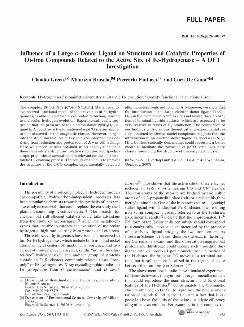
Scheme 1. Structures of the Fe-hydrogenase binuclear subsite in two different redox states, and geometry of the synthetic complex (µ-pdt)[Fe(CO)3]2.
pdt)[Fe(CO)3]2, the simplest functional model of the activesite of Fe-hydrogenases, all the carbonyl groups are in theterminal position.[7h] Such an arrangement of ligands willbe referred to as the “eclipsed conformation,” as opposedto the “rotated conformation” that can be observed in Fe-hydrogenases (Scheme 1).
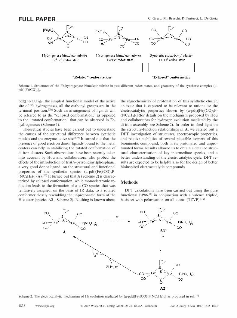
Theoretical studies have been carried out to understandthe causes of the structural difference between syntheticmodels and the enzyme active site.[8,9] It turned out that thepresence of good electron donor ligands bound to the metalcenters can help in stabilizing the rotated conformation ofdi-iron clusters. Such observations have been recently takeninto account by Hou and collaborators, who probed theeffects of the introduction of tris(N-pyrrolidinyl)phosphane,a very good donor ligand, on the structural and functionalproperties of the synthetic species (µ-pdt)[Fe2(CO)5P-(NC4H8)3] (A).[10] It turned out that A (Scheme 2) is charac-terized by eclipsed conformation, while monoelectronic re-duction leads to the formation of a µ-CO species that wastentatively assigned, on the basis of IR data, to a rotatedconformer closely resembling the unprotonated form of theH-cluster (species A2–, Scheme 2). Nothing is known about
Scheme 2. The electrocatalytic mechanism of H2 evolution mediated by (µ-pdt)[Fe2(CO)5P(NC4H8)3], as proposed in ref.[10]
www.eurjic.org © 2007 Wiley-VCH Verlag GmbH & Co. KGaA, Weinheim Eur. J. Inorg. Chem. 2007, 1835–18431836
the regiochemistry of protonation of this synthetic cluster,an issue that is expected to be relevant to rationalize theelectrocatalytic properties shown by (µ-pdt)[Fe2(CO)5P-(NC4H8)3] (for details on the mechanism proposed by Houand collaborators for hydrogen evolution mediated by thedi-iron assembly, see Scheme 2). In order to shed light onthe structure-function relationships in A, we carried out aDFT investigation of structures, spectroscopic properties,and relative stabilities of several plausible isomers of thisbiomimetic compound, both in its protonated and unpro-tonated forms. Results allowed us to obtain a detailed struc-tural characterization of key intermediate species, and abetter understanding of the electrocatalytic cycle. DFT re-sults are expected to be helpful also for the design of betterbioinspired electrocatalytic compounds.
Methods
DFT calculations have been carried out using the purefunctional BP86[11] in conjunction with a valence triple-ζbasis set with polarization on all atoms (TZVP).[12]
Properties of Di-Iron Compounds FULL PAPERCalculations have been carried out with the TURBO-
MOLE 5.7 suite[13] applying the resolution-of-the-identitytechnique.[14]
Stationary points of the energy hypersurface have beenlocated by means of energy gradient techniques and fullvibrational analysis has been carried out to further charac-terize each stationary point. In order to characterize iso-meric forms, DFT optimizations were repeated severaltimes, starting from different initial geometries.
Free energy (G) values have been obtained from the elec-tronic SCF energy considering three contributions to thetotal partition function (Q), namely qtranslational, qrotational,qvibrational, under the assumption that Q may be written asthe product of such terms.[15] In order to evaluate enthalpyand entropy contributions, the value for the temperature,pressure, and scaling factor for the SCF wavenumbers havebeen set to 298.15 K, 1 bar, and 0.9914, respectively. Rota-tions have been treated classically and vibrational modesdescribed according to the harmonic approximation.
The optimized structures of the complexes reported inthe present study always correspond to low spin states; high
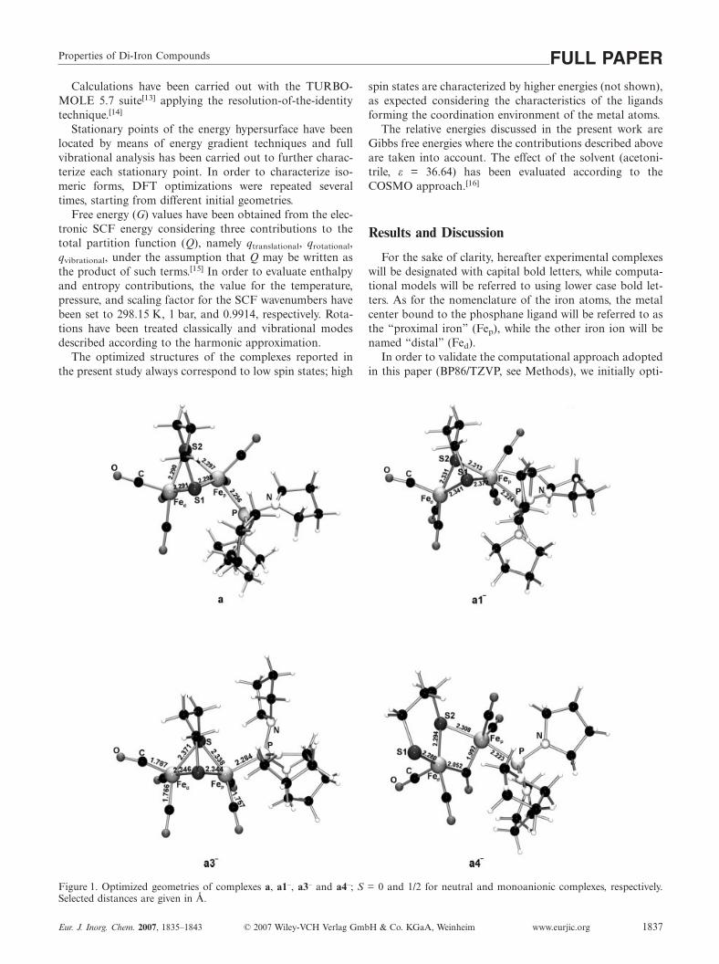
Figure 1. Optimized geometries of complexes a, a1–, a3– and a4–; S = 0 and 1/2 for neutral and monoanionic complexes, respectively.Selected distances are given in Å.
Eur. J. Inorg. Chem. 2007, 1835–1843 © 2007 Wiley-VCH Verlag GmbH & Co. KGaA, Weinheim www.eurjic.org 1837
spin states are characterized by higher energies (not shown),as expected considering the characteristics of the ligandsforming the coordination environment of the metal atoms.
The relative energies discussed in the present work areGibbs free energies where the contributions described aboveare taken into account. The effect of the solvent (acetoni-trile, ε = 36.64) has been evaluated according to theCOSMO approach.[16]
Results and Discussion
For the sake of clarity, hereafter experimental complexeswill be designated with capital bold letters, while computa-tional models will be referred to using lower case bold let-ters. As for the nomenclature of the iron atoms, the metalcenter bound to the phosphane ligand will be referred to asthe “proximal iron” (Fep), while the other iron ion will benamed “distal” (Fed).
In order to validate the computational approach adoptedin this paper (BP86/TZVP, see Methods), we initially opti-
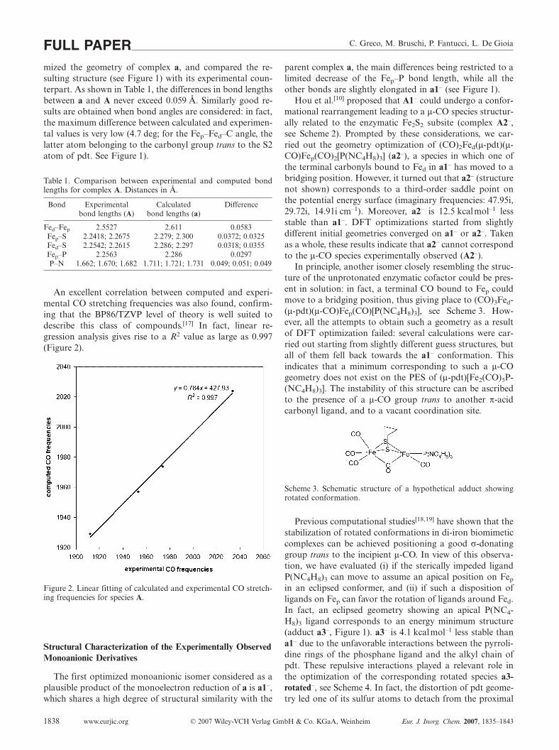
C. Greco, M. Bruschi, P. Fantucci, L. De GioiaFULL PAPERmized the geometry of complex a, and compared the re-sulting structure (see Figure 1) with its experimental coun-terpart. As shown in Table 1, the differences in bond lengthsbetween a and A never exceed 0.059 Å. Similarly good re-sults are obtained when bond angles are considered: in fact,the maximum difference between calculated and experimen-tal values is very low (4.7 deg; for the Fep–Fed–C angle, thelatter atom belonging to the carbonyl group trans to the S2atom of pdt. See Figure 1).
Table 1. Comparison between experimental and computed bondlengths for complex A. Distances in Å.
Bond Experimental Calculated Differencebond lengths (A) bond lengths (a)
Fed–Fep 2.5527 2.611 0.0583Fep–S 2.2418; 2.2675 2.279; 2.300 0.0372; 0.0325Fed–S 2.2542; 2.2615 2.286; 2.297 0.0318; 0.0355Fep–P 2.2563 2.286 0.0297P–N 1.662; 1.670; 1.682 1.711; 1.721; 1.731 0.049; 0.051; 0.049
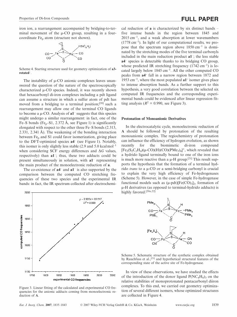
An excellent correlation between computed and experi-mental CO stretching frequencies was also found, confirm-ing that the BP86/TZVP level of theory is well suited todescribe this class of compounds.[17] In fact, linear re-gression analysis gives rise to a R2 value as large as 0.997(Figure 2).
Figure 2. Linear fitting of calculated and experimental CO stretch-ing frequencies for species A.
Structural Characterization of the Experimentally ObservedMonoanionic Derivatives
The first optimized monoanionic isomer considered as aplausible product of the monoelectron reduction of a is a1–,which shares a high degree of structural similarity with the
www.eurjic.org © 2007 Wiley-VCH Verlag GmbH & Co. KGaA, Weinheim Eur. J. Inorg. Chem. 2007, 1835–18431838
parent complex a, the main differences being restricted to alimited decrease of the Fep–P bond length, while all theother bonds are slightly elongated in a1– (see Figure 1).
Hou et al.[10] proposed that A1– could undergo a confor-mational rearrangement leading to a µ-CO species structur-ally related to the enzymatic Fe2S2 subsite (complex A2–,see Scheme 2). Prompted by these considerations, we car-ried out the geometry optimization of (CO)2Fed(µ-pdt)(µ-CO)Fep(CO)2[P(NC4H8)3] (a2–), a species in which one ofthe terminal carbonyls bound to Fed in a1– has moved to abridging position. However, it turned out that a2– (structurenot shown) corresponds to a third-order saddle point onthe potential energy surface (imaginary frequencies: 47.95i,29.72i, 14.91i cm–1). Moreover, a2– is 12.5 kcalmol–1 lessstable than a1–. DFT optimizations started from slightlydifferent initial geometries converged on a1– or a2–. Takenas a whole, these results indicate that a2– cannot correspondto the µ-CO species experimentally observed (A2–).
In principle, another isomer closely resembling the struc-ture of the unprotonated enzymatic cofactor could be pres-ent in solution: in fact, a terminal CO bound to Fep couldmove to a bridging position, thus giving place to (CO)3Fed-(µ-pdt)(µ-CO)Fep(CO)[P(NC4H8)3], see Scheme 3. How-ever, all the attempts to obtain such a geometry as a resultof DFT optimization failed: several calculations were car-ried out starting from slightly different guess structures, butall of them fell back towards the a1– conformation. Thisindicates that a minimum corresponding to such a µ-COgeometry does not exist on the PES of (µ-pdt)[Fe2(CO)5P-(NC4H8)3]. The instability of this structure can be ascribedto the presence of a µ-CO group trans to another π-acidcarbonyl ligand, and to a vacant coordination site.
Scheme 3. Schematic structure of a hypothetical adduct showingrotated conformation.
Previous computational studies[18,19] have shown that thestabilization of rotated conformations in di-iron biomimeticcomplexes can be achieved positioning a good σ-donatinggroup trans to the incipient µ-CO. In view of this observa-tion, we have evaluated (i) if the sterically impeded ligandP(NC4H8)3 can move to assume an apical position on Fep
in an eclipsed conformer, and (ii) if such a disposition ofligands on Fep can favor the rotation of ligands around Fed.In fact, an eclipsed geometry showing an apical P(NC4-H8)3 ligand corresponds to an energy minimum structure(adduct a3–, Figure 1). a3– is 4.1 kcalmol–1 less stable thana1– due to the unfavorable interactions between the pyrroli-dine rings of the phosphane ligand and the alkyl chain ofpdt. These repulsive interactions played a relevant role inthe optimization of the corresponding rotated species a3-rotated–, see Scheme 4. In fact, the distortion of pdt geome-try led one of its sulfur atoms to detach from the proximal
Properties of Di-Iron Compounds FULL PAPERiron ion, a rearrangement accompanied by bridging-to-ter-minal movement of the µ-CO group, resulting in a four-coordinate Fep atom (structure not shown).
Scheme 4. Starting structure used for geometry optimization of a3-rotated–.
The instability of µ-CO anionic complexes leaves unan-swered the question of the nature of the spectroscopicallycharacterized µ-CO species. Indeed, it was recently shownthat hexacarbonyl di-iron complexes including a pdt ligandcan assume a structure in which a sulfur atom of pdt hasmoved from a bridging to a terminal position;[20] such arearrangement may allow one of the terminal CO ligandsto become a µ-CO. Analysis of a1– suggests that this speciesmight undergo a similar rearrangement: in fact, one of theFe–S bonds (Fep–S1, 2.372 Å, see Figure 1) is significantlyelongated with respect to the other three Fe–S bonds (2.313,2.331, 2.341 Å). The weakening of the bonding interactionbetween Fep and S1 could favor isomerization, giving placeto the DFT-optimized species a4– (see Figure 1). Notably,this isomer is only slightly less stable (2.9 and 5.0 kcalmol–1
when considering SCF energy differences and ∆G values,respectively) than a1–; thus, these two adducts could bepresent simultaneously in solution, with a1– representingthe main product of the monoelectronic reduction of a.
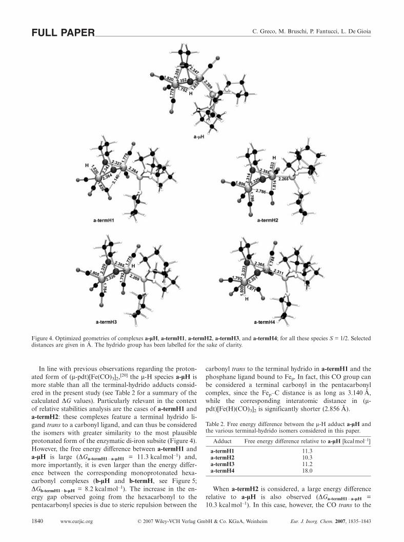
The co-existence of a4– and a1– is also supported by thecomparison between the computed CO stretching fre-quencies of these two species and the experimental IRbands: in fact, the IR spectrum collected after electrochemi-
Figure 3. Linear fitting of the calculated and experimental CO fre-quencies for the anionic adducts coming from monoelectronic re-duction of A.
Eur. J. Inorg. Chem. 2007, 1835–1843 © 2007 Wiley-VCH Verlag GmbH & Co. KGaA, Weinheim www.eurjic.org 1839
cal reduction of a is characterized by six distinct bands –five intense bands in the region between 1845 and2015 cm–1, and a weak absorption at lower wavenumbers(1778 cm–1). In light of our computational results, we pro-pose that the spectrum region above 1850 cm–1 is domi-nated by the stretching modes of the five terminal carbonylsincluded in the main reduction product a1–; the less stablea4– species is detectable thanks to its bridging CO group,whose predicted IR stretching frequency (1742 cm–1) is lo-cated largely below 1845 cm–1. All the other computed COpeaks from a4– fall in a narrow region between 1872 and1953 cm–1, where the most populated a1– isomer gives placeto intense absorption bands. As a further support to thishypothesis, a very good correlation between the selected sixcomputed IR frequencies and the corresponding experi-mental bands could be evidenced after linear regression fit-ting analysis (R2 = 0.990, see Figure 3).
Protonation of Monoanionic Derivatives
In the electrocatalytic cycle, monoelectronic reduction ofA should be followed by protonation of the resultingmonoanionic complex. The regiochemistry of protonationcan influence the efficiency of hydrogen evolution, as shownrecently for the biomimetic di-iron compound[Fe2(S2C2H4)(µ-CO)(H)(CO)(PMe3)4]+, which revealed thata hydrido ligand terminally bound to one of the iron ionsis much more reactive than a µ-H group.[21] This result sup-ports the hypothesis that the formation of a terminal hyd-rido trans to a µ-CO or a semi-bridging carbonyl is crucialto explain the very high efficiency of Fe-hydrogenases(Scheme 5). However, in the case of simple Fe-hydrogenasefunctional models such as (µ-pdt)[Fe(CO)3]2, formation ofµ-H derivatives (as opposed to terminal-hydrido adducts) ishighly favored.[20a,22]
Scheme 5. Schematic structure of the synthetic complex obtainedby Rauchfuss et al.,[21] and hypothetical structural features of thecorresponding state of the active site of Fe-hydrogenase.
In view of these observations, we have studied the effectsof the introduction of the donor ligand P(NC4H8)3 on therelative stabilities of monoprotonated pentacarbonyl diironcomplexes. To this end, we carried out geometry optimiza-tion of several different isomers, whose optimized structuresare collected in Figure 4.
C. Greco, M. Bruschi, P. Fantucci, L. De GioiaFULL PAPER
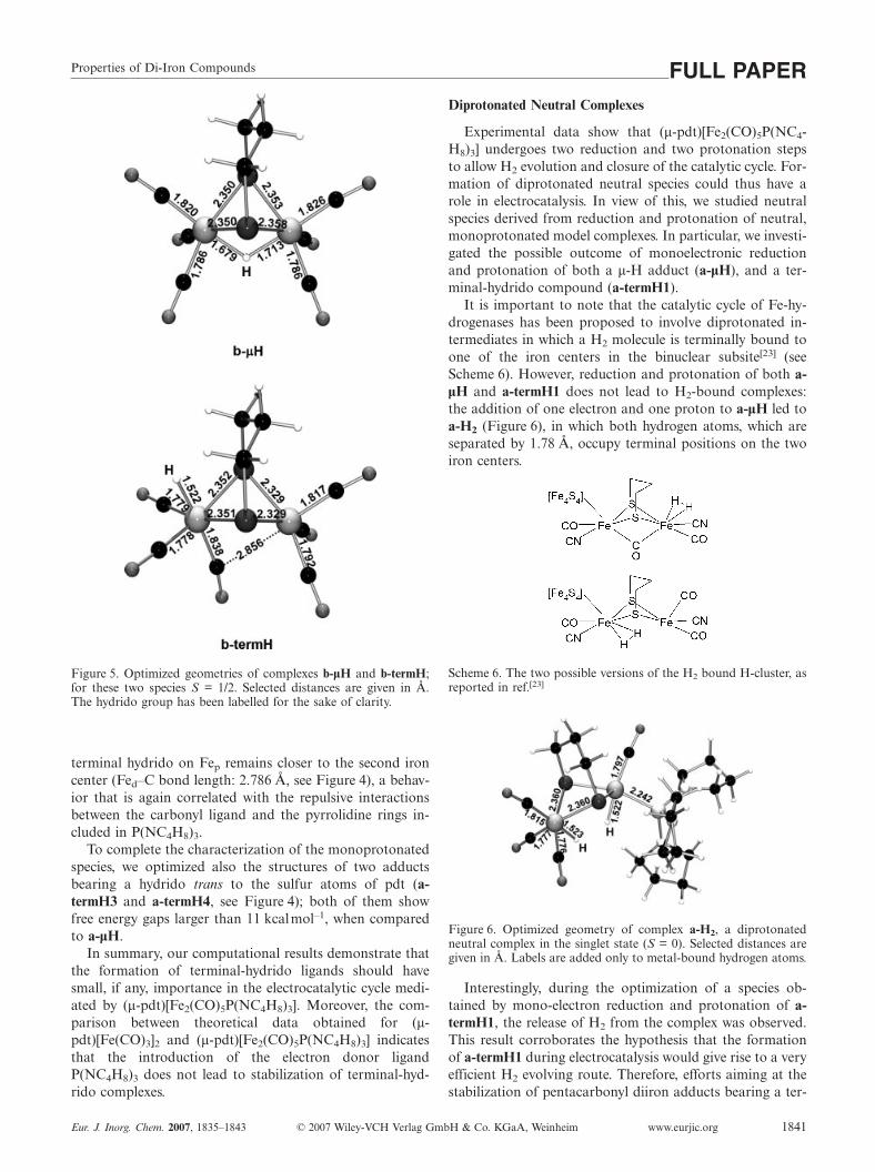
Figure 4. Optimized geometries of complexes a-µH, a-termH1, a-termH2, a-termH3, and a-termH4; for all these species S = 1/2. Selecteddistances are given in Å. The hydrido group has been labelled for the sake of clarity.
In line with previous observations regarding the proton-ated form of (µ-pdt)[Fe(CO)3]2,[20] the µ-H species a-µH ismore stable than all the terminal-hydrido adducts consid-ered in the present study (see Table 2 for a summary of thecalculated ∆G values). Particularly relevant in the contextof relative stabilities analysis are the cases of a-termH1 anda-termH2: these complexes feature a terminal hydrido li-gand trans to a carbonyl ligand, and can thus be consideredthe isomers with greater similarity to the most plausibleprotonated form of the enzymatic di-iron subsite (Figure 4).However, the free energy difference between a-termH1 anda-µH is large (∆Ga-termH1–a-µH1 = 11.3 kcalmol–1) and,more importantly, it is even larger than the energy differ-ence between the corresponding monoprotonated hexa-carbonyl complexes (b-µH and b-termH, see Figure 5;∆Gb-termH1–b-µH = 8.2 kcalmol–1). The increase in the en-ergy gap observed going from the hexacarbonyl to thepentacarbonyl species is due to steric repulsion between the
www.eurjic.org © 2007 Wiley-VCH Verlag GmbH & Co. KGaA, Weinheim Eur. J. Inorg. Chem. 2007, 1835–18431840
carbonyl trans to the terminal hydrido in a-termH1 and thephosphane ligand bound to Fep. In fact, this CO group canbe considered a terminal carbonyl in the pentacarbonylcomplex, since the Fep–C distance is as long as 3.140 Å,while the corresponding interatomic distance in (µ-pdt)[Fe(H)(CO)3]2 is significantly shorter (2.856 Å).
Table 2. Free energy difference between the µ-H adduct a-µH andthe various terminal-hydrido isomers considered in this paper.
Adduct Free energy difference relative to a-µH [kcalmol–1]
a-termH1 11.3a-termH2 10.3a-termH3 11.2a-termH4 18.0
When a-termH2 is considered, a large energy differencerelative to a-µH is also observed (∆Ga-termH1–a-µH =10.3 kcalmol–1). In this case, however, the CO trans to the
Properties of Di-Iron Compounds FULL PAPER
Figure 5. Optimized geometries of complexes b-µH and b-termH;for these two species S = 1/2. Selected distances are given in Å.The hydrido group has been labelled for the sake of clarity.
terminal hydrido on Fep remains closer to the second ironcenter (Fed–C bond length: 2.786 Å, see Figure 4), a behav-ior that is again correlated with the repulsive interactionsbetween the carbonyl ligand and the pyrrolidine rings in-cluded in P(NC4H8)3.
To complete the characterization of the monoprotonatedspecies, we optimized also the structures of two adductsbearing a hydrido trans to the sulfur atoms of pdt (a-termH3 and a-termH4, see Figure 4); both of them showfree energy gaps larger than 11 kcalmol–1, when comparedto a-µH.
In summary, our computational results demonstrate thatthe formation of terminal-hydrido ligands should havesmall, if any, importance in the electrocatalytic cycle medi-ated by (µ-pdt)[Fe2(CO)5P(NC4H8)3]. Moreover, the com-parison between theoretical data obtained for (µ-pdt)[Fe(CO)3]2 and (µ-pdt)[Fe2(CO)5P(NC4H8)3] indicatesthat the introduction of the electron donor ligandP(NC4H8)3 does not lead to stabilization of terminal-hyd-rido complexes.
Eur. J. Inorg. Chem. 2007, 1835–1843 © 2007 Wiley-VCH Verlag GmbH & Co. KGaA, Weinheim www.eurjic.org 1841
Diprotonated Neutral Complexes
Experimental data show that (µ-pdt)[Fe2(CO)5P(NC4-H8)3] undergoes two reduction and two protonation stepsto allow H2 evolution and closure of the catalytic cycle. For-mation of diprotonated neutral species could thus have arole in electrocatalysis. In view of this, we studied neutralspecies derived from reduction and protonation of neutral,monoprotonated model complexes. In particular, we investi-gated the possible outcome of monoelectronic reductionand protonation of both a µ-H adduct (a-µH), and a ter-minal-hydrido compound (a-termH1).
It is important to note that the catalytic cycle of Fe-hy-drogenases has been proposed to involve diprotonated in-termediates in which a H2 molecule is terminally bound toone of the iron centers in the binuclear subsite[23] (seeScheme 6). However, reduction and protonation of both a-µH and a-termH1 does not lead to H2-bound complexes:the addition of one electron and one proton to a-µH led toa-H2 (Figure 6), in which both hydrogen atoms, which areseparated by 1.78 Å, occupy terminal positions on the twoiron centers.
Scheme 6. The two possible versions of the H2 bound H-cluster, asreported in ref.[23]
Figure 6. Optimized geometry of complex a-H2, a diprotonatedneutral complex in the singlet state (S = 0). Selected distances aregiven in Å. Labels are added only to metal-bound hydrogen atoms.
Interestingly, during the optimization of a species ob-tained by mono-electron reduction and protonation of a-termH1, the release of H2 from the complex was observed.This result corroborates the hypothesis that the formationof a-termH1 during electrocatalysis would give rise to a veryefficient H2 evolving route. Therefore, efforts aiming at thestabilization of pentacarbonyl diiron adducts bearing a ter-
C. Greco, M. Bruschi, P. Fantucci, L. De GioiaFULL PAPERminal hydrido ligand could assume great relevance for thedevelopment of better electrocatalytic materials.
Conclusions
In the first part of this work, we investigated the struc-tural properties of several plausible isomers obtained bymonoelectron reduction of the biomimetic complex (µ-pdt)[Fe2(CO)5P(NC4H8)3]. Our computational study al-lowed us to show that complexes a1– and a4– should co-exist in solution. The former adduct corresponds to a “clas-sical” eclipsed conformation, while the second one is a µ-CO species structurally not related to the Fe-hydrogenasesbinuclear subsite. Therefore, the possibility that a rotatedconformer closely resembling the H-cluster geometry canbe formed during electrocatalysis is excluded. The ineffec-tiveness of P(NC4H8)3 in stabilizing the rotated form of thesynthetic assembly can be rationalized in light of recenttheoretical results regarding monosubstituted derivatives of(µ-pdt)[Fe(CO)3]2 (i.e., adducts in which only one carbonylhas been replaced by a good σ-donor).[18,19] In fact, it hasbeen shown that a good electron-donating ligand (L) is ableto stabilize the rotated conformation of (µ-pdt)[Fe(CO)3-Fe(CO)2(L)]– complexes, when L is trans to the incipientbridging CO group.[18,19] Nevertheless, if L corresponds toP(NC4H8)3, it is not possible to place the σ-donor trans tothe incipient µ-CO, since in such a configuration the alkylchain of pdt would cause steric clashes with the pyrrolidinerings included in P(NC4H8)3. These observations suggestthat the introduction of an electron donor ligand as good asP(NC4H8)3, but less sterically demanding, could represent abetter choice to facilitate the formation of more stable ro-tated adducts.
In the second part of the study, we have shown that themost stable protonated, uncharged form of (µ-pdt)-[Fe2(CO)5P(NC4H8)3] corresponds to a µ-H adduct (a-µH),and that terminal-hydrido complexes – which turned out tobe highly reactive according to DFT results – are not in-volved in the electrocatalytic cycle. In fact, all the terminal-hydrido species here investigated are less stable than the hy-drido-bridged isomer a-µH by at least 10 kcalmol–1. Thesteric repulsion between the P(NC4H8)3 ligand and the car-bonyl ligands plays a relevant role in determining such largeenergy gaps.
In conclusion, DFT results allowed us to rationalize whythe synthetic complex (µ-pdt)[Fe2(CO)5P(NC4H8)3] is sig-nificantly less efficient then the enzyme in H2 pro-duction,[10,24] and might contribute to the design and syn-thesis of novel functional models of the active site of Fe-hydrogenase.
[1] M. Y. Darensboug, Nature 2005, 433, 589.[2] S. P. Albracht, Biochim. Biophys. Acta 1994, 167, 1188; E. G.
Graf, R. K. Thauer, FEBS Lett. 1981, 136, 165; M. W. W. Ad-ams, Biochim. Biophys. Acta 1990, 115, 1020; R. Cammack,Nature 1999, 397, 214; Y. Nicolet, B. J. Lemon, J. C. Fontecilla-Camps, J. W. Peters, Trends Biochem. Sci. 2000, 25, 138; J. W.Peters, Curr. Opin. Struct. Biol. 1999, 9, 670; D. S. Horner, B.
www.eurjic.org © 2007 Wiley-VCH Verlag GmbH & Co. KGaA, Weinheim Eur. J. Inorg. Chem. 2007, 1835–18431842
Heil, T. Happe, T. M. Embley, Trends Biochem. Sci. 2002, 27,148; Y. Nicolet, C. Cavazza, J. C. Fontecilla-Camps, Inorg. Bi-ochem. 2002, 91, 1; R. Cammack, M. Frey, R. Robson, Hydro-gen as Fuel – Learning from Nature, Taylor and Francis, Lon-don, 2001; F. A. Armstrong, Curr. Opin. Chem. Biol. 2004, 8,133.
[3] E. J. Lyon, S. Shima, G. Buurman, S. Chowdhuri, A. Bats-chauer, K. Steinbach, R. K. Thauer, Eur. J. Biochem. 2004, 271,195; O. Pilak, B. Mamat, S. Vogt, C. H. Hagemeier, R. K.Thauer, S. Shima, C. Vonrhein, E. Warkentin, U. Ermler, J.Mol. Biol. 2006, 358, 798.
[4] J. W. Peters, W. N. Lanzilotta, B. J. Lemon, L. C. Seefeldt, Sci-ence 1998, 282, 1853.
[5] Y. Nicolet, C. Piras, P. Legrand, C. E. Hatchikian, J. C. Fontec-illa-Camps, Structure 1999, 7, 13.
[6] Y. Nicolet, A. L. de Lacey, X. Vernede, V. M. Fernandez, E. C.Hatchikian, J. C. Fontecilla-Camps, J. Am. Chem. Soc. 2001,123, 1596.
[7] a) W. Gao, J. Liu, C. Ma, L. Weng, K. Jin, C. Chen, B. Akerm-ark, L. Sun, Inorg. Chim. Acta 2006, 359, 1071; b) L.-C. Song,C. Jea, J. Yan, H.-T. Wang, X.-F. Liu, X. Q.-M. Hu, Organome-tallics 2006, 25, 1544; c) S. Jiang, J. Liu, L. Sun, Inorg. Chem.Commun. 2006, 9, 290; d) J.-F. Capon, F. Gloaguen, P.Schollhammer, J. Talarmin, Coord. Chem. Rev. 2005, 249, 1664;e) C. Tard, X. Liu, S. K. Ibrahim, M. Bruschi, L. De Gioia,S. C. Daview, X. Yang, L.-S. Wang, G. Sawers, C. J. Pickett,Nature 2005, 433, 610; f) L.-C. Song, Z.-Y. Yang, H.-Z. Bian,Y. Liu, H.-T. Wang, X.-F. Liu, Q.-M. Hu, Organometallics2005, 24, 6126; g) J. W. Tye, J. Lee, H.-W. Wang, R. Mejia-Rodriguez, J. H. Reibenspies, M. B. Hall, M. Y. Darensbourg,Inorg. Chem. 2005, 44, 5550; h) S. J. Borg, T. Behrsing, S. P.Best, M. Razavet, X. Liu, C. J. Pickett, J. Am. Chem. Soc. 2004,126, 16988; i) S. Ott, M. Kritikos, B. Akermark, L. Sun, R.Lomoth, Angew. Chem. Int. Ed. 2004, 43, 1006; j) T. Liu, M.Wang, Z. Shi, H. Cui, W. Dong, J. Chen, B. Akermark, L. Sun,Chem. Eur. J. 2004, 10, 4474; k) R. Mejia-Rodriguez, D.Chong, J. H. Reibenspies, M. P. Soriaga, M. Y. Darensborg, J.Am. Chem. Soc. 2004, 126, 12004; l) J.-F. Capon, F. Gloaguen,P. Schollhammer, J. Talarmin, J. Electroanal. Chem. 2003, 566,241; m) D. Chong, I. P. Georgakaki, R. Mejia-Rodriguez, J.Sanabria-Chinchilla, M. P. Soriaga, M. Y. Darensbourg, Dal-ton Trans. 2003, 4158; n) F. Gloaguen, J. D. Lawrence, T. B.Rauchfuss, M. Bénard, M.-M. Rohmer, Inorg. Chem. 2002, 41,6573; o) F. Gloaguen, J. D. Lawrence, T. B. Rauchfuss, J. Am.Chem. Soc. 2001, 123, 9476.
[8] M. Bruschi, P. Fantucci, L. De Gioia, Inorg. Chem. 2004, 43,3733.
[9] J. W. Tye, M. Y. Darensbourg, M. B. Hall, Inorg. Chem. 2006,45, 1552.
[10] J. Hou, X. Peng, Z. Zhou, S. Sun, X. Zhao, S. Gao, J. Org.Chem. 2006, 71, 691, 4633.
[11] A. D. Becke, Phys. Rev. A 1988, 38, 3098; J. P. Perdew, Phys.Rev. B 1986, 33, 8822.
[12] A. Schafer, C. Huber, R. Ahlrichs, J. Chem. Phys. 1994, 100,5829.
[13] R. Ahlrichs, M. Bar, M. Haser, H. Horn, C. Kolmel, Chem.Phys. Lett. 1989, 162, 165.
[14] K. Eichkorn, F. Weigend, O. Treutler, R. Ahlrichs, Theor.Chem. Acc. 1997, 97, 119.
[15] F. Jensen, Introduction to Computational Chemistry, JohnWiley & Sons, Chichester, England.
[16] A. Klamt, J. Phys. Chem. 1995, 99, 2224; A. Klamt, J. Phys.Chem. 1996, 100, 3349.
[17] G. Zampella, M. Bruschi, P. Fantucci, M. Razavet, C. J. Pick-ett, L. De Gioia, Chemistry 2005, 11, 509.
[18] M. Y. Darensbourg, E. J. Lyon, X. Zhao, I. P. Georgakaki,Proc. Natl. Acad. Sci. USA 2003, 100, 3683; I. P. Georgakaki,L. M. Thomson, E. J. Lyon, M. B. Hall, M. Y. Darensbourg,Coord. Chem. Rev. 2003, 238–239, 255.

Properties of Di-Iron Compounds FULL PAPER[19] It is interesting to note that a similar stabilizing effect exerted
by the ligand trans to the µ-CO could be operative also in theenzyme active site. In fact, the Fe4S4 subcluster included in theH-cluster proved to be a good electron-donating group (seeref.[7e]) and it has been demonstrated that good donors local-ized at the position occupied by the Fe–S cubane can favor therotation of ligands bound to the distal iron center (see ref.[8]).
[20] S. J. Borg, J. W. Tye, M. B. Hall, S. P. Best, Inorg. Chem. 2007,46, 384; C. Greco, G. Zampella, L. Bertini, M. Bruschi, P. Fan-tucci, L. De Gioia, Inorg. Chem. 2007, 46, 108.
Eur. J. Inorg. Chem. 2007, 1835–1843 © 2007 Wiley-VCH Verlag GmbH & Co. KGaA, Weinheim www.eurjic.org 1843
[21] J. I. van der Vlugt, T. B. Rauchfuss, C. M. Whaley, S. R. Wil-son, J. Am. Chem. Soc. 2005, 127, 16012.
[22] T. Zhou, Y. Mo, A. Liu, Z. Zhou, K. R. Tsai, Inorg. Chem.2004, 43, 923.
[23] M. Bruschi, G. Zampella, P. Fantucci, L. De Gioia, Coord.Chem. Rev. 2005, 249, 1620.
[24] J. N. Butt, M. Filipiak, W. R. Hagen, Eur. J. Biochem. 1997,245, 116.
Received: November 21, 2006Published Online: March 14, 2007


![[FeFe]‐Hydrogenase Mimic Employing κ2‐C,N‐Pyridine ... · DOI: 10.1002/ejic.201900405 Full Paper Proton Reduction Catalysts [FeFe]-Hydrogenase Mimic Employing κ2-C,N-Pyridine](https://img.pdfslide.tips/doc/110x75/60cf254691c2d1101b09b0e4/fefeahydrogenase-mimic-employing-2acnapyridine-doi-101002ejic201900405.jpg)