Embed Size (px)
Citation preview

Ingegneria delle tecnologie per la salute
Fondamenti di anatomia e istologia
aa. 2019-20
LEZIONE 3 ISTOLOGIA ed EMBRIOLOGIA

Anatomia umana Edizione italiana a cura di Fabrizio Michetti MCKINLEY - O'LOUGHLIN Data di pubblicazione: maggio 2014 Prezzo: 49,50 € ISBN: 978-88-299-2655-8 Codice Piccin: 1312165 Libro in italiano

Atlante di anatomia Edizione italiana a cura di Elena Donetti KAMINA Data di pubblicazione: settembre 2014 Prezzo: 45,00 € ISBN: 978-88-299-2668-8 Codice Piccin: 1100850 Libro in italiano

Colorare l'anatomia Edizione italiana a cura di Raffaele De Caro e Sergio Galli KAPIT - ELTON Data di pubblicazione: marzo 2016 Prezzo: 20,00 € ISBN: 978-88-299-2747-0 Codice Piccin: 1101200 Libro in italiano

Lezione 3. Cenni di embriologia ed istologia generale.
https://human.biodigital.com/index.html
https://www.openstaxcollege.org/files/textbook_version/hi_res_pdf/13/col11496-1.7_20150715-OP.pdf
https://lectureug5.files.wordpress.com/2014/02/difiores-atlas-of-histology-with-functional-correlations-11th-ed.pdf
THE TISSUE LEVEL OF ORGANIZATION
After studying this chapter, you will be able to: • Identify the main tissue types and discuss their roles in the human body • Identify the 4 types of tissue membranes and the characteristics of each that make them functional • Explain the functions of various epithelial tissues and how their forms enable their functions • Explain the functions of various connective tissues and how their forms enable their functions • Describe the characteristics of muscle tissue and how these enable function • Discuss the characteristics of nervous tissue and how these enable information processing and control of muscular and glandular activities

THE TISSUE LEVEL OF ORGANIZATION
our body: at least 200 distinct cell types
same internal structures, but vary enormously in shape and function
occur in organized layers: tissue
starts as a single cell
at fertilization and it
gives rise to trillions
of cells, each built
from the same
blueprint, but
organizing into tissues
and becoming
irreversibly committed
to a developmental
pathway

Tissue = a group of cells found together in the body, that share a common and morphological features and are arranged in an orderly pattern that achieves the tissue’s functions. many types of cells but organized into 4 broad categories of tissues: 1. epithelial 2. connective 3.muscle 4. nervous with a specific functions that contribute to the overall health and maintenance of the body.
Types of Tissues
Histology = microscopic study of tissue appearance, organization, and function.

Histology is a visual, as well as a very colorful, science
that is studied with the aid of a light microscope.
The prepared specimens for examination are thinly sliced,
placed on a glass slide, stained with a variety of stains,
and examined with a light microscope via a light beam that
passes through the tissues that are fixed on the slide.
Tissue Preparation—Light Microscopy
Antonie van Leeuwenhoek (1632–1724) is credited with bringing the microscope to the attention of biologists

essential in order to permanently preserve structural and molecular composition of specimen.
to further accelerate penetration and proper fixation process, tissue specimen is first cut into small pieces and then immersed into fixative.
hardens specimen for sectioning, causes cross-linkage of macromolecules within the cells, reduces cellular degeneration, preserves integrity of cells and tissues, and increases their affinity to take up different stains.
most commonly used fixative for light microcopy is neutral-buffered FORMALDEHYDE.
Tissue Preparation—Light Microscopy
Aldehyde fixatives form crosslinks between proteins.
FIXATION = prompt immersion of the specimen with different chemical solutions, to preserve a section of tissue or organ for histologic examination

POSTFIXATION After fixation, water must first be removed by
passing it through a series of ascending ALCOHOL (ethanol) concentrations, usually from 70 to 100%
then specimen must be cleared of alcohol by passing it through several changes of such clearing agents as XYLENE
Once the specimen is impregnated with the clearing agent xylene, it is then placed in a warm mold containing melted PARAFFIN. Once removed from the heat source, the paraffin in the mold cools, solidifies, and encases the specimen.
paraffin block then trimmed to the size of the specimen and mounted in an instrument called a MICROTOME [precisely advances the paraffin block so that the sections are cut at specific and predetermined increments with a steel knife] = sections are normally cut at 3-10 μm thickness.
thin paraffin sections are then collected and floated in a warm water bath and placed onto a glass slide that has been covered with a thin layer of albumen, which serves as an adhesive medium for the specimen.
Tissue Preparation—Light Microscopy

hydrated sections can then be stained with a variety of water-soluble stains, which selectively stain various components of the specimen
Most of the stains used for histologic slide preparations act like acidic or basic compounds: structures that stain most readily with basic stains are called BASOPHILIC, and those that stain with acidic stains are called ACIDOPHILIC [most common stains are hematoxylin and eosin stains]
Staining of Sections
paraffin sections on the glass slide are colorless needs to be stained.
paraffin must first be dissolved from the specimen with solvents, such as xylene, and the sections rehydrated with a series of decreasing alcohol concentrations.

Staining of Sections

Staining of Sections

Staining of Sections

Staining of Sections

Staining of Sections

Staining of Sections

Staining of Sections

Staining of Sections

Staining of Sections

Interpretation of Histologic Sections
the most challenging and difficult aspects of histology: interpretation of what the 2-dimensional histology sections represent in 3 dimensions.
Histologic sections = thin, flat slices of fixed and stained tissues or organs mounted on flat glass slides.
sections normally composed of cellular, fibrous, and tubular structures cut in different planes variety of shapes, sizes, and layers may be visible, depending on the plane of section.
Fibrous structures are solid and are found in connective, nervous, and muscle tissues.
Tubular structures are hollow and represent various types of blood vessels, lymph vessels, glandular ducts, and glands of the body.

Planes of Section of a Round, Solid Object

Planes of Section Through a Hollow Structure or a Tube

Planes of Section Through a Hollow Structure or a Tube

Transmission and Scanning Electron Microscopy

Cell microscopic anatomy (TEM)

Ciliated and nonciliated cells

Junctional complex

Basal region

Basal region in ions transporting cell

Cilia and microvilli

Nuclear envelope and pores

Mitochondria

RER

SER

Golgi apparatus

Lysosomes

Mitosis

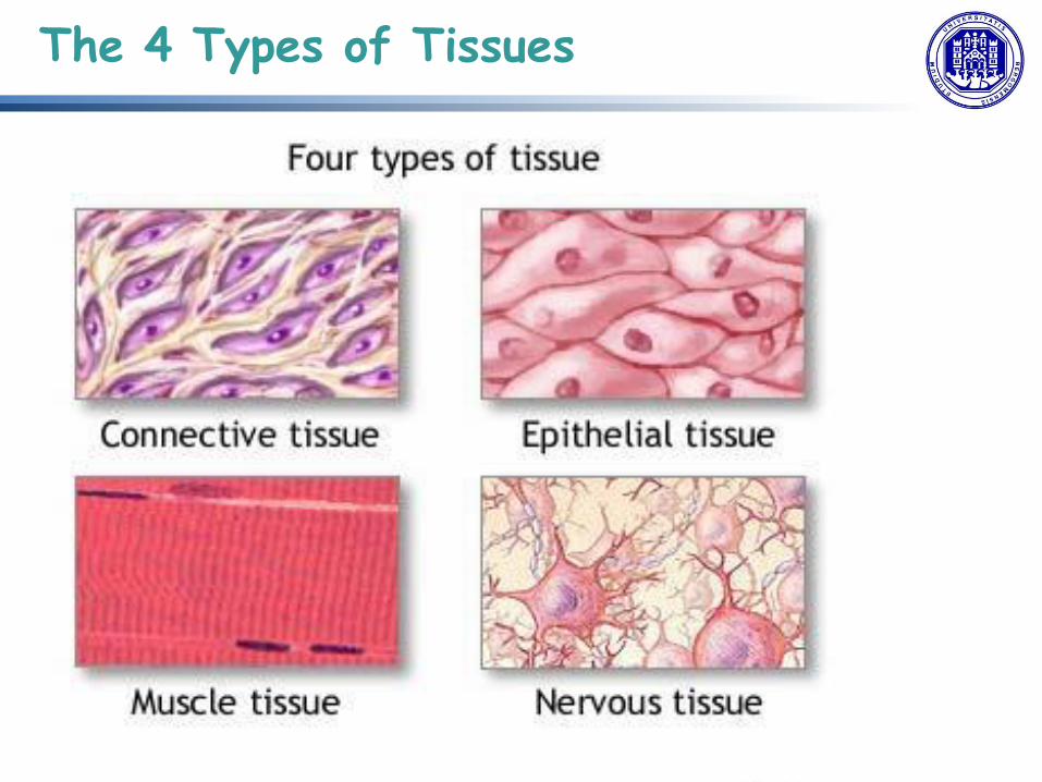
The 4 Types of Tissues
1. Epithelial tissue (epithelium) = sheets of cells that cover exterior surfaces of the body, internal cavities and passageways, and forms certain glands.
2. Connective tissue = binds cells and organs together (functions in the protection, support, and integration of all parts of the body)
3. Muscle tissue = excitable, responding to stimulation and contracting to provide movement, and occurs as 3 major types: skeletal (voluntary) muscle, smooth muscle, and cardiac muscle in the heart.
4. Nervous tissue = also excitable, allowing the propagation of electrochemical signals in the form of nerve impulses .
The 4 Types of Tissues

Organs are made of many different tissues…of the 4 fundamental types
sections through 4 different organs: Intestines, Skin, Lung, & Trachea. (each organ is made of multiple tissues and that their are variations on how the tissues are designed)

Embryonic Origin of Tissues
totipotent
three major cell lineages established within the embryo

Embryonic Origin of Tissues
3 lineages of embryonic cells forms 3 distinct germ layers identified by its relative position: 1. ectoderm (ecto-= “outer”), 2. mesoderm (meso- = “middle”), 3. endoderm (endo- = “inner”).

Embryonic Origin of Tissues
!!! epithelial tissue originates in all three layers, whereas nervous tissue derives primarily from the ectoderm and muscle tissue from mesoderm.

Tissue membrane = thin layer or sheet of cells that covers the outside of the body (for example, skin), the organs (for example, pericardium), internal passageways that lead to the exterior of the body (for example, abdominal mesenteries), and the lining of the moveable joint cavities.
Tissue Membranes
2 basic types
1. connective tissue 2. epithelial membranes

Tissue Membranes
1. Connective Tissue Membranes = formed solely from connective tissue, encapsulate organs, and line our movable joints (synovial membrane)
2. Epithelial Membranes = composed of epithelium attached to a layer of connective tissue i. mucous membrane (mucosae) = line the body cavities and hollow
passageways that open to the external environment, and include the digestive, respiratory, excretory, and reproductive tracts. Mucous, produced by the epithelial exocrine glands, covers the epithelial layer. The underlying connective tissue, called the lamina propria, supports the epithelial layer.
ii. serous membrane = composed of mesodermally derived epithelium called the mesothelium that is supported by connective tissue, line coelomic cavities (do not open to outside).
iii. cutaneous membrane (skin) = stratified squamous epithelial membrane resting on top of connective tissue.

Epithelial Tissue
share structural/ functional features: highly cellular, with little or no extracellular
matrix between cells cell junction = specialized intercellular
connection between cell polarity = differences in structure and
function between the exposed or apical facing surface of the cell and the basal surface
basal lamina = (a mixture of glycoproteins and collagen) provides an attachment site for the epithelium, separating it from underlying connective tissue and attaches to a reticular lamina, which is secreted by the underlying connective tissue, forming a basement membrane that hold it all together
nearly completely avascular capable of rapidly replacing damaged and dead
cells
= essentially large sheets of cells covering all the surfaces of the body exposed to the outside world + lining the outside of organs + much of the glandular tissue of the body;

Epithelial Tissue
Generalized Functions of Epithelial Tissue • provide the body’s first line of protection from physical, chemical, and biological wear and
tear, controlling permeability and allowing selective transfer of materials across a physical barrier
• are sometimes capable of secretion and release mucous and specific chemical compounds onto their apical surfaces.
The Epithelial Cell typically characterized by the polarized distribution of organelles and membrane-bound proteins between their basal and apical surfaces: certain organelles are segregated to the basal sides, whereas other organelles and extensions, such as cilia (microscopic extensions of the apical cell membrane that are supported by microtubules), when present, are on the apical surface and beat in unison and move fluids as well as trapped particles.

Epithelial Tissue
Cell to Cell Junctions Cells of epithelia are closely
connected and are not
separated by extracellular
material. 3 basic types of
connections allow varying
degrees of interaction between
the cells:
1. tight junctions,
2. anchoring junctions, 3. gap junctions

Epithelial Tissue
Classification of Epithelial Tissues
classified according to the shape
of the cells and number of the cell
layers formed. Transitional
describes a form of specialized
stratified epithelium in which the shape of the cells can vary.

Epithelial Tissue
Classification of Epithelial Tissues
classified according to the shape of the cells and number of the cell layers
formed. Transitional describes a form of specialized stratified epithelium in which the shape of the cells can vary.

Epithelial Tissue
Both simple and pseudostratified columnar epithelia
are heterogeneous epithelia because they include
additional types of cells interspersed among the
epithelial cells. For example, a goblet cell is a mucous-secreting
unicellular “gland” interspersed between the columnar epithelial cells of mucous membranes
goblet cell

Epithelial Tissue
different categories of epithelial cell tissue cells

Epithelial Tissue
different categories of epithelial cell tissue cells

Epithelial Tissue
Glandular Epithelium gland = a structure made up of one or more cells modified to synthesize and secrete chemical substances; most glands consist of groups of epithelial cells. 1. Endocrine Glands a ductless gland that releases secretions (hormones) directly into surrounding tissues and fluids (endo- = “inside”), that are part of regulatory system 2. Exocrine Glands gland whose secretions leave through a duct that opens directly, or indirectly, to the external environment (exo- = “outside”). through a tubular duct that leads to the epithelial surface

Epithelial Tissue
Exocrin Glandular Structure Exocrine glands classified: • unicellular = scattered
single cells (goblet cells)
• multicellular (classified by
structure)

Epithelial Tissue
Exocrin Glandular Methods and Types of Secretion Exocrine glands classified
• mode of secretion
• nature of the substances released
serous gland watery, blood-plasma-like secretions mucous gland watery to viscous products rich in the glycoprotein mucin. mixed glands both serous and mucous glands and release both types of secretions.

Epithelial Tissue
Sebaceous Glands secrete oils that lubricate and protect the skin and
are holocrine glands (are destroyed after releasing their contents, new glandular cells form to replace the cells that are lost).

Epithelial Tissue
Type of Tissue Function Location
Pseudostratified columnar removing dust and particles from airways, has cilia
lines the respiratory passageways

Epithelial Tissue
Simple Columnar Absorption lines the uterus and most organs of the digestive tract

Epithelial Tissue
Simple Cuboidal Secretion and Absorption glands, kidney tubules, ovaries

Epithelial Tissue
Simple Squamous Diffusion and Filtration lungs, walls of capillaries and vessels

Epithelial Tissue
Stratified Squamous Protects underlying cells Skin (keratinized) and the throat, vagina, mouth (soft)

Epithelial Tissue
Stratified Cuboidal Protection lines ducts of the mammary glands, sweat glands, pancreas

Epithelial Tissue
Stratified Columnar Protection, secretion male urethra and vas deferens, parts of the pharynx

Epithelial Tissue
Transitional (unstretched) Specialized to become distended urinary tract

Connective Tissue
General structure of CT
cells are dispersed in a matrix
matrix = a large amount of extracellular
material produced by the CT cells and plays a
major role in the functioning
matrix component = ground substance often
crisscrossed by protein fibers
ground substance usually fluid, but it can also
be mineralized and solid (bones)
CTs = vast variety of forms, but typically 3
characteristic components: cells, large
amounts of amorphous ground substance,
and protein fibers.

Connective Tissue
In connective tissue, the ground substance is an amorphous gel-like substance surrounding the cells. In a tissue, cells are surrounded and supported by an extracellular matrix. Ground substance traditionally does not include fibers (collagen and elastic fibers), but does include all the other components of the extracellular matrix. The components of the ground substance vary depending on the tissue. Ground substance is primarily composed of water, glycosaminoglycans (most notably hyaluronan), proteoglycans, and glycoproteins. Usually it is not visible on slides, because it is lost during the preparation process.
GROUND SUBSTANCE

Connective Tissue
Functions of Connective Tissues Support and connect other tissues Protection (fibrous capsules and bones that protect delicate organs and, of course, the
skeletal system). Transport of fluid, nutrients, waste, and chemical messengers is ensured by specialized
fluid connective tissues, such as blood and lymph. Adipose cells store surplus energy in the form of fat and contribute to the thermal
insulation of the body.
Embryonic Connective Tissue All connective tissues derive from the mesodermal layer of the embryo. The first connective tissue to develop in the embryo is mesenchyme, the stem cell line from which all connective tissues are later derived. Clusters of mesenchymal cells are scattered throughout adult tissue and supply the cells needed for replacement and repair after a connective tissue injury. A second type of embryonic connective tissue forms in the umbilical cord, called mucous connective tissue or Wharton’s jelly. This tissue is no longer present after birth, leaving only scattered mesenchymal cells throughout the body.

Connective Tissue
Classification of CTs 3 broad categories of CT are classified according to the characteristics of their ground substance and the types of fibers found within the matrix

Connective Tissue
Connective Tissue Proper CELLS Fibroblasts present in all CT proper Fibrocytes, adipocytes, and mesenchymal cells are fixed cells (remain
within the connective tissue). Other cells move in and out in response to chemical signals: macrophages,
mast cells, lymphocytes, plasma cells, and phagocytic cells (actually part of the immune system)

Connective Tissue
Connective Tissue Proper Connective Tissue Fibers and Ground Substance (all secreted by fibroblasts)
3 main types : • Collagen fiber = made from fibrous protein subunits linked together to form a
long and straight fiber, while flexible, have great tensile strength, resist stretching, and give ligaments and tendons their characteristic resilience and strength.
• Elastic fiber = protein elastin (that after being stretched or compressed, it will return to its original shape) along with lesser amounts of other proteins and glycoproteins.
• Reticular fiber = also formed from the same protein subunits as collagen fibers, but arrayed in a branching network.
• All of these fibers embedded in ground substance = made of polysaccharides, specifically hyaluronic acid, and proteins (combined to form a proteoglycan with a protein core and polysaccharide branches). The proteoglycan attracts and traps available moisture forming a clear, viscous, colorless matrix.

Connective Tissue
Connective Tissue Proper Loose Connective Tissue found between many organs where it acts both to absorb shock and bind tissues togethe + allows water, salts, and various nutrients to diffuse through to adjacent or imbedded cells and tissues. 1. Adipose tissue = mostly of fat storage cells, with little extracellular matrix. White fat contributes mostly to lipid storage and can serve as insulation from cold temperatures and mechanical injuries. Brown adipose tissue is more common in infants (“baby fat”) and is thermogenic

Connective Tissue
Connective Tissue Proper Loose Connective Tissue 2. Areolar tissue shows little specialization and fills the spaces between muscle fibers, surrounds blood and lymph vessels, and supports organs in the abdominal cavity. Areolar tissue underlies most epithelia and represents the connective tissue component of epithelial membranes. 3. Reticular tissue = mesh-like, supportive framework for soft organs such as lymphatic tissue, spleen, and liver.

Connective Tissue
Supportive Connective Tissues allow the body to maintain its posture and protect internal organs + 2 major forms:
1. Cartilage The distinctive appearance of cartilage is due to polysaccharides called chondroitin sulfates, which bind with ground substance proteins to form proteoglycans. Embedded within the cartilage matrix are chondrocytes and the space they occupy are called lacunae (singular = lacuna). A layer of dense irregular connective tissue, the perichondrium, encapsulates the cartilage. avascular very slow healing. 3 main types:
2. Bone the hardest CT with rigid extracellular matrix contains mostly collagen fibers embedded in a mineralized ground substance containing hydroxyapatite. Osteocytes are located within lacunae. highly vascularized tissue.

Connective Tissue
Fluid Connective Tissue = blood and lymph where cells circulate in a liquid extracellular matrix

Connective Tissue
The following types of connective tissue are covered in this activity:
1. Loose (areolar) connective tissue (delicate thin layers between
tissues; present in all mucous membranes)
2. Adipose tissue (fat)
3. Dense connective tissue (tendons/ligaments)
4. Hyaline cartilage (nose/ends of long bones/ribs)
5. Elastic cartilage (outer ear/epiglottis)
6. Fibrocartilage (between vertebrae/knee joints/pubic joint)
7. Bone (skeletal system)
8 Blood (bloodstream)

Comparison of Classes of Connective Tissues (1 of 2)
Connective Tissue

Connective Tissue
Comparison of Classes of Connective Tissues (2 of 2)

Copyright © 2010 Pearson Education, Inc.
(a) Connective tissue proper: loose connective tissue, areolar
Description: Gel-like matrix with all three fiber types; cells: fibroblasts, macrophages, mast cells, and some white blood cells.
Function: Wraps and cushions organs; its macrophages phagocytize bacteria; plays important role in inflammation; holds and conveys tissue fluid.
Location: Widely distributed under epithelia of body, e.g., forms lamina propria of mucous membranes; packages organs; surrounds capillaries.
Photomicrograph: Areolar connective tissue, a soft packaging tissue of the body (300x).
Epithelium
Lamina propria
Fibroblast nuclei
Elastic fibers
Collagen fibers
Connective Tissue

Copyright © 2010 Pearson Education, Inc.
Areolar connective tissue: A prototype (model) connective tissue.
Macrophage
Fibroblast
Lymphocyte
Fat cell
Mast cell
Neutrophil
Capillary
Cell types Extracellular
matrix
Fibers
• Collagen fiber • Elastic fiber • Reticular fiber
Ground substance

Copyright © 2010 Pearson Education, Inc.
(b) Connective tissue proper: loose connective tissue, adipose
Description: Matrix as in areolar, but very sparse; closely packed adipocytes, or fat cells, have nucleus pushed to the side by large fat droplet.
Function: Provides reserve food fuel; insulates against heat loss; supports and protects organs.
Location: Under skin in the hypodermis; around kidneys and eyeballs; within abdomen; in breasts.
Photomicrograph: Adipose tissue from the subcutaneous layer under the skin (350x).
Nucleus of fat cell
Vacuole containing fat droplet
Adipose tissue
Mammary glands
Connective Tissue

Copyright © 2010 Pearson Education, Inc.
(c) Connective tissue proper: loose connective tissue, reticular
Description: Network of reticular fibers in a typical loose ground substance; reticular cells lie on the network.
Function: Fibers form a soft internal skeleton (stroma) that supports other cell types including white blood cells, mast cells, and macrophages.
Location: Lymphoid organs (lymph nodes, bone marrow, and spleen).
Photomicrograph: Dark-staining network of reticular connective tissue fibers forming the internal skeleton of the spleen (350x).
Spleen
White blood cell (lymphocyte)
Reticular
fibers
Connective Tissue

Copyright © 2010 Pearson Education, Inc.
(d) Connective tissue proper: dense connective tissue, dense regular
Description: Primarily parallel collagen fibers; a few elastic fibers; major cell type is the fibroblast.
Function: Attaches muscles to bones or to muscles; attaches bones to bones; withstands great tensile stress when pulling force is applied in one direction.
Location: Tendons, most ligaments, aponeuroses.
Photomicrograph: Dense regular connective tissue from a tendon (500x).
Shoulder joint
Ligament
Tendon
Collagen fibers
Nuclei of fibroblasts
Connective Tissue

Copyright © 2010 Pearson Education, Inc.
(e) Connective tissue proper: dense connective tissue, dense irregular
Description: Primarily irregularly arranged collagen fibers; some elastic fibers; major cell type is the fibroblast.
Function: Able to withstand tension exerted in many directions; provides structural strength.
Location: Fibrous capsules of organs and of joints; dermis of the skin; submucosa of digestive tract.
Photomicrograph: Dense irregular connective tissue from the dermis of the skin (400x).
Collagen
fibers
Nuclei of
fibroblasts
Fibrous
joint
capsule
Connective Tissue
Copyright © 2010 Pearson Education, Inc.
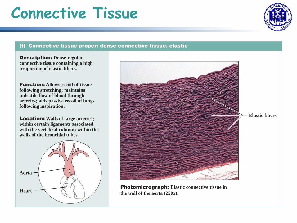
(f) Connective tissue proper: dense connective tissue, elastic
Description: Dense regular connective tissue containing a high proportion of elastic fibers.
Function: Allows recoil of tissue following stretching; maintains pulsatile flow of blood through arteries; aids passive recoil of lungs following inspiration.
Location: Walls of large arteries; within certain ligaments associated with the vertebral column; within the walls of the bronchial tubes.
Elastic fibers
Aorta
Heart Photomicrograph: Elastic connective tissue in
the wall of the aorta (250x).
Connective Tissue

Copyright © 2010 Pearson Education, Inc.
(g) Cartilage: hyaline
Description: Amorphous but firm matrix; collagen fibers form an imperceptible network; chondroblasts produce the matrix and when mature (chondrocytes) lie in lacunae.
Function: Supports and reinforces; has resilient cushioning properties; resists compressive stress.
Location: Forms most of the embryonic skeleton; covers the ends of long bones in joint cavities; forms costal cartilages of the ribs; cartilages of the nose, trachea, and larynx.
Photomicrograph: Hyaline cartilage from the
trachea (750x).
Costal cartilages
Chondrocyte in lacuna
Matrix
Connective Tissue

Copyright © 2010 Pearson Education, Inc.
(h) Cartilage: elastic
Description: Similar to hyaline
cartilage, but more elastic fibers
in matrix.
Function: Maintains the shape
of a structure while allowing
great flexibility.
Location: Supports the external
ear (pinna); epiglottis.
Photomicrograph: Elastic cartilage from
the human ear pinna; forms the flexible
skeleton of the ear (800x).
Chondrocyte
in lacuna
Matrix
Connective Tissue

Copyright © 2010 Pearson Education, Inc.
(i) Cartilage: fibrocartilage
Description: Matrix similar to but less firm than that in hyaline cartilage; thick collagen fibers predominate.
Function: Tensile strength with the ability to absorb compressive shock.
Location: Intervertebral discs; pubic symphysis; discs of knee joint.
Photomicrograph: Fibrocartilage of an
intervertebral disc (125x). Special staining
produced the blue color seen.
Intervertebral
discs
Chondrocytes
in lacunae
Collagen
fiber
Connective Tissue

Copyright © 2010 Pearson Education, Inc.
(j) Others: bone (osseous tissue)
Description: Hard, calcified matrix containing many collagen fibers; osteocytes lie in lacunae. Very well vascularized.
Function: Bone supports and protects (by enclosing); provides levers for the muscles to act on; stores calcium and other minerals and fat; marrow inside bones is the site for blood cell formation (hematopoiesis).
Location: Bones
Photomicrograph: Cross-sectional view of bone (125x).
Lacunae
Lamella
Central canal
Connective Tissue

Copyright © 2010 Pearson Education, Inc.
(k) Others: blood
Description: Red and white
blood cells in a fluid matrix
(plasma).
Function: Transport of
respiratory gases, nutrients,
wastes, and other substances.
Location: Contained within
blood vessels.
Photomicrograph: Smear of human blood (1860x); two
white blood cells (neutrophil in upper left and lymphocyte
in lower right) are seen surrounded by red blood cells.
Neutrophil
Red blood cells
Lymphocyte
Plasma
Connective Tissue

EXAMPLES
Can you name?
First, the tissue type
Second, where in the body the tissue is found
Connective Tissue

What kind of tissue does this represent?
Where in the body can you find this tissue?
delicate thin layers between tissues; present in all mucous membranes
Loose (areolar) connective tissue
Connective Tissue

What kind of tissue does this represent?
Where in the body can you find this tissue?
Adipose tissue
fat
Connective Tissue

What kind of tissue does this represent?
Where in the body can you find this tissue?
Dense connective tissue
tendons; ligaments
Connective Tissue

What kind of tissue does this represent?
Where in the body can you find this tissue?
Hyaline cartilage
nose; ends of long bones; ribs
Connective Tissue

What kind of tissue does this represent?
Where in the body can you find this tissue?
Elastic cartilage
outer ear; epiglottis
Connective Tissue

What kind of tissue does this represent?
Where in the body can you find this tissue?
Fibrocartilage
between vertebrae; knee joints; pubic joint
Connective Tissue

What kind of tissue does this represent?
Where in the body can you find this tissue?
Bone
skeletal system
Connective Tissue

What kind of tissue does this represent?
Where in the body can you find this tissue?
Blood
bloodstream
Connective Tissue

• characterized by properties that allow movement.
• muscle cells are excitable (=respond to a stimulus) + contractile (=can shorten and
generate a pulling force)
• some muscle movement is voluntary (=under conscious control) other involuntary
(ie contraction of your pupil) • classified into 3 types according to structure and function
Muscle Tissue

Muscle Tissue
• Skeletal muscle makes possible locomotion, facial expressions, posture, and other voluntary movements (40% body mass), participate in thermal homeostasis: myocyte (from myoblasts, mesoderm) and their numbers remain relatively constant throughout life. Arranged in bundles surrounded by connective tissue. striated (due to the regular alternation of the contractile proteins actin and myosin), with many nuclei squeezed along the membranes (as a result of the fusion of the many myoblasts to form each long muscle fiber).

Muscle Tissue
Skeletal muscle cells (fibers), with cross-striations and peripheral nuclei.

Muscle Tissue
Higher power of skeletal muscle for details of cross-striations. Notice thin Z discs and heavy A bands. From one Z disc to the next is a sarcomere, the unit of muscle contraction. In the upper muscle cell notice shadowy myofibrils running longitudinally.

Muscle Tissue
EM of several myofibrils running longitudinally through skeletal muscle cell. Between individual myofibrils lie the mitochondria (M) and glycogen (G) of the cytoplasm. Within each myofibril are the typical striations: A= A band; I= I band; Z= Z line; and H= H band. The banding is formed by the arrangement of myosin and actin filaments.

Muscle Tissue
• Smooth muscle responsible for involuntary movements in the internal organs. It forms the contractile component of the digestive, urinary, and reproductive systems as well as the airways and arteries. Each cell is spindle shaped with a single nucleus and no visible striations

Muscle Tissue
Smooth muscle - long, slender central nuclei, lying within narrow, fusiform cells that lie parallel to each other in a smooth arrangement. (Muscle cells are often referred to as muscle fibers because of their narrowness and length.)

Muscle Tissue
Smooth muscle - with cells more separated so as to see their extent and shape better, and the central position of their nuclei. A loose, irregular connective tissue (endomysium) lies between the cells. Nuclei seen in this c.t. belong to fibroblasts mainly.

Muscle Tissue
Smooth muscle with wrinkled nuclei due to contraction of cells.

Muscle Tissue
EM of smooth muscle showing typical "hairy" look of primarily filaments in the cytoplasm. Part of the cytoplasm is clear of filaments and shows mitochondria and polyribosomes. The cell membrane is at the lower right of the field and shows a few pinocytotic vesicles toward the extreme right. The left-hand extent of that same membrane seems darker and denser: probably a plaque, where filaments attach. The fuzzy density just outside the cell membrane is the basal lamina.

Muscle Tissue
• Cardiac muscle (contractile walls of the heart), cardiomyocytes, also striated single cells typically with a single centrally located nucleus, contract on their own intrinsic rhythms without any external stimulation and attach to one another with specialized cell junctions = intercalated discs (both anchoring junctions and gap junctions) long, branching cardiac muscle fibers that are, essentially, a mechanical and electrochemical syncytium with synchronized actions that pumps blood under involuntary control.

Muscle Tissue
Cardiac muscle with cross-striations, dark intercalated discs, and centrally located nuclei. Notice too that the nuclei are stubby in appearance, and that they lie in a rather granular cytoplasm. Some of the intercalated discs form a straight line across muscle fibers; others make a step-like arrangement.

Muscle Tissue
EM of intercalated disc between the ends of two cardiac muscle cells. Both desmosomes (1) and fasciae adheretes (2) are identified. Notice mitochondria and glycogen particles lying between myofibrils.

Muscle Tissue
Another view of cardiac muscle showing wavy connective tissue (endomysium) between muscle cells. Also, notice capillaries with r.b.c.'s; muscle is a highly vascularized tissue. Some yellow granular cytoplasm can be seen inside the lower muscle cells, where myofibrils are parted. This picture also gives some indication of the branching of cardiac fibers.

• excitable and capable of sending and receiving electrochemical signals that provide the
body with information. • 2 main classes of cells: neuron (propagate information via electrochemical impulses,
called action potentials, which are biochemically linked to the release of chemical signals) and neuroglia (play an essential role in supporting neurons and modulating their information propagation)
Nervous Tissue

Nervous Tissue
Neurons = distinctive morphology role as conducting cells, with 3 parts: 1. the cell body includes most of the cytoplasm, the organelles, and the nucleus. 2. dendrites branch off the cell body and appear as thin extensions. 3. a long “tail,” the axon, extends from the neuron body and can be wrapped in an insulating
layer known as myelin, which is formed by accessory cells. The synapse is the gap between nerve cells, or between a nerve cell and its target, for example, a muscle or a gland, across which the impulse is transmitted by chemical compounds known as neurotransmitters.

Nervous Tissue
Neurons categorized: multipolar, bipolar and unipolar. When a neuron is sufficiently stimulated, it generates an action potential that propagates down the axon towards the synapse. If enough neurotransmitters are released at the synapse to stimulate the next neuron or target, a response is generated.

Nervous Tissue
The second class of neural cells: neuroglia or glial cells, (from the Greek word for glue).
Astrocyte cells, (star shape) abundant in the CNS, have many functions: regulation of ion
concentration in the intercellular space, uptake and/or breakdown of some neurotransmitters,
and formation of the blood-brain barrier. Microglia protect the nervous system against
infection (related to macrophages). Oligodendrocytes produce myelin in the CSN (brain and
spinal cord) while the Schwann cell produces myelin in the peripheral nervous system