Embed Size (px)
Citation preview
ORIGINAL ARTICLE
Inhibition of Transforming GrowthFactor-�/Smad Signaling ImprovesRegeneration of Small-for-Size Rat Liver GraftsZhi Zhong,1 Shigeki Tsukada,3 Hasibur Rehman,1 Christopher J. Parsons,3 Tom P. Theruvath,1
Richard A. Rippe,3 David A. Brenner,4 and John J. Lemasters1,2
Departments of 1Pharmaceutical and Biomedical Sciences and 2Biochemistry and Molecular Biology,Medical University of South Carolina, Charleston, SC; 3Department of Medicine, University of NorthCarolina, Chapel Hill, NC; and 4Department of Medicine, University of California, San Diego, CA
Transforming growth factor-� (TGF-�) is a potent inhibitor of cell proliferation. This study investigated whether overexpressionof Smad7, which blocks TGF-�–induced activation of Smad2/3, could prevent the suppression of regeneration of small-for-sizeliver grafts. Rats were intravenously given adenoviruses (2 � 109 pfu/rat) carrying the LacZ gene or the Smad7 gene(Ad-Smad7) 3 days prior to liver harvesting. Half-size livers were implanted into recipients of the same weight or twice the donorweight, and this resulted in half-size or quarter-size liver grafts. Cell proliferation, detected by 5-bromo-2�-deoxyuridine (BrdU)incorporation, increased to 23% in half-size grafts at 38 hours after implantation but was only 4% in quarter-size grafts. Graftweight did not increase after 38 hours in full-size and quarter-size grafts but increased 28% in half-size grafts. Ad-Smad7restored BrdU labeling to 32%, and the graft weight increased to 43% in quarter-size grafts. Serum total bilirubin increasedapproximately 30-fold after the implantation of quarter-size grafts. Ad-Smad7 blunted hyperbilirubinemia by 80%. The basalhepatic TGF-�1 level was 7 ng/g of liver wet weight, and this increased to 30 ng/g at 1.5 hours after the transplantation offull-size grafts but decreased rapidly afterwards. After the transplantation of quarter-size grafts, however, TGF-�1 progressivelyincreased to 159 ng/g in 38 hours. Nuclear phosphorylated Smad2/3 was barely detectable, and p21Cip1 expression wasnegligible in full-size grafts but increased markedly in quarter-size grafts. Ad-Smad7 blocked Smad2/3 activation andexpression of p21Cip1. Together, these data show that TGF-� is responsible, at least in part, for the defective liver regenerationin small-for-size grafts by activating the Smad signaling pathway. Liver Transpl 16:181-190, 2010. © 2010 AASLD.
Received April 13, 2009; accepted September 26, 2009.
Living donor and split liver transplantation (LT) hasbecome more widely used in recent years to alleviate themortality resulting from the scarcity of suitable livergrafts for transplantation.1,2 Donor-recipient graft sizedisparity leading to small-for-size graft dysfunction andfailure is an important issue limiting the wider use ofpartial LT for adults.3 The mechanisms underlying thedysfunction and failure of small-for-size grafts remainunclear. Previous studies have indicated that liver re-generation is markedly suppressed in small-for-size
liver grafts, and this appears to contribute to graft fail-ure.4-6
Growth factors such as hepatic growth factor (HGF),transforming growth factor-� (TGF-�), epidermalgrowth factor (EGF), and vascular endothelial growthfactor and the cytokines interleukin-6 (IL-6) and tumornecrosis factor-� (TNF�) stimulate liver regeneration.7-9
TGF-� is a potent inhibitor of hepatocyte proliferationthat counterbalances the stimulatory effects of mito-gens during liver regeneration.10-12 In many cell types,
Abbreviations: Ad-LacZ, adenovirus carrying LacZ; Ad-Smad7, adenovirus carrying human Smad7; BrdU, 5-bromo-2�-deoxyuridine;CDK, cyclin-dependent kinase; CDKI, cyclin-dependent kinase inhibitor; EGF, epidermal growth factor; HA, hemagglutinin; HGF,hepatic growth factor; IL-6, interleukin 6; LT, liver transplantation; p53, tumor protein p53; PCNA, proliferating cell nuclear antigen;SnoN, Ski-like oncogene; TGF, transforming growth factor; TNF�, tumor necrosis factor-�.This study was supported in part by grants DK70844 and DK037034 from the National Institutes of Health.
Address reprint requests to Zhi Zhong, Department of Pharmaceutical and Biomedical Sciences, Medical University of South Carolina, Charleston,SC 29425. Telephone: 843-792-2163; FAX: 843-792-1617; E-mail: [email protected]
DOI 10.1002/lt.21966Published online in Wiley InterScience (www.interscience.wiley.com).
LIVER TRANSPLANTATION 16:181-190, 2010
© 2010 American Association for the Study of Liver Diseases.
TGF-� is the most potent growth-inhibitory polypeptidecurrently known.13 However, whether TGF-� plays arole in the suppression of small-for-size liver graft re-generation remains unclear.
TGF-� elicits its biological effects by signalingthrough a heteromeric receptor complex consisting oftype I and type II receptors. The binding of TGF-� totype II receptors leads to the recruitment, phosphory-lation, and activation of type I receptors, which subse-quently phosphorylate proteins of the Smad family,particularly Smad2 and Smad3.14,15 PhosphorylatedSmad2 and Smad3 form a complex with Smad4, moveinto the nucleus, and activate target genes expressingregulatory proteins for cell proliferation, differentiation,and cell death.14,16 TGF-� also regulates mitogen-acti-vated protein kinase–mediated signaling pathways andother signaling proteins, such as protein kinase A, pro-tein kinase C, phospholipase C, and nuclear factor-�B,in different cell types.17,18
Another Smad family member, Smad7, associatesstably with the TGF-� receptor complex, which inhibitsTGF-�–induced phosphorylation of Smad2 and Smad3and blocks TGF-�–dependent signaling.19 This studyexamined the effect of the adenoviral delivery of Smad7on liver regeneration after LT with small-for-size grafts.Our results show that TGF-�1 increases sharply afterthe transplantation of small-for-size liver grafts in as-sociation with Smad2/3 phosphorylation and nucleartranslocation. Smad7 adenoviral expression blocksSmad2/3 activation, promotes liver regeneration, andimproves graft function.
MATERIALS AND METHODS
Animals and LT
A recombinant adenovirus containing the transgene foreither �-galactosidase (Ad-LacZ) or human Smad7 (Ad-Smad7) was prepared at the University of North Caro-lina as described previously.20 Ad-LacZ, which containsthe �-galactosidase gene driven by the cytomegaloviruspromoter, was used as a control viral vector. The Ad-Smad7 virus expresses a C-terminal hemagglutinin(HA)-tagged human Smad7 protein. The human Smad7complementary DNA was obtained from Dr. Wrana(Hospital for Sick Children, Toronto, Canada) andcloned into the pGI-AdCMV5 transfer vector (Qbiogene,Carlsbad, CA). Viral amplification was performed in 293cells and cesium chloride purified by the standardmethodology. Viral titer estimates were performed byoptical density measurements. The virus (2 � 109 pfu)was diluted in 0.5 mL of normal saline and injected intothe tail vein of donor rats (male Lewis rats, 170-200 g).In preliminary studies from this laboratory, the infec-tion rates of liver cells were over 80% when doses ofadenovirus ranging from 1 to 3 � 109 pfu were used.
Our previous studies showed that adenoviral proteinexpression peaks in 2 to 3 days and persists for about 3weeks.21 Therefore, 3 days after the viral infection, liv-ers were explanted and reduced in size by the removalof the left lateral lobe, the left portion of the median
lobe, and the anterior and posterior caudate lobes exvivo as described.5 This procedure decreased liver massby approximately 50%.22 Explants were stored in Uni-versity of Wisconsin solution at 0 to 1°C for 6 hours andrinsed with lactated Ringer’s solution (Abbott Labora-tories, North Chicago, IL) just prior to implantation.
Reduced-size liver explants were implanted into re-cipients of similar (170-200 g) or greater body weights(350-430 g), and this resulted in graft weight/standardliver weight (defined as 4% of body weight) values ofapproximately 50% (half-size) and approximately 25%(quarter-size), respectively.23 Unreduced livers wereimplanted into recipients of similar body weights (170-200 g) as full-size controls. The hepatic artery and thebile duct were anastomosed with intraluminal splints.Implantation procedures usually took about 50 min-utes. For sham operations, the abdominal wall wasopened, and ligaments around the liver were freed. Theabdominal wall was then closed with running suturesafter 50 minutes. Under these conditions, survival was100% for full-size grafts, 80% for half-size grafts, and30% for quarter-size grafts.23 The graft weight/stan-dard liver weight values were on average 24.9% � 6% inthe quarter-size group pretreated with saline, 26.1% �4% in the quarter-size group treated with Ad-LacZ, and26.0% � 3% for the quarter-size grafts treated withAd-Smad7 (P � 0.10 versus the saline-pretreated andAd-LacZ–pretreated quarter-size groups). The graftweight was measured just prior to cold storage andafter harvesting at various times after implantation. Allanimals were given humane care in compliance withinstitutional guidelines using protocols approved by theInstitutional Animal Care and Use Committee.
Bilirubin
Blood samples were collected from the tail vein at 18,24, and 38 hours after implantation. Total bilirubin insera was determined with analytical kits from PointeScientific (Uncoln Park, MI) to assess liver function.
Immunohistochemical Staining for 5-Bromo-2�-Deoxyuridine (BrdU), Proliferating CellNuclear Antigen (PCNA), PhosphorylatedSmad2/3, and �-Galactosidase
BrdU was injected intraperitoneally (100 mg/kg) 1 hourprior to liver harvesting to detect cells synthesizingDNA. At 38 hours after implantation, livers were har-vested, and BrdU incorporation and expression ofPCNA, another marker of cell proliferation, were deter-mined immunohistochemically in liver sections as de-scribed elsewhere.5 Cells positive and negative for BrdUand PCNA were counted in 10 randomly selected fieldsunder a light microscope with a 20� objective lens.Immunohistochemistry of phosphorylated Smad2/3,Ski-like oncogene (SnoN), and �-galactosidase was per-formed with primary antibodies (Santa Cruz Biotech-nology, Santa Cruz, CA) at a concentration of 1:200 for1 hour, 1 hour, and 20 minutes at room temperature,respectively. Phosphorylated Smad2/3-positive and
182 ZHONG ET AL.
LIVER TRANSPLANTATION.DOI 10.1002/lt. Published on behalf of the American Association for the Study of Liver Diseases
Smad2/3-negative cells were counted in 10 randomlyselected fields under a light microscope with a 40�objective lens.
Western Blotting
Livers were harvested, snap-frozen in liquid nitrogen,homogenized in a buffer containing 40 mM trishy-droxymethylaminomethane (pH 7.6), 140 mM NaCl,and 1% protease and phosphatase inhibitor cocktails(Sigma, St. Louis, MO), and centrifuged at 1000g for 10minutes at 4°C. The supernatant was analyzed for HAprotein, Smad7, SnoN, p21Cip1, p27Kip1, p15Ink4B,p16Ink4A, and tumor protein p53 (p53) by westernblotting6 using specific antibodies against the proteinsof interest (Santa Cruz Biotechnology) at a dilution of1:300 to 1:500 at 4°C overnight followed by incubationwith an appropriate secondary antibody at 1:1000 to1:3000 for 1 hour. Chemiluminescence was detectedwith an ECL Plus western blotting detection system(Amersham Biosciences, Little Chalfont, United King-dom). To confirm equal loading, blots were reprobedwith anti-actin primary antibody at 1:3000 for 1 hour(ICN, Costa Mesa, CA). The protein concentration wasdetermined with the Bio-Rad protein assay (Bio-Rad,Hercules, CA).
Detection of TGF-�1 in Liver Tissue
Liver tissue was collected into liquid nitrogen, stored at�80°C, and homogenized after thawing in a buffer (pH7.5) containing 20 mM trishydroxymethylaminometh-ane, 0.25 M sucrose, 2 mM ethylene diamine tetraaceticacid, 10 mM ethylene glycol tetraacetic acid, 1% TritonX-100, and 1% protease inhibitor cocktail (Sigma). Thehomogenates were centrifuged at 100,000g at 4°C for 1hour. Total TGF-�1 in the supernatant, including bothprecursor and biologically active cleaved forms, wasdetermined with a TGF-�1 Emax immunoassay systemfrom Promega (Madison, WI) according to the manufac-turer’s instructions.
Statistical Analysis
All groups were compared with an analysis of varianceor Kruskal-Wallis test as appropriate. Numbers in eachgroup for different parameters are shown in the figurelegends. Differences were considered significant at P 0.05.
RESULTS
TGF-�1 Increased in Small-For-Size LiverGrafts
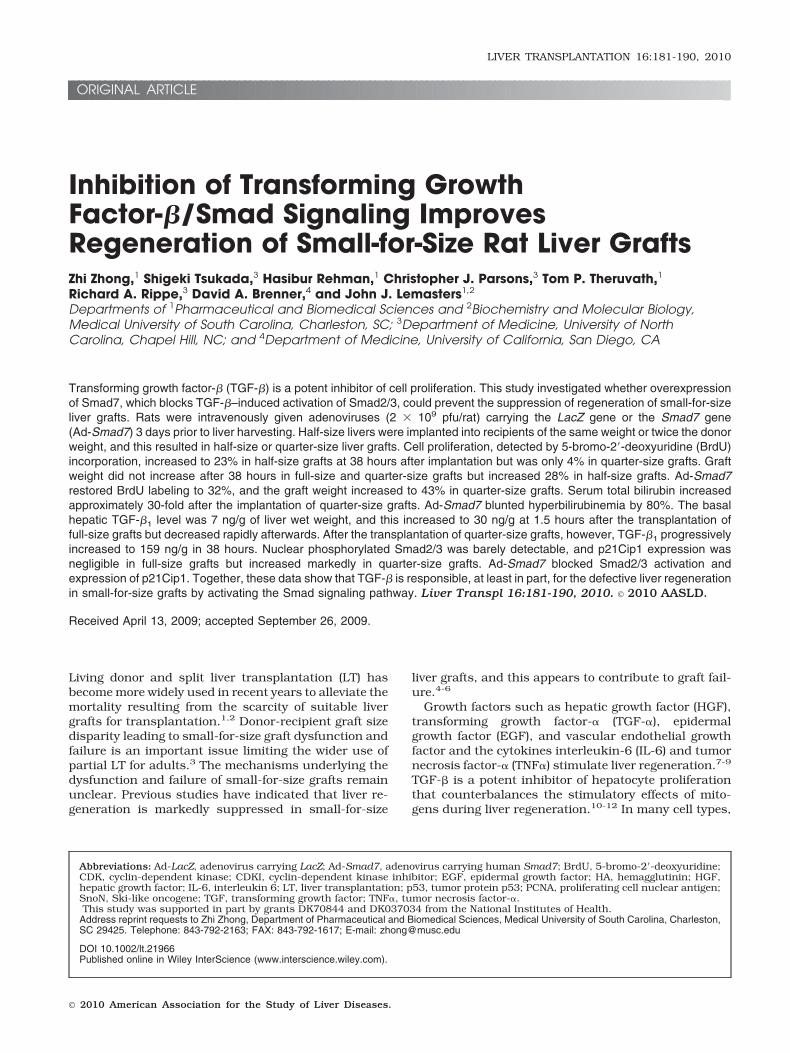
TGF-� is a potent inhibitor of cell proliferation. Accord-ingly, we measured total TGF-�1 in livers before andafter LT. Before LT, TGF-�1 was 7 ng/g of liver wetweight. After full-size LT, TGF-�1 increased to 30 ng/gat 1.5 hours but decreased afterwards to close to pre-transplant levels at 38 hours after the operation (Fig. 1).After the transplantation of half-size grafts, a progres-
sive increase of TGF-�1 occurred with a maximum of 79ng/g of liver at 18 hours, and it then declined gradually.An even larger sustained increase occurred after thetransplantation of quarter-size grafts, with TGF-�1 lev-els rising to 143 ng/g after 18 hours and to 159 ng/gafter 38 hours (Fig. 1).
Expression of �-Galactosidase and HA Proteinin the Liver After Viral Gene Delivery
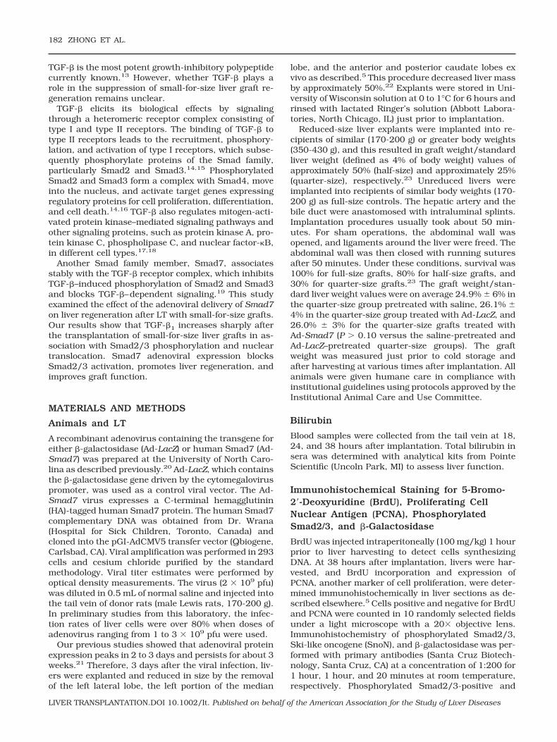
Rats were infected with Ad-LacZ, which carries the genefor �-galactosidase. At 3 days after infection with Ad-LacZ, over 80% of hepatocytes expressed �-galactosi-dase (Fig. 2A, right). By contrast, �-galactosidase wasnot detectable in livers from rats receiving normal sa-line (Fig. 2A, left). These results indicate that the ade-novirus effectively infects liver cells.
The Ad-Smad7 virus expresses a C-terminal, HA-tagged human Smad7 protein. The HA tag is a generalepitope tag in expression vectors and does not interferewith the bioactivity or biodistribution of recombinantproteins. The HA tag is present only in the Smad7 pro-teins encoded by the Smad7 gene carried by the adeno-viral vector. Therefore, detection of the HA tag indicatesexpression of the Smad7 gene transfected by adenoviralvectors. The HA tag was not detectable by western blot-ting in livers from rats given saline (Fig. 2B). By con-trast, livers from rats infected with Ad-Smad7 showedhigh levels of the HA tag, indicating Smad7 proteinexpression (Fig. 2B). We also detected Smad7 expres-sion by western blotting. Smad7 was barely detectablein livers from saline-treated rats but increased overtlyin livers from rats infected with Ad-Smad7 (Fig. 2B).
Figure 1. TGF-�1 production increased after partial livertransplantation. Livers were harvested at 1.5, 5, 18, and 38hours after full-size (100%, n � 4), half-size (50%, n � 4), andquarter-size (25%, n � 5) liver transplantation, and totalTGF-�1 was determined. Values are means and standard er-rors of the mean (P < 0.001 by 2-way analysis of variance).aP < 0.001 versus full-size grafts and bP < 0.001 versushalf-size grafts.
TGF-� AND SMALL-FOR-SIZE LIVER TRANSPLANTATION 183
LIVER TRANSPLANTATION.DOI 10.1002/lt. Published on behalf of the American Association for the Study of Liver Diseases
Therefore, this Smad7 expression is possibly mainlydue to gene transfection.
Liver Regeneration Was Suppressed After theTransplantation of Small-for-Size Liver Grafts:Reversal by Smad7
Liver regeneration was evaluated by BrdU incorpora-tion, expression of PCNA, and increases in graft weight.Our previous studies showed that after the implanta-tion of half-size livers, BrdU labeling first increased atabout 18 hours postoperatively in both periportal andmidzonal regions of the liver lobule and was maximalafter 38 hours. Proliferating cells were predominantlyhepatocytes.5
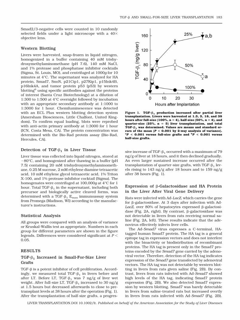
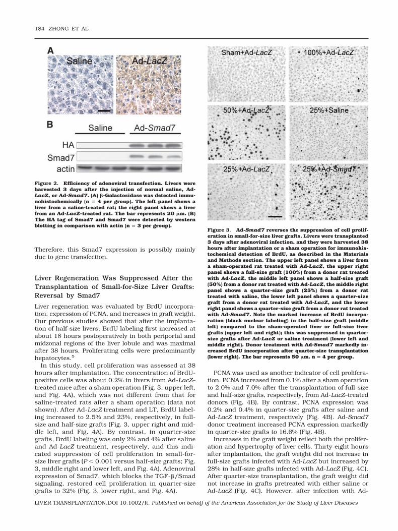
In this study, cell proliferation was assessed at 38hours after implantation. The concentration of BrdU-positive cells was about 0.2% in livers from Ad-LacZ–treated mice after a sham operation (Fig. 3, upper left,and Fig. 4A), which was not different from that forsaline-treated rats after a sham operation (data notshown). After Ad-LacZ treatment and LT, BrdU label-ing increased to 2.5% and 23%, respectively, in full-size and half-size grafts (Fig. 3, upper right and mid-dle left, and Fig. 4A). By contrast, in quarter-sizegrafts, BrdU labeling was only 2% and 4% after salineand Ad-LacZ treatment, respectively, and this indi-cated suppression of cell proliferation in small-for-size liver grafts (P 0.001 versus half-size grafts; Fig.3, middle right and lower left, and Fig. 4A). Adenoviralexpression of Smad7, which blocks the TGF-�/Smadsignaling, restored cell proliferation in quarter-sizegrafts to 32% (Fig. 3, lower right, and Fig. 4A).
PCNA was used as another indicator of cell prolifera-tion. PCNA increased from 0.1% after a sham operationto 2.0% and 7.0% after the transplantation of full-sizeand half-size grafts, respectively, from Ad-LacZ–treateddonors (Fig. 4B). By contrast, PCNA expression was0.2% and 0.4% in quarter-size grafts after saline andAd-LacZ treatment, respectively (Fig. 4B). Ad-Smad7donor treatment increased PCNA expression markedlyin quarter-size grafts to 16.6% (Fig. 4B).
Increases in the graft weight reflect both the prolifer-ation and hypertrophy of liver cells. Thirty-eight hoursafter implantation, the graft weight did not increase infull-size grafts infected with Ad-LacZ but increased by28% in half-size grafts infected with Ad-LacZ (Fig. 4C).After quarter-size transplantation, the graft weight didnot increase in grafts pretreated with either saline orAd-LacZ (Fig. 4C). However, after infection with Ad-
Figure 2. Efficiency of adenoviral transfection. Livers wereharvested 3 days after the injection of normal saline, Ad-LacZ, or Ad-Smad7. (A) �-Galactosidase was detected immu-nohistochemically (n � 4 per group). The left panel shows aliver from a saline-treated rat; the right panel shows a liverfrom an Ad-LacZ–treated rat. The bar represents 20 �m. (B)The HA tag of Smad7 and Smad7 were detected by westernblotting in comparison with actin (n � 3 per group).
Figure 3. Ad-Smad7 reverses the suppression of cell prolif-eration in small-for-size liver grafts. Livers were transplanted3 days after adenoviral infection, and they were harvested 38hours after implantation or a sham operation for immunohis-tochemical detection of BrdU, as described in the Materialsand Methods section. The upper left panel shows a liver froma sham-operated rat treated with Ad-LacZ, the upper rightpanel shows a full-size graft (100%) from a donor rat treatedwith Ad-LacZ, the middle left panel shows a half-size graft(50%) from a donor rat treated with Ad-LacZ, the middle rightpanel shows a quarter-size graft (25%) from a donor rattreated with saline, the lower left panel shows a quarter-sizegraft from a donor rat treated with Ad-LacZ, and the lowerright panel shows a quarter-size graft from a donor rat treatedwith Ad-Smad7. Note the marked increase of BrdU incorpo-ration (black nuclear labeling) in the half-size graft (middleleft) compared to the sham-operated liver or full-size livergrafts (upper left and right); this was suppressed in quarter-size grafts after Ad-LacZ or saline treatment (lower left andmiddle right). Donor treatment with Ad-Smad7 markedly in-creased BrdU incorporation after quarter-size transplantation(lower right). The bar represents 50 �m. n � 4 per group.
184 ZHONG ET AL.
LIVER TRANSPLANTATION.DOI 10.1002/lt. Published on behalf of the American Association for the Study of Liver Diseases
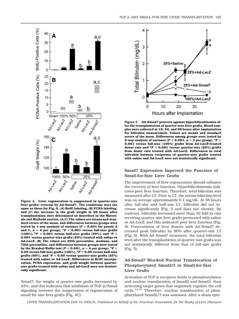
Smad7, the weight of quarter-size grafts increased by43%, and this indicates that inhibition of TGF-�/Smadsignaling reverses the suppression of regeneration ofsmall-for-size liver grafts (Fig. 4C).
Smad7 Expression Improved the Function ofSmall-for-Size Liver Grafts
The improvement of liver regeneration should enhancethe recovery of liver function. Hyperbilirubinemia indi-cates poor liver function. Therefore, total bilirubin wasmeasured after LT. Prior to LT, the serum bilirubin levelwas on average approximately 0.1 mg/dL. At 38 hoursafter full-size and half-size LT, bilirubin did not in-crease significantly (Fig. 5 and data not shown). Bycontrast, bilirubin increased more than 30-fold in ratsreceiving quarter-size liver grafts pretreated with salineor Ad-LacZ, and this indicated poor liver function (Fig.5). Pretreatment of liver donors with Ad-Smad7 de-creased peak bilirubin by 80% after quarter-size LT(Fig. 5). With Ad-Smad7 treatment, the total bilirubinlevel after the transplantation of quarter-size grafts wasnot statistically different from that of full-size grafts(Fig. 5).
Ad-Smad7 Blocked Nuclear Translocation ofPhosphorylated Smad2/3 in Small-for-SizeLiver Grafts
Activation of TGF-� receptors leads to phosphorylationand nuclear translocation of Smad2 and Smad3, thusactivating target genes that negatively regulate the cellcycle.14,16 Therefore, nuclear translocation of phos-phorylated Smad2/3 was assessed. After a sham oper-
Figure 4. Liver regeneration is suppressed in quarter-sizeliver grafts: reversal by Ad-Smad7. The conditions were thesame as those for Fig. 3. (A) BrdU labeling, (B) PCNA labeling,and (C) the increase in the graft weight at 38 hours aftertransplantation were determined as described in the Materi-als and Methods section. (A,C) The values are means and stan-dard errors of the mean, and differences between groups weretested by 1-way analysis of variance (P < 0.001 for panels Aand C, n � 4 per group). aP < 0.001 versus full-size grafts(100%), bP < 0.001 versus half-size grafts (50%), and cP <0.001 versus quarter-size grafts (25%) treated with saline orAd-LacZ. (B) The values are 25th percentiles, medians, and75th percentiles, and differences between groups were testedby the Kruskal-Wallis test (P < 0.001, n � 4 per group). aP <0.05 versus full-size grafts (100%), bP < 0.05 versus half-sizegrafts (50%), and cP < 0.05 versus quarter-size grafts (25%)treated with saline or Ad-LacZ. Differences in BrdU incorpo-ration, PCNA expression, and graft weight between quarter-size grafts treated with saline and Ad-LacZ were not statisti-cally significant.
Figure 5. Ad-Smad7 protects against hyperbilirubinemia af-ter the transplantation of quarter-size liver grafts. Blood sam-ples were collected at 18, 24, and 38 hours after implantationfor bilirubin measurement. Values are means and standarderrors of the mean. Differences among groups were tested by2-way analysis of variance (P < 0.001, n � 4 per group). aP <0.001 versus full-size (100%) grafts from Ad-LacZ–treateddonor rats and bP < 0.001 versus quarter-size (25%) graftsfrom donor rats treated with Ad-LacZ. Differences in totalbilirubin between recipients of quarter-size grafts treatedwith saline and Ad-LacZ were not statistically significant.
TGF-� AND SMALL-FOR-SIZE LIVER TRANSPLANTATION 185
LIVER TRANSPLANTATION.DOI 10.1002/lt. Published on behalf of the American Association for the Study of Liver Diseases
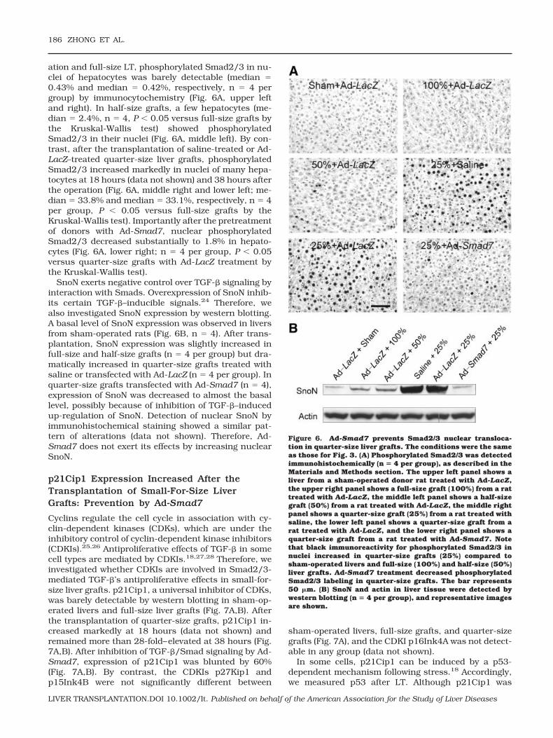
ation and full-size LT, phosphorylated Smad2/3 in nu-clei of hepatocytes was barely detectable (median 0.43% and median 0.42%, respectively, n 4 pergroup) by immunocytochemistry (Fig. 6A, upper leftand right). In half-size grafts, a few hepatocytes (me-dian 2.4%, n 4, P 0.05 versus full-size grafts bythe Kruskal-Wallis test) showed phosphorylatedSmad2/3 in their nuclei (Fig. 6A, middle left). By con-trast, after the transplantation of saline-treated or Ad-LacZ–treated quarter-size liver grafts, phosphorylatedSmad2/3 increased markedly in nuclei of many hepa-tocytes at 18 hours (data not shown) and 38 hours afterthe operation (Fig. 6A, middle right and lower left; me-dian 33.8% and median 33.1%, respectively, n 4per group, P 0.05 versus full-size grafts by theKruskal-Wallis test). Importantly after the pretreatmentof donors with Ad-Smad7, nuclear phosphorylatedSmad2/3 decreased substantially to 1.8% in hepato-cytes (Fig. 6A, lower right; n 4 per group, P 0.05versus quarter-size grafts with Ad-LacZ treatment bythe Kruskal-Wallis test).
SnoN exerts negative control over TGF-� signaling byinteraction with Smads. Overexpression of SnoN inhib-its certain TGF-�–inducible signals.24 Therefore, wealso investigated SnoN expression by western blotting.A basal level of SnoN expression was observed in liversfrom sham-operated rats (Fig. 6B, n 4). After trans-plantation, SnoN expression was slightly increased infull-size and half-size grafts (n 4 per group) but dra-matically increased in quarter-size grafts treated withsaline or transfected with Ad-LacZ (n 4 per group). Inquarter-size grafts transfected with Ad-Smad7 (n 4),expression of SnoN was decreased to almost the basallevel, possibly because of inhibition of TGF-�–inducedup-regulation of SnoN. Detection of nuclear SnoN byimmunohistochemical staining showed a similar pat-tern of alterations (data not shown). Therefore, Ad-Smad7 does not exert its effects by increasing nuclearSnoN.
p21Cip1 Expression Increased After theTransplantation of Small-For-Size LiverGrafts: Prevention by Ad-Smad7
Cyclins regulate the cell cycle in association with cy-clin-dependent kinases (CDKs), which are under theinhibitory control of cyclin-dependent kinase inhibitors(CDKIs).25,26 Antiproliferative effects of TGF-� in somecell types are mediated by CDKIs.18,27,28 Therefore, weinvestigated whether CDKIs are involved in Smad2/3-mediated TGF-�’s antiproliferative effects in small-for-size liver grafts. p21Cip1, a universal inhibitor of CDKs,was barely detectable by western blotting in sham-op-erated livers and full-size liver grafts (Fig. 7A,B). Afterthe transplantation of quarter-size grafts, p21Cip1 in-creased markedly at 18 hours (data not shown) andremained more than 28-fold–elevated at 38 hours (Fig.7A,B). After inhibition of TGF-�/Smad signaling by Ad-Smad7, expression of p21Cip1 was blunted by 60%(Fig. 7A,B). By contrast, the CDKIs p27Kip1 andp15Ink4B were not significantly different between
sham-operated livers, full-size grafts, and quarter-sizegrafts (Fig. 7A), and the CDKI p16Ink4A was not detect-able in any group (data not shown).
In some cells, p21Cip1 can be induced by a p53-dependent mechanism following stress.18 Accordingly,we measured p53 after LT. Although p21Cip1 was
Figure 6. Ad-Smad7 prevents Smad2/3 nuclear transloca-tion in quarter-size liver grafts. The conditions were the sameas those for Fig. 3. (A) Phosphorylated Smad2/3 was detectedimmunohistochemically (n � 4 per group), as described in theMaterials and Methods section. The upper left panel shows aliver from a sham-operated donor rat treated with Ad-LacZ,the upper right panel shows a full-size graft (100%) from a rattreated with Ad-LacZ, the middle left panel shows a half-sizegraft (50%) from a rat treated with Ad-LacZ, the middle rightpanel shows a quarter-size graft (25%) from a rat treated withsaline, the lower left panel shows a quarter-size graft from arat treated with Ad-LacZ, and the lower right panel shows aquarter-size graft from a rat treated with Ad-Smad7. Notethat black immunoreactivity for phosphorylated Smad2/3 innuclei increased in quarter-size grafts (25%) compared tosham-operated livers and full-size (100%) and half-size (50%)liver grafts. Ad-Smad7 treatment decreased phosphorylatedSmad2/3 labeling in quarter-size grafts. The bar represents50 �m. (B) SnoN and actin in liver tissue were detected bywestern blotting (n � 4 per group), and representative imagesare shown.
186 ZHONG ET AL.
LIVER TRANSPLANTATION.DOI 10.1002/lt. Published on behalf of the American Association for the Study of Liver Diseases
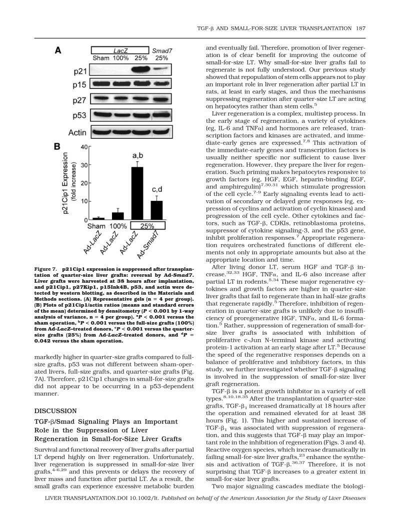
markedly higher in quarter-size grafts compared to full-size grafts, p53 was not different between sham-oper-ated livers, full-size grafts, and quarter-size grafts (Fig.7A). Therefore, p21Cip1 changes in small-for-size graftsdid not appear to be occurring in a p53-dependentmanner.
DISCUSSION
TGF-�/Smad Signaling Plays an ImportantRole in the Suppression of LiverRegeneration in Small-for-Size Liver Grafts
Survival and functional recovery of liver grafts after partialLT depend highly on liver regeneration. Unfortunately,liver regeneration is suppressed in small-for-size livergrafts,4-6,29 and this prevents or delays the recovery ofliver mass and function after partial LT. As a result, thesmall grafts can experience excessive metabolic burden
and eventually fail. Therefore, promotion of liver regener-ation is of clear benefit for improving the outcome ofsmall-for-size LT. Why small-for-size liver grafts fail toregenerate is not fully understood. Our previous studyshowed that repopulation of stem cells appears not to playan important role in liver regeneration after partial LT inrats, at least in early stages, and thus the mechanismssuppressing regeneration after quarter-size LT are actingon hepatocytes rather than stem cells.5
Liver regeneration is a complex, multistep process. Inthe early stage of regeneration, a variety of cytokines(eg, IL-6 and TNF�) and hormones are released, tran-scription factors and kinases are activated, and imme-diate-early genes are expressed.7,8 This activation ofthe immediate-early genes and transcription factors isusually neither specific nor sufficient to cause liverregeneration. However, they prepare the liver for regen-eration. Such priming makes hepatocytes responsive togrowth factors (eg, HGF, EGF, heparin-binding EGF,and amphiregulin)7,30,31 which stimulate progressionof the cell cycle.7-9 Early signaling events lead to acti-vation of secondary or delayed gene responses (eg, ex-pression of cyclins and activation of cyclin kinases) andprogression of the cell cycle. Other cytokines and fac-tors, such as TGF-�, CDKIs, retinoblastoma proteins,suppressor of cytokine signaling-3, and the p53 gene,inhibit proliferation responses.7 Appropriate regenera-tion requires orchestrated functions of different ele-ments not only in appropriate amounts but also at theappropriate location and time.
After living donor LT, serum HGF and TGF-� in-crease.32,33 HGF, TNF�, and IL-6 also increase afterpartial LT in rodents.5,34 These major regenerative cy-tokines and growth factors are higher in quarter-sizeliver grafts that fail to regenerate than in half-size graftsthat regenerate rapidly.5 Therefore, inhibition of regen-eration in quarter-size grafts is unlikely due to insuffi-ciency of proregenerative HGF, TNF�, and IL-6 forma-tion.5 Rather, suppression of regeneration of small-for-size liver grafts is associated with inhibition ofproliferative c-Jun N-terminal kinase and activatingprotein-1 activation at an early stage after LT.5 Becausethe speed of the regenerative responses depends on abalance of proliferative and inhibitory factors, in thisstudy, we further investigated whether TGF-� signalingis involved in the suppression of small-for-size livergraft regeneration.
TGF-� is a potent growth inhibitor in a variety of celltypes.8,10,18,35 After the transplantation of quarter-sizegrafts, TGF-�1 increased dramatically at 18 hours afterthe operation and remained elevated for at least 38hours (Fig. 1). This higher and sustained increase ofTGF-�1 was associated with suppression of regenera-tion, and this suggests that TGF-� may play an impor-tant role in the inhibition of regeneration (Figs. 3 and 4).Reactive oxygen species, which increase dramatically infailing small-for-size liver grafts,23 enhance the synthe-sis and activation of TGF-�.36,37 Therefore, it is notsurprising that TGF-� increases to a greater extent insmall-for-size liver grafts.
Two major signaling cascades mediate the biologi-
Figure 7. p21Cip1 expression is suppressed after transplan-tation of quarter-size liver grafts: reversal by Ad-Smad7.Liver grafts were harvested at 38 hours after implantation,and p21Cip1, p27Kip1, p15Ink4B, p53, and actin were de-tected by western blotting, as described in the Materials andMethods sections. (A) Representative gels (n � 4 per group).(B) Plots of p21Cip1/actin ratios (means and standard errorsof the mean) determined by densitometry (P < 0.001 by 1-wayanalysis of variance, n � 4 per group). aP < 0.001 versus thesham operation, bP < 0.001 versus the full-size grafts (100%)from Ad-LacZ–treated donors, cP < 0.001 versus the quarter-size grafts (25%) from Ad-LacZ–treated donors, and dP �0.042 versus the sham operation.
TGF-� AND SMALL-FOR-SIZE LIVER TRANSPLANTATION 187
LIVER TRANSPLANTATION.DOI 10.1002/lt. Published on behalf of the American Association for the Study of Liver Diseases
cal and pathological effects of TGF-�, namely, theSmad and Ras/mitogen-activated protein kinasepathways.18 Smads can be divided into 3 groups onthe basis of their structure and function: the recep-tor-activated Smads, including Smads 1 to 3, 5, and8; the common-partner Smads, including Smad4,Medea, and Sma-4; and the inhibitory Smads, includ-ing Smads 6/7 and Dad.14,16,38-41 Smads mediatethe signaling of several different members of theTGF-� superfamily.14,15,42 Activation of TGF-� recep-tor-I leads to phosphorylation of receptor-activatedSmads (Smad2 and Smad3), which form a complexwith Smad4, a common-partner Smad. The Smad2/3-Smad4 complex translocates into the nucleus andactivates target genes that negatively regulate the cellcycle.16 Smad7, an inhibitory Smad, associates sta-bly with the TGF-� receptor complex and inhibitsTGF-�–dependent phosphorylation of Smad2 andSmad3.14,19
In the present study, we delivered the Smad7 gene byan adenoviral vector to overexpress Smad7 in the liver(Fig. 2). Without Smad7 gene delivery, liver regenera-tion was inhibited in quarter-size liver grafts, as shownby suppression of BrdU incorporation, PCNA expres-sion, and graft weight gain (Figs. 3 and 4). Suppressionof liver regeneration was associated with translocationof phosphorylated Smad2/3 to the nucleus (Fig. 6).Gene delivery of Smad7 largely blocked Smad2/3 acti-vation and nuclear translocation (Fig. 6) and markedlyimproved the regeneration and functional recovery ofquarter-size liver grafts (Figs. 3, 4, and 5). These resultsare consistent with the conclusion that TGF-�/Smadsignaling plays an important role in the suppression ofregeneration of small-for-size liver grafts.
SnoN is a negative regulator of TGF-� signaling.Therefore, we investigated whether Ad-Smad7 exerts iteffects by increasing SnoN. To the contrary, we ob-served an increase in SnoN in small-for-size grafts thatwas decreased by Ad-Smad7. Therefore, prevention ofthe suppression of small-for-size liver graft regenera-tion by Ad-Smad7 is not mediated by increasing nu-clear SnoN. The relation of SnoN and TGF-� is complex.In the absence of TGF-�, SnoN interacts directly withSmad2/Smad3-Smad4 complexes and recruits the nu-clear hormone receptor corepressor/mSin3A/histonedeacetylase complex to Smads, thus repressing TGF-�signaling.24,43 With TGF-� treatment, SnoN is rapidlydegraded via the ubiquitin-proteasome pathway,44,45
and this leads to the dissociation of SnoN from theSmads, thus allowing the TGF-� signal to pass through.However, a longer TGF-� treatment induces SnoN mes-senger RNA and increases SnoN expression46; this in-dicates that TGF-� signaling also controls SnoN expres-sion. This may exert a negative feedback to limit TGF-�’s effects. Increased SnoN expression in small-for-sizegrafts may reflect a response of the liver to increasedTGF-� levels in an attempt to limit TGF-�’s effects. Be-cause Smad7 blocks TGF-� signaling, it likely also de-creases TGF-�–dependent induction of SnoN.
Role of CDKIs in the Suppression ofRegeneration of Small-for-Size Liver Grafts
Progress through the cell cycle is controlled by cyclinsand protein kinase complexes of CDKs, which phos-phorylate their downstream targets on serines andthreonines.47,48 Cyclin/CDKs hyperphosphorylate ret-inoblastoma gene products, leading to the transcriptionof a number of genes required for cell cycle progres-sion.49,50 CDKIs inhibit cyclin/CDKs, leading to cellcycle arrest. In some cells, TGF-� up-regulates the ex-pression of the CDKIs p15Ink4B, p27Kip1, andp21Cip1.36,51 p21Cip1, a potent universal growth in-hibitor, forms complexes with cyclin D–Cdk4/6, cyclinE–Cdk2, and cyclin A–Cdk2 to inhibit their activi-ties.52,53 Expression of p21Cip1 depends on p53 insome cell lines but is independent of p53 in some othercell lines.54-57
In this study, we investigated the effects of Ad-Smad7on CDKI expression after LT. CDKIs p27Kip1,p15Ink4B, and p16Ink4A were not different betweensham-operated livers, full-size liver grafts, and quarter-size grafts (Fig. 7A). By contrast, p21Cip1was barelydetectable in sham-operated livers and full-size graftsbut increased markedly in quarter-size grafts (Fig. 7).After treatment with Ad-Smad7 to block TGF-�/Smadsignaling, expression of p21Cip1 was blunted (Fig.7A,B). Expression of p53 was not altered in all groupsstudied. These results indicate that TGF-� inhibits re-generation of small-for-size liver grafts, most likely byup-regulating CDKI p21Cip1 in a p53-independentmanner. This up-regulation of p21Cip1 by TGF-� ismediated by the Smad signaling pathway.
Taken together, our results indicate that TGF-� in-creases after the transplantation of small-for-size livergrafts and likely plays an important role in the suppres-sion of liver regeneration. Failure of liver regeneration islikely mediated by activation of the Smad signalingpathway that up-regulates CDKI p21Cip1, leading tocell cycle arrest. Therefore, anti–TGF-� therapy holdspromise as a new strategy for improving the regenera-tion of small-for-size grafts clinically. However, TGF-� isa cytokine that has a variety of physiological and patho-physiological effects. Although inhibition of TGF-�could be therapeutic for some situations in which over-production of TGF-� leads to diseases (eg, liver fibrosisand suppressed liver regeneration after massive liverresection and small-for-size LT), caution should be paidto the potential adverse effects of overexpression ofSmad7 related to the beneficial effects of TGF-�, suchas wound healing and suppression of tumor growth,especially in small-for-size LT patients with a previoushistory of hepatic carcinoma because a previous studyshowed that small-for-size LT increases the risk of tu-mor invasion and migration.58 In our short-term stud-ies, we did not observe adverse effects. However, long-term survival studies would be needed in the future toinvestigate any potential adverse effects of overexpres-sion of Smad7.
Because protein expression of adenoviral gene deliv-ery peaks at 2 to 3 days, whereas TGF-� increases
188 ZHONG ET AL.
LIVER TRANSPLANTATION.DOI 10.1002/lt. Published on behalf of the American Association for the Study of Liver Diseases
within 18 hours after small-for-size LT, delivery of Ad-Smad7 at the same time as LT or after small-for-sizesyndrome develops would likely not achieve protectionas satisfactory as that achieved by predelivery of thegene. Nonetheless, our study illustrates the importantrole played by TGF-� in the suppression of regenerationof small-for-size liver grafts. On the basis of this obser-vation, TGF-� inhibitors and neutralizing antibodiesmay prove to be effective as therapy against small-for-size liver syndrome. Future studies will be needed todetermine the appropriate dose and time frame for suchtherapeutic use of TGF-� inhibitors.
REFERENCES
1. Renz JF, Emond JC, Yersiz H, Ascher NL, Busuttil RW.Split-liver transplantation in the United States: outcomesof a national survey. Ann Surg 2004;239:172-181.
2. Strong RW. Living-donor liver transplantation: an over-view. J Hepatobiliary Pancreat Surg 2006;13:370-377.
3. Sugawara Y, Makuuchi M, Takayama T, Imamura H,Dowaki S, Mizuta K, et al. Small-for-size grafts in living-related liver transplantation. J Am Coll Surg 2001;192:510-513.
4. Tian Y, Graf R, Jochum W, Clavien PA. Arterialized partialorthotopic liver transplantation in the mouse: a new modeland evaluation of the critical liver mass. Liver Transpl2003;9:789-795.
5. Zhong Z, Schwabe RF, Kai Y, He L, Yang L, Bunzendahl H,et al. Liver regeneration is suppressed in small-for-sizeliver grafts after transplantation: involvement of JNK, cy-clin D1 and defective energy supply. Transplantation2006;82:241-250.
6. Rehman H, Connor HD, Ramshesh VK, Theruvath TP,Mason RP, Wright GL, et al. Ischemic preconditioning pre-vents free radical production and mitochondrial depolar-ization in small-for-size rat liver grafts. Transplantation2008;85:1322-1331.
7. Michalopoulos GK. Liver regeneration. J Cell Physiol 2007;213:286-300.
8. Fausto N, Campbell JS, Riehle KJ. Liver regeneration.Hepatology 2006;43:S45–S53.
9. Streetz KL, Luedde T, Manns MP, Trautwein C. Interleukin6 and liver regeneration. Gut 2000;47:309-312.
10. Scotte M, Masson S, Lyoumi S, Hiron M, Teniere P, Leb-reton JP, Daveau M. Cytokine gene expression in liverfollowing minor or major hepatectomy in rat. Cytokine1997;9:859-867.
11. Bissell DM, Wang S, Jarnagin WR, Roll FJ. Cell-specificexpression of transforming growth factor-beta in rat liver.Evidence for autocrine regulation of hepatocyte prolifera-tion. J Clin Invest 1995;96:447-455.
12. Nakamura T, Tomita Y, Hirai R, Yamaoka K, Kaji K, Ichi-hara A. Inhibitory effect of transforming growth factor-beta on DNA synthesis of adult rat hepatocytes in primaryculture. Biochem Biophys Res Commun 1985;133:1042-1050.
13. Barnard JA, Lyons RM, Moses HL. The cell biology oftransforming growth factor beta. Biochim Biophys Acta1990;1032:79-87.
14. Lonn P, Moren A, Raja E, Dahl M, Moustakas A. Regulat-ing the stability of TGFbeta receptors and Smads. Cell Res2009;19:21-35.
15. Derynck R. TGF-beta-receptor-mediated signaling. TrendsBiochem Sci 1994;19:548-553.
16. Nakao A, Imamura T, Souchelnytskyi S, Kawabata M,Ishisaki A, Oeda E, et al. TGF-beta receptor-mediated sig-
nalling through Smad2, Smad3 and Smad4. EMBO J1997;16:5353-5362.
17. Zhang YE. Non-Smad pathways in TGF-beta signaling.Cell Res 2009;19:128-139.
18. Yue J, Mulder KM. Transforming growth factor-beta signaltransduction in epithelial cells. Pharmacol Ther 2001;91:1-34.
19. Nakao A, Afrakhte M, Moren A, Nakayama T, Christian JL,Heuchel R, et al. Identification of Smad7, a TGFbeta-in-ducible antagonist of TGF-beta signalling. Nature 1997;389:631-635.
20. Tsukada S, Westwick JK, Ikejima K, Sato N, Rippe RA.SMAD and p38 MAPK signaling pathways independentlyregulate alpha1(I) collagen gene expression in unstimu-lated and transforming growth factor-beta-stimulated he-patic stellate cells. J Biol Chem 2005;280:10055-10064.
21. Zhong Z, Froh M, Wheeler MD, Smutney O, Lehmann TG,Thurman RG. Viral gene delivery of superoxide dismutaseattenuates experimental cholestasis-induced liver fibrosisin the rat. Gene Ther 2002;9:183-191.
22. Omura T, Ascher NL, Emond JC. Fifty-percent partial livertransplantation in the rat. Transplantation 1996;62:292-293.
23. Zhong Z, Connor HD, Froh M, Bunzendahl H, Lind H,Lehnert M, et al. Free radical-dependent dysfunction ofsmall-for-size rat liver grafts: prevention by plant polyphe-nols. Gastroenterology 2005;129:652-664.
24. Sun Y, Liu X, Eaton EN, Lane WS, Lodish HF, WeinbergRA. Interaction of the Ski oncoprotein with Smad3 regu-lates TGF-� signaling. Mol Cell 1999;4:499-509.
25. Xiong Y, Hannon GJ, Zhang H, Casso D, Kobayashi R,Beach D. p21 is a universal inhibitor of cyclin kinases.Nature 1993;366:701-704.
26. Grana X, Reddy EP. Cell cycle control in mammalian cells:role of cyclins, cyclin dependent kinases (CDKs), growthsuppressor genes and cyclin-dependent kinase inhibitors(CKIs). Oncogene 1995;11:211-219.
27. Moustakas A, Pardali K, Gaal A, Heldin CH. Mechanismsof TGF-beta signaling in regulation of cell growth anddifferentiation. Immunol Lett 2002;82:85-91.
28. Robson CN, Gnanapragasam V, Byrne RL, Collins AT,Neal DE. Transforming growth factor-beta1 up-regulatesp15, p21 and p27 and blocks cell cycling in G1 in humanprostate epithelium. J Endocrinol 1999;160:257-266.
29. Zhong Z, Theruvath TP, Currin RT, Waldmeier PC, Lemas-ters JJ. NIM811, a mitochondrial permeability transitioninhibitor, prevents mitochondrial depolarization in small-for-size rat liver grafts. Am J Transplant 2007;7:1103-1111.
30. Webber EM, Godowski PJ, Fausto N. In vivo response ofhepatocytes to growth factors requires an initial primingstimulus. Hepatology 1994;19:489-497.
31. Fitzgerald MJ, Webber EM, Donovan JR, Fausto N. RapidDNA binding by nuclear factor kappa B in hepatocytes atthe start of liver regeneration. Cell Growth Differ 1995;6:417-427.
32. Asakura T, Ohkohchi N, Satomi S. Changes of serumcytokines associated with hepatic regeneration after liv-ing-related liver transplantation. Transplant Proc 2000;32:2199-2203.
33. Ninomiya M, Harada N, Shiotani S, Hiroshige S, MinagawaR, Soejima Y, et al. Hepatocyte growth factor and trans-forming growth factor beta1 contribute to regeneration ofsmall-for-size liver graft immediately after transplanta-tion. Transpl Int 2003;16:814-819.
34. Tian Y, Jochum W, Georgiev P, Moritz W, Graf R, ClavienPA. Kupffer cell-dependent TNF-alpha signaling mediatesinjury in the arterialized small-for-size liver transplanta-tion in the mouse. Proc Natl Acad Sci U S A 2006;103:4598-4603.
TGF-� AND SMALL-FOR-SIZE LIVER TRANSPLANTATION 189
LIVER TRANSPLANTATION.DOI 10.1002/lt. Published on behalf of the American Association for the Study of Liver Diseases
35. Michalopoulos GK, DeFrances MC. Liver regeneration.Science 1997;276:60-66.
36. Biasi F, Mascia C, Poli G. TGFbeta1 expression in colonicmucosa: modulation by dietary lipids. Genes Nutr 2007;2:233-243.
37. Koli K, Myllarniemi M, Keski-Oja J, Kinnula VL. Trans-forming growth factor-beta activation in the lung: focus onfibrosis and reactive oxygen species. Antioxid Redox Sig-nal 2008;10:333-342.
38. Massague J. TGF-beta signal transduction. Annu Rev Bio-chem 1998;67:753-791.
39. Piek E, Heldin CH, Ten DP. Specificity, diversity, and reg-ulation in TGF-beta superfamily signaling. FASEB J 1999;13:2105-2124.
40. Heldin CH, Miyazono K, Ten DP. TGF-beta signalling fromcell membrane to nucleus through SMAD proteins. Nature1997;390:465-471.
41. Massague J, Gomis RR. The logic of TGFbeta signaling.FEBS Lett 2006;580:2811-2820.
42. Attisano L, Wrana JL. Signal transduction by members ofthe transforming growth factor-beta superfamily. Cyto-kine Growth Factor Rev 1996;7:327-339.
43. Akiyoshi S, Inoue H, Hanai J, Kusanagi K, Nemoto N,Miyazono K, Kawabata M. c-Ski acts as a transcriptionalco-repressor in transforming growth factor-beta signalingthrough interaction with Smads. J Biol Chem 1999;274:35269-35277.
44. Wan Y, Liu X, Kirschner MW. The anaphase-promotingcomplex mediates TGF-� signaling by targeting SnoN fordestruction. Mol Cell 2001;8:1027-1039.
45. Akiyoshi S, Inoue H, Hanai J, Kusanagi K, Nemoto N,Miyazono K, Kawabata M. TGF-� induces assembly of aSmad2-Smurf2 ubiquitin ligase complex that targetsSnoN for degradation. Nat Cell Biol 2001;3:587-595.
46. Stroschein SL, Wang W, Zhou S, Zhou Q, Luo K. Negativefeedback regulation of TGF-� signaling by the SnoN onco-protein. Science 1999;286:771-774.
47. Roberts JM, Koff A, Polyak K, Firpo E, Collins S, OhtsuboM, Massague J. Cyclins, Cdks, and cyclin kinase inhibi-tors. Cold Spring Harb Symp Quant Biol 1994;59:31-38.
48. Morgan DO. Principles of CDK regulation. Nature 1995;374:131-134.
49. Sherr CJ, Roberts JM. Inhibitors of mammalian G1 cyclin-dependent kinases. Genes Dev 1995;9:1149-1163.
50. Nelsen CJ, Rickheim DG, Timchenko NA, Stanley MW,Albrecht JH. Transient expression of cyclin D1 is suffi-cient to promote hepatocyte replication and liver growth invivo. Cancer Res 2001;61:8564-8568.
51. Hannon GJ, Beach D. p15INK4B is a potential effector ofTGF-beta-induced cell cycle arrest. Nature 1994;371:257-261.
52. Sherr CJ, Roberts JM. CDK inhibitors: positive and neg-ative regulators of G1-phase progression. Genes Dev1999;13:1501-1512.
53. Harper JW, Adami GR, Wei N, Keyomarsi K, Elledge SJ.The p21 Cdk-interacting protein Cip1 is a potent inhibitorof G1 cyclin-dependent kinases. Cell 1993;75:805-816.
54. el-Deiry WS, Tokino T, Velculescu VE, Levy DB, ParsonsR, Trent JM, et al. WAF1, a potential mediator of p53tumor suppression. Cell 1993;75:817-825.
55. el-Deiry WS, Harper JW, O’Connor PM, Velculescu VE,Canman CE, Jackman J, et al. WAF1/CIP1 is induced inp53-mediated G1 arrest and apoptosis. Cancer Res 1994;54:1169-1174.
56. Alpan RS, Pardee AB. p21WAF1/CIP1/SDI1 is elevatedthrough a p53-independent pathway by mimosine. CellGrowth Differ 1996;7:893-901.
57. Michieli P, Chedid M, Lin D, Pierce JH, Mercer WE, GivolD. Induction of WAF1/CIP1 by a p53-independent path-way. Cancer Res 1994;54:3391-3395.
58. Man K, Lo CM, Xiao JW, Ng KT, Sun BS, Ng IO, et al. Thesignificance of acute phase small-for-size graft injury ontumor growth and invasiveness after liver transplantation.Ann Surg 2008;247:1049-1057.
190 ZHONG ET AL.
LIVER TRANSPLANTATION.DOI 10.1002/lt. Published on behalf of the American Association for the Study of Liver Diseases