Embed Size (px)
Citation preview

1
André Denault FRCPC, CSPQ, ABIM-CCM Professeur agrégé de cliniqueDépartement d’anesthésiologie
Institut de Cardiologie de MontréalService des soins intensifs
Centre Hospitalier Universitaire de Montréal
Instabilité hémodynamique
Cours de science de base2007
Fondation de la Recherche en Santé du QuébecCAS/Abbott Laboratories Ltd Career in Anesthesia
Montreal Heart Institute Foundation
Consultant for Actelion
Support and disclosure
Étiology of hemodynamic
instability in cardiac surgery

2
0
20
40
60
80
100
PASP RAP PCWP RAP / PCWP
mm
Hg
RV Dysfunction (n = 17)RV and LV Dysfunction (19)LV Dysfunction (n = 18)Normal (n = 21)
0
20
40
60
80
100
%
Davila-Roman VG, Ann Thorac Surg 1995;60:1081-6
Right Ventricular Dysfunction in Low Output Syndrome After Cardiac Operations: Assessment by TEE
*
*
N = 75
Critical Care Medicine 2002

3
Diagnostic concordance
Admission SICU 2hr SICU 4hr0.33 0.47 0.28Kappa Kappa Kappa
The hemodynamically unstable patient in the intensive care unit:
Hemodynamic vs TEE monitoring
Limitations of the Swan-Ganz catheterHypovolemiaHypovolemia
LowLow PaPPaP
Pseudo-normal PapSwan Ganz
LV LV dysfunctiondysfunction
ElevatedElevated PaPPaP
Echographic diagnosis
48 evaluations had2 causes of hemodynamic instability or more (82%)
72% diastolic dysfunction45% RV systolic dysfunction38% LV systolic dysfunction
29% hypovolemia
Our hypothesis

4
2005
LV hypertrophy: wall thickness

5
Systole Diastole
1 2 3 4 5 6 7
200
100
80
60
40
20
0 LeftLeft ventricularventricularpressurepressure
Left atrial pressure LeftLeft atrial pressure atrial pressure
EKGEKG
ArterialArterial pressurepressure
Adapted from Bettex D. Échocardiographie transoesophagienne en anesthésie-réanimation 1997
Pre
ssur
e
Volume
Diastolic function
Systolic function
Pre
ssur
e
Volume
Pressure-Volume loop
LVEDVLVESV
LVESP
LVEDP
←Stroke volume→
6
SV = LVEF x LVEDV
Cardiac performance
Systolic function
Filling
Diastolic function
LVEF = SVLVEF = SVLVEDVLVEDV
Pre
ssur
e
Volume
Strokevolume
LVEDVLVESV
LVESP
LVEDP
Pre
ssio
n
Volume
Pressure-volume loop
Adapted from Bettex D. Échocardiographie transoesophagienne en anesthésie-réanimation 1997
1 2 3 4 5 6 7
200
100
80
60
40
20
0LeftLeft ventricularventricularpressurepressure
Left atrial pressure LeftLeft atrial pressure atrial pressure
EKGEKG
ArterialArterial pressurepressure
Stroke Stroke workwork
Pre
ssur
e
Volume
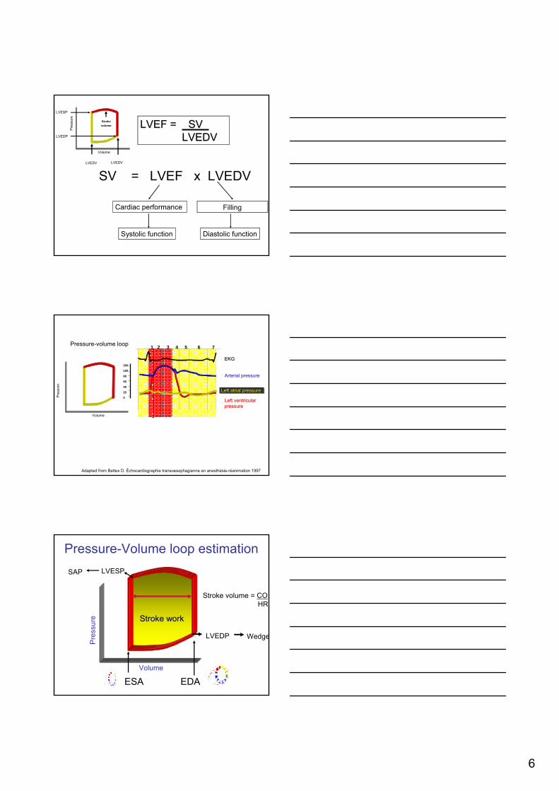
Pressure-Volume loop estimation
ESA EDA
Stroke volume = COHR
LVEDP Wedge
LVESPSAP
7
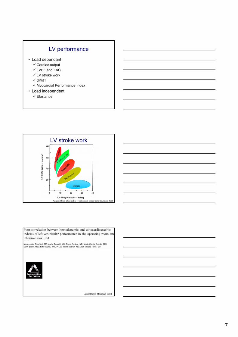
LV performance
• Load dependantCardiac outputLVEF and FACLV stroke workdP/dTMyocardial Performance Index
• Load independentElastance
Hype
r rea
ctor
Health
y, yo
ung
Depressed
Adequ
ate
Shock
Adapted from Shoemaker Textbook of critical care Saunders 1989
LV stroke work
Critical Care Medicine 2004

8
200 60 80 10040
Bouchard et al CCM 2004
Normal range
Myocardial dysfonction after sucessfulresuscitation from cardiac arrest
Gazmuri RJ et al. CCM 1996
Pres
sure
Volume
Left ventricular dysfunction

9
Volume (ml)40 100 160
100
10
Stroke volume 60 ml 60 mlLVSW (X 0.0136) 60 X (100-10) 60 X (100-10)
Limitation of LVSW measurement
Pres
sure
(mm
Hg)
Stroke Work Stroke Work
Stroke Work Stroke Work
LVEF 60 ml/100ml 60 ml/160ml= 60% = 40%
Limitation of LVSW measurement
Volume (ml)40 100 160
100
10
Pres
sure
(mm
Hg)
Contractility
Pre
ssur
e
Volume
Pre
ssur
e
Volume

10
Gorcsan J. Circulation 1994;89:180-90
Assessment of the immediate effects of cardiopulmonary bypass on left ventricular
performance by on-line pressure-area relations
dP/dT
1-Estimation of LV function in mitral regurgitation2-Estimation of left atrial systolic pressure3-Estimation of systolic arterial pressure: is your radial arterial pressure reliable?
Aortique et radiale
Radial pressure
Femoral pressure
69 yo woman after MVR and AVR

11
Radial pressure = 111 mmHg
Presssure gradient = 111 mmHg = LVESP - LAP
LV systolic pressure = PG + LAP ≈ Aortic pressure= 111 mmHg + pulmonary artery wedge pressure (v wave)= 111 mmHg + 18 mmHg
Aortic pressure = 129 mmHg
Aortic pressure measured by surgeon = 125 mmHg
Prevalence of a central to radial pressure gradient in high-risk surgery
Dr Antonio Su
Percentage of central-to-radial gradients in complex surgeries
N = 60

12
René Magritte
Why is this patient hemodynamically unstable?
13
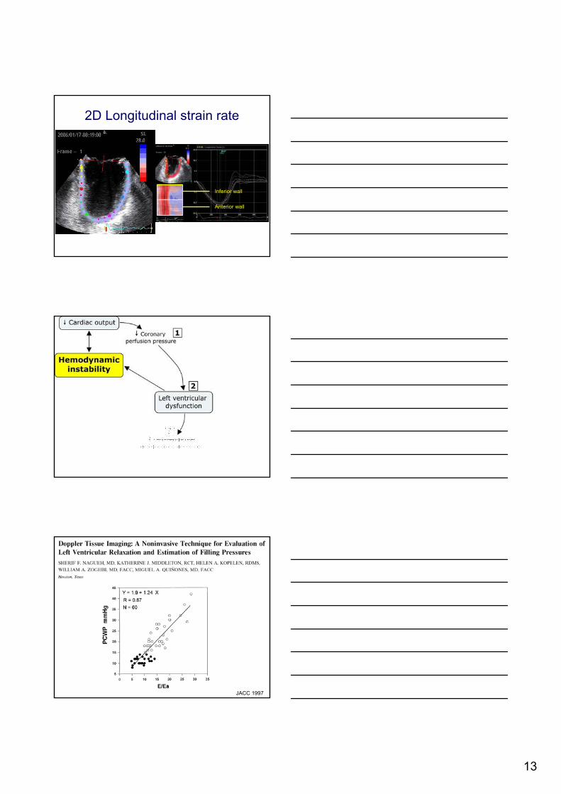
2D Longitudinal strain rate
Inferior wall
Anterior wall
JACC 1997

14
Salem
British Journal Anesthesia 2006N = 3024
Volume
Pre
ssur
e
Diastolic dysfunction
End-systolic areaEnd-diastolic area
Wedge = 5 mmHg

15
Adapté de Khoury et al JASE 2004
Summary
Evaluation of diastolic fillingof left ventricle in healthand disease: Doppler
echocardiography is the clinician's Rosetta Stone
Nishimura RA JACC 1997

16

17
Anesth Analgesia 2001;92:291-8
Canadian J Anesthesia 2006
Grade I: relaxation abnormality
LVV
LVP

18
76 yo woman with aortic stenosis
A A wavewave
EmAm
EA
EmAm
SAPSAP
PAPPAPCVPCVP
Grade III: Increased Chamber Stifness
LVV
LVP
62 yo patient unstable after AVR

19
Beginnning post-bypass
End post-bypass
Chamber Dilatation
LVV
LVP
Systolic Failure
Normal
Normal diastolicchamberdistensibility
Left
Ven
tricu
larP
ress
ure
Left Ventricular Volume

20
Impaired Left Ventricular Filling Due to Right Ventricular Pressure Overload inPrimary Pulmonary Hypertension*Noninvasive Monitoring Using MRI
Marcus JT Chest 2001;119:1761-5
Pericardial Restraint
LVV
LVPPericardialThoracic
Abdominal
Kaplan Cardiac anesthesia 1998
Effet de la fréquence cardiaque sur la fonction diastolique
Volume (mL /m2)
Pres
sure
(mm
Hg)
50
40
30
20
10
50
0 20 40 60 80
84 bmp110 bmp135 bmp

21
Hemodynamically unstableafter cardiac surgery: paced
Slow down the pacemaker
Hemodynamically unstableafter cardiac surgery

22
Noradrenaline 4ug/min Noradrenaline 0.5 ug/min
Homme de 70 ans instable après RVA
100 mmHg 100 mmHg
Unexpected hemodynamicinstability. What happened?

23
LVOT obstruction
Courtesy of Gaudiani
Normally

24
Fearful consequence
ME
With permission from Lang et al JASE 2005;18:1440-1463

25
JASE 2007 (In press)
Right ventricular pressure monitoring
ME: Normal RV waveform

26
HVF: RV dysfunctionPra
Prv
RA and RV pressure waveform correlation
HVF
A wave
V wave Abnormal RV diastolic slope
56 yo woman: CABG, MVR and LV remodeling
*
Effect of adrenalinand thoracic closure
End of CPB Adrenalin Thoracic closure

27
Évolution ScO2
Effect of adrenalin upon weaning from CPB
RV dysfunction impact
Another confounding factor

28
Right ventricular outflow tract obstruction
Right ventricular outflow tract obstruction
Right ventricular outflow tract obstruction

29
Septal interaction

30
31
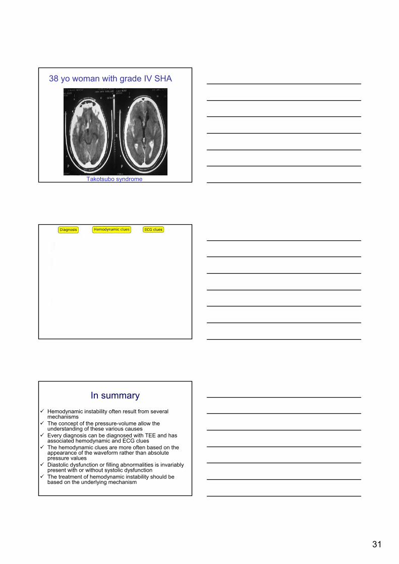
38 yo woman with grade IV SHA
Takotsubo syndrome
In summaryHemodynamic instability often result from several mechanismsThe concept of the pressure-volume allow the understanding of these various causesEvery diagnosis can be diagnosed with TEE and has associated hemodynamic and ECG cluesThe hemodynamic clues are more often based on the appearance of the waveform rather than absolute pressure valuesDiastolic dysfunction or filling abnormalities is invariably present with or without systolic dysfunctionThe treatment of hemodynamic instability should be based on the underlying mechanism

32
L’équipe de recherche:
Denis Babin MSc Inh
Jean-Claude Tardif MD FRCP
Alain Deschamps MD FRCPC PhD
Jean Lambert PhDBiostatistics
Thesis director
Pierre Couture MD FRCPC
Louis P.PerraultMD PhD FRCSC

![Epuration extra rénale.ppt [Mode de compatibilité] › pdf_ppt_docs › college2014 › epurationextrarenale.pdf‐IRA avec instabilité hémodynamique sévère ‐Désordres hydroéléctrolytiquesmettant](https://img.pdfslide.tips/doc/110x75/60d94fa14677cd24300cee43/epuration-extra-rnaleppt-mode-de-compatibilit-a-pdfpptdocs-a-college2014.jpg)