Embed Size (px)
Citation preview
Isoform Patterns of Chitinase and â-1,3-Glucanase in Maturing CornKernels (Zea mays L.) Associated with Aspergillus flavus Milk StageInfection
C. Ji,*,† R. A. Norton,‡ D. T. Wicklow,‡ and P. F. Dowd‡
National Center for Agricultural Utilization Research, Bioactive Agents Research, Agricultural ResearchService, U.S. Department of Agriculture, 1815 North University, Peoria, Illinois 61604, and Department of
Crop Science, University of Illinois at Champaign-Urbana, Urbana, Illinois 61801
Isoform patterns of chitinase and â-1,3-glucanase of maturing kernels of yellow dent corn (Pioneer3394) infected with Aspergillus flavus at the milk stage were investigated through polyacrylamidegel electrophoresis (PAGE). Proteins on the sodium dodecyl sulfate (SDS) gel with an apparentmolecular mass range of 23-46 kDa were differentially present in the kernels infected with bothaflatoxin-producing and non-aflatoxin-producing strains of A. flavus. From in-gel (native PAGE)enzyme activity assays, three bands corresponding to chitinase isoforms and two bands correspondingto â-1,3-glucanase isoforms were detected in the infected kernels. One chitinase isoform of 29 kDawas present only in the infected kernels, and another one of 28 kDa was present in both infectedand noninfected kernels. They were judged to be acidic on the basis of their migration on anacrylamide isoelectric focusing (IEF) gel. For the â-1,3-glucanase, one isoform of 35 kDa was presentin both infected and noninfected kernels, but another one, a 33 kDa isoform, was present only inthe infected kernels. Both acidic and basic â-1,3-glucanase isoforms were detected in the IEF gel.The results of this study are the first to demonstrate patterns of enhanced or inducible proteins inmaturing corn kernels in response to A. flavus infection at the milk stage. The results also indicatethat only particular isoforms of the two hydrolytic enzymes are involved in the maturing corn kernelsinfected at the milk stage with A. flavus.
Keywords: Isoform; â-1,3-glucanase; chitinase; corn kernels; Aspergillus flavus; aflatoxin
INTRODUCTION
Sporadic outbreaks of aflatoxin contamination ofpreharvest corn (Zea mays L.) can be a serious problemin the Midwestern corn belt (Hurburgh, 1991). Aflatox-ins, produced by the fungus Aspergillus flavus, are toxicand carcinogenic to humans and other animals (Smithand Moss, 1985). Current attempts to prevent A. flavusinfection and aflatoxin contamination include isolationand formulation of competitive microbes for use inbiocontrol; characterization of structural, chemical, andbiochemical factors associated with the resistance ofcorn varieties to A. flavus infection, aflatoxin accumula-tion, and/or insect damage to the ear; and identificationof genes through marker-assisted gene mapping respon-sible for A. flavus resistance (Brown et al., 1999).Antimicrobial factors of protein or peptide nature havebeen extensively investigated because such factors couldbe rapidly introduced into corn through genetic engi-neering. Several families of pathogenesis-related (PR)proteins are recognized as biochemical factors possess-ing antimicrobial activity in vitro or the ability toenhance disease resistance when overexpressed intransgenic plants (Bushnell et al., 1998; Ryals et al.,1996). Some of these proteins are hydrolytic, includingchitinase and â-1,3-glucanase (Boller, 1985; Nasser etal., 1988, 1990). Studies with various plant systemshave indicated that several isoforms of the two hydro-
lytic enzymes tend to appear in response to pathogeninfection or other stress conditions (Darnetty et al.,1993; Ryals et al., 1996; Yi and Hwang, 1996), and onlycertain isoforms have defensive roles against particularfungal pathogens (Sela-Buurlage et al., 1993; Sticheret al., 1997). Activities of chitinase and â-1,3-glucanasehave recently been reported to exist and inhibit thegrowth of A. flavus in mature corn kernels (Neucere etal., 1991, 1995; Lozovaya et al., 1998). However, littleinformation has been published on what isoforms of thetwo hydrolytic enzymes in the corn kernels are inducedby A. flavus infection.
In this paper, we provide evidence that proteins andparticularly isoforms of chitinase and â-1,3-glucanasewere differentially induced in maturing corn kernelsinfected with A. flavus. By utilizing in-gel (nativepolyacrylamide gel) enzyme activity assays (Trudel andAsselin, 1989), we further observed that certain isoformsof both chitinase and â-1,3-glucanase were associatedwith infection of A. flavus but not with infection ofanother fungus, Fusarium moniliforme, or with droughtstress.
MATERIALS AND METHODS
Corn Inoculations. A commercial corn hybrid (Pioneer3394) was grown in 1997 to maturity in experimental irriga-tion control plots at the 40-acre Illinois River Valley Sand Field(IRVSF), Kilbourne, IL. Within each plot, up to 20 corn earsin the late-milk to early-dough stage of kernel maturity (21days after silking; August 7, 1997) were inoculated at threepoints separated by 4 cm, in two vertical rows on opposite sides
† University of Illinois at Champaign-Urbana.‡ U.S. Department of Agriculture.
507J. Agric. Food Chem. 2000, 48, 507−511
10.1021/jf9905119 CCC: $19.00 © 2000 American Chemical SocietyPublished on Web 01/04/2000
of the ear by inserting a sterile wooden toothpick through thehusk to wound underlying kernels. A second toothpick con-taminated with A. flavus NRRL A-27837 (105 spores/mL), anaflatoxin-producing strain isolated from corn grown at IRVSF,was then inserted into each wound site and left until thenaturally dried ears (15.5% moisture content) were handharvested and shelled on October 6, 1997. Grain samples wereselected from two irrigation control treatments: A, full-seasonirrigation; H, full-season nonirrigation. A single row of 20 cornplants under continuous irrigation was wound-inoculated witha non-aflatoxin-producing strain of A. flavus, NRRL A-27668(105 spores/mL), to contrast the enzymatic response of infectedkernels. The grain samples were examined for bright greenishyellow fluorescent (BGYF) kernels (Shotwell et al., 1972) usinga black light at 365 nm and then grouped into three catego-ries: category 1, intact non-BGYF kernels that were consid-ered to be symptom-free kernels; category 2, intact kernelswith limited fluorescence to the germ and endosperm (8000-17000 ppb of aflatoxin); category 3, kernels with full BGYF/discolored and shriveled appearance (28000-30000 ppb ofaflatoxin). All kernel samples were stored at -20 °C. Inselecting BGYF kernels for analyses we know that the fungushas gained entry to the seed proper and infected the germ orendosperm, sites of aflatoxin production. Infected kernels(Pioneer 3394) from ears inoculated with Fusarium monili-forme (strain M-3125) and having visible mold growth wereprovided by R. D. Plattner at the USDA-ARS, National Centerfor Agricultural Utilization Research, Peoria, IL (Desjardinsand Plattner, 1998).
Fungal Growth and Harvest. A. flavus strains thatproduced aflatoxin (NRRL A-27837) or did not produce afla-toxin (NRRL A-27668) were grown separately for 7 days (25°C) on potato dextrose agar (PDA) (Difco Laboratories, Detroit,MI). Portions of the colony were removed using a spatula forprotein extraction. Each fungal strain was also inoculated ontosoaked (12 h) and autoclaved corn kernels (25 g of dw; 50%moisture) and incubated for 7 days (25 °C). Following incuba-tion, the molded kernels were extracted for proteins using theprocedure described below.
Aflatoxin Analyses. All samples were analyzed for afla-toxin by the Aflatest (Vicam Inc., Waterown, MA) procedureaccording to the manufacturer’s instructions, but adjusted toaccommodate 10-g samples of ground corn. The standardprocedure could detect aflatoxin as low as 1 ppb. Aflatoxin-containing wastes were immersed in Clorox bleach (regular)for 1 h to destroy the aflatoxin and then autoclaved.
Protein Extraction. The whole kernels or the lower thirdsof the kernels were milled in a mini-miller (model IDS-50, Mr.Coffee, Inc.), and 1 g of the resulting powder was homogenizedwith a blender (Polytron) by adding 10 mL of an acetate buffer(pH 5.1), which contained 0.05% (w/v) sodium acetate, 4 mMof ascorbic acid, 2 mM PMSF, and 2% (w/v) PVPP. Proteinswere extracted with gentle shaking at 4 °C overnight. Thehomogenates were then centrifuged at 15000g for 60 min at 4°C. The isolated supernatants were concentrated ∼10 timesby filtering through ultrafiltration membranes (NMWL5,000,Millipore). The protein filtrates were then dialyzed against thesame buffer for 8 h at 4 °C. The final protein extracts werestored at -20 °C and analyzed within 2 weeks. The sameprocedure was used to extract proteins of A. flavus or F.moniliforme, but the extraction was started by adding 10 mLof the acetate buffer to 5 g (fresh weight) of fungi. Proteinconcentration was determined using the Bio-Rad protein assaykit using bovine γ-globulin as standard (Bradford, 1976).
Electrophoretic Separation of Proteins. SDS-polyacryl-amide gel electrophoresis (PAGE) and PAGE under nativeconditions were performed according to previously describedmethods (Trudel and Asselin, 1989; Ji and Kuc, 1995, 1996).The SDS-PAGE contained 12.5% (w/v) polyacrylamide and0.1% SDS. Stacking gels were made of 4.5% (w/v) polyacryl-amide containing 0.1% (w/v) SDS. The native PAGE contained15% (w/v) polyacrylamide in the resolving gel and 5% poly-acylamide in the stacking gel.
Isoelectric focusing (IEF) was performed using precast widerange gels (pH 3.5-9.5, Pharmacia-LKB), as previously de-scribed (Dowd, 1994).
In-Gel Chitinase and â-1,3-Glucanase Isoform Assays.Detection of chitinase isoforms after native PAGE and IEFwas based on the use of an overlay gel containing 0.01% (w/v)glycol chitin as substrate (Trudel and Asselin, 1989). After 2h of incubation at 37 °C, the overlay gel was stained withfluorescent brightener 28 (Sigma) for 5 min. Lytic zonescorresponding to chitinase activity on the gels were visualizedby placing the gels on a Chromato-Vue C-62 transilluminator(UV Products). The fluorescent background could be reducedby washing the gel several times with distilled water. Thebands on the gels were then photographed with Polaroid film(type 107) with UV-haze and 02 orange filters. Detection ofâ-1,3-glucanase isoforms after native PAGE and IEF wasperformed as described by Pan (1989) and Ji and Kuc (1995).Gels were incubated in a solution containing 1% of laminarinfor 90 min at 40 °C. â-1,3-Glucanase activity in the gels wasthen visualized by staining the gels for 5-10 min at 100 °C ina 1 M NaOH solution containing 0.3% (w/v) 2,3,5-triphenyl-tetrazolium chloride (Sigma).
Partial Purification of Chitinase and â-1,3-Glucanase.After a single electrophoretic separation of proteins in theprotein extracts by the native PAGE or IEF, all bands thatcorresponded to kernel chitinase or â-1,3-glucanase were cutout of the native PAGE. The proteins in the bands were elutedwith an Electro-Eluter (model 422, Bio-Rad) and concentratedaccording to the manufacturer’s instructions. The enzymaticnature of the partially purified proteins was checked byperforming enzyme activity assays (Ji and Kuc, 1995).
RESULTS
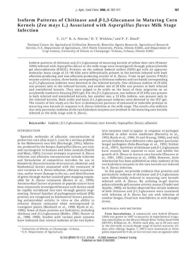
Proteins from maturing corn kernels that are associ-ated with A. flavus infection were analyzed on an SDS-polyacrylamide gel (PAGE) as shown in Figure 1. Therewere at least four corn kernel proteins enhanced orinduced in response to A. flavus infection (Figure 1,lanes 2-5, 7, and 8) in contrast to the proteins fromsymptom-free kernels without infection (Figure 1, lanes1 and 6). The four proteins, indicated by arrows, hadapparent molecular masses of 23-46 kDa. The inducibleproteins were similar among kernels infected with theaflatoxin-producing strain (Figure 1, lanes 2 and 3) orthe non-aflatoxin-producing strain (Figure 1, lanes 4
Figure 1. Proteins of maturing corn kernels associated withA. flavus infection. Proteins were separated by 12.5% SDS-PAGE and stained with GelCode Blue stain reagent (Pierce).Each lane was loaded with 100 µg of proteins from the lowerthird of the kernels. Samples were from symptom-free kernels(lanes 1 and 6), intact kernels with limited BGYF distribution(lanes 2, 4, and 7), kernels with full BGYF distribution (lanes3, 5, and 8), kernels from irrigation treatment (lanes 1-5),kernels of nonirrigation treatment (lanes 6-8), and kernelsinfected with A. flavus strain NRRL A-27837 (lanes 2, 3, 7,and 8) and strain NRRL A-27668 (lanes 4 and 5). Mr,molecular mass markers.
508 J. Agric. Food Chem., Vol. 48, No. 2, 2000 Ji et al.
and 5) and among kernels from irrigated (Figure 1,lanes 2 and 3) or nonirrigated (Figure 1, lanes 7 and 8)treatments.
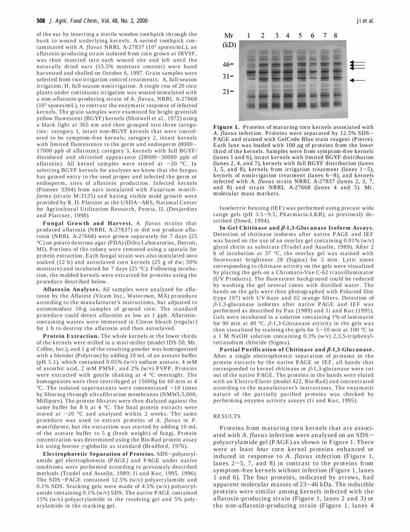
Results in Figure 2 show chitinase activity in a nativePAGE after a single electrophoretic separation of theproteins. Three bands indicating different chitinaseisoforms were detected in the kernels infected with A.flavus (Figure 2, lanes 2-8) or F. moniliforme (Figure2, lane 12). On the basis of the intensity of each band,the two upper bands appeared to be inducible orenhancible, whereas the lowest band exhibited littledifference among samples from both symptom-free(Figure 2, lanes 1 and 11) and infected kernels. Thesecond inducible band (Figure 2, lane 12) by F. monili-forme was slightly different in migration rate from theone induced by A. flavus. Because proteins of fungi inthe infected kernels might also contribute to the de-tected chitinase activity, protein extracts from A. flavusand F. moniliforme were used as controls. One bandfrom either A. flavus (Figure 2, lane 9) or F. moniliforme(Figure 2, lane 10) was observed, and it appeared to bedifferent from the kernel inducible chitinase isoforms.
Three bands corresponding to â-1,3-glucanase iso-forms were also observed in the protein extracts ofkernels infected with A. flavus (Figure 3, lanes 2, 3, 5,and 6). One band of â-1,3-glucanase at the lowestposition was not associated with A. flavus infectionbecause this band could also be detected in the symptom-free corn kernels (Figure 3, lanes 1 and 9). Another bandof â-1,3-glucanase at the middle position might not beof corn kernel origin becuase this band was present inthe protein extracts of A. flavus (Figure 3, lane 7). Thethird band at the highest position of the gel seemedspecifically associated with A. flavus infection. This wassupported by the observation that the third band wasdifferent from the one at a similar migration positiondetected in the corn kernels infected with F. moniliforme(Figure 3, lane 10).
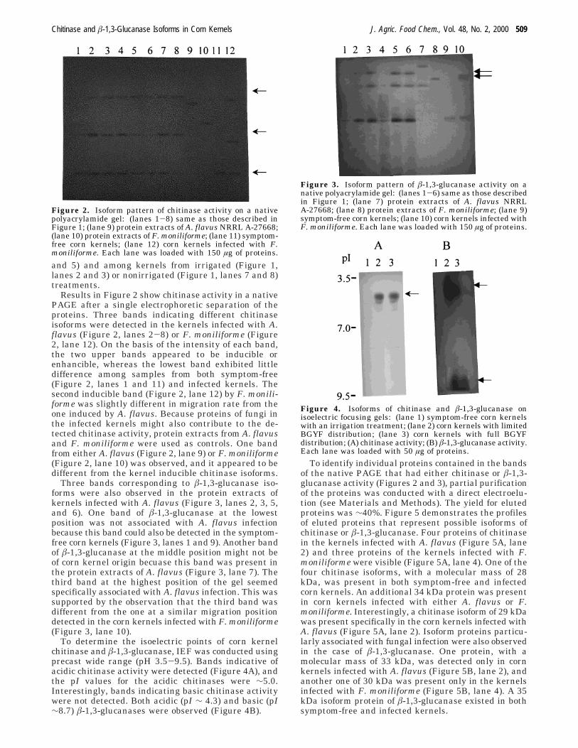
To determine the isoelectric points of corn kernelchitinase and â-1,3-glucanase, IEF was conducted usingprecast wide range (pH 3.5-9.5). Bands indicative ofacidic chitinase activity were detected (Figure 4A), andthe pI values for the acidic chitinases were ∼5.0.Interestingly, bands indicating basic chitinase activitywere not detected. Both acidic (pI ∼ 4.3) and basic (pI∼8.7) â-1,3-glucanases were observed (Figure 4B).
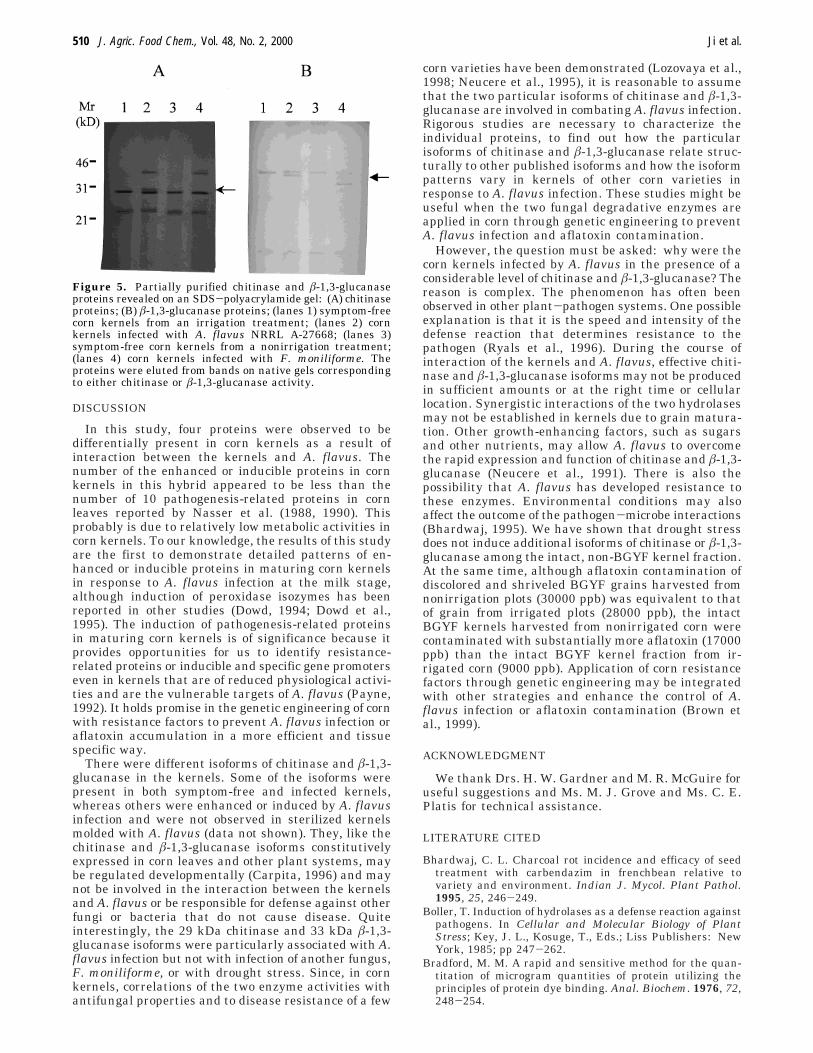
To identify individual proteins contained in the bandsof the native PAGE that had either chitinase or â-1,3-glucanase activity (Figures 2 and 3), partial purificationof the proteins was conducted with a direct electroelu-tion (see Materials and Methods). The yield for elutedproteins was ∼40%. Figure 5 demonstrates the profilesof eluted proteins that represent possible isoforms ofchitinase or â-1,3-glucanase. Four proteins of chitinasein the kernels infected with A. flavus (Figure 5A, lane2) and three proteins of the kernels infected with F.moniliforme were visible (Figure 5A, lane 4). One of thefour chitinase isoforms, with a molecular mass of 28kDa, was present in both symptom-free and infectedcorn kernels. An additional 34 kDa protein was presentin corn kernels infected with either A. flavus or F.moniliforme. Interestingly, a chitinase isoform of 29 kDawas present specifically in the corn kernels infected withA. flavus (Figure 5A, lane 2). Isoform proteins particu-larly associated with fungal infection were also observedin the case of â-1,3-glucanase. One protein, with amolecular mass of 33 kDa, was detected only in cornkernels infected with A. flavus (Figure 5B, lane 2), andanother one of 30 kDa was present only in the kernelsinfected with F. moniliforme (Figure 5B, lane 4). A 35kDa isoform protein of â-1,3-glucanase existed in bothsymptom-free and infected kernels.
Figure 2. Isoform pattern of chitinase activity on a nativepolyacrylamide gel: (lanes 1-8) same as those described inFigure 1; (lane 9) protein extracts of A. flavus NRRL A-27668;(lane 10) protein extracts of F. moniliforme; (lane 11) symptom-free corn kernels; (lane 12) corn kernels infected with F.moniliforme. Each lane was loaded with 150 µg of proteins.
Figure 3. Isoform pattern of â-1,3-glucanase activity on anative polyacrylamide gel: (lanes 1-6) same as those describedin Figure 1; (lane 7) protein extracts of A. flavus NRRLA-27668; (lane 8) protein extracts of F. moniliforme; (lane 9)symptom-free corn kernels; (lane 10) corn kernels infected withF. moniliforme. Each lane was loaded with 150 µg of proteins.
Figure 4. Isoforms of chitinase and â-1,3-glucanase onisoelectric focusing gels: (lane 1) symptom-free corn kernelswith an irrigation treatment; (lane 2) corn kernels with limitedBGYF distribution; (lane 3) corn kernels with full BGYFdistribution; (A) chitinase activity; (B) â-1,3-glucanase activity.Each lane was loaded with 50 µg of proteins.
Chitinase and â-1,3-Glucanase Isoforms in Corn Kernels J. Agric. Food Chem., Vol. 48, No. 2, 2000 509
DISCUSSION
In this study, four proteins were observed to bedifferentially present in corn kernels as a result ofinteraction between the kernels and A. flavus. Thenumber of the enhanced or inducible proteins in cornkernels in this hybrid appeared to be less than thenumber of 10 pathogenesis-related proteins in cornleaves reported by Nasser et al. (1988, 1990). Thisprobably is due to relatively low metabolic activities incorn kernels. To our knowledge, the results of this studyare the first to demonstrate detailed patterns of en-hanced or inducible proteins in maturing corn kernelsin response to A. flavus infection at the milk stage,although induction of peroxidase isozymes has beenreported in other studies (Dowd, 1994; Dowd et al.,1995). The induction of pathogenesis-related proteinsin maturing corn kernels is of significance because itprovides opportunities for us to identify resistance-related proteins or inducible and specific gene promoterseven in kernels that are of reduced physiological activi-ties and are the vulnerable targets of A. flavus (Payne,1992). It holds promise in the genetic engineering of cornwith resistance factors to prevent A. flavus infection oraflatoxin accumulation in a more efficient and tissuespecific way.
There were different isoforms of chitinase and â-1,3-glucanase in the kernels. Some of the isoforms werepresent in both symptom-free and infected kernels,whereas others were enhanced or induced by A. flavusinfection and were not observed in sterilized kernelsmolded with A. flavus (data not shown). They, like thechitinase and â-1,3-glucanase isoforms constitutivelyexpressed in corn leaves and other plant systems, maybe regulated developmentally (Carpita, 1996) and maynot be involved in the interaction between the kernelsand A. flavus or be responsible for defense against otherfungi or bacteria that do not cause disease. Quiteinterestingly, the 29 kDa chitinase and 33 kDa â-1,3-glucanase isoforms were particularly associated with A.flavus infection but not with infection of another fungus,F. moniliforme, or with drought stress. Since, in cornkernels, correlations of the two enzyme activities withantifungal properties and to disease resistance of a few
corn varieties have been demonstrated (Lozovaya et al.,1998; Neucere et al., 1995), it is reasonable to assumethat the two particular isoforms of chitinase and â-1,3-glucanase are involved in combating A. flavus infection.Rigorous studies are necessary to characterize theindividual proteins, to find out how the particularisoforms of chitinase and â-1,3-glucanase relate struc-turally to other published isoforms and how the isoformpatterns vary in kernels of other corn varieties inresponse to A. flavus infection. These studies might beuseful when the two fungal degradative enzymes areapplied in corn through genetic engineering to preventA. flavus infection and aflatoxin contamination.
However, the question must be asked: why were thecorn kernels infected by A. flavus in the presence of aconsiderable level of chitinase and â-1,3-glucanase? Thereason is complex. The phenomenon has often beenobserved in other plant-pathogen systems. One possibleexplanation is that it is the speed and intensity of thedefense reaction that determines resistance to thepathogen (Ryals et al., 1996). During the course ofinteraction of the kernels and A. flavus, effective chiti-nase and â-1,3-glucanase isoforms may not be producedin sufficient amounts or at the right time or cellularlocation. Synergistic interactions of the two hydrolasesmay not be established in kernels due to grain matura-tion. Other growth-enhancing factors, such as sugarsand other nutrients, may allow A. flavus to overcomethe rapid expression and function of chitinase and â-1,3-glucanase (Neucere et al., 1991). There is also thepossibility that A. flavus has developed resistance tothese enzymes. Environmental conditions may alsoaffect the outcome of the pathogen-microbe interactions(Bhardwaj, 1995). We have shown that drought stressdoes not induce additional isoforms of chitinase or â-1,3-glucanase among the intact, non-BGYF kernel fraction.At the same time, although aflatoxin contamination ofdiscolored and shriveled BGYF grains harvested fromnonirrigation plots (30000 ppb) was equivalent to thatof grain from irrigated plots (28000 ppb), the intactBGYF kernels harvested from nonirrigated corn werecontaminated with substantially more aflatoxin (17000ppb) than the intact BGYF kernel fraction from ir-rigated corn (9000 ppb). Application of corn resistancefactors through genetic engineering may be integratedwith other strategies and enhance the control of A.flavus infection or aflatoxin contamination (Brown etal., 1999).
ACKNOWLEDGMENT
We thank Drs. H. W. Gardner and M. R. McGuire foruseful suggestions and Ms. M. J. Grove and Ms. C. E.Platis for technical assistance.
LITERATURE CITED
Bhardwaj, C. L. Charcoal rot incidence and efficacy of seedtreatment with carbendazim in frenchbean relative tovariety and environment. Indian J. Mycol. Plant Pathol.1995, 25, 246-249.
Boller, T. Induction of hydrolases as a defense reaction againstpathogens. In Cellular and Molecular Biology of PlantStress; Key, J. L., Kosuge, T., Eds.; Liss Publishers: NewYork, 1985; pp 247-262.
Bradford, M. M. A rapid and sensitive method for the quan-titation of microgram quantities of protein utilizing theprinciples of protein dye binding. Anal. Biochem. 1976, 72,248-254.
Figure 5. Partially purified chitinase and â-1,3-glucanaseproteins revealed on an SDS-polyacrylamide gel: (A) chitinaseproteins; (B) â-1,3-glucanase proteins; (lanes 1) symptom-freecorn kernels from an irrigation treatment; (lanes 2) cornkernels infected with A. flavus NRRL A-27668; (lanes 3)symptom-free corn kernels from a nonirrigation treatment;(lanes 4) corn kernels infected with F. moniliforme. Theproteins were eluted from bands on native gels correspondingto either chitinase or â-1,3-glucanase activity.
510 J. Agric. Food Chem., Vol. 48, No. 2, 2000 Ji et al.
Brown, R. L.; Chen, Z. Y.; Cleveland, T. E.; Russin, J. S.Advances in the development of host resistance in corn toaflatoxin in field maize. Phytopathol. Rev. 1999, 89, 113-117.
Bushnell, W. R.; Somers, D. A.; Giroux, R. W.; Szabo, L. J.;Zeyen, R. J. Genetic engineering of disease resistance incereals. Can. J. Plant Pathol. 1998, 20 (2), 137-149.
Carpita, N. C. Structure and biogenesis of the cell walls ofgrasses. Annu. Rev. Plant Physiol. Mol. Biol. 1996, 47, 445-476.
Cordero, M. J.; Raventos, D.; Segundo, B. S. Differentialexpression and induction of chitinases and â-1,3-glucanasesin response to fungal infection during germination of maizeseeds. Mol. Plant-Microbe Interact. 1994, 7 (1), 23-31.
Darnetty, J. F. L.; Muthukrishnan, S.; Swegle, M.; Vigers, A.J.; Selitrennikoff, C. P. Variability in antifungal proteinsin the grains of maize, sorghum and wheat. Physiol. Plant.1993, 88, 339-349.
Desjardins, A. E.; Plattner, R. D. Distribution of fumonisinsin maize ears infected with strains of Fusarium moniliformethat differ in fumonisin production. Plant Dis. 1998, 82,953-958.
Dowd, P. F. Enhanced maize (Zea mays L.) pericarp brown-ing: associations with insect resistance and involvement ofoxidizing enzymes. J. Chem. Ecol. 1994, 20, 2777-2803.
Dowd, P. F.; Bennett, G. A.; Richard, J. L. IPM of aflatoxin inthe corn belt-FY 1995 results. Presented at the AflatoxinElimination Workshop, Atlanta, GA, Oct 23-24, 1995; pp76-78.
Hurburgh, C. R., Jr. Aflatoxin in midwestern corn. In Aflatoxinin Corn: New Perspectives; Shotwell, O. L., Hurburgh, C.R., Jr., Eds.; Research Bullettin 599; Iowa Agriculture andHome Economics Experiment Station: Ames, IA, 1991; pp343-350.
Ji, C.; Kuc, J. Purification and characterization of an acidicâ-1,3-glucanase from cucumber and its relationship tosystemic disease resistance induced by Colletotrichum la-genarium and tobacco necrosis virus. Mol. Plant-MicrobeInteract. 1995, 8, 899-905.
Ji, C.; Kuc, J. Antifungal activity of cucumber â-1,3-glucanaseand chitinase. Physiol. Mol. Plant Pathol. 1996, 49, 257-265.
Lozovaya, V. V.; Waranyuwat, A.; Widholm, J. M. â-1,3-Glucanase and Resistance to Aspergillus flavus Infection inMaize. Crop Sci. 1998, 38, 1255-1260.
Nasser, W.; Tapia, M.; Kauffmann, S.; Montasser-Kouhsari,S.; Burkard, G. Identification and characterization of maizepathogenesis-related proteins. Four maize PR proteins arechitinases. Plant Mol. Biol. 1988, 11, 529-538.
Nasser, W.; Tapia, M.; Burkard, G. Maize pathogenesis-relatedproteins: Characterization and cellular distribution of â-1,3-glucanases and chitinases induced by brome mosaic virusinfection or mercuric chloride treatment. Physiol. Mol. PlantPathol. 1990, 36, 1-14.
Neucere, J. N.; Cleveland, T. E.; Dischinger, C. Existence ofchitinase activity in maturing corn kernels (Zea mays L.).J. Agric. Food Chem. 1991, 39, 1326-1328.
Neucere, J. N.; Brown, R. L.; Cleveland, T. E. Correlation ofantifungal properties and â-1,3-glucanase in aqueous ex-tracts of kernels from several varieties of corn. J. Agric. FoodChem. 1995, 43, 275-276.
Pan, S. Q.; Ye, X. S.; Kuc, J. Direct detection of â-1,3-glucanaseisozymes on polyacrylamide electrophoresis and isoelectro-focusing gels. Anal. Biochem. 1989, 182, 136-140.
Payne, G. A. Aflatoxin in maize. Crit. Rev. Plant Sci. 1992, 10(5), 423-440.
Ryals, J.; Neuenschwander, U.; Willits, M.; Molina, A.; Steiner,H. Y.; Hunt, M. Systemic acquired resistance. Plant Cell.1996, 8, 1809-1819.
Sela-Buurlage, M. B.; Ponstein, A. S.; Bres-Vloemans, S. A.;Melchers, L. S.; van den Elzen, P. J. M.; Cornelissen, B. J.C. Only specific tobacco (Nicotiana tabacum) chitinases andâ-1,3-glucanases exhibit antifungal activity. Plant Physiol.1993, 101, 857-863.
Shotwell, O. L.; Goulden, M. L.; Hesseltine, C. W. Aflatoxincontamination: association with foreign material and char-acteristic fluorescence in damaged corn kernels. CerealChem. 1972, 49, 458-465.
Smith, J. E.; Moss, M. O. Mycotoxins: Formation, Analysis,and Significance; Wiley: New York, 1985; pp 1-148.
Sticher, L.; Mauch-Mani, B.; Metraux, J. P. Systemic acquiredresistance. Annu. Rev. Phytopathol. 1997, 35, 235-270.
Trudel, J.; Asselin, A. Detection of chitinase acitivity afterpolyacrylamide gel electrophoresis. Anal. Biochem. 1989,178, 362-366.
Yi, S. Y.; Hwang, B. K. Differential induction and accumulationof â-1,3-glucanase and chitinase isoforms in soybean hypo-cotyls and leaves after compatible and incompatible infectionwith Phytophthora megasperma f.sp. glycinea. Physiol. Mol.Plant Pathol. 1996, 48, 179-192.
Received for review May 14, 1999. Revised manuscript receivedOctober 28, 1999. Accepted November 11, 1999. This researchwas supported in part by Specific Cooperative Agreement 58-3620-5-148 between the University of Illinois and the ARS.
JF9905119
Chitinase and â-1,3-Glucanase Isoforms in Corn Kernels J. Agric. Food Chem., Vol. 48, No. 2, 2000 511


![Chondrichthyes Chitinase: Molecular Cloning, Distribution ... · Chondrichthyes, or the stomach of Latimeria chalmnae, classified as Sarcoptery-gii [12]. These findings suggested](https://img.pdfslide.tips/doc/110x75/5cfc746a88c993a30c8bf07c/chondrichthyes-chitinase-molecular-cloning-distribution-chondrichthyes.jpg)