Embed Size (px)
Citation preview

ACNRISSN 1473-9348 Volume 6 Issue 6 January/February 2007
Advances in Clinical Neuroscience & Rehabilitation
Rustam Al-Shahi Saman, Henning Mast, Christian Stapf
Unruptured Arteriovenous Malformations of the Brain
David Baker, Samuel J JacksonModels of Multiple Sclerosis
Oleh Hornykiewicz Dopamine, Levodopa and Parkinson’s Disease
Conference and Society News • Journal Reviews • Diary of Events
www.acnr.co.uk

The first and only transdermal patch for early-stage Parkinson’s disease
Neupro® Rotigotine.Prescribing information.Presentation: Neupro® is a thin, matrix-typesquare transdermal patch.Neupro 2 mg/24 h transdermal patch:Releases 2 mg rotigotine over 24 hours.10 cm2 patch contains 4.5 mg rotigotine.Neupro 4 mg/24 h transdermal patch:Releases 4 mg rotigotine over 24 hours.20 cm2 patch contains 9.0 mg rotigotine.Neupro 6 mg/24 h transdermal patch:Releases 6 mg rotigotine over 24 hours.30 cm2 patch contains 13.5 mg rotigotine.Neupro 8 mg/24 h transdermal patch:Releases 8 mg rotigotine over 24 hours.40 cm2 patch contains 18.0 mg rotigotine.Indications: To treat the signs and symptoms ofearly-stage idiopathic Parkinson’s disease withoutconcomitant levodopa therapy. Dosage: Neupro isapplied to the skin once a day.The patch remains onthe skin for 24 hours and will then be replaced by a
new one at a different application site. Treatment isinitiated with a single daily dose of 2 mg/24 h.Increase dose by 2 mg/24 h each week (e.g. 2 mg/24 hin Week 1, 4 mg/24 h in Week 2, 6 mg/24 h in Week 3 and 8 mg/24 h in Week 4), until an effectivedose is reached. Maximal dose is 8 mg/24 h.Contraindications: Hypersensitivity to rotigotineor to any of the excipients. Neupro should beremoved prior to Magnetic Resonance Imaging (MRI)or cardioversion to avoid burns. Warnings andPrecautions: External heat should not be appliedto the patch. Dopamine agonists are known to causehypotension, and monitoring of blood pressure is recommended. Where somnolence or suddensleep onset occurs, or where there is persistent,spreading or serious skin rash at the application site, consider dose reduction or termination oftherapy. Rotate the site of patch application to minimise the risk of skin reactions. In case ofgeneralised skin reaction associated with use of Neupro, discontinue treatment. Avoid exposure
to direct sunlight until the skin is healed. If treatmentis to be withdrawn, it should be gradually reduced toavoid symptoms of neuroleptic malignant syndrome.Compulsive behaviours and hallucinations have been reported in patients treated with Neupro.Do not administer neuroleptics or dopamineantagonists to patients taking dopamine agonists.Caution is advised when treating patients with severe hepatic impairment, and in patients takingsedating medicines or other depressants incombination with rotigotine. Switching to anotherdopamine agonist may be beneficial for thosepatients who are insufficiently controlled byrotigotine. Undesirable Effects: Very commonside effects include nausea, vomiting, somnolence,dizziness and application site reactions. Common sideeffects include anorexia, hallucinations, sleep attacks,insomnia, abnormal dreams, headache, dyskinesia,lethargy, orthostatic hypotension, hypertension,hiccup, cough, constipation, diarrhoea, dry mouth,dyspepsia, hyperhydrosis, erythema, pruritus, asthenic
conditions and peripheral oedema. Uncommonly,syncope, loss of consciousness, visual disturbances,or hypotension may occur. Rarely, psychoticdisorders, increased libido or convulsion may occur.Basic NHS Cost: Starter Pack: £110.342 mg Continuation Pack of 28 patches: £77.244 mg Continuation Pack of 28 patches: £88.286 mg Continuation Pack of 28 patches: £110.348 mg Continuation Pack of 28 patches: £142.79Legal Category: POM. Product LicenceNumber: EU/1/05/331/001-013. Product LicenceHolder: SCHWARZ PHARMA Ltd, ShannonIndustrial Estate, Shannon, Co. Clare, Ireland. Date ofPreparation: March 2006 (3484). Neupro® is a registered trademark. Prescribers should consultthe Summary of Product Characteristics for the full information on side-effects, warnings andprecautions. Further information is available fromSCHWARZ PHARMA Limited, Schwarz House, EastStreet, Chesham, Bucks HP5 1DG, United Kingdom.Date of Literature Preparation: March 2006.
Information about adverse event reporting can be found at www.yellowcard.gov.uk Adverse events should be reported to the Drug Safety department at SCHWARZ PHARMA Limited (UK) on 01494 797 500 or [email protected]
References: 1. Neupro Summary of Product Characteristics. 2. Braun M et al. Poster presented at EFNS 2005. 3. Watts RL et al. Poster presented at MDS 2004. Abstract P737.
NEU3637
• Once-daily non-ergolinic dopamine agonist1
• Steady-state plasma concentration profile
over 24 hours2
• Proven efficacy in early Parkinson’s disease1,3
SWZ2052 297x210 ACNR.qxp 8/3/06 10:48 Page 1
ACNR • VOLUME 6 NUMBER 6 • JANUARY/FEBRUARY 2007 I 3
Editorial
ACNR is published by Whitehouse Publishing, 1 The Lynch, Mere, Wiltshire, BA12 6DQ.Publisher: Rachael Hansford • Email: [email protected]
Advertising and Editorial: Patricia McDonnellEmail: [email protected]/Fax: 0288 289 7023Design & Production Email: [email protected] by: Warners Midlands PLC, Tel. 01778 391000
Copyright: All rights reserved; no part of this publication may be reproduced, stored in a retrieval system or transmitted in any form or by anymeans, electronic, mechanical, photocopying, recording or otherwise without either the prior written permission of the publisher or a license permitting restricted photocopying issued in the UK by the Copyright Licensing Authority. Disclaimer: The publisher, the authors and editors accept no responsibility for loss incurred by any person acting or refraining from action as aresult of material in or omitted from this magazine. Any new methods and techniques described involving drug usage should be followed only inconjunction with drug manufacturers' own published literature.This is an independent publication - none of those contributing are in any way supported or remunerated by any of the companies advertising init, unless otherwise clearly stated. Comments expressed in editorial are those of the author(s) and are not necessarily endorsed by the editor, editorial board or publisher. The editor's decision is final and no correspondence will be entered into. Cover picture of neuron
courtesy of Stockxpert.
ACNR Journal reviewers - reviews start on page 28
Heather Angus-Leppan, Royal Free & Barnet Hospitals;Chrystalina Antoniades, Cambridge Centre for Brain Repair.Roger Barker, Cambridge Centre for Brain Repair;Alasdair Coles, Cambridge University;
Andrew Larner, Walton Centre, Liverpool;Mark Manford, Addenbrooke’s Hospital, Cambridge and Bedford Hospitals;Wendy Phillips, Addenbrooke’s Hospital, Cambridge;Robert Redfern, Morriston Hospital, Swansea;Ailie Turton, University of Bristol.
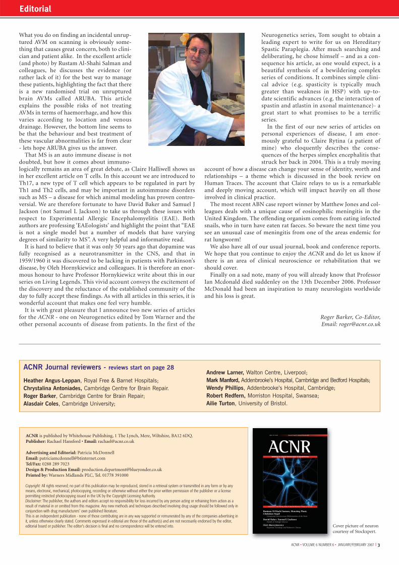
What you do on finding an incidental unrup-tured AVM on scanning is obviously some-thing that causes great concern, both to clini-cian and patient alike. In the excellent article(and photo) by Rustam Al-Shahi Salman andcolleagues, he discusses the evidence (orrather lack of it) for the best way to managethese patients, highlighting the fact that thereis a new randomised trial on unrupturedbrain AVMs called ARUBA. This articleexplains the possible risks of not treatingAVMs in terms of haemorrhage, and how thisvaries according to location and venousdrainage. However, the bottom line seems tobe that the behaviour and best treatment ofthese vascular abnormalities is far from clear- lets hope ARUBA gives us the answer.
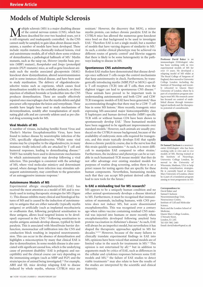
That MS is an auto immune disease is notdoubted, but how it comes about immuno-logically remains an area of great debate, as Claire Halliwell shows usin her excellent article on T cells. In this account we are introduced toTh17, a new type of T cell which appears to be regulated in part byTh1 and Th2 cells, and may be important in autoimmune disorderssuch as MS – a disease for which animal modeling has proven contro-versial. We are therefore fortunate to have David Baker and Samuel JJackson (not Samuel L Jackson) to take us through these issues withrespect to Experimental Allergic Encephalomyelitis (EAE). Bothauthors are professing ‘EAEologists’ and highlight the point that “EAEis not a single model but a number of models that have varyingdegrees of similarity to MS”. A very helpful and informative read.
It is hard to believe that it was only 50 years ago that dopamine wasfully recognised as a neurotransmitter in the CNS, and that in1959/1960 it was discovered to be lacking in patients with Parkinson’sdisease, by Oleh Hornykiewicz and colleagues. It is therefore an enor-mous honour to have Professor Hornykiewicz write about this in ourseries on Living Legends. This vivid account conveys the excitement ofthe discovery and the reluctance of the established community of theday to fully accept these findings. As with all articles in this series, it iswonderful account that makes one feel very humble.
It is with great pleasure that I announce two new series of articlesfor the ACNR - one on Neurogenetics edited by Tom Warner and theother personal accounts of disease from patients. In the first of the
Neurogenetics series, Tom sought to obtain aleading expert to write for us on HereditarySpastic Paraplegia. After much searching anddeliberating, he chose himself – and as a con-sequence his article, as one would expect, is abeautiful synthesis of a bewildering complexseries of conditions. It combines simple clini-cal advice (e.g. spasticity is typically muchgreater than weakness in HSP) with up-to-date scientific advances (e.g. the interaction ofspastin and atlastin in axonal maintenance)- agreat start to what promises to be a terrificseries.
In the first of our new series of articles onpersonal experiences of disease, I am enor-mously grateful to Claire Rytina (a patient ofmine) who eloquently describes the conse-quences of the herpes simplex encephalitis thatstruck her back in 2004. This is a truly moving
account of how a disease can change your sense of identity, worth andrelationships – a theme which is discussed in the book review onHuman Traces. The account that Claire relays to us is a remarkableand deeply moving account, which will impact heavily on all thoseinvolved in clinical practice.
The most recent ABN case report winner by Matthew Jones and col-leagues deals with a unique cause of eosinophilic meningitis in theUnited Kingdom. The offending organism comes from eating infectedsnails, who in turn have eaten rat faeces. So beware the next time yousee an unusual case of meningitis from one of the areas endemic forrat lungworm!
We also have all of our usual journal, book and conference reports.We hope that you continue to enjoy the ACNR and do let us know ifthere is an area of clinical neuroscience or rehabilitation that weshould cover.
Finally on a sad note, many of you will already know that ProfessorIan Mcdonald died suddenley on the 13th December 2006. ProfessorMcDonald had been an inspiration to many neurologists worldwideand his loss is great.
Roger Barker, Co-Editor, Email: [email protected]

4 I ACNR • VOLUME 6 NUMBER 6 • JANUARY/FEBRUARY 2007
Contents4 Editorial
6 Review ArticleUnruptured Arteriovenous Malformations of the BrainRustam Al-Shahi Saman, Henning Mast, Christian Stapf
8 Immunology PrimerImmunology News: a New Type of T Cell is DiscoveredClaire Helliwell
10 Review ArticleModels of Multiple SclerosisDavid Baker, Samuel J Jackson
14 Living LegendsDopamine, Levodopa and Parkinson’s DiseaseOleh Hornykiewicz
16 Neurogenetics ArticleHereditary Spastic ParaplegiaTom Warner
18 Personal Experiences ofNeurological DisordersMy Life Post HSAClaire Rytina
20 Case Report - ABN WinnerEosinophillic Meningitis Due to AngiostrongylusCantonensis: First Reported Case Matthew Jones, Rajiv Mohanraj, Sandip Shaunak
22 Conference News7th International Congress of Neuroimmunology;75th Anniversary Celebrations of the Associationof British Neurologists;17th Meeting of the European NeurologicalSociety.
24 Events Diary
25 Courses & Conferences
28 Journal Reviews
30 Book ReviewsStroke Treatment and Prevention: an Evidence-based Approach;Human Traces.
31 News
January/February 2007
Contents
Deadlines:January/February - 5 DecemberMarch/April - 5 February May/June - 5 AprilJuly/August - 5 JuneSeptember/October - 5 AugustNovember/December - 5 October
Letters to the Editor
Have your say about issues relating to your specialty, or in response to articles which appear inthe magazine. Send your news and views to ACNR:[email protected]
NEW
NEW

ABBREVIATED PRESCRIBING INFORMATION(Please consult the Summary of Product Characteristics (SPC) before prescribing.)KEPPRA® film-coated tablets 250 mg, 500 mg, 750 mg, 1000 mg KEPPRA® 100 mg/ml oral solutionKEPPRA® 100 mg/ml concentrate for solution for infusionActive Ingredient: Tablets: levetiracetam 250, 500, 750 and 1,000 mg. Oral Solution: levetiracetam 100 mg per ml. Infusion levetiracetam 100 mg per ml. Uses: Monotherapy for partial onset seizures with or without secondary generalisation in patients from 16 years of age with newly diagnosed epilepsy. Adjunctive therapy for partial onset seizures with or without secondary generalisation in adults and children from 4 years of age and for myoclonic seizures in adults and adolescents from 12 years of age with Juvenile Myoclonic Epilepsy. Infusion: an alternative for patients when oral administration is temporari ly not feasible. Dosage and Administrat ion: Oral solution should be diluted prior to use. Infusion: Keppra concentrate must be diluted in at least 100 ml of a compatible diluent and administered intravenously as a 15-minute infusion. Monotherapy (adults and adolescents from 16 years): Recommended starting dose of 250 mg twice daily which should be increased to an initial therapeutic dose of 500 mg twice daily after two weeks. The dose can be further increased by 250 mg twice daily every two weeks depending upon the clinical response. The maximum dose is 1500 mg twice dai ly. Adjunctive therapy: Adults and adolescents older than 12 years or weighing 50 kg or more: 500 mg twice daily can be increased up to 1,500 mg twice daily. Dose changes can be made in 500 mg twice daily increases or decreases every two to four weeks. Elderly: Adjustment of the dose is recommended in patients with compromised renal function. Children aged 4 to 11 years and adolescents (12 to 17 years) of less than 50 kg: 10 mg/kg twice daily, increased up to 30 mg/kg twice daily. Do not exceed increases or decreases of 10 mg/kg twice daily every two weeks. The lowest effective dose should be used. (For full dosage recommendations see SPC.) Patients with renal impairment: Adjust dose according to creatinine clearance as advised in SPC. Patients with hepatic impairment: No dose adjustment with mild to moderate hepatic impairment. With severe hepatic impairment (creatinine clearance <70ml/min) a 50% reduction of the daily maintenance dose is recommended. Contraindications, Warnings etc.: Contraindications: Hypersensitivity to levetiracetam, other pyrrolidone derivatives or excipients. Precautions: If discontinuing treatment reduce dose gradually as advised in SPC. Due to its excipients, the oral solution may cause allergic reactions (possibly delayed). Infusion: Keppra concentrate
contains 7.196 mg of sodium per vial. To be taken into consideration by patients on a controlled sodium diet. Interactions: Keppra did not affect serum concentrations of phenytoin, carbamazepine, valproic acid, phenobarbital, lamotrigine, gabapentin or primidone. Drugs excreted by active tubular secretion could reduce the renal clearance of the metabolite. Levetiracetam 1,000 mg daily did not affect the pharmacokinetics of oral contraceptives (ethinyl-estradiol and levonorgestrel). Levetiracetam 2,000 mg daily did not affect the pharmacokinetics of digoxin and warfarin and prothrombin times were not modified. Pregnancy and lactation: Should not be used during pregnancy unless clearly
necessary. Breast-feeding not recommended. Driving, etc: Caution recommended when performing skilled tasks, e.g. driving vehicles or operating machinery. Adverse Effects: Incidence of undesirable effects considered to be at least poss ib ly re la ted in controlled clinical studies: Very common (≥10%): a s t h e n i a / f a t i g u e , somnolence. Common (between 1%–10%): GI disturbances, anorexia, weight increase, accidental injury, headache, dizziness, hyperkinesia, tremor, ataxia, convulsion, amnesia, balance disorder, d isturbances in a t tent ion, memory impairment, emotional lability, hostility, depression, insomnia, nervousness/i r r i tabi l i ty, agi ta t ion, personal i ty disorders, thinking abnormal, vertigo, rash, eczema, pruritus, diplopia, vision blurred, m y a l g i a , i n f e c t i o n , nasopharyngitis, cough increased, thrombocytopenia. Consult SPC in relationto o ther s ide e f fec ts . P h a r m a c e u t i c a l Precautions: Tablets: None. Oral solution: Store in original container. After first opening use within 2 months. Infusion: Use immediately after dilution. Legal Category: POM. Marketing Authorisation Numbers: 250 mg x 60 tabs: EU/1/00/146/004. 500 mg x 60 tabs: EU/1/00/146/010. 750 mg x 60 tabs: EU/1/00/146/017. 1,000 mg x 60 tabs: EU/1/00/146/024. So lu t ion x 300 ml : EU/1/146/027, Infusion (500 mg/5 ml) x 10 vials: EU/1/00/146/030. NHS Cost: 250 mg x 60 tabs: £29.70. 500 mg x 60 tabs: £52.30. 750 mg x 60 tabs: £89.10. 1,000 mg x 60 tabs: £101.10. Solution x 300 ml: £71.00, Infusion (500 mg/ 5ml) x 10 vials: £135.00. Name and Address of PL Holder: UCB S.A., Allée de la Recherche 60, B-1070 Bruxelles, Belgium. Further information is
available from: UCB Pharma Ltd., 208 Bath Road, Slough, Berkshire, SL1 3WE. Tel: 01753 534655.Fax: 01753 536632. Email: [email protected] Date of Revision: August 2006Information about adverse event reporting can be found at www.yellowcard.gov.uk Adverse events should also be reported to UCB Pharma Ltd© 2006 UCB Pharma Ltd.® Keppra is a registered trade mark of UCB Pharma Ltd.Printed in the UK Date of preparation: September 2006. 06KP0194b
Now indicated as monotherapy* in epilepsy.
Start now
* For partial onset seizures with or without secondary generalisation in patients from 16 years old with newly diagnosed epilepsy.
KEPP6042 AdcCnr A4.indd 1KEPP6042 AdcCnr A4.indd 1 14/9/06 7:46:36 pm14/9/06 7:46:36 pm
6 I ACNR • VOLUME 6 NUMBER 6 • JANUARY/FEBRUARY 2007
Unruptured, asymptomatic arteriovenous malfor-mations (AVMs) lurk in the brains of approxi-mately one person in every thousand; their
prevalence, based on four studies of magnetic resonanceimaging (MRI) of 7,359 people without brain disor-ders,1-4 was 0.1 % (95% confidence interval [CI] 0% to0.2%). Some of these brain AVMs may be discovered ifand when they cause intracranial haemorrhage, epilep-tic seizure(s), headache, or a focal neurological deficit,but many brain AVMs may potentially lie dormant fromthe cradle to the grave.
The detection of this reservoir of unruptured brainAVMs is likely to depend on the differences in availabil-ity, uptake, and indications for brain MRI betweencountries. Indirect evidence for this comes from the twoongoing population-based studies of the clinical epi-demiology of brain AVMs:5,6 in Scotland 54% of allbrain AVMs detected in an incidence study were unrup-tured at presentation,5 whereas this proportion was 62%in New York (difference -8%, 95% CI -19% to 4%).6 Thedetection rate of unruptured brain AVMs seems set torise with the increasing appropriate use of brain MRIfor investigating epilepsy and stroke, as well as moreindiscriminate uses such as ‘health check-ups’ pur-chased from private health screening companies.7
What’s the prognosis for an adult with an unruptured brain AVM?Only a few published studies are of sufficient quality toprovide reliable estimates of the prognosis for unrup-tured brain AVMs.8-10 Most cohorts have been small, ret-rospective, hospital-based, with short incomplete fol-low-up from an unclear inception point, using un-blinded assessment according to bespoke rather thangeneric outcome measures, without stratification by dif-ferences in treatment. Even in high quality studies, theoutcome described for unruptured brain AVMs that arenot treated is inevitably biased, since a conservativestrategy may be adopted either because of the ‘untreata-bility’ of the AVM, or due to the patient’s burden of dis-ability or co-morbidity.
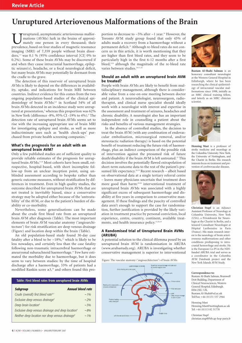
Nevertheless, some generalisations can be madeabout the crude first bleed rate from an unrupturedbrain AVM after diagnosis (Table). The most importantelements of brain AVM vascular anatomy (‘angioarchi-tecture’) for risk stratification are deep venous drainage(Figure) and location deep within the brain (Table).
An old population-based study found 30-day casefatality after a bleed to be ~18%,12 which is likely to beless nowadays, and certainly less than the case fatalityfollowing non-traumatic intracerebral haemorrhage oraneurysmal subarachnoid haemorrhage.13 Few have esti-mated the morbidity due to haemorrhage, but it doesseem to vary between studies: by the time of hospitaldischarge after a haemorrhage, 33% of patients had amodified Rankin score ≥3,10 and others found this pro-
portion to decrease to ~5% after ~1 year.14 However, theToronto AVM study group found that only 45% ofadults made a recovery from a haemorrhage without apermanent deficit.15 Although re-bleed rates do not con-cern us in this article, it is worth mentioning that theyare higher than first bleed rates, and they seem to beparticularly high in the first 6-12 months after a firstbleed,9,10 although the magnitude of the re-bleed ratevaries between studies.10,11,16
Should an adult with an unruptured brain AVMbe treated?People with brain AVMs are likely to benefit from mul-tidisciplinary management, although there is consider-able value from a one-on-one meeting between doctorand patient. A neuroradiologist, neurosurgeon, radio-therapist, and clinical nurse specialist should ideallywork with a neurologist with interest and expertise inthe assessment and treatment of seizures, headaches andchronic disability. A neurologist also has an importantindependent role in counselling a patient about therisks and benefits of various management strategies.
In the absence of controlled studies, the decision totreat the brain AVM (with any combination of endovas-cular embolisation, microsurgical removal, and/orstereotactic radiation therapy) is based on the potentialbenefit of treatment reducing the future risk of haemor-rhage, plus an indirect comparison of the possible riskof intervention against the presumed risk of futuredeath/disability if the brain AVM is left untreated.17 Thisdecision involves the potentially flawed extrapolation ofshort-term outcome data to the rest of the patient’s pre-sumed life expectancy.18,19 Recent research – albeit basedon observational data at a single tertiary referral centre– leaves many physicians uncertain that treatment doesmore good than harm:19,20 interventional treatment ofunruptured brain AVMs was associated with a highlysignificant excess of subsequent haemorrhage and dis-ability at five years in comparison to conservative man-agement. If these findings and the paucity of controlleddata aren’t enough to support the case for randomisa-tion, further justification is provided by the likely vari-ation in treatment practice by personal conviction, localexperience, centre, country, continent, available treat-ments, and health insurance policy.13,18,19
A Randomised trial of Unruptured Brain AVMs(ARUBA)A potential solution to the clinical dilemma posed by anunruptured brain AVM is randomisation in ARUBA(www.arubastudy.org). ARUBA is investigating whetherconservative management is superior to interventional
Unruptured Arteriovenous Malformations of the Brain
Review Article
Rustam Al-Shahi Salman is anhonorary consultant neurologistat the Western General Hospital inEdinburgh, where he has beenresearching the clinical epidemiol-ogy of intracranial vascular mal-formations since 1998, initially asan MRC clinical training fellow,and latterly as an MRC clinicianscientist.
Henning Mast is a professor ofstroke medicine and neurology atthe Columbia University New York,the University of Nottingham, andthe Charité in Berlin. His researchinterests focus on treatment and pre-vention of cerebrovascular diseases.
Christian Stapf is an AdjunctAssistant Professor of Neurology atColumbia University, New York(USA), a Privatdozent für Neuro -logie at the Charité in Berlin, and atenured Consultant Neurologist atHôpital Lariboisière in Paris(France). His main research inter-est is the neurology of brain arteri-ovenous malformations and otherconditions predisposing to intra -cranial hemorrhage and stroke. Heis the European Co-PI in the NIH-funded ARUBA trial and serves asa coordinator in the ColumbiaAVM Databank project and theNew York Islands AVM Study.
Correspondence to:Rustam Al-Shahi Salman, BramwellDott Building, Department ofClinical Neurosciences, WesternGeneral Hospital, Edinburgh. EH4 2XU. UK. [email protected]/Fax: +44 (0)131 537 2944
Henning [email protected]: +44 (0)115 82 31778
Christian [email protected]
Subgroup Annual bleed rate
Crude (overall) first bleed rate9,11 ~1%
Exclusive deep venous drainage9 ~2%
Deep brain location9 ~3%
Exclusive deep venous drainage and deep location9 ~8%
Neither deep location nor deep venous drainage9 ~1%
Table: First bleed rates from unruptured brain AVMs
Figure: The vascular anatomy (‘angioarchitecture’) of brain AVMs

ACNR • VOLUME 6 NUMBER 6 • JANUARY/FEBRUARY 2007 I 7
treatment for consenting adults aged ≥18years, with an unruptured brain AVM that ispotentially treatable, over a minimum follow-up period of five years, based on outcomeassessments by a neurologist. Although enrol-ment in this trial may prove challengingbecause of the instinctive difficulties somepatients (and doctors) may face in allowing
randomisation to decide whether their ‘timebomb’ is treated or not, fully informed con-sent – involving a sanguine discussion of therisks of intervention – is crucial.
ARUBA is funded by the National Institutesof Health (ISRCTN 44013133), with per-patient reimbursement. The trial has ethicalapproval in the UK, and a decision about trial
adoption by the UK Stroke Research Networkis pending. Interested investigators shouldcontact any of the authors, and may also con-sider participating in a similar trial randomis-ing people with an unruptured intracranialaneurysm to endovascular or conservativemanagement (www.teamstudy.org, ISRCTN62758344).
References
1. Weber F, Knopf H. Incidental findings in magnetic reso-nance imaging of the brains of healthy young men. Journalof the Neurological Sciences 2006;240(1-2):81-4.
2. Katzman GL, Dagher AP, Patronas NJ. Incidental find-ings on brain magnetic resonance imaging from 1000asymptomatic volunteers. JAMA 1999;282(1):36-9.
3. Yue NC, Longstreth WT Jr, Elster AD, Jungreis CA,O'Leary DH, Poirier VC. Clinically serious abnormali-ties found incidentally at MR imaging of the brain: datafrom the Cardiovascular Health Study. Radiology1997;202(1):41-6.
4. Illes J, Rosen AC, Huang L, Goldstein RA, Raffin TA,Swan G et al. Ethical consideration of incidental find-ings on adult brain MRI in research. Neurology2004;62(6):888-90.
5. Al-Shahi R, Bhattacharya JJ, Currie DG,Papanastassiou V, Ritchie V, Roberts RC et al.Prospective, Population-Based Detection of IntracranialVascular Malformations in Adults: The ScottishIntracranial Vascular Malformation Study (SIVMS).Stroke 2003;34(5):1163-9.
6. Stapf C, Mast H, Sciacca RR, Berenstein A, Nelson PK,Gobin YP et al. The New York Islands AVM Study:Design, Study Progress, and Initial Results. Stroke2003;34(5):E29-E33.
7. Al-Shahi Salman R, Whiteley WN, Warlow C.Screening using whole body MRI scanning: who wantsan incidentaloma? Journal of Medical Screening. Inpress 2007.
8. Al-Shahi R, Warlow C. A systematic review of the fre-quency and prognosis of arteriovenous malformations ofthe brain in adults. Brain 2001;124(Pt 10):1900-26.
9. Stapf C, Mast H, Sciacca RR, Choi JH, Khaw AV,Connolly ES et al. Predictors of hemorrhage in patientswith untreated brain arteriovenous malformation.Neurology 2006;66(9):1350-5.
10. Halim AX, Johnston SC, Singh V, McCulloch CE,Bennett JP, Achrol AS et al. Longitudinal risk ofintracranial hemorrhage in patients with arteriovenousmalformation of the brain within a defined population.Stroke 2004;35(7):1697-702.
11. Al-Shahi R, Vousden C, Warlow C. ScottishIntracranial Vascular Malformation Study (SIVMS)Steering Committee. Bias from requiring explicit con-sent from all participants in observational research:prospective, population based study. BMJ2005;331(7522):942-5.
12. Brown Jr RD, Wiebers DO, Torner JC, O'Fallon WM.Incidence and prevalence of intracranial vascular mal-formations in Olmsted County, Minnesota, 1965 to1992. Neurology 1996;46(4):949-52.
13. Al-Shahi R, Stapf C. The prognosis and treatment ofarteriovenous malformations of the brain. PracticalNeurology 2005;5:194-205.
14. Hartmann A, Mast H, Mohr JP, Koennecke HC,Osipov A, Pile-Spellman J et al. Morbidity of intracra-nial hemorrhage in patients with cerebral arteriovenousmalformation. Stroke 1998; 29(5):931-4.
15. Porter PJ, terBrugge KG, Montanera W, Kerr RG,Stefani MA, Willinsky RA et al. Outcome followinghaemorrhage from brain arteriovenous malformationsat presentation and during follow up: is it worse thanwe think? [abstract]. Journal of Neurosurgery1998;88:184A-5A.
16. Mast H, Young WL, Koennecke H-C, Sciacca RR,Osipov A, Pile-Spellman J et al. Risk of spontaneoushaemorrhage after diagnosis of cerebral arteriovenousmalformation. Lancet 1997;350(9084):1065-8.
17. Brown Jr RD, Kondziolka D. Simple risk predictions forarteriovenous malformation hemorrhage. Neurosurgery2000;46(4):1024.
18. Al-Shahi R, Warlow C. Arteriovenous malformations ofthe brain: ready to randomise? J Neurol NeurosurgPsychiatry 2005;76(10):1327-9.
19. Stapf C, Mohr JP, Choi JH, Hartmann A, Mast H.Invasive treatment of unruptured brain arteriovenousmalformations is experimental therapy. Curr OpinNeurol 2006;19(1):63-8.
20. Mohr JP, Stapf C, Sciacca RR, Khaw AV, Mast H,Connolly ES et al. Natural history versus treatmentoutcome in patients with unruptured brain arteriove-nous malformation (AVM). Stroke 2004:35:328.[Abstract]
Review Article
Roger Barker is co-editor of ACNR, and is Honorary Consultant inNeurology at The Cambridge Centre for Brain Repair. His mainarea of research is into neurodegenerative and movement disor-ders, in particular parkinson's and Huntington's disease. He is alsothe university lecturer in Neurology at Cambridge where he con-tinues to develop his clinical research into these diseases alongwith his basic research into brain repair using neural transplants.
Alasdair Coles is co-editor of ACNR. He has recently beenappointed to the new position of University Lecturer inNeuroimmunology at Cambridge University. He works on exper-imental immunological therapies in multiple sclerosis.
Stephen Kirker is the editor of the Rehabilitation section ofACNR and Consultant in Rehabilitation Medicine inAddenbrooke's NHS Trust, Cambridge. He trained in neurologyin Dublin, London and Edinburgh before moving to rehabilitationin Cambridge and Norwich. His main research has been into pos-tural responses after stroke. His particular interests are in pros-thetics, orthotics, gait training and neurorehabilitation.
David J Burn is the editor of our conference news section andConsultant and Reader in Movement Disorder Neurology at theRegional Neurosciences Centre, Newcastle-upon-Tyne. He runsMovement Disorders clinics in Newcastle-upon-Tyne. Researchinterests include progressive supranuclear palsy and dementiawith Lewy bodies. He is also involved in several drugs studies forParkinson's Disease.
Andrew Larner is the editor of our Book Review Section. He is aConsultant Neurologist at the Walton Centre for Neurology andNeurosurgery in Liverpool, with a particular interest in dementiaand cognitive disorders. He is also an Honorary Apothecaries'Lecturer in the History of Medicine at the University of Liverpool.
Alastair Wilkins is our Case Report Co-ordinator. He is Senior Lecturer inNeurology and Consultant Neurologist, University of Bristol. He trained inNeurology in Cambridge, Norwich and London. His research interests are thebasic science of axon degeneration and developing treatments for progressivemultiple sclerosis.
Roy O Weller is ACNR’s Neuropathology Editor. He is Emeritus Professor ofNeuropathology, University of Southampton. His particular research interestsare in the pathogenesis of Multiple Sclerosis, Alzheimer’s disease and CerebralAmyloid Angiopathy.
International editorial liaison committeeProfessor Riccardo Soffietti, Italy: Chairman of the Neuro-Oncology Service,Dept of Neuroscience and Oncology, University and S. Giovanni BattistaHospital, Torino, Italy. President of the Italian Association of Neuro-Oncology,member of the Panel of Neuro-Oncology of the EFNS and EORTC BrainTumour Group, and Founding member of the EANO (European Associationfor Neuro-Oncology).
Professor Klaus Berek, Austria: Head of the Neurological Department of theKH Kufstein in Austria. He is a member of the Austrian Societies of Neurology,Clinical Neurophysiology, Neurological and Neurosurgical Intensive CareMedicine, Internal and General Intensive Care Medicine, Ulrasound inMedicine, and the ENS.
Professor Hermann Stefan, Germany: Professor of Neurology / Epileptologyin the Department of Neurology, University Erlangen-Nürnberg, andspecialis-es in the treatment of epilepsies, especially difficult to treat types of epilepsyand presurgical evaluation, including Magnetic source imaging (MEG/EEG)and MR-Spectroscopy.
Professor Nils Erik Gilhus, Norway: Professor of Neurology at the University ofBergen and Haukeland University Hospital. Research Dean at the medical facul-ty, and Chairman for the Research Committee of the Norwegian MedicalAssociation. He chairs the scientist panel of neuroimmunology, EFNS, is a mem-ber of the EFNS scientific committee, the World Federation of Neurorehabilitationcouncil, and the European School of Neuroimmunology board. His mainresearch interests are neuroimmunology and neurorehabilitation.
Editorial Board and contributors

8 I ACNR • VOLUME 6 NUMBER 6 • JANUARY/FEBRUARY 2007
Immunology is a young, fast-moving, discipline.Today’s dogma is often disproved tomorrow. Butmost people would have thought that the funda-
mental division of helper cells into ‘Th1’ and ‘Th2’ wasconclusive. Not so, it turns out.
The DogmaT helper (Th) cells are an important part of the adaptiveimmune system. They express T cell receptors (TCRs)that recognise a specific protein bound to class II MHCmolecules and activation causes cytokine release. Theyare important in the defence against microbes but alsoinduce inflammation in immune-mediated diseases. Inthe 1980s, Mosmann showed that CD4+ T lymphocytescould be divided into ‘Th1’ and ‘Th2’.1
• The cytokine interleukin-12 (IL-12) promotes thedevelopment of Th1 cells, which secrete IFN-γ, IL-2and TNF-β (lymphotoxin); these drive cell-mediatedimmunity to eliminate intracellular pathogens.
• In contrast, T cells stimulated in the presence of IL-4turn into Th2 cells which secrete more IL-4, IL-5, IL-10 and IL-13, and up-regulate antibody-mediatedresponses for elimination of extracellular pathogens
Recently, regulatory T cells (Tregs) have been describedwhich are thought to inhibit unwanted immuneresponses to self antigens. When this regulation fails,autoimmune disease results. Multiple sclerosis wasthought to be a classic example of a disease driven byTh1 cells, whereas allergy was due to excessive Th2cytokine production.
A major plank of evidence for all of this came fromExperimental Autoimmune Encephalomyelitis (EAE).For instance, studies with IL-12 knock out mice (IL-12-/-),2 or using IL-12p40 neutralising antibodies,2,3 haveshown that IL-12 is necessary for disease expression;hence Th1 cells and IFN-γ drive EAE. All very tidy.
The problemAccording to all of this, mice which lack certain criticalcomponents of the Th1–IFN-γpathway (IFN- –/– 4 , IFN-R–/– 5 , IL-12Rβ2–/– 6 , and IL-12p35–/–7 mice) should notget EAE. Unfortunately however, they do.
The solutionThe first step in sorting all this out was the finding thatthe subunit p40 is shared by both IL-12 and a newly-dis-covered cytokine called IL-23.8 IL-23 is secreted by acti-vated dendritic cells and stimulates IFN-γ productionand proliferation of blast T cells and memory T cells.8
Becher et al7 showed conclusively that mice deficient inthe specific p35 subunit of IL-12 (p35-/-) develop severeEAE whereas those deficient to the common p40 sub-unit (p40-/-) were resistant to EAE. So IL-12 is notresponsible for EAE.
Daniel Cua, from Schering-Plough Biopharma, rea-soned that all the p40 knock-out experiments wereflawed and that deficits attributable to IL-12 deficiencymay have actually been due to lack of IL-23. His teamproved this by manipulating mice cells in vitro and see-ing if they induced EAE on transfer to naïve mice. Theresult was clear: T cells cultured in vitro with IL-23, butnot IL-12, caused severe clinical signs of EAE on trans-fer (Figure 1).9 In IL-23 deficient mice, Th1 cells invad-ed the CNS, but did not cause disease. So IL-12 and Th1cells do not drive EAE! This is a major paradigm shift….
What sort of T cells does IL-23 induce?CD4+ T cells from IL-23p19–/– knockout mice are
specifically unable to produce IL-17.10 So the thinking isthat IL-23 induces a new brand of helper T cells called‘Th17’ cells characterised by their production of IL-17.IL-17 has not had much of a press until now. It inducesthe secretion of pro-inflammatory cytokines tumournecrosis factor (TNF), IL-1 and IL-6 frommacrophages.11 IL-17 also induces production of IL-6,IL-8, prostaglandin E2 and granulocyte colony-stimulat-ing factor (GCSF) from rheumatoid synovial fibroblastsand IL-6 from a variety of stromal cells.12 Anti-IL-17treatment of wild type mice immunised with myelinprotein PLP are partially protected against EAE.9
How do Th17 cells develop?As well as IL-23, TGF-β1 is important in the develop-ment of Th17 cells. Mice over-expressing TGF-β1 hadincreased numbers of Th17 cells and worse autoim-mune disease.13 Th17 differentiation is inhibited by theproducts of Th1 and Th2 cells, IFN-γ and IL-4 respec-tively.14 Development of Th17 cells is promoted by thecombination of transforming growth factor (TGF-β1)and IL-6.13,15 These cytokines are produced by manycells. TGF-β1 alone induces the differentiation certainsubsets of Treg cells.16 When TGF-β1 is combined withIL-6 it inhibits the expression of FoxP3, a gene tran-scription factor essential for Treg development, thuspromoting Th17 and suppressing Treg cell develop-ment.13,15 In the steady state TGF-β1 will induce FoxP3+Tregs and maintain self-tolerance. When there is infec-tion or inflammation, IL-6 produced by the innateimmune system will suppress Tregs cells and inducepro-inflammatory response by Th17. So now, a schemelike this can be drawn (Figure 2).
Th17 cells drive autoimmunity and cancerSerum IL-17 is raised in patients with MS,17 SLE,18 asth-ma19 and in RA synovium.20 It has been shown that thereis increased IL-23 secretion from monocyte deriveddendritic cells from MS patients compared to healthycontrols and that there is increased IL-17 production by
Immunology News: A New Type of T Cell Is Discovered
Immunology Primer
Dr Claire Helliwell,Honorary Clinical Fellow,Deptartment of ClinicalNeurosciences,Addenbrooke's Hospital,Cambridge, UK.Email: [email protected]
Figure 1 shows CD4+ T cells cultured in vitro with IL-23 (■), but not IL-12 (▲) induce EAEpathogenesis, Langrish et al.9
3
2
1
06 10 14 18 22 26 30
Days post cell transfer
IL-23 CD4+
IL-12 CD4+
EA
E s
core

ACNR • VOLUME 6 NUMBER 6 • JANUARY/FEBRUARY 2007 I 9
stimulated CD4+ T cells from MS patients.2
IL23 may also play an important role intumours as IL-23p19 mRNA expression hasbeen shown to be increased in a variety ofhuman tumours.22 One mechanism for thismay be that IL-23 reduces the ability of CD8+T cells to infiltrate tumours as shown inmice.22 At present a clear role for the IL-23/IL-17 pathway in response to infection has notbeen identified. IL-23 knock-out mice are lessprone to some infections (tuberculosis andtoxoplasmosis) than IL-12 knock-outs,10,23 sug-gesting that it may not play an important role.
SummaryNewly described Th17 cells which produce IL-17 and are expanded in the presence of IL-23are likely to have an important role in thepathogenesis of autoimmune diseases andpossibly some cancers. Work done with micehas shown that EAE is prevented by the use ofanti-IL23p19 antibodies24 and that anti-IL 17antibodies give partial protection.9 In theorythe selective neutralisation of the IL-23/IL-17immune pathway (with an IL-23p19 or IL-17antibody) might reduce autoimmunity, yethave little detriment on the immune responseto infection.
References
1. Mosmann TR, Cherwinski H, Bond MW, Giedlin MA,and Coffman RL. Two types of murine helper T cellclone. I. Definition according to profiles of lymphokineactivities and secreted proteins. J. Immunol.1986;136:2348.
2. Segal BM, Dwyer BK, and Shevach EM. An interleukin(IL)-10/IL-12 immunoregulatory circuit controls suscep-tibility to autoimmune disease. J. Exp. Med.1998;187:537.
3. Leonard JP, Waldburger KE, and Goldman SJ.Prevention of experimental autoimmuneencephalomyelitis by antibodies against interleukin 12.J. Exp. Med. 1995;181:381.
4. Ferber IA, Brocke S, Taylor-Edwards C, Ridgway W,Dinisco C, Steinman L, Dalton D, and Fathman CG.Mice with a disrupted IFN-gamma gene are susceptibleto the induction of experimental autoimmuneencephalomyelitis (EAE). J. Immunol. 1996;156:5.
5. Willenborg DO, Fordham S, Bernard CC, CowdenWB, and Ramshaw IA. IFN-gamma plays a criticaldown-regulatory role in the induction and effector phaseof myelin oligodendrocyte glycoprotein-induced autoim-mune encephalomyelitis. J. Immunol. 1996;157:3223.
6. Zhang GX, Gran B, Yu S, Li J, Siglienti I, Chen X,Kamoun M, and Rostami A. Induction of experimentalautoimmune encephalomyelitis in IL-12 receptor-beta 2-deficient mice: IL-12 responsiveness is not required inthe pathogenesis of inflammatory demyelination in thecentral nervous system. J. Immunol. 2003;170:2153.
7. Becher B, Durell BG, and Noelle RJ. Experimentalautoimmune encephalitis and inflammation in theabsence of interleukin-12. J. Clin. Invest 2002;110:493.
8. Oppmann B, Lesley R, Blom B, Timans JC, Xu Y,Hunte B, Vega F, Yu N, Wang J, Singh K, Zonin F,Vaisberg E, Churakova T, Liu M, Gorman D, Wagner J,Zurawski S, Liu Y, Abrams JS, Moore KW, Rennick D,de Waal-Malefyt R, Hannum C, Bazan JF, andKastelein RA. 2000. Novel p19 protein engages IL-12p40to form a cytokine, IL-23, with biological activities simi-lar as well as distinct from IL-12. Immunity.2000;13:715.
9. Langrish CL, Chen Y, Blumenschein WM, Mattson J,Basham B, Sedgwick JD, McClanahan T, Kastelein RA,and Cua DJ. IL-23 drives a pathogenic T cell populationthat induces autoimmune inflammation. J. Exp. Med.2005;201:233.
10. Khader SA, Pearl JE, Sakamoto K,Gilmartin L, BellGK, Jelley-Gibbs DM, Ghilardi N, deSauvage F, andCooper AM. IL-23 compensates for the absence of IL-12p70 and is essential for the IL-17 response duringtuberculosis but is dispensable for protection and anti-gen-specific IFN-gamma responses if IL-12p70 is avail-able. J. Immunol. 2005;175:788.
11. Jovanovic DV, Di Battista JA, Martel-Pelletier J,Jolicoeur FC, He Y, Zhang M, Mineau F, and PelletierJP. IL-17 stimulates the production and expression ofproinflammatory cytokines, IL-beta and TNF-alpha, byhuman macrophages. J. Immunol. 1998;160:3513.
12. Fossiez F, Djossou O, Chomarat P, Flores-Romo L, it-Yahia S, Maat C, Pin JJ, Garrone P, Garcia E, SaelandS, Blanchard D, Gaillard C, Das MB, Rouvier E,Golstein P, Banchereau J, and Lebecque S. T cell inter-leukin-17 induces stromal cells to produce proinflamma-tory and hematopoietic cytokines. J. Exp. Med.1996:183:2593.
13. Bettelli E, Carrier Y, Gao W, Korn T, Strom TB, OukkaM, Weiner HL, and Kuchroo VK. Reciprocal develop-mental pathways for the generation of pathogenic effec-tor TH17 and regulatory T cells. Nature 2006:441:235.
14. Harrington LE, Hatton RD, Mangan PR, Turner H,Murphy TL, Murphy KM, and Weaver CT. Interleukin17-producing CD4+ effector T cells develop via a lineagedistinct from the T helper type 1 and 2 lineages. Nat.Immunol. 2005:6:1123.
15. Veldhoen M, Hocking RJ, Atkins CJ, Locksley RM, andStockinger B. TGFbeta in the context of an inflammato-ry cytokine milieu supports de novo differentiation ofIL-17-producing T cells. Immunity. 2006:24:179.
16. Chen W, Jin W, Hardegen N, Lei KJ, Li L, Marinos N,McGrady G, and Wahl SM. Conversion of peripheralCD4+. J. Exp. Med. 2003:198:1875.
17. Matusevicius D, Kivisakk P, He B, Kostulas N, OzenciV, Fredrikson S, and Link H. Interleukin-17 mRNAexpression in blood and CSF mononuclear cells is aug-mented in multiple sclerosis. Mult. Scler. 1999:5:101.
18. Wong CK, Ho CY, Li EK, and Lam CW. Elevation ofproinflammatory cytokine (IL-18, IL-17, IL-12) andTh2 cytokine (IL-4) concentrations in patients with sys-temic lupus erythematosus. Lupus 2000:9:589.
19. Wong CK, Ho CY, Ko FW, Chan CH, Ho AS, Hui DS,and Lam CW. Proinflammatory cytokines (IL-17, IL-6,IL-18 and IL-12) and Th cytokines (IFN-gamma, IL-4,IL-10 and IL-13) in patients with allergic asthma. Clin.Exp. Immunol. 2001:125:177.
20. Chabaud M, Fossiez F, Taupin JL, and Miossec P.Enhancing effect of IL-17 on IL-1-induced IL-6 andleukemia inhibitory factor production by rheumatoidarthritis synoviocytes and its regulation by Th2cytokines. J. Immunol. 1998:161:409.
21. Vaknin-Dembinsky A, Balashov K, and Weiner HL. IL-23 is increased in dendritic cells in multiple sclerosis anddown-regulation of IL-23 by antisense oligos increasesdendritic cell IL-10 production. J. Immunol.2006:176:7768.
22. Langowski JL, Zhang X, Wu L, Mattson JD, Chen T,Smith K, Basham B, McClanahan T, Kastelein RA, andOft M. IL-23 promotes tumour incidence and growth.Nature 2006:442:461.
23. Lieberman LA, Cardillo F, Owyang AM, Rennick DM,Cua DJ, Kastelein RA, and Hunter CA. IL-23 providesa limited mechanism of resistance to acute toxoplasmosisin the absence of IL-12. J. Immunol. 2004:173:1887.
24. Chen Y, Langrish CL, McKenzie B, Joyce-Shaikh B,Stumhofer JS, McClanahan T, Blumenschein W,Churakovsa T, Low J, Presta L, Hunter CA, KasteleinRA, and Cua DJ. Anti-IL-23 therapy inhibits multipleinflammatory pathways and ameliorates autoimmuneencephalomyelitis. J. Clin. Invest 2006:116:1317.
Undifferentiated T helper cell
IL-12
IL-4
TGF-β1IL-23
IL-6
TGF-β1
IFN-γ1 Elimination of intracellular pathogens
Upregulation of antibody responses forelimination of extracellular pathogens,allergy & asthma
Autoimmunity, induction ofpro-inflammatory cytokines
Immunosuppression
Th1
Th2
Th17
Treg
IL-4
IL-17
TGF-β1
Immunology Primer
10 I ACNR • VOLUME 6 NUMBER 6 • JANUARY/FEBRUARY 2007
Multiple sclerosis (MS) is a major disabling diseaseof the central nervous system (CNS), which hasbeen described for over two hundred years, yet it
is still enigmatic and inadequately controlled.1 As the CNScannot easily be sampled, to gain ideas about disease mech-anisms, a number of models have been developed. Theseinclude: myelin mutants, chemically-induced lesions, viraland autoimmune models, all of which show some evidenceof demyelination, a pathological hallmark of MS.2 Myelinmutants, such as the taiep rat, Shiverer (myelin basic pro-tein (MBP) mutant), Rumpshaker and Jimpy (proteolipidprotein (PLP) mutants) mice, as well as gene knockout ani-mals such the myelin associated glycoprotein (MAG)knockout show dysmyelination, altered neurotransmissionand in some instances clinical disease, and have been usedto study myelination. The delivery of oligodendrocyte-selective toxins such as cuprizone, which causes focaldemyelination notably to the cerebellar peduncle, or directinjection of ethidium bromide or lysolecithin into the CNSproduces demyelination. These are usually effectivelyrepaired once macrophages clear the myelin debris and glialprecursor cells repopulate the lesion and remyelinate. Thesemodels have largely been used to study mechanisms ofde/remyelination, notably after transplantation of myeli-nating glial cells and are currently seldom used as pre-clin-ical drug screening tools for MS.
Viral Models of MSA number of viruses, including Semliki Forest Virus andTheiler’s Murine Encephalomyelitis Virus, have beenfound to induce disease by neurotrophic infection of theCNS, specifically oligodendrocytes. Whilst some viralstrains may be cytopathic to the oligodendrocyte, in manyinstances virally-infected cells are attacked by T cell andhumoral responses, leading to demyelinating disease.2,3
TMEV has notably been used to demonstrate mechanismsby which autoimmunity may develop following a viralinfection. This paradigm is consistent with the aetiologyof MS, where viral molecular mimicry and determinantspread, where damage from infection may stimulate sub-sequent autoimmunity, may contribute to the generationof an autoaggressive immune response.3
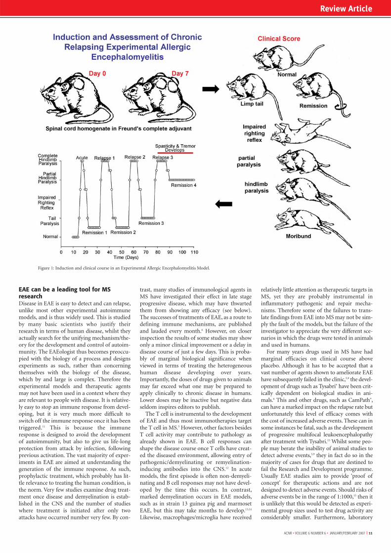
Autoimmune Models of MSExperimental allergic encephalomyelitis (EAE) hasreceived the most attention as a model of MS and is rou-tinely used in testing therapeutic strategies for MS (Figure1). This disease exhibits many clinical and histological fea-tures of MS and is caused by the induction of autoimmu-nity to antigens that are either naturally (typically myelinantigens) or artificially (such as implanted mycobacteriaor ovalbumin that, following peripheral sensitisation tothese antigens, allows local targeted lesions to be devel-oped) expressed in the CNS.2-4 Following sensitisation tomyelin antigens animals develop disease, typified by limbparalysis. This is associated with blood:brain barrier dys-function, mononuclear cell infiltration into the CNS andconduction block resulting in impaired neurotransmis-sion. This can occur in the absence of demyelination andhighlights a misconception by many that clinical EAE isdue to demyelination. In some models disease is also asso-ciated with significant axonal loss, which is the underlyingcause of persistent disability.2 EAE is polygenic and sus-ceptibility and the clinical course can vary depending onthe immunising antigen (such as MBP and PLP) and thestrain/species of animal being investigated.2,4 For example,ABH and SJL mice develop relapsing EAE to diseaseinduced by whole myelin, whereas C57BL/6 mice are
resistant.2 However, the discovery that MOG, a minormyelin protein, can induce chronic paralytic EAE in theC57BL/6 mice has allowed the numerous gene-knockoutmice bred on that background to be used to investigateEAE.2 Therefore EAE is not a single model, but a numberof models that have varying degrees of similarity to MS.2
As such, a similar clinical phenotype may be achieved viadifferent routes of genetic control2 and likewise suggeststhat there is likely to be some heterogeneity in the path-ways leading to disease in MS.
Spontaneous CNS autoimmunityRodent EAE studies have demonstrated that disease devel-ops once sufficient T cells escape the control mechanismsthat keep autoimmunity in check. Furthermore, by trans-genically introducing myelin (MBP, PLP or MOG)-specif-ic T cell receptors (TCR) into all T cells, then even theslightest trigger can lead to spontaneous CNS disease.2,4
These animals have proved to be important tools inunderstanding autoimmunity and both CD4+ and CD8+
TCR transgenic models of EAE have been generated,4 thusaccommodating thoughts that there may be a CD8+ T cellbias in some MS lesions.5 More recently, transgenic miceexpressing MS-associated major histocompatibility classII haplotypes and human derived myelin (MBP)-specificTCR with or without human CD4 have been shown tospontaneously develop EAE.7 These humanised modelshave been suggested to be significant improvements overstandard models.7 However, such animals are usually pro-duced on the C57BL/6 mouse background, because of theavailability of embryonic stem cells required for transgen-esis, and this strain typically develops EAE that rapidlyshows a chronic paralytic course, due to the nerve loss thatthis strain quickly accumulates.2,4 As such, it is more diffi-cult to manipulate EAE compared to other strains.2
Furthermore, the incidence and phenotype can be so vari-able in such humanised-TCR mouse models7 that they donot offer advantage over existing standard models forpurposes of routine drug screening, unless there is an apriori reason for testing agents that are specific for thesehuman components. Nevertheless, humanising models,such that they can accept MS-patient derived cells maylead to new tools for the future.7
Is EAE a misleading tool for MS research?MS appears to be a uniquely human condition and noother animal spontaneously develops a disease identicalto MS. Furthermore, it must be recognised that immuni-sation of mammals, including humans, with CNS pro-teins does not induce MS, but acute disseminatedencephalomyelitis. This was recognised over a centuryago when rabies vaccine containing residual CNS mate-rial was injected into humans or more recently whenencephalomyelitis developed following amyloid betaprotein vaccination in Alzheimer's disease.8 As such, EAEwill always be an imperfect model, but nevertheless it hasshaped the therapeutic approaches applied to MS fordecades.5,6,9,10 However, because of the many failures toclinically translate experimental findings in EAE intoMS, opinions have been voiced that animal models are oflimited value in the search for treatments in MS.5,9 Thisopinion is not entertained by all,6,10 but in addition toarguments made by critics of EAE, such as differences inthe cellular and cytokines responses between some EAEmodels and MS,5,9 the failure of EAE studies to detectviable treatments5,9 may also relate to how the results ofthe studies are interpreted by the scientific and clinicalfraternity.
Models of Multiple Sclerosis
Review Article
Professor David Baker is animmunologist (EAEologist) whohas been working with in vivoexperimental models of MS forover 15 years. He developed arelapsing model of MS whilst atThe Royal College of Surgeons ofEngland, then moved to UniversityCollege London including theInstitute of Neurology and recent-ly relocated to Queen MaryUniversity of London, where he ispart of a translation program aim-ing to treat MS. His research careerhas focused on control of estab-lished disease through immuno-logical methods and the therapeu-tic potential of cannabinoids.
Dr Samuel J Jackson is a neurosci-entist (EAEologist) who has beenworking with in vitro and in vivomodels of MS. Having trained atthe Institute of Neurology,University College London, hemoved to the University ofWisconsin, Madison, USA to helpdevelop remyelination strategies.He is currently based at QueenMary University of London, wherehe is part of a translation programaiming to treat progression in MS.
Correspondence to:David Baker and Samuel J JacksonNeuroimmunology Unit,Neuroscience Centre, Institute of Cell and MolecularScience,Barts and the London School ofMedicine, Queen Mary College London, 4 Newark Street, London, E1 2AT.Tel: 020 7882 2485Email: [email protected]
ACNR • VOLUME 6 NUMBER 6 • JANUARY/FEBRUARY 2007 I 11
EAE can be a leading tool for MSresearchDisease in EAE is easy to detect and can relapse,unlike most other experimental autoimmunemodels, and is thus widely used. This is studiedby many basic scientists who justify theirresearch in terms of human disease, whilst theyactually search for the unifying mechanism/the-ory for the development and control of autoim-munity. The EAEologist thus becomes preoccu-pied with the biology of a process and designsexperiments as such, rather than concerningthemselves with the biology of the disease,which by and large is complex. Therefore theexperimental models and therapeutic agentsmay not have been used in a context where theyare relevant to people with disease. It is relative-ly easy to stop an immune response from devel-oping, but it is very much more difficult toswitch off the immune response once it has beentriggered.11 This is because the immuneresponse is designed to avoid the developmentof autoimmunity, but also to give us life-longprotection from attack by infection, followingprevious activation. The vast majority of exper-iments in EAE are aimed at understanding thegeneration of the immune response. As such,prophylactic treatment, which probably has lit-tle relevance to treating the human condition, isthe norm. Very few studies examine drug treat-ment once disease and demyelination is estab-lished in the CNS and the number of studieswhere treatment is initiated after only twoattacks have occurred number very few. By con-
trast, many studies of immunological agents inMS have investigated their effect in late stageprogressive disease, which may have thwartedthem from showing any efficacy (see below).The successes of treatments of EAE, as a route todefining immune mechanisms, are publishedand lauded every month.6 However, on closerinspection the results of some studies may showonly a minor clinical improvement or a delay indisease course of just a few days. This is proba-bly of marginal biological significance whenviewed in terms of treating the heterogeneoushuman disease developing over years.Importantly, the doses of drugs given to animalsmay far exceed what one may be prepared toapply clinically to chronic disease in humans.Lower doses may be inactive but negative dataseldom inspires editors to publish.
The T cell is instrumental to the developmentof EAE and thus most immunotherapies targetthe T cell in MS.5 However, other factors besidesT cell activity may contribute to pathology asalready shown in EAE. B cell responses canshape the disease course once T cells have creat-ed the diseased environment, allowing entry ofpathogenic/demyelinating or remyelination-inducing antibodies into the CNS.12 In acutemodels, the first episode is often non-demyeli-nating and B cell responses may not have devel-oped by the time this occurs. In contrast,marked demyelination occurs in EAE models,such as in strain 13 guinea pig and marmosetEAE, but this may take months to develop.13,14
Likewise, macrophages/microglia have received
relatively little attention as therapeutic targets inMS, yet they are probably instrumental ininflammatory pathogenic and repair mecha-nisms. Therefore some of the failures to trans-late findings from EAE into MS may not be sim-ply the fault of the models, but the failure of theinvestigator to appreciate the very different sce-narios in which the drugs were tested in animalsand used in humans.
For many years drugs used in MS have hadmarginal efficacies on clinical course aboveplacebo. Although it has to be accepted that avast number of agents shown to ameliorate EAEhave subsequently failed in the clinic,5,9 the devel-opment of drugs such as Tysabri® have been crit-ically dependent on biological studies in ani-mals.6 This and other drugs, such as CamPath®,can have a marked impact on the relapse rate butunfortunately this level of efficacy comes withthe cost of increased adverse events. These can insome instances be fatal, such as the developmentof progressive multifocal leukoencephalopathyafter treatment with Tysabri.15 Whilst some peo-ple may berate the inability of animal studies todetect adverse events,5,9 they in fact do so in themajority of cases for drugs that are destined tofail the Research and Development programme.Usually EAE studies aim to provide ‘proof ofconcept’ for therapeutic actions and are notdesigned to detect adverse events. Should risks ofadverse events be in the range of 1:1000,15 then itis unlikely that this would be detected as experi-mental group sizes used to test drug activity areconsiderably smaller. Furthermore, laboratory
Review Article
Figure 1: Induction and clinical course in an Experimental Allergic Encephalomyelitis Model.

12 I ACNR • VOLUME 6 NUMBER 6 • JANUARY/FEBRUARY 2007
Review Article
animals are housed in environments free of animal pathogen, whichremoves the risk of infectious complications. However, when arrestingthe function of a significant portion of the immune system with potentimmunomodulatory drugs, it is not surprising that development ofinfection and tumours become more probable.
EAE and MS are not just about autoimmunityThe concept that MS is just a problem of autoimmunity has beenchampioned and directed by much EAE research, but immunothera-py has consistently failed in the clinic when progressive MS has beentargeted.1 Instead these studies suggest that while (auto)immunitymay drive blood:brain barrier dysfunction and relapsing disease, thisalso appears to create a CNS microenvironment that is permissive toneurodegenerative processes that are no longer dependent on, or sen-sitive to inhibitors, of autoimmunity.1 This has recently also beenshown to be the case in long-established EAE,11 which suggests thatmonotherapies solely targeting autoimmunity are insufficient to con-trol EAE, let alone MS.11 In addition, there is emerging evidence fromstudies employing chemical lesions or dysmyelinating genetic mutantsthat there is also ‘slow burning’ axonal loss in chronically demyelinat-ed tissue. This indicates that the autoimmune paradigm is insufficientto describe both progressive EAE and MS and may go some way toexplaining the clinical failures using anti-immunological therapies.Whilst aggressive immunotherapy early after diagnosis of MS may bedesirable, once sufficient damage has accumulated, the additional useof neuroprotective agents will be needed to treat progressive MS.Whilst EAE may be able to detect agents that inhibit immunity, wehave some way to go in determining which agents will inhibit(auto)immune-independent nerve loss and progression.
ConclusionExperimental models of human disease, whether directly relevant toMS or not, help one to understand the underlying biology. Withoutthese one would not have the base of knowledge to inform develop-ment of treatments towards the clinic. Importantly, they help providethe confidence to invest in the development of costly clinical trials thatmay ultimately lead to improvements in the lives of many people livingwith MS. It is important to realise that experimental models can onlygive sensible answers if we ask of them sensible and relevant questions!
References
1. Compston A & Coles A. Multiple sclerosis. Lancet. 2002;359:1221-31.
2. Lavi E & Constantinescu C (Eds). Experimental Models of Multiple Sclerosis. 2005Springer Science + Business Media, New York. ISBN 0-387-25517-6.
3. Ercolini AM & Miller SD. Mechanisms of immunopathology in murine models of central nervous system demyelinating disease. J Immunol. 2006; 176:3293-8.
4. Owens T. Animal models for multiple sclerosis. Adv Neurol. 2006;98:77-89.
5. Sriram S & Steiner I. Experimental allergic encephalomyelitis: a misleading model ofmultiple sclerosis. Ann Neurol. 2005;58:939-45.
6. Steinman L, Zamvil SS. How to successfully apply animal studies in experimental allergic encephalomyelitis to research on multiple sclerosis. Ann Neurol. 2006;60:12-21.
7. Friese MA, Montalban X, Willcox N, Bell JI, Martin R, Fugger L. The value of animalmodels for drug development in multiple sclerosis. Brain. 2006; 129:1940-52.
8. Broytman O, & Malter JS. Anti-Abeta: The good, the bad, and the unforeseen.J Neurosci Res. 2004;75:301-6.
9. Ransohoff RM. EAE: pitfalls outweigh virtues of screening potential treatments formultiple sclerosis. Trends Immunol. 2006;27:167-8.
10. Gold R, Linington C & Lassmann H. Understanding pathogenesis and therapy ofmultiple sclerosis via animal models: 70 years of merits and culprits in experimentalautoimmune encephalomyelitis research. Brain. 2006; 129:1953-71.
11. Pryce G, O'Neill JK, Croxford JL, Amor S, Hankey DJ, East E, Giovannoni G, &Baker D. Autoimmune tolerance eliminates relapses but fails to halt progression in amodel of multiple sclerosis. J Neuroimmunol. 2005; 165:41-52.
12. Martin Mdel P & Monson NL. Potential role of humoral immunity in the pathogene-sis of multiple sclerosis (MS) and experimental autoimmune encephalomyelitis (EAE).Front Biosci. 2007;12:2735-49.
13. Lassman H. (1983) Comparative neuropathology of chronic experimental allergicencephalomyelitis and multiple sclerosis. Springer Verlag Berlin Heidelberg.
14. Hart BA, Bauer J, Brok HP, Amor S. Non-human primate models of experimentalautoimmune encephalomyelitis: Variations on a theme. J Neuroimmunol. 2005;168:1-12.
15. Aksamit AJ. Review of progressive multifocal leukoencephalopathy and natalizumab.Neurologist. 2006;12:293-8.

mother: 365 days a yearms patient: 15 minutes every friday
puts time between injectionsputs time before progression
Prescribing information can be found on the adjacent page
7961 Avo JNNP A4.indd 17961 Avo JNNP A4.indd 1 16/8/06 1:24:41 pm16/8/06 1:24:41 pm

14 I ACNR • VOLUME 6 NUMBER 6 • JANUARY/FEBRUARY 2007
Dopamine, Levodopa and Parkinson’s Disease
Our first report, from Vienna’s UniversityInstitute of Pharmacology, about thebrain dopamine (DA) deficit in
Parkinson’s Disease (PD) came out in print inDecember 1960.1 Eleven months later, inNovember 1961, we published the results of ourfirst clinical levodopa trials in PD patients.2
Both articles were written in German; they werere-published in English translations, in 1974 ina book3 and in 1998 in a neurological journal.1,2
How did all this come about? What was thestatus at that time of DA as a substance of bio-logical importance? When in August 1957Kathleen Mongatu, in England, reported thediscovery that DA occurred in the mammalianbrain,4 DA was generally regarded as beingmerely a metabolic intermediate in the forma-tion of the catecholamines noradrenaline andadrenaline in the body. However, already in theautumn of 1956, Hermann Blaschko ofOxford’s pharmacology department, had pro-posed that DA, in addition to being a metabol-ic intermediate, may have “some regulatoryfunctions of its own which are not yet known”.5
At that time, I was working as a visiting scien-tist in Blaschko’s laboratory, trying to define thenature of DA’s action on the guinea-pig bloodpressure. The results of my study confirmedBlaschko’s idea, indicating that DA had indeedits own biological activity; levodopa, DA’simmediate precursor substance, had the sameeffects as DA.6
I finished my experiments shortly before thepublication, between October 1957 and May1958, of a cluster of animal studies related tolevodopa’s central effects, showing that, inchronological order, levodopa caused centralexcitation and abolished the ‘hypnotic’ effect ofhexobarbital (Peter Holtz, Germany); abolishedthe ‘tranquilising’ effect of reserpine (ArvidCarlsson, Sweden); increased the brain cate-cholamine levels (Alfred Pletscher,Switzerland); and increased the brain DA levelsreduced by reserpine (Arvid Carlsson, Sweden;Hans Weil-Malherbe, England). Interestingly,of the researchers involved in these studies, onlyHoltz came forward with the conclusion thatthe amine responsible for levodopa’s centralactions must be “the hydroxytyramine [DA]formed from dopa in the brain”.7
For me, now back in Vienna, theidea of DA having central effectsappeared quite exciting. I switchedmy research from the periphery tothe brain, and in 1958 examinedin the rat, together with GeorgHolzer, the effect on brain DA ofcentrally acting drugs, amongthem chlorpromazine – the firstdrug to produce a reversible neu-rological syndrome in humansvery much like PD. To do thestudy, I had to develop a chemicalDA assay applicable to brain tis-sue. This proved very useful when, early in1959, Bertler and Rosengren, in Sweden, andSano, in Japan, discovered that DA was highlyconcentrated in the striatal/basal ganglia nuclei– in particular the caudate and putamen.8,9 In aflash, I saw the connection between the striatallocalisation of DA, its central stimulant effect,the DA depleting effect of reserpine (like chlor-promazine a parkinsonism-inducing agent)and human PD, a well-known disorder of stri-atal function. And rather than trying to use ani-mal models of the disease, like many others did,I felt that the best way to test my idea was to godirectly to the human brain and see whether inPD there was a DA deficit or not. After arrivingat this conclusion, what remained to be donewas simple: to arrange, together with my col-laborator in training Herbert Ehringer, for thecollection and dissection of freshly autopsiedhuman brains; then process the tissue samplesand analyse them for DA – with the chemicalDA assay already in my hands.
We started the work in February/March 1959and published the full paper in December 1960.1
We included a total of 20 adult controls; six PDbrains; six cases with extrapyramidal (basal gan-glia) symptoms of unknown aetiology; and twoHuntington’s disease brains. Of the fourteencases with basal ganglia symptomatology, onlythe six PD cases had a severe DA deficit in thecaudate and putamen. The results of the study,remarkable for its completeness, were immedi-ately accepted and never put in doubt. Theyhave become common textbook knowledge. Forthe first time, a specific chemical abnormalitywas found in a specific brain region in a specif-
ic degenerative brain disorder – amodel for all current research intothe causes and treatments of neu-rodegenerative diseases.
The most important immediateconsequence of the DA work wasthe step “from brain homogenateto DA replacement”.10 InNovember 1960, I proposed to theneurologist Walter Birkmayer aclinical trial with slow i.v. injec-tions of levodopa. Being aware ofthe literature about levodopa,including my 1957 Oxford study,
replacement of the missing DA with levodopaappeared to me the most rational thing to do.We started the first trials in July 1961 and pub-lished the results in November 1961. In most ofthe 20 patients studied, the antiparkinson effectof levodopa was spectacular. As stated in ourreport, “for short periods of time, the patientswere able to perform motor activities whichcould not be prompted to any comparabledegree by any other known drug”.2
However, our observations were receivedwith some reservations. Many neurologists sus-pected a placebo effect of the i.v. injections,ignoring the fact that we also had shown, usingthe same patients, the ineffectiveness of i.v.injected substances related to levodopa.11
Finally, in 1967 George Cotzias, in New York,gave D,L-dopa orally in large, gradually increas-ing doses chronically and showed that the effectwas not only dramatic but also sustained.12
Nonetheless, some – among them ratherprominent10 – brain scientists were reluctant toadmit that the ‘miraculous’ therapeutic effect oflevodopa was actually due to the DA formedfrom it, thereby undermining the whole DAreplacement concept as the rational basis onwhich our first levodopa trials had hinged. Thedoubts were eventually silenced in 1974 byDonald Calne, in England, who demonstratedthat the direct DA receptor agonist bromocrip-tine had a clinical antiparkinson effect qualita-tively identical with that of levodopa.13 At pres-ent levodopa remains the single most potentdrug for PD and the reference standard for anynew approaches to the treatment of this com-mon debilitating movement disorder.
Living Legends
Oleh Hornykiewicz, MD, Professor (pictured), Medical University of Vienna, Centre for Brain Research, Division of Biochemistry and Molecular Biology, Spitalgasse 4, A-1090 Vienna,Austria. Tel: +43 1 4277 62887, Fax: +43 1 4277 62899.
References
1. Ehringer H, Hornykiewicz O. Distribution of noradrena-line and dopamine (3-hydroxytyramine) in human brain:Their behaviour in extrapyramidal system diseases. (InGerman) Klin.Wochenschr., 1960;38:1236-9. (Re-pub-lished in English translation in Parkinsonism andRelated Disorders, 1998;4:53-57.)
2. Birkmayer W, Hornykiewicz O. The effect of 3,4-dihy-droxyphenylaline (=DOPA) on Parkinsonian akinesia. (InGerman) Wien.Klin.Wochenschr., 1961;73:787-8. Re-published in English translation in Parkinsonism andRelated Disorders, 1998;4:59-60.
3. Marks J. The treatment of Parkinsonism with L-Dopa.New York: American Elsevier Publ.Co. 1974:165 pages.
4. Montagu KA. Catechol compounds in rat tissues and inbrains of different animals. Nature, 1957;180:244-5.
5. Blaschko H. Metabolism and storage of biogenic amines.Experientia, 1957;13:9-12.
6. Hornykiewicz O. The action of dopamine on the arterialblood pressure of the guinea-pig. Br.J.Pharmacol.,1958;13:91-4.
7. Holtz P, Balzer H, Westermann E, Wezler E.Beefinflussung der Evinnarkose durch Resperin, Iproniazidund biogene Amine. Arch.Exp.Path.Pharmakol.,1957;231:333-48.
8. Bertler Å, Resengren E. Occurrence and distribution ofdopamine in brain and other tissues. Experientia,1959;15:10-11.
9. Sano I, Gamo T, Kakimoto Y, Taniguchi K, Takesada M,Nishinuma K. Distribution of catechol compounds inhuman brain. Biochim. Biophys. Acta, 1959;32:586-7.
10. Hornykiewicz O. Dopamine miracle: from brainhomogenate to dopamine replacement. Mov.Disord.,2002;17:501-8.
11. Birkmayer W, Hornykiewicz O. Der-Dioxyphenylalanin(=L-Dopa) – Effekt beim Parkinsonsyndrom desMenschen: zur Pathogenese und Behandlung derParkinson-Akinese. Arch.Psychiatr.Gesamte Neurol.,1962;203:560-74.
12. Cotzias Gc, Van Woert MH, Schiffer IM. Aromaticamono acids and modification of parkinsonism.N.Engl.J.Med., 1967;276:374-9.
13. Calne DB, Teychenne PF, Claveria LE, Eastman R,Greenacre JK, Petrie A. Bromocriptine in parkinsonism.Br.Med.J., 1974;4:442-4.

COPAXONE® (glatiramer acetate) Pre-Filled Syringe Prescribing InformationPresentation: Glatiramer acetate 20mg solution for injection in 1mlPre-filled Syringe. Indication: Reduction of frequency of relapses inrelapsing-remitting multiple sclerosis in ambulatory patients whohave had at least two relapses in the preceding two years beforeinitiation of therapy. Dosage and administration: 20mg ofglatiramer acetate (one pre-filled syringe) administered sub-cutaneously once daily. Children (<18 years): Not recommended.Elderly: No specific data. Impaired renal function: No specificstudies. Monitor renal function during treatment and considerpossibility of deposition of immune complexes. Contra-indications:Known allergy to glatiramer acetate or mannitol (excipient).Pregnancy. Special warnings and precautions: Sub-cutaneous useonly. Initiation to be supervised by neurologist or experiencedphysician. Supervise first self-injection and for 30 minutes after.One or more of vasodilatation, chest pain, dyspnoea, palpitations ortachycardia may occur within minutes after injection. Thesegenerally resolve spontaneously after a short time. If severe, treatsymptomatically. Caution in patients with pre-existing cardiacdisorders and review such patients regularly. Rarely convulsions and/or anaphylactic or allergic reactions. Rarely, hypersensitivity
(bronchospasm, anaphylaxis or urticaria). If severe, treatappropriately and discontinue Copaxone. Interactions: No formalevaluation. Increased incidence of injection-site reactions withconcurrent corticosteroids. Theoretical potential to affectdistribution of protein-bound drugs, therefore concomitant use ofthese should be monitored. Pregnancy and lactation: Not to beused in pregnancy. Consider contraceptive cover. No data onexcretion in human milk. Undesirable effects: Injection sitereactions (erythema, pain, mass, pruritus, oedema, inflammation,hypersensitivity, lipoatrophy). An immediate post-injection reaction(one or more of vasodilation, chest pain, dyspnoea, palpitation,tachycardia) may occur within minutes, reported at least once by41% of patients receiving Copaxone compared to 20% of patientsreceiving placebo. >1%: Nausea, anxiety, rash, sweating, chills, faceoedema, syncope, vomiting, lymphadenopathy, oedema,nervousness, tremor, herpes simplex, skin benign neoplasm, eyedisorder, vaginal moniliasis. Rarely: Anaphylactoid reactions,convulsions, shifts in white blood cell counts, elevated liver enzymes(with no evidence of clinical significance) and skin necrosis atinjection sites. Please refer to the SPC for a full list of adverseevents. Overdose: Monitor, treat symptomatically. PharmaceuticalPrecautions: Store Copaxone in refrigerator (2ºC to 8ºC). If the pre-
filled syringes cannot be stored in a refrigerator, they can be storedat room temperature (15ºC to 25ºC) once for up to 7 days. LegalCategory: POM. Package Quantity and Basic NHS Cost: 28 pre-filled syringes of Copaxone: £545.59. Product Licence Number:10921/0023. Further Information: Further medical informationavailable on request from Teva Pharmaceuticals Limited, The GateHouse, Gatehouse Way, Aylesbury, Bucks, HP19 8DB. Date ofPreparation of PI: January 2006.
References:1. Sorensen PS et al. Neurology 2005; 65: 33-39.2. The PRISMS-4 Study Group and the University of British
Columbia MS/MRI Analysis Group. Neurology 2001; 56: 1628-1636.
3. Kappos L et al. Neurology 2005; 65: 40-47.4. Johnson KP et al. Acta Neurol Scand 2005; 111: 42-47.
Date of preparation: April 2006. C0406/348
Information about adverse event reporting can be foundat www.yellowcard.gov.uk. Adverse events should also bereported to Teva Pharmaceuticals Ltd on telephonenumber: 01296 719768.
g l a t i r a m e r a c e t a t e
Recent data shows that up to 41% of IFN-β treated patients repeatedlytest positive for neutralising antibodies (NAbs).1
Unfortunately, IFN-β treated patientswho are persistently NAb positive have
– more frequent relapses2,3
– increased T2 active lesions2
– a worsening of EDSS score3
compared to those who test negative for NAbs.
COPAXONE® however, does notstimulate the production of neutralisingantibodies. It delivers sustained efficacy4
that’s free from NAbs.
NAbs – it’s time to test
Sustained efficacy, free from NAbs
6620 ANCR 20/7/06 10:21 Page 1

16 I ACNR • VOLUME 6 NUMBER 6 • JANUARY/FEBRUARY 2007
Hereditary Spastic Paraplegia
Hereditary spastic paraplegia (HSP) is a group ofclinically and genetically heterogeneous condi-tions, with prevalence of 10 cases per 100,000
population. The key diagnostic clinical findings are oflower limb spasticity and pyramidal weakness, hyper-reflexia and extensor plantar responses.1 HSP can bedivided into uncomplicated (pure) or complicated HSPdepending on the presence of other neurological featuresin addition to spastic paraparesis. Even amongst individ-uals categorised as having uncomplicated HSP, mild sen-sory abnormalities of the lower limbs (e.g. reduced vibra-tion sense), urinary symptoms, pes cavus, and mild tomoderate cognitive decline are recognised.2 However, cra-nial nerves are almost never involved in HSP.
Complicated HSP comprises a large number of condi-tions in which spasticity is accompanied by other featuresincluding muscle wasting (amyotrophy), optic atrophy, pig-mentary retinopathy, mental retardation, extrapyramidaldisease, ataxia, dementia, deafness, icthyosis, peripheral neu-ropathy and epilepsy. Perhaps unsurprisingly, this has led tooverlap in classification schemes, leading to rather confusingdual classification: hereditary motor neuronopathy withspasticity (HMN type V) and SPG17 (HSP with distal amy-otrophy) describe the same condition. Complicated formsof HSP are usually autosomal recessive and rare.
Clinical features HSP has onset from early childhood onwards, with insid-ious development of leg stiffness and/or abnormal wear ofthe shoes. There often appears to be relative preservationof power despite dramatically increased tone in the legs.Important clues to the cause of spastic paraplegia are ageand nature of onset, progression of symptoms, family his-tory and presence of other clinical features. It is helpful toask about athletic ability in childhood, as poor perform-ance or lack of interest in sport may indicate a longstand-ing motor disability. There is a high incidence of urinarysymptoms in HSP, reported in <40% of cases, but is rarelymarked in early disease.2
The differential diagnoses according to age of onset arelisted in Table 1. Spastic paraplegia developing over theage of 20 years is a relatively frequent clinical problem inneurological practice. It is likely that a significant propor-tion of cases of undiagnosed paraplegia are of genetic ori-gin and detailed family investigations are critical, andsometimes leads to identification of an asymptomaticaffected individual. The presence of a slowly progressivegait disorder with few sensory symptoms and signsfavours HSP. Sudden onset of spasticity favours a vascular,inflammatory or mechanical cause, and in these casesthere is frequently more marked weakness, sensory signs,and spinal or referred pain.
A family history compatible with autosomal dominanttransmission in the context of adult onset spastic paraple-gia, is almost always due to HSP. However, HSP can showautosomal dominant, recessive and X-linked inheritance.1
For the apparently sporadic case of spastic paraplegia,HSP is a diagnosis of exclusion (see Table 1).
InvestigationsThe diagnosis of pure HSP in a family in which severalmembers have typical clinical features presents few diffi-culties. For an isolated case with young adult onset, MRIscanning of brain, cervical and thoracic cord is importantto exclude the main differential diagnoses. In HSP, themost common MRI abnormality is of thinning of the cer-vical and thoracic spinal cord. Studies in dominant HSPkindreds have also suggested that there is loss of volume
of the corpus callosum and a higher incidence of cerebralwhite matter lesions. In most cases of pure HSP, nerveconduction studies and EMG are normal, but centralmotor conduction times can be delayed or unrecordablefrom the lower limbs, and lower limb somatosensoryevoked potentials small. Blood investigations (vitaminB12, very long chain fatty acids, serology where appropri-ate) may be required. CSF analysis is usually normal inHSP. The mapping and cloning of HSP genes has led tospecific molecular genetic tests which will allow morefocused investigation of potential cases of HSP.
Genetic subtypes of HSPHSP can be inherited as an autosomal dominant, recessiveor X-linked recessive trait and currently 33 SPG loci havebeen mapped. Autosomal dominant HSP is the mostprevalent form and represents around 70% of cases.1 Mostcases of pure HSP are autosomal dominant, whilst com-plicated forms tend to be autosomal recessive. The morecommon genetic forms are considered below:
Autosomal dominant HSP (AD-HSP)
SPG4 HSP:The locus on chromosome 2p22-p23 (SPG4) is the mostimportant and accounts for around 45% of AD-HSP kin-dreds. The gene encodes the protein spastin and has 17exons spanning about 90kb.3 Most pedigrees with spastinmutations have pure HSP, with onset from childhood toold age (typical age of onset 26-35 years). SPG4 HSP can-not be reliably differentiated from other forms of AD-HSP by clinical features alone. Cognitive impairment,dementia and epilepsy have been reported in some fami-lies.4 The severity and age of onset can vary markedly,even within one family, suggesting the effect of modifyinggenes or environmental factors.
The function of spastin is unknown, but it is a member ofa group of proteins known as the ATPases Associated withdiverse cellular Activities (AAA). These AAA proteins act invarious cellular functions, including cell cycle regulation,protein degradation, organelle biogenesis and vesicle medi-ated protein function. Mutations within the spastin genethat have been identified include missense, nonsense and
Neurogenetics Article
Tom Warner is Reader in ClinicalNeurosciences and HonoraryConsultant Neurologist at RoyalFree & University College MedicalSchool. His main research interestsare the molecular genetics andpathogenesis of movement disor-ders, particularly HSP, dystoniaand Huntington’s disease.
Correspondence to:Department of ClinicalNeurosciences,Royal Free & University CollegeMedical School,Rowland Hill Street,London NW3 2PF.Tel: 020 7830 2951,Fax: 020 7472 6829,Email:[email protected]
Childhood Onset:
Diplegic cerebral palsy
Structural: Chiari malformation, atlanto-axial subluxation
Hereditary spastic paraplegia
Leukodystrophy: e.g Krabbe’s
Metabolic: arginase deficiency, abetalipoproteinemia
Dopa-responsive dystonia
Infection: myelitis
Adult Onset:
Cervical spine degenerative disease
Multiple sclerosis
Motor neurone disease
Neoplasm: primary/secondary tumour, parasagittal meningioma
Dural arteriovenous malformation
Chiari malformation
Adrenoleukodystrophy
Hereditary spastic paraplegia
Spinocerebellar ataxias
Vitamin deficiency: B12 and E
Lathyrism
Dopa-responsive dystonia
Infection: syphilis, HTLV1, HIV
Table 1: Differential diagnoses for childhood and adult onset spastic paraplegia

ACNR • VOLUME 6 NUMBER 6 • JANUARY/FEBRUARY 2007 I 17
splice site mutations in various exons which usu-ally lead to major amino acid sequence changesin the AAA domain or truncation of the protein.5
Deletions have become increasingly recognised.This implies that there is a loss of function and athreshold level of spastin required to maintainaxonal integrity, although there is also evidencefor a dominant negative effect.
Other forms of AD-HSPThe SPG3A gene on chromosome 14q11.2-q24.3encodes the protein atlastin, in which a numberof missense mutations have been identified.6 Thephenotype is of pure HSP with childhood onset(usually < 10 years of age) and a relatively benigncourse, such that most affected individualsremain ambulant. It has been estimated to cause10% of AD-HSP. A small proportion of familieswith uncomplicated AD-HSP are caused bymutations in the SPG6 and SPG10 genes, whichencode the proteins NIPA17 and neuronal kinesinheavy chain (KIF5A)8 respectively.
Complicated forms of AD-HSP are all rare.SPG17 (Silver syndrome) describes HSP plusamyotrophy of the small muscles of the handsand feet with onset usually in the second to 4thdecades. Mutations were identified in the BSCL2gene on chromosome 11q12-q14 and can alsocause HMN typeV.9
Autosomal recessive HSP (AR-HSP)AR-HSP is rarer than AD-HSP, many of thegenetic loci relate to single consanguineous fam-ilies. The more common forms are:• SPG5A: This locus on chromosome 8p12-q13
causes pure HSP with onset in first twodecades.10
• SPG7: Mutations in SPG7 gene leads to acomplicated form of AR-HSP with additionalneurological features of ataxia, dysarthria,optic disc pallor, axonal neuropathy. Onset isbetween 20 and 40 years of age, leading toprogressive disability. The gene encodes para-plegin, a mitochondrial ATPase protein.11
Studies suggest that SPG7 HSP accounts for<10% of AR-HSP.12
• SPG11: Mapped to chromosome 15q13-q15,this locus cause a characteristic phenotypewith onset in first two decades associated withmental retardation and agenesis of the corpuscallosum.13
• SPG15: Another characteristic (Kjellin) syn-drome maps to chromosome 14q22-q24 andthe HSP is associated with a retinal degenera-tion and mental retardation.14
X-linked HSP X-linked recessive HSP is very rare, althoughSPG1 and SPG2 were the first HSP genes cloned.SPG1 encodes the L1 cell adhesion molecule(L1CAM) and mutations cause a complicatedform of HSP with mental retardation andabsence of the extensor pollicis muscle.15 SPG2mutations are within the proteolipoprotein geneand can cause both pure and complicated formsof HSP. Mutations (usually duplications) of thisgene also give rise to the dysmyelinating condi-tion Pelizaeus-Merzbacher disease (PMD), whichis characterised by congenital hypotonia, psy-chomotor deterioration and progressive pyrami-dal, dystonic and cerebellar signs.16
PathophysiologyThe main neuropathological finding in HSP isaxonal degeneration of the terminal portions ofthe long descending (corticospinal tracts) andascending (dorsal columns) pathways in thespinal cord. There have also been reports ofdegeneration of spinocerebellar tracts and loss ofBetz cells in motor cortex layer V. Any pathophys-iological mechanism must explain why the bruntof the disease falls upon the longest neurons inthe spinal cord. The current hypothesis is that thedifferent mutant proteins disrupt axonal trans-port of macromolecules and organelles, whichselectively affects the distal axon.17 This view hascome from study of a number of genes, in partic-ular SPG4. Spastin (SPG4) appears to play a keyrole in the dynamics of microtubule turnover,which make up the intracellular cytoskeleton,and along which axonal transport occurs.18,19 Wildtype spastin can sever microtubules, a propertylost in mutant forms, and recent studies suggestthis leads to excessive amounts of stabilised MTsoccurring distally in terminal axons leading toaltered distribution of organelles and other axon-al cargoes.20,21 A tantalising finding was that, in aDrosophila model of SPG4 HSP, the abnormalphenotype and pathological changes were ame-liorated by vinblastine, a drug which destabilisesmicrotubules.21
Atlastin (SPG3A) is a dynamin expressed inthe Golgi, and may be involved in vesicular trans-port. Recent evidence suggests its pathogeniceffect is mediated by an interaction withspastin.22,23 Transgenic mouse models of otherforms of HSP, including mutant paraplegin(SPG7)24 and proteolipoprotein (SPG1)25 alsoappear to disrupt axonal transport.
Management and testingThere is no disease modifying therapy currentlyavailable for HSP. Physiotherapy is important tomaximise function and prevent complicationssuch as contractures. Anti-spasticity drugs such asbaclofen, tizanidine, and to a lesser extentdiazepam and dantrolene, can be helpful, as canbotulinum toxin injections into specific muscles.Footdrop can be helped by orthoses. Occasionallysurgery is required to release contractures or ten-dons. Early referral to continence advisory clinicsis helpful to deal with urinary problems.
In appropriate circumstances, and with ade-quate counseling, molecular genetic testing canbe performed and prevent extensive investiga-tions. Testing is available for SPG4 (spastin),SPG3A (atlastin) and SPG6 (NIPA1) for familieswith pure AD-HSP. Other testing (e.g SPG7 andSPG17) may be available on a research basis.Testing for mutations in spastin (SPG4) shouldalso be considered in cases of sporadic progres-sive spastic paraplegia where no cause has beenidentified, as mutations have been detected in<10% of apparently sporadic cases of pure HSP.
SummaryThe HSPs are a heterogeneous group of degener-ative disorders with a common pathogenetictheme of abnormalities of axonal transport lead-ing to axonal dying back of long spinal neurons.Greater understanding their aetiology will shedlight on the function of these long spinal neuronsin both health and disease.
References1. Fink J. Hereditary spastic paraplegias: nine genes and count-
ing. Arch Neurol 2003;60:1045-9.
2. Harding AE. Hereditary “pure” spastic paraplegic: clinicaland genetic study of 22 families. J Neurol NeurosurgPsychiatry 1981;44:871-83.
3. Hazan J et al. Spastin, a new AAA protein, is altered in themost frequent from of autosomal dominant spastic paraple-gia. Nature Genet 1999;23:296-303.
4. McDermott et al. Clinical Features of hereditary spasticparaplegia due to spastin. Neurology 2006;67:45-51.
5. Feki I et al. Spectrum of SPG4 mutations in autosomaldominant spastic paraplegia. Hum Mol Genet 2000;9:637-44.
6. Namekawa M et al. SPG3A is the most frequent cause ofhereditary spastic paraplegia with onset before age 10 years.Neurology 2006;66:112-4.
7. Rainier S et al. NIPA1 mutations cause autosomal domi-nant hereditary spastic paraplegia (SPG6). Am J HumGenet 2003;73:967-71.
8. Reid E et al. A kinesin heavy chain (KIF5A) mutation inhereditary spastic paraplegia (SPG10). Am J Hum Genet2002;71:1189-94.
9. Windpassinger C et al. Heterozygous missense mutations inBSCL2 are associated with distal hereditary motor neuropa-thy and Silver syndrome. Nature Genet 2004 36: 271-6.
10. Wilkinson PA et al. A clinical and genetic study of SPG5Alinked to autosomal recessive hereditary spastic paraplegia.Neurology 2003;61:235-8.
11. Casari G et al. Spastic paraplegia and OXPHOS impair-ment caused by mutations in paraplegin, a nuclear-encodedmitochondrial metalloprotease. Cell 1998;93:973-83.
12. Wilkinson PA et al. A clinical, genetic and biochemicalstudy of SPG7 mutations in hereditary spastic paraplegia.Brain 2004;127:973-80.
13. Shibasaki Y et al. Linkage of autosomal recessive hereditaryspastic paraplegia with mental impairment and thin corpuscallosum to chromosome 15q13-15. Ann. Neurol.2000;48:108-12.
14. Hughes CA et al. SPG15, a new locus for autosomal reces-sive complicated HSP on chromosome 14q. Neurology2001;56:1230-3.
15. Jouet, M et al. X-linked spastic paraplegia (SPG1), MASAsyndrome and X-linked hydrocephalus result from muta-tions in the L1 gene. Nature Genet.1994;7:402-7.
16. Saugier-Veber P et al. X-linked spastic paraplegia andPelizaeus-Merzbacher disease are allelic disorders at the pro-teolipid protein locus. Nature Genet.1994;6:257-62.
17. Reid E. Science in motion: common molecular pathologicalthemes emerge in the hereditary spastic paraplegias. J MedGenet 2003:40:81-6.
18. Errico A, Ballabio A, Rugarli EI. Spastin, the protein mutat-ed in autosomal dominant hereditary spastic paraplegia, isinvolved in microtubule dynamics. Hum Mol Genet2002;11:153-63.
19. McDermott CJ et al. Hereditary spastic paraparesis: dis-rupted intracellular transport associated with spastin muta-tion. Ann Neurol 2003;54:748-59.
20. Evans KJ, Gomes ER, Reisenweber SM, Gundersen GG,Lauring BP. Linking axonal degeneration to microtubuleremodeling by Spastin-mediated microtubule severing. J CellBiol 2005;168:599-606.
21. Orso et al. Disease related phenotypes in a Drosophila modelof hereditary spastic paraplegia are ameliorated by treat-ment with vinblastine. J Clin Invest 2005;115:3026-34.
22. Evans K, Keller C, Pavur K, Glasgow K, Conn B, LauringB. Interaction of two hereditary spastic paraplegia geneproducts, spastin and atlastin, suggests a common pathwayfor axonal maintenance. Proc Natl Acad Sci USA2006;103:10666-71.
23. Sanderson CM et al. Spastin and atlastin, two proteinsmutated in autosomal-dominant hereditary spastic paraple-gia, are binding partners. Hum Mol Genet 2006;15:307-18.
24. Ferreirinha F et al. Axonal degeneration in paraplegin-defi-cient mice is associated with abnormal mitochondria andimpairment of axonal transport. J. Clin. Invest.2004;113:231-42.
25. Edgar JM et al. Oligodendroglial modulation of fast axonaltransport in a mouse model of hereditary spastic paraplegia.J. Cell. Biol. 2004;166:121–31.
Neurogenetics Article
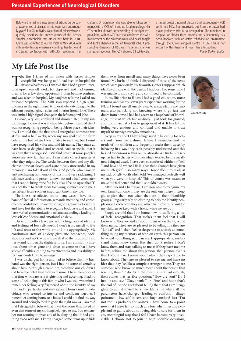
18 I ACNR • VOLUME 6 NUMBER 6 • JANUARY/FEBRUARY 2007
The first I knew of my illness with herpes simplexencephalitis was being told I had been in hospital forsix and a half weeks. I am told that I had a gastro-intes-
tinal upset, was off work, felt depressed and had unusualdreams for a few days. Apparently I then became confusedand was taken to hospital. My daughter tells me I called myhusband Stephanie. The MRI scan reported a high signalintensity in the right mesial temporal lobe extending into theadjacent basal ganglia, insular and inferior frontal lobe. Therewas limited high signal change in the left temporal lobe.
I awoke, very lost, confused and disorientated in my sur-roundings. I know it was a while later before I realised that Iwas not recognising people, or being able to place their iden-tity. I am told that the first time I recognised someone wasat five and a half weeks, when my son spoke to me frombehind the bed where I was unable to see him, but I musthave recognised his voice and said his name. They must allhave been so delighted and relieved. And so special that itwas him that I recognised. I still find now that some people’svoices are very familiar and I can make correct guesses atwho they might be. The weeks between then and my dis-charge home, at eleven weeks, are mostly unrecorded in mymemory. I am told about all the kind people who came tovisit me, having no memory of this I find very saddening. Istill have cards and presents, now two and a half years later,that I cannot bring myself to look at as I feel so upset that “I”was not there to thank them for caring so much about me. Ifeel absent from such an important time in my life.
The illness has affected me in many ways: I have lost abank of factual information, semantic memory, and conse-quently confidence. I have prosopagnosia, have had a seizureand have lost the ability to recognise both taste and smell. Ihave verbal communication misunderstandings leading tolow self-confidence and emotional anxiety.
These difficulties leave me with a huge loss of identityand feelings of low self-worth. I don’t trust myself to livelife and react to the world around me appropriately. Mycontinuous state of anxiety gives me headaches, back,shoulder and neck ache a great deal of the time and I amnervy and jump at the slightest noise. I am constantly anx-ious about times gone and times to come so that I havesleep difficulties leading to overtiredness and less ability tofeel any confidence to manage.
I was discharged home and had to believe that my hus-band was the right person, but I had no sense of certaintyabout him. Although I could not recognise our children Idid have the belief that they were mine. I have memories ofthat time which are very frightening and upsetting. I had nosense of belonging to this family who I was told was mine. Iremember feeling very frightened about the identity of myhusband in particular and very separate from a unit of indi-viduals who seemed so intense and confident together. Iremember coming home to a house I could not find my wayaround and being helped to go to the right rooms. I am toldthat I struggled to believe that it was the home that I lived in,even that some of my clothing belonged to me. I do remem-ber not wanting to wear any of it, denying that it had any-thing to do with me. I know I bagged some items up to hide
them away from myself and many things have never beenfound. My husband thinks I disposed of most of the itemswhich were previously my favourites, ones I suppose whichidentified more with the person I had lost. For some time Iwas unable to stop crying and continued to be confused.
In my life prior to illness I had a good education, nursetraining and twenty-seven years experience working for theNHS. I found myself unable even to name plants and ani-mals, also panicking not knowing where to go just twodoors from home. I had had access to a huge bank of knowl-edge, most of which like anybody I just took for granted,and find myself at a loss to grasp most of it. It has left mefeeling very anxious and confused and unable to trustmyself to manage everyday situations.
Deep in my heart I have a huge need to be caring for oth-ers and I now feel a dismal failure. I misunderstand theneeds of my children and frequently make them upset bybehaving in a way they can’t possibly understand and thishas led to tensions within our relationships. Our family set-up has had to change with roles which worked before my ill-ness being adjusted. I have been so confused within my “self“ and how and where I fit in, that these changes have givenme much grief in so many ways. How difficult to readjustmy lack of self-worth when told ”we managed perfectly wellwhen you were in hospital.” This of course was meant tomake me feel better and that I shouldn’t worry.
After two and a half years, I am now able to recognise myown family at home if they are the only ones there. I strug-gle to pick them out when they are in their friendshipgroups. I regularly rely on clothing to help me identify peo-ple once I know who they are, which helps me mind out formy children or keep with a friend while we are out.
People are told that I am home now but suffering a lossof facial recognition. That makes them feel that I willknow who they are and all about them when they give metheir name. They are so pleased to be telling me they are“Linda!” and I then feel so desperate to snatch at some-thing to jog my memory of who on earth this person canbe – just something so I can react appropriately, under-stand them, know them. But they don’t realise I don’tknow them and start talking to me as if they have met mebefore, telling me about this person, that person, eventsthat I would have known about which they expect me toknow about. They are so pleased to see me and have noidea that they feel like a complete stranger to me. They aresomeone who knows so much more about the person thatwas me, than “I” do. As if the meeting isn’t bad enough,then comes that terrible question: “How are you?” Do Ijust lie and say: “Okay thanks” or “Fine” and hope that’sthe end of it or do I set about telling them that I am strug-gling to adjust myself to a new life, a life where all theparameters have changed, leading to confusion, disap-pointment, low self-esteem and huge anxiety? Just “I’mnot me” is probably the answer. I have come to a pointnow that I have felt so much at a loss when meeting peo-ple and so guilty about not being able to care for them inany meaningful way, that I feel I have become very unso-ciable. I choose not to join in with sociable occasions,
My Life Post Hse
Personal Experiences of Neurological Disorders
Claire Rytina
Below is the first in a new series of articles on person-al experiences of disease. In this issue, I am enormous-ly grateful to Claire Rytina (a patient of mine) who elo-quently describes the consequences of the herpessimplex encephalitis that struck her back in 2004.Claire was admitted to our hospital in May 2004 witha three day history of nausea, vomiting, headache andincreasing confusion with difficulty recognising her
children. On admission she was able to follow com-mands with a GCS of 14 and no focal neurology. HerCT scan that showed some swelling of the right tem-poral lobe, with an MRI scan that confirmed this withinvolvement of the adjacent insula and basal gangliaand subtle changes in the left temporal lobe. A pre-sumptive diagnosis of HSE was made and she wasstarted on acyclovir. Her CSF showed 32 white cells,
a raised protein, normal glucose and subsequently PCRconfirmed HSV. She improved, but from the outset hadmajor problems with facial recognition. She remained inhospital for almost three months and subsequently hasbeen involved with an active rehabilitation programmethrough the Oliver Zangwill Centre in Ely. This is heraccount of this illness and how it has affected her.
Roger Barker, Editor.

ACNR • VOLUME 6 NUMBER 6 • JANUARY/FEBRUARY 2007 I 19
keep in a back seat if I do and make sure I nevermake eye contact with anyone in case they startto speak to me. Expecting I’ll fail again.
I feel very oddly separated from the idea of‘myself ’ so that I can’t feel I properly belong toany group anymore. I don’t feel like I am thewife, the mum, the nurse, the friend that Ibelieve I was before. I am fearful that I willnever fit into these roles again.
I struggle to understand what people are tellingme and feel very anxious about misunderstand-ing what is happening, what is expected of meand how I will manage. I can’t concentrate forvery long on things which need me to think andwork things through in a logical meaningful way.I get very tired just trying to manage simpleeveryday things. I am my own worst enemy forhelping myself relax. I don’t do stop. I’ve led abusy working and home life in which relaxinghad very little part, and nor did I want it to. Istruggle to make the new myself do it, and can’twind down because I’m anxious about all thethings I’m not doing.
I feel very sad most of the time, and evenwhen I’m not tired, anxious or stressed and Ishould relax and have some enjoyment, I justcan’t. My family are fun-loving in a full-onway and I can’t relax enough to have muchenjoyment even with them. They do tease meto try and make me laugh, and sometimes Idon’t recognise this, so it upsets me. More andmore now I am able to recognise the repeatedteasing for what it is, just fun, and react morenormally. I don’t manage to relax and have funvery much, especially when out with specialfriends as their expectations of me to be the‘me’ they made friends with feel threatening tothe mixed up feelings I have about myself. Ifind it a lot easier to talk to people and be‘myself?’ if I have never met them before andthey have no expectations of who I am, theyjust take me for the person that I appear to be.In that situation I can relax and almost feelokay about the person that I am now. I can stillbe sociable, polite, helpful, even enjoy a bit ofa joke. I can relax and not feel threatened bythe muddle of who I was. It’s like I’m startingagain, as the new me, and it feels okay. This hasmade it a lot easier for me to take friendshipand support from the other people with braindifficulties, and the therapists whom I havemet along the way. It has been easier for me tospend time with them and feel like a personaccepted for who I am.
I feel completely separate from the world Ithink I used to live in, I suppose I just carried onwith life; wife, mother to four children, nurse,close friend, a person known, loved and valued byso many people. I feel now that I am no more theperson I was and I have a huge difficulty feelingany sense of belonging in this new world.
I know I am not the same person that I wasbefore my illness and that my life has changedcompletely. I have to take ten deep breaths beforeI venture out into the village in case someonespeaks to me, I avoid meeting up with my closefriends and keep a low profile when I do, and Istruggle hugely emotionally with my family.Since having my illness I have found social con-tact much easier to manage by emails. I don’tneed any facial recognition and I have time to
think about what to say, and reply, without anybody language or facial nuances to try to under-stand. I am able to re-read post messages to jogmy memory and then write and say as much or aslittle as I like at that time. I can even change mymind about what I have said which is easy to doand doesn’t involve a whole lot of shock and guilt,laughing or teasing. I can take those three deepbreaths, and choose a moment when I am calmand able to manage the communication positive-ly and properly. None of these are possible at thevillage post office or when answering the phone.I never answer the phone unless I have to.
I have been encouraged by my rehabilitationtherapists to use relaxation methods: specific CDto listen quietly to, using breathing techniquesand taking proper rest time.
I have had support to help me face the difficul-ty of asking people who they are; polite and prac-tical methods of trying to know who they are andways of explaining my difficulties to them. It feelsso rude and un-caring to have to ask. Many peo-ple now just come straight up to my face andannounce their name with great pleasure and Ido get lots of hugs.
I have always said, “I was born a nurse” andhave worked my heart out for the NHS for 27years, so recently having my contract terminatedon ill-health grounds has broken my heart. Iknow I can’t do the job I did because of my mem-ory and confidence problems but the one thingthat has kept me going was the OccupationalHealth Consultant saying to me “I’m not sayingthat you can’t do anything”.
My poor family have had to live with methroughout my illness, struggling hugely with mysense of negativity and my loss of any feelings ofbelonging, even to them. All our interactions arecomplicated both practically and emotionallyand as much as they have tried to help me, I havemisunderstood and felt rejected, pushed out bylosing my own sense of any worth. I feel my hus-band organises the family and that the childrenrefer to him for everything, quite fairly as I feel Ican’t be trusted to give the correct answer – evendown to the basic question “When’s tea?”
I am feeling almost like I am one of the chil-dren at times. I have been unable to drive forthe last year, following a tonic-clonic seizure,and don’t do any of the household shopping,but I can do household jobs and cooking – noweven without a timing list, so I am improving. Ican even do a roast dinner now, let alone dippyeggs using a timer. I have even coped recentlywith my husband teasing me, and calling me “ahousewife” which I not only recognised as fun,but I managed to tell myself should feel posi-tive, not negative.
I do need to remember that, although my lifehas turned upside down, it has also been a hugeemotional upset for my family. The sooner Iaccept that going backwards isn’t going to help,the more positive I will feel about going forwards.My family have always been there to help me,even when it hasn’t quite felt like it, and I am avery privileged and thankful person. I want torelax and enjoy life again, enjoy being with themand I have been very lucky to have the support ofspecialist people to help not just me, but all thefamily to support us managing. I know that Ihave to face what has happened and try to move
forwards and not dwell on my difficulties. I needto do this for myself and all my family andfriends.
I have recently accepted the idea that rehabil-itation is not synonymous with recovery – andthat I can’t expect to get back to be the person Iwas before the illness. I will do my best to usethe strategies I have been given to continue tohelp myself to become a new happy, meaning-ful, belonging, valued and loving person, caringand sharing. A year ago I had never heard of astrategy application, let alone cognitive think-ing! I know there are things which may nevermend – like the part of my brain which tells methe truth about taste and smell and that I maynever taste chocolate again or be able to smellroses. But I do know that brains are good atlearning and I am busy teaching mine aboutwho I know and how I know them, what pathsin our lives have crossed. I get great pleasurefrom writing a friendship book where I writeinformation down to re-read. I use face-onphotographs with names. I make photo-scrap-books and watch old home videos with myfamily to help me improve my sense of belong-ing. My husband and children enjoyed theseand I became able to relax and laugh at theirpleasure shouting out “There’s you Mum!!” “Wehad a storm that night!!” Many bits of memorywere happily jogged so that I coped with thebits missing without too much heartache.
My brain has been taught to use a Filofaxand not to scribble on millions of scraps ofold paper and then lose them. And I havelearnt not to hide any more official letters ordocuments so they can never be found – andI keep my clothing.
Right from coming home from hospital Ihave used a pack of cards to gain some relax-ation and feel like I am getting my headtogether. I have recently read about CliveWearing and was amazed that I am playingpatience almost addictively, rather like he did.I pester others to play card games with me andhave played on many train journeys to passthe time and reduce agitation and worryabout the journey and what the day may havein store. I have been lucky to travel with otherclients to our centre for rehabilitation butstrangers on the train have often joined in acard game very happily. It’s an easy type ofsocial communication which I cope well withand feel enjoyment and sharing out of.
Over a long period of wonderful rehabilita-tion I have realised that we only have one life,and that I should live it. They have helped meto form strategies to do this. They can’t do itfor me. No-one can. I have to do it for myself.No-one can mend my brain.
How lucky I am to be able to say that myheart is in the right place, even if my headisn’t.
I give huge thanks to the heads and heartsof my family, my friends and all the caringtherapists who have helped me so muchthroughout my illness and who still give meunderstanding, caring and a more positiveoutlook for my future.
Claire Rytina,December 2006.
Personal Experiences of Neurological Disorders

20 I ACNR • VOLUME 6 NUMBER 6 • JANUARY/FEBRUARY 2007
Eosinophillic Meningitis due to Angiostrongylus Cantonensis:First Reported Case in the UK
Angiostrongylus cantonensis, the rat lungworm, isthe commonest cause of eosinophilic meningitisworldwide.1 The organism is endemic in Southeast
Asia and the Caribbean, although an increase in worldtravel has seen cases occurring outside of these areas.Cases have been documented in the USA,2 Switzerland,3
Australia,4 and New Zealand.5 There are no previousreported cases of angiostrongyliasis in the UK.Eosinophilic meningitis is rare in the UK and awarenessof parasitosis as an emergent cause of infectious diseaseremains relatively low. We report a case of A. cantonensisinfection in a female patient returning to the UK fromThailand.
Case reportA thirty-year-old Thai woman, living in the UK, attendedher local district general hospital with five days of worsen-ing headache and meningism. The patient had been a res-ident of the UK for two years but had visited Thailandrecently, returning ten days prior to her presentation tohospital. On her admission she was pyrexial with a tem-perature of 38oC. She had nuchal rigidity and photopho-bia, but no focal neurological deficits. A CT brain scan wasreported as normal and lumbar puncture revealed slightlyturbid CSF with 626 WBC per mm3 (90% lymphocytes,10% polymorphs). CSF protein was 0.55g/L (normal 0.15-0.45). No organisms were seen on Gram stain and therewas no growth on bacterial culture. The patient was treat-ed symptomatically with a presumptive diagnosis of viralmeningitis and discharged after seven days. At home, thepatient’s headache worsened and she developed diplopiaand hyperaesthesia of the right lower limb. She was read-mitted to her district general hospital nine days later andunderwent further lumbar puncture. This revealed slight-ly turbid CSF with 483 WBC per mm3 (70% lymphocytes,30% polymorphs). CSF protein was 0.75g/L. She was com-menced on intravenous Aciclovir and an MRI of the brainordered. MR scanning revealed multiple hyperintensewhite matter lesions and the patient was then transferred
to the regional neurology centre.On arrival, the patient was pyrexial (38oC) with signs of
nuchal rigidity and photophobia. She had a right sixthcranial nerve palsy and a patch of altered sensation overthe lateral border of her right lower limb. No other abnor-malities were evident on neurological examination.Review of her MRI scans revealed multiple white matterhyperintense lesions in the deep cerebral white matter,periventricular regions and in the corpus callosum, whichdid not enhance with gadolinium contrast (Figures 1 and2). There was no enhancement of the pachymeninges.Lumbar puncture was repeated and revealed an openingpressure of 28cm of water, 361 WBC per mm3 (70% lym-phocytes), protein of 0.73g/L, glucose of 2.61mmol/L(serum glucose 5.3mmol/L) and was negative for AAFB.
Further questioning revealed that whilst recently inThailand she had visited Bangkok and the Northernregion of Isaan. She had eaten snails, which she believed tobe cooked, as part of a salad in the village from where sheoriginally came. She had no specific risk factors for HIVinfection and said she had tested negative six monthsprior to this illness. Review of her blood results from theoriginal hospital admission revealed an eosinophilia of1.35*109/L (total white cell count 10.1*109/L), which hadpersisted into her second admission. This prompted re-examination of the original CSF samples for eosinophils.Cytological analysis revealed a significant eosinophilia(Figure 3). In light of this, a parasitic infection was con-sidered likely, in particular ‘rat lungworm’ meningitiscaused by Angiostrongylus cantonensis.
While further microbiological investigations were beingperformed, the patient was treated with isoniazid, rifam-pacin, pyrazinamide and ethambutol with dexametha-sone to cover the possibility of tuberculous meningitis.We discussed the case with the Department ofParasitology at the Hospital for tropical diseases inLondon, who arranged to send the patients serum andCSF samples to Bangkok to test for Angiostrongylus canto-nensis and Gnathostomia spinigerum antibodies.
ABN Case Report Winner
Matthew Jones, Rajiv Mohanraj, Sandip ShaunakDepartment of Neurology, Royal Preston Hospital, Preston, Lancashire, UK.
Correspondence to:Matthew Jones,Department of Neurology,Royal Preston Hospital,Preston,Lancashire, PR2 9HT.Tel: 01772 716565,Email:[email protected]
Figure 1: MR scan of the brain showing high signal lesion on the corpuscallosum on T1 weighted images.
Figure 2: High signal lesion in the deep white matter on T2 weightedimages.
This is the ABN CaseReport Winner andwe congratulate theauthors on thisachievement.
If you would like toreport similar interesting case reportsthen do contact us at:[email protected]

ACNR • VOLUME 6 NUMBER 6 • JANUARY/FEBRUARY 2007 I 21
Polymerase Chain Reaction (PCR) for herpessimplex virus, varicella zoster virus, enterovirusand parechovirus nucleic acids were negative inthe CSF. PCR for Mycobacterium tuberculosiswas also negative. Serum HIV test was negativeand cryptococcal antigen was not detected inthe CSF. Stool and urine were negative for ova,cysts and parasites. As she was originally froman endemic area for Strongyloides stercoralisand was receiving steroids as part of the antitu-berculosis treatment, she was treated withIvermectin to prevent strongyloides hyperinfes-tation. ELISA testing for Strongyloides was pos-itive. Her symptoms however, were not thoughtto be attributable to Strongyloides infection.
The patient’s symptoms improved withreduction in severity of headache, resolution ofmeningism and improvement in diplopia.Repeat MRI scan of the brain showed almostcomplete resolution of the white matter lesionsseen on the initial scan. Thirteen days afteradmission she was discharged home. Uponreview seventeen days later her headache hadconsiderably improved and she had no diplop-ia. Neurological examination was normal.Repeat lumbar puncture showed 170 WBC,predominantly lymphocytes.
Serum and CSF results confirmed infectionwith Angiostrongylus cantonensis. Anti tuber-culosis chemotherapy was stopped and the dex-amethasone tapered at a rate of 1mg per weekuntil stopped. One month later she returnedcomplaining of increasing headache and neckstiffness. Repeat LP revealed 506 WBC per mm3
(80% eosinophils). She was then treated with afurther reducing course of dexamethasone. Thepatient’s symptoms improved and her CSFWBC returned to normal.
DiscussionAngiostrongylus cantonensis is the most com-mon cause of eosinophilic meningitis world-wide. This helminth is endemic in SoutheastAsia and the Caribbean, and the disease is wellrecognised in these areas. The rat is the primaryhost of A. cantonensis. First stage larvae hatchin the lungs of rats and migrate into rat faecesvia the trachea and gut. Molluscs that feed onrat excrement become intermediate hosts andwithin these organisms the first stage larvaemoult twice to become the infective third stagelarvae. Humans who eat the snails may thenbecome infected. Vegetables contaminated withmollusc slime can also be a source of infection,as can transport hosts such as freshwaterprawns, fish or crabs. In humans, larvaemigrate via the bloodstream to the CNS wherethey cause an inflammatory response. Man is
an accidental end stage host in which the larvaeare unable to complete their life cycle and even-tually die.1,6
Symptoms occur 4-23 days after infection.7
The illness is characterised by headache andmeningism.2 Hyperaesthesia and paraesthesiaare well described.1 Other symptoms mayinclude visual disturbance, fever, fatigue andcognitive impairment.7,8 On examination theremay be photophobia, nuchal rigidity, sensorydisturbance, encephalopathy, and cranial nerve,including abducens and facial nerve, palsy.2,7
In this case, finding a peripheral eosinophil-ia prompted the search for parasitic causes andnecessitated re-examination of the CSF foreosinophils. If parasitosis is clinically suspect-ed the CSF should be specifically examined foreosinophils. It may be possible to visualise theworms on direct microscopy. The diagnosiscan be confirmed by serological tests on serumand CSF.
The MRI scan in the patient described abovedemonstrated high signal lesions in deep whitematter and corpus callosum, reminiscent ofdemyelination. A wide variety of MRI abnormal-ities have been reported in Angiostrongyliasis.High T2 signal intensity lesions, contrast enhanc-ing lesions, meningeal enhancement and highsignal intensity in the globus pallidus have allbeen reported.9,10,11
We gave our patient no specific treatment forA. cantonensis infection. Treatment is primarilysupportive, with repeated lumbar puncturethought to relieve symptoms of persistent
headache due to raised intracranial pressure.Anthelmintics can theoretically worsen symp-toms due to an inflammatory reaction to dyingworms. One randomised, double blind, placebocontrolled trial has assessed the use of corticos-teroids in eosinophilic meningitis.12 This trialconcluded that a two week course of pred-nisolone, 60mg per day, significantly reducedheadache and the need for repeat lumbar punc-ture. No adverse effects were reported. In ourpatient, we believe that the initial improvementin symptoms and CSF findings was due to dex-amethasone which she received as part of anti-tuberculous treatment. Her symptoms recurredand CSF eosninophilia worsened after discon-tinuing the steroids, but a further, more pro-longed, course of steroids had a beneficial effect.
In cases of meningitis in travellers returningfrom endemic areas, it is vital to specificallylook for eosinophils in the CSF. A. cantonensisis the commonest cause of eosinophilicmeningitis worldwide, and subacute meningi-tis with associated hyperaesthesia and paraes-thesia should alert the clinician to this organ-ism. MRI scan appearances in this conditioncan be normal or widely variable. The hyper-intense T2 signal lesions can appear demyeli-nating in character and location. This is, toour knowledge, the first reported case of A.cantonensis meningitis in the UK. Withincreasing international travel, it is likely thatcases will be encountered more frequently inthe future.
References
1. Slom T, Johnson S. Eosinophilic Meningitis. Curr Infect Dis Rep 2003;5(4):322-8.
2. Slom T, Cortese MM, Gerber SI, et al. An outbreak of eosinophilic meningitis caused byAngiostrongylus cantonensis in travelers returning from the Caribbean. N Engl J Med2002;346(9):668-75.
3. Bartschi E, Bordmann G, Blum J, et al. Eosinophilic meningitis due to Angiostrongylus can-tonensis in Switzerland. Infection 2004;32(2):116-8.
4. Prociv P. Angiostrongyliasis in Australia. Rev Infect Dis 1990;12:160-1.
5. Lo Re V 3rd, Gluckman SJ. Eosinophilic meningitis due to Angiostrongylus cantonensis in areturned traveler: Case report and review of the literature. Clin Infect Dis 2001;33(9):112-5.
6. Lo Re V 3rd, Gluckman SJ. Eosinophilic meningitis. Am J Med 2003;114(3):217-23.
7. Tsai HC, Liu YC, Kunin CM, et al. Eosinophilic meningitis caused by Angiostrongylus can-tonensis: Report of 17 cases. Am J Med 2001;111(2):109-14.
8. Chau TTH, Thwaites GE, Chuong LV, et al. Headache and confusion: the dangers of a raw
snail supper. Lancet 2003;361:1866.
9. Jin E, Ma D, Liang Y, et al. MRI findings of eosinophilic myelomeningoencephalitis due to
Angiostrongylus cantonensis. Clin Radiol 2005;60(2):242-50.
10. Tsai HC, Liu YC, Kunin CM, et al. Eosinophilic meningitis caused by Angiostrongylus can-
tonensis associated with eating raw snails: Correlation of brain magnetic resonance imaging
scans with clinical findings. Am J Trop Med 2003;68(3):281-5.
11. Kanpittaya J, Jitpimolmard S, Tiamkao S, et al. MR findings of eosinophilic meningoen-
cephalitis attributed to Angiostrongylus cantonensis. Am J Neuroradiol 2000;21:1090-4.
12. Chotmongkol V, Swanyawisuth K, Thavornpitak Y. Corticosteroid treatment of eosinophilic
meningitis. Clin Infect Dis 2000;31:660-2.
Figure 3: CSF eosinophilia.
ABN Case Report Winner

22 I ACNR • VOLUME 6 NUMBER 6 • JANUARY/FEBRUARY 2007
Conference News
For five days, on alternate years, theInternational Society of Neuro -immunology (ISNI) brings together
research groups from all over the globe that arededicated to understanding the relationshipbetween the central and peripheral nervous sys-tem and the immune system in both health anddisease. This year the conference was held inNagoya - Japan’s fourth largest city located onthe Pacific coast; one of Japan’s major ports.The newly opened conference centre housedsome 2000 delegates attending distinguishedreview talks, plenary sessions, workshops, sem-inars, poster sessions and satellite symposia.There were many highlights; a handful of whichare described below. I cannot however fail toapplaud the traditional Japanese folk music‘minyo’, to which the delegates were treated onthe penultimate evening (compelling most tolearn some new moves on the dance floor).Singers were accompanied by the 3 stringed luteknown as the shamisen, taiko drums and a 13stringed zither known as the koto.
IL17/23 cellsThe breaking story of T cell immunology overthe last two years has been the discovery of anew type of helper T cell, called the Th17 cell.(It is called this not because Th1 through toTh16 have already been defined, but becausethese secrete interleukin-17). In fact, likeHelicobacter, these cells were always there, buttheir presence has been misunderstood. All theexciting news about this cell are summarised inClaire Helliwell’s immunology primer in thisissue of ACNR (page 8).
Now this scheme has to be redrawn. It turnsout that a key experiment was flawed. The IL-12receptor subunit knock-out in these animals alsoforms part of the IL-23 receptor. So, both IL-12and IL-23 function was being neutralised. IL-23stimulates a new kind of cell which secretes highlevels of IL-17 and low levels of IFN-γ or IL-4. Itis these cells, called ‘Th17 cells’, which drive EAE,not IFN-γ secreting Th1 cells. Animals that trulylack IL-12, but retain IL-23 and Il-17, still getEAE. Daniel Cua from Shering-PloughBiopharma, USA - also showed that the tran-scription factor that drives naïve cells to becomeIL-17 cells is called retinoid orphan receptor(ROR). This, is in turn promoted by a cytokinemilieu in which there is IL-6 and IGF-β.
Burkhard Becher (Zurich) showed that Th17cells secrete IL-22 as well as Il-17; but IL-22knock-outs are not resistant to EAE and EAEbrains do not contain IL-22 when examinedhistologically. However, VJ Kuchroo has shownthat IL-22 is expressed in the EAE model ofneuromyelitis optica developed by Wekerle andhimself.
A new treatment for Guillain-BarréHuw Willison’s group in Glasgow have studiedthe neuromuscular effect of the anti-ganglio-side antibody, anti-GD1a, that is normally pro-duced in the motor axonal form of Guillain-
Barré. They showed this antibody destroys themotor nerve endplate, by a process that isdependent upon the presence of complement.
Very happily, there is now a drug that canblock complement in humans. A monoclonalantibody against complement, C5, (ecluzimab)has been used effectively as a treatment forparoxysmal nocturnal haemoglobinuria.Willison and colleagues are attempting tosecure funding for a trial of ecluzimab inGuilllain-Barre.
Predicting who needs treatment in MSThe factors that determine the localisation ofmultiple sclerosis lesions are far from under-stood. It was interesting, therefore, to hearMichael Pender present data suggesting thatproteolipid protein (PLP) reactivity drives thedevelopment of brainstem and cerebellarlesions.
His group performed HLA-DR and HLA-DQtyping and examined T cell reactivity (prolifer-ation assays) to myelin proteins in 121 patientswith MS, 71 healthy controls and 47 patientswith other neurological disorders. Patients wereassessed clinically by a blinded assessor andgadolinium enhanced MRI scans were per-formed. They found that nearly 50% of patientswith brainstem and or cerebellar disease hadincreased T cell responses to the immuno-dom-inant region of myelin PLP184-209, but not toother myelin proteins, compared to 10% of MS
subjects without brainstem or cerebellarlesions, 11% of healthy controls and 19% ofpatient with other CNS disease.
They also found that 79% of patients withbrainstem and or cerebellar lesions carried theHLA-DR4, DR7 or DR13 alleles compared to29% of patients with disease elsewhere. In addi-tion, the majority of patients with HLA-DR4,DR7 or DR13 alleles had increased T cell reac-tivity to PLP184-209 compared to only 12% ofpatients not carrying these alleles.
They concluded that reactivity to PLP184-209
drives the development of brainstem and cere-bellar lesions and suggested that patients carry-ing the susceptibility alleles HLA-DR4, DR7 orDR13 may benefit from therapy with alteredpeptide ligands based on PLP184-209.
How the immune system protectsagainst autoimmunity - a step closer.The demands on our immune system are great:the system must defend against incomingpathogens, repair damaged tissue, recogniseand remove abnormal cells, all without causingdisease (autoimmunity). Naturally occurringregulatory T cells (Tregs) play an essential rolein the maintenance of peripheral self-tolerance.Tregs express elevated levels of the high affinityinterleukin-2 (IL-2) receptor alpha subunit(CD25); neutralisation of IL-2 reduces the sizeof the CD4+CD25+ compartment inducingautoimmune disease in mice. Tregs also expressFoxP3, a transcription factor which is critical inthe development and function of Tregs.Transduction of FoxP3 into effector T cellsresults in these cells differentiating into Tregs,however, the mechanism by which FoxP3 pre-vents the emergence of autoimmune diseaseshave remained unsolved until now.
Shimon Sakaguchi has shown that FoxP3interacts with acute myeloid leukaemia 1(AML1 also known as Runx1). AML1 regu-lates the expression of a variety ofhaematopoietic genes and is a critical regula-tor of haematopoietic development, includingT cell development. A variety of post-transla-tional modifications can modulate AML1activity and these determine whether AML1acts as a transcriptional repressor or activatorof gene expression.
AML1 binds to the IL-2 promoter upon Tcell receptor (TCR) stimulation. Sakaguchishowed that FoxP3 binds to AML1 and sup-presses AML1 enhanced IL-2 production. Tcells that do not express AML1 fail to bind wildtype FoxP3, do not suppress IL-2 productionand fail to up-regulate CD25, CTLA-4 andGITR, markers of Tregs; these cells were alsoshown to be less suppressive than those thatcould bind AML1 in a functional assay.Sakaguchi also demonstrated that singlenucleotide polymorphisms (SNPs) affecting theAML1/FoxP3 interaction resulted in suscepti-bility to rheumatoid arthritis, systemic lupuserythematosus, psoriasis and other autoim-mune diseases.
The 7th International Congress of Neuroimmunology Nagoya, Japan, 15-19 October 2006.

ACNR • VOLUME 6 NUMBER 6 • JANUARY/FEBRUARY 2007 I 23
Neurology: Learning, knowledge,progress and the future
Key lectures:• Axonal protection in chronic
inflammatory neurological disorders• Mitochondrial disorders• Repair of brain and spinal cord
injuries• Stroke prevention
The ENS 2007 scientific programme includes 4symposia featuring lectures by experts dis-cussing new advances in neurology and clinicalneurosciences. The first symposium onMonday is the presidential symposium onaxonal protection in chronic inflammatoryneurologic disorders, chaired by G Comi. Thissymposium will include lectures on mecha-nisms between inflammation and axonaldegeneration, the implication of stem-cells forregeneration as well as new therapeuticalavenues.
On Tuesday, two symposia will run in paral-lel. The first symposium deals with mitochon-drial diseases and will be chaired by P Chinnery
and includes lectures on the clinical investiga-tion of mitochondrial disorders, the discoveryof novel nuclear genes, the management ofmitochondrial disorders and the implication ofmitochondrial dysfunction in common neuro-logical disorders.
The other symposium - chaired by V Dietz -is devoted to the contribution of repair of brainand spinal cord injuries and includes lectureson neuronal plasticity of the spinal cord injury,achievements in basic research on regenerationand advances in experimental spinal cordinjury.
On Wednesday, the symposium focussing onstroke prevention will be chaired D Leys andincludes lectures on preventing early recur-rences after cerebral ischaemic event, newdevelopment in antiplatelet therapy, stroke pre-vention in women and stroke prevention inyoung adults.
This will be followed by the best of free com-munications 2007 which will give a summary ofthe best papers presented during the meeting.
In addition to the symposia, the scientificprogramme includes 5 poster sessions and 16oral sessions of free communication. There willbe an integrated programme on the manage-ment of MS patients, on the treatment of acutestroke and of motoneuron disease. After thesuccessful introduction of poster walks lastyear, we will once again have these to display theposters in a more lively and interesting format.Experts will lead a review of selected posterspromoting discussion with the authors.
Prof A Steck, Executive Committee.
Visit the ENS 2007 website www.ensinfo.com featuring:• Continuously updated scientific
programme• Online abstract submission
(deadline February 7, 2007)• Online registration as well as hotel &
tour registration• Information about Rhodes
This work is crucial in our understanding ofthe regulation of self-tolerance and the mecha-nisms that control the critical balance that ourimmune system has to strike between defense,repair and autoimmunity. Importantly, it offersa possible target for future treatments that canhope to prevent autoimmunity without causingimmunosuppression.
The enhancement of neurogenesis poststroke - a new role for rosiglitazone?The formation and maintenance of centralnervous system circuit integrity has been afocus of Theo Palmer’s work for over ten years.Now as assistant professor in Neurosurgery atStanford University, he presented recent dataexamining the roles of inflammation on neuro-genesis. He showed that in a post-stroke ratmodel, neurogenesis is enhanced, andmicroglial activation is suppressed, by non-steroidal drugs such as indomethacin. However,these results were overshadowed by the superi-or results seen with rosiglitazone. This peroxi-some proliferator-activated receptor gammaagonist, used clinically in diabetics to enhanceglycaemic control, is emerging as a new neuro-protective agent. How it does this remains to bedefined, but is sure to involve modulation ofpro-inflammatory cytokines and attenuation ofan otherwise activated immune system.
Amanda Cox, Alasdair ColesDr Jo Jones,
Dr Vicki Robertson and Dr Ben Wright.
Rhodes, Greece, 16 – 20 June, 2007.
PREVIEW: 17th Meeting of the European Neurological Society
Conference News
The Association of British Neurologists willvisit Cambridge on April 11-13th 2007 andthis will form part of the 75th anniversarycelebrations of the Association.
The main symposium on the Thursdaywill outline the history of Neurology in theUnited Kingdom during the 20th century,focusing on activities of the Association.The programme for Thursday also includesa guest lecture on spinal cord injury andrepair by Professor James Fawcett. One ofthe meeting highlights will be the unveilingof the ABN Coat of Arms on the Thursdayevening.
The Conference Dinner is at KingsCollege with music from members of theCollege Choir. Our principal guest is
Baroness Onora O’Neill – Reith lecturer in2002, and an authority on bioethics.
We shall be building upon the previous suc-cess of the case presentation competition andagain putting greater focus upon poster pre-sentations. The scientific events close with adebate with the motion ‘Modernising MedicalCareers for patients, trainees and the practiceof neurology,’ proposed by Chris Clough, andopposed by Professor Compston.
We are looking forward to seeing every-one in Cambridge and hope to make this anenjoyable, memorable and instructive meet-ing of the Association, fit for our 75thanniversary.
Association of British Neurologists.
Cambridge, UK, 11 – 13 April, 2007.
PREVIEW: 75th Anniversary Celebrations ofthe Association of British Neurologists
Homerton College, Cambridge.

24 I ACNR • VOLUME 6 NUMBER 6 • JANUARY/FEBRUARY 2007
2007
JanuaryNEWUnderstanding and treating cognitive problemsafter brain injury.12-13 January, 2007; London, UKW. www.braintreetraining.co.uk E. [email protected]
NEWEncephalitis Society Professional Seminar16 January, 2007; Liverpool; UKT. 01653 692 583 F. 01653 604 369 E. [email protected] W. www.encephalitis.info
BISWG South Wales and the West CountryRegional Meeting18 January, 2007; CardiffKate Coles T. 02920 224871 E. [email protected]
35th National Conference Indian Association ofPMR Specialists19-21 January 2007; Patna, IndiaE. [email protected]@yahoo.com
NEWMultiple Sclerosis Trust Masterclass in PosturalManagement30 January, 2007; London, UK W. www.mstrust.org.uk T. 01462 476704 E. [email protected]
FebruaryThink Ahead Think Success - OT SupportWorkers 2nd Biennial National Conference6 February, 2007; York, UKJulie HawkinsT. 020 7450 2337F. 020 7450 2349E. [email protected]
NEWRSM Schizophrenia 7 February, 2007; Cardiff, UKT. 020 7290 2965 E. [email protected]
35th Annual INS Meeting7-10 February, 2007; Portland, Oregon, USAInternational Neuropsychological Society T. + (614) 263-4200 F. + (614) 263-4366 E. [email protected]
NEWMultiple Sclerosis Trust General Study Day inMS8 February, 2007; Stoke, UKW. www.mstrust.org.uk T. 01462 476704 E. [email protected]
The Society for Research in RehabilitationWinter Meeting8 February, 2007; Sheffield, UKW. www.srr.org.uk E. [email protected]
Global Conference on Neuroprotection &Neuroregeneration14-16 February, 2007; Garmisch-Partenkirchen,GermanyW. www.gcnpnr.org
NEWRCSE Intracranial and Spinal Anatomy forNeurosurgeons19-23 February, 2007; London, UKT. 020 7869 6332 F. 020 7869 6329 E. [email protected]
3rd Annual Update Symposium on ClinicalNeurology and Neurophysiology 19-21 February; 2007, Tel Aviv, IsraelW. www.neurophysiology-symposium.com
1st East Mediterran Epilepsy Congress21-24 February, 2007; Luxor, EgyptT. +353 1 205 6720 F. +353 1 205 6156 E. [email protected]
British Neuropsychiatry Association 2007Meeting - Parkinson’s Disease, Epilepsy, Mindand Brain: The next 20 years22-23 February 2007; London, UKT/F. 01621 843334 E. [email protected] W. www.bnpa.org.uk
March NEWMyelination, Demyelination and MultipleSclerosis 20072 March, 2007; London, UKW. www.abcam.com/myelin2007
1st Congress on Epilepsy, Mind & Brain2-4 March, 2007; Prague, Czech RepublicW. www.kenes.com/epilepsy
NEWMultiple Sclerosis Trust Annual Meeting forSpecialist Nurses6-7 March, 2007; York, UKW. www.mstrust.org.uk T. 01462 476704, E. [email protected]
International Congress on Neurology andRehabilitation (ICNR)9-11 March, 2007; New Delhi, IndiaE. [email protected] W. www.iamst.com
23rd Annual Pacific Rim Conference onDisabilities12-13 March, 2007; Waikiki, HawaiiE. [email protected] W. www.pacrim.hawaii.edu
NEWFirst London Colloquium on Status Epilepticus12-15 April, 2007; London, UKE. [email protected] T. 01323 740 612 / 01691 650 290
4th Annual Meeting of the Global College ofNeuroprotection & Neuroregeneration14-16 March, 2007; Garmisch-Partenkirchen,GermanyW. www.gcnpnr.org E. [email protected] T. +44 115 969 2016, F. +44 115 969 2017
8th International Conference AD/PD 2007 14-18 March, 2007; Salzburg, AustriaW. www.kenes.com/adpd/
NEWRSM “Growth factors and psychiatric disor-ders” 23 March, 2007; London, UKT 0207 290 2965 E. [email protected]
1st International Congress on Epilepsy, Mind &Brain29-31 March, 2007; Prague, Czech RepublicT. +41 22 908 0488F. +41 22 732 2850E. [email protected]
AprilNEWInternational Symposium on ‘Brain Ageing andDementia in Developing Countries’10-13 April, 2007; Nairobi, KenyaSamantha Tannahill, c/o Prof RN Kalaria T. +44 191 256 3206 F. +44 191 256 3011 E. [email protected] W. http://advascular.ncl.ac.uk
Certificate Course in NeurologicalRehabilitation10-27 April, 2007; Newcastle upon Tyne, UK E: [email protected]
ABN Spring Scientific Meeting11-13 April, 2007; Cambridge, UKInfo: [email protected]
NEW1st London Colloquium on Status Epilepticus12-15 April, 2007; London, UKT. 01323 740 612 / 01691 650 290 F. 01691 670 302.
Alzheimer's Disease: Update on Research,Treatment, and Care12-13 April, 2007; San Diego, California, USAW. htttp://cme.ucsd.edu
NEW
Understanding and treating insight problemsafter brain injury20-21 April, 2007; London, UKW. www.braintreetraining.co.uk E. [email protected]
The Challenges of Commissioning for BrainInjury Services-A National Conference25 April, 2007; London, UKPatti Simonson T. 0208 780 4530 F. 0208 780 4530 E. [email protected]
NEWSynaptic Plasticity in Pain27 April, 2007; London, UKW. http://www.abcam.com/pain07
NEW5th Staffordshire Conference On ClinicalBiomechanics27 - 28 April, 2007; Stoke-on-Trent, UKCheryl Blewitt E. [email protected]. 01782 294341
59th Annual Meeting of the American Academyof Neurology28 April-5 May, 2007; Boston, USAW. www.aan.com
American Academy of Neuroscience Nursing(AANN) Annual Meeting29 April – 2 May, 2007; Orlando, USAE. [email protected]
MayNEWManagement of Parkinson's disease - Joint con-ference of the Royal College of Physicians andthe Parkinson's Disease Society3 May, 2007; London, UKRCP Conference Dept T. 0207 935 1174 ext 252/300/436E. [email protected]
NEWVisual Perceptual Dysfunction and braininjury, Part 29 - 11May, 2007; London, UKW. www.braintreetraining.co.uk E. [email protected]
NEWBrain Development 200710 May, 2007; London, UKW. www.abcam.com/brain2007
EFNS Academy for Young Neurologists 8thCourse10-13 May, 2007; Staré Splavy, Czech RepublicE. [email protected] or [email protected] T/F. +420 2 6716 35 63
NEWPrimary Care Neurology Society 2007Conference17 May, 2007; Birmingham, UKW. www.p-cns.org.uk
NEWMultiple Sclerosis Trust Masterclass for MSSpecialist Nurses22 May, 2007; London, UKW. www.mstrust.org.uk T. 01462 476704 E. [email protected]
2nd Biennial Vocational Outcomes inTraumatic Brain Injury Conference24-26 May, 2007; Vancouver, BC CanadaE. [email protected] W. www.tbicvancouver.com
8th European Neuro-Ophthalmology SocietyMeeting26-29 May, 2007; IstanbulPinar Aydin O'dwyerE. [email protected]. www.Eunos2007.Org
XVI European Stroke Congress29 May – 1 June, 2007; Glasgow, UKE. [email protected]
Consortium of Multiple Sclerosis Centers(CMSC)30 May – 3 June, 2007; Washington DC, USAE. [email protected]
June 39th International Danube Symposium forNeurological Sciences and ContinuingEducation in conjunction with the 1stInternational Congress on ADHD2-5 June, 2007; Wurzburg, GermanyW. www.danube-wuerzburg.de or www.adhd-wuerzburg.de
1st International Congress on ADHD: FromChildhood to Adult Disease3-7 June, 2007; Wuerzburg, GermanyE. [email protected]
11th International Congress of Parkinson'sDisease and Movement Disorders3-7 June, 2007; Istanbul, TurkeyW. www.movementdisorders.org/meetings/index.shtml
NEWMultiple Sclerosis Trust Study Day in MS –Focus on Health Professionals in theResidential Environment5 June, 2007; UKW. www.mstrust.org.uk T. 01462 476704 E. [email protected]
Kuopio Stroke Symposium6-8 June, 2007; Kuopio, FinlandE. [email protected] W. www.uku.fi/stroke2007
2nd International Congress on NeuropathicPain 7-10 June 2007; Berlin, GermanyT. +41 22 908 0488 F. +41 22 732 2850 E. [email protected] W. www.kenes.com/neuropain
4th World Congress of the InternationalSociety of Physical Medicine andRehabilitation10-14 June, 2007; Seoul, KoreaE. [email protected] W. www.isprm2007.org
NEWMS Frontiers - MS Society flagship researchevent presenting key themes in internationalMS research14-15 June 2007; London, UKT. 020 8438 0809 F. 020 8438 0877 E. [email protected]
Workshop: Prediction of Outcome15-16 June, 2007; Erlangen, GermanyProf. Dr. Hermann Stefan E. [email protected]
17th Meeting of the European NeurologicalSociety16-20 June, 2007; Rhodes, GreeceW. www.ensinfo.com T. +41 61 686 77 11 F. +41 61 686 77 88 E. [email protected]
Advances in NeurorehabilitationPart of The Festival of InternationalConferences on Caregiving, Disability, Agingand Technology (FICCDAT)16-19 June, 2007; Toronto, CanadaE. [email protected] W. www.ficdat.ca
International Society for Stem Cell ResearchMeeting17 –22 June, 2007; CairnsInfo: International Society for Stem CellResearchT. +847-509-1944 F. +847-480-9282 E. [email protected]
2nd Neurorehabilitation PanamericanCongress18-20 June, 2007; Buenos Aires, ArgentinaE. [email protected] W. www.ineba.net
Canadian Neurological Sciences Federation(CNSF) 42nd Annual Scientific Meeting19-22 June, 2007; Alberta, CanadaW. www.ccns.org
Events Diary
To list your event in this diary, email brief details to Patricia McDonnell at [email protected] by January 22nd, 2007

Seventeenth Meeting of the European Neurological Society
June 16-20, 2007 – Rhodes/Greece
Young neurologists programme 2007Following the great demand in 2006, ENS is pleased to offer once again a limited number of grantsproviding free accommodation from Saturday to Wednesday, including registration and admission toteaching courses at the ENS 2007 meeting in Rhodes to European Neurologists in training, born onor after January 1, 1972. This programme is not dependent on submitting an abstract.Applications should provide a letter from chairman of their department to certify that they are intraining as well as a copy of their passport.The applicants must also select the 3 courses they want to attend. Application is only available online at www.akm.ch/ens2007.Attendance to courses is compulsory and no-shows will be excluded from this offer in the future andwill be charged for their attendance.Deadline for applications: 18 January 2007. Applications without passport copy or arriving after thisdate will not be considered.Good qualitiy accommodation has been reserved this year and will be on a double room occupancybasis. We invite applicants to mention on their registration form the name of another youngneurologist they would wish to share the room with.For further information please check our regularly updated website: www.ensinfo.comPlease apply online on www.ensinfo.com – ENS 2007 the online application will be available fromOctober onwards.
Separately to this offer, young colleagues who have an abstract accepted for presentation at the ENSmeeting may also apply for a travel grant. Please see further details on the congress website.
Visit the ENS 2007 website featuring:• Continuously updated scientific programme
• Online abstract submission (deadline February 7, 2007)
• Online registration as well as hotel & tour reservations
• Option to compose your personal congress programme
• Details about the industrial exhibition
• Information on the congress venue and the island of Rhodes
For further information please contact:
Administrative Secretariat: 17th ENS 2007, c/o AKM Congress ServiceP.O. Box, CH-4005 Basel / SwitzerlandPhone +41 61 686 77 11 Fax +41 61 686 77 88 E-mail [email protected]
www.ensinfo.com
Neurologists in training offerDeadline January 18, 2007
ENS07_T_210x297mm 23.10.2006 11:41 Uhr Seite 1

26 I ACNR • VOLUME 6 NUMBER 6 • JANUARY/FEBRUARY 2007
…is a simple and fun way for European neuroscientists to get involved in communicatingwhat you do to the public. Using a disposable camerato document your day, we want to show the publicand young people in particular what the everydaylives of neuroscientists entail: highlighting the important research work as well as showing thehuman side to a career in neuroscience.
The project is run by The Manchester ScienceGroup www.manchesterscience.blogspot.comand sponsored by the European Dana Alliance forthe Brain.
To take part, email your postal address to: [email protected] with A DAY IN THE LIFE in the subject line and you will receive a disposable camera pack and instruc-tions.
A Dayin theLife…
BRAIN AWARENESS WEEK
12-18 MARCH 2007A global celebration of the latest brainscienceBrain Awareness Week is an annual celebration dedicated toraising public awareness of brain research. Coordinated by theEuropean Dana Alliance for the Brain, and the Dana Alliancefor Brain Initiatives in the US, Brain Awareness Week hasbecome a major international collaboration.
You can become involved in Brain Awareness Week by organis-ing an activity. By registering as a partner, and then posting yourevent, your organisation will be listed on our worldwide BrainAwareness Week calendar of events, and you will receive information on running and publicising your programmes.
Please join us in this major international campaign
To organise an event or get involved:Contact EDAB on 020 7019 4914 or by email at [email protected]
www.edab.net
MANAGEMENT OF PARKINSON’S DISEASE
Thursday 3 May 2007
at the Royal College of Physicians, 11 St Andrews Place, Regent’s Park, London NW1
With the ageing of the population, the prevalence of Parkinson's disease is set to increase dramatically in the next20 years. Research in this important condition has led tomany significant developments in the last few years. This con-ference, organised by the Royal College of Physicians and the Parkinson’s Disease Society will summarise recent develop-ments in relation to the recently introduced NICE guidelinesfor the diagnosis and management of Parkinson's disease.
The programme and booking forms are available on-line: www.rcplondon.ac.uk/conferences or from:Conference Department, Royal College of PhysiciansTel: 020 7935 1174 Ext. 436/252/300 Fax: 020 7224 0719Email: [email protected]
UCL Institute of Child Health and Great Ormond Street Hospital for Children NHS Trust
Practical Neurology Study Days 200719th – 23rd March 2007Leolin Price Lecture Theatre, Institute of Child HealthOrganisers:Vijeya Ganesan, Sarah Aylett, Carlos de Sousa, Catherine DeVile, andRobert Surtees.These study days are to help clinicians update their knowledgeregarding common paediatric neurology problems, develop practicalskills in dealing with common acute neurology problems and considermanagement of neurological illness in different settings.The course uses a combination of lectures, interactive sessions, casepresentations, slides and videos. All the lecturers are currently practisingpaediatric neurologists.Monday Overview of Paediatric Neurology & Epilepsy Tuesday Headaches, Hydrocephalus, Brain Tumours & Spinal
Cord Wednesday Neuromuscular DisordersThursday Acute NeurologyFriday Neurogenetic & Neurometabolic Disorders
The course is intended for:Consultant hospital and Community Paediatricians, Trainees inPaediatrics, Neurology and NeurodisabilityThere are limited places available so book now to avoiddisappointment!Fees: £645 or £149 daily rate
For further information, please contact: Courses and Conferences Office, Institute of Child Health,30 Guilford Street, London WC1N 1EH, UKTelephone: +44 (0)20 7829 8692, +44 (0)20 7905 2135; Fax: +44 (0)20 7831 6902; e-mail: [email protected];Website: www.ichevents.com
Great Ormond StreetHospital for Children
NHS Trust

www.efns.org/efns2007
Special Sessions, Short Communications as well as the following Main Topics:
• RTMS and neuroplasticity
• Scientific basis of headache
• Frontiers in movement disorders
• The “more-than-6-hours” stroke
• Neuromuscular diseases
• Multiple sclerosis (MS) treatment: how and when?
• New disease modifying treatment approaches in Alzheimer’s disease
• Status epilepticus: toward European treatment guidelines
President of EFNS Management Committee: Jacques L. De Reuck, Belgium
Chairperson of the Congress Programme Committee: Gian Luigi Lenzi, Italy
Chairperson of the Local Arrangements Committee: Jean Schoenen, Belgium
DEADLINE FOR SUBMISSION OF ABSTRACTS: FEBRUARY 15, 2007
11th CONGRESS OF THEEUROPEAN FEDERATION OFNEUROLOGICAL SOCIETIES
BRUSSELS, BELGIUM, AUGUST 25-28, 2007
ACNR ND06:Layout 1 24/10/06 21:19 Page 32

28 I ACNR • VOLUME 6 NUMBER 6 • JANUARY/FEBRUARY 2007
Journal Reviews
HUNTINGTON’S DISEASE: new cells for oldThe role of neural stem cells in neurodegenerative disorders hasbeen something of a passion of mine for many years, as abnormali-ties in this process may have important implications for therapyboth in terms of innate repair and exogenous cell transplants in dis-orders of the CNS. Furthermore, understanding the processes ofneurogenesis in diseased brains may also provide us with a betterunderstanding of abnormal signalling pathways involved in diseasepathogenesis. This new paper from the van der Kooy group raisessome very interesting questions on how disease processes may influ-ence stem cell behaviour in one of the neurogenic regions of theadult brain (the subventricular zone [SVZ]) in the R6/2 transgenicmouse model of Huntington’s disease. They report that in thismouse, which contains the expanded exon 1 CAG repeat of humanHuntington’s disease gene:1. There is an increase in neural precursor cell/neurospheres gen-
erated from the SVZ in R6/2 mice compared to wild type withincreasing age and the development of symptoms.
2. There is an increase in BrdU in the SVZ 30 days after injectionin the R6/2 mice compared to the wild type, implying there is anincrease in stem cells at this site which is confirmed using EMand the demonstration of more precursor ‘B’ cells in the SVZ inthe R6/2 mice.
3. There is an increase in proliferation with age which occurs invitro with cloning but only at a time when such abnormalities inproliferation have been seen in the R6/2 mouse in vivo.
4. The increase which is seen in vitro is clearly primed in vivo andcannot be mimicked by growing early stage neurospheres forlonger periods of time in vitro.
5. There is no change in the fate potential of the precursor cells invivo or in vitro.
6. There is a decrease in olfactory bulb BrdU positive cells with anincrease the BrdU positive cells in the striatum, suggesting aswitch in migration of the stem cells to sites of more pathology.
These series of observations suggest that, with disease progression inthis mouse model of Huntington’s disease, there is a steady increasein the number of stem cells in the SVZ which, once primed andinduced in vivo, is maintained in vitro. This suggests that the stemcell changes its properties with the evolving disease process in thebrain. This would therefore imply that, in neurodegenerative disor-ders of the CNS, there are signals that cause a fundamental switch inthe properties of proliferating neural stem cells which attempts tocompensate for the pathology. Obviously if this process can be prop-erly harnessed, then it will have important implications for CNSrepair, but the key question remains as to what that signal is and howcan we manipulate it to effect better repair in conditions such asHuntington’s disease. - RABBatista CM, Kippin TE, Willaime-Morawek S, Shimabukuro MK,Akamatsu W, van der Kooy D.A progressive and cell non-autonomous increase in striatal neuralstem cells in the Huntington’s disease R6/2 mouse.JOURNAL OF NEUROSCIENCE 2006;26:10452-60.
HUNTINGTON’S DISEASE: up and down or side to side?Huntington’s chorea is characterised by cognitive decline and chorea, butpatients often display other deficits, including abnormalities of eye move-ment. Blekher et al have carried out a detailed and sensitive series of oculo-motor tests in a group of patients with HD. Clinical evaluation was carriedout using the standardised clinical rating scale UHDRS (UnifiedHuntington’s Disease Rating Scale). The study included 215 individualsgrouped into four classes depending on the presence or absence of motorabnormalities representative of HD. All the individuals in this study had aparent affected with HD and followed one of two patterns: they were eitherat risk of HD but not yet diagnosed or they had been diagnosed with HDwithin the past two years. Both vertical and horizontal saccadic readings wererecorded using a video eye tracking system. Eye movement measures includ-ed visually-guided, anti-saccade, predictable, memory-guided (two versions)and fixation tasks. None of the participants reported significant eye problemsand all had normal or corrected visual acuity. The authors showed that HD
gene carriers – but not nongene carriers - had impaired saccades and were par-ticularly poor in anti-saccade and memory-guided tasks, and this when stan-dard clinical assessments currently used in HD were normal. The improvedsensitivity provided by these quantitative saccadic measures may open a newchapter in the diagnosis of disease onset and also may enable researchers toascertain whether disease modifying therapies are just that. - CABlekher T, Johnson SA, Marshall J, White K, Hui S, Weaver M, Gray J, Yee R,Stout JC, Beristain X, Wojcieszek J, Foroud T. Saccades in presymptomatic and early stages of Huntington disease.NEUROLOGY2006;67:394-9.
STROKE: a pit of vipersIf you get bitten by a Malaysian pit viper, you may bleed to death. A key com-ponent of the venom is Ancrod which splits fibrinopeptide A from fibrino-gen. Ancod has been used in several trials as a treatment for ischaemic stroke.The previous North American Stroke Treatment with Ancrod Trial (STAT)showed that Ancrod outperformed placebo if given within three hours of astroke. This study looks at its ability to improve stroke outcome if given later,within six hours. The result, perhaps unsurprisingly given what we know ofTPA, is that later treatment is not beneficial. Normally such a pedestrianstudy would not make it onto the review pages of ACNR, but it is the vipersin the last paragraph of the report that merit attention: “Preliminary data from this trial were first presented at the World Federationof Neurology Congress in London in 2001. Soon thereafter, the sponsor com-pany, which was always supportive of the scientific outcomes of this trial, wassold. In the meantime, data from the study were not fully available to theinvestigators, and further analysis was difficult. Only with the support ofmany dedicated investigators and after careful reassessment of the materialfinally provided, could the members of the executive and safety committeesprepare this report on behalf of the investigators. This situation illustrates theunderstandable but often regrettable divergences between sponsor and inves-tigators’ interests, leading to scientific losses and unethical waste of patients’and investigators’ efforts. The bias towards easier publication of successfultrials, sometimes at too early a stage, is another important issue to consider:the publication of this report is therefore an important recognition of the sci-entific and medical value of all clinical trials.”The moral is that it is critical that investigators have free access to data accu-mulated in their trial. It has always amazed me that some investigators areprepared to work in any other way. - AJCHennerici MG, Kay R, Bogousslavsky J, Lenzi GL, Verstraete M, OrgogozoJM; ESTAT investigators. Intravenous ancrod for acute ischaemic stroke in the European StrokeTreatment with Ancrod Trial: a randomised controlled trial.LANCET2006 Nov 25;368(9550):1871-8.
PARKINSON’S DISEASE: First gene therapy trialThis very short abstract buried at the back of an issue of MovementDisorders is one that was presented at the 20th Annual Symposium on theAetiology, Pathogenesis and Treatment of Parkinson’s Disease and OtherMovement Disorders. This gives a very brief account (as one would expect)on 12 patients who have received open label unilateral subthalamic viral vec-tor (AAV-GAD) injection as a treatment for their Parkinson’s disease. Thisopen label study involves 12 patients with an average age of 58.2 years and inthis abstract the authors report on their FDG PET studies using thisapproach. The strategy involves trying to convert the excitatory subthalamicnucleus to an inhibitory one by changing the phenotype of the projectionneurons from this structure and builds on earlier experimental work report-ed in Science in 2002 (see ACNR 2.5). Five patients appeared to respond tothis virally delivered therapy and seven did not. In those that responded,there were clearly changes in FDG metabolism as one would anticipate,namely a decline in the internal globus pallidum and ventral lateral thalamusipsilateral to the delivery of the viral vector and increased metabolism in thepremotor and supplementary motor regions. These changes appear to corre-late with improvements in their UPDRS scores. Whilst this is an abstract, itis important because this is the first ever gene therapy trial in Parkinson’s dis-ease and clearly gives us hope that this approach may have a future. - RABFeigin A, Tang C, During M et al.Gene therapy for Parkinson’s disease with subthalamic nucleus AAV-GAD:FDG PET results.MOVEMENT DISORDERS2006;21:1543-4.
EDITOR’S CHOICE

ACNR • VOLUME 6 NUMBER 6 • JANUARY/FEBRUARY 2007 I 29
PARKINSON’S DISEASE: Get the rhythmThe role of dopamine in the control of movement is well known (see for exam-ple the historical article in this issue of the ACNR by Oleh Hornykiewicz), but howexactly dopamine achieves this is not altogether clear. The classical model of basalganglia function has nigral dopamine stimulating the direct and indirect path-ways through the striatum by activation of the D1 and D2 receptors on the pro-jection neurons respectively. This then changes the outflow of the basal ganglia(the internal part of the globus pallidum and the substantia nigra pars reticula-ta) to the thalamus and so cortex, especially motor areas and by so doing con-tributes to the initiation of movement. As a result, in diseases such as Parkinson’sdisease (PD) there is a loss of the nigrostriatal dopaminergic projection with adecrease in activation of cortical areas, causing akinesia amongst other symp-toms, presumably through a failure of motor cortical activation. However, arecent paper suggests that it may not be as simple as this and that dopamine maybe more important in coordinating activity between the striatum and motor cor-tex, so that in PD the problem is more a loss of this synchronous activity. Thework leading to this suggestion uses a dopamine transporter (DAT) knock outmouse in which dopamine levels can be easily manipulated such that: a) a hyperdopaminergic state with hyperactivity can be achieved by placing
the DAT knock out mouse in a novel environment;b) a hypodopaminergic state with akinesia can be achieved by blocking
dopamine synthesis using alpha methyl-p-tyrosine (AMPT) [as there areno dopamine stores to compensate for this synthetic block];
c) restoration of dopamine in the hypodopaminergic state can be achievedusing L-dopa.
Using this system the authors show that there are differences in firing frequency(both at the single level and local field potential level) in the striatum and motorcortex with differing levels of dopaminergic tone. More importantly though, theyshow that with dopamine depletion the degree of synchrony between striatumand cortex is enhanced whilst the hyperdopaminergic state is characterised byasynchronous activity in corticostriatal networks. This is an interesting studygiven the recent work on the mechanisms underlying the beneficial effects of deepbrain stimulation in PD and its postulated functions on changing corticostriatalrhythmicity and synchrony. However, this paper more importantly raises funda-mental questions about how diffusely projecting neurotransmitters may work inthe normal brain and how they fail in disease across networks and as such thiswork has implications on how best to treat conditions such as PD. - RABCosta RM, Lin SC, Sotnikova TD, Cyr M, Gainetdinov RR, Caron MG,Nicolelis MA. Rapid alterations in corticostriatal ensemble coordination during acutedopamine-dependent motor dysfunction.NEURON 2006;52:359-69.
nvCJD: Its in the blood
Almost anything, however trivial, about variant CJD gets straight into the media,but this report probably deserves to, because it is very worrying. It describes thecase of a 22-year-old man who, after several years of symptoms of inflammatorybowel disease, had an ileostomy. Complications of its reversal some time laterwere so severe that he required 22 units of blood transfusion. Unfortunately oneof the donors of this blood died 20 months afterwards of variant CJD. The recip-ients were all informed there was a slight risk of developing CJD, which cannothave been an easy message to relay or receive. All was well for six years, and thenthe patient developed progressive limb pain, balance and cognitive deficits. Hedied nearly nine years after the vCJD implicated transfusion (age 32 years). Priorto death, a MRI had shown the pulvinar sign, which had not been seen on a scandone at the onset of symptoms. Deposition of type 4 PrP, the hallmark of vCJD,was seen post mortem throughout the cortex and cerebellum, and likewise in thetonsils. There are two other such cases in the literature, which does not amountto a whole lot of beans until you consider that the long incubation period ofvCJD, and lack of reliable blood test, means that much of the transfusion stockmay be contaminated. As Collinge told listeners of Radio 4’s Today programme,we really need to know the results of the National Anonymous Tonsil Archive’sscreening of 100 000 tonsils for disease-associated PrP. It is still not clear whethervCJD is going to be a ghastly pandemic or a fascinating rarity. - AJCWroe SJ, Pal S, Siddique D, Hyare H, Macfarlane R, Joiner S, Linehan JM,Brandner S, Wadsworth JD, Hewitt P, Collinge J.Clinical presentation and pre-mortem diagnosis of variant Creutzfeldt-Jakob disease associated with blood transfusion: a case report.LANCET2006 Dec 9;368(9552):2061-7.
EPILEPSY: The cost of misdiagnosisYou all know that in everybody else’s clinic, not your own of course, thechronic misdiagnosis of epilepsy is 20-25%. You all want to help thoseother neurologists resolve their diagnostic problems and the only way to doso is to have video-EEG-telemetry. You go to your friendly, local health carecommissioner and tell them that epilepsy misdiagnosis causes misery, psy-chosocial deterioration, inappropriate treatment, lack of appropriate treat-ment and all those clinical things that medical practice is supposed to help.The response is: “That is all very interesting but how will you save money?”So here is the answer. Using standard prevalence and misdiagnosis rates,these authors calculate a misdiagnosis rate of 1,769 cases per million pop-ulation. The cost of medical resources allocated to these patients was takenfrom another paper, including inappropriate admissions (45%), AEDs(26%), outpatient attendances (16%) and GP care (8%). The total theycame to is £316 per patient. This is similar to the NICE guidelines calcula-tion of £263 and works out as £559,076 per million population per year. Ireckon it is still an underestimate, considering that a new AED for a yearcosts about £1,000. What we don’t know from this calculation, is how muchit will cost to treat the patient’s true diagnosis, but so far my managers havenot thought of this and I am not going to tell them. So now you have thedata you can help all those other guys who can’t diagnose fits. Good luck.Any other tips on managerial bamboozling, please email to [email protected]. - MRAMJuarez-Garcia A, Stokes T, Shaw B, Camosso-Stefinovic, Baker R. The costs of epilepsy misdiagnosis in England and Wales. SEIZURE2006;15:598-605.
EPILEPSY: Sliced resistance to carbamazepine
Antiepileptic drugs often don’t work and epilepsy associated with hip-pocampal sclerosis is particularly resistant to medical treatment.Hypotheses for the mechanism of this resistance include failure of drugs tocross the blood brain barrier, but perhaps the varying effect of differentdrugs in different types of epilepsy make an alteration intrinsic to the tis-sue more likely. This study explored that possibility by applying carba-mazepine directly to the mesial temporal lobe slices from 28 patients withmesial temporal lobe epilepsy and from 6 patients with extrahippocampaltumours. The resected tissue was sliced and stimulated until spontaneousdischarges occurred. Then carbamazepine was applied and finally washedoff. Different types of epileptic activity were induced in different slices. Theslices from patients with mesial temporal lobe epilepsy showed little changein spike activity when carbamazepine was applied but tissue from thosewith extrahipocampal tumours showed a reduction of abnormal activity byabout 90%. Tissue showing lower drug resistance generally came frompatients with a shorter duration of epilepsy and clinically less resistantepilepsy, suggesting that this model is clinically relevant. Seven per cent ofpatients who were carbamazepine-sensitive in the model, where the drugcould gain access to tissue, had been resistant in vivo, suggesting that thissubgroup may have had a different mechanism of resistance, perhaps at theblood brain barrier. The authors speculated on the mechanism of resist-ance. They found from voltage clamp experiments that there was reducedsensitivity of voltage dependent sodium channels to the effects of carba-mazepine in resistant slices. The authors argue that, since carbamazepineacts on the sodium channel on the outer surface of the cell, cellular trans-porters which increase efflux of the drug are unlikely to have any impact onresistance. They speculated on the possibility that synaptic reorganisationin the hippocampus may be a factor in drug resistance but had no directevidence for this hypothesis. This new finding moves a step closer to under-standing why the drugs we use act so differently in different patients, butclearly this kind of work is limited by the availability of human experimen-tal tissue. - MRAMJandova K, Pasler D, Antonio LL, Raue C, Ji S, Njunting M, Kann O, KovacsR, Meencke HJ, Cavalheiro EA, Heinemann U, Gabriel S, Lehmann TN. Carbamazepine resistance in the dentate gyrus of human hippocampalslices. BRAIN2006;129:3290-306.
WWWW RECOMMENDED
WWW RECOMMENDED
For a list of Journal reviewers please turn to page 03
Journal Reviews

30 I ACNR • VOLUME 6 NUMBER 6 • JANUARY/FEBRUARY 2007
This book represents a typically thorough and conscien-tious approach by Graeme Hankey to gather together thebest of the evidence, where it exists, to help the clinicianmanage patients with cerebrovascular disease. It is a bookof just over 500 pages and 18 chapters, all following a sim-ilar structure. The first two chapters briefly discuss, first-ly ‘the size of the problem of stroke’ and, secondly, under-standing the evidence. The latter is a particularly usefulchapter, leading one through the currently standardisedlevels of evidence, helping the reader to understand andevaluate critically randomised trials, and also, and morepertinent to this book, meta-analyses and systematicreviews.
Then the book proper begins. Each chapter, looking atdifferent clinical questions or dilemmas (e.g. acute throm-bolysis, neuroprotection, anticoagulation, blood pressurelowering, carotid artery revascularisation, etc), follows asimilar theme. The first part of each chapter is entitled‘rationale’ and describes the arguments believing that acertain treatment might work. Different treatments arethen described, the evidence for or against in relation toplacebo or other existing therapies, with accompanyingforest plots of all included trials. Then, most importantlythere is a comment regarding interpretation of the evi-dence and implications for practice. The latter is largely acomment based upon analysis of the previous evidence,
but where such evidence is lacking or controversial,Graeme Hankey provides the reader with potentialoptions to take. Finally, each chapter finishes with a sec-tion titled ‘implications for research’ which brings thereader completely up to date with where a given field washeading at the time of writing and where it might go inthe future. There is a final summary for each chapterwhich is an excellent means of getting to grips with thedata in a very convenient way.
Each chapter is of course fully referenced, as are all thestudies included in each meta-analysis, and as a rapidsource of reference to any clinician dealing with stroke itis the sort of book that really should be on the office shelf.
I would very much hope that in five years time GraemeHankey will have the energy to update the book as it is anexcellent format and he deserves enormous credit for hiscommitment to gathering such a wealth of trial evidence.
Although much of the data is available through theCochrane Library (which Graeme Hankey is the first toacknowledge in his preface) his interpretation of the evi-dence, and his ability to highlight where the data are lacking,incomplete, or need further work, is what makes this work avery worthy addition to the cerebrovascular literature.
Peter Martin, Addenbrooke’s Hospital, Cambridge, UK.
Book Reviews
Stroke Treatment and Prevention: An Evidence-based Approach
“I am suffering from the limits of my mind,” he said. “Thereis a simple enough problem that I have set out to solve. Howour minds work…..”
“Yes, I suppose for simplicity’s sake, you might say that hisguiding light is Charcot and mine is Darwin.”
“ So a man may first have deduced the existence of hisown consciousness by imagining it in others”
“.... perhaps for quite simple reasons connected to thelimits of their ability to reason, human beings could live outtheir whole long lives without ever knowing what sort ofcreatures they really were. Perhaps it did not matter; perhapswhat was important was to find serenity in not knowing.”
Many writers have grappled with the fundamentals of whatconstitutes the human mind, consciousness and the soul ofa person. Sebastian Faulks in his latest novel explores suchissues in the context of the late 19th century running from1870 to just after the First World War – a time of greatchange in many differing spheres of life and science. InHuman Traces, Faulks uses two fictional characters tounwrap the arguments that formed the kernel of the debatein neurological-psychiatric circles in Europe at the turn ofthe last century, and grapples in particular with the questionof whether mental illness can best be understood through apsychological approach fashioned through experiences or amore deterministic one in nature with a basis in genes andevolutionary pressures. These two stand points are adoptedby the two main characters- a French psychiatrist JacquesRebière trained in the ways of Charcot at the Salpetrière inParis and Thomas Midwinter, an English physician influ-enced by his experiences in a London lunatic asylum. Thesetwo doctors enter their respective practices with the ambi-tion that they can begin to understand the basis of mentalillness, and through this offer cures as well as providinginsights into consciousness and what it is to be human.These ambitions are driven by different influences and guid-
ing philosophies- Jacques is possessed by a desire to cure hisschizophrenic brother, Olivier, within whom the only mem-ories of his mother are trapped whilst Thomas is encour-aged by his desire to understand the creative genius of greatartists. After spending formative years in Paris and Londonrespectively, the two aspiring doctors unite and combinetheir efforts in a sanatorium they set up in the Austrianmountains, and the relationship thickens as Jacques marriesthe influential older sister of Thomas, Sonia, whilst Thomasmarries a patient of Jacques, Kitty. This, however, is no ordi-nary patient, as she comes to define the difference betweenthe two men and contributes to the unravelling of theirfriendship and their ultimate destinies. Initially Kitty is seenby Jacques, who explains her problems through some tortu-ous psychological reasoning, which he regards as being soseminal that he wishes to present her case to a scientificmeeting and asks Thomas to read the case history he haswritten on her. His reading of it, far from confirming thevalidity and insight of Jacques approach, alerts him to thereal problem of her condition and the physical, organiccause of her complaints. The failure of Jacques to recognisehis error in this case, coupled to his introspective outlookand philosophy on mental illness, leads him to becomemore isolated and distant from the kindly, more physicallyminded and egregious Thomas. Their relationship foundersand survives in no small part because of their spouses andtheir over-arching but crumbling ambitions.
This tension remains despite the sanatorium movinglocation, and is heightened by the loss of Olivier (and laterDaniel, the only son of Jacques and Sonia). In contrast,Thomas and Kitty have two thriving twin girls and seemdestined to live out their days happily despite Thomas near-ly dying in Africa during a 3-month sabbatical. However,the arrival of the 1st World War causes them to return totheir respective countries and, after time apart, the final actis played out. The two become reconciled as Thomas enters
If you would like to review books for ACNR, please contact Andrew Larner, Book Review Editor, c/o [email protected]
Author: Graeme J HankeyPublished by: Cambridge University Press, 2005ISBN: 0521827191Price: £80.00
Human Traces
Author: Sebastian FaulksPublished by: Hutchinson,London. 2005.ISBN: 0091794552Price: £17.99

ACNR • VOLUME 6 NUMBER 6 • JANUARY/FEBRUARY 2007 I 31
Book Reviews
a new final phase of his life, and Jacques accepts the loss-es and false hopes of his work and family.
Throughout this whole novel the scientific theories thatdominated the neurological world and formed the founda-tion of psychiatry at this time are laid out, often in greatdetail. Whilst of interest, especially to those wishing tounderstand these formative years of psychiatry, they canarrest the flow of the story and impede the unfolding of thenarrative. Indeed some reviewers have criticised the book asbeing more concerned with describing research than being anovel. Whilst this is true in parts, and there are many diver-sions which seem to be not wholly necessary – for examplethe trips to America and Africa by Jacques and Thomasrespectively- there is nevertheless an underlying rhythm tothe work which drives the reader on to find out how thesedifferent philosophies will work themselves out in the lives ofthe main characters. En route the reader is invited to engagewith a range of interesting questions, not least of which iswhy mental illness is so common given its destructive natureon patients and families and what it can tell us about con-
sciousness and the human mind and soul. Whilst Jacquesand Thomas try to answer this question from different per-spectives, Kitty and Sonia seek to understand such thingsusing less rigorous approaches and in many ways seem tohave a greater, more profound understanding of what it is tobe human. This they would argue is through relationshipsand love with their attendant memories and a sense oftomorrow and looking beyond oneself. Thus the true humantraces are those left by our effect on and interactions withothers, physically, mentally and spiritually. This is no betterencapsulated than in the moving speech by Thomas at theend of the book, where, guided by his wife Kitty at the finalfamily reunion, Thomas describes his own evaluation of hislife: as a doctor he feels he has achieved nothing (althoughothers disagree) but in love he has been rich.
Roger A Barker,Honorary Consultant,
The Cambridge Centre for Brain Repair,Cambridge, UK.
News Review
Trigeminal Neuralgia AssociationUK will hold its first two dayconference at Keele Universityon 30 June-1 July 2007. Theprogramme will include emi-nent speakers from the USAand the UK and this event willinclude a Continuing EducationProgramme for healthcare practi-tioners. All interested professionalsand non-professionals are welcome to attend the con-ference as it will be an opportunity to meet TrigeminalNeuralgia patients and encourage two-way communi-cation about the difficulties faced by patients. Fromprevious conference evaluations, patients and profes-sionals have valued the interaction that this type ofconference encourages.
The proposed healthcare professionals’ conference isaimed at dentists, local GPs, neurologists and neuro-surgeons. The fee for the conference will include fullrefreshments, and accommodation is available ifrequired. Full details will be published in future edi-tions of ANCR, but please make a note of the date.
The aim of the programme is to increase healthcareprofessionals’ awareness of trigeminal neuralgia, espe-cially its diagnosis and up-to-date management. Fromrecent data produced by Hall et al (Pain, 2006) on theepidemiology of neuropathic pain in the UK, it wouldappear that trigeminal neuralgia is more common thanpreviously thought in UK medical practices. Its inci-dence has been put at 26.8 per 100,000, whereasthis was thought to be 4-6 in 100,000.Carbamazepine is the drug of choice but this is onlyprescribed to 58% of patients.
For further information contact: Trigeminal Neuralgia Association UK, Tel: 020 8462 9122, Web: www.tna-uk.org.uk
Trigeminal Neuralgia AssociationUK – first 2-day conference
The Primary Care Neurology Society willbe holding one annual conference inEngland and one conference in Scotlandin 2007. The first conference will be onthe 17th of May at the BirminghamHippodrome. The date and venue for theScotland conference is still to befinalised. Over 180 people attended theirconferences in 2006 with 93% rating itgood or excellent. “Brilliant conference –I’ve learnt a lot and will approach my
neurological patients with more confi-dence” and “Excellent lecturers and con-tent – thank-you” were just two of themany positive comments received. TheSociety is keen to develop a 2007 pro-gramme that reflects the interests of theirmembers and is offering a 40% discounton the conference delegate fee on com-pletion of the pre-conference question-naire which you can access from www.p-cns.org.uk.
Are you or someone you know interested in tak-ing part in a once in a lifetime trek or cycle ridewhilst at the same time raising money forHeadway – the brain injury association?
In 2007 Headway are offering the following trips:
15-25 Feb - Cuba Cycle Ride17-25 Mar - Sahara Trek27 Apr-6 May - China Trek13-17 Jun - London to Paris Cycle Ride18-26 Aug - Iceland Trek14-23 Sep - Peru Trek27 Oct - 8 Nov - Vietnam Cycle Ride
We will take care of all event organisation andadministration, leaving you to concentrate onraising sponsorship and preparing for the trip.
For further details please contact:Rachel Broughton, Events and Conferences Officer,Tel: +44 (0)115 924 0800 or Email: [email protected]
Primary Care Neurology Society Conference
A 2007 Challenge…

Because every day is precious
we don’t waste a day
Only with Aricept is the first dose a therapeutic dose1-6
Continuing Commitment To Alzheimer’s
®
donepezil hydrochloride
ARICEPT® IS INDICATED FOR THE SYMPTOMATIC TREATMENT OF MILD TO MODERATELY SEVERE ALZHEIMER’S DEMENTIA.
ABBREVIATED PRESCRIBING INFORMATIONARICEPT® (donepezil hydrochloride) Please refer to the SmPC before prescribing ARICEPT 5 mg or ARICEPT 10 mg. Indication: Symptomatic treatment of mild to moderately severe Alzheimer’s dementia. Dose and administration: Adults/elderly; 5 mg daily which may be increased to 10 mg once daily after at least one month. Treatment should be initiated and supervised by a physician with experience of Alzheimer’s dementia. A caregiver should be available to monitor compliance. Monitor regularly to ensure continued therapeutic benefi t, consider discontinuation when evidence of a therapeutic effect ceases. No dose adjustment necessary for patients with renal impairment. Dose escalation, according to tolerability, should be performed in patients with mild to moderate hepatic impairment. Children; Not recommended. Contra-Indications: Hypersensitivity to donepezil, piperidine derivatives or any excipients used in ARICEPT. Pregnancy: Aricept should not be used unless clearly necessary. Lactation: Excretion into human breast milk unknown. Women on donepezil should not breast feed. Warnings and Precautions: Exaggeration of succinylcholine-type muscle relaxation. Avoid concurrent use of anticholinesterases, cholinergic agonists, cholinergic antagonists. Possibility of vagotonic effect on the heart which may be particularly important with “sick sinus syndrome”, and supraventricular conduction conditions. There have been reports of syncope and seizures - in such patients the possibility of heart block or long sinusal pauses should be considered. Careful monitoring of patients at risk of ulcer disease including those receiving NSAIDs. Cholinomimetics may cause bladder outfl ow obstruction. Seizures occur in Alzheimer’s disease and cholinomimetics have the potential to cause seizures and they may also have the potential to exacerbate or induce extrapyramidal symptoms. Care in patients suffering from asthma and obstructive pulmonary disease. No data available for patients with severe hepatic impairment. Should not be used in patients with rare hereditary problems of galactose intolerance, the Lapp lactase defi ciency or glucose-galactose malabsorption. Donepezil has minor or moderate infl uence on ability to drive/use machines so this should be routinely evaluated. Drug Interactions: Experience of use with concomitant medications is limited, consider possibility of as yet unknown interactions. Interaction possible with inhibitors
or inducers of cytochrome P450; use such combinations with care. May interfere with anticholinergic agents. Possible synergistic activity with succinylcholine-type muscle relaxants, beta-blockers, cholinergic agents. Side effects: Most commonly diarrhoea, muscle cramps, fatigue, nausea, vomiting, and insomnia. Common effects (>1/100, <1/10): common cold, anorexia, hallucinations, agitation, aggressive behaviour, syncope, dizziness, insomnia, diarrhoea, vomiting, nausea, abdominal disturbance, rash, pruritis, muscle cramps, urinary incontinence, headache, fatigue, pain, accident. Uncommon effects (>1/1,000, <1/100): seizure, bradycardia, gastrointestinal haemorrhage, gastric & duodenal ulcers, minor increases in serum creatine kinase. Rare (>1/10,000, <1/1,000): extrapyramidal symptoms, sino-atrial block, atrioventricular block, liver dysfunction including hepatitis. Presentation and basic NHS cost: Blister packed in strips of 14. ARICEPT 5 mg; white, fi lm coated tablets marked 5 and Aricept, packs of 28 £63.54 ARICEPT 10 mg; yellow, fi lm coated tablets marked 10 and Aricept, packs of 28 £89.06 Marketing authorisation numbers:ARICEPT 5 mg; PL 10555/0006. ARICEPT 10 mg; PL 10555/0007. Marketing authorisation holder: Eisai Ltd. Further Information from/Marketed by: Eisai Ltd, Hammersmith International Centre, 3 Shortlands, London, W6 8EE and Pfi zer Limited, Walton Oaks, Dorking Road, Tadworth, Surrey KT20 7NS.Legal category: POM Date of preparation: July 2006
References: 1. Aricept SmPC. 2. Aricept Evess SmPC 3. Rivastigmine SmPC 4. Galantamine SmPC5. Galantamine XL SmPC 6. Memantine SmPCDate of preparation: November 2006 A968-ARI955-10-06
Information about adverse event reporting can be found at www.yellowcard.gov.uk Adverse events should also be reported
to Eisai Ltd on 0208 600 1400 or [email protected]
ARIC6015 ACNR 297x210.indd 1ARIC6015 ACNR 297x210.indd 1 2/1/07 18:55:372/1/07 18:55:37