Embed Size (px)
Citation preview
Journal meeting
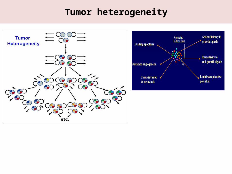
Tumor heterogeneity
Tumor heterogeneity
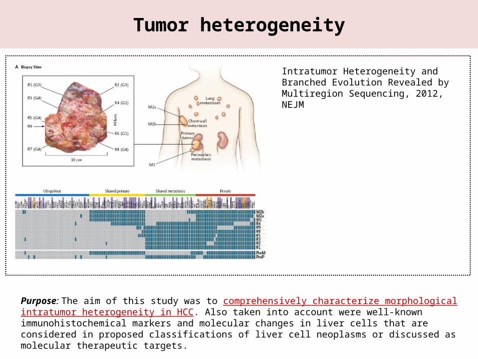
Purpose: The aim of this study was to comprehensively characterize morphological intratumor heterogeneity in HCC. Also taken into account were well-known immunohistochemical markers and molecular changes in liver cells that are considered in proposed classifications of liver cell neoplasms or discussed as molecular therapeutic targets.
Intratumor Heterogeneity and Branched Evo-lution Revealed by Multiregion Sequencing, 2012, NEJM
Introduction & Materials and Methods
Introduction:
► Hepatocellular carcinoma (HCC) generally arises in the context of chronic liver diseases, including chronic viral hepati-tis, alcohol-induced liver injury, or other metabolic, dietary or toxic factors such as fatty liver disease or aflatoxin inges-tion and ranks number three among the leading causes of cancer mortality worldwide.
► It is well-known that HCC frequently display heterogeneous growth patterns and/or cytological features within one and the same tumor. This might either reflect plasticity of phenotypes or intratumor genetic heterogeneity. Both have been described in several solid tumor entities, including skin, breast, and kidney cancer.
Materials and methods:
► Patients and tissues23 patients with an initial diagnosis of a primary hepatocellular carcinoma (HCC) receiving surgical treatment be-tween 2003 and 2009 at the University Hospital Zurich were included in this study. Excluded were cases with any kind of pretreatment (e.g. chemotherapy, TACE) and cases with substantial tumor necrosis (>20%).
► Morphology and definition of tumor areas
► ImmunohistochemistryAll slides were stained with antibodies against cytokeratin 7 (CK7), cytokeratin 19 (CK19), CD34, CD44, epithelial cell adhesion molecule (EpCAM), α-fetoprotein (AFP), β-catenin and glutamine synthetase (GS), a downstream target of β-catenin
► Sanger sequencing & Deep sequencing
ResultsOverall frequency of intratumor heterogeneity in hepatocellular carcinoma
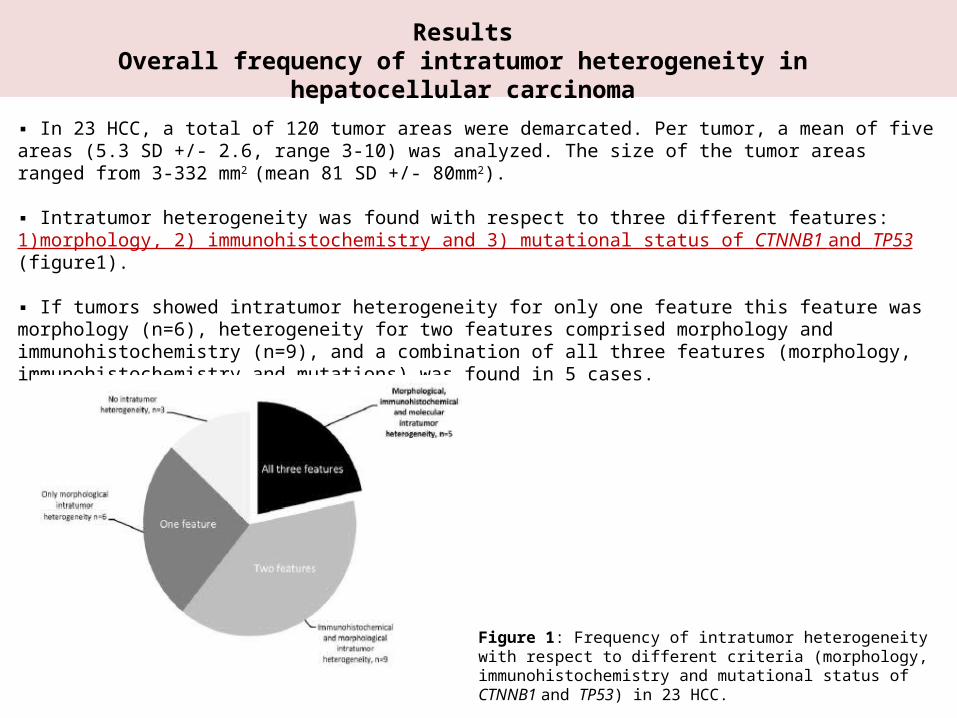
▪ In 23 HCC, a total of 120 tumor areas were demarcated. Per tumor, a mean of five areas (5.3 SD +/- 2.6, range 3-10) was analyzed. The size of the tumor areas ranged from 3-332 mm2 (mean 81 SD +/- 80mm2).
▪ Intratumor heterogeneity was found with respect to three different features: 1)morphology, 2) immunohistochemistry and 3) mutational status of CTNNB1 and TP53 (figure1).
▪ If tumors showed intratumor heterogeneity for only one feature this feature was morphology (n=6), heterogeneity for two features comprised morphology and immunohistochemistry (n=9), and a combination of all three features (morphol-ogy, immunohistochemistry and mutations) was found in 5 cases.
Figure 1: Frequency of intratumor heterogeneity with respect to dif-ferent criteria (morphology, immunohistochemistry and mutational status of CTNNB1 and TP53) in 23 HCC.
ResultsClinico-pathological findings
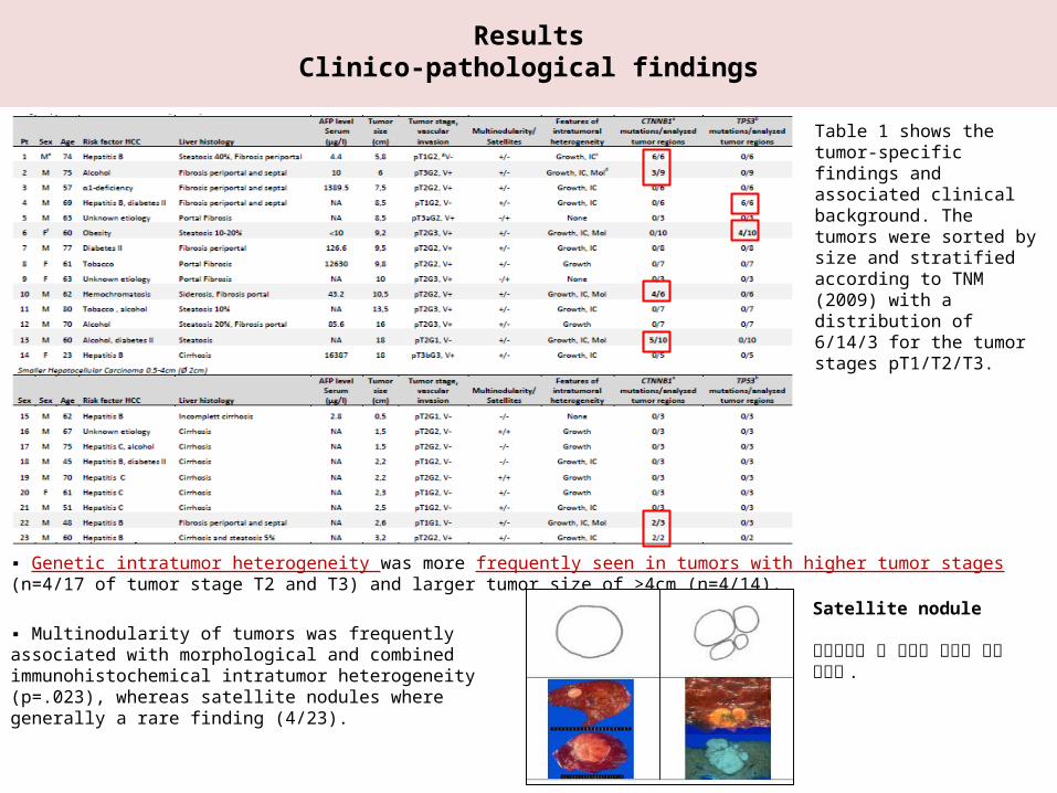
▪ Genetic intratumor heterogeneity was more frequently seen in tumors with higher tumor stages (n=4/17 of tumor stage T2 and T3) and larger tumor size of >4cm (n=4/14).
Table 1 shows the tumor-spe-cific findings and associated clinical background. The tu-mors were sorted by size and stratified according to TNM (2009) with a distribution of 6/14/3 for the tumor stages pT1/T2/T3.
▪ Multinodularity of tumors was frequently associated with morpho-logical and combined immunohistochemical intratumor heterogeneity (p=.023), whereas satellite nodules where generally a rare finding (4/23).
Satellite nodule
위성결절은 주 종양에 근접한 작은 결절임 .
ResultsClinico-pathological findings
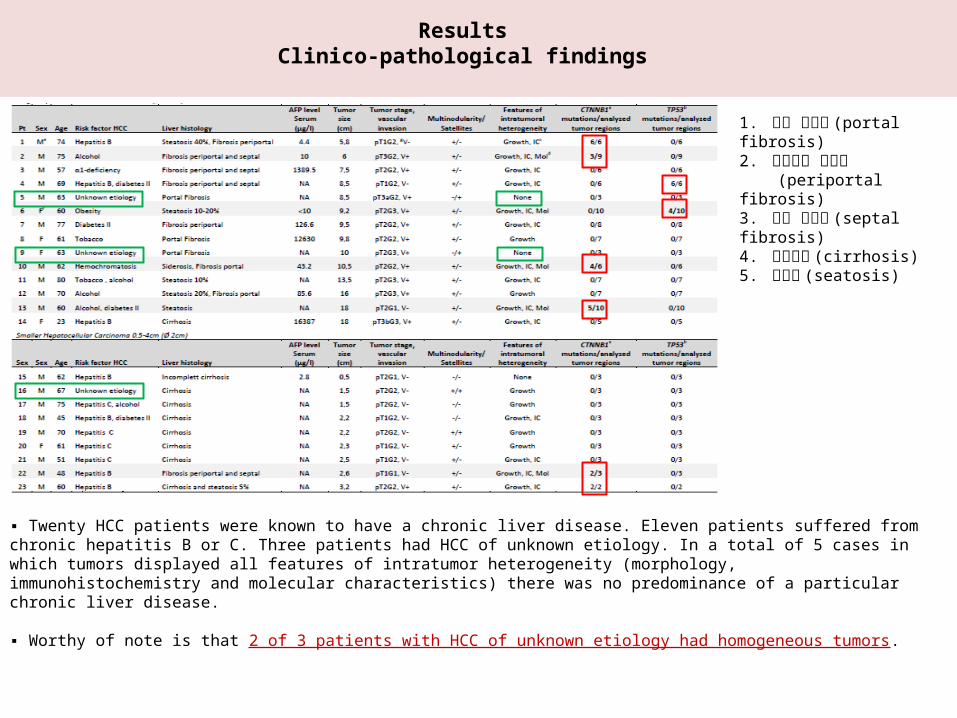
▪ Twenty HCC patients were known to have a chronic liver disease. Eleven patients suffered from chronic hepatitis B or C. Three patients had HCC of unknown etiology. In a total of 5 cases in which tumors displayed all features of intratumor heterogeneity (morphology, immunohis -tochemistry and molecular characteristics) there was no predominance of a particular chronic liver disease.
▪ Worthy of note is that 2 of 3 patients with HCC of unknown etiology had homogeneous tumors.
1. 문맥 섬유화 (portal fibrosis) 2. 문맥주변 섬유화 (periportal fibrosis) 3. 격벽 섬유화 (septal fibrosis)4. 간경변증 (cirrhosis)5. 지방간 (seatosis)
ResultsIntratumor heterogeneity of immune phenotypes
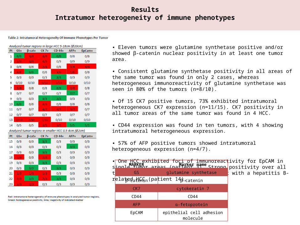
▪ Eleven tumors were glutamine synthetase positive and/or showed β-catenin nuclear positivity in at least one tumor area.
▪ Consistent glutamine synthetase positivity in all areas of the same tumor was found in only 2 cases, whereas heterogeneous immunoreactivity of glutamine synthetase was seen in 80% of the tumors (n=8/10).
▪ Of 15 CK7 positive tumors, 73% exhibited intratumoral heterogeneous CK7 expression (n=11/15). CK7 positivity in all tumor areas of the same tumor was found in 4 HCC.
▪ CD44 expression was found in ten tumors, with 4 showing intratumoral heterogeneous expression.
▪ 57% of AFP positive tumors showed intratumoral heterogeneous expression (n=4/7).
▪ One HCC exhibited foci of immunoreactivity for EpCAM in single tumor areas (patient 3). Strong positivity over all tumor areas was seen in a young patient with a hepatitis B-re-lated HCC (patient 14).
MARKER Marker name
GS glutamine synthetase
β-catenin β-catenin
CK7 cytokeratin 7
CD44 CD44
AFP α-fetoprotein
EpCAM epithelial cell adhesion molecule
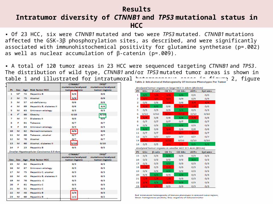
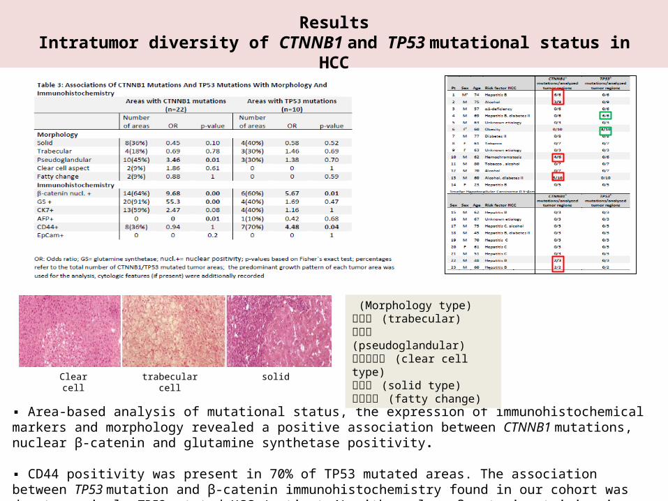
ResultsIntratumor diversity of CTNNB1 and TP53 mutational status in HCC
▪ Of 23 HCC, six were CTNNB1 mutated and two were TP53 mutated. CTNNB1 mutations affected the GSK-3β phosphory-lation sites, as described, and were significantly associated with immunohistochemical positivity for glutamine syn-thetase (p=.002) as well as nuclear accumulation of β-catenin (p=.009).
▪ A total of 120 tumor areas in 23 HCC were sequenced targeting CTNNB1 and TP53. The distribution of wild type, CTNNB1 and/or TP53 mutated tumor areas is shown in table 1 and illustrated for intratumoral heterogeneous cases in figure 2, figure 3.
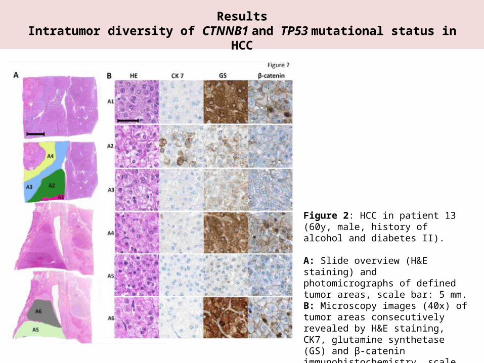
ResultsIntratumor diversity of CTNNB1 and TP53 mutational status in HCC
Figure 2: HCC in patient 13 (60y, male, his-tory of alcohol and diabetes II).
A: Slide overview (H&E staining) and pho-tomicrographs of defined tumor areas, scale bar: 5 mm. B: Microscopy images (40x) of tumor areas consecutively revealed by H&E staining, CK7, glutamine synthetase (GS) and β-catenin immunohistochemistry, scale bar: 40 μm.
ResultsIntratumor diversity of CTNNB1 and TP53 mutational status in HCC
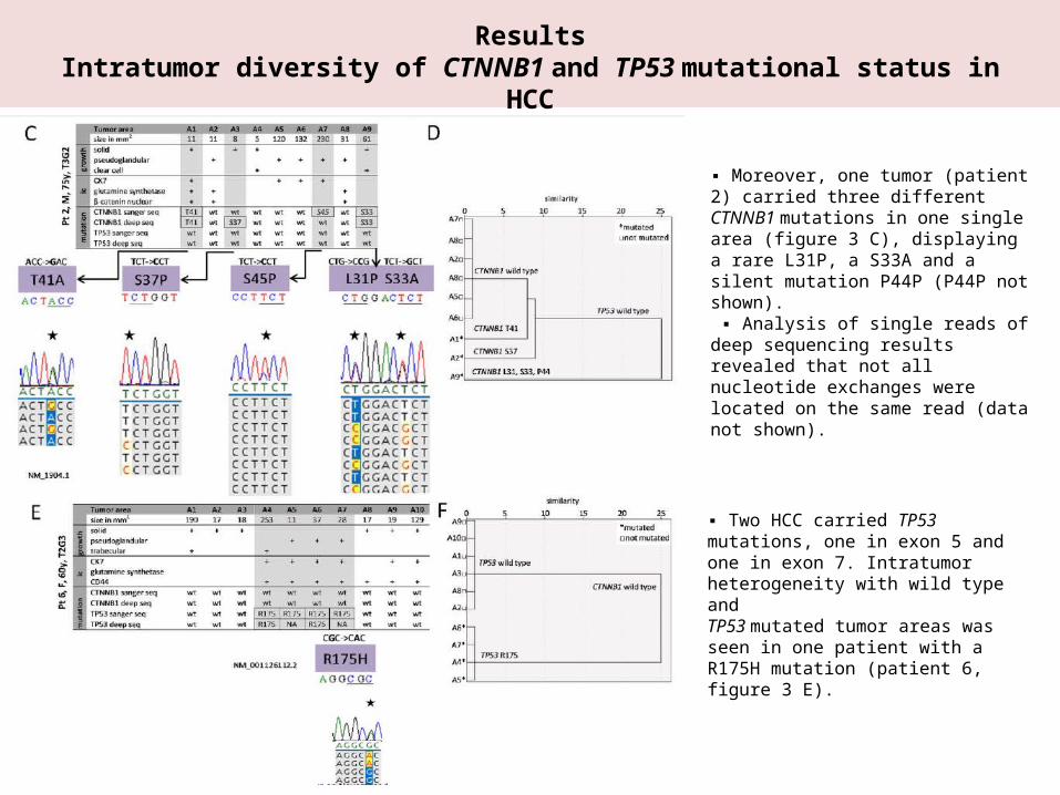
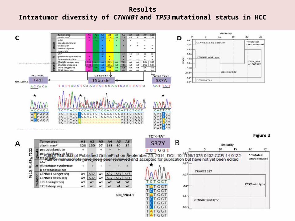
▪ Moreover, one tumor (patient 2) carried three different CTNNB1 mutations in one single area (figure 3 C), displaying a rare L31P, a S33A and a silent mutation P44P (P44P notshown). ▪ Analysis of single reads of deep sequenc-ing results revealed that not all nucleotide exchanges were located on the same read (data not shown).
▪ Two HCC carried TP53 mutations, one in exon 5 and one in exon 7. Intratumor het-erogeneity with wild type andTP53 mutated tumor areas was seen in one patient with a R175H mutation (patient 6, figure 3 E).
ResultsIntratumor diversity of CTNNB1 and TP53 mutational status in HCC
ResultsIntratumor diversity of CTNNB1 and TP53 mutational status in HCC
▪ Area-based analysis of mutational status, the expression of immunohistochemical markers and morphology revealed a positive association between CTNNB1 mutations, nuclear β-catenin and glutamine synthetase positivity.
▪ CD44 positivity was present in 70% of TP53 mutated areas. The association between TP53 mutation and β-catenin im-munohistochemistry found in our cohort was due to a single TP53 mutated HCC (patient 4) with nuclear β-catenin stain-ing in all tumor areas (table 3).
(Morphology type) 육주형 (trabecular) 위선형 (pseudoglandular) 투명세포형 (clear cell type)고형형 (solid type) 지방변화 (fatty change)Clear cell trabecular cell solid
Discussion
▪ In this study they systematically characterized intratumor heterogeneity in hepatocellular carcinoma (HCC) on the level of morphology, immune phenotype, or mutational status of CTNNB1 and TP53, respectively.
▪ With this approach, they found tumor heterogeneity of at least one feature in the majority of HCC (20/23; 87%).
▪ Although based on a relatively small number of cases, our findings have implications for HCC biology, HCC classifica-tion and HCC targeted therapy in the era of personalized medicine.
▪ Not unexpectedly, they observed that the frequency of morphological intratumor heterogeneity was associated with larger tumor size and higher tumor stage, although it did not reach statistical significance, most likely due to a small sample size.
▪ Studying TP53 and CTNNB1, they found a heterogeneous intratumor mutational status in 22% of HCC. Different types of CTNNB1 mutations in one tumor sample were also reported before by Van Nieuh et al. and Huang et al. Park et al. analyzed HCC.
▪ These findings show that CTNNB1 mutations, generally considered as driver mutations in HCC, are not uniformly present in all tumor regions within the same tumor, and reflect a rather late event in hepatocarcinogenesis.
▪ This finding might be the reason for some of the challenges in developing a robust classification of HCC as well as a molecular targeted therapy for this tumor entity.