Embed Size (px)
Citation preview
Research ArticleKaempferide Protects against Myocardial Ischemia/ReperfusionInjury through Activation of the PI3K/Akt/GSK-3β Pathway
Dong Wang,1 Xinjie Zhang,1 Defang Li,2 Wenjin Hao,2 Fanqing Meng,3 Bo Wang,2
Jichun Han,2 and Qiusheng Zheng2
1Department of Cardiac Surgery, Shandong Provincial Qianfoshan Hospital, Shandong University, Jinan 250014, China2Binzhou Medical University, Yantai, Shandong 264003, China3Weifang Medical University, Weifang, Shandong 261053, China
Correspondence should be addressed to Qiusheng Zheng; [email protected]
Received 24 January 2017; Revised 17 June 2017; Accepted 16 July 2017; Published 27 August 2017
Academic Editor: Hermann Gram
Copyright © 2017 Dong Wang et al. This is an open access article distributed under the Creative Commons AttributionLicense, which permits unrestricted use, distribution, and reproduction in any medium, provided the original work isproperly cited.
The aim of this study is to investigate both the efficacy and mechanism of action of kaempferide (Kae) as a therapy for thetreatment of cardiovascular disease. A rat model of myocardial ischemia/reperfusion (I/R) injury was established by ligationof the left anterior descending coronary artery for 30min followed by a 2 h perfusion. In our study, we show that Kaeremarkably improved cardiac function, alleviated myocardial injury via a decrease in myocardial enzyme levels, andattenuated myocardial infarct size in a dose-dependent manner. In addition, preconditioning treatment with Kae was foundto significantly decrease serum TNF-α, IL-6, C-reactive protein (CRP), MDA, and ROS levels, while it was found to increaseserum levels of SOD. Nuclear factor erythroid 2-related factor 2 (Nrf2) and cleaved caspase-3 expression levels wereobserved to be downregulated, while phospho-Akt (p-Akt) and phospho-glycogen synthase kinase-3β (p-GSK-3β) expressionlevels were upregulated. However, cotreatment with LY294002 (a PI3K inhibitor) or TDZD-8 (a GSK-3β inhibitor) wasfound to abolish the above cardioprotective effects observed with the Kae treatment. The data presented in this studyprovides evidence that Kae attenuates I/R-induced myocardial injury through inhibition of the Nrf2 and cleaved caspase-3signaling pathways via a PI3K/Akt/GSK 3β-dependent mechanism.
1. Introduction
Acute myocardial infarction (AMI) is a prevalent diseaseassociated with high morbidity and mortality rates [1].In China alone, over 700,000 people reportedly die fromthis disease every year [2]. Currently, the most successfultherapeutic strategy involves the pharmacological ormechanical restoration of coronary blood flow to preserveviable myocardium following AMI [3]. While reperfusiontherapy has brought new hope for the reduction of myo-cardial damage, reperfusion itself has been shown topotentially induce a localized oxidative burst and aregional inflammatory response, resulting in cell damageand, in some cases, death. This pathophysiologic process
has been defined in the field as an ischemia/reperfusioninjury (I/R injury) [4]. In order to improve clinical out-comes in the case of AMI, it is critical to develop novelpharmacological agents for the prevention of myocardialI/R injury.
I/R injury is an intricate process involving numerousmechanisms. Oxidative damage has been demonstrated toplay an important role in I/R injury progression [5]. Inaddition, antiapoptosis and anti-inflammation mechanismshave been reported to play protective roles for the heart inthe case of I/R injury. This provides further evidence thatapoptosis, inflammation, and oxidative injury all play arole in I/R injury [6, 7]. Therefore, the modulation ofapoptosis, inflammation, oxidative damage, and related
HindawiMediators of InflammationVolume 2017, Article ID 5278218, 11 pageshttps://doi.org/10.1155/2017/5278218
cascade responses is considered crucial therapeutic strategiesfor the treatment of cardiovascular I/R disease.
Phosphoinositide 3-kinases (PI3K) and their down-stream target, protein kinase B (Akt), have been shown tobe involved in the regulation of oxidation, inflammatoryresponses, and apoptosis [8–10]. Previous studies haveindicated that the PI3K/Akt/GSK-3β signaling pathwaymay function as an endogenous negative feedback regulatorthat generates a compensatory mechanism to limit proin-flammatory and apoptotic events in response to harmfulstimuli [11, 12]. Activation of PI3K/Akt/GSK3β-dependentsignaling has been demonstrated to result in the attenuationof myocardial I/R injury [13].
Flavonoids possess unique antioxidant properties andother pharmacological activities that may be relevant inprotecting the heart from I/R injury; studies have alsofound that many flavonoids can protect against myocardialI/R injury [14, 15]. Kaempferide (3,5,7-trihydroxy-4′-meth-oxyflavone, Kae) is a naturally occurring flavonoid that isisolated from the roots of Alpinia officinarum (lesser galan-gal) [16]. Like other flavonoids, Kae also has a very goodantioxidant properties and Kae can effectively reduce 1,1-diphenyl-2-picrylhydrazyl (DPPH) [17]. Studies have shownthat Kae has anticancer [18, 19] and antihypertension effects[20]. However, little is known regarding the mechanism ofaction of Kae as a therapeutic agent for the treatment ofcardiovascular disease. Thus, in this current study, we aimto investigate (1) whether Kae protects rat myocardium fromI/R injury in an in vivo rat model and (2) the possible role ofPI3K/Akt/GSK-3β signaling in the protective effects of Kaeagainst myocardial I/R injury.
2. Material and Methods
2.1. Ethics Statement. The animals were handled, and allprocedures were performed in accordance with the regula-tions of the Guide for the Care and Use of LaboratoryAnimals, approved by the Animal Care and Use Committeeof Shandong University (publication number SYXK (Lu)20130001, revised 2013).
2.2. Test Compounds, Chemicals, and Reagents. Kaempferide(purity≥ 98%) was purchased from Chengdu Must Bio-Technology Co. LTD. (Chengdu, China). 1,1,3,3-Tetra-methoxypropane was obtained from Fluka Chemical Co.(Ronkonkoma, NY). 2,3,5-Triphenyltetrazolium chloride,oxidized glutathione, and reduced glutathione were pur-chased from Sigma Chemical Co. (St. Louis, MO). Thecreatine kinase (CK) kits, lactate dehydrogenase (LDH) kits,superoxide dismutase (SOD) kits, malondialdehyde (MDA)kits, reactive oxygen species (ROS) kits, C-reactive protein(CRP) kits, interleukin-6 (IL-6) kits, and tumor necrosisfactor-α (TNF-α) kits were purchased from Tsz Biosciences(Greater Boston, USA). All other chemicals and reagentswere of analytical grade.
2.3. Animals. Adult Sprague Dawley (SD) rats were obtainedfrom Jinan Pengyue Experimental Animal Breeding Co.Ltd. (license number: SCXK (lu) 2014-0007). Animals were
between 250–300 g in weight and were housed in a roomat 22–25°C, with a relative humidity between 50–60%, anda 12 h light/12 h dark cycle. All experimental protocols wereapproved by the Institutional Animal Care and Use Com-mittee of Shandong University. Methods were carried outin accordance with the approved guidelines.
2.4. Ischemia/Reperfusion (I/R) Rat Model. We performedcoronary artery ligation (CAL) to induce I/R injury in SDrats. Our modeling method mainly refers to the reference[21] and makes corresponding improvements. In brief, ratswere anesthetized using 10% chloral hydrate (300mg/kgbody weight) via intraperitoneal injection and were ventilatedwith a HX-300S animal respirator (Chengdu Technology &Market Co. Ltd., Chengdu, China) (tidal volume, 6–8ml/kg;ventilator frequency, 80 breaths/min). The heart was exposedvia a left thoracotomy, and the left anterior descending (LAD)coronary artery was ligated for 30min followed by a 2 hreperfusion. Muscle tone and tail/pedal withdrawal responsewere used to monitor anesthesia adequacy. At the completionof the experiment, rats were sacrificed by the administrationof pentobarbital sodium (200mg/kg) via intraperitonealinjection and phlebotomy.
2.5. Experimental Groups. Rats were randomly divided into7 groups: (1) sham group, rats underwent sham surgery;(2) I/R group, rats were subjected to a 30min LAD coronaryartery ligation followed by a 2 h reperfusion; (3) L-Kae+ I/Rgroup, rats were injected with a low dose of Kae (L-Kae;0.1mg/kg body weight) 30min prior to I/R, then subjectedto a 30min LAD coronary artery ligation followed by a 2 hreperfusion; (4) M-Kae + I/R group, rats were injected witha moderate dose of Kae (M-Kae; 0.3mg/kg body weight)30min prior to I/R, then subjected to a 30min LAD coronaryartery ligation followed by a 2 h reperfusion; (5) H-Kae+ I/Rgroup, rats were injected with a high dose of Kae (H-Kae;1mg/kg body weight) 30min prior to I/R, then subjected toa 30min LAD coronary artery ligation followed by a 2 hreperfusion; (6) LY+H-Kae + I/R group, rats were injectedwith LY294002 (0.3mg/kg body weight) and H-Kae (1mg/kgbody weight) 30min prior to I/R, then subjected to a 30minLAD coronary artery ligation followed by a 2 h reperfusion;and (7) TD+H-Kae+ I/R group, rats were injected withTDZD-8 (0.3mg/kg body weight) and H-Kae (1mg/kg bodyweight) 30min prior to I/R, then subjected to a 30min LADcoronary artery ligation followed by a 2 h reperfusion.
2.6. Determination of Cardiac Function. After a 2 h reperfu-sion, a Philips IE33 ultrasound system (Philips Healthcare,Amsterdam, The Netherlands) was used to measure ejectionfraction (EF), fractional shortening (FS), left ventricularend-diastolic pressure (LVEDP), and left ventricular systolicpressure (LVSP) of rats in the study.
2.7. Determination of Myocardial Infarct. To evaluate the sizeof I/R-related myocardial infarct in rat hearts, we used TTCstaining to measure the myocardial infarct size. The experi-ments were performed as described previously [14, 22–24].
2 Mediators of Inflammation
2.8. Determination of CK and LDH Activities in Serum.After a 2 h reperfusion, serum levels of CK and LDH weremeasured using commercial kits according to the manu-facturer’s instructions.
2.9. Assay of Oxidative Stress and Inflammatory Factors.After a 2 h reperfusion, SOD activity, MDA levels,ROS levels, CRP levels, IL-6 levels, and TNF-α levelswere determined using commercial kits following themanufacturer’s instructions.
2.10. TUNEL. Terminal deoxynucleotidyl transfer-mediateddUTP nick end labeling (TUNEL) was carried out using anIn Situ Cell Death Detection Kit, POD (Roche, Germany)according to the manufacturer’s instructions. The experi-ments were performed as described previously [14, 22–24].
2.11. Western Blot Analysis. Proteins levels of total glycogensynthase kinase-3β (GSK-3β), phospho-GSK-3β (P-GSK-3β), Akt, phospho-Akt (p-Akt), Nrf2, cleaved caspase-3,and caspase-3 were measured by Western blot. Followingperfusion with the Langendorff apparatus, the same part ofthe rat heart was cut and collected from each sample, homog-enized in buffer (50mM Tris-HCl, pH7.6, 0.5% Triton X-100, 20% glycerol), and centrifuged at 15,000×g for 15minat 4°C. The supernatant was then collected and boiled for15min in order to denature the proteins. The resulting wholecell protein extracts were separated by electrophoresis on a12% SDS polyacrylamide gel and subsequently transferredto nylon membrane using an electrophoretic transfer system.Membranes were then incubated first with the followingprimary antibodies: rabbit anti-rat GSK-3β, rabbit anti-ratP-GSK-3β, rabbit anti-rat Akt, rabbit anti-rat p-Akt, rabbitanti-rat Nrf2, rabbit anti-rat cleaved caspase-3, rabbitanti-rat caspase-3, and rabbit anti-rat β-actin polyclonalantibodies (Cell Signaling, Beverly, MA, USA) at 4°C over-night. Membranes were then washed with TBS-T bufferand incubated with horseradish peroxidase-conjugated sec-ondary antibody (Cell Signaling, Beverly, MA, USA). Finally,membranes were developed using ECL-plus reagent tovisualize protein bands and imaged using the Bio-Rad GelDoc 2000 imaging system. Bio-Rad Gel Doc 2000 imagingsoftware was used to calculate the integrated absorbance(IA) of the bands. IA= area× average density. Followingnormalization to β-actin levels, the ratios of the IAs ofGSK-3β, P-GSK-3β, Akt, p-Akt, Nrf2, cleaved caspase-3,and caspase-3 to the IA of β-actin were used to representrelative levels of activated GSK-3β, P-GSK-3β, Akt, p-Akt,Nrf2, cleaved caspase-3, and caspase-3, respectively.
2.12. Statistical Analysis. Data are presented as the mean±standard deviation from at least six independent experi-ments. Statistical differences were determined using analysisof variance (ANOVA), where P < 0 05 was considered statis-tically significant. The analyses were performed using theStatistical Program for Social Sciences Software (IBM SPSS,International Business Machines Corporation, Armonk City,New York, USA).
3. Results
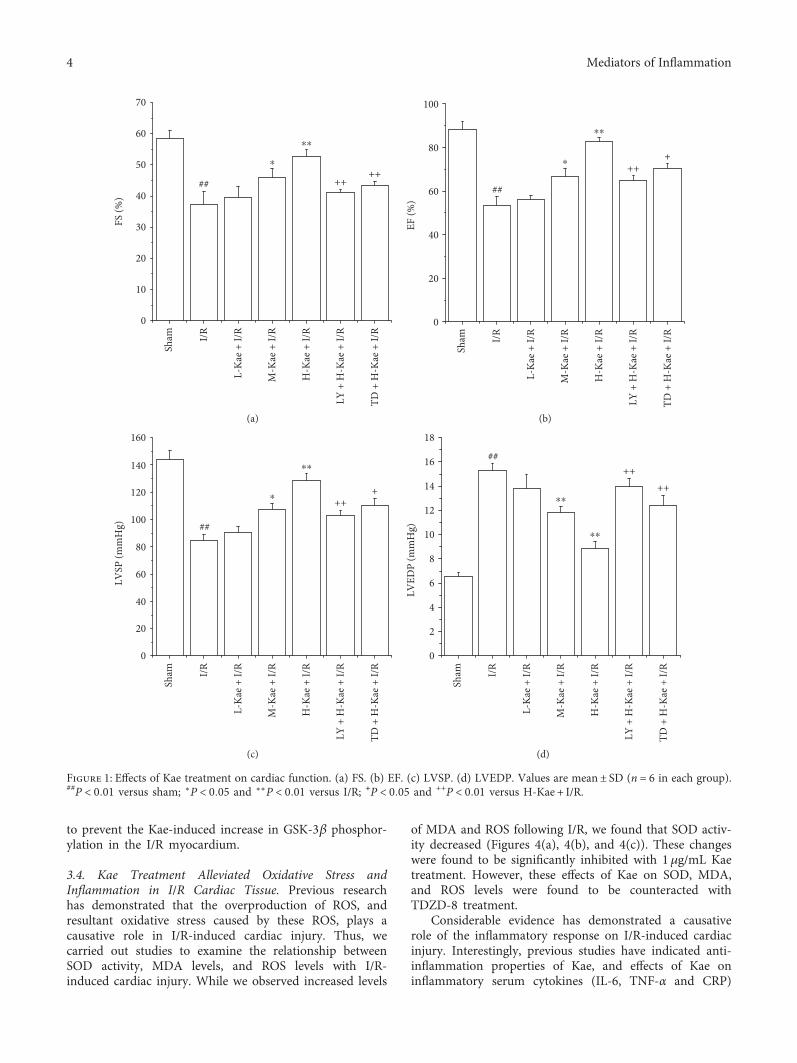
3.1. The Effect of Kae on Cardiac Function. We first inves-tigated the effect of Kae on cardiac function. As shown inFigure 1, I/R injury was found to greatly decrease FS(Figure 1(a)), EF (Figure 1(b)), and LVSP (Figure 1(c)).However, I/R injury was found to increase LVEDP(Figure 1(d)) in the I/R group (P < 0 01). Compared to theI/R group hearts, the M-Kae + I/R and H-Kae + I/R groups’hearts exhibited a significant functional recovery, with theefficacy of H-Kae on cardiac function found to be greaterthan that of M-Kae. However, this protective effect of Kaewas found to be reversed by cotreatment with LY294002or TDZD-8.
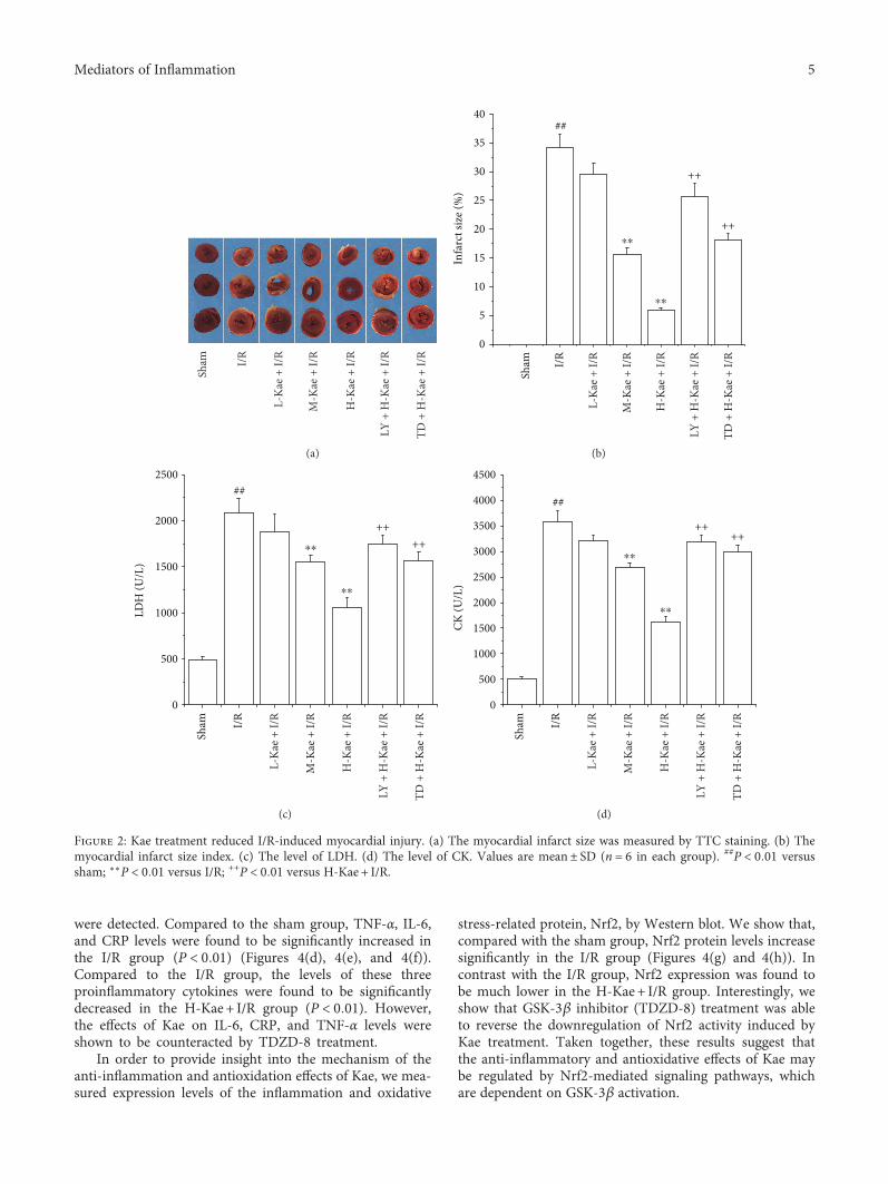
3.2. Kae Decreased Myocardial Injury in I/R Rats.Myocardialinfarct size, LDH, and CK levels were measured in order todetermine whether Kae treatment resulted in a reductionin myocardial injury. We show that while hearts that under-went myocardial ischemia for 30min followed by 2h ofreperfusion (I/R group) exhibited a significant increase inmyocardial infarct size, pretreatment with M-Kae + I/R andH-Kae + I/R significantly decreased the myocardial infarctsize induced by I/R, with a more significant effect observedwith H-Kae compared with M-Kae (Figures 2(a) and 2(b)).Compared with the H-Kae + I/R group, pretreatment withTDZD-8 and LY294002 was found to counteract the effectsof Kae on myocardial infarct size.
We demonstrate a marked increase in the leakage ofmyocardial isoenzyme (LDH and CK) in the I/R groupafter 30min of ischemia, followed by 2h of reperfusion,compared to that of the sham group (Figures 2(c) and2(d)). In contrast, M-Kae and H-Kae pretreatments werefound to significantly reduce the I/R-induced leakage ofLDH and CK, with the inhibitory effect of H-Kae greaterthan that of M-Kae. However, the protective effect of Kaewas demonstrated to be reversed by cotreatment with eitherLY294002 or TDZD-8. Considering the significant cardio-protective effects observed with Kae treatment at 1mg/kgbody weight (H-Kae), this concentration was chosen forsubsequent assays.
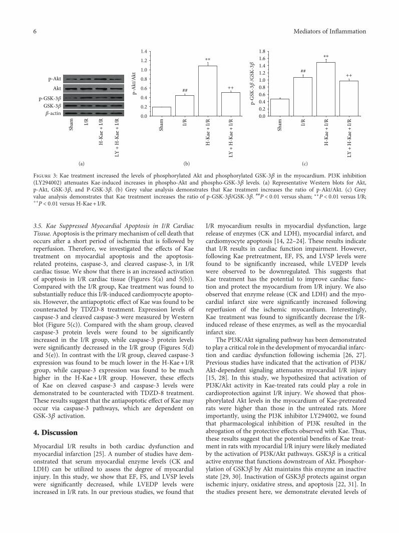
3.3. Kae Protects against Myocardial I/R Injury by theActivation of PI3K/Akt/GSK-3β Pathways. Because thecardioprotective effects of Kae were found to be reversed bycotreatment with LY294002 (a PI3K inhibitor) or TDZD-8(a GSK-3β inhibitor), we further examined the regulationof Kae on the PI3K signaling pathway. Expression levels ofGSK-3β, p-GSK-3β, Akt, and p-Akt were measured usingWestern blot. As depicted in Figure 3, Kae treatment wasfound to result in a significant increase in myocardiallevels of pAkt in the I/R group compared with those inthe untreated I/R group. However, administration of thePI3K inhibitor, LY294002, was found to significantly attenu-ate the Kae-induced upregulation of p-Akt. In addition, Kaetreatment was found to result in a significant increase inp-GSK-3β levels, a downstream kinase of Akt, in the myo-cardium compared with untreated I/R hearts. However,administration of the PI3K inhibitor, LY294002, was found
3Mediators of Inflammation
to prevent the Kae-induced increase in GSK-3β phosphor-ylation in the I/R myocardium.
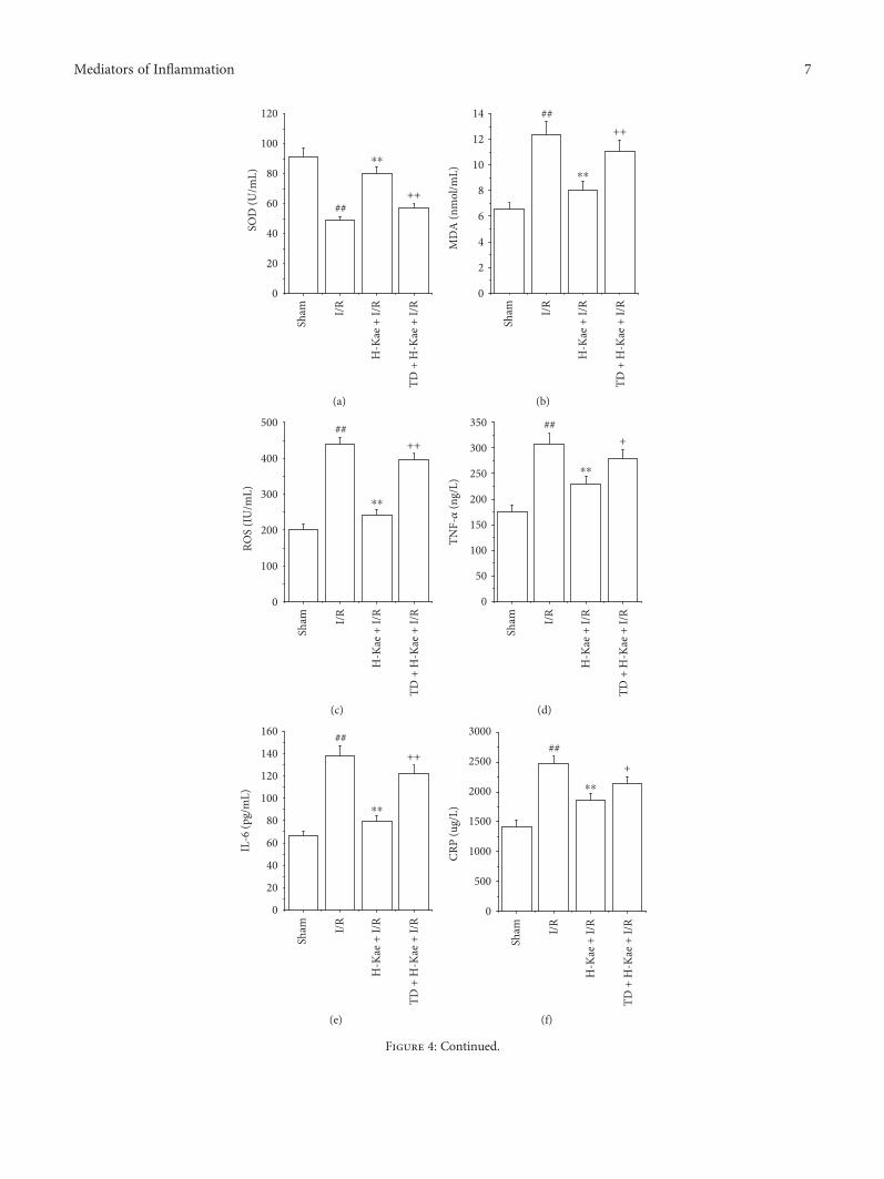
3.4. Kae Treatment Alleviated Oxidative Stress andInflammation in I/R Cardiac Tissue. Previous researchhas demonstrated that the overproduction of ROS, andresultant oxidative stress caused by these ROS, plays acausative role in I/R-induced cardiac injury. Thus, wecarried out studies to examine the relationship betweenSOD activity, MDA levels, and ROS levels with I/R-induced cardiac injury. While we observed increased levels
of MDA and ROS following I/R, we found that SOD activ-ity decreased (Figures 4(a), 4(b), and 4(c)). These changeswere found to be significantly inhibited with 1μg/mL Kaetreatment. However, these effects of Kae on SOD, MDA,and ROS levels were found to be counteracted withTDZD-8 treatment.
Considerable evidence has demonstrated a causativerole of the inflammatory response on I/R-induced cardiacinjury. Interestingly, previous studies have indicated anti-inflammation properties of Kae, and effects of Kae oninflammatory serum cytokines (IL-6, TNF-α and CRP)
0
10
20
30
40
50
60
70
++
FS (%
)
## ++Sh
am I/R
TD +
H-K
ae +
I/R
LY +
H-K
ae +
I/R
H-K
ae +
I/R
M-K
ae +
I/R
L-Ka
e + I/
R
⁎
⁎⁎
(a)
0
20
40
60
80
100
+++
EF (%
)
##
Sham I/R
TD +
H-K
ae +
I/R
LY +
H-K
ae +
I/R
H-K
ae +
I/R
M-K
ae +
I/R
L-Ka
e + I/
R
⁎
⁎⁎
(b)
0
20
40
60
80
100
120
140
160
+++
LVSP
(mm
Hg) ##
Sham I/R
⁎
⁎⁎
TD +
H-K
ae +
I/R
LY +
H-K
ae +
I/R
H-K
ae +
I/R
M-K
ae +
I/R
L-Ka
e + I/
R
(c)
0
2
4
6
8
10
12
14
16
18
++++
LVED
P (m
mH
g)
##Sh
am I/R
⁎⁎
⁎⁎
TD +
H-K
ae +
I/R
LY +
H-K
ae +
I/R
H-K
ae +
I/R
M-K
ae +
I/R
L-Ka
e + I/
R
(d)
Figure 1: Effects of Kae treatment on cardiac function. (a) FS. (b) EF. (c) LVSP. (d) LVEDP. Values are mean± SD (n = 6 in each group).##P < 0 01 versus sham; ∗P < 0 05 and ∗∗P < 0 01 versus I/R; +P < 0 05 and ++P < 0 01 versus H-Kae + I/R.
4 Mediators of Inflammation
were detected. Compared to the sham group, TNF-α, IL-6,and CRP levels were found to be significantly increased inthe I/R group (P < 0 01) (Figures 4(d), 4(e), and 4(f)).Compared to the I/R group, the levels of these threeproinflammatory cytokines were found to be significantlydecreased in the H-Kae + I/R group (P < 0 01). However,the effects of Kae on IL-6, CRP, and TNF-α levels wereshown to be counteracted by TDZD-8 treatment.
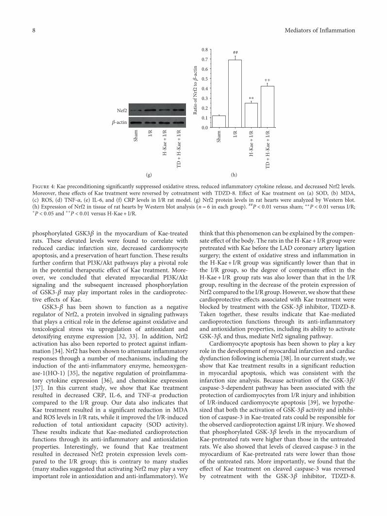
In order to provide insight into the mechanism of theanti-inflammation and antioxidation effects of Kae, we mea-sured expression levels of the inflammation and oxidative
stress-related protein, Nrf2, by Western blot. We show that,compared with the sham group, Nrf2 protein levels increasesignificantly in the I/R group (Figures 4(g) and 4(h)). Incontrast with the I/R group, Nrf2 expression was found tobe much lower in the H-Kae + I/R group. Interestingly, weshow that GSK-3β inhibitor (TDZD-8) treatment was ableto reverse the downregulation of Nrf2 activity induced byKae treatment. Taken together, these results suggest thatthe anti-inflammatory and antioxidative effects of Kae maybe regulated by Nrf2-mediated signaling pathways, whichare dependent on GSK-3β activation.
Sham I/R
TD +
H-K
ae +
I/R
LY +
H-K
ae +
I/R
H-K
ae +
I/R
M-K
ae +
I/R
L-Ka
e + I/
R
(a)
0
5
10
15
20
25
30
35
40
++
++
Infa
rct s
ize (
%)
##
Sham I/R
⁎⁎
⁎⁎
TD +
H-K
ae +
I/R
LY +
H-K
ae +
I/R
H-K
ae +
I/R
M-K
ae +
I/R
L-Ka
e + I/
R
(b)
0
500
1000
1500
2000
2500
++++
LDH
(U/L
)
##
Sham I/R
⁎⁎
⁎⁎
TD +
H-K
ae +
I/R
LY +
H-K
ae +
I/R
H-K
ae +
I/R
M-K
ae +
I/R
L-Ka
e + I/
R
(c)
0
500
1000
1500
2000
2500
3000
3500
4000
4500
++++
CK (U
/L)
##Sh
am I/R⁎⁎
⁎⁎
TD +
H-K
ae +
I/R
LY +
H-K
ae +
I/R
H-K
ae +
I/R
M-K
ae +
I/R
L-Ka
e + I/
R
(d)
Figure 2: Kae treatment reduced I/R-induced myocardial injury. (a) The myocardial infarct size was measured by TTC staining. (b) Themyocardial infarct size index. (c) The level of LDH. (d) The level of CK. Values are mean± SD (n = 6 in each group). ##P < 0 01 versussham; ∗∗P < 0 01 versus I/R; ++P < 0 01 versus H-Kae + I/R.
5Mediators of Inflammation
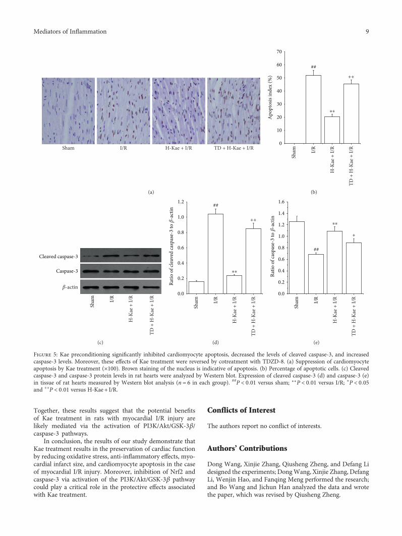
3.5. Kae Suppressed Myocardial Apoptosis in I/R CardiacTissue.Apoptosis is the primary mechanism of cell death thatoccurs after a short period of ischemia that is followed byreperfusion. Therefore, we investigated the effects of Kaetreatment on myocardial apoptosis and the apoptosis-related proteins, caspase-3, and cleaved caspase-3, in I/Rcardiac tissue. We show that there is an increased activationof apoptosis in I/R cardiac tissue (Figures 5(a) and 5(b)).Compared with the I/R group, Kae treatment was found tosubstantially reduce this I/R-induced cardiomyocyte apopto-sis. However, the antiapoptotic effect of Kae was found to becounteracted by TDZD-8 treatment. Expression levels ofcaspase-3 and cleaved caspase-3 were measured by Westernblot (Figure 5(c)). Compared with the sham group, cleavedcaspase-3 protein levels were found to be significantlyincreased in the I/R group, while caspase-3 protein levelswere significantly decreased in the I/R group (Figures 5(d)and 5(e)). In contrast with the I/R group, cleaved caspase-3expression was found to be much lower in the H-Kae + I/Rgroup, while caspase-3 expression was found to be muchhigher in the H-Kae + I/R group. However, these effectsof Kae on cleaved caspase-3 and caspase-3 levels weredemonstrated to be counteracted with TDZD-8 treatment.These results suggest that the antiapoptotic effect of Kae mayoccur via caspase-3 pathways, which are dependent onGSK-3β activation.
4. Discussion
Myocardial I/R results in both cardiac dysfunction andmyocardial infarction [25]. A number of studies have dem-onstrated that serum myocardial enzyme levels (CK andLDH) can be utilized to assess the degree of myocardialinjury. In this study, we show that EF, FS, and LVSP levelswere significantly decreased, while LVEDP levels wereincreased in I/R rats. In our previous studies, we found that
I/R myocardium results in myocardial dysfunction, largerelease of enzymes (CK and LDH), myocardial infarct, andcardiomyocyte apoptosis [14, 22–24]. These results indicatethat I/R results in cardiac function impairment. However,following Kae pretreatment, EF, FS, and LVSP levels werefound to be significantly increased, while LVEDP levelswere observed to be downregulated. This suggests thatKae treatment has the potential to improve cardiac func-tion and protect the myocardium from I/R injury. We alsoobserved that enzyme release (CK and LDH) and the myo-cardial infarct size were significantly increased followingreperfusion of the ischemic myocardium. Interestingly,Kae treatment was found to significantly decrease the I/R-induced release of these enzymes, as well as the myocardialinfarct size.
The PI3K/Akt signaling pathway has been demonstratedto play a critical role in the development of myocardial infarc-tion and cardiac dysfunction following ischemia [26, 27].Previous studies have indicated that the activation of PI3K/Akt-dependent signaling attenuates myocardial I/R injury[15, 28]. In this study, we hypothesized that activation ofPI3K/Akt activity in Kae-treated rats could play a role incardioprotection against I/R injury. We showed that phos-phorylated Akt levels in the myocardium of Kae-pretreatedrats were higher than those in the untreated rats. Moreimportantly, using the PI3K inhibitor LY294002, we foundthat pharmacological inhibition of PI3K resulted in theabrogation of the protective effects observed with Kae. Thus,these results suggest that the potential benefits of Kae treat-ment in rats with myocardial I/R injury were likely mediatedby the activation of PI3K/Akt pathways. GSK3β is a criticalactive enzyme that functions downstream of Akt. Phosphor-ylation of GSK3β by Akt maintains this enzyme an inactivestate [29, 30]. Inactivation of GSK3β protects against organischemic injury, oxidative stress, and apoptosis [22, 31]. Inthe studies present here, we demonstrate elevated levels of
Sham I/R
H-K
ae +
I/R
LY +
H-K
ae +
I/R
�훽-actinGSK-3�훽
p-GSK-3�훽
Akt
p-Akt
(a)
0.0
0.2
0.4
0.6
0.8
1.0
1.2
1.4
p-A
kt/A
kt
##
⁎⁎
++
Sham I/R
H-K
ae +
I/R
LY +
H-K
ae +
I/R
(b)
0.00.20.40.60.81.01.21.41.61.8
p-G
SK-3�훽
/GSK
-3�훽
##++
Sham I/R
⁎⁎
H-K
ae +
I/R
LY +
H-K
ae +
I/R
(c)
Figure 3: Kae treatment increased the levels of phosphorylated Akt and phosphorylated GSK-3β in the myocardium. PI3K inhibition(LY294002) attenuates Kae-induced increases in phospho-Akt and phospho-GSK-3β levels. (a) Representative Western blots for Akt,p-Akt, GSK-3β, and P-GSK-3β. (b) Grey value analysis demonstrates that Kae treatment increases the ratio of p-Akt/Akt. (c) Greyvalue analysis demonstrates that Kae treatment increases the ratio of p-GSK-3β/GSK-3β. ##P < 0 01 versus sham; ∗∗P < 0 01 versus I/R;++P < 0 01 versus H-Kae + I/R.
6 Mediators of Inflammation
0
20
40
60
80
100
120
SOD
(U/m
L)
++##
⁎⁎
Sham I/R
H-K
ae +
I/R
TD +
H-K
ae +
I/R
(a)
0
2
4
6
8
10
12
14
MD
A (n
mol
/mL)
++##
⁎⁎
Sham I/R
H-K
ae +
I/R
TD +
H-K
ae +
I/R
(b)
0
100
200
300
400
500
ROS
(IU
/mL)
++##
⁎⁎
Sham I/R
H-K
ae +
I/R
TD +
H-K
ae +
I/R
(c)
0
50
100
150
200
250
300
350+
TNF-�훼
(ng/
L)
##
⁎⁎
Sham I/R
H-K
ae +
I/R
TD +
H-K
ae +
I/R
(d)
0
20
40
60
80
100
120
140
160
++
IL-6
(pg/
mL)
##
⁎⁎
Sham I/R
H-K
ae +
I/R
TD +
H-K
ae +
I/R
(e)
0
500
1000
1500
2000
2500
3000
CRP
(ug/
L)
+##
⁎⁎
Sham I/R
H-K
ae +
I/R
TD +
H-K
ae +
I/R
(f)
Figure 4: Continued.
7Mediators of Inflammation
phosphorylated GSK3β in the myocardium of Kae-treatedrats. These elevated levels were found to correlate withreduced cardiac infarction size, decreased cardiomyocyteapoptosis, and a preservation of heart function. These resultsfurther confirm that PI3K/Akt pathways play a pivotal rolein the potential therapeutic effect of Kae treatment. More-over, we concluded that elevated myocardial PI3K/Aktsignaling and the subsequent increased phosphorylationof GSK3-β may play important roles in the cardioprotec-tive effects of Kae.
GSK3-β has been shown to function as a negativeregulator of Nrf2, a protein involved in signaling pathwaysthat plays a critical role in the defense against oxidative andtoxicological stress via upregulation of antioxidant anddetoxifying enzyme expression [32, 33]. In addition, Nrf2activation has also been reported to protect against inflam-mation [34]. Nrf2 has been shown to attenuate inflammatoryresponses through a number of mechanisms, including theinduction of the anti-inflammatory enzyme, hemeoxygen-ase-1(HO-1) [35], the negative regulation of proinflamma-tory cytokine expression [36], and chemokine expression[37]. In this current study, we show that Kae treatmentresulted in decreased CRP, IL-6, and TNF-α productioncompared to the I/R group. Our data also indicates thatKae treatment resulted in a significant reduction in MDAand ROS levels in I/R rats, while it improved the I/R-inducedreduction of total antioxidant capacity (SOD activity).These results indicate that Kae-mediated cardioprotectionfunctions through its anti-inflammatory and antioxidationproperties. Interestingly, we found that Kae treatmentresulted in decreased Nrf2 protein expression levels com-pared to the I/R group; this is contrary to many studies(many studies suggested that activating Nrf2 may play a veryimportant role in antioxidation and anti-inflammatory). We
think that this phenomenon can be explained by the compen-sate effect of the body. The rats in the H-Kae + I/R group werepretreated with Kae before the LAD coronary artery ligationsurgery; the extent of oxidative stress and inflammation inthe H-Kae + I/R group was significantly lower than that inthe I/R group, so the degree of compensate effect in theH-Kae + I/R group rats was also lower than that in the I/Rgroup, resulting in the decrease of the protein expression ofNrf2 compared to the I/R group. However, we show that thesecardioprotective effects associated with Kae treatment wereblocked by treatment with the GSK-3β inhibitor, TDZD-8.Taken together, these results indicate that Kae-mediatedcardioprotection functions through its anti-inflammatoryand antioxidation properties, including its ability to activateGSK-3β, and thus, mediate Nrf2 signaling pathway.
Cardiomyocyte apoptosis has been shown to play a keyrole in the development of myocardial infarction and cardiacdysfunction following ischemia [38]. In our current study, weshow that Kae treatment results in a significant reductionin myocardial apoptosis, which was consistent with theinfarction size analysis. Because activation of the GSK-3β/caspase-3-dependent pathway has been associated with theprotection of cardiomyocytes from I/R injury and inhibitionof I/R-induced cardiomyocyte apoptosis [39], we hypothe-sized that both the activation of GSK-3β activity and inhibi-tion of caspase-3 in Kae-treated rats could be responsible forthe observed cardioprotection against I/R injury. We showedthat phosphorylated GSK-3β levels in the myocardium ofKae-pretreated rats were higher than those in the untreatedrats. We also showed that levels of cleaved caspase-3 in themyocardium of Kae-pretreated rats were lower than thoseof the untreated rats. More importantly, we found that theeffect of Kae treatment on cleaved caspase-3 was reversedby cotreatment with the GSK-3β inhibitor, TDZD-8.
Sham I/R
Nef2
�훽-actin
H-K
ae +
I/R
TD +
H-K
ae +
I/R
(g)
0.0
0.1
0.2
0.3
0.4
0.5
0.6
0.7
0.8
Ratio
of N
rf2 to
�훽‑a
ctin
##
++
⁎⁎
Sham I/R
H-K
ae +
I/R
TD +
H-K
ae +
I/R
(h)
Figure 4: Kae preconditioning significantly suppressed oxidative stress, reduced inflammatory cytokine release, and decreased Nrf2 levels.Moreover, these effects of Kae treatment were reversed by cotreatment with TDZD-8. Effect of Kae treatment on (a) SOD, (b) MDA,(c) ROS, (d) TNF-α, (e) IL-6, and (f) CRP levels in I/R rat model. (g) Nrf2 protein levels in rat hearts were analyzed by Western blot.(h) Expression of Nrf2 in tissue of rat hearts by Western blot analysis (n = 6 in each group). ##P < 0 01 versus sham; ∗∗P < 0 01 versus I/R;+P < 0 05 and ++P < 0 01 versus H-Kae + I/R.
8 Mediators of Inflammation
Together, these results suggest that the potential benefitsof Kae treatment in rats with myocardial I/R injury arelikely mediated via the activation of PI3K/Akt/GSK-3β/caspase-3 pathways.
In conclusion, the results of our study demonstrate thatKae treatment results in the preservation of cardiac functionby reducing oxidative stress, anti-inflammatory effects, myo-cardial infarct size, and cardiomyocyte apoptosis in the caseof myocardial I/R injury. Moreover, inhibition of Nrf2 andcaspase-3 via activation of the PI3K/Akt/GSK-3β pathwaycould play a critical role in the protective effects associatedwith Kae treatment.
Conflicts of Interest
The authors report no conflict of interests.
Authors’ Contributions
Dong Wang, Xinjie Zhang, Qiusheng Zheng, and Defang Lidesigned the experiments; DongWang, Xinjie Zhang, DefangLi, Wenjin Hao, and Fanqing Meng performed the research;and Bo Wang and Jichun Han analyzed the data and wrotethe paper, which was revised by Qiusheng Zheng.
Sham I/R H-Kae + I/R TD + H-Kae + I/R
(a)
0
10
20
30
40
50
60
70
Apo
ptos
is in
dex
(%)
##
++
Sham I/R
⁎⁎
H-K
ae +
I/R
TD +
H-K
ae +
I/R
(b)
Sham I/R
Cleaved caspase-3
Caspase-3
�훽-actin
H-K
ae +
I/R
TD +
H-K
ae +
I/R
(c)
0.0
0.2
0.4
0.6
0.8
1.0
1.2 ##
++
Ratio
of c
leav
ed ca
spas
e-3
to �훽
-act
in
Sham I/R
⁎⁎
H-K
ae +
I/R
TD +
H-K
ae +
I/R
(d)
0.0
0.2
0.4
0.6
0.8
1.0
1.2
1.4
1.6
##
+
Ratio
of c
aspa
se-3
to �훽
-act
in
Sham I/R
⁎⁎
H-K
ae +
I/R
TD +
H-K
ae +
I/R
(e)
Figure 5: Kae preconditioning significantly inhibited cardiomyocyte apoptosis, decreased the levels of cleaved caspase-3, and increasedcaspase-3 levels. Moreover, these effects of Kae treatment were reversed by cotreatment with TDZD-8. (a) Suppression of cardiomyocyteapoptosis by Kae treatment (×100). Brown staining of the nucleus is indicative of apoptosis. (b) Percentage of apoptotic cells. (c) Cleavedcaspase-3 and caspase-3 protein levels in rat hearts were analyzed by Western blot. Expression of cleaved caspase-3 (d) and caspase-3 (e)in tissue of rat hearts measured by Western blot analysis (n = 6 in each group). ##P < 0 01 versus sham; ∗∗P < 0 01 versus I/R; +P < 0 05and ++P < 0 01 versus H-Kae + I/R.
9Mediators of Inflammation
Acknowledgments
This study was supported by a funding from the BinzhouMedical University (BY2014KYQD30) to Wenjin Hao.
References
[1] J. Zhu, X. Su, G. Li, J. Chen, B. Tang, and Y. Yang, “The inci-dence of acute myocardial infarction in relation to overweightand obesity: a meta-analysis,” Archives of Medical Science,vol. 10, no. 5, pp. 855–862, 2014.
[2] P. Wang, B. Zhang, L. Jin, H. Liao, and T. Dong, “Associationof various risk factors with prognosis and hospitalization costin Chinese patients with acute myocardial infarction: a clinicalanalysis of 627 cases,” Experimental and Therapeutic Medicine,vol. 9, no. 2, pp. 603–611, 2014.
[3] L. Lapointe-Shaw and C. M. Bell, “Acute myocardial infarc-tion,” British Medical Journal, vol. 348, article F7696, 2014.
[4] K. Ueda, H. Takano, Y. Niitsuma et al., “Sonic hedgehog is acritical mediator of erythropoietin-induced cardiac protectionin mice,” The Journal of Clinical Investigation, vol. 120, no. 6,pp. 2016–2029, 2010.
[5] J. K. Du, B. H. Cong, Q. Yu et al., “Upregulation of microRNA-22 contributes to myocardial ischemia-reperfusion injury byinterfering with the mitochondrial function,” Free RadicalBiology & Medicine, vol. 96, pp. 406–417, 2016.
[6] Q. Zhang, M. Shang, M. Zhang et al., “Microvesicles derivedfrom hypoxia/reoxygenation-treated human umbilical veinendothelial cells promote apoptosis and oxidative stress inH9c2 cardiomyocytes,” BMC Cell Biology, vol. 17, no. 1,p. 25, 2016.
[7] Y. Nakano, T. Matoba, M. Tokutome et al., “Nanoparticle-mediated delivery of irbesartan induces cardioprotection frommyocardial ischemia-reperfusion injury by antagonizingmonocyte-mediated inflammation,” Scientific Reports, vol. 6,article 29601, 2016.
[8] A. Patruno, S. Franceschelli, M. Pesce et al., “Novelaminobenzyl-acetamidine derivative modulate the differentialregulation of NOSs in LPS induced inflammatory response:role of PI3K/Akt pathway,” Biochimica et Biophysica Acta(BBA) - General Subjects, vol. 1820, no. 12, pp. 2095–2104,2012.
[9] D. Kumar, B. Das, R. Sen et al., “Andrographolide analogueinduces apoptosis and autophagy mediated cell death inU937 cells by inhibition of PI3K/Akt/mTOR pathway,” PLoSOne, vol. 10, no. 10, article e0139657, 2015.
[10] H. Zhang, Z. Xiong, J. Wang et al., “Glucagon-like peptide-1protects cardiomyocytes from advanced oxidation proteinproduct-induced apoptosis via the PI3K/Akt/bad signalingpathway,” Molecular Medicine Reports, vol. 13, no. 2,pp. 1593–1601, 2016.
[11] T. Zheng, X. Yang, D. Wu et al., “Salidroside amelioratesinsulin resistance through activation of a mitochondria-associated AMPK/PI3K/Akt/GSK3β pathway,” British Journalof Pharmacology, vol. 172, no. 13, pp. 3284–3301, 2015.
[12] Y. Zhang, Z. Zhang, H. Wang et al., “Neuroprotective effect ofginsenoside Rg1 prevents cognitive impairment induced byisoflurane anesthesia in aged rats via15 antioxidant, anti-inflammatory and anti-apoptotic effects mediated by thePI3K/AKT/GSK-3β pathway,” Molecular Medicine Reports,vol. 14, no. 3, pp. 2778–2784, 2016.
[13] C. W. Liu, F. Yang, S. Z. Cheng, Y. Liu, L. H. Wan, andH. L. Cong, “Rosuvastatin postconditioning protects isolatedhearts against ischemia-reperfusion injury: the role of radicaloxygen species, PI3K-Akt-GSK-3β pathway and mitochon-drial permeability transition pore,” Cardiovascular Therapeu-tics, vol. 12, no. 11, pp. 45–50, 2016.
[14] D. Qu, J. Han, H. Ren et al., “Cardioprotective effects ofastragalin against myocardial ischemia/reperfusion injury inisolated rat heart,” Oxidative Medicine and Cellular Longevity,vol. 2016, Article ID 8194690, 11 pages, 2016.
[15] H. Li, F. Song, L. R. Duan et al., “Paeonol and danshensucombination attenuates apoptosis in myocardial infarcted ratsby inhibiting oxidative stress: roles of Nrf2/HO-1 and PI3K/Akt pathway,” Scientific Reports, vol. 6, article 23693, 2016.
[16] H. Matsuda, S. Nakashima, Y. Oda, S. Nakamura, and M.Yoshikawa, “Melanogenesis inhibitors from the rhizomes ofAlpinia officinarum in B16 melanoma cells,” Bioorganic &Medicinal Chemistry, vol. 17, no. 16, pp. 6048–6053, 2009.
[17] Q. Y. Bian, S. Y. Wang, L. J. Xu, C. O. Chan, D. K. Mok,and S. B. Chen, “Two new antioxidant diarylheptanoidsfrom the fruits of Alpinia oxyphylla,” Journal of Asian NaturalProducts Research, vol. 15, no. 10, pp. 1094–1099, 2013.
[18] V. S. Nguyen, L. Shi, F. Q. Luan, and Q. A. Wang, “Synthesis ofkaempferide Mannich base derivatives and their antiprolifera-tive activity on three human cancer cell lines,” Acta BiochimicaPolonica, vol. 62, no. 3, pp. 547–552, 2015.
[19] V. Martineti, I. Tognarini, C. Azzari et al., “Inhibition ofin vitro growth and arrest in the G0/G1 phase of HCT8 linehuman colon cancer cells by kaempferide triglycoside fromDianthus caryophyllus,” Phytotherapy Research, vol. 24,no. 9, pp. 1302–1308, 2010.
[20] H. Maruyama, Y. Sumitou, T. Sakamoto, Y. Araki, andH. Hara, “Antihypertensive effects of flavonoids isolatedfrom Brazilian green propolis in spontaneously hypertensiverats,” Biological & Pharmaceutical Bulletin, vol. 32, no. 7,pp. 1244–1250, 2009.
[21] D. Lin, J. Ma, Y. Xue, and Z. Wang, “Penehyclidine hydrochlo-ride preconditioning provides cardioprotection in a rat modelof myocardial ischemia/reperfusion injury,” PLoS One, vol. 10,no. 12, article e0138051, 2015.
[22] M. Zhou, H. Ren, J. Han, W. Wang, Q. Zheng, and D. Wang,“Protective effects of Kaempferol against myocardial ische-mia/reperfusion injury in isolated rat heart via antioxidantactivity and inhibition of glycogen synthase kinase-3β,”Oxidative Medicine and Cellular Longevity, vol. 2015, ArticleID 481405, 8 pages, 2015.
[23] X. T. Tian, C. L. Liu, H. L. Jiang et al., “Cardioprotection pro-vided by Echinatin against ischemia/reperfusion in isolated rathearts,” BMC Cardiovascular Disorders, vol. 16, p. 119, 2016.
[24] J. Han, D. Wang, B. Yu et al., “Cardioprotection against ische-mia/reperfusion by licochalcone B in isolated rat hearts,” Oxi-dative Medicine and Cellular Longevity, vol. 2014, Article ID134862, 11 pages, 2014.
[25] Y. Qiu, N. Cong, M. Liang, Y. Wang, and J. Wang, “Systemspharmacology dissection of the protective effect of Myricetinagainst acute ischemia/reperfusion-induced myocardial injuryin isolated rat heart,” Cardiovascular Toxicology, vol. 17, no. 3,pp. 277–286, 2017.
[26] S. Liu, Q. Ai, K. Feng, Y. Li, and X. Liu, “The cardioprotectiveeffect of dihydromyricetin prevents ischemia-reperfusion-induced apoptosis in vivo and in vitro via the PI3K/Akt and
10 Mediators of Inflammation
HIF-1α signaling pathways,” Apoptosis, vol. 21, no. 12,pp. 1366–1385, 2016.
[27] B. Yang, P. Yan, H. Gong et al., “TWEAK protects cardiomyo-cyte against apoptosis in a PI3K/AKT pathway dependentmanner,” American Journal of Translational Research, vol. 8,no. 9, pp. 3848–3860, 2016.
[28] X. Y. Cheng, X. Y. Gu, Q. Gao, Q. F. Zong, X. H. Li, and Y.Zhang, “Effects of dexmedetomidine postconditioning onmyocardial ischemia and the role of the PI3K/Akt-dependentsignaling pathway in reperfusion injury,” Molecular MedicineReports, vol. 14, no. 1, pp. 797–803, 2016.
[29] T. Ichikawa, S. Nakahata, T. Tamura, N. Manachai, and K.Morishita, “The loss of NDRG2 expression improves depres-sive behavior through increased phosphorylation of GSK3β,”Cellular Signalling, vol. 27, no. 10, pp. 2087–2098, 2015.
[30] S. Jalali, Y. Huang, D. J. Dumont, and K. Hynynen, “Focusedultrasound-mediated bbb disruption is associated with anincrease in activation of AKT: experimental study in rats,”BMC Neurology, vol. 10, p. 114, 2010.
[31] T. Liu, Y. Fang, S. Liu et al., “Limb ischemic preconditioningprotects against contrast-induced acute kidney injury in ratsvia phosphorylation of GSK-3β,” Free Radical Biology &Medicine, vol. 10, no. 509, p. 31, 2015.
[32] X. Chen, Y. Liu, J. Zhu et al., “GSK-3β downregulates Nrf2 incultured cortical neurons and in a rat model of cerebral ische-mia-reperfusion,” Scientific Reports, vol. 6, article 20196, 2016.
[33] A. M. Byrne, A. M. Ruiz-Lopez, S. L. Roche, J. N. Moloney,A. C. Wyse-Jackson, and T. G. Cotter, “The syntheticprogestin norgestrel modulates Nrf2 signaling and acts asan antioxidant in a model of retinal degeneration,” RedoxBiology, vol. 10, pp. 128–139, 2016.
[34] Z. Dong, H. Shang, Y. Q. Chen, L. L. Pan, M. Bhatia, and J. Sun,“Sulforaphane protects pancreatic acinar cell injury by modu-lating Nrf2-mediated oxidative stress and NLRP3 inflamma-tory pathway,” Oxidative Medicine and Cellular Longevity,vol. 2016, Article ID 7864150, 12 pages, 2016.
[35] Y. Song, L. Huang, and J. Yu, “Effects of blueberry anthocya-nins on retinal oxidative stress and inflammation in diabetesthrough Nrf2/HO-1 signaling,” Journal of Neuroimmunology,vol. 301, pp. 1–6, 2016.
[36] Y. Li, G. Yu, S. Yuan et al., “14,15-Epoxyeicosatrienoic acidsuppresses cigarette smoke condensate-induced inflammationin lung epithelial cells by inhibiting autophagy,” AmericanJournal of Physiology Lung Cellular and Molecular Physiology,vol. 311, no. 5, pp. L970–L980, 2016.
[37] D. Serra, L. M. Almeida, and T. C. Dinis, “Anti-inflammatoryprotection afforded by cyanidin-3-glucoside and resveratrol inhuman intestinal cells via Nrf2 and 17 PPAR-γ: comparisonwith 5-aminosalicylic acid,” Chemico-Biological Interactions,vol. 260, pp. 102–109, 2016.
[38] M. Chen, X. Zhou, L. Yu et al., “Low-level vagus nerve stimu-lation attenuates myocardial ischemic reperfusion injury byantioxidative stress and antiapoptosis reactions in canines,”Journal of Cardiovascular Electrophysiology, vol. 27, no. 2,pp. 224–231, 2016.
[39] Y. H. Pei, J. Chen, L. Xie et al., “Hydroxytyrosol protectsagainst myocardial ischemia/reperfusion injury through aPI3K/Akt-dependentmechanism,”Mediators of Inflammation,vol. 2016, Article ID 1232103, 9 pages, 2016.
11Mediators of Inflammation

Submit your manuscripts athttps://www.hindawi.com
Stem CellsInternational
Hindawi Publishing Corporationhttp://www.hindawi.com Volume 2014
Hindawi Publishing Corporationhttp://www.hindawi.com Volume 2014
MEDIATORSINFLAMMATION
of
Hindawi Publishing Corporationhttp://www.hindawi.com Volume 2014
Behavioural Neurology
EndocrinologyInternational Journal of
Hindawi Publishing Corporationhttp://www.hindawi.com Volume 2014
Hindawi Publishing Corporationhttp://www.hindawi.com Volume 2014
Disease Markers
Hindawi Publishing Corporationhttp://www.hindawi.com Volume 2014
BioMed Research International
OncologyJournal of
Hindawi Publishing Corporationhttp://www.hindawi.com Volume 2014
Hindawi Publishing Corporationhttp://www.hindawi.com Volume 2014
Oxidative Medicine and Cellular Longevity
Hindawi Publishing Corporationhttp://www.hindawi.com Volume 2014
PPAR Research
The Scientific World JournalHindawi Publishing Corporation http://www.hindawi.com Volume 2014
Immunology ResearchHindawi Publishing Corporationhttp://www.hindawi.com Volume 2014
Journal of
ObesityJournal of
Hindawi Publishing Corporationhttp://www.hindawi.com Volume 2014
Hindawi Publishing Corporationhttp://www.hindawi.com Volume 2014
Computational and Mathematical Methods in Medicine
OphthalmologyJournal of
Hindawi Publishing Corporationhttp://www.hindawi.com Volume 2014
Diabetes ResearchJournal of
Hindawi Publishing Corporationhttp://www.hindawi.com Volume 2014
Hindawi Publishing Corporationhttp://www.hindawi.com Volume 2014
Research and TreatmentAIDS
Hindawi Publishing Corporationhttp://www.hindawi.com Volume 2014
Gastroenterology Research and Practice
Hindawi Publishing Corporationhttp://www.hindawi.com Volume 2014
Parkinson’s Disease
Evidence-Based Complementary and Alternative Medicine
Volume 2014Hindawi Publishing Corporationhttp://www.hindawi.com