Embed Size (px)
Citation preview

La diagnostica delle
piastrinopatie congenite
Paolo GreseleDipartimento di Medicina
Università di Perugia
SISET Training Center: CORSO MALATTIE EMORRAGICHEFirenze, 26-30 settembre 2016
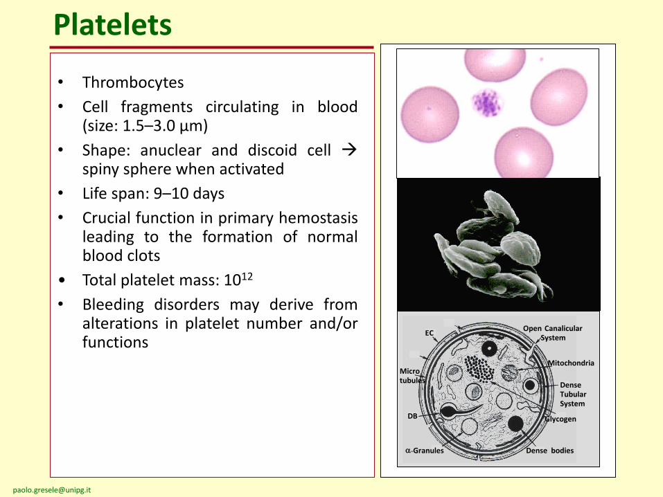
Platelets
EC
Microtubules
DB
a-Granules
Glycogen
Open CanalicularSystem
Dense bodies
Mitochondria
Dense TubularSystem
• Thrombocytes
• Cell fragments circulating in blood(size: 1.5–3.0 µm)
• Shape: anuclear and discoid cell spiny sphere when activated
• Life span: 9–10 days
• Crucial function in primary hemostasisleading to the formation of normalblood clots
• Total platelet mass: 1012
• Bleeding disorders may derive fromalterations in platelet number and/orfunctions
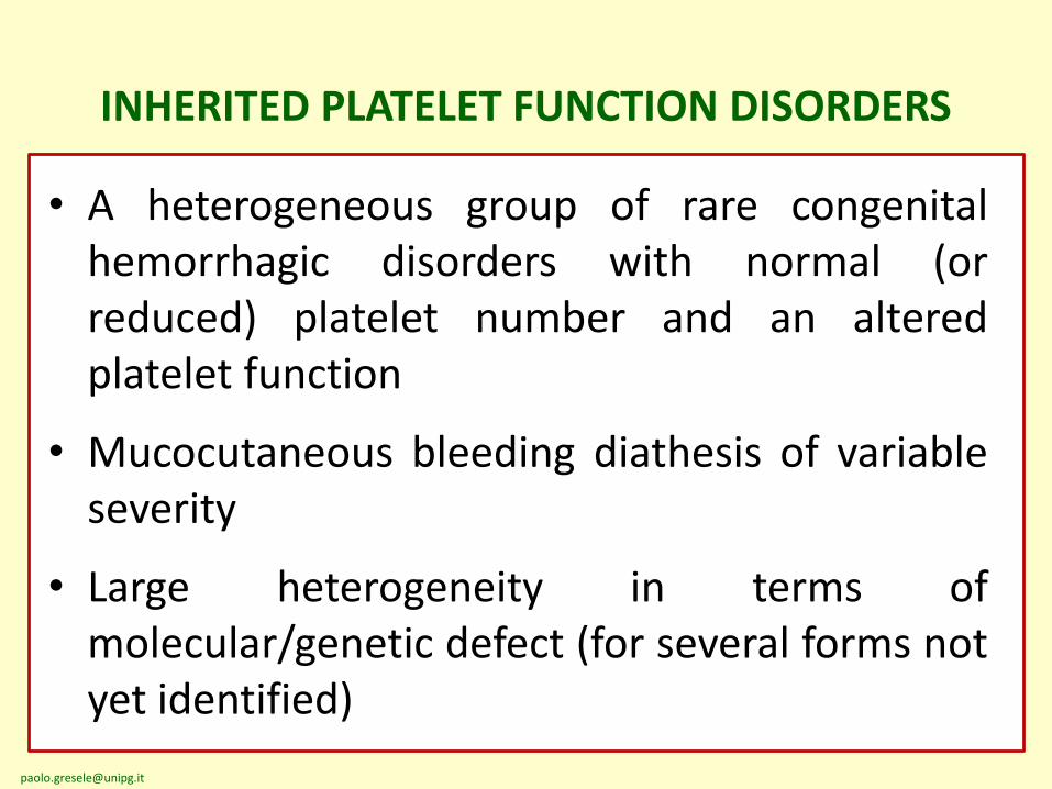
INHERITED PLATELET FUNCTION DISORDERS
• A heterogeneous group of rare congenitalhemorrhagic disorders with normal (orreduced) platelet number and an alteredplatelet function
• Mucocutaneous bleeding diathesis of variableseverity
• Large heterogeneity in terms ofmolecular/genetic defect (for several forms notyet identified)
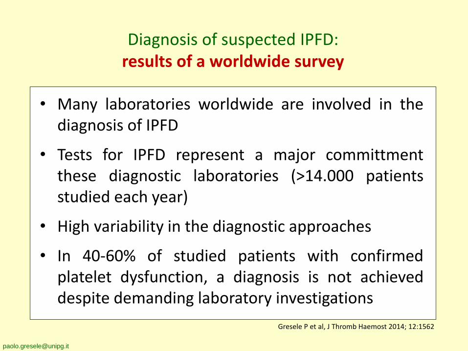
Diagnosis of suspected IPFD: results of a worldwide survey
• Many laboratories worldwide are involved in thediagnosis of IPFD
• Tests for IPFD represent a major committmentthese diagnostic laboratories (>14.000 patientsstudied each year)
• High variability in the diagnostic approaches
• In 40-60% of studied patients with confirmedplatelet dysfunction, a diagnosis is not achieveddespite demanding laboratory investigations
Gresele P et al, J Thromb Haemost 2014; 12:1562
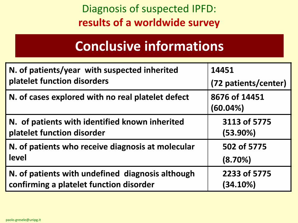
N. of patients/year with suspected inherited platelet function disorders
14451
(72 patients/center)
N. of cases explored with no real platelet defect 8676 of 14451 (60.04%)
N. of patients with identified known inherited platelet function disorder
3113 of 5775 (53.90%)
N. of patients who receive diagnosis at molecular level
502 of 5775
(8.70%)
N. of patients with undefined diagnosis although confirming a platelet function disorder
2233 of 5775 (34.10%)
Diagnosis of suspected IPFD: results of a worldwide survey
Conclusive informations
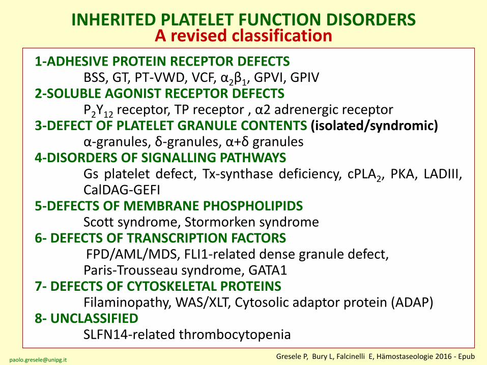
1-ADHESIVE PROTEIN RECEPTOR DEFECTSBSS, GT, PT-VWD, VCF, α2β1, GPVI, GPIV
2-SOLUBLE AGONIST RECEPTOR DEFECTSP2Y12 receptor, TP receptor , α2 adrenergic receptor
3-DEFECT OF PLATELET GRANULE CONTENTS (isolated/syndromic)α-granules, δ-granules, α+δ granules
4-DISORDERS OF SIGNALLING PATHWAYSGs platelet defect, Tx-synthase deficiency, cPLA2, PKA, LADIII,CalDAG-GEFI
5-DEFECTS OF MEMBRANE PHOSPHOLIPIDSScott syndrome, Stormorken syndrome
6- DEFECTS OF TRANSCRIPTION FACTORSFPD/AML/MDS, FLI1-related dense granule defect,Paris-Trousseau syndrome, GATA1
7- DEFECTS OF CYTOSKELETAL PROTEINSFilaminopathy, WAS/XLT, Cytosolic adaptor protein (ADAP)
8- UNCLASSIFIEDSLFN14-related thrombocytopenia
INHERITED PLATELET FUNCTION DISORDERSA revised classification
Gresele P, Bury L, Falcinelli E, Hämostaseologie 2016 - [email protected]
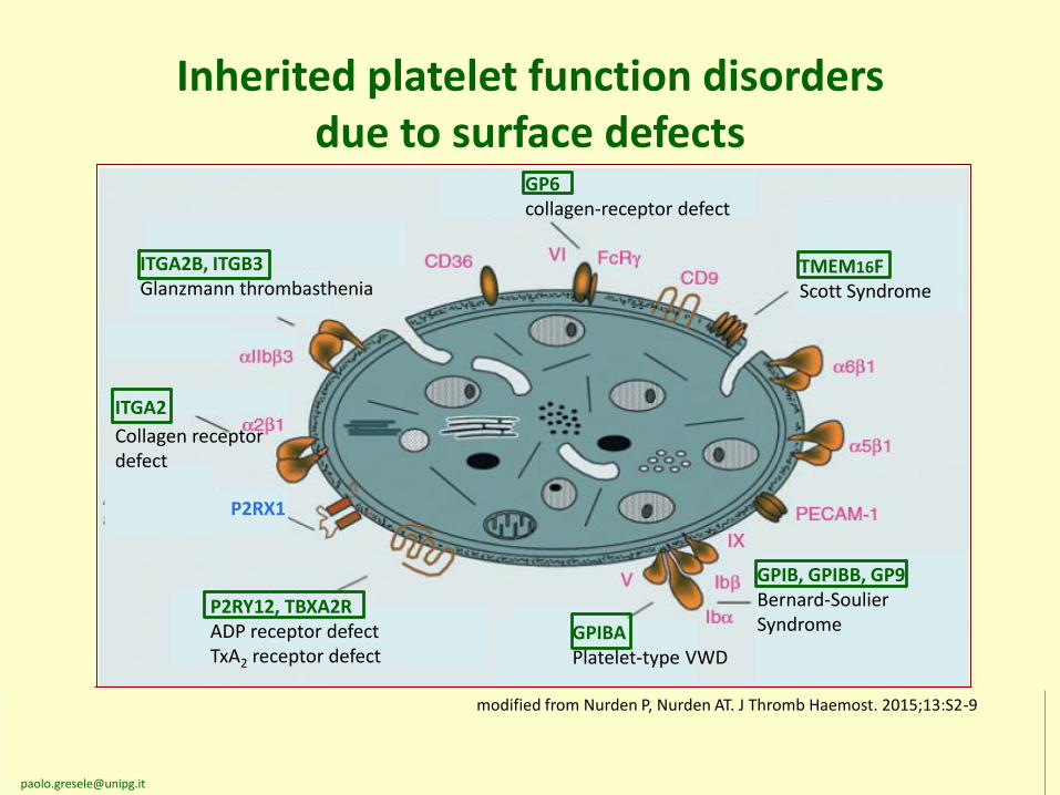
Inherited platelet function disorders due to surface defects
ITGA2B, ITGB3Glanzmann thrombasthenia
P2RX1
P2RY12, TBXA2RADP receptor defectTxA2 receptor defect
GPIBAPlatelet-type VWD
GPIB, GPIBB, GP9Bernard-SoulierSyndrome
GP6collagen-receptor defect
TMEM16FScott Syndrome
ITGA2
Collagen receptordefect
modified from Nurden P, Nurden AT. J Thromb Haemost. 2015;13:S2-9
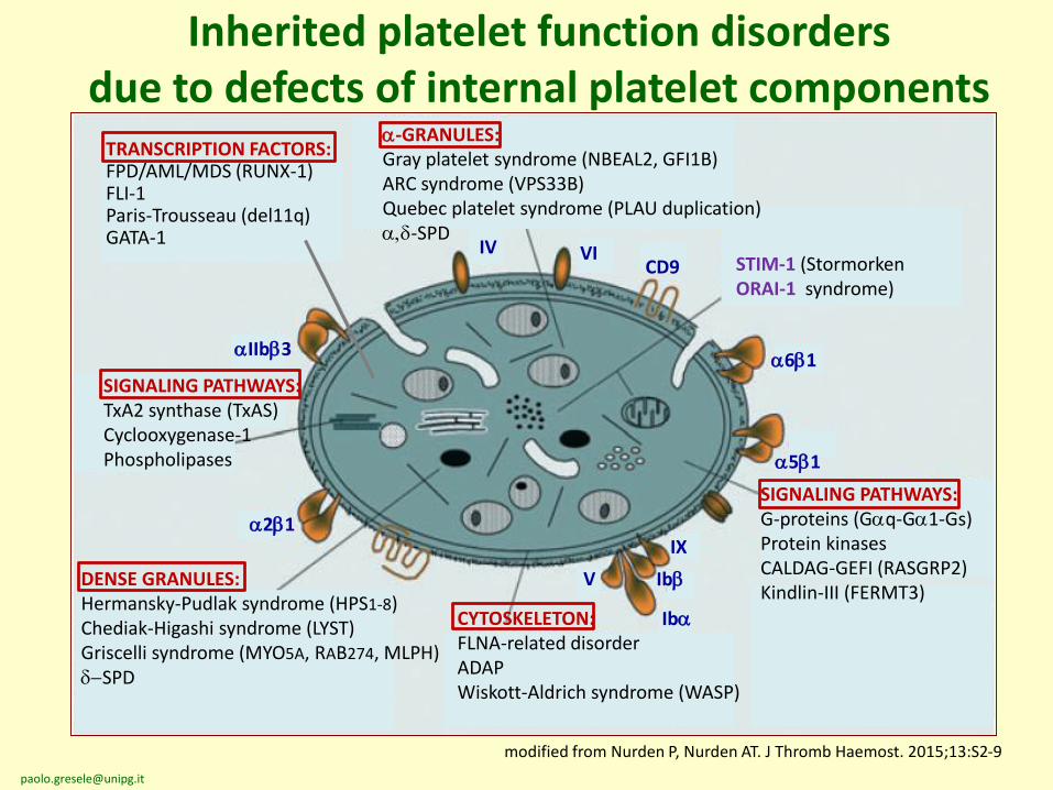
Inherited platelet function disorders due to defects of internal platelet components
modified from Nurden P, Nurden AT. J Thromb Haemost. 2015;13:S2-9
a-GRANULES:Gray platelet syndrome (NBEAL2, GFI1B)ARC syndrome (VPS33B)Quebec platelet syndrome (PLAU duplication)a,d-SPD
SIGNALING PATHWAYS:TxA2 synthase (TxAS)Cyclooxygenase-1Phospholipases
DENSE GRANULES:Hermansky-Pudlak syndrome (HPS1-8)Chediak-Higashi syndrome (LYST)Griscelli syndrome (MYO5A, RAB274, MLPH)d-SPD
SIGNALING PATHWAYS:G-proteins (Gaq-Ga1-Gs)Protein kinasesCALDAG-GEFI (RASGRP2)Kindlin-III (FERMT3)
CYTOSKELETON:FLNA-related disorderADAPWiskott-Aldrich syndrome (WASP)
aIIbb3
IV VICD9
a6b1
a5b1
a2b1IX
Ibb
Iba
V
STIM-1 (StormorkenORAI-1 syndrome)
TRANSCRIPTION FACTORS:FPD/AML/MDS (RUNX-1)FLI-1Paris-Trousseau (del11q)GATA-1
10
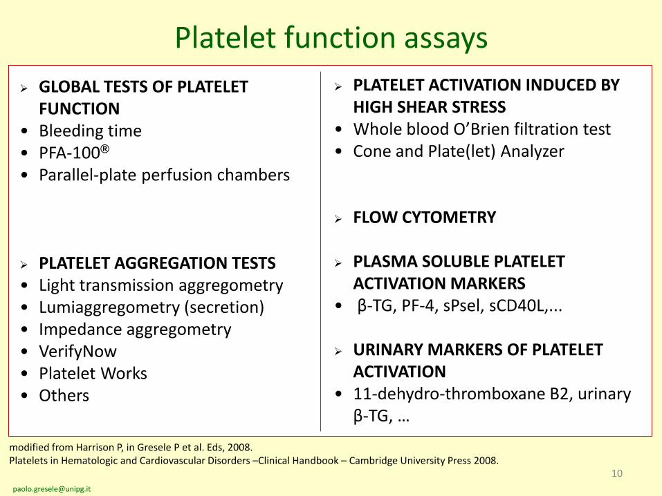
Platelet function assays
GLOBAL TESTS OF PLATELET FUNCTION
• Bleeding time • PFA-100®
• Parallel-plate perfusion chambers
PLATELET AGGREGATION TESTS• Light transmission aggregometry• Lumiaggregometry (secretion)• Impedance aggregometry• VerifyNow• Platelet Works• Others
PLATELET ACTIVATION INDUCED BY HIGH SHEAR STRESS
• Whole blood O’Brien filtration test• Cone and Plate(let) Analyzer
FLOW CYTOMETRY
PLASMA SOLUBLE PLATELET ACTIVATION MARKERS
• β-TG, PF-4, sPsel, sCD40L,...
URINARY MARKERS OF PLATELET ACTIVATION
• 11-dehydro-thromboxane B2, urinary β-TG, …
modified from Harrison P, in Gresele P et al. Eds, 2008. Platelets in Hematologic and Cardiovascular Disorders –Clinical Handbook – Cambridge University Press 2008.
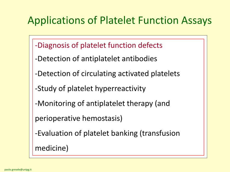
Applications of Platelet Function Assays
-Diagnosis of platelet function defects
-Detection of antiplatelet antibodies
-Detection of circulating activated platelets
-Study of platelet hyperreactivity
-Monitoring of antiplatelet therapy (and
perioperative hemostasis)
-Evaluation of platelet banking (transfusion
medicine)
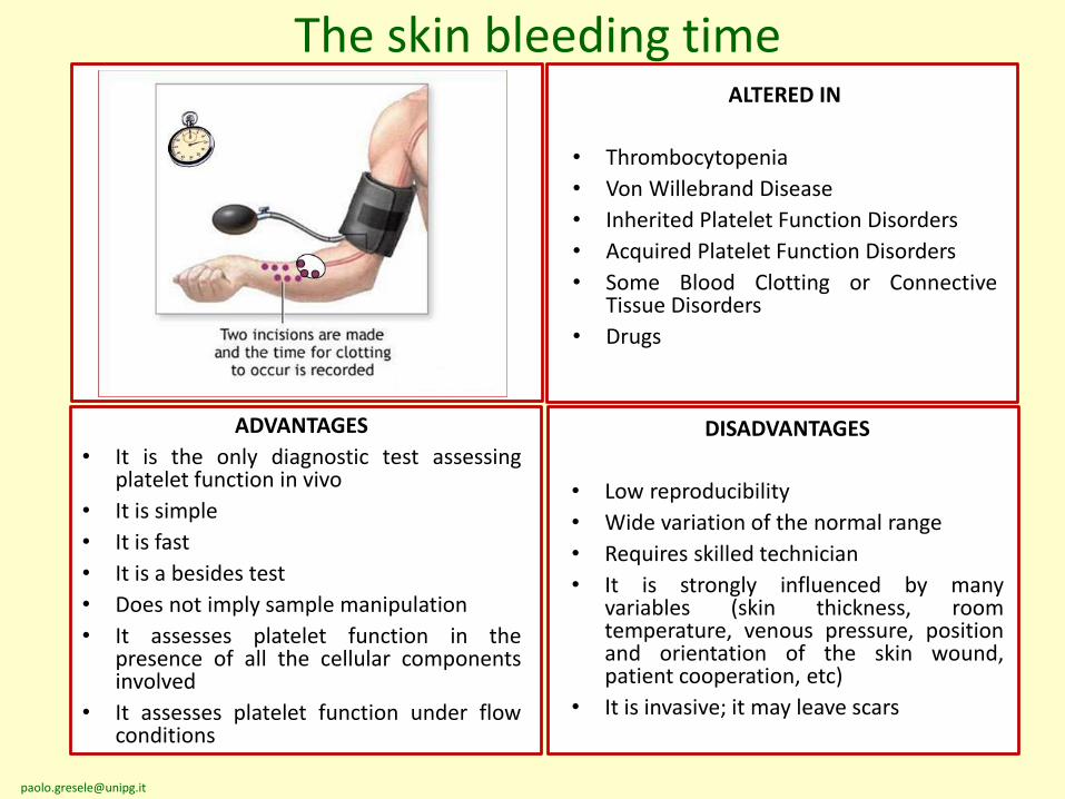
ADVANTAGES
• It is the only diagnostic test assessingplatelet function in vivo
• It is simple
• It is fast
• It is a besides test
• Does not imply sample manipulation
• It assesses platelet function in thepresence of all the cellular componentsinvolved
• It assesses platelet function under flowconditions
The skin bleeding timeALTERED IN
• Thrombocytopenia
• Von Willebrand Disease
• Inherited Platelet Function Disorders
• Acquired Platelet Function Disorders
• Some Blood Clotting or ConnectiveTissue Disorders
• Drugs
DISADVANTAGES
• Low reproducibility
• Wide variation of the normal range
• Requires skilled technician
• It is strongly influenced by manyvariables (skin thickness, roomtemperature, venous pressure, positionand orientation of the skin wound,patient cooperation, etc)
• It is invasive; it may leave scars
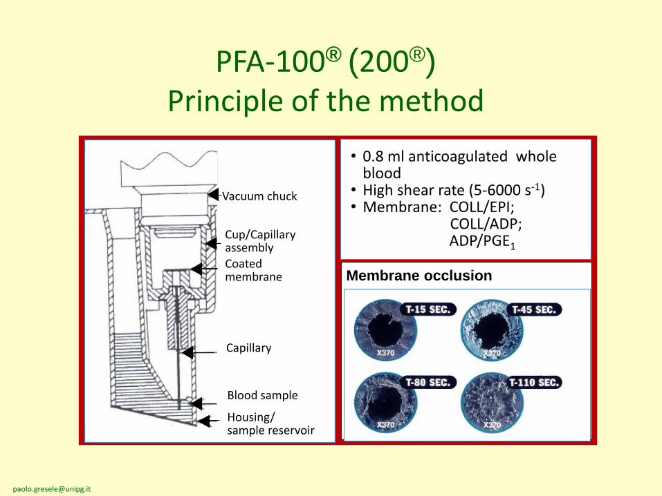
PFA-100® (200®)
Principle of the method
Vacuum chuck
Cup/CapillaryassemblyCoated membrane
Capillary
Blood sample
Housing/sample reservoir
• 0.8 ml anticoagulated whole blood
• High shear rate (5-6000 s-1)• Membrane: COLL/EPI;
COLL/ADP;ADP/PGE1
Membrane occlusion
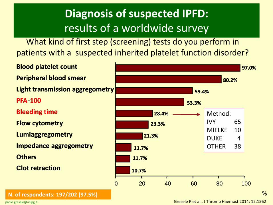
Diagnosis of suspected IPFD:results of a worldwide survey
What kind of first step (screening) tests do you perform in patients with a suspected inherited platelet function disorder?
N. of respondents: 197/202 (97.5%) %Gresele P et al., J Thromb Haemost 2014; 12:1562
Blood platelet count
Peripheral blood smear
Light transmission aggregometry
PFA-100
Bleeding time
Flow cytometry
Lumiaggregometry
Impedance aggregometry
Others
Clot retraction
0
21.3%
11.7%
11.7%
23.3%
10.7%
97.0%
80.2%
28.4%
53.3%
59.4%
20 40 60 80 100
Blood platelet count
Peripheral blood smear
Light transmission aggregometry
PFA-100
Bleeding time
Flow cytometry
Lumiaggregometry
Impedance aggregometry
Others
Clot retraction
0
21.3%
11.7%
11.7%
23.3%
10.7%
97.0%
80.2%
28.4%
53.3%
59.4%
20 40 60 80 100
Method:IVY 65MIELKE 10DUKE 4OTHER 38
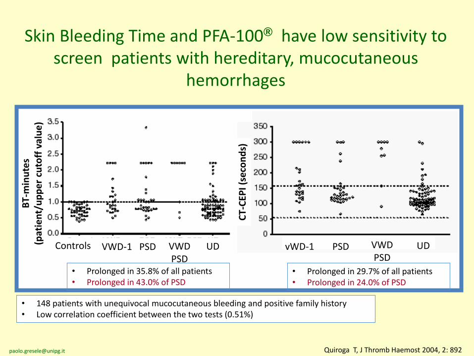
Quiroga T, J Thromb Haemost 2004, 2: 892
Controls VWD-1 PSD VWDPSD
UD vWD-1 PSD VWDPSD
UD
BT-
min
ute
s(p
atie
nt/
up
pe
r cu
toff
val
ue
)
CT-
CEP
I (se
con
ds)
Skin Bleeding Time and PFA-100® have low sensitivity to screen patients with hereditary, mucocutaneous
hemorrhages
• Prolonged in 35.8% of all patients• Prolonged in 43.0% of PSD
• Prolonged in 29.7% of all patients• Prolonged in 24.0% of PSD
• 148 patients with unequivocal mucocutaneous bleeding and positive family history• Low correlation coefficient between the two tests (0.51%)
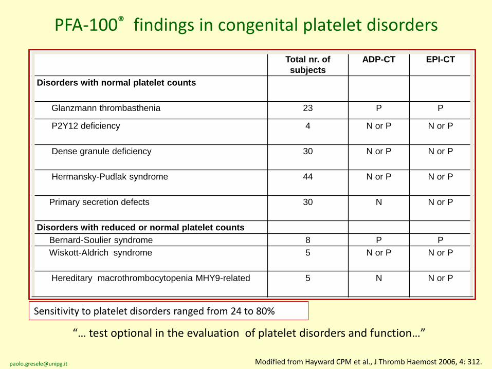
PFA-100® findings in congenital platelet disorders
Total nr. of
subjects
ADP-CT EPI-CT
Disorders with normal platelet counts
Glanzmann thrombasthenia 23 P P
P2Y12 deficiency 4 N or P N or P
Dense granule deficiency 30 N or P N or P
Hermansky-Pudlak syndrome 44 N or P N or P
Primary secretion defects 30 N N or P
Disorders with reduced or normal platelet counts
Bernard-Soulier syndrome 8 P P
Wiskott-Aldrich syndrome 5 N or P N or P
Hereditary macrothrombocytopenia MHY9-related 5 N N or P
Modified from Hayward CPM et al., J Thromb Haemost 2006, 4: 312.
“… test optional in the evaluation of platelet disorders and function…”
Sensitivity to platelet disorders ranged from 24 to 80%
17
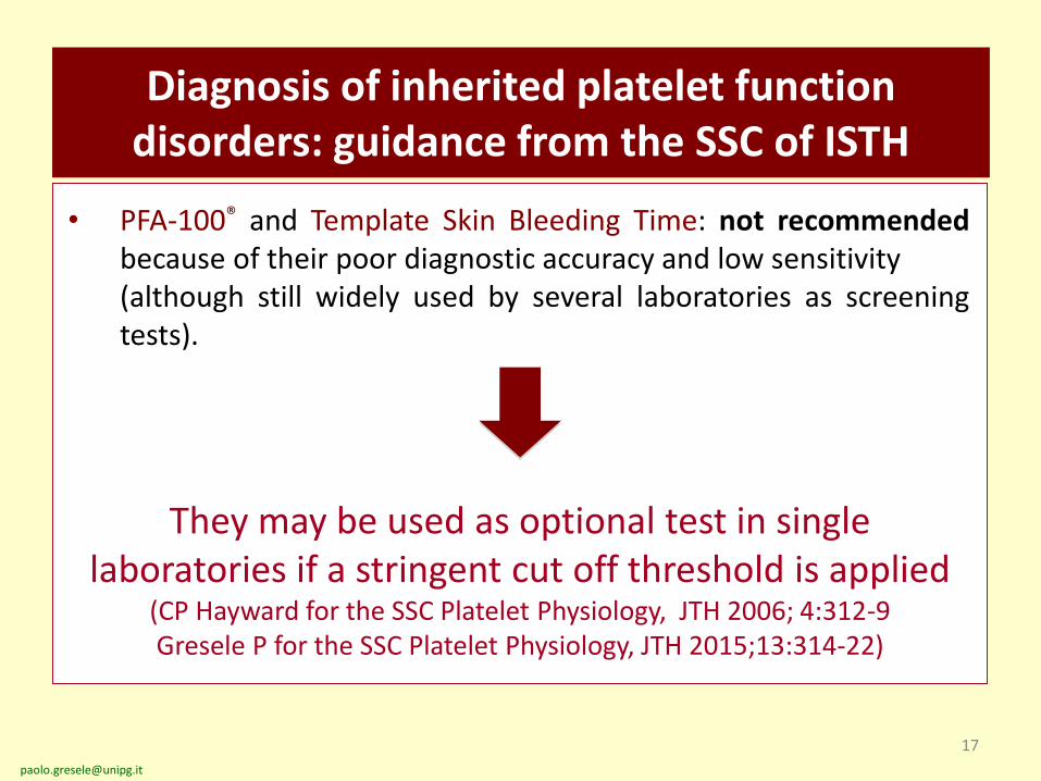
• PFA-100® and Template Skin Bleeding Time: not recommendedbecause of their poor diagnostic accuracy and low sensitivity(although still widely used by several laboratories as screeningtests).
Diagnosis of inherited platelet function disorders: guidance from the SSC of ISTH
They may be used as optional test in single laboratories if a stringent cut off threshold is applied
(CP Hayward for the SSC Platelet Physiology, JTH 2006; 4:312-9Gresele P for the SSC Platelet Physiology, JTH 2015;13:314-22)
18
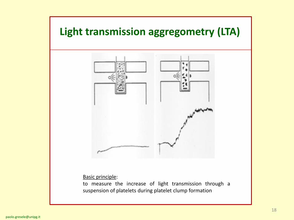
Light transmission aggregometry (LTA)
Basic principle:to measure the increase of light transmission through asuspension of platelets during platelet clump formation
19
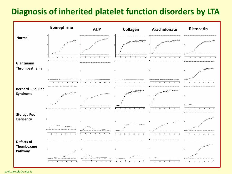
Diagnosis of inherited platelet function disorders by LTA
ADP Collagen Arachidonate Ristocetin
Glanzmann
Thrombasthenia
Bernard – Soulier Syndrome
Storage Pool Deficency
Defects of Thomboxane Pathway
Normal
Epinephrine
20
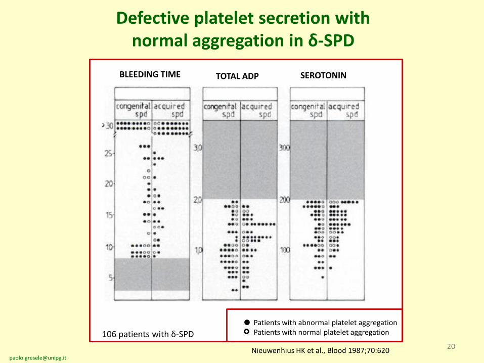
Defective platelet secretion with normal aggregation in δ-SPD
BLEEDING TIME TOTAL ADP SEROTONIN
1
2
3
4
5
6
2
4
6
8
10
12
µm
ol/
10
11
plt
s
nm
ol/
10
11
plt
s
Nieuwenhius HK et al., Blood 1987;70:620
106 patients with δ-SPD
Patients with abnormal platelet aggregationPatients with normal platelet aggregation
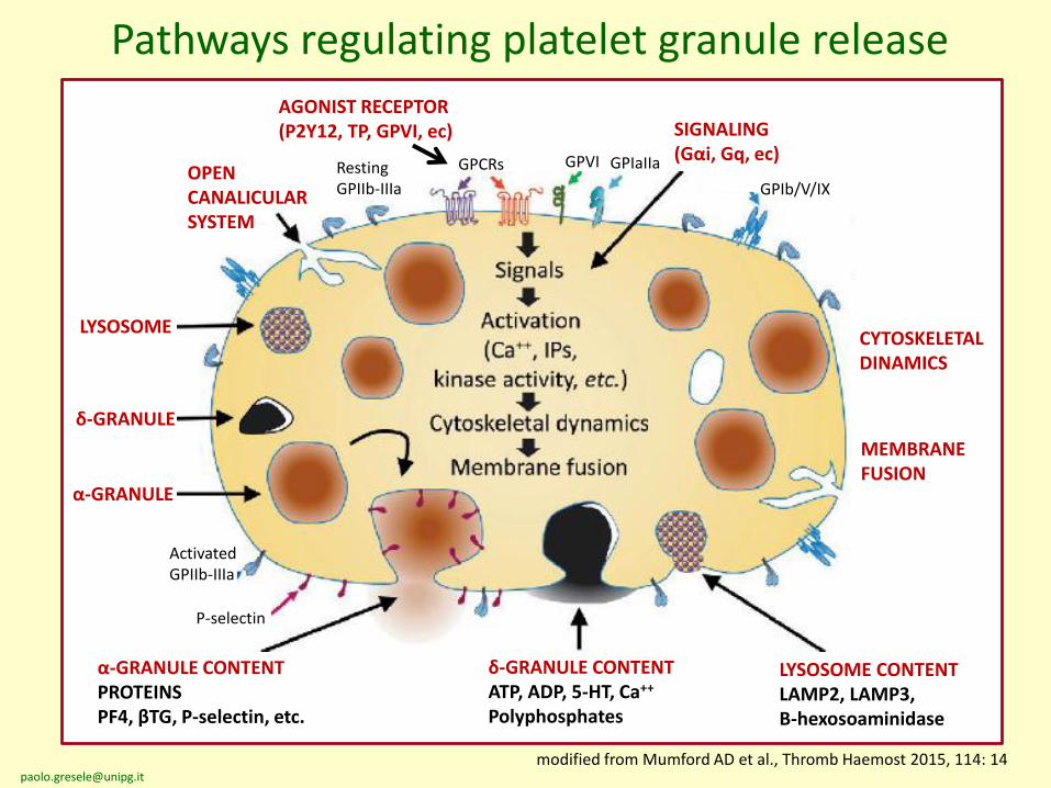
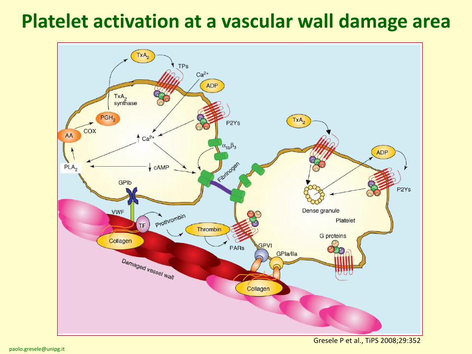
Pathways regulating platelet granule release
Resting GPIIb-IIIa
GPCRs GPVI GPIaIIa
GPIb/V/IX
ActivatedGPIIb-IIIa
P-selectin
AGONIST RECEPTOR (P2Y12, TP, GPVI, ec) SIGNALING
(Gαi, Gq, ec)
CYTOSKELETALDINAMICS
MEMBRANEFUSION
OPEN CANALICULAR SYSTEM
LYSOSOME
δ-GRANULE
α-GRANULE
modified from Mumford AD et al., Thromb Haemost 2015, 114: 14
α-GRANULE CONTENT PROTEINSPF4, βTG, P-selectin, etc.
δ-GRANULE CONTENTATP, ADP, 5-HT, Ca++
Polyphosphates
LYSOSOME CONTENTLAMP2, LAMP3,Β-hexosoaminidase
22
Mumford AD et al., Thromb Haemost 2015, 114: 14
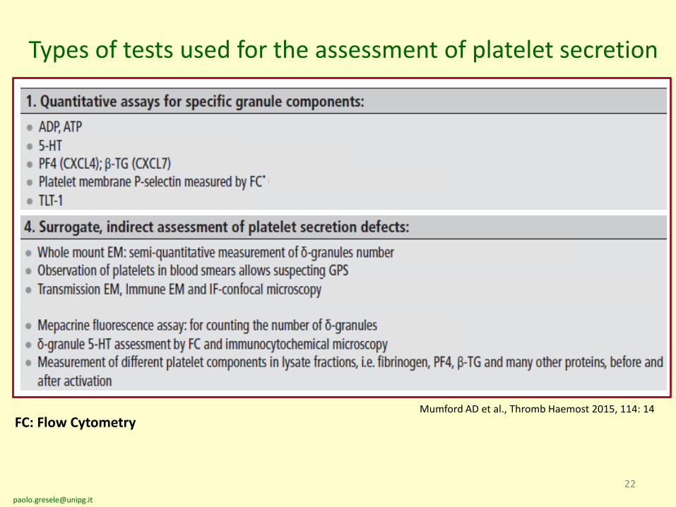
Types of tests used for the assessment of platelet secretion
FC: Flow Cytometry
23
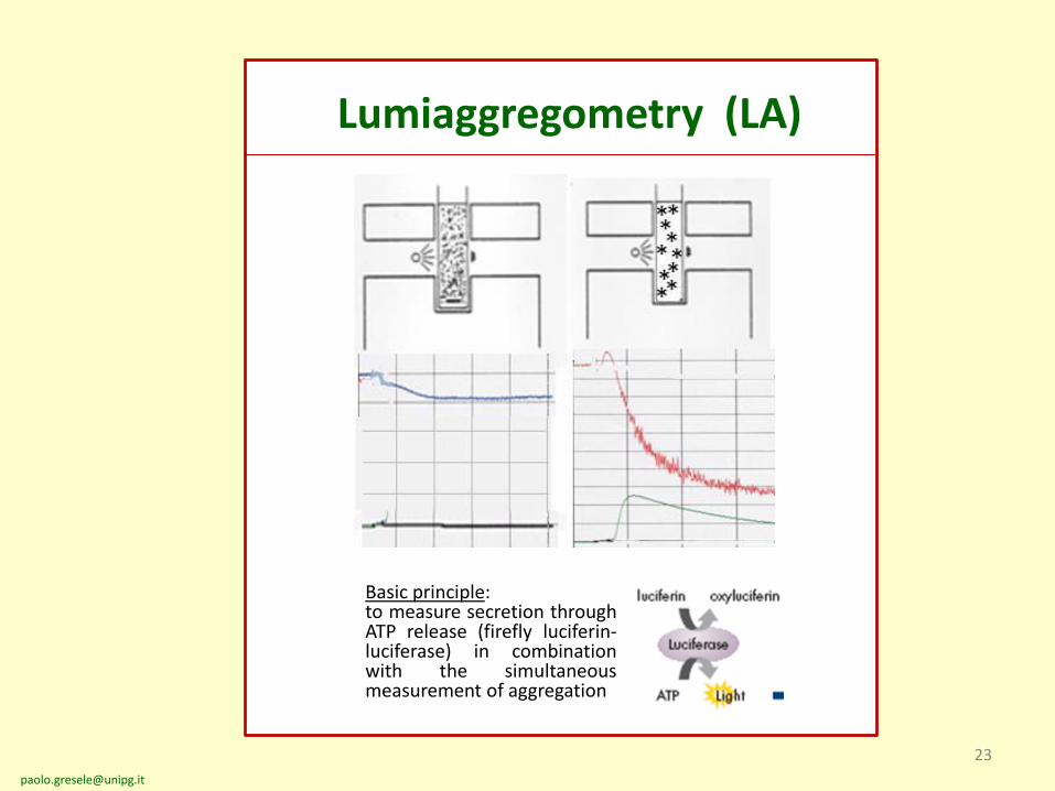
Lumiaggregometry (LA)
Basic principle:to measure secretion throughATP release (firefly luciferin-luciferase) in combinationwith the simultaneousmeasurement of aggregation
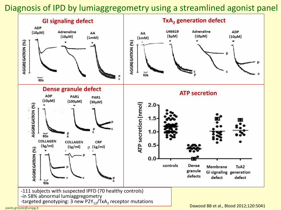
Dawood BB et al., Blood 2012;120:5041
Diagnosis of IPD by lumiaggregometry using a streamlined agonist panel
-111 subjects with suspected IPFD (70 healthy controls)-in 58% abnormal lumiaggregometry-targeted genotyping: 3 new P2Y12/TxA2 receptor mutations
26
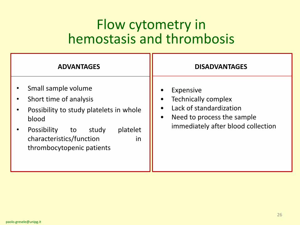
Flow cytometry in hemostasis and thrombosis
ADVANTAGES
• Small sample volume
• Short time of analysis
• Possibility to study platelets in wholeblood
• Possibility to study plateletcharacteristics/function inthrombocytopenic patients
DISADVANTAGES
• Expensive• Technically complex • Lack of standardization• Need to process the sample
immediately after blood collection
27
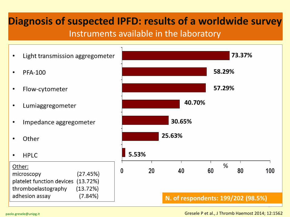
Gresele P et al., J Thromb Haemost 2014; 12:1562
Diagnosis of suspected IPFD: results of a worldwide surveyInstruments available in the laboratory
28
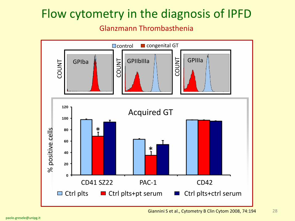
control congenital GT
Flow cytometry in the diagnosis of IPFD Glanzmann Thrombasthenia
Giannini S et al., Cytometry B Clin Cytom 2008, 74:194
0
20
40
60
80
100
120
% p
osi
tive
cel
ls
CD41 SZ22 PAC-1 CD42
Ctrl plts Ctrl plts+pt serum Ctrl plts+ctrl serum
*
*
GPIba GPIIbIIIa GPIIIaC
OU
NT
CO
UN
T
CO
UN
T
Acquired GT
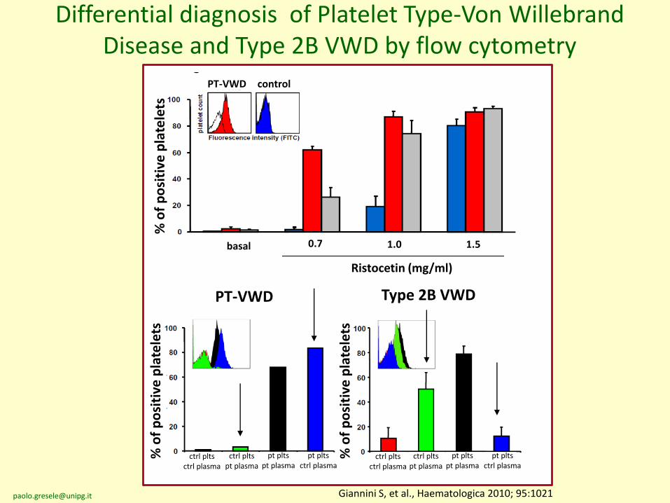
PT-VWD Type 2B VWD
PT-VWD control
Differential diagnosis of Platelet Type-Von WillebrandDisease and Type 2B VWD by flow cytometry
Giannini S, et al., Haematologica 2010; 95:1021
ctrl pltsctrl plasma
ctrl pltspt plasma
pt pltspt plasma
pt pltsctrl plasma
ctrl pltsctrl plasma
ctrl pltspt plasma
pt pltspt plasma
pt pltsctrl plasma
% o
f p
osi
tive
pla
tele
ts
% o
f p
osi
tive
pla
tele
ts
% o
f p
osi
tive
pla
tele
ts
Ristocetin (mg/ml)
basal 0.7 1.0 1.5
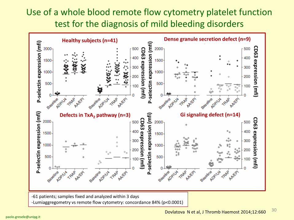
30Dovlatova N et al, J Thromb Haemost 2014;12:660
Use of a whole blood remote flow cytometry platelet function test for the diagnosis of mild bleeding disorders
P-s
ele
ctin
exp
ress
ion
(m
fi)
P-s
ele
ctin
exp
ress
ion
(m
fi)
P-s
ele
ctin
exp
ress
ion
(m
fi)
P-s
ele
ctin
exp
ress
ion
(m
fi) C
D6
3 exp
ressio
n (m
fi)
CD
63
expre
ssion
(mfi)
CD
63
expre
ssion
(mfi)
Healthy subjects (n=41) Dense granule secretion defect (n=9)
Defects in TxA2 pathway (n=3) Gi signaling defect (n=14)
-61 patients; samples fixed and analyzed within 3 days-Lumiaggregometry vs remote flow cytometry: concordance 84% (p<0.0001)
CD
63
expre
ssion
(mfi)
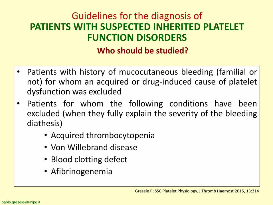
Who should be studied?
Guidelines for the diagnosis ofPATIENTS WITH SUSPECTED INHERITED PLATELET
FUNCTION DISORDERS
• Patients with history of mucocutaneous bleeding (familial ornot) for whom an acquired or drug-induced cause of plateletdysfunction was excluded
• Patients for whom the following conditions have beenexcluded (when they fully explain the severity of the bleedingdiathesis)
• Acquired thrombocytopenia
• Von Willebrand disease
• Blood clotting defect
• Afibrinogenemia
Gresele P; SSC Platelet Physiology, J Thromb Haemost 2015, 13:314
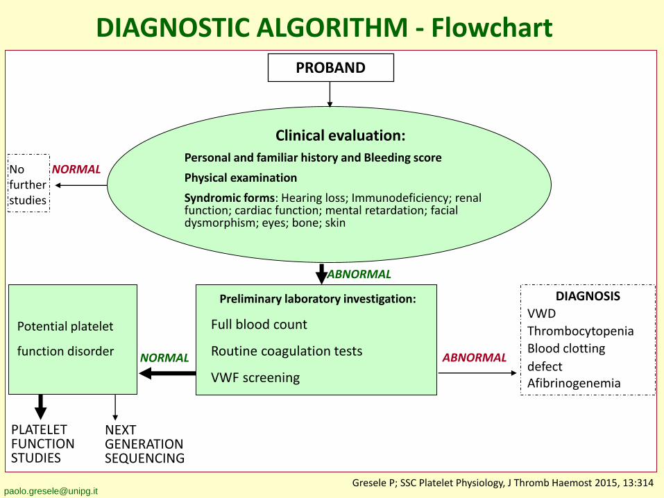
DIAGNOSTIC ALGORITHM - FlowchartPROBAND
Clinical evaluation:
Personal and familiar history and Bleeding score
Physical examination
Syndromic forms: Hearing loss; Immunodeficiency; renal function; cardiac function; mental retardation; facial dysmorphism; eyes; bone; skin
NORMAL ABNORMAL
DIAGNOSIS
VWD
Thrombocytopenia
Blood clotting
defectAfibrinogenemia
Potential platelet
function disorder
ABNORMAL
NORMALNo further studies
PLATELET FUNCTIONSTUDIES
NEXTGENERATIONSEQUENCING
Preliminary laboratory investigation:
Full blood count
Routine coagulation tests
VWF screening
[email protected] P; SSC Platelet Physiology, J Thromb Haemost 2015, 13:314
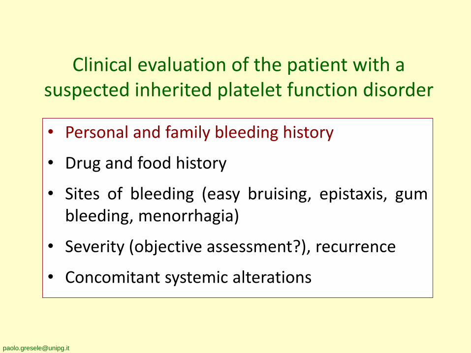
Clinical evaluation of the patient with a suspected inherited platelet function disorder
• Personal and family bleeding history
• Drug and food history
• Sites of bleeding (easy bruising, epistaxis, gumbleeding, menorrhagia)
• Severity (objective assessment?), recurrence
• Concomitant systemic alterations
The ISTH Bleeding Assessment Toolfor the evaluation of IPFDs
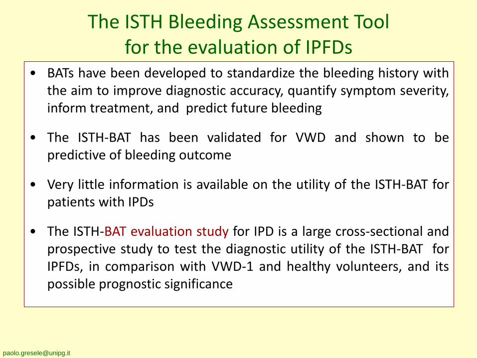
• BATs have been developed to standardize the bleeding history withthe aim to improve diagnostic accuracy, quantify symptom severity,inform treatment, and predict future bleeding
• The ISTH-BAT has been validated for VWD and shown to bepredictive of bleeding outcome
• Very little information is available on the utility of the ISTH-BAT forpatients with IPDs
• The ISTH-BAT evaluation study for IPD is a large cross-sectional andprospective study to test the diagnostic utility of the ISTH-BAT forIPFDs, in comparison with VWD-1 and healthy volunteers, and itspossible prognostic significance
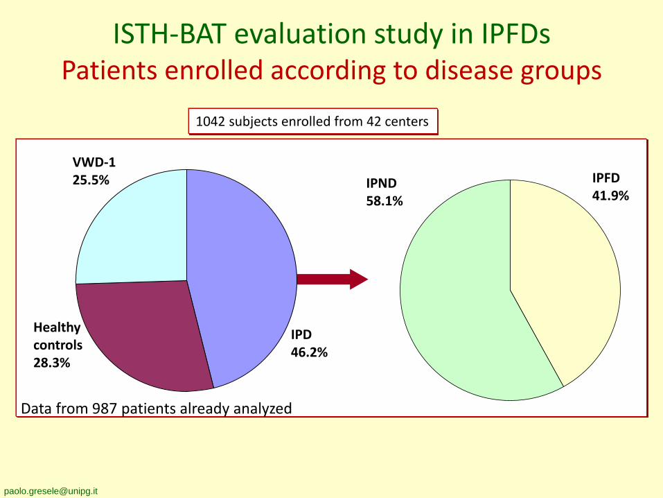
ISTH-BAT evaluation study in IPFDsPatients enrolled according to disease groups
1042 subjects enrolled from 42 centers
Data from 987 patients already analyzed
IPFD41.9%
IPND58.1%
VWD-125.5%
Healthycontrols28.3%
IPD46.2%
0
2
4
6
8
10
12
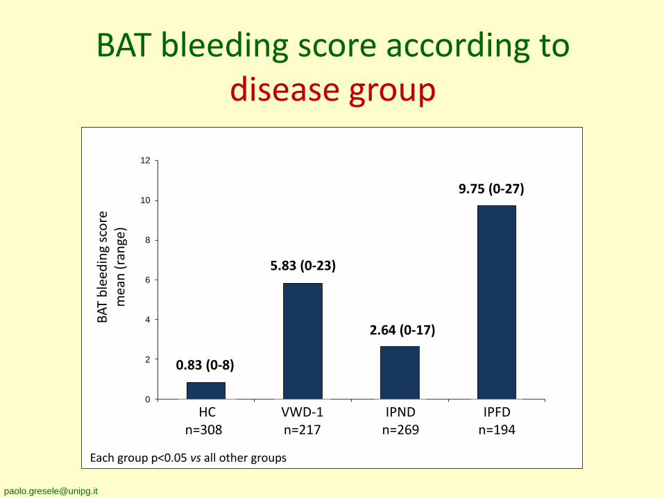
BAT bleeding score according to disease group
HCn=308
BA
T b
leed
ing
sco
rem
ean
(ra
nge
)
IPFDn=194
0.83 (0-8)
VWD-1n=217
5.83 (0-23)
9.75 (0-27)
IPNDn=269
2.64 (0-17)
Each group p<0.05 vs all other groups
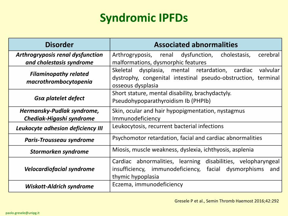
Syndromic IPFDs
Disorder Associated abnormalitiesArthrogryposis renal dysfunction
and cholestasis syndromeArthrogryposis, renal dysfunction, cholestasis, cerebralmalformations, dysmorphic features
Filaminopathy related macrothrombocytopenia
Skeletal dysplasia, mental retardation, cardiac valvulardystrophy, congenital intestinal pseudo-obstruction, terminalosseous dysplasia
Gsα platelet defectShort stature, mental disability, brachydactyly.Pseudohypoparathyroidism Ib (PHPIb)
Hermansky-Pudlak syndrome, Chediak-Higashi syndrome
Skin, ocular and hair hypopigmentation, nystagmusImmunodeficiency
Leukocyte adhesion deficiency III Leukocytosis, recurrent bacterial infections
Paris-Trousseau syndrome Psychomotor retardation, facial and cardiac abnormalities
Stormorken syndrome Miosis, muscle weakness, dyslexia, ichthyosis, asplenia
Velocardiofacial syndromeCardiac abnormalities, learning disabilities, velopharyngealinsufficiency, immunodeficiency, facial dysmorphisms andthymic hypoplasia
Wiskott-Aldrich syndrome Eczema, immunodeficiency
Gresele P et al., Semin Thromb Haemost 2016;42:292
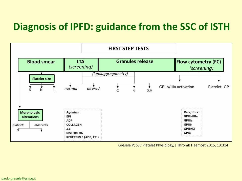
Diagnosis of IPFD: guidance from the SSC of ISTH
Blood smear LTA(screening)
Granules release Flow cytometry (FC)(screening)
Platelet size
Morphologic alterations
Gresele P; SSC Platelet Physiology, J Thromb Haemost 2015, 13:314
Agonists:α-ThrTRAP6U46619CRPCVXPAR4-apPMAA23187Inhibition by
Iloprost or PGE1
Receptors:GPIa/IIaGPIVGPVI
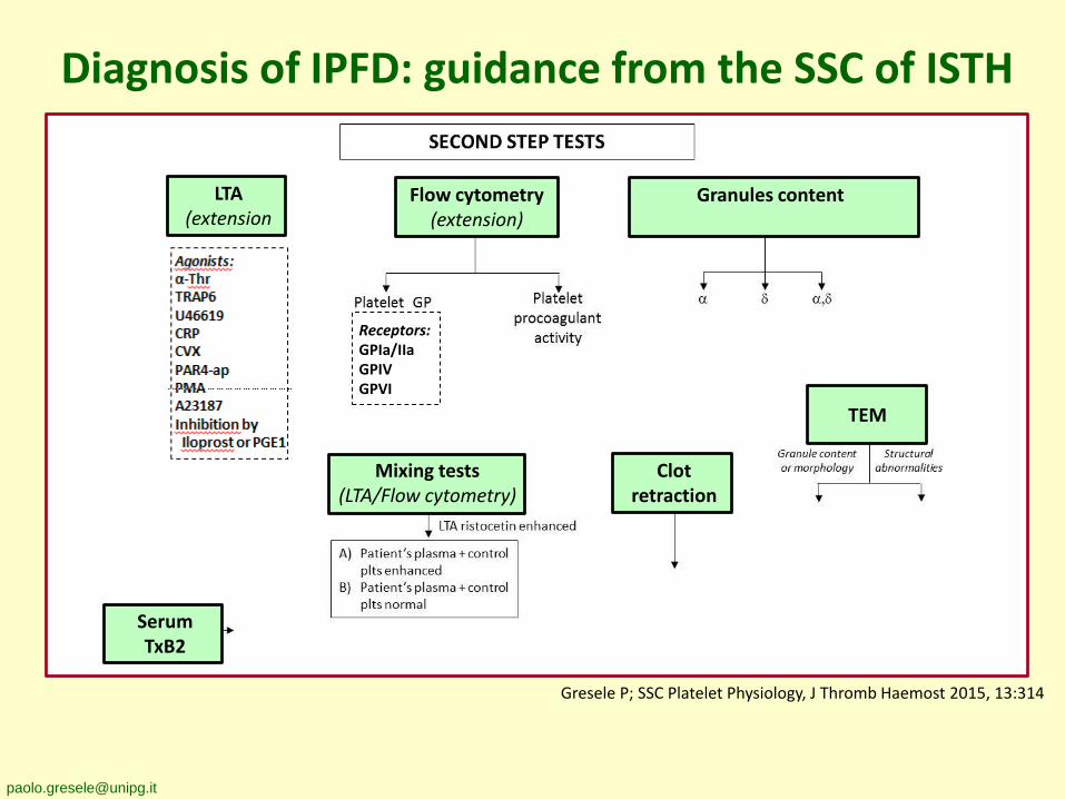
Diagnosis of IPFD: guidance from the SSC of ISTH
LTA(extension
Flow cytometry(extension)
Granules content
Serum TxB2
Mixing tests(LTA/Flow cytometry)
Clotretraction
TEM
Gresele P; SSC Platelet Physiology, J Thromb Haemost 2015, 13:314
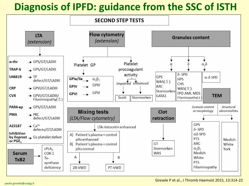
Gresele P et al., J Thromb Haemost 2015, 13:314-22
Diagnosis of IPFD: guidance from the SSC of ISTH
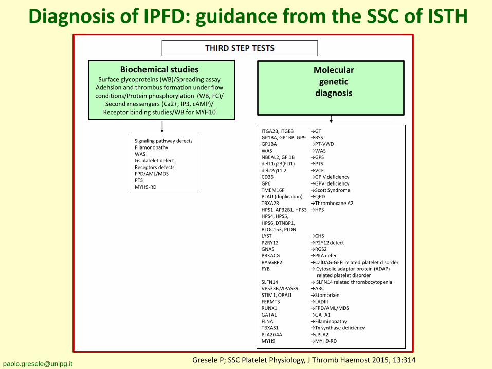
Diagnosis of IPFD: guidance from the SSC of ISTH
ITGA2B, ITGB3 →GTGP1BA, GP1BB, GP9 →BSSGP1BA →PT-VWDWAS →WASNBEAL2, GFI1B →GPSdel11q23(FLI1) →PTSdel22q11.2 →VCFCD36 →GPIV deficiencyGP6 →GPVI deficiencyTMEM16F →Scott SyndromePLAU (duplication) →QPDTBXA2R →Thromboxane A2HPS1, AP32B1, HPS3 →HPSHPS4, HPS5, HPS6, DTNBP1,BLOC153, PLDNLYST →CHSP2RY12 →P2Y12 defectGNAS →RGS2PRKACG →PKA defectRASGRP2 →CalDAG-GEFI related platelet disorderFYB → Cytosolic adaptor protein (ADAP)
related platelet disorderSLFN14 → SLFN14 related thrombocytopeniaVPS33B,VIPAS39 →ARCSTIM1, ORAI1 →StomorkenFERMT3 →LADIIIRUNX1 →FPD/AML/MDSGATA1 →GATA1FLNA →FilaminopathyTBXAS1 →Tx synthase deficiencyPLA2G4A →cPLA2MYH9 →MYH9-RD
Signaling pathway defectsFilamonopathyWASGs platelet defectReceptors defectsFPD/AML/MDSPTSMYH9-RD
Biochemical studiesSurface glycoproteins (WB)/Spreading assay
Adehsion and thrombus formation under flow conditions/Protein phosphorylation (WB, FC)/
Second messengers (Ca2+, IP3, cAMP)/Receptor binding studies/WB for MYH10
Molecular genetic
diagnosis
Gresele P; SSC Platelet Physiology, J Thromb Haemost 2015, 13:314
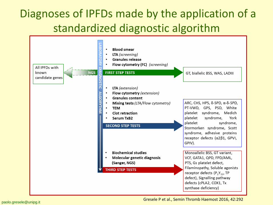
Diagnoses of IPFDs made by the application of a standardized diagnostic algorithm
[email protected] P et al., Semin Thromb Haemost 2016, 42:292
• Next Generation Sequencing enables the simultaneous analysis of large groupsof candidate genes, allowing the rapid identification of a mutation in a knowngene.
• Diagnosis of some IPFD can be reached only after genetic analysis (e.g. GTVariants).
• For some disorders (e.g. MYH9-RD) a phenotype/genotype correlation exists.
• We need to consider that several centers still do not have access to moleculartesting.
• Genetic testing (especially WES) is a potentially valuable investigation for genediscovery only if backed up by good phenotyping.
• Ethics of predictive testing should be considered (e.g. diagnosis of FPD/AML)
• It can not be considered yet as an initial diagnostic test, but rather ascomplementary and/or confirmatory.
Genetic Diagnosis: pros and cons
however
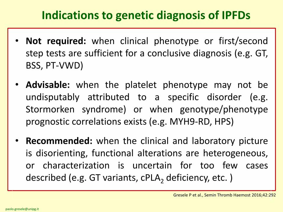
Indications to genetic diagnosis of IPFDs
• Not required: when clinical phenotype or first/secondstep tests are sufficient for a conclusive diagnosis (e.g. GT,BSS, PT-VWD)
• Advisable: when the platelet phenotype may not beundisputably attributed to a specific disorder (e.g.Stormorken syndrome) or when genotype/phenotypeprognostic correlations exists (e.g. MYH9-RD, HPS)
• Recommended: when the clinical and laboratory pictureis disorienting, functional alterations are heterogeneous,or characterization is uncertain for too few casesdescribed (e.g. GT variants, cPLA2 deficiency, etc. )
Gresele P et al., Semin Thromb Haemost 2016;42:292
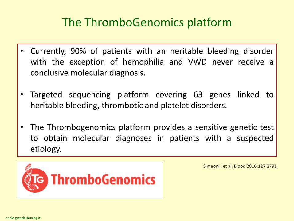
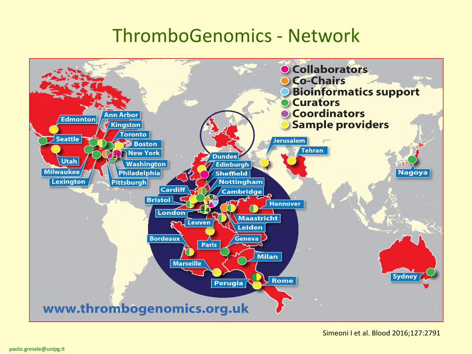
• Currently, 90% of patients with an heritable bleeding disorderwith the exception of hemophilia and VWD never receive aconclusive molecular diagnosis.
• Targeted sequencing platform covering 63 genes linked toheritable bleeding, thrombotic and platelet disorders.
• The Thrombogenomics platform provides a sensitive genetic testto obtain molecular diagnoses in patients with a suspectedetiology.
Simeoni I et al. Blood 2016;127:2791
The ThromboGenomics platform
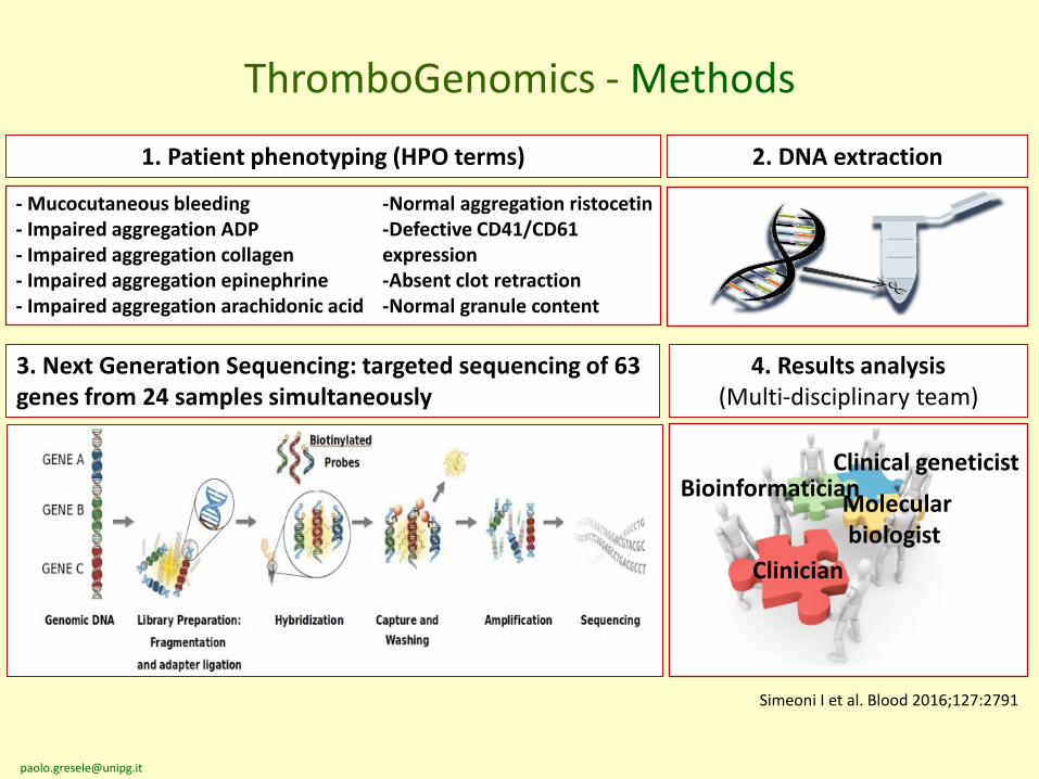
2. DNA extraction
3. Next Generation Sequencing: targeted sequencing of 63 genes from 24 samples simultaneously
4. Results analysis (Multi-disciplinary team)
Molecularbiologist
Clinician
BioinformaticianClinical geneticist
ThromboGenomics - Methods
1. Patient phenotyping (HPO terms)
- Mucocutaneous bleeding- Impaired aggregation ADP- Impaired aggregation collagen- Impaired aggregation epinephrine- Impaired aggregation arachidonic acid
-Normal aggregation ristocetin-Defective CD41/CD61 expression-Absent clot retraction-Normal granule content
Simeoni I et al. Blood 2016;127:2791
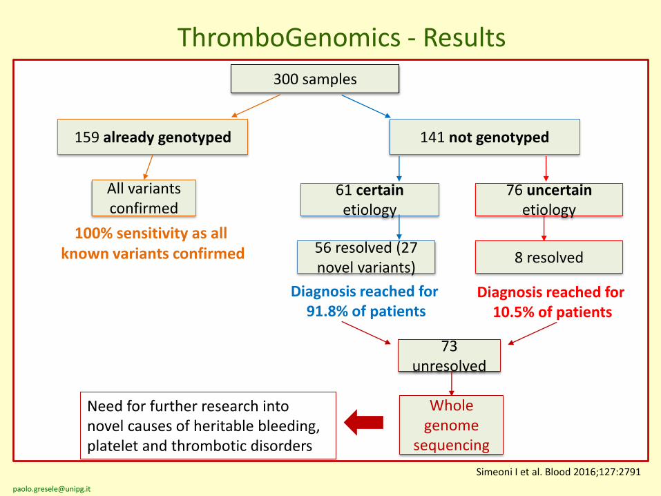
300 samples
159 already genotyped 141 not genotyped
61 certain etiology
76 uncertain etiology
All variants confirmed
56 resolved (27 novel variants)
Diagnosis reached for 91.8% of patients
8 resolved
100% sensitivity as all known variants confirmed
Diagnosis reached for 10.5% of patients
73 unresolved
Whole genome
sequencing
Need for further research into novel causes of heritable bleeding, platelet and thrombotic disorders
ThromboGenomics - Results
Simeoni I et al. Blood 2016;127:2791
Conclusions• IPFDs are a heterogeneous group of bleeding diseases
which represent a significant fraction of all the bleedingdiatheses
• A careful clinical evaluation and a rational diagnosticalgorithm based on a streamlined panel of tests allowsdiagnosis in a large part of the cases.
• Genetic diagnosis is becoming a conceivable alternative toextensive platelet function testing for many IPFDs
• IPFDs are associated with a significant bleeding risk
• Correct diagnosis and the use of prompt and appropriatetreatment may minimize bleeding risk

• Excessive bleeding during invasive procedures is a fearedcomplication in patients with IPFD.
• However, very few studies have evaluated the bleeding riskassociated with surgery in patients with IPFDs and most datacome from case reports or small case series.
• The exact bleeding risk of surgery and the most appropriatemanagement options in IPFDs are therefore unknown
• SPATA Study: retrospective, multicentre, worldwide studyinvolving clinical centers managing IPFDs.
• Participants were asked to enroll all IPFD patients they had onfile who had undergone surgery and to examine their records.
Bleeding risk of surgery in IPFD
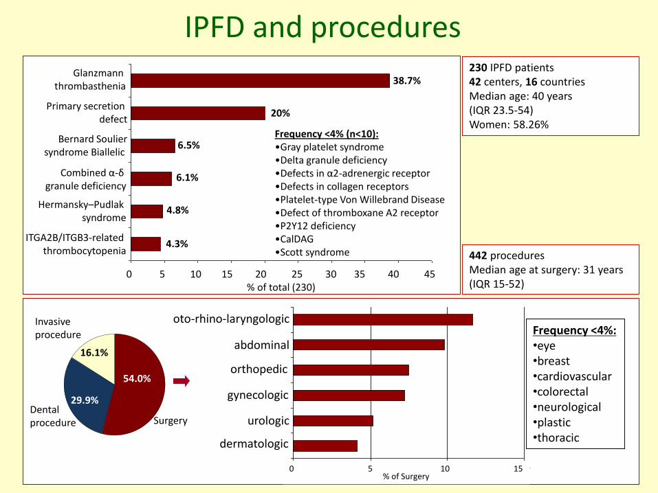
IPFD and procedures230 IPFD patients42 centers, 16 countriesMedian age: 40 years (IQR 23.5-54)Women: 58.26%
442 procedures Median age at surgery: 31 years(IQR 15-52)
Glanzmann thrombasthenia
Primary secretion defect
Bernard Souliersyndrome Biallelic
Combined α-δgranule deficiency
Hermansky–Pudlak syndrome
ITGA2B/ITGB3-related thrombocytopenia
0 5 10 15 20 25 30 35 40 45% of total (230)
38.7%
20%
6.5%
6.1%
4.8%
4.3%
Frequency <4% (n<10):•Gray platelet syndrome •Delta granule deficiency •Defects in α2-adrenergic receptor•Defects in collagen receptors•Platelet-type Von Willebrand Disease •Defect of thromboxane A2 receptor•P2Y12 deficiency •CalDAG•Scott syndrome
54.0%
29.9%
16.1%
Invasive procedure
Dentalprocedure
0% 5% 10% 15%
dermatologic surgery
urological surgery
gynecological
surgery
orthopedic surgery
abdominal surgery
otorinolaringoiatric
surgeryFrequency <4%:•eye•breast•cardiovascular•colorectal•neurological•plastic•thoracic
0 5 10 15% of Surgery
oto-rhino-laryngologic
abdominal
orthopedic
gynecologic
urologic
dermatologic
Surgery
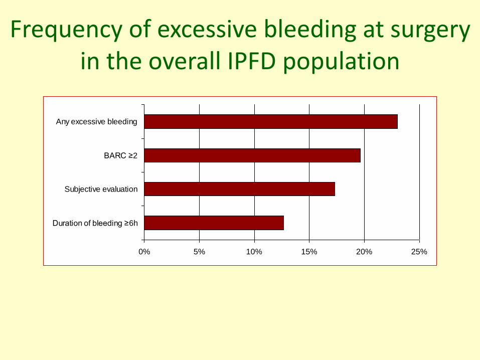
Frequency of excessive bleeding at surgeryin the overall IPFD population
0% 5% 10% 15% 20% 25%
Duration of bleeding ≥6h
Subjective evaluation
BARC ≥2
Any excessive bleeding
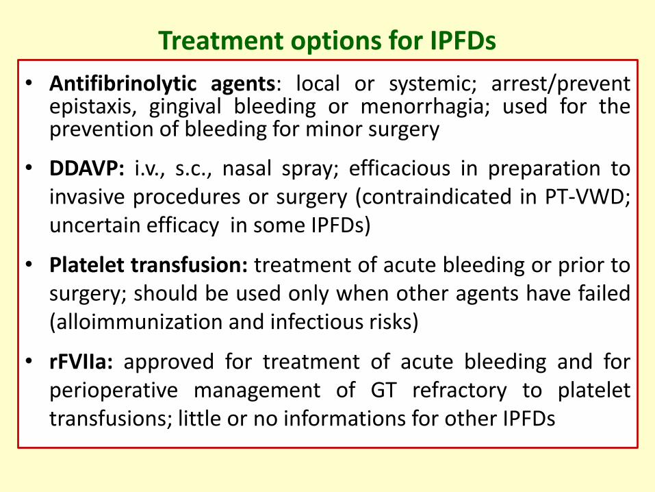
Treatment options for IPFDs
• Antifibrinolytic agents: local or systemic; arrest/preventepistaxis, gingival bleeding or menorrhagia; used for theprevention of bleeding for minor surgery
• DDAVP: i.v., s.c., nasal spray; efficacious in preparation toinvasive procedures or surgery (contraindicated in PT-VWD;uncertain efficacy in some IPFDs)
• Platelet transfusion: treatment of acute bleeding or prior tosurgery; should be used only when other agents have failed(alloimmunization and infectious risks)
• rFVIIa: approved for treatment of acute bleeding and forperioperative management of GT refractory to platelettransfusions; little or no informations for other IPFDs
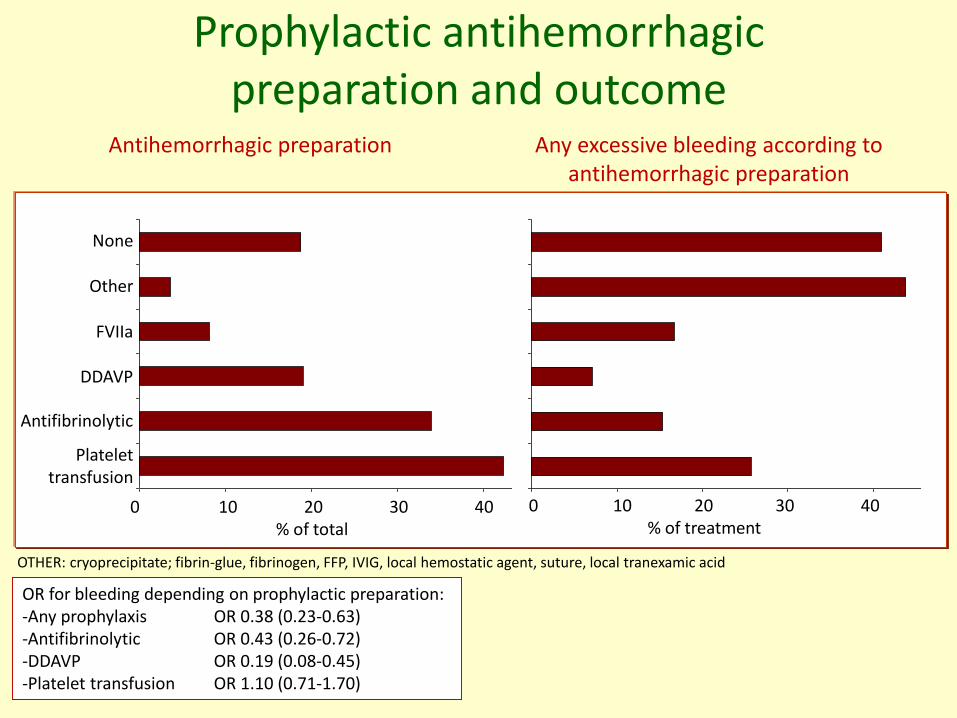
Prophylactic antihemorrhagicpreparation and outcome
OTHER: cryoprecipitate; fibrin-glue, fibrinogen, FFP, IVIG, local hemostatic agent, suture, local tranexamic acid
Antihemorrhagic preparation Any excessive bleeding according to antihemorrhagic preparation
OR for bleeding depending on prophylactic preparation: -Any prophylaxis OR 0.38 (0.23-0.63)-Antifibrinolytic OR 0.43 (0.26-0.72)-DDAVP OR 0.19 (0.08-0.45)-Platelet transfusion OR 1.10 (0.71-1.70)
None
Other
FVIIa
DDAVP
Antifibrinolytic
Platelettransfusion
0 10 20 30 40% of total
0 10 20 30 40% of treatment
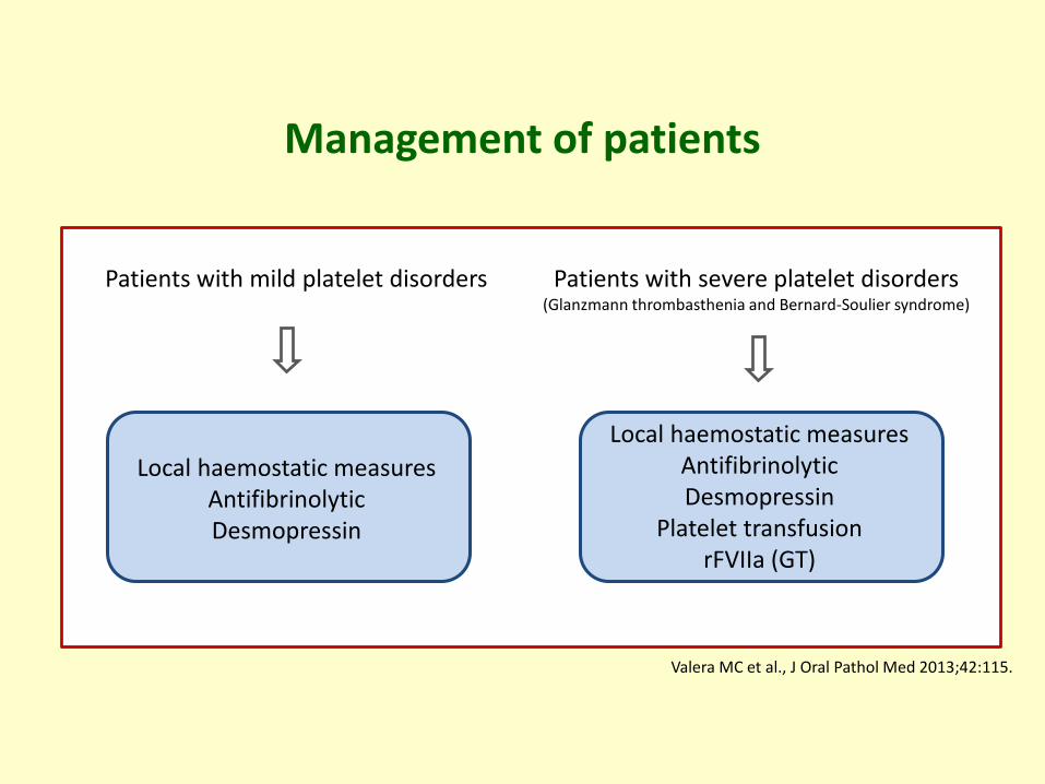
Management of patients
Patients with mild platelet disorders Patients with severe platelet disorders(Glanzmann thrombasthenia and Bernard-Soulier syndrome)
Local haemostatic measuresAntifibrinolyticDesmopressin
Local haemostatic measuresAntifibrinolyticDesmopressin
Platelet transfusionrFVIIa (GT)
Valera MC et al., J Oral Pathol Med 2013;42:115.
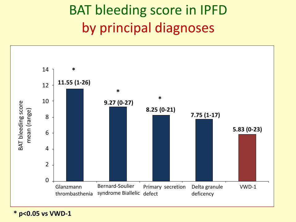
BAT bleeding score in IPFD by principal diagnoses
BA
T b
leed
ing
sco
rem
ean
(ra
nge
)
Glanzmann thrombasthenia
Delta granule deficency
Bernard-Souliersyndrome Biallelic
Primary secretion defect
VWD-1
14
12
10
8
6
4
2
0
11.55 (1-26)
9.27 (0-27)8.25 (0-21)
7.75 (1-17)
*
**
5.83 (0-23)
* p<0.05 vs VWD-1
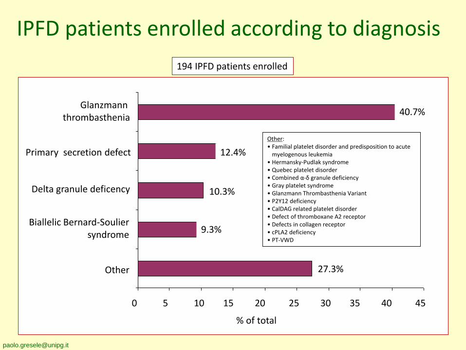
IPFD patients enrolled according to diagnosis
Glanzmann thrombasthenia
Delta granule deficency
Biallelic Bernard-Souliersyndrome
Primary secretion defect
Other
0 5 10 15 20 25 30 35 40 45
194 IPFD patients enrolled
Other:• Familial platelet disorder and predisposition to acute
myelogenous leukemia• Hermansky-Pudlak syndrome• Quebec platelet disorder• Combined α-δ granule deficiency• Gray platelet syndrome• Glanzmann Thrombasthenia Variant• P2Y12 deficiency• CalDAG related platelet disorder• Defect of thromboxane A2 receptor• Defects in collagen receptor• cPLA2 deficiency• PT-VWD
9.3%
27.3%
% of total
10.3%
40.7%
12.4%
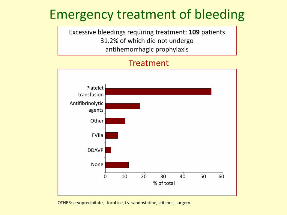
Emergency treatment of bleedingExcessive bleedings requiring treatment: 109 patients
31.2% of which did not undergo antihemorrhagic prophylaxis
OTHER: cryoprecipitate, local ice, i.v. sandostatine, stitches, surgery.
Treatment
Antifibrinolyticagents
None
Other
FVIIa
DDAVP
Platelettransfusion
0 10 20 30 40 50 60% of total
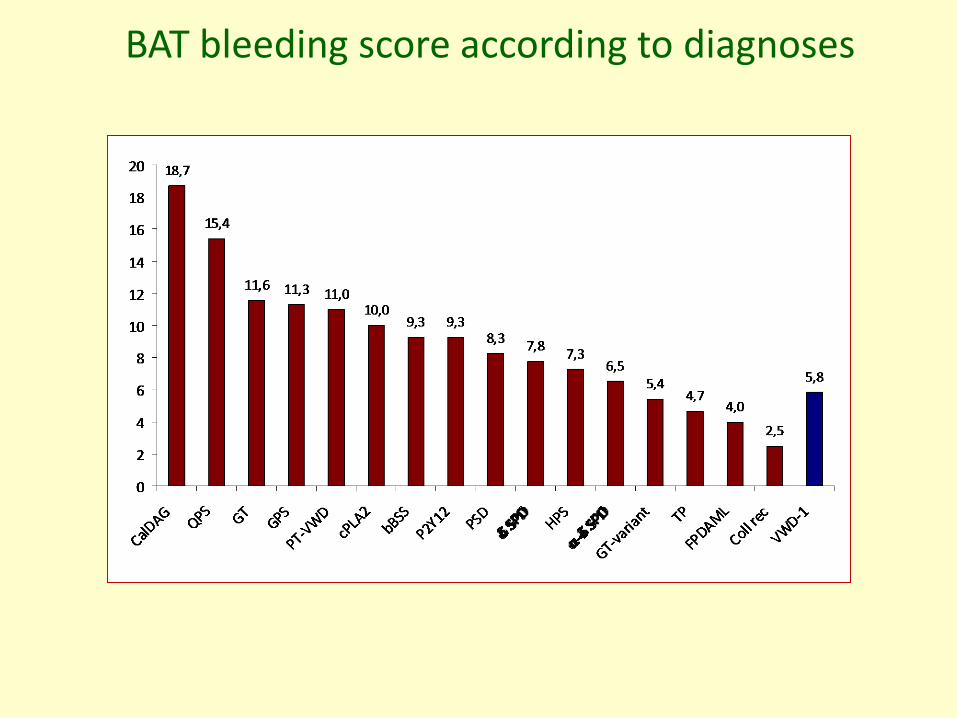
BAT bleeding score according to diagnoses