Embed Size (px)
Citation preview

La PET nel Linfoma di Hodgkin in età pediatrica: nuove metodologie per la valutazione della risposta terapeutica
Stefano Panareo
UOC Medicina Nucleare
Azienda Ospedaliero - Universitaria di Ferrara
(Direttore F.F.: Corrado Cittanti)
[email protected] www.ospfe.it

Tomografi PET/CT • Imaging funzionale e morfologico
insieme
• Migliore risoluzione spaziale (5 mm)
• Imaging “whole body”
• Tempo di acquisizione limitato (12-15’)
• Minor dose al paziente
• Ridotta esposizione del paziente a
radiazioni

Marcatori biologici nei tumori con PET
• Perfusione
• Metabolismo 18F-FDG
• Ipossia
• Espressione recettoriale
• Sintesi DNA
18F-FDG nei tumori
• La trasformazione maligna della maggior parte
delle linee cellulari si associa ad elevato
consumo di glucosio:
• sovraespressione dei trasportatori di
glucosio (Glut1 e Glut3)
• aumento dell’attività esochinasica.

Glucose
Transporter
Hexokinase
Glucose-6-
phosphatase
Glycogen FDG FDG FDG-6-PO4
Glycolysis
Glucose-6P-
isomerase
GLUT 1-3
18F-FDG nei tumori

• Maggiore accumulo di FDG nelle lesioni a più
rapida crescita e/o nei tumori più aggressivi.
• FDG rispecchia il numero di cellule tumorali vitali.
L’accumulo di FDG nei tumori in vivo dipende anche da
vari parametri fisiologici (es. ossigenazione, flusso
ematico, flogosi peritumorale)
18F-FDG nei tumori

18F-FDG E METABOLISMO
• 18F-FDG captato da cellule in attiva proliferazione.
• fornisce caratteristiche di crescita e di metabolismo tumorale piuttosto che di morfologia della neoplasia (TC, RM).
• descrive estensione locale o a distanza del tumore.
• riesce a predire ed interpretare le modificazioni che si hanno in seguito a trattamenti terapeutici.

18F-FDG PET
Ricostruzione 3D
Indication %
Lung 24,2%
Head/neck/chest 14,5%
Lymphoma 14,2%
Colorectal 10,1%
Thyroid 8,4%
Gynecological/breast 7,7%
Other 6,6%
Liver 2,8%
Sarcoma 2,7%
Esophageal 2,7%
Testicular/prostate 2,6%
Melanoma 1,7%
Brain 0,9%
Unknown primary 0,8%
Total 99,9%
FDG PET/CT IN ONCOLOGIA
• Sensibilità 92-94% • Specificità 93% • Accuratezza diagnostica 93%
PET in oncologia – Linee Guida AIMN

SUV
(Standardized Uptake Value)
• Indice semiquantitativo che misura la intensità di captazione tumorale del radiofarmaco.
• SUV alto è espressione di elevato metabolismo.
• I tumori con elevato SUV sono più proliferanti e maggiormente aggressivi.
• SUV come valore prognostico (risposta alla terapia).

2.5

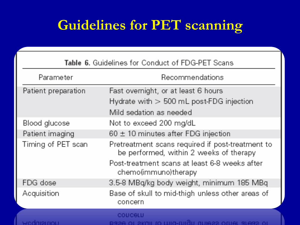
Guidelines for PET scanning

BIODISTRIBUTION
fegato
vescica
intestino
mediastino
reni

• PRE-TREATMENT STAGING
• RESTAGING:
– END-TREATMENT
– INTERIM/MID-TREATMENT
• FOLLOW-UP
• Further indications:
– Pre and post transplant
– Pre and post Radio Immunotherapy (RIT)
– Aggressive transformation
– Radiotherapy planning
INDICATIONS

False positive
False negative
FDG is taken up in any process with increased glycolysis:
• INFLAMMATION
• INFECTION
• NECROSIS
• GRANULOMATOUS DISEASE (including sarcoidosis)
• THYMIC HYPERPLASIA
• BROWN FAT
FP occures with the use of RITUXIMAB [Han et al]
• NON FDG AVID HISTOLOGIES
• SMALL DIMENSION (< 5mm)
• UNCORRECT TIMING OF PET SCANNING
• POST CHEMOTHERAPY STUNNING

Spaepen, B.J. Haemat., 2001
60 HL.
Follow-up: 3 years
End-treatment

• High NPV: 90%
(similar to spiral CT)
10-20% FN
inability to detect
microscopic disease
resulting in relapse
End-treatment
• Lower and more variable PPV:
75% in NHL (vs 40% CT)
65% in HL (vs 20% CT) FP post radiation inflammatory changes
FP thymic hyperplasia in younger patients

End-treatment
CT
End-treatment
PET

HL nodular sclerosis (stage III ): A) after CHT, B) post CHT
End-treatment

Female, 7yo, HL (lymphocytic prevalence) CHT: CVP
End-treatment
post CHT
after CHT
24/06/2015
27/08/2015

Response assessment at therapy conclusion is typically performed:
• At least 3 WEEKS AFTER CHEMOTHERAPY (6-8)
• 8-12 WEEKS AFTER RADIOTHERAPY
Initial staging with PET, for assessment of response after treatment:
• Not mandatory but strongly recommended for:
HL, DLBCL, FL, MCL (routinely FDG avid)
• Mandatory for:
T-cell lymphomas and indolent NHL other than FL
(variably FDG avid)
PET+ at all disease sites >1.5cm in diameter noted by CECT
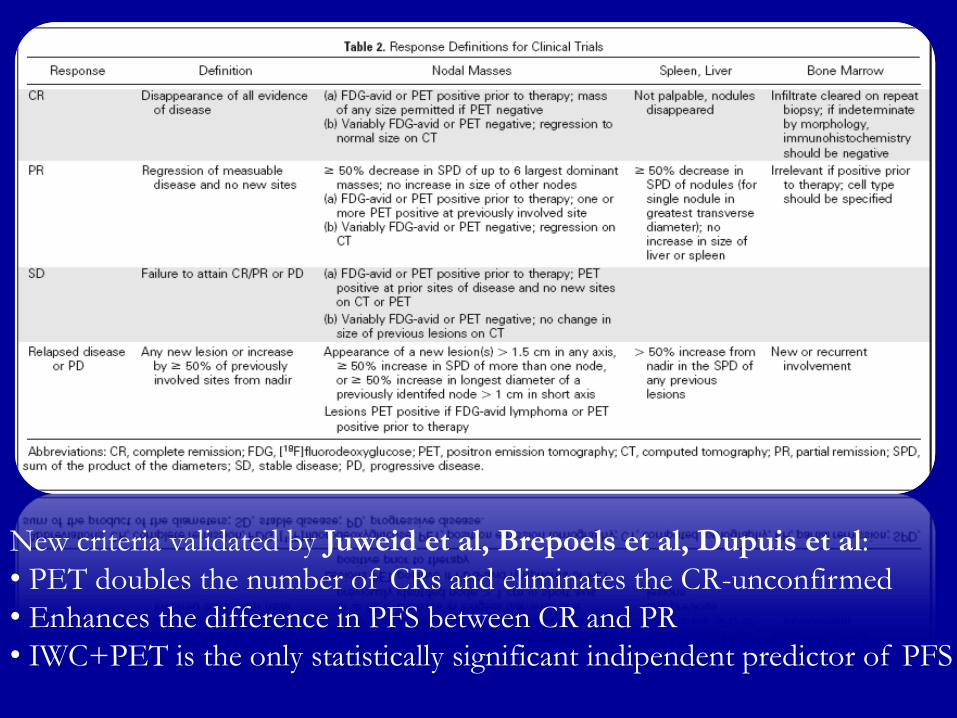
New criteria validated by Juweid et al, Brepoels et al, Dupuis et al:
• PET doubles the number of CRs and eliminates the CR-unconfirmed
• Enhances the difference in PFS between CR and PR
• IWC+PET is the only statistically significant indipendent predictor of PFS

[Gallamini et al] and [Cerci et al]:
Interim PET (dynamic) is the most important prognostic factor,
more powerful than the IPS (static)
ALBUMIN Hb SEX AGE STAGE WBC
count
Lympho
cyte
IPS <4
g/dl
<10.5
g/dl
Male >45y IV >15000/
mm2
<600/m
m2 or
<8%wbc
Interim

9y PFS:
PET- 94,7%
vs
PET+ 31,3%
(p=0,0000)
9y OS:
PET- 100%
vs
PET+ 85,2%
(p=0,0001)

HL (A) STAGING: IV (spleen + bone marrow) (B) PET interim: P.R. (C) PET end-treatment: MRU. 2 year-follow up: C.R.

Male, 26yo patient diagnosed with HL.
A: Initial staging FDG-PET present multiple bilateral cervical, bilateral supraclavicular, left
axilary, mediastinum lymph nodes and also spleen lesions.
B: After 2 cycles of ABVD patient was submitted to interim FDG-PET, with partial metabolic
response, with markedly reduced metabolism in all lymph node chains.
C: After four cycles of ABVD patient was reevaluated and presented progressive disease (PD).

Male, 16yo, HL (nodular sclerosis)
CTH: first l. COPP/ABV + 2 cycles IEP; second l. OPPA + DHAP
07/01/2015
09/03/2015
18/05/2015
19/11/2015
staging
interim
Change CHT - interim
End CHT
HL: clinical case

2007:
• [JUWEID ET AL.]
CONSENSUS OF THE IMAGING
SUBCOMMITTEE OF INTERNATIONAL
HARMONIZATION PROJECT (IHP)
• [CHESON ET AL.]
REVISED RESPONSE CRITERIA FOR
MALIGNANT LYMPHOMA
2009:
•[GALLAMINI et al.] THE EDUCATION PROGRAM FOR
THE ANNUAL CONGRESS OF THE EUROPEAN
HEMATOLOGY ASSOCIATION
N° and intensity residual sites relative to baseline scan


2008:
• [GUY’S et al.] LONDON FDG UPTAKE 5-POINT SCALE:
1. No uptake
2. Uptake > mediastinum
3. Uptake > mediastinum but <liver
4. Uptake moderately increased compared to the liver at any site
5. Uptake markedly increased compared to the liver at any site or/and new
sites of disease
2009:
• FIRST INTERNATIONAL WORKSHOP ON INTERIM PET IN
LYMPHOMA: validation 5-point scale as DEAUVILLE SCORE
2010:
• Criteria publication by [BARRINGTON et al] and [MEIGNAN et al] after
the SECOND INTERNATIONAL WORKSHOP (in Menton -France)
A score of 4 or more was the positivity threshold


LINFOMA DI HODGKIN

Corrado
Cittanti
Licia
Uccelli
Ilaria
Rambaldi
Fabrizio
Cocciolillo Ivan
Santi
Stefano
Panareo