Embed Size (px)
Citation preview

ORIGINAL PAPER
Lactobacillus acidophilus regulates STAT3 and STAT5signaling in bovine β-lg-sensitized mice model
Yun Zhang1 & Ai-li Li1 & Yi-qiao Sun1& Peng Du1
&
Li-bo Liu1& Shuang Li1 & Chao Zhang1
Received: 13 October 2015 /Revised: 12 January 2016 /Accepted: 26 February 2016 /Published online: 23 March 2016# INRA and Springer-Verlag France 2016
Abstract Our previous study has shown that oral supplementation with LactobacillusacidophilusKLDS 1.0738 could inhibit β-lactoglobulin (β-lg) allergy. In this study, weinvestigated the effect of L. acidophilus on the balance between T helper type 17(Th17) cells and regulatory T cells (Treg) in allergic mouse model and explored theparticipation of related signal transducers and activator of transcription (STAT) in thisprocess. Bovine β-lg-sensitized mice received strains KLDS 1.0738 for 3 weeks. Afterthe allergen challenge, the percentages of Treg and Th17 cells, cytokine and STATmRNA expression, and pSTAT protein levels were detected by flow cytometry,quantitative RT-PCR, and western blot, respectively. The results showed that stimula-tion with β-lg increased the levels of IL-6, pSTAT3, and Th17 cells, but decreased thelevels of IL-2, pSTAT5, and Treg cells compared to the controls (P<0.05). However,oral administration of L. acidophilus KLDS 1.0738 suppressed β-lg-induced inflam-matory and improved the Treg/Th17 imbalance. In addition, L. acidophilus-treatedgroup presented decrease in pSTAT3 activation, SOCS3, and IL-6 level, but increase inSTAT5a/b, CD25, and IL-2 mRNA expression. These findings suggest thatL. acidophilus could regulate IL-6/STAT3 and IL-2/STAT5 pathway, which may beresponsible for the Treg/Th17 imbalance in β-lg-sensitized mice.
Keywords Lactobacillus acidophilus . Milk allergy . Treg/Th17 imbalance . STAT3/STAT5 signaling pathway
Dairy Sci. & Technol. (2016) 96:501–512DOI 10.1007/s13594-016-0284-3
* Ai-li [email protected]
1 Key laboratory of Dairy Science, Ministry of Education, College of Food Science, NortheastAgriculture University, No.59, Mucai Street, Xiangfang District, Harbin,Heilongjiang 150030, China
1 Introduction
Cow’s milk allergy (CMA) is a complex inflammatory disease in which CD4+
T helper (Th) cells play a central role (Giovanna et al. 2012). In addition to thegeneral paradigm of Th1/Th2 immune regulation, the current study showed thattwo additional Th cell subsets, interleukin 17 (IL-17)-producing Th17 cells andregulatory T cells (Treg), were also partially responsible for the development ofallergic inflammation (Palomares et al. 2010; Zhao et al. 2010). Therefore, abetter understanding of the roles of Treg and Th17 cells may provide insightsinto effective CMA control.
Most evidence demonstrated that the STAT pathways were involved in Thcell differentiation (Saleh et al. 2009; Chen et al. 2007) and correlated with theinflammatory process (Minegishi et al. 2007). Among the STAT family ofproteins, STAT3 was reported to promote Th17 differentiation, including theinduction of Th17-related cytokine production, RORγt and RORα (Harris et al.2007). In addition, STAT3 might be activated by most proinflammatory agentsin the proallergic milieu, such as IL-6 (Yang et al. 2007). Mori et al. (2011)showed that the IL-6/STAT3 pathway was critical in chronic intestinal inflam-mation by facilitating Th17 cells and restraining Treg. In contrast, STAT5 wasactivated by IL-2 and was indispensable in maintaining Treg homeostasis andself-tolerance (Laurence et al. 2007; Yao et al. 2006). Burchill et al. (2007)indicated that STAT5a/b promoted Treg development and maintenance throughregulation of both forkhead box P3 (Foxp3) and CD25 expression. Morerecently, Wang et al. (2012) suggested that the Th17/Treg imbalance causedby nasal polyposis was positively correlated with STAT3/STAT5 activation.However, it is not yet known whether IL-6/STAT3 and IL-2/STAT5 pathwaysmight contribute to the Th17/Treg imbalance in CMA.
Lactobacilli have beneficial effects on T-cell-mediated inflammatory diseases.Previous research reported that probiotics could prevent food allergy by im-proving the Th1/Th2 imbalance (Kim et al. 2008). Recent studies indicated thatapplication of lactobacilli could attenuate airway hyperreactivity by induction ofTregs in a mouse model of asthma (Jan et al. 2012; Karimi et al. 2009). Wealso showed Lactobacillus acidophilus strain KLDS 1.0738 had the ability tosuppress the β-lg allergic symptoms and Th17 cytokine production (Li et al.2013). Moreover, some studies focused on the effect of probiotics on activatingthe cytokine-mediated STAT pathways to regulate the immune responses.Miettinen et al. (2000) reported that L. rhamnosus GG ATCC 53103 couldinduce Th1 cytokine production through the NF-κB and STAT pathways in themacrophages. Jandu et al. (2009) found that L. helveticus prevented IFN-γ-Jak1and 2-STAT-1 activation in Escherichia coli O157:H7-infected cells. However,whether lactobacilli might modulate STAT signaling in response to theTh17/Treg development in β-lg-sensitized mice is currently unknown. There-fore, the aim of this research is to evaluate the relationship between STATpathways and the imbalance of Treg/Th17 in β-lg sensitized mice and toanalyze the effect of L. acidophilus on the activations of STAT3 and STAT5.
502 Y. Zhang et al.
2 Materials and methods
2.1 Mice and bacteria
Six-week-old BALB/c mice were purchased from the Harbin Veterinary ResearchInstitute (Harbin, China) and raised in normal husbandry environment. The mice werefed a milk-free standard diet and provided with water ad libitum. All animal experi-ments complied with the rules of the Care and Use of Laboratory Animals of NortheastAgricultural University.
The L. acidophilus KLDS 1.0738 used in this study was obtained from the KeyLaboratory of Dairy Science, Ministry of Education (Northeast Agriculture University,China). The bacteria were incubated at 37 °C in Man-Rogosa-Sharpe broth until theyreached a concentration of 5×10⁹ CFU/mL. The bacteria cells were collected bycentrifugation (4000 g, 10 min), washed three times with sterile distilled water, andheat-killed at 100 °C for 20 min. Then, the treated organisms were lyophilized andsuspended in 0.9% sterile saline.
2.2 Experimental groups
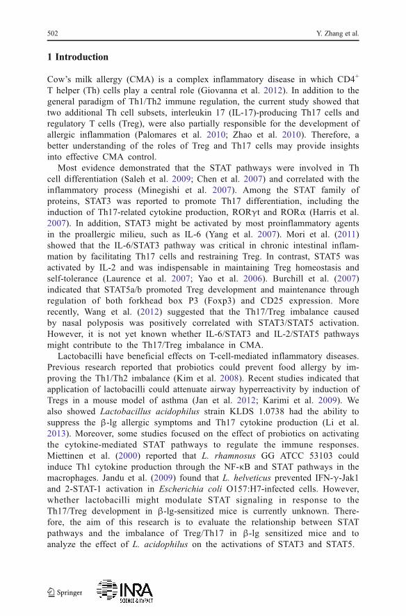
Animal experiments were carried out as described previously with minor modifications(Li et al. 2013). As shown in Fig. 1, BALB/c mice were randomly divided into threegroups: β-lg allergy mice (β-Lg) were sensitized by intraperitoneal injection of 50 μgβ-lg (Sigma-Aldrich, USA) adsorbed on 2 mg aluminum hydroxide (Sigma) at days 7,14, and 21; L. acidophilus-treated mice (LR) were intragastrically administered with200 μL of L. acidophilus suspension (2.5 mg/animal) three times a week from days 1 to21; control mice (Con) were treated with 0.9% sterile saline. On day 25, the mice wereorally challenged twice with 20 mg β-lg or saline solution, and then all the animalswere sacrificed in 2 h after the last β-lg challenge. The blood, lung, colon, and spleentissues were subsequently collected.
Oral administration of L. acidophilus suspension
BALB/c 0 7 14 21 25 days
6-weeks-old
Sensitization with 50 µg β-lg + 2mg Al(OH)3
Oral challenge
Fig. 1 Schedule for immunization with β-lg and oral administration of Lactobacillus acidophilus KLDS1.0738. Mice were sensitized by intraperitoneal injection of with β-lg once a week for 3 weeks. L. acidophilusKLDS 1.0738 (dose of 2.5 mg/mouse) was administered orally three times a week from days 1 to 21. As acontrol, 200 μL of distilled water was administered. Oral challenge with and without β-lg was carried out onday 25
Lactobacillus acidophilus regulates STAT3 and STAT5 signaling 503
2.3 Eosinophil counts
The blood samples were collected and the plasma was obtained. The cell counts wereperformed using a hemacytometer under light microscope after staining with eosin-acetone diluent.
2.4 Histological analysis of the colon and lung tissues
The colon and lung tissues were removed and fixed in 10% formalin, and then thespecimens were dehydrated and embedded in paraffin. The paraffin sections werestained with hematoxylin and eosin (HE). Pathological alterations in the lung andcolon tissues were assessed under a light microscope.
2.5 Flow cytometric analysis
Single-cell suspensions were prepared from spleens by injecting PBS into the tissues,and the erythrocytes were lysed before the samples were subjected to flow cytometricanalysis. To detect the Treg, FITC-labeled anti-CD4 and PE-labeled anti-CD25(eBioscience, USA) were used for cell surface staining, and APC-labeled anti-Foxp3(eBioscience, USA) was applied for intracellular staining after fixation and perme-abilization. To detect the Th17 cells, the cells were first surface-stained with FITC-conjugated CD4 Abs, then fixed and permeabilized according to the kit manual, andfinally stained intracellularly with APC-conjugated IL-17A Abs. After washing threetimes with PBS, the cells were resuspended and processed with a FACS flow cytometerequipped with Cell Quest software (BD FACS Aria™ Cell Sorter, USA).
2.6 RNA isolation and real-time PCR
Total RNA was isolated from the spleen tissues using the RNA simple Total RNA kit(Tiangen, China), and cDNAwas synthesized using the cDNA RT reagent kit (Takara,China). The STAT3, STAT5a/b, SOCS3, CD25, IL-2, and IL-6 message expressionlevels were quantified using the ABI 7500 Real-Time PCR System (Applied Biosystems, USA). Amplification was performed in a total volume of 25 μL for 40 cycles,and the products were detected using SYBR Premix Ex Taq™II (Takara, China). Thesequences of the specific primers used in the PCR are shown in Table 1 and the mRNAexpression in each group were normalized to the level of β-actin housekeeping genesusing the 2−△△Ct method and represented as the fold induction.
2.7 Extraction of the tissue protein and western blot analysis
Proteins from the CD4 T cell fraction were extracted with RIPA lysis buffer (Solarbio,China) and the concentration was detected using BCA kit (Solarbio, China) followingthe manufacturer’s protocol. The samples containing 25 μg of protein were boiled,separated by polyacrylamide gel electrophoresis, and transferred to a polyvinylidenefluoride membrane. The membrane was allowed to react with polyclonal anti-GAPDH,rabbit anti-STAT3, anti-phospho-STAT3 (Tyr), anti-STAT5, and anti-phospho-STAT5(Tyr) (Cell Signaling, USA). After incubation with a horseradish peroxidase (HRP)-
504 Y. Zhang et al.
conjugated anti-rabbit antibody, the membrane was incubated with ECL chemilumi-nescence reagent (TransGen Biotech, China), and the film was exposed to themembrane.
2.8 Statistical analysis
All data in the text and figures were presented as the means± standard deviation(means±SD). One-way ANOVAwas performed for detection of significant differencesamong the groups. The statistical analysis was carried out using the multiple compar-ison test (SPSS17.0 software). P values<0.05 were considered significantly different.
3 Results
3.1 Effect of L. acidophilus on eosinophil counts and histologic characteristicsof β-lg-sensitized mice
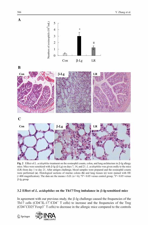
As shown in Fig. 2a, β-lg allergen caused eosinophil-rich inflammation in allergicmice. In contrast, treatment with L. acidophilus decreased the percentages of eosino-phils in the blood of sensitized mice.
The severity of allergy symptoms was further investigated by means of histopathol-ogy. Bovine β-lg challenge led to inflammatory cell infiltrates into the lung and thecolon tissues of sensitized mice. However, decreased inflammatory responses wereobserved in L. acidophilus-treated group (Fig. 2b, c).
Table 1 Primers used for real-time PCR
Name Sequence (5′→ 3′)
STAT5a Forward primer AAGATCAAGCTGGGGCACTA
Reverse primer ATGGGACAGCGGTCATAC
STAT5b Forward primer CGAGCTGGTCTTTCAAGTCA
Reverse primer CTGGCTGCCGTGAACAAT
STAT3 Forward primer CAAAACCTCAAGAAGCCAAGG
Reverse primer TCACTCACAATGCTTCTCCGC
CD25 Forward primer ACACCTGTAAGCCCAGCTCT
Reverse primer TGGAAAGGTTGAGGGGTAAG
SOCS3 Forward primer GAAGACCAAGTTCATCTGTGTG
Reverse primer GTAGCACACTCCGAGGTCAGAT
IL-6 Forward primer GATGCTACCAAACTGGATATAATC
Reverse primer GGTCCTTAGCCACTCCTTCTGTG
IL-2 Forward primer CACATTTGAGTGCCAATTCGAT
Reverse primer GCGCTTACTTTGTGCTGTCCTA
β-actin Forward primer CGCAAAGACCTGTATGCCAAT
Reverse primer GGGCTGTGATCTCCTTCTGC
Lactobacillus acidophilus regulates STAT3 and STAT5 signaling 505
3.2 Effect of L. acidophilus on the Th17/Treg imbalance in β-lg-sensitized mice
In agreement with our previous study, the β-lg challenge caused the frequencies of theTh17 cells (CD4+IL-17+⁄CD4+ T cells) to increase and the frequencies of the Treg(CD4+CD25+Foxp3+ T cells) to decrease in the allergic mice compared to the controls
*
0
1
2
3
4
5
Con β-Lg LR
Num
bers
of e
osin
ophi
ls (1
0 6 /m
L)A
LRCon β-Lg
B
C
Con β-Lg LR
Fig. 2 Effect of L. acidophilus treatment on the eosinophil counts, colon, and lung architecture in β-lg allergymice. Mice were sensitized with β-lg (β-Lg) on days 7, 14, and 21. L. acidophiluswas given orally to the mice(LR) from day 1 to day 21. After antigen challenge, blood samples were prepared and the eosinophil countswere performed (a). Histological sections of murine colons (b) and lung tissues (c) were stained with HE(×400 magnification). The data are the means ± S.D. (n = 6); *P < 0.05 versus control group, #P < 0.05 versusβ-lg group
506 Y. Zhang et al.
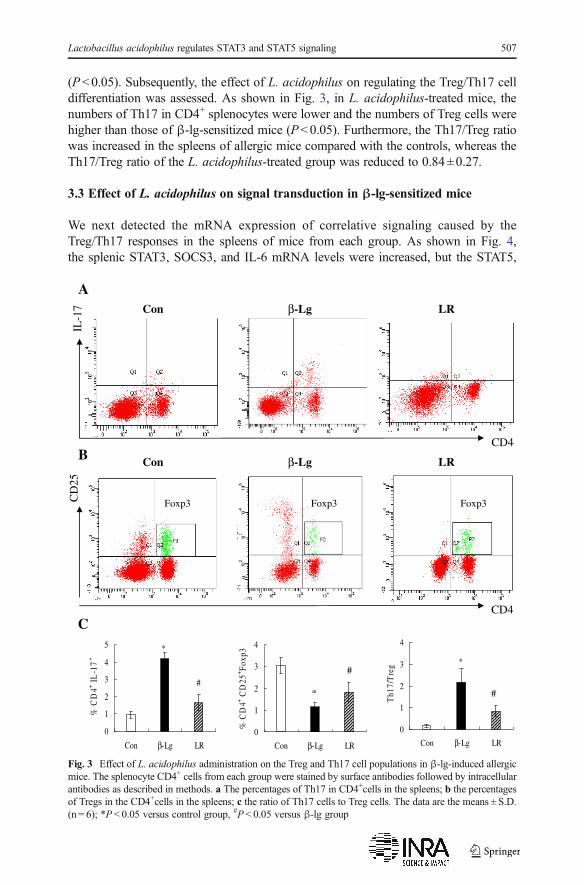
(P<0.05). Subsequently, the effect of L. acidophilus on regulating the Treg/Th17 celldifferentiation was assessed. As shown in Fig. 3, in L. acidophilus-treated mice, thenumbers of Th17 in CD4+ splenocytes were lower and the numbers of Treg cells werehigher than those of β-lg-sensitized mice (P<0.05). Furthermore, the Th17/Treg ratiowas increased in the spleens of allergic mice compared with the controls, whereas theTh17/Treg ratio of the L. acidophilus-treated group was reduced to 0.84±0.27.
3.3 Effect of L. acidophilus on signal transduction in β-lg-sensitized mice
We next detected the mRNA expression of correlative signaling caused by theTreg/Th17 responses in the spleens of mice from each group. As shown in Fig. 4,the splenic STAT3, SOCS3, and IL-6 mRNA levels were increased, but the STAT5,
Con β-Lg LR
Con β-Lg LR
*
#
0
1
2
3
4
5
Con β-Lg LR
% C
D4+ I
L-1
7+
0
1
2
3
4
Con β-Lg LR
% C
D4+ C
D25
+ Fox
p3
*
0
1
2
3
4
Con β-Lg LR
Th1
7/T
reg
CD4
IL-1
7
A
B
CD4
CD
25
*
#
C
#
3pxoF3pxoF3pxoF
Fig. 3 Effect of L. acidophilus administration on the Treg and Th17 cell populations in β-lg-induced allergicmice. The splenocyte CD4+ cells from each group were stained by surface antibodies followed by intracellularantibodies as described in methods. a The percentages of Th17 in CD4+cells in the spleens; b the percentagesof Tregs in the CD4+cells in the spleens; c the ratio of Th17 cells to Treg cells. The data are the means ± S.D.(n = 6); *P < 0.05 versus control group, #P < 0.05 versus β-lg group
Lactobacillus acidophilus regulates STAT3 and STAT5 signaling 507
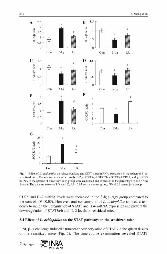
CD25, and IL-2 mRNA levels were decreased in the β-lg allergy group compared tothe controls (P<0.05). However, oral consumption of L. acidophilus showed a ten-dency to inhibit the upregulation of STAT3 and IL-6 mRNA expression and prevent thedownregulation of STAT5a/b and IL-2 levels in sensitized mice.
3.4 Effect of L. acidophilus on the STAT pathways in the sensitized mice
First, β-lg challenge induced a transient phosphorylation of STAT3 in the spleen tissuesof the sensitized mice (Fig. 5). The time-course examination revealed STAT3
*
0
0.5
1
1.5
2
2.5
Con β-Lg LR
IL-6
/β-a
ctin
*
0
0.5
1
1.5
Con β-Lg LR
IL-2
/β-a
ctin
*
0
0.5
1
1.5
Con β-Lg LR
STA
T5a
/β-a
ctin
*
0
0.5
1
1.5
Con β-Lg LRST
AT
5b/β
-act
in
*
0
0.5
1
1.5
2
Con β-Lg LR
STA
T3/
β-ac
tin
*
0
1
2
3
4
5
Con β-Lg LR
CD
25/β
-act
in
*
0
5
10
15
20
25
Con β-Lg LR
SOC
S3/β
-act
in
A B
C D
E F
G
Fig. 4 Effect of L. acidophilus on related cytokine and STAT signal mRNA expression in the spleen of β-lg-sensitized mice. The relative levels of a IL-6, b IL-2, c STAT5a, d STAT5b, e STAT3, f CD25, and g SOCS3mRNA in the spleens of mice from each group were calculated and expressed at the percentage of mRNA toβ-actin. The data are means ± S.D. (n = 6); *P < 0.05 versus control group, #P < 0.05 versus β-lg group
508 Y. Zhang et al.
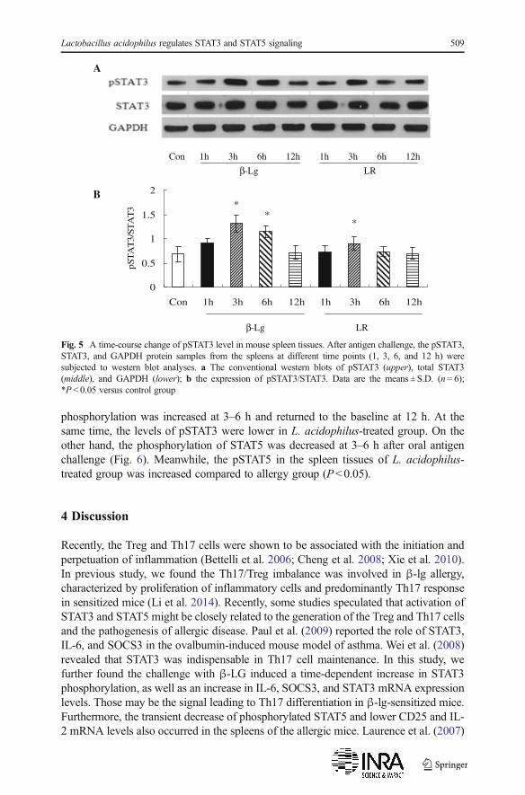
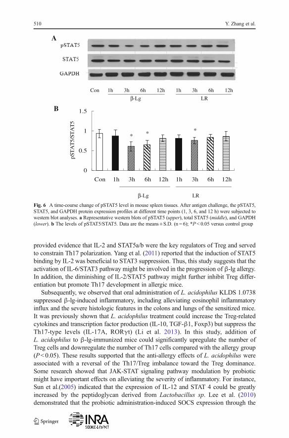
phosphorylation was increased at 3–6 h and returned to the baseline at 12 h. At thesame time, the levels of pSTAT3 were lower in L. acidophilus-treated group. On theother hand, the phosphorylation of STAT5 was decreased at 3–6 h after oral antigenchallenge (Fig. 6). Meanwhile, the pSTAT5 in the spleen tissues of L. acidophilus-treated group was increased compared to allergy group (P<0.05).
4 Discussion
Recently, the Treg and Th17 cells were shown to be associated with the initiation andperpetuation of inflammation (Bettelli et al. 2006; Cheng et al. 2008; Xie et al. 2010).In previous study, we found the Th17/Treg imbalance was involved in β-lg allergy,characterized by proliferation of inflammatory cells and predominantly Th17 responsein sensitized mice (Li et al. 2014). Recently, some studies speculated that activation ofSTAT3 and STAT5 might be closely related to the generation of the Treg and Th17 cellsand the pathogenesis of allergic disease. Paul et al. (2009) reported the role of STAT3,IL-6, and SOCS3 in the ovalbumin-induced mouse model of asthma. Wei et al. (2008)revealed that STAT3 was indispensable in Th17 cell maintenance. In this study, wefurther found the challenge with β-LG induced a time-dependent increase in STAT3phosphorylation, as well as an increase in IL-6, SOCS3, and STAT3 mRNA expressionlevels. Those may be the signal leading to Th17 differentiation in β-lg-sensitized mice.Furthermore, the transient decrease of phosphorylated STAT5 and lower CD25 and IL-2 mRNA levels also occurred in the spleens of the allergic mice. Laurence et al. (2007)
Con 1h 3h 6h 12h 1h 3h 6h 12h
β-Lg LR
**
*
0
0.5
1
1.5
2
Con 1h 3h 6h 12h 1h 3h 6h 12h
pST
AT
3/ST
AT
3
β-Lg LR
A
B
Fig. 5 A time-course change of pSTAT3 level in mouse spleen tissues. After antigen challenge, the pSTAT3,STAT3, and GAPDH protein samples from the spleens at different time points (1, 3, 6, and 12 h) weresubjected to western blot analyses. a The conventional western blots of pSTAT3 (upper), total STAT3(middle), and GAPDH (lower); b the expression of pSTAT3/STAT3. Data are the means ± S.D. (n = 6);*P < 0.05 versus control group
Lactobacillus acidophilus regulates STAT3 and STAT5 signaling 509
provided evidence that IL-2 and STAT5a/b were the key regulators of Treg and servedto constrain Th17 polarization. Yang et al. (2011) reported that the induction of STAT5binding by IL-2 was beneficial to STAT3 suppression. Thus, this study suggests that theactivation of IL-6/STAT3 pathway might be involved in the progression of β-lg allergy.In addition, the diminishing of IL-2/STAT5 pathway might further inhibit Treg differ-entiation but promote Th17 development in allergic mice.
Subsequently, we observed that oral administration of L. acidophilus KLDS 1.0738suppressed β-lg-induced inflammatory, including alleviating eosinophil inflammatoryinflux and the severe histologic features in the colons and lungs of the sensitized mice.It was previously shown that L. acidophilus treatment could increase the Treg-relatedcytokines and transcription factor production (IL-10, TGF-β1, Foxp3) but suppress theTh17-type levels (IL-17A, RORγt) (Li et al. 2013). In this study, addition ofL. acidophilus to β-lg-immunized mice could significantly upregulate the number ofTreg cells and downregulate the number of Th17 cells compared with the allergy group(P<0.05). These results supported that the anti-allergy effects of L. acidophilus wereassociated with a reversal of the Th17/Treg imbalance toward the Treg dominance.Some research showed that JAK-STAT signaling pathway modulation by probioticmight have important effects on alleviating the severity of inflammatory. For instance,Sun et al.(2005) indicated that the expression of IL-12 and STAT 4 could be greatlyincreased by the peptidoglycan derived from Lactobacillus sp. Lee et al. (2010)demonstrated that the probiotic administration-induced SOCS expression through the
Con 1h 3h 6h 12h 1h 3h 6h 12h
β-Lg LR
* * *
0
0.5
1
1.5
Con 1h 3h 6h 12h 1h 3h 6h 12h
pST
AT
5/ST
AT
5
β-Lg LR
B
A
Fig. 6 A time-course change of pSTAT5 level in mouse spleen tissues. After antigen challenge, the pSTAT5,STAT5, and GAPDH protein expression profiles at different time points (1, 3, 6, and 12 h) were subjected towestern blot analyses. a Representative western blots of pSTAT5 (upper), total STAT5 (middle), and GAPDH(lower). b The levels of pSTAT5/STAT5. Data are the means ± S.D. (n = 6); *P < 0.05 versus control group
510 Y. Zhang et al.
STAT1/STAT3 pathway could inhibit Helicobacter pylori infection. This study showedthat application of L. acidophilus led to enhancement of CD25 and IL-2 mRNAexpressions, accompanied with upregulation of STAT5 mRNA. In contrast, IL-6 andSTAT3 expression in L. acidophilus-treated group were lower than those in allergygroup. Similar to the results of mRNA trends, the phosphorylation of both STAT3 andSTAT5 transiently occurred in response to the distinct cytokines after L. acidophilusstimulation. Low levels of pSTAT3 and high levels of pSTAT5 were consistent withincreased Treg numbers and decreased Th17/IL-17 numbers in L. acidophilus-treatedgroup. Therefore, IL-2/STAT5 pathway induced by L. acidophilus may be a potentnegative regulator of Th17 differentiation in CMA.
In conclusion, the increase of the IL-6/STAT3 pathway may contribute to theTh17/Treg imbalance caused by β-lg allergy. The Treg-dominant immunity stimulatedby L. acidophilus may be positively correlated to the IL-2/STAT5 pathway. However,this study was limited because the JAK-STAT pathways served as a major cytokinesignaling pathway which was involved in many biological processes. Therefore, furtherstudies are recommended to explore the anti-inflammatory mechanism ofL. acidophilus, which will be helpful in application of probiotics to prevent variousallergies.
Acknowledgments This work was financially supported by the Education Department of HeilongjiangProvince backbone teachers (1254G008) and the Natural Science Foundation of Heilongjiang Province(C201423).
References
Bettelli E, Carrier Y, Gao W, Korn T, Strom TB, Oukka M, Weiner HL, Kuchroo VK (2006) Reciprocaldevelopmental pathways for the generation of pathogenic effector TH17 and regulatory T cells. Nature441(7090):235–238
Burchill MA, Yang J, Vogtenhuber C, Blazar BR, Farrar MA (2007) IL–2 receptor β–dependent STAT5activation is required for the development of Foxp3+ regulatory T cells. J Immunol 178(1):280–290
Chen Z, Laurence A, O’Shea JJ (2007) Signal transduction pathways and transcriptional regulation in thecontrol of Th17 differentiation. Semin Immunol 19(6):400–408
Cheng X, Yu X, Ding YJ, Fu QQ, Xie JJ, Tang TT, Yao R, Chen Y, Liao YH (2008) The Th17/Treg imbalancein patients with acute coronary syndrome. Clin Immunol 127(1):89–97
Giovanna V, Carla C, Alfina C, Domenico PA, Elena L (2012) The immunopathogenesis of cow’s milkprotein allergy (CMPA). Ital J Pediatr 38:35
Harris TJ, Grosso JF, Yen HR, Xin H, Kortylewski M, Albesiano E, Hipkiss EL, Getnet D, Goldberg MV,Maris CH, Housseau F, Yu H, Pardoll DM, Drake CG (2007) Cutting edge: an in vivo requirement forSTAT3 signaling in TH17 development and TH17–dependent autoimmunity. J Immunol 179(7):4313–4317
Jan RL, Yeh KC, Hsieh MH, Lin YL, Kao HF, Li PH, Chang YS, Wang JY (2012) Lactobacillus gasserisuppresses Th17 pro–inflammatory response and attenuates allergen–induced airway inflammation in amouse model of allergic asthma. Brit J Nutr 108(1):130–139
Jandu N, Zeng ZJ, Johnson-Henry KC, Sherman PM (2009) Probiotics prevent enterohaemorrhagicEscherichia coli O157:H7–mediated inhibition of interferon-gamma- induced tyrosine phosphorylationof STAT-1. J Microbiol 155(Pt 2):531–540
Karimi K, Inman MD, Bienenstock J, Forsythe P (2009) Lactobacillus reuteri–induced regulatory T cellsprotect against an allergic airway response in mice. Am J Resp Crit Care 179(3):186–193
Lactobacillus acidophilus regulates STAT3 and STAT5 signaling 511
Kim JY, Choi YO, Ji GE (2008) Effect of oral probiotics (Bifidobacterium lactis AD011 and Lactobacillusacidophilus AD031) administration on ovalbumin–induced food allergy mouse model. J MicrobiolBiotechnol 18(8):1393–1400
Laurence A, Tato CM, Davidson TS, Kanno Y, Chen Z, Yao Z, Blank RB, Meylan F, Siegel R, HennighausenL, Shevach EM, O’Shea JJ (2007) Interleukin–2 signaling via STAT5 constrains T helper 17 cellgeneration. Immunity 26(3):371–381
Lee JS, Paek NS, Kwon OS, Hahm KB (2010) Anti-inflammatory actions of probiotics through activatingsuppressor of cytokine signaling (SOCS) expression and signaling in helicobacter pylori infection: a novelmechanism. J Gastroen Hepatol 25(1):194–202
Li AL, Meng XC, Duan CC, Huo GC, Zheng QL, Li D (2013) Suppressive effects of oral administration ofheat–killed Lactobacillus acidophilus on Th17 immune responses in a bovine β-lg-sensitized micemodel. Biol Pharm Bull 36(2):202–207
Li AL, Meng XC, Huo GC, Duan CC, Huo GC, Zheng QL, Li D (2014) The Treg/Th17 imbalance in bovineβ–lg–sensitized mice. Int Dairy J 34(2):257–262
MiettinenM, Lehtonen A, Julkunen I, Matikainen S (2000) Lactobacilli and streptococci activate NF–kappa Band STAT signaling pathways in human macrophages. J Immunol 164(7):3733–3740
Minegishi Y, Saito M, Tsuchiya S, Tsuge I, Takada H, Hara T, Kawamura N, Ariga T, Pasic S, Stojkovic O,Metin A, Karasuyama H (2007) Dominant–negative mutations in the DNA– binding domain of STAT3cause hyper–IgE syndrome. Nature 448(7157):1058–1062
Mori T, Miyamoto T, Yoshida H, Asakawa M, Kawasumi M, Kobayashi T, Morioka H, Chiba K, Toyama Y,Yoshimura A (2011) IL–1β and TNFα-initiated IL–6–STAT3 pathway is critical in mediating inflam-matory cytokines and RANKL expression in inflammatory arthritis. Int Immunol 23(11):701–712
Palomares O, Yaman G, Azkur AK, Akkoc T, Akdis M, Akdis CA (2010) Role of Treg in immune regulationof allergic diseases. Eur J Immunol 40(5):1232–1240
Paul B, Mishra V, Chaudhury B, Awasthi A, Das AB, Saxena U, Saxena A, Chauhan LK, Kumar P, RaisuddinS (2009) Status of Stat3 in an ovalbumin–induced mouse model of asthma: analysis of the role of Socs3and IL–6. Int Arch Allergy Immunol 148(2):99–108
Saleh A, Shan L, Halayko AJ, Kung S, Gounni AS (2009) Critical role for STAT3 in IL–17A–mediatedCCL11 expression in human airway smooth muscle cells. J Immunol 182(6):3357–3365
Sun J, Shi YH, Le GW, Ma XY (2005) Distinct immune response induced by peptidoglycan derived fromLactobacillus sp. World J Gastroenterol 11(40):6330–6337
Wang XQ, Hu G, Kou W, Shen Y, Kang HY, Hong SL (2012) Reciprocal roles of STAT3 and STAT5 in nasalpolyposis. Am J Otolaryngol 33(6):741–752
Wei L, Laurence A, O’Shea JJ (2008) New insights into the roles of Stat5a/b and Stat3 in T cell developmentand differentiation. Semin Cell Dev Biol 19(4):394–400
Xie JJ, Wang J, Tang TT, Chen J, Gao XL, Yuan J, Zhou ZH, Liao MY, Yao R, Yu X, Wang D, Cheng Y, LiaoYH, Cheng X (2010) The Th17/Treg functional imbalance during atherogenesis in ApoE(−/−)mice.Cytokine 49(2):185–193
Yang XO, Panopoulos AD, Nurieva R, Chang SH, Wang D, Watowich SS, Dong C (2007) STAT3 regulatescytokine-mediated generation of inflammatory helper T cells. J Biol Chem 282(13):9358–363
Yang XP, Ghoreschi K, Steward-Tharp SM, Rodriguez-Canales J, Zhu J, Grainger JR, Hirahara K, Sun HW,Wei L, Vahedi G, Kanno Y, O’Shea JJ, Laurence A (2011) Opposing regulation of the locus encoding IL–17 through direct, reciprocal actions of STAT3 and STAT5. Nat Immunol 12(3):247–254
Yao Z, Cui Y, Watford WT, Bream JH, Yamaoka K, Hissong BD, Li D, Durum SK, Jiang Q, Bhandoola A,Hennighausen L, O’Shea JJ (2006) Stat5a/b are essential for normal lymphoid development anddifferentiation. Proc Natl Acad Sci 103(4):1000–1005
Zhao Y, Yang J, Gao YD, Guo W (2010) Th17 immunity in patients with allergic asthma. Int Arch AllergyImmunol 151(4):297–307
512 Y. Zhang et al.


![-Thalassemia:HiJAKingIneffectiveErythropoiesisand IronOverloaddownloads.hindawi.com/journals/ah/2010/938640.pdf · 2019-07-31 · the Jak2-Stat5 pathway in erythroid cells [35]. Since](https://img.pdfslide.tips/doc/110x75/5e61a711f943864ec2353be9/thalassemiahijakingineffectiveerythropoiesisand-i-2019-07-31-the-jak2-stat5-pathway.jpg)