Embed Size (px)
Citation preview
' UNIVERSITA DEGLI STUDI DI TRIESTE
DIPARTIMENTO DI BIOCHIMICA, BIOFISICA E CHIMICA DELLE MACROMOLECOLE
DOTTORATO DI RICERCA IN BIOCHIMICA VII CICLO
TESI DI DOTTORATO
LE CATELICIDINE, UNA NUOVA FAMIGLIA DI PRECURSORI DI PEPTIDI ANTIMICROBICI
DEI NEUTROFILI Caratterizzazione Molecolare e Strutturale
Dottoranda: Dr.ssa Paola STORICI
Re latore: Prof. Domenico ROMEO Università degli Studi di Trieste
Coordinatore: Prof. Benedetto DE BERNARD Università degli Studi di Trieste
ANNO ACCADEMICO 1994- 1995

' UNIVERSITA DEGLI STUDI DI TRIESTE
DIPARTIMENTO DI BIOCHIMICA, BIOFISICA E CHIMICA DELLE MACROMOLECOLE
DOTTORATO DI RICERCA IN BIOCHIMICA VII CICLO
TESI DI DOTTORATO
LE CATELICIDINE, UNA NUOVA FAMIGLIA DI PRECURSORI DI PEPTIDI ANTIMICROBICI
DEI NEUTROFILI Caratterizzazione Molecolare e Strutturale
Dottoranda: Dr.ssa Paola STORICI
Re latore: Prof. Domenico ROMEO -~~iversità degli ~i di Trieste
<.-:"'7_~··iv ~~ Coordinatore: Prof. Benedetto DE BERNARD
)j:'Jversità d i Studi di Trieste
(;~- @1~
ANNO ACCADEMICO 1994- 1995
RINGRAZIAMENTI
Desidero ringraziare il prof. Domenico Romeo per il prezioso supporto
scientifico fornitomi con costante incoraggiamento e proficue opportunità di accesso ad
ambienti di ricerca sempre molto stimolanti e competitivi. Ringrazio inoltre la prof.ssa
Margherita Zanetti per avermi guidato nelle fasi formative del mio lavoro e il prof.
Renato Gennaro per le vivaci discussioni scientifiche, nonché per la sua paziente
disponibilità. Infine voglio ringraziare Alex, Lorella, Michela e tutti i colleghi del
laboratorio per avermi permesso di lavorare in un ambiente sereno e di simpatica
collaborazione.
Immunità Innata e Peptidi Antimicrobici l
Capitolo l
Immunità innata e peptidi antimicrobici
1.1 Introduzione
In tutti gli organismi viventi, dai protozoi ali 'uomo, i processi evolutivi hanno
sviluppato meccanismi di difesa più o meno sofisticati, atti a contrastare l'aggressione da
agenti patogeni. Lo straordinario grado di complessità raggiunto da questi sistemi è
apprezzabile soprattutto negli organismi superiori, in cui si distinguono effettori cellulari e
umorali generati dall'azione cooperativa se non sinergica di due tipi di immunità, innata (non
adattativa) e acquisita (adattativa).
n sistema immunitario acquisito si avvale di meccanismi umorali (anticorpi, citochine,
linfochine) e cellulari (linfociti T, linfociti B, cellule che presentano l'antigene), che
costituiscono uno straordinario sistema di difesa, basato su principi di specificità e di
memoria nel riconoscimento del self dal non-se !f. Questo sistema è presente solo nei
vertebrati e richiede un certo periodo di tempo (da alcuni giorni a settimane) per esplicare la
sua azione. n sistema immunitario innato, che compare filogeneticamente già negli
invertebrati più antichi (Turner, 1994 ), è basato su meccanismi attivi fin dalla nascita mediati
da effettori molecolari e cellulari. A differenza del sistema immunitario acquisito, quello
innato utilizza effettori a più bassa specificità ma attivi già durante le prime fasi di un
processo infettivo. Nei vertebrati quindi l'immunità innata agisce anticipando e sopportando
l'azione di quella acquisita, mentre negli organismi inferiori essa costituisce l 'unico sistema
complesso di difesa presente.
Immunità Innata e Peptidi Antimicrobici 2
Tra gli effettori umorali dell'immunità innata si annoverano le proteine del
complemento. La loro attivazione a cascata conduce alla formazione di un complesso
transmembrana che lisa le cellule bersaglio, liberando proteine chemiotattiche per i fagociti
nonché opsonine che si legano alla superficie dei microrganismi favorendone la fagocitosi.
Altri effettori sono alcune lectine, la proteina che lega il mannosio, la proteina che lega il
lipopolisaccaride e la proteina C-reattiva.
Nei mammiferi, gli effettori cellulari dell'immunità innata sono rappresentati dai
fagociti professionali e dai linfociti citotossici NK (Natural Killer). Essi hanno origine nel
midollo osseo dai rispettivi precursori e, una volta differenziati, sono presenti a livello
ematico o nei tessuti, soprattutto in corrispondenza delle vie di comunicazione con l'esterno.
Anche negli insetti vi è una controparte cellulare, rappresentata dagli emociti, ed una
umomle, costituita dalla cascata proteolitica che porta alla coagulazione e alla melanizzazione.
Una componente di rilievo dell'immunità innata è rappresentata da un'ampia varietà di
peptidi e polipeptidi capaci di esercitare selettivamente e direttamente effetti battericidi o
batteriostatici contro agenti esogeni. L'interesse per lo studio di queste biomolecole è
rapidamente cresciuto negli ultimi quindici anni, tanto che, attualmente, sono stati isolati e
caratterizzati da vari organismi pluricellulari più di un centinaio di peptidi antimicrobici
diversi. Peptidi antimicrobici vengono anche comunemente prodotti dai batteri, che li
utilizzano per arrestare la crescita di ceppi antagonisti.
A questo proposito vale la pena ricordare che la storia dei peptidi antibiotici ebbe inizio
nei primi anni '40 con la scoperta di Hotchkiss e Dubos (1942), che purificarono da Bacillus
brevis due composti battericidi costituiti da L- e D-aminoacidi, che furono chiamati
Tirocidina e Gramicidina. In seguito fu dimostrato che si trattava di prodotti del metabolismo
secondario dei batteri e che la loro sintesi avveniva mediante reazioni enzimatiche a più stadi.
Entrambi questi peptidi dimostrarono effetti tossici nei confronti dell'ospite e quindi il loro
uso nel trattamento di infezioni fu molto limitato. Successivamente a questa scoperta, molti
Immunità Innata e Peptidi Antimicrobici 3
antibiotici peptidici di origine batterica furono caratterizzati e alcuni di essi sono entrati
nell'uso comune in qualità di agenti terapeutici antinfettivi.
Le prime indicazioni che (poli)peptidi antimicrobici endogeni fossero presenti anche
negli animali si ebbero dagli studi di Hirsch negli anni '50. A quel tempo egli dimostrò che
gli estratti acidi grezzi dei leucociti polimorfonucleati di coniglio contengono svariati
polipeptidi cationici che inattivano la crescita di batteri Gram positivi e Gram negativi
(Hirsch, 1956). In seguito fu dimostrato che i componenti attivi di questa frazione sono
associati ai granuli citoplasmatici dei neutrofili (Cohn and Hirsch, 1960a, b). Queste
osservazioni pionieristiche furono riprese dopo una quindicina di anni, quando le tecniche di
frazionamento permisero una migliore separazione dei componenti degli estratti cellulari.
Furono così purificati diversi peptidi antimicrobici a basso peso molecolare (ca. 4 kDa) sia
da macrofagi (Patterson-Delafield et al., 1980) che da neutrofili di coniglio (Selsted et al.,
1984 ). Questi furono i primi peptidi antibatterici caratterizzati nei mammiferi e furono
chiamati defensine, per sottolineare il loro ruolo di difesa dell'ospite.
N egli stessi anni, un gruppo svedese stava studiando la risposta immunitaria degli
insetti in pupe di un lepidottero, Hyalophora cecropia. I loro studi portarono alla scoperta
che la maggior parte dei fattori battericidi indotti dalla stimolazione delle pupe con batteri
erano molecole peptidiche che furono chiamate cecropine (Hultmark et al., 1980).
Sulla scia di alcune osservazioni effettuate negli anni '60 sulla presenza di peptidi
bioattivi nella pelle degli anfibi, Zasloff descrisse per primo i peptidi antibatterici da Xenopus
laevis, che chiamò magainine (Zasloff~ 1987). Successivamente un enorme numero di nuovi
peptidi furono isolati sia dalla pelle che dallo stomaco di varie specie di rane e rospi.
Queste indagini, seppur sviluppate indipendentemente, avevano sempre quale interesse
primario la comprensione di meccanismi di difesa che sono presenti in animali
filogeneticamente molto lontani (dagli insetti ai mammiferi), la cui divergenza evolutiva risale
a circa 600 milioni di anni fa. Questi studi hanno aperto un nuovo campo delle scienze
biomediche, quello dei peptidi antimicrobici di origine animale. In questi ultimi anni la
Immunità Innata e Peptidi Antimicrobici 4
conoscenza di questi importanti effettori dell'immunità animale si è notevolmente ampliata e
sta attirando l 'interesse di molte compagnie biotecnologiche per i possibili sviluppi
applicativi.
L'intenzione di questa introduzione è di fornire un quadro generale dello stato dell'arte
in questo campo, descrivendo alcuni tra i tanti peptidi antimicrobici noti e dando particolare
rilievo ai peptidi di mammifero presenti nei neutroftli, in quanto i precursori di alcuni di essi
sono stati il tema del lavoro descritto in questa tesi.
1.2 I distretti fisiologici che producono i peptidi antimicrobici
negli animali
I peptidi antimicrobici hanno un ruolo fondamentale nel contrastare in tempi molto
brevi l'invasione batterica Per questo motivo è necessario che siano espressi a livello dei siri
anatomici maggiormente esposti all'infezione microbica quali la cute, le vie respiratorie e
l'apparato digerente. Inoltre, per essere rapidamente mobilitati a livello di distretti infetti essi
devono o essere secreti direttamente nei fluidi interni oppure essere immagazzinati nei granuli
di cellule fagocitarle.
La pelle e le mucose costituiscono delle vere e proprie barriere ali' entrata di agenti
microbici che in caso di lesioni quali tagli o ferite, diventano però facili vie di accesso ai
microbi. Per contrastare questa invasione, gli organismi secernono svariati peptidi
antimicrobici a livello delle superfici epiteliali. Un esempio di questa forma di difesa si ha
negli insetti, in cui gli epiteli del tratto riproduttivo sia dei maschi che delle femmine
producono dei peptidi antibatterici sesso-specifici che giocano un ruolo di protezione di
questi delicati distretti anatomici. Anche negli epiteli degli anfibi e dei mammiferi sono stati
individuati svariati tipi di agenti antimicrobici. Nella rana vi sono particolari strutture dermali
chiamate ghiandole granulari che, in seguito ad uno stimolo adrenergico, secernono sulla
superficie esterna svariate sostanze tra cui peptidi antimicrobici (Zasloff, 1992; Bevins &
Zasloff, 1990; Simmaco et al., 1991; Simmaco et al., 1994). Strutture ghiandolari analoghe
Immunità Innata e Peptidi Antimicrobici 5
si hanno anche a livello delle mucose gastriche che secernono altri peptidi attivi (Zasloff,
1992). Le cellule del Paneth sono cellule granulari dell'intestino tenue dei mammiferi che si
trovano alla base delle cripte di Liberkuhn. Nel topo e nell'uomo esse esprimono alcuni
peptidi antimicrobici che, una volta secreti nel lume intestinale, contribuiscono a controllare
la carica batterica della mucosa intestinale (Selsted et al., 1992a; Jones & Bevins, 1992 e
1993). Anche dall'intestino di maiale sono stati isolati due diversi peptidi antimicrobici, ma a
differenza dei precedenti, le cellule epiteliari deputate alla loro sintesi non sono mai state
individuate. Nell'epitelio tracheale e linguale del bovino sono presenti cellule non granulari
che secemono peptidi antimicrobici il cui livello di espressione è indotto dallipopolisaccaride
o dalla lacerazione del tessuto (Diamond et al., 1993; Schonwetter et al., 1995).
I peptidi antimicrobici compongono un importante strumento dei meccanismi di difesa
non-ossidativi dei fagociti nei mammiferi (Lehrer e Ganz, 1990; Gennaro et al., 1991;
Weiss, 1994). I neutrofili e i macrofagi conservano nei loro granuli citoplasmatici un vero e
proprio arsenale di agenti antimicrobici che vengono mobilitati a livello dei siti di infezione.
Durante un evento di fagocitosi, i granuli citoplasmatici rilasciano il loro contenuto
all'interno del fagosoma dove è stato inglobato il batterio. Altri potenziali siti d'azione di
queste biomolecole, che vengono secrete in seguito ad una stimolazione (Zanetti et al. 1991),
sono i fluidi extracellulari quali il fluido ascitico e gli essudati infiannnatori.
Recentemente, un peptide antibatterico chiamato NK-lisina, isolato inizialmente
dall'intestino tenue di maiale, è stato scoperto essere prodotto da linfociti T citotossici e da
cellule NK, suggerendo quindi che potrebbero essere le molecole effettrici di questi tipi di
cellule (Andersson et al., 1995).
1.3 I fagociti professionali
Il meccanismo della fagocitosi è stato descritto per la prima volta nel 1883 dallo
zoologo russo Eli e Metchnikoff. Dali' inizio del secolo questo meccanismo di difesa
cellulare, seppur universalmente accettato, è stato quasi dimenticato dai ricercatori, tanto che
Immunità Innata e Peptidi Antimicrobici 6
dopo i primi studi pionieristici, molto poco è stato aggiunto alla conoscenza del fenomeno.
Solo in tempi recenti la fagocitosi è stata riconsiderata come uno degli eventi centrali della
difesa immunitaria. La sua importanza è legata al fatto che le cellule fagocitarle sono in grado
di operare durante le primissime fasi di un evento infiammatorio, inglobando e distruggendo
i microrganismi che invadono l'organismo.
I fagociti professionali sono divisi in due tipi principali: i macrofagi, localizzati in tutti i
principali distretti dell'organismo, ed i granulociti polimorfonucleati circolanti.
I macrofagi sono dei grandi fagociti localizzati a livello di molti tessuti connettivi delle
pareti delle cavità sierose e dei polmoni. Sono cellule a lunga vita e possono rimanere nei
tessuti per anni. Altri tipi di macrofagi ricircolano attraverso gli organi linfoidi secondari,
milza e linfonodi, dove agiscono come "Antigen Presenting Cells".
I granulociti polimorfonucleati si distinguono per il nucleo polilobato e per l'elevato
contenuto di granuli citoplasmatici. A seconda del tipo di colorazione a cui sono sensibili i
loro granuli, queste cellule si classificano in tre gruppi: neutrofili, basofili ed eosinofili,
ognuno dei quali assolve a funzioni diverse. Gli eosinofili costituiscono meno del 2% dei
leucociti e sono particolarmente attivi nell'eliminazione di parassiti; i loro granuli contengono
una notevole varietà di enzimi, proteine cationiche antiparassitarie e sostanze come
l'istaminasi e l'arilsulfatasi che modulano le risposta infiammatoria. I basofili sono meno del
0.5% dei leucociti e hanno granuli particolarmente ricchi di istamina ed eparina;
funzionalmente sono molto simili alle mast cellule in quanto hanno il recettore per l 'Fc delle
Ig E e sono coinvolti nelle reazioni di tipo allergico. I neutrofili rappresentano circa il70%
dei leucociti circolanti e sono le prime cellule che si accumulano sul sito d'infezione,
migrando per diapedesi attraverso le pareti dei vasi sanguigni in risposta a stimoli
chemiotattici prodotti sia dagli agenti patogeni endogeni (illipopolisaccaride della parete
esterna dei batteri Gram negativi), che derivati dali' attivazione di altri sistemi di difesa (i
fattori C5a e C3a del complemento, la citochina IL-8 e illeucotriene B4, derivato dal
metabolismo dell'acido arachidonico ).
Immunità Innata e Peptidi Antimicrobici 7
1.4 I granulociti neutrofili
I granulociti neutrofili si formano nel midollo osseo a partire da cellule staminali che
seguono vie diverse di differenziamento, originando i vari tipi di cellule del sangue. n primo
stadio del loro differenziamento è rappresentato dai mieloblasti, piccole cellule con un grosso
nucleo, che contengono numerosi mitocondri e un abbondante reticolo endoplasmico: in esse
ha luogo una fervente attività di sintesi proteica. Nei due stadi successivi, promielocita e
mielocita, si ha la formazione e il completamento del corredo granulare che costituisce una
delle caratteristiche peculiari dei neutrofili. Successivamente, allo stadio di metamielocita, la
cellula perde la capacità di dividersi e diminuisce notevolmente la sintesi proteica Si ha
quindi il differenziamento in neutrofilo maturo, che solo a questo stadio della maturazione
può essere liberato nel circolo sanguigno, in quanto le cellule progenitrici sono troppo rigide
per uscire dal midollo osseo.
A differenza dei macrofagi, i granulociti neutrofili sono cellule a vita breve: nel
momento in cui vengono rilasciati dal midollo osseo, presentano un corredo proteico
completo e praticamente monouso. Generalmente essi trascorrono circa 48 ore in circolo
prima di migrare per diapedesi nei tessuti extravasali richiamati da stimoli chemiotattici di
vario tipo. I neutrofili inoltre presentano il recettore per la porzione Fc delle
immunoglobuline, il che facilita la loro adesione a particelle esogene riconosciute dagli
anticorpi.
Durante il meccanismo della fagocitosi avviene un'invaginazione della membrana del
fagocita stesso dovuta ad una contrazione dei filamenti di actina e miosina ancorati ai
microtubuli del citoscheletro, con formazione del cosiddetto fagosoma. In questo modo il
microrganismo viene a trovarsi rinchiuso in un vacuolo interno alla cellula. Durante questo
processo si ha un forte aumento della respirazione cellulare. Contemporaneamente, a livello
della membrana del fagosoma, si fondono i granuli citoplasmatici, rilasciando il loro
contenuto di fattori deputati all'uccisione e digestione del microrganismo fagocitato, quali
enzimi idrolitici e svariati fattori antibatterici.
Immunità Innata e Peptidi Antimicrobici 8
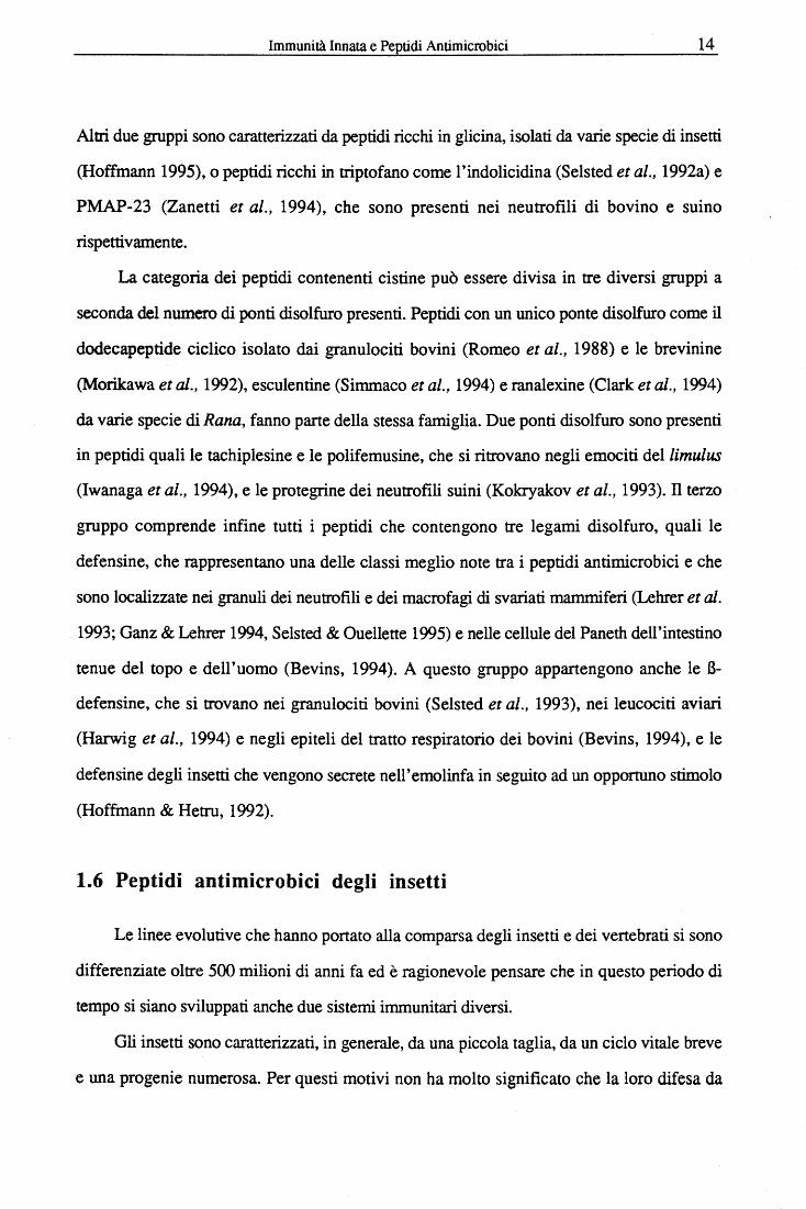
Nei neutrofili umani sono presenti almeno tre, se non quattro, diverse popolazioni di
granuli citoplasmatici (Boxer & Smolen, 1988). I granuli azzurrofùi o primari costituiscono
la prima componente granulare durante il processo mielopoietico, in quanto essi si formano
allo stadio promielocitico. Sono considerati in genere come uno speciale tipo di lisosomi
primari che emergono dalla superficie concava del complesso del Golgi. Oltre allisozima e
alla mieloperossidasi, che è considerata un marker specifico per questa popolazione di
granuli, essi contengono anche varie idrolasi acide, proteasi e proteine o peptidi cationici
(tabella 1.1). I granuli secondari o specifici, che sono più piccoli e meno densi, emergono
dal lato convesso del complesso del Golgi, sia durante lo stadio mielocitico che
metamielocitico. Essi, oltre a contenere lisozima, contengono collagenasi, lattoferrina,
Vitamin B12-binding-protein, citocromo be l'attivatore del plasiminogeno (tabella 1.1). Sia i
granuli azzurrofili che quelli specifici possono essere suddivisi in diverse sottopopolazioni
(Rice et al., 1987). Inoltre, nei neutrofili umani è stato identificato un terzo tipo di granuli,
simili ai secondari, ma meno densi, e ricchi in gelatinasi e eparanasi (Kjeldsen et al., 1992)
(tabella 1.1).
Nei neutrofili dei ruminanti, ed in particolare in quelli bovini, è stata scoperta una
popolazione di granuli più grandi e più densi degli altri (Gennaro et al., 1983a; Baggiolini et
al., 1985), che sono stati chiamati di conseguenza granuli "large". Essi sono privi dei
componenti tipici dei granuli azzurrofili, quali mieloperossidasi, proteasi neutre e idrolasi
acide, mentre hanno in comune con i granuli specifici solo la lattoferrina. La loro
caratteristica principale è quella di contenere un vero e proprio arsenale di peptidi cationici
dotati di attività antimicrobica (tabella 1.1). Questa popolazione ha origine durante uno stadio
di sviluppo intermedio tra il promielocita e il mielocita.
I granuli dei neutrofili possono essere considerati dei veri e propri arsenali di fattori
antimicrobici, generalmente di natura polipeptidica, costituendo il sistema di difesa ossigeno
indipendente che risulta importantissimo in condizioni di bassa tensione di ossigeno, quali
spesso si verificano durante un processo infiammatorio.
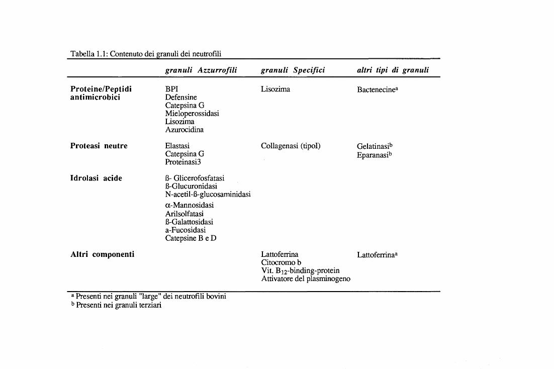
Tabella 1.1: Contenuto dei granuli dei neutrofili
Proteine/Peptidi antimicrobici
Proteasi neutre
Idrolasi acide
Altri componenti
granuli Azzurro/ili
BPI Defensine Catepsina G Mieloperossidasi Lisozima Azurocidina
Elastasi Catepsina G Proteinasi3
B- Glicerofosfatasi B-Glucuronidasi N -acetii-B-glucosaminidasi a-Mannosidasi Arilsolfatasi B-Galattosidasi a-Fucosidasi Catepsine B e D
a Presenti nei granuli "large" dei neutrofili bovini b Presenti nei granuli terziari
granuli Specifici
Lisozima
Collagenasi (ti poi)
altri tipi di granuli
Bactenecinea
Gelatinasib Eparanasib
Lattoferrina Lattoferrina a Citocromo b Vit. B12-binding-protein Attivatore del plasminogeno
Immunità Innata e Peptidi Antimicrobici 10
1.5 I peptidi antimicrobici animali
I peptidi antimicrobici animali fmo ad ora conosciuti presentano tra loro significative
diversità di sequenza, struttura e spettro d'azione. Tuttavia, si riconoscono almeno due
caratteristiche comuni alla maggioranza di essi, che possono essere correlate alla loro
funzione di molecole capaci di perturbare le membrane biologiche: (i) la significativa
prevalenza di residui basici che conferiscono, a pH neutro, una carica netta positiva; (ii) la
tendenza a strutturarsi con una conformazione di natura antipatica.
Conseguentemente alla notevole varietà e al crescente numero di (poli)peptidi
conosciuti la scelta dei parametri di classificazione può essere diversa. Un modo abbastanza
comune di ragrupparli ·in categorie è quello di dividerli in base alle specie di origine,
distinguendo quindi tra peptidi di insetti, di protozoi, di anfibi o di mammiferi. Questo
metodo però non permette un facile confronto tra peptidi che sono simili a livello di sequenza
o struttura, ma sono prodotti da organismi evolutivamente molto lontani. Un altro modo di
classificare i peptidi antimicrobici tiene conto di alcune analogie di sequenza e di struttura,
quali la presenza o assenza di ponti disolfuro: seguendo questo criterio, i peptidi
antimicrobici sono divisi in due grossi gruppi, che a loro volta contengono più sottogruppi
(tabella 1.2). Le principali caratteristiche dei membri rappresentativi di entrambe le queste
categorie sono riassunte nelle tabelle 1.3 e 1.4.
Uno dei gruppi principali tra i peptidi lineari privi di ponti disolfuro comprende tutti
quei peptidi che presentano una struttura ad a-elica anfipatica. A questa categoria
appartengono famiglie di peptidi molto ben caratterizzati quali le magainine di rana
(Berkowitz et al., 1990) e le cecropine di insetto (Boman et al., 1991). Un secondo gruppo
comprende i peptidi lineari con elevato contenuto di prolina e arginina. Questo gruppo
include le apidecine, le abecine e la drosocina, isolate dall'emolinfa degli insetti (Cociancich
et al., 1994a; Hoffmann 1995), e Bac5, Bac7 (Frank et al., 1990), PR-39, (Agerberth et al.,
1991) e le profenine (Pungercar et al., 1993; Harwig et al., 1995), tutti espressi nelle cellule
mieloidi dei mammiferi.
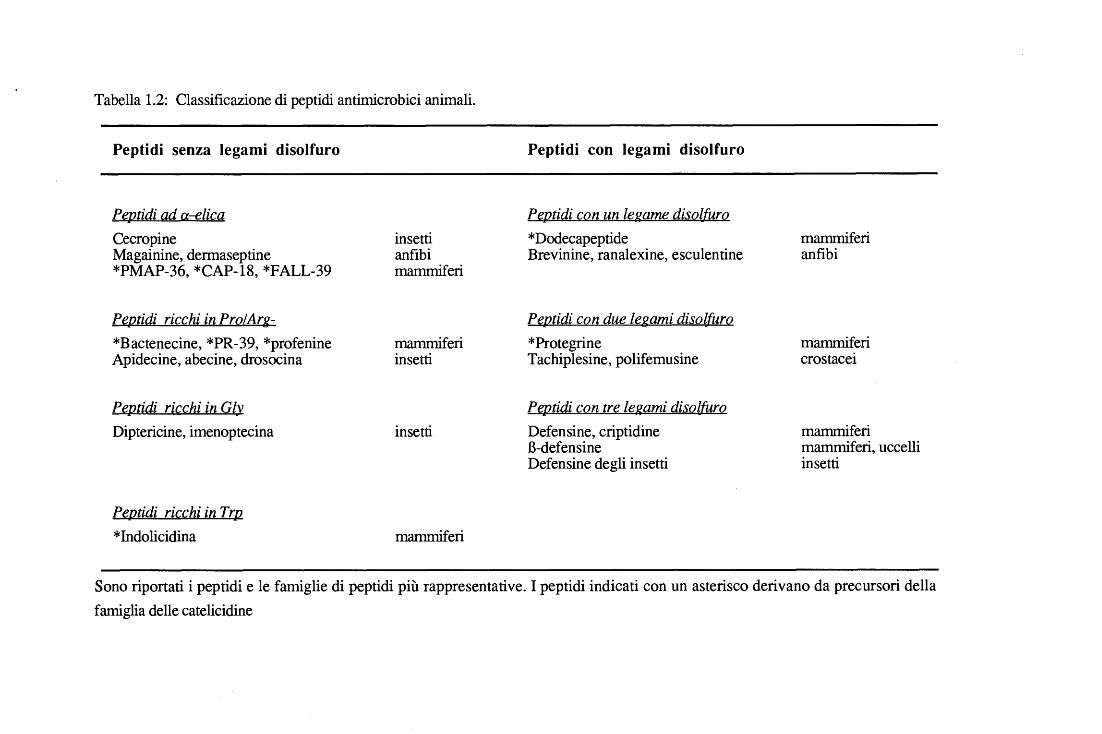
Tabella 1.2: Classificazione di peptidi antimicrobici animali.
Peptidi senza legami disolfuro
Peptidi ad a-elica Cecropine Magainine, dermaseptine *PMAP-36, *CAP-18, *FALL-39
PeJ].tidi ricç_hi in PrQLArg-*Bactenecine, *PR-39, *profenine Apidecine, abecine, drosocina
PeJ].lid.i ricchi ia Gl~ Diptericine, imenoptecina
Peptidi ricchi in Trp *Indolicidina
insetti anfibi mammiferi
mammiferi insetti
insetti
mammiferi
Peptidi con legami disolfuro
Pe.fl.tid.i CQll ualegame disol[urQ *Dodecapeptide mammiferi Brevinine, ranalexine, esculentine anfibi
Pe.r2tid.i con due le.gami dis.ol[urQ *Protegrine mammiferi Tachiplesine, polifemusine crostacei
Pep.tid.i ç_on tre. legami dis.QlfurQ Defensine, criptidine mammiferi B-defensine mammiferi, uccelli Defensine degli insetti insetti
Sono riportati i peptidi e le famiglie di peptidi più rappresentative. I peptidi indicati con un asterisco derivano da precursori della famiglia delle catelicidine
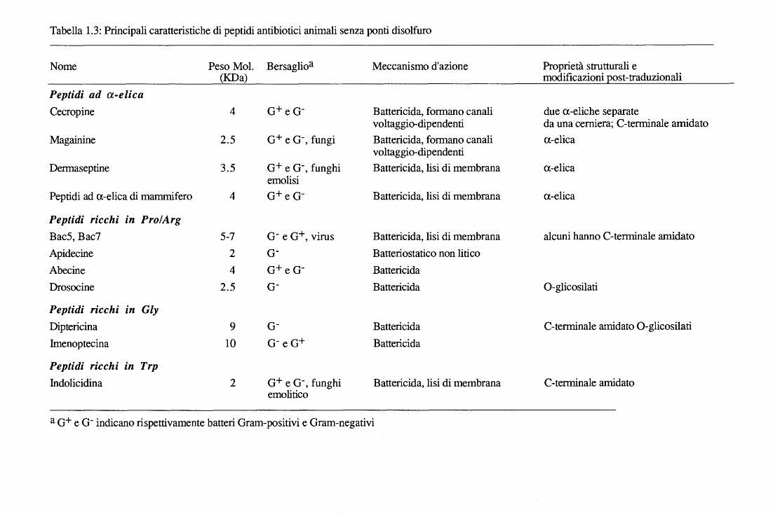
Tabella 1.3: Principali caratteristiche di peptidi antibiotici animali senza ponti disolfuro
Nome Peso Mol. Bersaglioa Meccanismo d'azione Proprietà strutturali e (KDa) modificazioni post-traduzionali
Peptidi ad a-elica Cecropine 4 o+ e o- Battericida, formano canali due a-eliche separate
voltaggio-dipendenti da una cerniera; C-terminale amidato Magainine 2.5 o+ e o-' fungi Battericida, formano canali a-elica
voltaggio-dipendenti Dermaseptine 3.5 o+ e o-, funghi Battericida, lisi di membrana a-elica
emolisi Peptidi ad a-elica di mammifero 4 o+ e o- Battericida, lisi di membrana a-elica
Peptidi ricchi in Pro/Arg Bac5, Bac7 5-7 o- e o+, virus Battericida, lisi di membrana alcuni hanno C-terminale amidato Apidecine 2 o- Batteriostatico non lirico Abecine 4 o+ e o- Battericida Drosocine 2.5 o- Battericida 0-glicosilati
Peptidi ricchi in Gly Dipteri cina 9 o- Battericida C-terminale amidato 0-glicosilati
Imenoptecina 10 o-e o+ Battericida
Peptidi ricchi in Trp lndolicidina 2 o+ e o-, funghi Battericida, lisi di membrana C-terminale ami dato
emolitico
a o+ e o- indicano rispettivamente batteri Oram-positivi e Oram-negativi
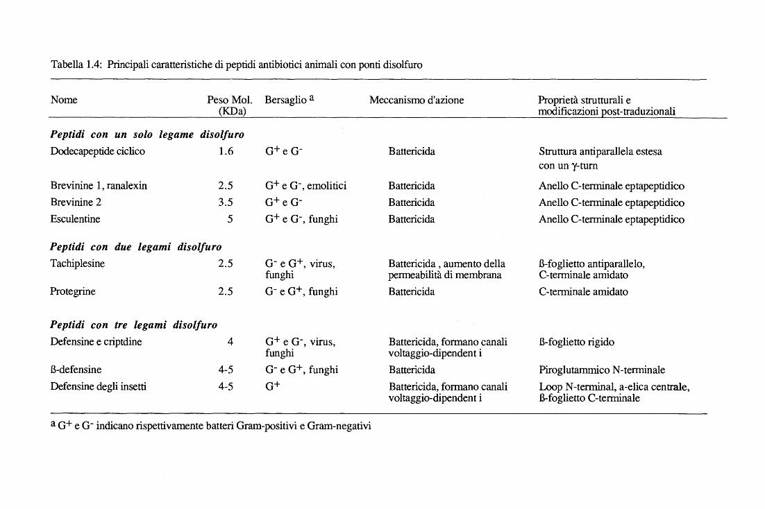
Tabella 1.4: Principali caratteristiche di peptidi antibiotici animali con ponti disolfuro
Nome Peso Mol. Bersaglio a Meccanismo d'azione Proprietà strutturali e (KDa) modificazioni post-traduzionali
Peptidi con un solo legame disolfuro Dodecapeptide ciclico 1.6 o+ e o- Battericida Struttura antiparallela estesa
con un y-turn
Brevinine l, ranalexin 2.5 o+ e o-' emolitici Battericida Anello C-terminale eptapeptidico Brevinine 2 3.5 o+ e o- Battericida Anello C-terminale eptapeptidico Esculentine 5 o+ e o-' funghi Battericida Anello C-terminale eptapeptidico
Peptidi con due legami disolfuro Tachiplesine 2.5 o- e o+, virus, Battericida, aumento della B-foglietto antiparallelo,
funghi permeabilità di membrana C-terminale amidato Protegrine 2.5 o- e o+' funghi Battericida C-terminale amidato
Peptidi con tre legami disolfuro Defensine e criptdine 4 o+ e o-, virus, Battericida, formano canali B-foglietto rigido
funghi voltaggio-dipendent i B-defensine 4-5 o- e o+, funghi Battericida Piro glutammico N-terminale Defensine degli insetti 4-5 o+ Battericida, formano canali Loop N-terminai, a-elica centrale,
voltaggio-dipendent i B-foglietto C-terminale
a o+ e o- indicano rispettivamente batteri Oram-positivi e Oram-negativi
Immunità Innata e Peptidi Antimicrobici 14
Altri due gruppi sono caratterizzati da peptidi ricchi in glicina, isolati da varie specie di insetti
(Hoffmann 1995), o peptidi ricchi in triptofano come l'indolicidina (Selsted et al., 1992a) e
PMAP-23 (Zanetti et al., 1994), che sono presenti nei neutrofili di bovino e suino
rispettivamente.
La categoria dei peptidi contenenti cistine può essere divisa in tre diversi gruppi a
seconda del numero di ponti disolfuro presenti. Peptidi con un unico ponte disolfuro come il
dodecapeptide ciclico isolato dai granulociti bovini (Romeo et al., 1988) e le brevinine
(Morikawa et al., 1992), esculentine (Simmaco et al., 1994) e ranalexine (Clark et al., 1994)
da varie specie di Rana, fanno parte della stessa famiglia. Due ponti disolfuro sono presenti
in peptidi quali le tachiplesine e le polifemusine, che si ritrovano negli emociti dellimulus
(lwanaga et al., 1994), e le protegrine dei neutrofili suini (Kokryakov et al., 1993). n terzo
gruppo comprende infine tutti i peptidi che contengono tre legami disolfuro, quali le
defensine, che rappresentano una delle classi meglio note tra i peptidi antimicrobici e che
sono localizzate nei granuli dei neutrofili e dei macrofagi di svariati mammiferi (Lehrer et al.
1993; Ganz & Lehrer 1994, Selsted & Ouellette 1995) e nelle cellule del Paneth dell'intestino
tenue del topo e dell'uomo (Bevins, 1994). A questo gruppo appartengono anche le B-
defensine, che si trovano nei granulociti bovini (Selsted et al., 1993), nei leucociti aviari
(Harwig et al., 1994) e negli epiteli del tratto respiratorio dei bovini (Bevins, 1994), e le
defensine degli insetti che vengono secrete nell'emolinfa in seguito ad un opportuno stimolo
(Hoffmann & Hetru, 1992).
1.6 Peptidi antimicrobici degli insetti
Le linee evolutive che hanno portato alla comparsa degli insetti e dei vertebrati si sono
differenziate oltre 500 milioni di anni fa ed è ragionevole pensare che in questo periodo di
tempo si siano sviluppati anche due sistemi immunitari diversi.
Gli insetti sono caratterizzati, in generale, da una piccola taglia, da un ciclo vitale breve
e una progenie numerosa. Per questi motivi non ha molto significato che la loro difesa da
Immunità Innata e Peptidi Antimicrobici 15
agenti nocivi sia basata su un sistema immunitario adattativo e con memoria Per essi è molto
più utile un sistema di difesa precostituito, che pur essendo meno specifico, permetta una
risposta rapida e ad ampio spettro (Boman, 1991). Nella realtà tale sistema risulta tanto
efficiente da rendere gli insetti particolarmente resistenti alle infezioni batteriche. Alla fine
dell'ottocento questa resistenza veniva attribuita alla fagocitosi, ma studi eseguiti dopo il
1920 dimostrarono che un'infezione primaria (iniezione di una dose subletale di batteri)
poteva indurre protezione verso un'infezione secondaria altrimenti letale, e che tale
protezione indotta era correlabile alle presenza di molecole solubili nell'emolinfa
1.6.1 Cecropine
Le prime due molecole antibatteriche indotte, di natura peptidica, furono isolate nel
1980 dallepidottero Hyalophora cecropia, e per questo chiamate "cecropine" (A e B)
(Steiner et al., 1981). Le cecropine rappresentano una delle classi più note di peptidi
antibiotici (Boman et al., 1991; Cociancich et al., 1994). Hanno sequenze di 35-39 residui
con una regione N-terminale fortemente basica, seguita da una sequenza prevalentemente
idrofobica e con il residuo C-terminale sempre amidato. Allineando le sequenze di tutte le
cecropine note si osserva che almeno il 30% dei residui sono perfettamente conservati e che
buona parte dei residui idrofobici occupano determinate posizioni. Sorprendentemente, un
peptide con un'alta similarità con le cecropine di insetto è stato isolato dall'intestino di maiale
(Lee et al., 1989) ed è stato chiamato cecropina Pl. Resta ancora da stabilire se questo
peptide possa aver avuto un'origine comune alle cecropine di insetto o se sia da considerare
come un interessante ed inusuale caso di convergenza evolutiva La struttura delle cecropine
è stata inizialmente studiata mediante spettroscopia CD, da cui risulta che queste molecole
non sono strutturate in soluzione acquosa (random coil), mentre in solvente a minore polarità
(30% esafluoro-2-propanolo in acqua) acquisiscono una conformazione con un alto
contenuto di a -elica (Steiner, 1982). Questo comportamento è considerato tipico di un'elica
antipatica. Studi strutturali ad alta risoluzione mediante 2D-NMR (Holak et al., 1988),
hanno dimostrato che le cecropine sono costituite da due a -eliche distinte, separate da un
Immunità Innata e Peptidi Antimicrobici 16
cardine Gly-Pro (residui 24-26). L'elica N-terminale è fortemente cationica e anfipatica,
mentre quella C-terminale è meno carica e più idrofobica. Anche la struttura della cecropina
di mammifero è stata chiarita, e risulta essere costituita da un 'unica a -elica mancante quindi
del tratto a cardine, tipico di tutti i congeneri di insetto.
In generale le cecropine sono attive verso molti ceppi di batteri Gram negativi, mentre
solo alcuni Gram positivi sono suscettibili alla loro azione. In particolare, le cecropine A e B
di Hyalophora mostrano una potente azione contro vari ceppi di Escherichia coli, Salmonella
typhimurium, Acinetobacter calcoaceticus e P seudomonas aeruginosa, con una
concentrazione letale (LC) dell'ordine di 0.2-1.4 JJ.M. Alcuni di questi valori di LC sono
inferiori di circa un ordine di grandezza rispetto a quelli relativi alle tetracicline in analoghe
condizioni di saggio. Tra i batteri Gram positivi, il più sensibile è il Bacillus megaterium,
mentre non sono stati osservati effetti citotossici contro funghi e altre cellule eucariote.
Sempre dagli insetti sono stati isolati altri peptidi con struttura ad a -elica simile alle
cecropine, ma questi, a differenza dei precedenti, non vengono secreti nell'emolinfa, bensì
vengono prodotti costitutivamente nei tessuti degli organi riproduttivi e la loro espressione
viene indotta durante l'accoppiamento. Il gene dell' andropina, un peptide antimicrobico di 32
residui, è stato infatti caratterizzato in Drosofila (Samakovlis et al., 1991) e viene espresso
nel maschio adulto solo a livello nel dotto eiaculatorio. Due peptidi sono stati isolati da
femmine di Cerratitis capitata, da cui il nome di ceratotossine A e B (Marchini et al., 1993),
che oltre ad avere attività antibatterica sono anche emolitiche.
Molti altri peptidi antibiotici sono stati isolati e caratterizzati in vari ordini di insetti
quali lepidotteri, ditteri, imenotteri e coleotteri (Hoffmann & Hetru, 1992; Cociancich et al.,
1994; Hoffmann, 1995). Tra questi vi sono le apidecine, le abecine e la drosocina, ricchi in
prolina e attivi prevalentemente contro batteri Gram negativi; le attacine, le sarcotossine n, le
diptericine e la coleoptericina che costituiscono un gruppo di peptidi di 8-27 kDa, cationici e
ricchi in glicina e che sono prevalentemente attivi contro batteri Gram negativi (tabella 1.3); e
Immunità Innata e Peptidi Antimicrobici 17
infine le defensine di insetti, le uniche tra i peptidi menzionati a possedere residui cisteinici,
che sono impegnati nella formazione di ben tre ponti disolfuro intramolecolari (tabella 1.4).
1.6.2 Defensine di insetto
Le defensine di insetto sono particolarmente attive su batteri Gram positivi e, per ora,
rappresentano il gruppo di peptidi antibiotici più diffuso tra gli insetti. Sono state chiamate
defensine sia per la presenza di sei cisteine, che sono caratteristiche delle defensine di
mammifero, che per una omologia del 50% riscontrata a livello della sequenza compresa tra i
residui 15 e 34 di una defensina di un dittero (Phormia te"anovae) e il frammento 4-24 della
defensina NP-1 di coniglio (Hoffmann & Hetru, 1992). Oggi sappiamo che le defensine
degli insetti e dei mammiferi costituiscono due famiglÌe distinte, in quanto la loro struttura
terziaria differisce notevolmente, il che suggerisce che le somiglianze osservate siano
piuttosto il frutto di importanti fenomeni di convergenza evolutiva.
Le defensine degli insetti sono lunghe 38-43 residui e sono moderatamente cationiche
(pl = 8-8.5). La struttura terziaria della defensina A di Phormia te"anovae è stata determinata
mediante studi di spettroscopia NMR in soluzione acquosa e risulta composta da tre domini
ben distinguibili: un loop flessibile N-terminale, comprendente i residui da l a 13, un a-elica
antipatica centrale dal residuo 14 a124 e un B-foglietto antiparallelo C-terminale dal residuo
27 al 40, con un ripiegamento B a livello degli aminoacidi 32-34. L'a-elica è stabilizzata da
due ponti di solfuro con il secondo tratto del B-foglietto, mentre il primo tratto è legato da un
ponte disolfuro con illoop N-terminale (Hoffmann & Hetru, 1992).
Le defensine degli insetti mostrano somiglianze strutturali con la caribdotossina, un
peptide presente nel veleno dello scorpione che è attivo nel blocco dei canali del potassio
nelle membrane eucariotiche. Sembra plausibile che esse derivino da un gene ancestrale
comune che si è duplicato e modificato nel corso dell'evoluzione e che è completamente
diverso da quello delle defensine di mammifero (Hoffmann & Hetru, 1992).
Immunità Innata e Peptidi Antimicrobici 18
l. 7 Peptidi antimicrobici degli anfibi
Negli ultimi trent'anni numerosi composti bioattivi sono stati isolati dalle secrezioni
cutanee degli anfibi. La maggior parte di essi sono ormoni e neurotrasmettitori quali amine
biogeniche, per lo più derivate dalla serotonina, e peptidi farmacologicamente attivi quali
bradichinina, ceruleina, bombesina e dermorfina (Bevins & Zasloff, 1990). La maggior
parte di queste molecole hanno controparti simili o identiche nel sistema nervoso o nel tratto
gastrointestinale dei mammiferi. In anni più recenti è stato dimostrato che queste secrezioni
contengono anche una moltitudine di peptidi antimicrobici. La presenza di composti
altamente attivi spiega il motivo per cui la pelle di rane e rospi è sempre stata considerata
nell'antichità come un preparato dalle doti "magiche".
Le prime ipotesi sull'esistenza di sostanze antimicrobiche negli anfibi vennero
formulate sulla base dell'osservazione che lo Xenopus Iaevis, un anfibio anuro, usato
normalmente in laboratorio per esperimenti di biologia cellulare, era in grado di guarire
rapidamente da gravi ferite cutanee senza alcun segno apparente di infezione, anche in
ambienti non sterili come l'acqua stagnante. Nel 1987 Zasloff suggerì che potessero esservi
fattori antisettici cicatrizzanti nella pelle degli anfibi e isolò da X. Iaevis due peptidi di 2000 e
3000 Da, che chiamò "magainine" dalla parola ebraica "magain" che significa "scudo"
(Zasloff, 1987).
1.7.1 Magainine
Attualmente si conoscono più di una dozzina di peptidi isolati da varie specie di anfibi
che appartengono a questa famiglia e che vengono secreti sia sulla pelle che nel tratto
gastrointestinale (Bevins & Zasloff, 1990). Le magainine hanno sequenze lineari di 21-27
residui prevalentemente cationici e spaziati con regolarità da residui idrofobici o neutri.
Mediante studi di dicroismo circolare e NMR bidimensionale é stato osservato che le
magainine non sono strutturate in soluzione acquosa, mentre assumono una conformazione
ad a -elica anfipatica in seguito ali' aggiunta di un solvente organico che abbassa la polarità
Immunità Innata e Peptidi Antimicrobici 19
del solvente, mimando l'ambiente idrofobico delle membrane (Bechinger et al., 1993). Le
magainine, infatti, perturbano le membrane batteriche provocando un'alterazione della
permeabilità, dissipando il potenziale elettrochimico e impedendo la regolazione osmotica e le
funzioni di trasporto (Jacob & Zasloff, 1994).
Lo spettro antimicrobico delle magainine si estende contro batteri, protozoi e funghi a
concentrazioni tra 5 e 50 Jlg/ml. Inoltre sono in grado di agire selettivamente su cellule
trasformate a concentrazioni 5-10 volte inferiori rispetto a quelle tossiche per cellule
eucariotiche normali (Jacob & Zasloff, 1994).
Le basi molecolari della selettività d'azione delle magainine per diversi bersagli cellulari ~
(microrganismi e cellule tumorali vs. cellule normali) sono presumibilmente da ricercare nella
diversa composizione delle membrane suscettibili. A differenza dei batteri e di molte cellule
neoplastiche che presentano fosfolipidi anionici nel foglietto esterno delle membrane e
mancano o sono carenti, nel caso di cellule trasformate, di colesterolo, le normali cellule
dell'ospite hanno membrane ricche di colesterolo e i fosfolipidi anionici sono, in generale,
confinati nel foglietto interno del doppio strato lipidico (Jacob & Zasloff, 1994). Una
descrizione più approfondita del meccanismo d'azione verrà ripresa nel paragrafo specifico.
Oltre alle magainine diX.laevis, che senza dubbio rappresentano una delle famiglie di
peptidi antimicrobici più studiate, negli ultimi anni molti altri peptidi sono stati isolati da
secrezioni cutanee di varie specie di anfibi quali Bombina (B. variegata e B. orientalis),
Phyllomedusa (P. sauvagii e P. bicolor), Litoria (L. splendida e L. caerulea) e Rana (R.
pipiens, R. brevipoda, R. esculenta).
l. 7.2 Bombinine e H-bombinine
Le bombinine (Simmaco et al., 1991; Gibson et al., 1991) e le H-bombinine
rappresentano due diverse famiglie di peptidi isolati da Bombina. Le sequenze delle
bombinine sono state dedotte dal sequenziamento dei rispettivi cD NA e tutte risultano essere
costituite da una sequenza di 27 aminoacidi in cui la porzione idrofobica N -terminale è
Immunità Innata e Peptidi Antimicrobici 20
altamente variabile (residui 1-13) mentre la porzione cationica C-terminale è costante (residui
14-27) e presenta sempre il residuo C-terminale amidato (Simmaco et al., 1991). L'attività
antibatterica è stata valutata sintetizzando i peptidi dedotti dalle sequenze nucleotidiche stesse,
da cui risulta che le bombinine hanno una potente attività antibatterica su ,vari ceppi di
Staphylococchi a concentrazioni tra 2 e l O ~' mentre non presentano alcuna capacità lirica
degli eritrociti.
Da B. orientalis sono stati caratterizzati altri quatto peptidi cationici simili alle
bombinine, noti come BLP-1, -4 (Gibson et al., 1991), che cono composti da 25-27
aminoacidi con il residuo C-terminale amidato. Essi possono assumere una conformazione
ad a -elica anfipatica e agiscono associandosi alle membrane e alterandone la permeabilità.
Sono attivi a una concentrazione 8 J.LM contro batteri non enterici Gram negativi quali
Neisseria meningitis, gonorrhoeae, lactamica e cinerea. Mentre esercitano un'azione molto
blanda contro batteri enterici inclusi Escherichia, Pseudomonas, Enterobacter e Klebsiella. A
differenza dalle bombinine, questi peptidi sono emolitici a concentrazioni superiori a 40 J.LM,
suggerendo una selettività per membrane procariotiche piuttosto che eucariotiche.
Dal sequenziamento di cONA codificanti per i precursori pre-propeptidici di varie
bombinine, è stata predetta la sequenza corrispondente a un nuovo peptide antimicrobico di
20 residui (Simmaco et al., 1991). Successivamente è stato dimostrato che oltre a questo,
altri peptidi simili sono presenti nelle secrezioni cutanee di B. variegata (Mignogna et al.,
1993) e sono stati chiamati bombinine H in quanto sono più idrofobici. Alcune bombinine H
(H3-H5) contengono una D-allo-isoleucina in posizione due della loro sequenza. La
presenza di D-aminoacidi è piuttosto comune nei peptidi antibiotici batterici, mentre non lo è
affatto tra i peptidi antibiotici di origine animale (Kreil, 1994 ). Le bombinine H sono attive
sia contro batteri Gram positivi che Gram negativi con una potenza simile alle magainine, ma
a differenza di quest'ultime, le bombinine H presentano una forte capacità emolitica e non
assumono una conformazione ad a -elica anfipatica. Questi dati suggeriscono, quindi, un
meccanismo d'azione diverso da quello proposto per i peptidi ad a -elica
Immunità Innata e Peptidi Antimicrobici 21
l. 7.3 Dermaseptine
La famiglia delle dennaseptine (Mor et al., 1994a; Kreil, 1994) attualmente comprende
sette peptidi isolati dalle secrezioni cutanee di alcune rane arboree del genere P hyllomedusa
(Mor et al., 1991). Cinque dennaseptine (S1- S5) sono state purificate in P. sauvagii e due
(adenoregulina e dermaseptina b) da P. bicolor (Amiche et al., 1993; Mor et al., 1994a).
Strutturalmente ricordano le cecropine per la presenza di un residuo Trp ali' estremità N-
terminale e un dominio idrofobico C-terminale. Studi di CD hanno dimostrato che anche
questi peptidi si strutturano come a -eliche antipatiche in solventi organici (Mor et al., 1991,
1994a). Le dermaseptine esercitano un'attività antibiotica ad ampio spettro con differenze
significative tra i vari congeneri. A concentrazioni tra 0.5 e 30 J.LM, inibiscono
irreversibilmente la crescita di batteri Gram negativi e Gram positivi (es. E. coli, Aeromonas
caviae, S. aureus, Enterococcusfaecalis, B. subtilis, Nocardia brasiliensis), e sono attive
anche su lieviti e funghi filamentosi (es. Cryptococcus neoformans, S. cerevisiae, Candida
albicans, Aspergillus niger, Aspergillusfumigatus) (MorA et al., 1991, 1994a, b; Mor &
Nicolas 1994b ). Le dennaseptine, inoltre, presentano un'attività sinergica (Mor A et al.,
1994b): infatti, miscele peptidiche risultano nettamente più attive dei peptidi singoli.
Nonostante l 'importanza di questo sinergismo, sia sul piano biologico che applicativo, il
meccanismo d'azione non è ancora noto. E' stato proposto un modello simile alle magainine,
poiché anche questi peptidi inducono una rapida lisi cellulare (Pouny et al., 1992). Gli
appartenenti a questa famiglia mostrano diversa attività nei confronti di cellule eucariote. In
particolare, le dennaseptine S l e S5 non lisano gli eritrociti neppure a concentrazioni elevate.,
mentre la dermaseptina S4 causa totale emolisi a concentrazioni di l J.LM (MorA et al.,
1994b).
1.7.4 Brevinine ed Esculentine
Un altro gruppo di peptidi antimicrobici, le brevinine e le brevinine E sono state isolate
rispettivamente in Rana brevipoda e Rana esculenta (Morikawa et al., 1992; Simmaco et al.,
Immunità Innata e Peptidi Antimicrobici 22
1993). Questi peptidi sono fortemente cationici, hanno una sequenza di 24-34 aminoacidi e
due cisteine a livello C-terminale separate da sei residui che formano un ponte disolfuro,
originando così una struttura ad anello. La loro attività è rivolta prevalentemente contro
batteri Gram positivi e Gram negativi, inoltre, la brevinina lE risulta avere una fortissima
capacità emolitica. Sempre da Rana esculenta è stata isolata un'altra famiglia di peptidi, le
esculentine (Simmaco et al., 1994 ), che contano 37-44 residui e che presentano un
meccanismo d'azione molto simile a quello delle cecropine. La esculentina-1 è inoltre attiva
verso Pseudomonas aeruginosa, che è solitamente molto resistente ai peptidi antimicrobici, e
verso i funghi Candida albicans e Saccharomyces cerevisiae. Un altro peptide di 20 residui,
la ranalexina (Clark, 1994) è stato isolato dalla Rana catesbiana, e presenta, come le
esculentine, un "loop" C-terminale per la presenza di un ponte disolfuro. Questo peptide é
attivo sia contro batteri Gram negativi (E. coli, P. aeruginosa) che Gram positivi (S. aureus).
1.8 Peptidi antimicrobici dei mammiferi
La maggior parte dei (poli)peptidi antimicrobici noti nei mammiferi sono presenti in
cellule specializzate della difesa immunitaria, quali i fagociti professionali e i linfociti
citotossici NK (Spitznagel, 1990; Gennaro et al., 1991; Gabay & Almeida, 1993; Weiss,
1993; Andersson et al., 1995). Più recentemente si è scoperto che alcuni membri di queste
famiglie vengono espressi anche in cellule epiteliali che rivestono le vie respiratorie ed
intestinali (Gabay & Almeida, 1993; Lehrer et al., 1993; Ganz & Lehrer, 1994),
contribuendo così a costituire uno sbarramento locale a potenziali agenti invasivi.
Nei paragrafi seguenti verranno descritte le principali caratteristiche di alcuni tra i più
significativi (poli)peptidi antimicrobici dei mammiferi quali (i) peptidi con uno o più ponti
disolfuro come le defensine e le B-defensine; (ii) peptidi ricchi in prolina e arginina come le
bactenecine o ricchi in triptofano come l'indolicidina; (iii) peptidi con strutture ad a -elica
antipatica come il CAP-18 ed infine (iv) delle vere e proprie proteine antibatteriche come la
BPI ("bactericidal/penneability protein") o le serprocidine.
Immunità Innata e Peptidi Antimicrobici
1.8.1 Peptidi con ponti disolfuro
Defensine e Criptidine
Le defensine furono descritte per la prima volta circa 30 anni fa da Zeya e
Spitznagel (1966) che osservarono la presenza di "proteine cationiche lisosomiali" in
leucociti polimorfonucleati di mammifero. A quel tempo essi non poterono purificare e
caratterizzare dettagliatamente questi peptidi e i loro studi furono ripresi alla fine degli
anni settanta dal gruppo di Lehrer (Selsted et al., 1985), che riuscì a purificare da
neutrofili di coniglio ben sei peptidi diversi che chiamò NP numerandoli da l a 5 (NP =
neutrophil peptide ). Attualmente si conoscono almeno 20 diverse defensine che sono
prodotte sia dai neutrofili umani (Ganz et al. 1985; Selsted et al., 1985), di coniglio
(Selsted et aL, 1984), di cavia (Selsted & Harwig, 1987) e di ratto (Eisenhauer et al.,
1989), sia dai macrofagi alveolari del coniglio (Patterson-Delafield et al., 1980), sia
dalle cellule del Paneth umane (Jones & Bevins 1992, 1993) e murine (Ouellette et al.,
1989, 1992 e 1994; Eisenhauer & Lehrer, 1992; Selsted et al., 1992b). Le defensine
sono peptidi cationici di 29 - 35 residui, ricchi in arginina e caratterizzati dalla presenza
di sei residui cisteinici che sono implicati nella formazione di tre ponti disolfuro
intramolecolari che risultano "strategici" nel ripiegamento strutturale del peptide (per una
rassegna vedi: Lehrer et al., 1993; Ganz & Lehrer, 1994; Selsted & Ouellette 1995).
Nonostante la completa conservazione delle sei cisteine e la presenza di altri cinque
residui pressoché immutati, l'identità di sequenza che si osserva varia dal27% al45%.
Tuttavia, la determinazione strutturale di alcune defensine umane e di coniglio mediante
NMR e cristallografia a raggi-X (Bach et al., 1987; Pardi et al. 1988 e 1992; Hill et al.,
1991; Zhang et al., 1992) dimostra che tutte presentano un motivo strutturale comune,
costituito da tre filamenti a B-foglietto an ti parallelo, stabilizzati dai tre ponti disolfuro
intramolecolari. Ne risulta, quindi, una conformazione molto compatta che è
notevolmente resistente ali' azione delle proteasi del fagosoma. La struttura cristallina
della defensina umana HNP-3 risulta essere costituita da un dimero stabile non-
23
Immunità Innata e Peptidi Antimicrobici
covalente che ha caratteristiche anfipatiche. La conformazione del dimero è stata
paragonata ad un "cestello" con una base idrofobica e un bordo idrofilico e cationico
(Hill et al., 1991). Sulla base di queste caratteristiche strutturali sono stati proposti
diversi meccanismi per spiegare l'attività antimicrobica e citotossica delle defensine
(Wimley et al., 1994; White et al., 1995).
24
Di recente sono state scoperte alcune defensine enteriche, che vengono espresse
costitutivamente dalle cellule del Paneth, che sono localizzate alla base delle cripte
dell'intestino tenue del topo e dell'uomo, e sono state denominate criptidine (Ouellette et al.,
1992; Selsted et al., 1992; Eisenhauer et al., 1992). Sei di questi peptidi sono stati isolati
dall'intestino murino, ma dal clonaggio dei cDNA ottenuti da questo tessuto risulterebbe che
in ogni singola cripta siano espressi almeno 17 mRN A codificanti per diverse criptidine
( Ouelette et al., 1994 ). N eli' uomo invece sono state clonate solo due sequenze
corrispondenti a delle defensine enteriche (Jones & Bevins, 1992 e 1993). E' interessante
notare che, a differenza di quanto si osserva per l 'intestino, i neutrofili murini non
contengono affatto delle defensine o peptidi omologhi (Eisenhauer & Lehrer, 1992).
L'attività biologica delle defensine è notevole e varia (Lehrer et al. 1993): a
concentrazioni comprese tra 2 e 20 J.l.M esse inattivano un ampio spettro di batteri sia Gram
positivi che Gram negativi, micobatteri non tubercolari (M. avium intracellulare), spirochete
(Treponema pallidum), alcuni funghi (tra cui C. albicans, C. neoformans, A.fumigatus),
protozoi (Giardia lamblia). Inoltre, esse neutralizzano alcuni virus con mantello (p.e. Herpex
virus l e~ il virus della stomatite vescicolare e i virus influenzali) (Daher et al., 1986) ed
esercitano un'attività citotossica aspecifica su svariate cellule normali e tumorali (Lichtenstein
et al,. 1988; Lichtenstein, 1991). Tra le varie defensine note si osserva una sostanziale
variabilità nello spettro e, generalmente, nella potenza d'azione. Le defensine mieloidi sono
più tossiche contro i batteri Gram positivi rispetto ai Gram negativi, mentre l'opposto
avviene per le criptidine. n meccanismo d'azione è stato studiato con più metodiche, da cui
risulta che le defensine si legano elettrostaticamente alle membrane e formano pori
Immunità Innata e Peptidi Antimicrobici 25
multimerici che permeabilizzano sequenzialmente le membrane dei batteri (White et
al.,1995).
In aggiunta ali' attività antimicrobica, alcune defensine mostrano in vitro altre funzioni
che potrebbero avere un ruolo di regolazione nei processi infiammatori e di riparo delle
ferite. Le HPN infatti, risultano essere chemiotattiche per i monociti (Territo et al., 1989), le
defensine dei macrofagi di coniglio hanno capacità opsonizzante (Fleischmann et al., 1985) e
altre defensine sono mito geniche per i fibroblasti, e ciò. indicherebbe una capacità di
accelerare il riparo delle ferite (Murphy et al., 1993).
jJ-defensine
Nei neutroftli bovini è stata caratterizzata una nuova famiglia di peptidi che ha molte
analogie con quella delle defensine: sia a livello strutturale che funzionale. Questi nuovi
peptidi sono stati chiamati B-defensine e hanno sequenze di 38-42 aminoacidi, caratterizzate
dalla presenza di sei cisteine. I tre ponti disolfuro che formano in seguito ai legami
intramolecolari tra le cisteine sono disposti in modo diverso da quello osservato nelle
defensine classiche (Tang & Selsted, 1993). Nonstante questa sostanziale differenza, la
struttura terziaria delle B-defensine risulta essere simile a quella delle defensine
(Zimmermann et al., 1995) e ciò spiega anche le analogie funzionali tra i componenti delle
due famiglie. Le B-defensine sono attive a concentrazioni J..Lmolari contro batteri Gram
negativi (E. coli, K. pneumoniae), Gram positivi (S. aureus, L. monocytogenes) e funghi
(C. albicans e tropicalis ).
Peptidi omologhi alle B-defensine bovine sono stati isolati da cellule dell'epitelio
tracheale (trachea! antimicrobial peptide,TAP) e linguale (lingua! antimicrobial peptide, LAP)
del bovino (Diamond et al. 1991; Schonwetter et al., 1995) ed anche da leucociti eterofili di
pollo (Harwig et al., 1994) e di tacchino (Evans et al., 1994). Queste due ultime scoperte
indicano, quindi, che l'origine delle B-defensine risale a più di 250 milioni di anni fa, prima
della divergenza evolutiva tra uccelli e mammiferi.
Immunità Innata e Peptidi Antimicrobici 26
Protegrine
Una nuova famiglia di peptidi antimicrobici, detti protegrine, è stata recentemente
identificata nei leucociti suini. Tre membri, noti come PG-1, PG-2 e PG-3, sono stati isolati
e sequenziati (Kokryakov et al. 1993), mentre la sequenza di un quarto congenere PG-4, è
stata dedotta dal cDNA codificante per il suo precursore (Zhao et al. 1994). L'analisi
sequenziale mostra che questi peptidi hanno alcune caratteristiche comuni alle tachiplesine:
peptidi antimicrobici dellimulus (Korneva & Lehrer,1993; Iwanaga et al., 1994); sono
infatti piccoli peptidi cationici ( 17-18 residui) che contengono quattro cisteine che formano
due ponti disolfuro; hanno inoltre l'aminoacido C-terminale ami dato. Tuttavia, il
posizionamento delle cisteine nelle protegrine è completamente diverso da quello delle
tachiplesine e ciò indica una netta separazione tra le due famiglie. E' interessante invece
notare che le spaziatura tra le prime tre Cys nelle protegrine è identica a quella delle prime tre
Cys nelle defensine e che il tratto 4-13 della sequenza aminoacidica di PG-3 è quasi identico
al tratto N-terminale 1-10 della defensina NP-3a di coniglio (otto residui sono identici e gli
altri due sono simili).
Lo spettro d'azione delle protegrine è simile a quello delle tachiplesine e comprende
batteri Gram negativi e Gram positivi, funghi e alcuni virus con mantello (Kokryakov et al.,
1993). Inoltre, le protegrine legano illipopolisaccaride (LPS) con un affinità comparabile
alla polimixina B (Bevins, 1994).
Dodecapeptide
n dodecapeptide è il primo peptide antimicrobico che è stato isolato dai granuli "large"
dei neutrofùi bovini (Romeo et al., 1988) ed è anche il più piccolo tra i peptidi antimicrobici
noti negli animali. Esso è costituito da una sequenza di 12 aminoacidi -RLCRIVVIRVCR- in
cui sono presenti quattro arginine che conferiscono alla molecola una carica netta positiva,
mentre i residui idrofobici contribuiscono a conferire l'anfipaticità necessaria per l'attività
antibatterica del peptide. Le due cisteine presenti non sono libere, ed è stato ipotizzato che si
Immunità Innata e Peptidi Antimicrobici 27
leghino tra loro dando così una struttura ciclica al peptide. Il dodecapeptide è stato anche
sintetizzato chimicamente ed è stato dimostrato che la struttura ciclica è decisamente più attiva
di quella linearizzata (Gallis et al., 1990). Il dodecapeptide sintetico presenta un'attività
comparabile al peptide naturale che è battericida contro E.coli che S.aureus a concentrazioni
inferiori a 10 JJ.g/ml (Romeo et al,. 1988). Inoltre inibisce la crescita di alcuni funghi (C.
albicans) ed è citotossico nei confronti di cellule neuronali, gliali e dei linfociti T
(Radermacher et al.,1993; Schluesener et al.,1993).
1.8.2 Peptidi con elevata percentuale di particolari aminoacidi
Peptidi ricchi in Prolina e Arginina: Bactenecine, PR-39 e Profenine
I primi due peptidi antimicrobici con un'elevato contenuto di arginine (>45%) e proline
(>20%) sono stati isolati dai granuli "large" dei neutrofili bovini e sono stati chiamati
bactenecine, dal latino bacterium e necare (Gennaro et al., 1983b; Gennaro et al., 1989). Le
due bactenecine, dette anche Bac5 e Bac7, si distinguono oltre che per il diverso peso
molecolare, rispettivamente di 5 e 7 kDa, anche per la sequenza primaria che nel Bac5 è
composta della ripetizione a tandem del motivo Arg-Pro-Pro-X, mentre nel Bac7 il motivo
ripetuto è Pro-Arg-Pro-X, dove X indica un residuo idrofobico ingombrante (Frank et al.,
1990). Entrambi i peptidi vengono sintetizzati come pre-probactenecine di 21 kDa (pre-
proBac5) e 23.5 kDa (pre-proBac7) rispettivamente, che dopo la rimozione del peptide
segnale, vengono indirizzate e accumulate nei granuli "large"dei neutrofùi come proforme
(proBac5 di 15 kDa e proBac7 di 20 kDa)(Zanetti et al. 1990). Queste, a differenza di quanto
si osserva per i peptidi maturi, non presentano attività antimicrobica (Scocchi et al., 1992).
La liberazione dei peptidi maturi avviene ad opera di un taglio proteolitico catalizzato
dali' elastasi presente nei granuli azzurrofili. n meccanismo di rilascio dei peptidi attivi è
subordinato quindi ad un evento di fagocitosi, durante il quale la cellula attivata rilascia nel
fagosoma il contenuto di diverse popolazioni di granuli permettendo così l 'incontro
dell'enzima con il suo bersaglio.
Immunità Innata e Peptidi Antimicrobici 28
Bac5 e Bac7 sono attivi prevalentemente contro batteri Gram negativi: a concentrazioni
comprese tra 10 e 50 J!g/ml essi uccidono efficacemente microrganismi quali Salmonella
typhimurium, Klebsiella pneumonie e Escherichia coli e arrestano la crescita di Enterobacter
cloacae e Pseudomonas aeruginosa. Tra i batteri Gram positivi, gli unici ad essere sensibili
sono Bacillus megaterium e in misura minore Staphylococcus epidermidis (Skerlavaj et al.;
1990). Bac7 è inoltre capace di inattivare in vitro il virus Herpes simplex (Zerial et al.,
1987). Entrambi i peptidi sono anche attivi contro alcune spirochete come Leptospira
interrogans e Leptospira bifle:xa, mentre due linee di Borrelia burgdorferi sono resistenti alla
loro azione tossica (Scocchi et al., 1993).
Mediante esperimentiin vitro , utilizzando il ceppO di E. coli ML-35, è stato dimostrato
che l'effetto letale di Bac5 e Bac7 nei confronti dei batteri, è mediato da una rapida
permeabilizzazione della membrana esterna ed interna, che si osserva misurando l'aumento
di accessibilità al substrato per due enzimi, la ~-lattamasi periplasmica e la 6-galattosidasi
citoplasmatica (Skerlavaj et al., 1990). L'elevato grado di specificità per le membrane
procariotiche è dimostrato anche dal fatto che questi peptidi, anche a concentrazioni elevate
(50 J!M), non lisano gli eritrociti umani. Per quanto riguarda il loro meccanismo d'azione, si
suppone che le bactenecine si leghino alla membrana esterna dei batteri mediante interazioni
elettrostatiche e che, una volta entrate in contatto con l'ambiente idrofobico più interno,
assumano una conformazione anfipatica in grado di perturbare il doppio strato lipidico
(Skerlavaj et al., 1990). Entro pochi minuti dall'interazione con le membrane, infatti, Bac5 e
Bac7 provocano il collassamento del gradiente elettrochimico protonico, necessario per la
sintesi di ATP, e causano l'alterazione del trasporto di precursori del DNA e delle proteine e
la conseguente fuoriuscita dei metaboliti cellulari. Allo scopo di chiarire il rapporto
struttura/funzione ed individuare il dominio responsabile per l'attività antimicobica, entrambi
questi peptidi, così come alcuni frammenti corrispondenti a varie loro porzioni, sono stati
sintetizzati chimicamente mediante il metodo Fmoc in fase solida. Da esperimenti condotti sia
contro batteri Gram negativi che Gram positivi risulta che la porzione N-terminale cationica è
Immunità Innata e Peptidi Antimicrobici 29
essenziale per l'attività antibiotica e che è necessario una peptide di almeno 16-18 residui per
provocare la lisi della membrana (Gennaro et al., manoscritto in preparazione). La struttura
secondaria di questi due peptidi non è stata ancora chiarita: per il momento le uniche
indicazioni derivano da esperimenti condotti mediante CD e spettrometria a infrarosso che
suggeriscono strutture simili a poliproline.
Altri due peptidi ricchi in prolina e arginina sono stati isolati da maiale e sono stati
denominati PR-39 e profenina-1. Il primo è stato isolato dall'intestino tenue del maiale
(Agerberth et al., 1991) ed ha una sequenza in cui si distingue un tratto N-terminale simile a
Bac7 (85% di identità), mentre il resto della molecola è costituito da un modulo ripetuto di
X-Pro-Pro-Y, dove X è un residuo idrofobico e Y molto spesso è un'arginina, richiamando
la sequenza di Bac5. Studi di dicroismo circolare e di spettroscopia all'infrarosso
suggeriscono anche per questo peptide una struttura del tipo poliprolina (Cabiaux et al.,
1994). Per quanto riguarda lo spettro d'azione, PR-39 agisce a concentrazioni micromolari
sia contro batteri Gram negativi (E. coli, S. typhimurium e A. calcoaceticus) che Gram
positivi (B. megaterium, B. subtilis e S. pyogenes), bloccandone la sintesi proteica e del
DNA e provocando la loro degradazione (Agerberth et al., 1991; Boman et al., 1993). Di
recente è stato scoperto che il PR-39 è presente negli essudati circostanti le ferite e che
provoca a livello della superficie cellulare un aumento dell'espressione di due proteoglicani
(sindecanina-1 e -4)(Gallo et al. 1994); questo risultato suggerisce un ruolo specifico del
PR-39 nel meccanismo di riparo dei tessuti.
La profenina-1 è un peptide di 79 residui, ricco in prolina (53%) e fenilal~a (19%),
che è stato purificato dai leucociti di maiale (Harwig et al., 1995) e che è caratterizzato
anch'esso da una sequenza ripetuta. In particolare il tratto tra il residui 2 e 61 è costituito dal
decamero FPPPNFPGPR che è ripetuto consecutivamente sei volte, in modo pressoché
perfetto. Questo peptide ha una potente attività antibatterica contro E. coli, mentre è
debolmente attivo contro Listeria monocytogenes.
Immunità Innata e Peptidi Antimicrobici 30
Un peptide ricco in Triptofano: Indolicidina
Dalla componente granulare dei neutrofili bovini è stato isolato un peptide di soli 14
residui: l'indolicidina (Selsted et al. 1992a). Il nome deriva dall'elevata percentuale di
triptofani presenti nella sequenza (5 su 13 aminoacidi); una così alta percentuale di Trp non è
attualmente nota in nessun'altra proteina. L' indolicidina è attiva a concentrazioni tra 0.1-1
J.1M contro un ampio spettro di batteri (Gram positivi e Gram negativi), e contro alcuni
funghi (C. albicans and C. neoformans)(Selsted et al. 1992a; V an Abel et al. 1995). A
queste concentrazioni esso provoca la penneabilizzazione della membrana esterna ed interna
dell'E. coli e risulta lirico per gli ertrociti oltreché tossi~o per i linfociti T (Schluesener et al.,
1993).
1.8.3 Peptidi ad a -elica
CAP18
Da granulociti peritoneali di coniglio è stato isolato e clonato un peptide,
denominato CAP18(106-142) che inizialmente era stato caratterizzato per la sua capacità
di legare/inattivare illipopolisaccaride (Larrick et al., 1991). Poichè la sequenza della
pre-proregione, dedotta dal clonaggio del cD N A, mostrava un'elevata omologia con le
pre-proregioni di peptidi antimicrobici, il CAP18 e suoi analoghi di sintesi sono stati
saggiati come agenti antibatterici. Questo peptide, infatti, è molto attivo sia contro batteri
Gram positivi che Gram negativi, e la sua azione dipende dalla possibilità di assumere
una conformazione ad a-elica. Inoltre, mediante la sintesi di analoghi del CAP18 e di
alcuni frammenti è stato dimostrato che l'attività antibatterica risiede nella regione N-
terminale (primi 20 residui), che è cationica e forma un elica anfipatica (Tossi et al.,
1994), mentre il frammento C-terminale (ultimi 26 residui) non ha attività antibiotica
(Larrick et al., 1993). Di recente è stato determinato il modello della struttura del
dominio N-terminale del CAP18 in 30% trifluoroetanolo, ottenuto mediante NMR, da
cui risulta una struttura rigida ad a-elica anfipatica (Chen et al., 1995). Per quanto
Immunità Innata e Peptidi Antimicrobici
riguarda il meccanismo d'azione, il framento N-terminale del CAP18(106-125) esercita
la sua attività intemgendo con la membrana dei batteri sensibili e provocandone la lisi
con una cinetica dose dipendente. Un congenere umano del CAP18 di coniglio,
denominato CAP 18h/F ALL39 è stato caratterizzato e clonato da due diversi laboratori
(Larrick et al., 1995; Agerberth et al., 1995) e risulta anche essere espresso sia nei
neutrofili che nei testicoli.
La capacità di inattivare l 'LPS, osservata per il peptide di coniglio è conservata
anche dal peptide umano (Larrick et al. 1994; Larrick et al., 1995). Questi dati
suggeriscono quindi possibili applicazioni terapeutiche per il trattamento dello shock
settico.
1.8.4 Proteine antibatteriche dei neutrofili
Bactericidal/permeability-increasing protein ( BP l)
31
Questa proteina, nota anche con il nome di CAP57 o BP, è una proteina di 50-60 k:Da
che è presente nei granuli primari dei neutrofili umani, bovini e del coniglio (Weiss et al.,
1978; Elsbach et al., 1979). Le sequenze corrispondenti sono state ottenute dal clonaggio del
cDNA di questi tre animali e mostrano un grado di omologia del 65% e una simile
distribuzione dei residui carichi e idrofobici. In generale si distinguono due regioni: una N-
terminale ricca di lisine e con caratteristiche anfipatiche; una C-terminale piuttosto idrofobica
e poco carica. Tra i due domini vi è una regione cerniera, molto sensibile alla proteolisi, che
presenta le caratteristiche tipiche delle regioni di snodo (presenza di proline e di aminoacidi
idrofobici). Il trattamento della proteina con una serin-proteasi permette di ottenere un
frammento N-terminale di 25 kDa che preserva le stesse capacità antibatteriche e di
inattivazione dellipopolisaccaride dimostrate dalla o lo-proteina. Sia la BPI che il frammento
N-terminale, infatti, sono fortemente attivi contro un ampio spettro di batteri Gram negativi,
mentre sono innocue contro batteri Gram positivi e cellule eucariote. Inoltre è stato
dimostrato che sia la proteina intera che il frammento N-terminale si legano con elevata
Immunità Innata e Peptidi Antimicrobici 32
affinità allipopolisaccaride circolante e ne bloccano l'attività biologica sia in vitro che in
vivo. Questa specificità per l 'LPS è stata suggerita dali' omologia di sequenza che si osserva
tra la BPI e la LPS-binding protein (LBP), una proteina caratteristica della fase acuta
dell'infiammazione il cui livello plasmatico aumenta considerevolmente durante un processo
d'infezione (Tobias et al., 1988). Di recente è stato anche dimostrato che la BPI può essere
direttamente secreta nell'ambiente extracellulare, a livello del sito d 'infezione.
Per quanto concerne il suo meccanismo d'azione antibatterico, sono distinguibili due
fasi: (i) una prima fase in cui si ha il legame alla membrana esterna del batterio con un
aumento della sua permeabilità e l'attivazione delle fosfolipasi A batteriche periplasmatiche
che provocano idrolisi dei fosfolipidi e l'arresto reversibile della crescita; (ii) una seconda
fase in cui si ha l'alterazione della permeabilità della membrana interna che porta ad uno
squilibrio del metabolismo energetico e al blocco irreversibile della crescita (Elsbach &
Weiss, 1993).
Attualmente, vista la considerevole capacità della BPI di legare l'endotossina e di
inattivare i suoi effetti indesiderati, questa proteina è sottoposta ad un intenso studio
finalizzato all'utilizzo terapeutico nello shock settico.
S erprocidine
A questa famiglia appartiene un gruppo di glicoproteine cationiche di peso molecolare
compreso tra 25 e 29 kDa. Tre di esse posseggono attività serin-proteasica (è il caso di
elastasi, catepsina G e proteinasi 3), mentre un quarto membro, noto come azurocidina o
CAP37, è cataliticamente inattivo a causa di due sostituzioni aminoacidiche (His~Ser e
Ser~Gly) a livello del sito catalitico, tipico di questi enzimi (Gabay, 1994).
Le serprocidine vengono sintetizzate come pre-proproteine e sono presenti, oltre che
nei granuli azzurrofili dei neutrofili, anche nei monociti. La loro attività battericida è dovuta
alla prevalente cationicità ed è indipendente dali' eventuale attività proteolitica
Immunità Innata e Peptidi Antimicrobici 33
La catepsina G è la più potente della famiglia, per quanto concerne l'attività
antibatterica a pH neutro. Essa viene sintetizzata come pre-procatepsina G, con una sequenza
segnale di 18 aminoacidi e una proregione diaminoacidica che è importante nel mantenimento
dell'enzima in forma inattiva durante il processo di traslocazione dal reticolo endoplasmatico
al granulo azzurrofilo, dove sono localizzate tutte le serprocidine. La proteina matura è
costituita da 235 aminoacidi, contiene tre ponti disolfuro intramolecolari (Salvesen et al.,
1987) ed è presente in più forme isoenzimatiche che differiscono tra loro per il grado di
glicosilazione. A concentrazioni micromolari la catepsina G esercita una notevole attività
antimicrobica contro batteri Gram positivi (Streptococcus faecalis), Gram negativi e funghi
(Candida albicans) (Gabay e Almeida, 1993). Alle stesse concentrazioni è anche citotossica
per le cellule di mammifero. Entrambe queste attività sono indipendenti dall'attività
enzimatica, in quanto, dopo esposizione al calore la proteina perde la capacità catalitica, ma
conserva le proprietà battericide e citotossiche. Il meccanismo battericida proposto
comprende: una prima interazione elettrostatica con la superficie del bersaglio, seguita
dali' alterazione della membrana plasmatica con conseguente inibizione del trasporto attivo e
dell'incorporazione di precursori di nucleotidi e proteine (Gabay, 1994). La proteinasi 3 e
l' elastasi si comportano in modo analogo alla catepsina G, anche se la loro attività
antimicrobica è decisamente minore.
Mentre i tre polipeptidi già menzionati mostrano un optimum di attività antimicrobica a
valori di pH leggermente alcalini, l' azurocidina o CAP 37 risulta essere funzionale solo a pH
compresi tra 5 e 6 che corrispondono, tra l'altro, ai valori generalmente presenti nel
fagosoma alcuni minuti dopo l'inizio della de granulazione. Da ciò si deduce che la catepsina
G, l'elastasi e la proteinasi 3 funzionano in modo ottimale solo durante la fase iniziale della
fagocitosi, quando cioé il pH è ancora neutro, mentre l'azione dell'azurocidina si
esplicherebbe dopo che il pH è sceso a valori più acidi. Una proteina di 31kDa, analoga
all' azurocidina umana, è stata isolata dai neutro fili bovini mediante un metodo innovativo,

Immunità Innata e Peptidi Antimicrobici 34
basato sull'affinità di legame di alcuni (poli)peptidi alla superficie batterica (Litteri & Romeo,
1993).
Le serprocidine, in aggiunta al loro ruolo antibatterico diretto, presentano altre
proprietà biologiche sempre correlate al potenziamento della difesa dell'organismo. L'attività
enzimatica dell'elastaSi, della catepsina G e della proteinasi 3 hanno un'importante funzione
nei meccanismi di attivazione proteolitica di proteine o peptidi che sono immagazzinati in
forma inattiva in granuli diversi dagli azzurratili e che, in seguito a degranulazione,
diventano funzionali. A differenza dalle altre tre, la azurocidina, che non ha attività
enzimatica, presenta, a concentrazioni nM, una notevole capacità chemiotattica per i
monociti. Infine, anche per le serprocidine esiste un meccanismo proteolitico di attivazione
che è catalizzato da una tiolproteasi dipeptidil peptidasi I.
1.9 Meccanismi d 'azione dei peptidi antibiotici animali
L 'interesse per lo studio di peptidi e polipeptidi antimicrobici coinvolti nei meccanismi
dell'immunità innata degli animali è andato via via crescendo negli ultimi anni portando alla
scoperta di un numero elevato di nuove sequenze e strutture. Tuttavia, le conoscenze dei
meccanismi d'azione di queste biomolecole sono ancora incomplete. Indubbiamente
caratteristiche quali la cationicità e la struttura antipatica sono essenziali per svolgere un ruolo
di perturbazione delle membrane biologiche che costituiscono il primo sito bersaglio. La
canonicità sarebbe quindi determinante durante la fase iniziale, in cui avviene l'interazione
elettrostatica del peptide con la membrana batterica, le cui molecole di superficie sono cariche
negativamente. La struttura antipatica risulterebbe importante per la fase successiva di
penneabilizzazione del doppio strato lipidico.
Esistono diversi modi di studiare il meccanismo d'azione dei peptidi antibiotici: (i)
l'osservazione dell'alterazione di funzioni note n eli' organismo bersaglio quali, la
permeabilità di membrana e/o la sintesi proteica e del DNA; (ii) la modifica delle
caratteristiche del bersaglio mediante sostituzioni aminoacidiche (quando possibili) o
Immunità Innata e Peptidi Antimicrobici 35
mutazioni knock-out; (iii) la variazione della struttura dei peptidi mediante sintesi di analoghi
con aminoacidi sostituiti, D-enantiomeri o retro sintesi; (iv) la misurazione di parametri
biofisici per osservare la formazione di canali o la dissipazione del potenziale di membrana;
(v) l'utilizzo di membrane artificiali, liposomi o mitocondri come sistemi modello dei batteri.
Questi diversi approcci sono stati usati per studiare il comportamento di alcuni dei peptidi
noti. n problema maggiore è però quello di correlare i singoli risultati con l'evento finale di
morte o lisi cellulare, tenendo conto sia della diversa sensibilità dei ceppi batterici usati e
delle variabili relative ai possibili meccanismi di riparo usati dai batteri stessi. Inoltre, la
maggior parte dei peptidi isolati e studiati singolarmente in vitro, possono agire in modo
cooperativo con altri fattori in vivo, per cui il chiarimento dell'effettivo meccanismo con cui i
peptidi agiscono diventa ancora più difficile.
Studi elettrochimici, con membrane artificiali e liposomi, hanno dimostrato che vari
peptidi quali le defensine, le cecropine, le magainine e le defensine di insetto formano dei
canali voltaggio dipendenti (Christensen et al., 1988; Agawa et al., 1991; Cociancich et al.,
1993), causando una successiva lisi (Cociancich et al., 1993; Nakaijma et al., 1987; Steiner
et al., 1988; Fujii et al., 1993). Per le defensine, di cui è nota la struttura terziaria, è stato
proposto un modello secondo cui i dimeri antipatici a "cestello" (vedi paragrafo defensine)
interagiscono con la membrana batterica dando luogo ad una struttura ad anello con un canale
centrale di dimensioni tali da permettere il passaggio degli ioni. Secondo il modello costruito
sarebbero necessari sei oppure otto coppie di dimeri per costituire un canale.
Anche per i peptidi lineari ad a-elica quali cecropine e magainine sono stati proposti
dei modelli, ma in questo caso ci sono ancora molte incertezze e spesso le opinioni sono
contrastanti. Studi di spettrofluorimetria e NMR indicano che questi peptidi interagiscono
con la membrana, disponendosi con l'asse eli cale parallelo alla superficie, per poi
sprofondare il loro lato idrofobico nello strato lipidico. La teoria più accreditata al momento
(Merrifield et al., 1994) propone che i peptidi, la cui struttura in soluzione è disordinata,
inizialmente si leghino alla membrana mediante l'interazione elettrostatica dei loro residui
Immunità Innata e Peptidi Antimicrobici 36
ca ti onici con le teste anioniche dei fosfolipidi e che, in seguito, l'ambiente apolare della
membrana induca la formazione di un'a-elica antipatica che si orienta orizzontalmente alla
membrana, sprofondando il suo lato idrofobico nello strato lipidico. Successivamente i
peptidi si aggregano formando delle "zattere" o "dischi" che galleggiano nella membrana e,
spinti dalla forza elettromotrice della membrana stessa, alcuni di essi si orientano
verticalmente ad essa, formando così pori o canali che permettono il passaggio di ioni e
piccoli metaboliti.
In sintesi, le qualità essenziali affinché un peptide possa essere antimicrobico sono la
carica netta positiva e la struttura antipatica; oltre a ciò, svariati elementi di sequenza e
struttura hanno un ruolo sulla loro specificità e la potenza d'azione. E' comunque necessario
precisare che, almeno per i peptidi lineari ad a-elica ( cecropine e magainine ), si è osservato
che analoghi sintetici composti da residui enantiomerici (D-peptidi), da sequenze invertite
(retro-peptidi) o da entrambi (retro-enantiopeptidi) hanno attività comparabili e quelli naturali
(W ade et al., 1990). Queste osservazioni sono un'ulteriore prova del meccanismo proposto
in quanto escluderebbero la presenza di recettori stereospecifici. Questo modello non è valido
per le apidaecine, peptidi ricchi in prolina presenti nelle api, il cui meccanismo sembra
richiedere il coinvolgimento di un bersaglio proteico. Infatti, è stato dimostrato che gli
analoghi D-enantiomerici sono totalmente inattivi ed inoltre sono stati osservati fenomeni di
resistenza a questi peptidi in ceppi mutanti di E.coli (Casteels & Tempst, 1994).
In generale è riconosciuto che l'alterazione della permeabilità della membrana sia
responsabile della morte dei microrganismi sensibili, provocando perdita di metaboliti
cellulari e, nei batteri, l'abolizione del gradiente elettrochimico pro tonico transmembrana
necessario per la sintesi dell' ATP (Skerlavaj et al., 1990).
1.10 Biosintesi e maturazione di peptidi antibiotici animali
A differenza dagli antibiotici classici, che vengono prodotti attraverso complesse
sintesi enzimatiche a più stadi, i peptidi antimicrobici di origine animale sono codificati da
Immunità Innata e Peptidi Antimicrobici 37
geni e sono il prodotto della maturazione proteolitica di precursori pre-propeptidici, dai quali
vengono rimossi sequenzialmente il peptide segnale e la pro-sequenza. Quest'ultima si trova
solitamente in posizione N-terminale rispetto al peptide maturo e presenta un elevato
contenuto di residui anionici. La biosintesi dei peptidi antimicrobici più noti, come le
defensine, le bactenecine, le cecropine e le magainine è stata studiata in modo approfondito
sia con esperimenti in vitro sia su modelli cellulari. La struttura dei pre-propeptidi dedotta dal
sequenziamento dei cDNA risulta seguire, nella maggior parte dei casi, una tipologia
comune, con un peptide segnale idrofobico di 20-30 residui cui fa seguito una proregione
prevalentemente anionica, mentre il peptide cationico si trova nella porzione C-terminale.
Solo nel caso delle magainine, delle tachiplesine e delle apidecine questa struttura non è
rispettata. Le prime, prodotte da cellule ghiandolari di X.laevis, vengono sintetizzate come
poli proteine in cui i domini antimicrobici sono intervallati da tratti di sequenze "pro"
anioniche (Terry et al. 1988). Le pre-protachiplesine, sintetizzate negli emociti di limulus
(Tachypleus tridentatus), hanno la proregione anionica in posizione C-terminale rispetto al
peptide che invece si trova di seguito al peptide segnale (Shigenaga et al. 1990). Anche le
apidecine sono generate in più copie da un unico grande precursore in cui alla vera e propria
pre-proregione fa seguito una sequenza ripetuta da quattro a nove volte. Questo motivo
ripetitivo contiene il peptide maturo, preceduto da un dipeptide Arg-Arg e da una sequenza
"spaziatrice" acida composta da 3 o 4 dipeptidi. Il peptide dibasico è rimosso da una
carbossipeptidasi specifica, mentre lo spaziatore da una dipeptidil aminopeptidasi che toglie i
dipeptidi in modo consecutivo.
Le tachiplesine sono localizzate in forma matura nei granuli citoplasmatici degli emociti
dellimulus T. tridentatus e la loro maturazione avviene mediante un meccanismo proteolitico
a più stadi che porta alla rimozione del peptide segnale e della proregione C-terminale e infine
modifica il residuo C-terminale amidandolo.
Per quanto riguarda i meccanismi di maturazione in cellule granulari di organismi più
complessi, quali i granulociti neutrofili nei mammiferi, gli studi più approfonditi sono stati
Immunità Innata e Peptidi Antimicrobici 38
fatti sulle defensine (Ganz, 1994) e sulle bactenecine bovine (Zanetti et al., 1990 e 1991). La
defensine sono sintetizzate a livello di promielociti come pre-propeptidi di 94-11 O residui,
con una pre-proregione altamente conservata. Il peptide segnale (19 aminoacidi) viene
rapidamente rimosso subito dopo la traduzione, mentre la proregione anionica viene tagliata
con un processo a due o più stadi che dura parecchie ore, fino a liberare il peptide maturo
(Valore & Ganz, 1992). Questo processo è accoppiato con il corretto indirizzamento del
peptide nei granuli azzurrofili. E' stato dimostrato che questo meccanismo è molto efficiente
solo nelle cellule mieloidi, infatti, esprimendo artificialmente le defensine in diverse linee
cellulari, la maturazione e la localizzazione dei peptidi maturi avviene correttamente solo nelle
cellule di origine mieloide (leucociti polimorfonucleaii e alcuni macrofagi), mentre in altre
cellule si ha la rimozione del solo peptide segnale e non della proregione; le prodefensine
vengono quindi immagazzinate come tali nei granuli oppure secrete nel mezzo estracellulare,
nel caso in cui siano espresse in cellule mancanti di granuli citoplasmatici.
Lo studio della biosintesi e maturazione di due peptidi antimicrobici (Bac5 e Bac7)
isolati da neutrofili bovini è stato condotto nel laboratorio dove ho condotto le mie
ricerchenegli anni antecedenti all'inizio del mio lavoro di dottorato (Zanetti et al., 1990 e
1991). Bac5 e Bac7 vengono sintetizzati da cellule mieloidi immature in concomitanza
ali' assemblaggio dei granuli "large" in cui sono localizzati. Esperimenti di traduzione in vitro
mostrano che essi sono sintetizzati come pre-propeptidi di 21 kDa (pre-proBac5) e 23.5 kDa
(pre-proBac7) e che dopo rimozione della sequenza segnale N-terminale e del segnale di
amidazione C-terminale, nel caso in cui sia presente (proBac5), vengono immagazzinati nei
granuli "large" come proforme di circa 15 kDa (proBac5) e 20 kDa (proBac7). Il rilascio del
peptide antibiotico avviene ad opera di un taglio enzimatico catalizzato dali' elastasi (Scocchi
et al., 1992), una serio protesi neutra localizzata nei granuli azzurrofili del neutrofilo. La
maturazione delle bactenecine avviene in seguito al rilascio concomitante del contenuto dei
granuli "large" e azzurrofili a livello del vacuolo di fagocitosi. E' stato dimostrato inoltre che
le proforme intere possono essere secrete nel mezzo extracellulare in seguito a opportuni
Immunità Innata e Peptidi Antimièrobici 39
stimoli che inducono la de granulazione solo di granuli secondari e "large" (Zanetti et al.,
1991).
In generale, il ruolo biologico delle proregioni dei vari peptidi antimicrobici noti non è
ancora stato chiarito. Le ipotesi più accreditate riguardano l'inattivazione del peptide attivo,
l'indirizzamento per la corretta localizzazione subcellulare, oppure "l'assistenza" nel corretto
ripiegamento del peptide. Nel caso di proBac5 e proBac7 e anche delle prodefensine è stato
dimostrato che la proregione scherma l'attività tossica dei peptidi; molto probabilmente
questo effetto è dovuto all 'interazione elettrostatica tra la proregione, generalmente anionica,
con il peptide, prevalentemente cationico. Mutazioni specifiche nella proregione delle
defensine (HNP-1) hanno dimostrato che il dominio idrofobico normalmente presente è
importante per il corretto indirizzamento subcellulare (Liu & Ganz, 1995). A questo
proposito è interessante notare che TAP, una B-defensina espressa in cellule epiteliari del
tratto respiratorio del bovino ha un proregione di dimensioni molto ridotte (Diamond et al.,
1991), mentre le criptidine che sono espresse nelle cellule del Paneth delle cripte intestinali
hanno una proregione di dimensioni paragonabili alle defensine mieloidi (Ouellette et al.,
1989; Jones & Bevins, 1992 e 1993). Questo fatto potrebbe essere messo in correlazione
con la rispettiva mancanza o presenza di granuli di immagazzinamento nei due tipi di cellule.
Per quanto riguarda la partecipazione della proregione nel favorire la corretta conformazione
del peptide maturo, non ci sono attualmente prove a riguardo; il gruppo di Ganz, che è
fortemente interessato allo studio delle proregioni delle defensine, sostiene fortemente questa
ipotesi. E' probabile, infatti, che peptidi con tre ponti disolfuro intermolecolari come le
defensine necessitino di un sistema di "folding" assistito.
Scopo e sviluppo della tesi 40
Capitolo 2
Scopo e sviluppo della tesi
Le ricerche della mia tesi di dottorato hanno av~to come obbiettivo iniziale quello di
caratterizzare a livello molecolare e strutturale i precursori di due peptidi antimicrobici dei
neutrofili bovini, Bac5 e Bac7, anche al fine di meglio comprendere le funzioni della loro
proregione. Questi studi hanno rivelato la presenza di precursori di alcuni peptidi
antimicrobici già noti, con una proregione molto simile. Questa osservazione, associata
ali' alto livello di riproducibilità e affidabilità delle tecniche di biologia molecolare messe a
punto, ha permesso di identificare a livello di cDNA una serie di precursori, sia in cellule
mieloidi di maiale che di bovino, tutti dotati di una pre-proregione con un elevato grado di
conservazione. Complessivamente ho caratterizzato a livello di cD NA dieci precursori.
Le mie ricerche hanno indotto esperimenti analoghi anche in altri laboratori, che hanno
via via fornito nuovi dati che integravano le mie scoperte. E' stata così caratterizzata una
famiglia di precursori di peptidi antimicrobici, che attualmente conta circa venti componenti e
a cui è stato dato il nome di "cathelicidins" o "catelicidine" (Zanetti et al., 1995).
Una volta identificati questi precursori, sono stati sintetizzati i peptidi C-terminali,
quando non già noti, per verificare che essi fossero effettivamente dei fattori antimicrobici e
per caratterizzarne in parte alcune proprietà strutturali.
Ricerche precedenti avevano messo in evidenza il fatto che nei neutrofili bovini i
precursori di alcuni peptidi antimicrobici rappresentano la forma in cui questi peptidi sono
Scopo e sviluppo della tesi 41
f· ...
depositati in una popolazione di granuli citoplasmatici (Zanetti et al., 1990). Un aspetto
importante della mia tesi è stato quindi quello relativo alla dimostrazione dell'effettiva
presenza di varie catelicidine (una sorta di pro-antibiotici) in neutrofili circolanti. Ho messo
così a punto metodi relativamente semplici per la purificazione di quattro di queste proteine,
di cui ho parzialmente caratterizzato la struttura, utilizzando principalmente analisi di
spettrometria di massa.
Sia per i peptidi sintetizzati, sia per le catelicidine purificate, è stata utilizzata un'ampia
serie di metodi di predizione di struttura, che hanno fornito dati convergenti. In sintesi,
questi dati indicano che le catelicidine contengono presumibilmente una sequenza N-
terminale strutturata ad a-elica, mentre il resto delle molecole ha una struttura estesa. Per
quanto riguarda le estremità C-terminali dotate di attività antimicrobica, nel caso di Bac5 e
Bac7 si è individuata la probabile presenza di una struttura simile a quella di una poliprolina.
Per la maggior parte degli altri peptidi (BMAP-27, -28 e PMAP-36, -37), analisi di
dicroismo circolare indicano che questi possono assumere una conformazione ad a-elica in
ambiente apolare. Queste ultime osservazioni sono rilevanti ai fini di spiegare il meccanismo
d'azione dei peptidi antimicrobici.
Interessanti, infine, i dati di confronto, a livello di sequenza e di struttura, con altre
proteine note. Questi dati hanno infatti evidenziato notevoli analogie delle proregioni delle
catelicidine con i domini cistatino-simili dei chininogeni, permettendo di inserire questo
nuovo gruppo di proteine nella superfamiglia delle cistatine (Turk & Bode, 1991).
In conclusione, affrontando le ricerche del dottorato ho potuto utilizzare numerose
tecniche di biologia molecolare e di biochimica strutturale, che hanno condotto alla scoperta
di una nuova famiglia di proteine che si inserisce nel quadro degli effettori dell'immunità
innata.
Metodiche Utilizzate 42
Capitolo 3
Metodiche utilizzate
Le metodiche utilizzate per lo svolgimento del lavoro descritto in questa tesi verranno
presentate in modo riassuntivo, in quanto si rimanda· alle pubblicazioni allegate per una
lettura più approfondita e per la visione delle relative figure.
Al fme di semplificare la lettura, il presente capitolo è stato diviso in due sezioni: nella
prima sono descritte le metodologie di biologia molecolare utilizzate per il clonaggio e la
caratterizzazione delle catelicidine dei rispettivi cDNA; nella seconda sono descritte le
tecniche biochimiche relative alla purificazione e alla caratterizzazione delle proteine stesse.
3 .l Clonaggio e caratterizzazione delle sequenze dedotte
3.1.1 Preparazione di cellule mieloidi immature da midollo osseo ed
estrazione di RNA totale.
Le cellule del midollo osseo vengono estratte dalla prima e seconda costola di bovini o
suini appena macellati. La metodologia utilizzata per l'estrazione è descritta in modo
approfondito nell'allegato M e prevede la lisi degli eritrociti in soluzione isotonica e una
centrifugazione in gradiente di Ficoll-Paque. In questo modo si ottiene una preparazione
arricchita di cellule mieloidi immature. Generalmente da una singola preparazione ( 4 costole)
si ottengono in media 3x l os cellule che vengono immediatamente sottoposte alla procedura
per l'estrazione dell 'RNA totale mediante guanidinio tiocianato (Chomczynski & Sacchi,
Metodiche Utilizzate 43
1987) che permette di ottenere con buona resa RNA di alta qualità e non degradato. La
medesima tecnica di estrazione è stata usata anche per ottenere RNA totale da tessuti quali
polmone, fegato, milza, appendice, cuore, intestino tenue, intestino crasso e trachea. Circa
l g di materiale di partenza, prelevato da animali appena macellati veniva omogeneizzato a
freddo (Politron) in guanidinio tiocianato. La purezza e la concentrazione dell'RNA ottenute
erano valutate mediante misura di assorbimento allo spettrofotometro a À = 260 nm.
3.1.2 Northern blot e ibridazione con sonde radioattive.
5-10 JJ.g di RNA totale sono stati separati mediante elettroforesi su gel di agarosio
all'l% in presenza di formaldeide al 6.7% (Lehrach et al., 1977) e trasferiti mediante
Nothern blot su membrana di nylon (GeneScreen Plus, Du Pont o Zeta-Probe GT, BioRad)
utilizzando il sistema di trasferimento a vuoto LKB 2016 (Pharmacia) oppure seguendo il
protocollo di trasferimento in ambiente alcalino, descritto da Chomczynski (1992). Mediante
trattamento con un UV-Stratalinker (Stratagene) l'RNA veniva legato covalentemente alla
membrana.
L'ibridazione è stata condotta in condizioni diverse a seconda del tipo di sonda
utilizzata di volta in volta, e si rimanda alla lettura delle relative pubblicazioni allegate (A - n. Come sonde sono stati utilizzati sia oligonucleotidi sintetici che frammenti di restrizione da
cDNA. Gli oligonucleotidi, sintetizzati mediante sintetizzatore automatico per RNA e DNA
(Applied Biosystem 394), venivano purificati dalla miscela di reazione e marcati con fosforo
radioattivo [32P] in posizione 5', come descritto in Sambrook et al. ( 1989). I frammenti di
restrizione, ottenuti da amplificazione con PCR, venivano purificati secondo il protocollo
standard (Sambrook et al., 1989) e marcati con [32p] mediante sintesi con random primer
(Phannacia).
Metodiche Utilizzate 44
3.1.3 Costruzione della genoteca di cDNA e vaglio con sonde
oligonucleotidiche degenerate
L'RNA messaggero poli(A +) è stato ottenuto per purificazione dell'RNA totale di
cellule mieloidi immature su una colonnina cromatografica di cellulosa-oligo(dT) (PolyA
Quick mRNA purification kit, Stratagene). Questo mRNA è stato utilizzato per costruire la
genoteca di cONA, ottenuta mediante un kit commerciale a random primer (You-Prime
cD NA synthesis kit, Phannacia). I cONA di dimensione inferiore a 2 kb sono stati inseriti in
un vettore fagico À. TI-TI/E-H (Grimaldi et al., 1987). Circa 6 x 10⁴ cloni sono stati vagliati
usando come sonda marcata un oligonucleotide antisenso degenerato, derivato dalla sequenza
aminoacidica di Bac5 (residui 9-19), vedi tabella I in allegato A. Le condizioni di ibridazione
erano come descritto da Church & Gilbert (1984). I cloni positivi sono stati subclonati in
vettore plasmidico pBluescript KS+ (Stratagene) e quindi sequenziati.
3.1.4 Amplificazione del cDNA di catelicidine
L'approccio molecolare messo a punto per l'amplificazione di sequenze cONA
codificanti per nuove proteine appartenenti alla famiglia delle catelicidine sfrutta l'elevata
conservazione delle pre-proregioni a livello nucleotidico. Questa strategia consiste in una
modificazione del protocollo di clonaggio dei terminali 3' e 5' di un cONA generalmente noto
come protocollo di RACE (Rapid Amplification of cDNA Ends). RACE è un metodo basato
sulla tecnica di amplificazione di cONA mediante PCR, che era stato originariamente
sviluppato per facilitare il clonaggio di sequenze complete a partire da frammenti parziali di
cONA ottenuti da genoteche (Frohman et al., 1988; Ohara et al., 1989; Schaefer, 1995).
Questo protocollo è stato modificato utilizzando una serie di primer catelicidina-specifici e
primer non specifici per retrotrascrivere le regioni 3' e 5' degli mRNA mieloidi che
contengono le pre-proregioni conservate delle catelicidine. I filamenti ottenuti sono poi stati
amplificati mediante PCR. Utilizzando un'opportuna serie di primer sono state ottenute
sequenze sovrapponibili dalle quali sono state dedotte le sequenze complete. Questo metodo,
Metodiche Utilizzate 45
non solo è molto più semplice e rapido del protocollo RACE originale, ma soprattutto
permette di evitare la necessità di costruire e vagliare una genoteca di cDNA, per
caratterizzare nuove sequenze di proteine appartenenti a questa famiglia.
n metodo seguito per amplificare il cDNA delle catelicidine è illustrato schematicamente
in fig. 3.1.
Amplificazione specifica dei terminali 3' di cDNA codificanti catelicidine:
RACE 3'
Le sequenze dei terminali al3' dei cDNA delle catelicidine sono state ottenute mediante
trascrizione inversa dell'RNA totale di cellule miel<?idi, utilizzando un oligonucleotide
innesco (primer) di 36 basi (primer l, vedi tabella 3.1) che contiene una sequenza di 18
desossitimidine e una sequenza "adaptor" con i siti di restrizione necessari per il
subclonaggio in vettore plasmidico. I protocollo dettagliato per questa reazione è descritto in
allegato M. Il cDNA viene poi amplificato mediante PCR utilizzando due oligonucleotidi
innesco (primer): quello "senso" che si appaia in modo specifico ad un tratto altamente
conservato della pre-proregione delle catelicidine (primer 2A o 2B o 2C in tabella 3.1), e
quello "antisenso" che si appaia alla sequenza "adaptor" del primer l (primer 3 in tabella 3.1)
Le sequenze nucleotidiche dei tre diversi primer "senso" utilizzati per amplificare le
varie catelicidine da bovino e suino sono riportate in tabella 3 .l, e corrispondono alle
sequenze altamente conservate nella proregione. Inoltre includono un sito di restrizione al
loro terminale 5'. Il Primer 2A è specifico per le sequenze delle catelicidine bovine. I Primer
2B e 2C sono stati utilizzati per amplificare le regioni al3' di catelicidine bovine e suine. Per
ogni amplificazione venivano effettuati 30 cicli, utilizzando uno strumento automatico,
Perkin Elmer Cetus Gene Amp 9600 PCR thermal cicler. Le condizioni di reazione sono
indicate con precisione nell'allegato M.
Il prodotto della reazione veniva controllato per mezzo di elettroforesi su gel di
agarosio e le singole bande corrispondenti ai specifici frammenti amplificati venivano estratte
secondo i protocolli standard (Sambrook et al., 1989) , quindi digeriti con opportuni enzimi
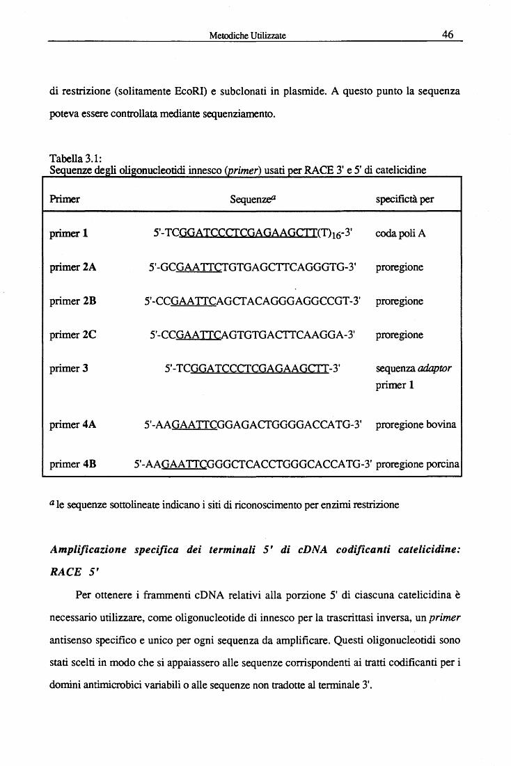
Metodiche Utilizzate 46
di restrizione (solitamente EcoRI) e subclonati in plasmide. A questo punto la sequenza
poteva essere controllata mediante sequenziamento.
Tabella 3.1: Sequenze degli oligonucleotidi innesco (primer) usati per RACE 3' e 5' di catelicidine
Primer Sequenz& specifictà per
primer 1 5'-TCGGATCCCfCGAGAAGCTI(T)16-3' coda poliA
primer 2A 5'-GCGAA TICTGTGAGCTICAGGGTG-3' proregione
primer 2B 5'-CCGAATICAGCTACAGGGAGGCCGT-3' proregione
primer2C 5'-CCGAATICAGTGTGACTTCAAGGA-3' proregione
primer 3 5'-TCGGATCCCTCGAGAAGCI I-3' sequenza adaptor primer l
primer4A 5'-AAGAATICGGAGACTGGGGACCATG-3' proregione bovina
primer 4B 5'-AAGAA TICGGGCTCACCTGGGCACCATG-3' proregione porcina
a le sequenze sottolineate indicano i siti di riconoscimento per enzimi restrizione
Amplificazione specifica dei terminali S' di cDNA codificanti catelicidine:
RACE 5'
Per ottenere i frammenti cDNA relativi alla porzione 5' di ciascuna catelicidina è
necessario utilizzare, come oligonucleotide di innesco per la trascrittasi inversa, un primer
antisenso specifico e unico per ogni sequenza da amplificare. Questi oligonucleotidi sono
stati scelti in modo che si appaiassero alle sequenze corrispondenti ai tratti codificanti per i
domini antimicrobici variabili o alle sequenze non tradotte al terminale 3'.
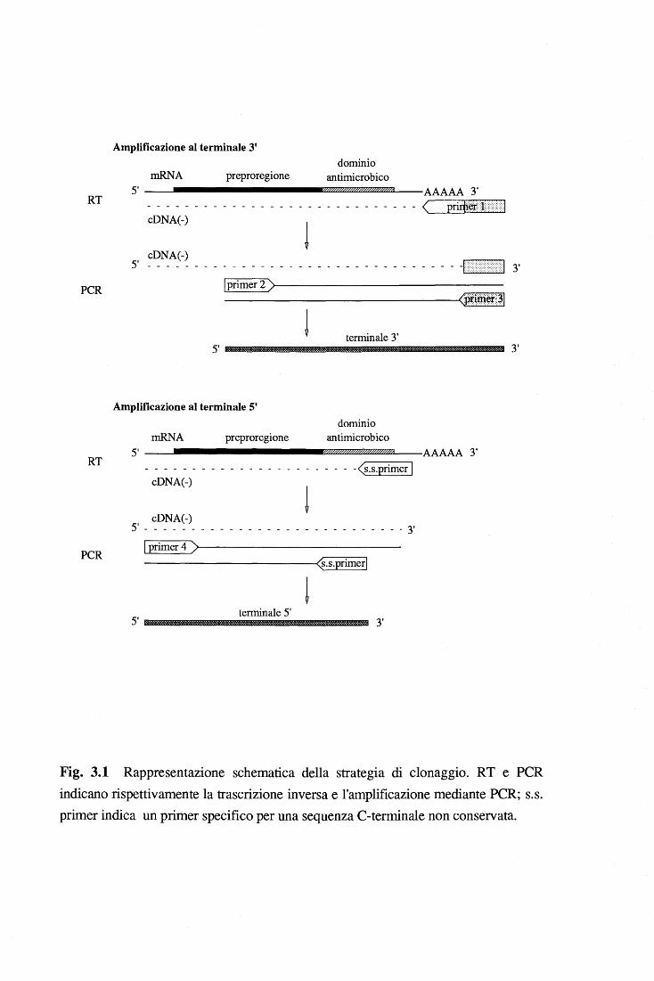
RT
PCR
RT
PCR
Amplificazione al terminale 3'
mRNA preproregione dominio
antimicrobico 5' -------------~;;.??z::z;u:z:zw/z::zrn:z:zwz::zrn:z:zw~;;;zm_;a __ AAAAA 3'
- - - - - --- ------ -- ------- -- ----< priÌij@J }} d cDNA(-)
j cDNA(-)
5' - - - - - - - - - - - - - - - - - - - - - - - - - - - - - - - - - -1 ::r:u::: ::::::::1 3'
lprimer 2 )>-----------------------------------'<P~~m:~::~l
terminale 3'
Amplificazione al terminale 5'
mRNA preproregione dominio
antimicrobico 5' ------------~z.~m~m~m~mmmmmmmm»?r~WAa_ __ AAAAA 3'
____ - - - - - - - - - - - - - - - - - - -(s.s.primer l cDNA(-)
j cDNA(-)
5' - - - - - - - - - - - - - - - - - - - - - - - - - - - - 3 l
l primer 4 )>----------------------------<(s.s.primerl
j terminale 5'
3'
3'
Fig. 3.1 Rappresentazione schematica della strategia di clonaggio. RT e PCR indicano rispettivamente la trascrizione inversa e l'amplificazione mediante PCR; s.s. primer indica un primer specifico per una sequenza C-terminale non conservata.
Metodiche Utilizzate 48
Le condizioni sperimentali usate per la trascrizione inversa sono simili a quelle usate per il
RACE 3' e sono descritte in dettaglio nell'allegato M. Il cDNA 5' viene amplificato
utilizzando ilprimer 4 (tabella 3.1) che corrisponde alla regione conservata della sequenza
non tradotta al 5' (fig. 3.1) e un primer antisenso sequenza-specifico che si appaia in un
tratto leggermente al 5' rispetto al tratto in cui si appaia il primer per la trascrizione inversa.
Sono stati disegnati due primer 4, leggermente diversi l'uno dall'altro: il primer 4A è stato
usato per amplificare il tratto di cD NA al 5' delle catelicidine bovine, mentre il primer 4B è
stato usato per amplificare il tratto di cDNA al 5' delle catelicidine suine. I frammenti
amplificati sono stati trattati come descritto per il RACE 3'.
Le sequenze complete delle catelicidine identificate sono state ottenute dalla
sovrapposizione delle sequenze dei prodotti di amplificazione al5' e al3'.
Vista l'elevata conservazione della sequenza al 5' non tradotto che si osserva tra la
famiglia delle catelicidine è stato quasi sempre possibile amplificare il terminale 5' delle varie
catelicidine utilizzando il protocollo sopra descritto, semplificando notevolmente la procedura
descritta nel metodo originale di Frohman per il RACE 5'. Tuttavia per amplificare il cDNA
5' di Bac5 è stato necessario ricorrere al metodo tradizionale (Frohman et al., 1988) in cui
viene aggiunta ad opera di una desossinucleotidil-transferasi terminale una coda
omopolimerica al3' del filamento cDNA ottenuto dalla trascrittasi inversa. Questo filamento
è poi amplificato mediante PCR utilizzando un oligonucleotide senso che contiene una
sequenza complementare alla coda omopolimerica, oltre al sito di restrizione "adaptor".
3.1.5 Sequenziamento del DNA e analisi delle sequenze
Il sequenziamento dei cloni inseriti in vettore plasmidico pBluescript KS+ è stato
effettuato secondo il metodo della reazione di terminazione didesossinucleotidica di Sanger
(1977) utilizzando un kit commerciale (T7 Sequencing kit, Pharmacia). In tutti i cD NA sono
stati sequenziati entrambi i filamenti, le regioni ad alto contenuto di C+G sono state
sequenziate in parallelo con deazaguanosina (Pharmacia). Molte delle sequenze sono state
Metodiche Utilizzate 49
sequenziate utilizzando un sequenziatore automatico a fluorescenza (EMBL Fluorescent DNA
Sequencer) presso il laboratorio nazionale del Consorzio Interuniversitario per le
Biotecnologie (AREA di Ricerca, Padri ciano - Trieste).
L'analisi delle sequenze è stata effettuata utilizzando i pacchetti bioinformatici messi a
disposizione dall'ICGEB-Net (GCG e IntelliGenetics Suite V 5.4). Per ogni nuova sequenza
clonata venivano condotte ricerche in banca dati (Swiss-Prot per proteine e EMBL per DNA)
utilizzando i programmi FastDB e Genalign del pacchetto IG, e il programma BLITZ
dell'EMBL.
3.1.6 Primer extension
L'oligonucleotide primer antisenso veniva incubato con 10 Jlg di RNA totale a 42 °C
per 15 min. Alla miscela venivano aggiunti i nucleotidi trifosfati + a-[32P]-dGTP e la
trascrittasi inversa (M-ML V RT); dopo l ora a 42 °C il prodotto finale dell'allungamento
veniva analizzato su gel di acrilamide al 6% e urea 8 M.
3.1. 7 Trascrizione e traduzione in vitro
n vettore pBluescript KS+ contenente la sequenza cD NA completa di una catelicidina
(p.es. proBac5, proindolicidina, prododecapeptide) è stato linearizzato, tagliando con un
opportuno enzima di restrizione a livello della regione corrispondente al3' non tradotto della
sequenza codificante. Il DNA linearizzato è stato trascritto dalla T7 RNA polimerasi (vedi
condizioni usate in allegato A). La reazione di traduzione è stata condotta utilizzando un lisato
di reticolociti di coniglio (Novagene) secondo le istruzioni del produttore. Per analizzare la
traslocazione di membrana, la traduzione è stata effettuata in presenza di un preparato
microsomiale pancreatico di cane (Du Pont-New England Nuclear). I prodotti di traduzione
sono stati immunoprecipitati con anticorpi specifici per il dominio antimicrobico seguendo il
protocollo decritto da Dasso & Jackson (1989) e sono stati successivamente analizzati
mediante SDS-PAGE su un gel di acrilamide al15%.
Metodiche Utilizzate 50
3.1.8 Southern blot
Dopo separazione elettroforetica su gel di agarosio all' l% il DNA viene trasferito su
membrana di nylon (Zeta-Probe GT, BioRad) secondo il protocollo classico descritto da
Southem (1975). Le membrane vengono quindi ibridate con opportuni ologonucleotidi,
marcati radioattivamente con [32p] secondo il protocollo descritto in Sambrook et al. (1989).
3.2 Purificazione e caratterizzazione strutturale
3.2.1 Isolamento di neutrofili bovini, estrazione proteica e iniziale
frazionamento mediante salting out.
I neutrofili sono stati purificati da sangue bovino mediante centrifugazione differenziale
seguita da lisi ipotonica degli eritrociti contaminanti (Mottola et al., 1980). Le cellule sono
state sospese alla concentrazione di lx109 cellule/mi in una soluzione di N a acetato 0.2 M,
pH 4.0, in presenza di inibitori delle serin-proteasi neutre (10 JJ.M 3,4-DCI, l mM PMSF) al
fine di impedire all'elastasi di digerire le catelicidine (Scocchi et al., 1992). In seguito le
cellule sono state lisate per trattamento con ultrasuoni (Branson Sonic Power S75) e
sottoposte ad estrazione, agitando per 2 ore a 4°C. Il materiale corpuscolato è stato
sedimentato centrifugando per 20 mina 30,000 x g a 4°C e quindi è stato riestratto come già
descritto. Alternativamente, è stata prima isolata la frazione granulare e poi si è proceduto
all'estrazione delle proteine. Le cellule, risospese (lxi08 cellule/mi) in "relaxation buffer"
(KCl 100mM, NaCl 3mM, MgCl2 35mM, Hepes10mM, ATP(Na)2 lmM, pH 7.3), sono
state lisate mediante decompressione in ambiente di azoto, seguendo le indicazioni del
costruttore (Parr Instrument Company) ed il protocollo descritto da Borregaard et al. (1983).
In seguito, illisato è stato sottoposto a centrifugazione per eliminare nuclei e cellule intere
(500xg, 10 min, 4°C) ed il sopranatante ricentrifugato a 12,000 rpm per 10 min, 4°C
(Rotore: Kontron A 8.24, R = 8.24 cm). I granuli, così sedimentati sono stati quindi estratti
in tampone N a acetato, pH 4.0 con la stessa metodica descritta per le cellule.
Metodiche Uùlizzate 51
Un frazionamento preliminare delle proteine contenute in entrambi gli estratti acidi, sia
di cellule totali che di granuli, è stato effettuato mediante aggiunta successiva di ammonio
solfato, rispettivamente al 25% e 45% di saturazione (salting out).
3.2.2 Purificazione di catelicidine bovine
Per purificare quattro diverse catelicidine da neutrofili bovini (proBac5, proBac7,
prododecapeptide e proBMAP-28) sono stati messi a punto diversi protocolli. I metodi che
hanno portato a migliori risultati sono riassunti e schematizzati nelle pagine successive.
Nel primo caso la frazione contenente le proteine precipitate con ammonio solfato saturo
al 25% è stata sospesa in 0.05% TFA (acido trifluoroacetico) e purificata in RP-HPLC
(colonna Resource-Pharmacia) con un gradiente da 0% a 50% di acetonitrile in acqua, in
presenza di 0.05% TFA. L'eluizione delle frazioni veniva seguita mediante monitoraggio a
214 nm. Le frazioni corrispondenti a ciascun picco risolto venivano riunite e ripurificate in
RP-HPLC e quindi liofilizzate. A questo punto, la frazione contenente proBac5 risultava
pura secondo i parametri di analisi utilizzati (SDS-PAGE e ES-MS), mentre le frazioni
contenenti prododecapeptide e proBPAM-28 sono state ulteriormente purificate mediante
FPLC su colonna Mono Sin tampone 20 mM N a acetato pH 5.5 in gradiente di sale (NaCl
da O a 1.5 M).
Le proteine precipitate in ammonio solfato al 45% di saturazione sono state separate
mediante gel filtrazione su colonna G-50 Sephadex (Pharmacia) (2x120 cm, flusso 15 cm/h)
equilibrata in N a acetato 50 mM, pH 4.5. L'eluizione veniva seguita spettrofotometricamente
a 280 nm e ogni seconda frazione veniva analizzata mediante dot blot. Le frazioni contenenti
proBac7 sono state ulteriormente purificate mediante RP-HPLC sequendo le condizioni
descritte sopra.
Alternativamente, le proteine ottenute dal frazionamento dell'estratto acido granulare
mediante precipitazione con ammonio solfato saturo al 25% sono state separate per gel
fùtrazione su colonna G-50 Sephadex (condizioni come sopra) equilibrata in N a acetato 50
Metodiche Utilizzate 52
mM, pH 5.5. Le frazioni sono state analizzate in Western blot e quelle contenenti proBac5 o
prododecapeptide, sono state ulteriormente purificate mediante FPLC su colonna Mono S
(Pharmacia) con un gradiente di N aCl da O a 1.5 M in tampone N a acetato 20 mM, pH 5.5.
n prododecapeptide eluiva ad una concentrazione 0.3 M di N aCl, mentre proBac5 a circa 0.9
M. Le due frazioni sono state desalate e concentrate per ultrafiltrazione in cella Amicon
utilizzando una membrana Diaflo YM5. A questo punto proBac5 risultava puro in base
all'analisi su gel SDS-PAGE colorato con colorazione argentica, mentre per il
prododecapeptide è stato necessario effettuare un ulteriore passaggio cromatografico in RP-
HPLC.
Con una procedura, sostanzialmente diversa dalle precedenti è stato possibile purificare
la sola proregione del dodecapeptide comprendente i residui da l a 114 della proteina intera.
In questo caso l'estratto granulare acido non è stato frazionato mediante salting out, ma è
stato diluito in tampone N a acetato 50 mM a pH 6.0 e fatto passare attraverso una colonna a
scambio cationico debole (CM-52). La porzione non legata è stata desalata mediante un
passaggio rapido su cartuccia monouso Sep-Pak C 18 (Pharmacia) e le proteine sono state
eluite con CH3CN a concentrazioni crescenti e in presenza di 0.05% TF A. Due delle frazioni
(30% e 40% CH3CN rispettivamente), risultavano particolarmente ricche di una proteina
riconosciuta in Western blot dall'anticorpo specifico per il tratto C-terminale della proregione
del dodecapeptide. Queste frazioni sono state ulteriormente purificate mediante C18 RP-
HPLC fino ad ottenere la proteina pura.
Tutti gli stadi di purificazione descritti sono stati condotti a 4°C ad eccezione del FPLC e
HPLC che sono stati eseguiti a temperatura ambiente.
3.2.3 Saggi analitici per proteine
La concentrazione proteica è stata misurata sia mediante il metodo colorimetrico con
acido bicinconinico (Smith et al., 1985), che con metodo di spettrofotometrico
all'ultravioletto (W addell, 1956), utilizzando BSA come proteina di riferimento.
Metodiche Utilizzate' 53
Le proteine sono state separate sia in condizioni riducenti che non riducenti mediante
SDS/P AGE su gel di acrilamide al 14% e colorati con Coomassie brilliant blue o con
colorazione argentica. I Western blot e dot blot sono stati condotti come descritto da Zanetti
et al. (1990). Gli anticorpi utilizzati per riconoscere le catelicidine purificate erano specifici
per le porzioni C-tenninali variabili. ed erano stati ottenuti immunizzando conigli con Bac5 o
Bac7 o BMAP-28 oppure con il frammento C-terminale della proregione del dodecapeptide
(SIRITKQPW APPQAA, residui l 00-114 in fig. 4.14 ). I primi due sono peptidi isolati da
neutrofili bovini, mentre gli altri sono stati sintetizzati chimicamente sulla base di sequenze
note dal cDNA.
La spettrometria di massa (ES-MS) è stata eseguita con uno spettrometro dotato di una
camera di ionizzazione spray a pressione ambiente (Perkin Elmer Sciex API-l), in cui i
campioni venivano iniettati direttamente dopo eluizione da HPLC oppure dopo risospensione
in acido formico 2%/acetonitrile 50% dei campioni precedentemente liofilizzati. Per ogni
misurazione venivano accumulati almeno dieci spettri di massa ad una risoluzione di 0.1
unità di massa, in un intervallo di rn/z da 400 a 2400. I valori di massa teorici sono stati
calcolati con il programma per PC Peptide Companion (CoshiSoft/Peptisearch).
3.2.4 Digestione con tripsina e modificazione chimica dei ponti disolfuro.
Circa 100 Jlg di proBac7 purificato, disciolti in 100 Jll di tampone 0.1 M Tris-HCl,
0.001% SDS, pH 8.5, sono stati digeriti con 5 Jlg di tripsina per 16 ore a 37°C. La miscela
dei frammenti generati è stata separata in RP-HPLC (0-50% acetonitrile, 0.1% TFA, 0.7
ml/min, SuperPac Pep-S column), dopo aggiunta di TFA al 0.1% (concentrazione finale).
Ciascun picco eluito è stato direttamente analizzato con lo spettrometro di massa, al fine di
individuare i possibili frammeni contenenti un ponte disolfuro. La presenza di Cys2 in questi
frammenti è stata confermata sia mediante ossidazione che S-acetilazione.
Metodiche Utilizzate 54
Nel primo caso, un'aliquota della frazione eluita da HPLC, contenente un legame
disolfuro, è stata trattata con acido perfonnico per 30 min seguendo un protocollo standard
(Picotag, Waters Millipore), e quindi liofilizzata ed analizzata mediante ES-MS.
Nel secondo caso, un'aliquota della frazione contenente il frammento è stata incubata
per 2 ore a 37°C in condizioni riducenti (10 mM DTT in tampone 0.5 M Tris-HCl, pH 8.5) e
quindi è stata aggiunta la iodoacetamide (50 mM concentrazione finale) e lasciata reagire al
buio per 20 min a temperatura ambiente. La reazione è stata bloccata con un eccesso di DTT,
desalata in RP-HPLC (0-H>O% acetonitrile, 0.1% TFA in 10 minuti; colonna analitica
SuperPac Pep-S) ed analizzata mediante ES-MS. In parallelo la stessa reazione è stata
condotta su un'altra aliquota del medesimo frammento in condizioni non riducenti, in modo
di accertare l'effettiva integrità del legame disolfuro.
3.2.5 Dicroismo circolare
Gli spettri CD sono stati registrati a 23°C con uno spettropolarimetro Jasco-600
utilizzando una cella con 0.1 cm di cammino ottico. Le analisi sono state condotte su
campioni disciolti ad una concentrazione finale 4 JJ.M sia in acqua MilliQ che in tampone N a
fosfato 5 mM, p H 7 .0. Per ogni campione sono stati accumulati almeno dieci spettri
nell'intervallo di lunghezza d'onda tra 190-240 nm. Il contenuto di strutture ad a-elica e B-
foglietto in ciascuna catelicidina in soluzione è stato valutato mediante il programma
CONTIN (Provencher & Glockner, 1981) e con il metodo di Chen et. al. (1974).
3.2.6 Idrolisi acida limitata
Per valutare il grado di suscettibilità ali 'idrolisi acida di alcuni legami peptidici Asp-Pro,
le catelicidine purificate sono state conservate per una settimana in ambiente blandamente
acido (da 0.05 a 0.5 % TFA) e l'incremento di idrolisi è stato confrontato con gli stessi
campioni incubati in 10 mM ammonio carbonato, pH 8.0. ProBac5 e proBac7 sono stati
Metodiche Utilizzate 55
trattati anche con acido formico al 75% per 16 ore a 37°C. Il grado di frammentazione
presente nei vari campioni è stata valutata mediante ES-MS.
3.2. 7 Ricerche in banche dati e predizioni di struttura
Ricerche di omologia di sequenza sono state condotte presso la banca dati SwissProt
utilizzando i programmi FastDB del pacchetto GCG e il programma BLITZ dell'EMBL
(Sturrock & Collins, 1993), con diversi valori di INDEL e PAM. La predizione della
struttura secondaria è stata condotta utilizzando il metodo "PhD profile network" (Rost &
Sanders, 1994) e il metodo "topology based TOPITS" (Rost, 1995) e mediante i programmi
THREADER (Jones et al.,1992).
3.2.8 Saggi di inibizione enzimatica
I saggi di inibizione della catepsina L sono stati effettuati secondo il protocollo descritto
da Barrette Kirschke (1981}, utilizzando Z-Phe-Arg-AMC (Novabiochem) come substrato,
e le concentrazioni dell'enzima e dell'inibitore erano come precedentemente descritto da
Verbanac et al. (1993). Per i saggi sono state utilizzate sia la catepsina L umana di fegato
reperibile commercialmente (Calbiochem) che la catepsina L umana di rene purificata nel
laboratorio del prof. V. Turk dell'Istituto J. Stefan di Lubiana (Turk et al., 1993).
I saggi di inibizione della papaina (Sigma) sono stati condotti come descrtitto in Barrett
(1972) utilizzando BANA (Sigma) come substrato.
I valori di Ki sono stati calcolati secondo il metodo Bieth (1984).
Risultati e Discussione 56 ! ~
'Capitolo 4
Risultati e Discussione
4.1 Clonaggio molecolare di cDNA e analisi della sequenza
aminoacidica dedotta
4.1.1 BacS
La conoscenza della sequenza peptidica di Bac5 ci ha permesso di progettare due
oligonucleotidi sintetici degenerati (tabella I in allegato A), di cui uno risultava
sufficientemente specifico da essere utilizzato come sonda per isolare il clone codificante
Bac5 da una genoteca a cDNA di cellule mieloidi di midollo osseo bovino. Il clone isolato
(Bac5A) conteneva un frammento di 393 bp, corrispondente alla sequenza centrale
(nucleotidi 69-461 in fig.14.1) del cDNA completo di Bac5. La strategia adottata per
ottenere la sequenza completa del cDNA (julllength) ha richiesto l'uso della tecnica RACE
(vedi capitolo 3, fig.3.1), che ha permesso di amplificare copie delle sequenze terminali 5' e
3' dell 'mRNA codificante Bac5, utilizzando oligonucleotidi primer interni alla sequenza di
Bac5A (capitolo 3, tabella 3.1). Mediante un esperimento di "primer extension" si è
dimostrato che la regione non tradotta al5' si estende per sole 14 bp prima del codone AUG
di inizio di trascrizione (fig. lB in allegato A). Questo dato ci ha permesso di affermare con
certezza che la sequenza ottenuta dal frammento amplificato mediante RACE 5' era
effettivamente completa (allegato A).
Risultati e Discussione 57
Dalla sovrapposizione di tutti i frammenti ottenuti è stata quindi ottenuta la sequenza
completa che è composta da 661 bp (fig. 4.1) e codifica per una proteina di 17 6 aminoacidi.
In essa si distingue un peptide segnale di 29 residui~ una sequenza "pro" di 101 residui,
seguita da una sequenza di 43 residui, corrispondenti al peptide antimicrobico Bac5
(sottolineato in fig. 4.1). La presenza di una valina quale ultimo aminoacido prima dell'inizio
della sequenza di Bac5, conferma l'esistenza di un sito di taglio specifico per l'elastasi,
enzima responsabile del rilascio del peptide maturo da proBac5. Infine, la sequenza di Bac5
è seguita da una coda C-terminale di tre residui -Gly-Arg-Arg che costituiscono un segnale
per l'amidazione della molecola, che avviene durante le prime fasi della maturazione. La
presenza di un gruppo a-amidico ali' estremità C-terminale è stata descritta in molti peptidi
(ad esempio neuropeptidi, ma anche peptidi antimicrobici) e la sua funzione sembra essere
quella di aumentare la loro biostabilità
Per verificare la correttezza della chiave di lettura della sequenza dedotta, il frammento
di 661 bp, corrispondente al cDNA completo, clonato in vettore pBluescript è stato trascritto
e tradotto in vitro. Come dimostrato dalle masse osservate in SDS-PAGE (fig. 5, allegato
A), il prodotto della traduzione viene immunoprecipitato con anticorpi anti Bac5 e se la
traduzione avviene in presenza di membrane microsomiali, si ha la rimozione del peptide
segnale. Inoltre, le masse osservate nei prodotti di trascrizione/traduzione sono confrontabili
a quelle riportate in precedenza da Zanetti et al. (1990).
Dali' analisi della sequenza di pre-proBac5 risulta che la pro-regione è prevalentemente
anioni ca (p I 4.1 )~ mentre il peptide è fortemente cationico (p l 13.4 ). Questo dato è in
accordo con l 'ipotesi che la proregione possa neutralizzare il peptide antimicrobico,
bloccandone l'attività battericida.
La sequenza dedotta di pre-proBac5 è stata quindi confrontata con altre proteine note,
mediante una ricerca in banca dati (Swiss-Prot data bank). Inaspettatamente abbiamo
scoperto l'esistenza di due proteine che hanno un elevato grado di omologia (> 70%) con la
pre-proregione di Bac5 (fig. 6, allegato A). La prima, detta catelina, è una proteina di 99
Risultati e Discussione 58
residui che è stata isolata dai leucociti del maiale e che inizialmente era stata descritta come
inibitore delle cistein-proteasi (Rijtonia et al., 1989; Kopitar et al., 1989); la seconda, detta
CAP18, è il precursore di un peptide antimicrobico dei granulociti di coniglio capace di
inattivare illipopolisaccaride (Larrick et al., 1991). In nessuna delle due proteine si trova
una sequenza simile al peptide antimicrobico Bac5.
4.1.2 lndolicidina e dodecapeptide
La tecpica RACE, che era stata inizialmente messa a punto per amplificare la sequenza
del terminale 3' di Bac5, ha portato alla scoperta di una nuova famiglia di precursori di
peptidi antimicrobici. Analizzando elettroforeticarriente in gel di agarosio il prodotto
dell'amplificazione effettuata secondo il protocollo descritto (vedi capitolo 3), si è osservata
la presenza di tre bande a peso molecolare diverso, che sono state successivamente clonate in
vettore plasmidico pBluescript Dopo sequenziamento risultava che solo la banda a più alto
peso molecolare conteneva, come atteso,. il frammento corrispondente alla porzione 3' del
cD NA di Bac5, mentre le altre due contenevano i frammenti 3' dei cD NA di altri due peptidi
antibiotici già noti: il dodecapeptide (allegato B) e l'indolicidina (allegato C). Il
dodecapeptide era stato precedentemente isolato dai granuli dei neutrofili nel nostro stesso
laboratorio (Romeo et al., 1989), mentre l 'indolicidina era stata isolata da leucociti bovini da
Selsted et al. (1992b). Per confermare l'esistenza degli mRNA di questi due peptidi sono
stati eseguiti dei controlli ibridando con sonde oligonucleotidiche specifiche un Northem blot
di RNA totale proveniente da diversi tessuti o cellule bovine (midollo osseo, polmone, rene,
stomaco, milza, cuore, fegato, intestino tenue, colon, neutrofili circolanti e macrofagi).
Come nel caso di Bac5, anche il dodecapeptide e l'indolicidina vengono espressi solo dalle
cellule mieloidi nel midollo osseo, infatti la sonda specifica per l'indolicidina riconosce in
queste cellule un mRNA di circa 0.6 kb (fig.1A, allegato B), mentre quella per il
dodecapeptide ne riconosce uno di circa 0.65 kb (fig.1, allegato C).
Risultati e Discussione 59
Il cONA full-length dell'indolicidina (550 bp) é stato generato amplificando l'mRNA
specifico mediante RACE 5' ed é stato inserito in vettore pBluescript. L'analisi della
sequenza aminoacidica dedotta rivela che l 'indolicidina è sintetizzata in forma di precursore
di 144 aminoacidi con un peptide segnale di 29 residui, seguito dalla pro-regione di 101
residui e dalla sequenza del tridecapeptide antibiotico che si trova nel tratto C-terminale (fig.
4.2). L 'ultimo aminoacido é una glicina che presumibilmente viene rimossa da un sistema di
modificazione post-traduzionale, dando luogo all'amidazione del COOH terminale. Inoltre,
come era già stato osservato per Bac5, l'ultimo aminoacido della pro-sequenza è una valina
che è un sito di taglio specifico per l' elastasi. Da ciò ipotizziamo che anche la maturazione da
proindolicidina a indolicidina sia catalizzata da questa serin proteasi neutra.
Il cONA del dodecapeptide codifica per una proteina di 155 aminoacidi in cui si
distingue il peptide segnale di 29 e una proregione di 114 residui, seguita dai 12 aminoacidi
della sequenza del peptide. Come si osserva in fig. 4.3, la proregione del dodecapeptide
contiene 13 residui in più rispetto alle altre due prosequenze e la valina corrispondente al sito
di taglio dell' elastasi non è conservata, in quanto al suo posto si trova una isoleucina.
L'ultimo residuo prima della sequenza del dodecapeptide è un' alanina, che è un buon sito di
taglio anche per l' elastasi.
L'identificazione di queste due nuove sequenze è stata del tutto inaspettata in quanto
nulla era noto dei precursori di questi due peptidi e tanto meno ci si attendeva che due peptidi
così dissimili avessero una pre-proregione altamente omologa, da far supporre un'origine
comune. La somiglianza strutturale tra queste proteine é, infatti, strettamente localizzata nella
regione N -terminale e include quattro cisteine altamente conservate che probabilmente hanno
un ruolo essenziale nella funzionalità dei precursori (fig.4, allegato C). Le regioni C-
terminali, che contengono i peptidi bioattivi, sono invece diverse sia nella lunghezza che
nella composizione aminoacidica.
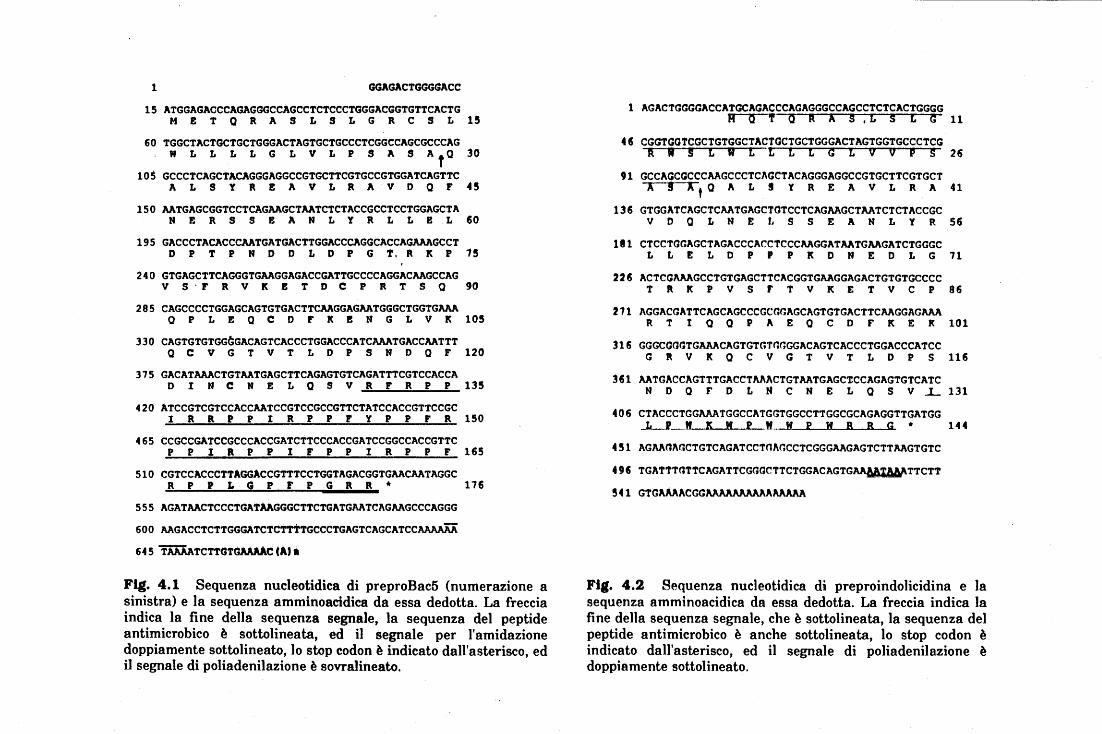
1 GGAGACTGGGGACC
15 ATGGAGACCCAGAGGGCCAGCCTCTCCCTGGGACGGTGTTCACTG M E T Q R A S L S L G R C S L 15
60 TGGCTACTGCTGCTGGGACTAGTGCTGCCCTCGGCCAGCGCCCAG W L L L L G L V L P S A S AfQ 30
105 GCCCTCAGCTACAGGGAGGCCGTGCTTCGTGCCGTGGJ\TCAGTTC A L S Y R E A V L R A V D Q F 45
150 AATGAGCGGTCCTCAGAAGCTAATCTCTACCGCCTCCTGGAGCTA N E R S S E A N L Y R L L E L 60
195 GACCCTACACCCAATGATGACTTGGACCCAGGCACCAGAAAGCCT D P T P N D D L D P G T. R K P 15
240 GTGAGCTTCAGGGTGAAGGAGACCGATTGCCCCAGGACAAGCCAG V S · F R V K E T D C P R T S 0 90
285 CAGCCCCTGGAGCAGTGTGACTTCAAGGAGAATGGGCTGGTGAAA 0 P L E 0 C D F K E N G L V K 105
330 CAGTGTGTGG~GACAGTCACCCTGGACCCATCAAATGACCAATTT O C V G T V T L D P S N D Q F 120
375 GACATAAACTGTAATGAGCTTCAGAGTGTCAGATTTCGTCCACCA D I N C N E L Q S V R F R P P 135
420 ATCCGTCGTCCACCAATCCGTCCGCCGTTCTATCCACCGTTCCGC I R k P P I R P P F Y P P F R 150
465 CCGCCGATCCGCCCACCGATCTTCCCACCGATCCGGCCACCGTTC P P I a P P l F P P l R P P F 165
510 CGTCCACCCTTAGGACCGTTTCCTGGTAGACGGTGAACAATAGGC R P P L G P F P G a R * 176
555 AGATAACTCCCTGATAAGGGCTTCTGATGAATCAGAAGCCCAGGG
600 AAGACCTCTTGGGATCTCTTtTGCCCTGAGTCAGCATCCAJ\AAAA
64 5 TAAMTCTT~TGAAAAC tAJ l
Fig. 4.1 Sequenza nucleotidica di preproBac5 (numerazione a sinistra) e la sequenza amminoacidica da essa dedotta. La freccia indica la fine della sequenza segnale, la sequenza del peptide antimicrobico è sottolineata, ed il segnale per l'amidazione doppiamente sottolineato, lo stop codon è indicato dall'asterisco, ed il segnale di poliadenilazione è sovralineato.
1 AGACTGGGGACCATGCAGACCCAGAGGGCCAGCCTCTCACTGGGG H o 'f o R X s ,L s L G 11
46 CGGTGGTCGCTGTGGCTACTGCTGCTGGGACTAGTGGTGCCCTCG R " s L w L t L L G t v v p s 26
91 GCCAGCGCCCAAGCCCTCAGCTACAGGGAGGCCGTGCTTCGTGCT A ! x, Q A L s y R E A v L R A 41
136 GTGGATCAGCTCAATGAGCTGTCCTCAGAAGCTAATCTCTACCGC v D o L N E r. s s E A N L y R 56
111 CTCCTGGAGCTAGACCCACCTCCCAAGGATAATGAAGATCTGGGC L L E L D p p p K D N E D L G 71
226 ACTCGAAAGCCTGTGAGCTTCACGGTGAAGGAGACTGTGTGCCCC T " l( p v s r T v K E T v c p 96
271 AGGACGATTCAGCAGCCCGCGGAGCAGTGTGACTTCAAGGAGAAA R T t o o p A E o c D F K E K 101
316 GGGCGGGTGAAACAGTGTGTGGGGACAGTCACCCTGGACCCATCC G R v l( o c v G T v T L D p s 116
361 AATGACCAGTTTGACCTAAACTGTAATGAGC~CCAGAGTGTCATC N D o F D L N c N E L o s v ...L 131
406 CTACCCTGGAAATGGCCATGGTGGCCTTGGCGCAGAGGTTGATGG _L_I_M lt lf ~ Jf._ lf ~ lf B B G • 144
451 AGAAUAGCTGTCAGATCCTOAGCCTCGGGAAGAGTCTTAAGTGTC
496 TGATTTGTTCAGATTCGGGCTTCTGGACAGTG~TTCTT
!41 GTGAAAACGGAAAAMAAAAAAAAA
Fig. 4.2 Sequenza nucleotidica di preproindolicidina e la sequenza amminoacidica da essa dedotta. La freccia indica la fine della sequenza segnale, che è sottolineata, la sequenza del peptide antimicrobico è anche sottolineata, lo stop codon è indicato dall'asterisco, ed il segnale di poliadenilazione è doppiamente sottolineato.
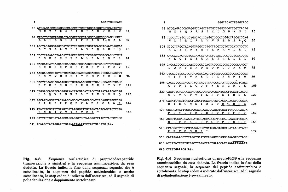
l AGACTGGGCACC
13 ATGGAGACCCCGAGGGCCAGCCTCTCCCTGGGACGGTGGTCACTGTGG M E T p R A s L s L G R w s L w 16
61 CTGCTGCTGCTGGGACTAGCGCTGCCCTCGGCCAGCGCCCAGGCCCTC L L L L G L A L p s A s At Q A L 32
109 AGCTACAGGGAGGCCGTGCTTCGTGCTGTGGATCAGCTCAATGAGCAA s y R E A v L R A v D Q L N E Q 48
157 TCCTCAGAACCTAACATTTACCGTCTTCTCGAGCTGGACCAGCCTCCT s s E p N I y R L L E L D o p p 64
205 CAGGATGATGAAGACCCAGACAGCCCGAAGCGGGTGAGCTTCAGGGTG Q D D E D p D s p K R v $ F R v 80
253 AAGGAGACCGTGTGCTCCAGGACCACCCAGCAGCCCCCCGAGCAGTGT K E T v c s R T T Q o p p E Q c 96
301 GACTTCAAGGAGAATGGGCTGCTGAAACGCTGTGAGGGGACAGTCACC D F K E N G L L K R c E G T v T 112
349 CTGGACCAGGTCAGGGGTAACTTCGACATCACCTGTAATAATCACCAG L D Q v R G N F D I T c N N H Q 128
397 AGCATCAGGATTACAAAGCAGCCATGGGCACCACCACAGGCAGCCCGA s I R I T K Q p w A p p Q A A.JL 144
445 TTATGTCGCATTGTAGTGATAAGGGTTTGCAGATAATCAGCTCTTGTA li ~ B l :i v l B v ~ B 1r 155
493 GATTTCTGTCATAAGCCAGCAGAGTCCTAAGGGTTTTCTTACTCTGCC
541 TCAAGCTACTGGATCTG~TTCTTGTGACATG(A)n
Fig. 4.3 Sequenza nucleotidica di preprododecapeptide (numerazione a sinistra) e la sequenza amminoacidica da essa dedotta. La freccia indica la fine della sequenza segnale, che è sottolineata, la sequenza del peptide antimicrobico è anche sottolineata, lo stop codon è indicato dall'asterisco, ed il segnale di poliadenilazione è doppiamente sottolineato
l GGGCTCACCTGGGCACC
18 ATGGAGACCCAGAGGGCCAGCCTGTGCCTGGGGCGCTGGTCACTG M E T o R A s L c L G R w s L 15
63 TGGCTTCTGCTGCTGGCACTCGTGGTGCCCTCGGCCAGCGCCCAG w L L L L A L v v p s A s AfO 30
108 GCCCTCAGCTACAGGGAGGCCGTGCTTCGTGCTGTGGATCGCCTC A L s y R E A v L R A v D R L 45
153 AACGAGCAGTCCTCGGAAGCTAATCTCTACCGCCTCCTGGAGCTG N E Q s s E A N L y R L L E L 60
198 GACCAGCCGCCCAAGGCCGACGAGGACCCGGGCACCCCGAAACCT D o p p K A D E D p G T p K p 75
243 GTGAGCTTCACGGTGAAGGAGACTGTGTGTCCCAGGCCGACCCGG v s F T v K E T v c p R p T R 90
288 CAGCCCCCGGAGCTGTGTGACTTCAAGGAGAATGGGCGAGTGAAG Q p p E L c D F K E N G R v K 105
333 CAGTGTGTGGGGACAGTCACCTTGAACCCATCCATTCACTCACTG Q c v G T v T L N p s I H s L 120
378 GACATCTCCTGTAATGAGATTCAGAGTGTCAGGAGACGTCCCCGA D I s c N E I Q s v R 'R. R p R 135
423 CCCCCATATTTGCCAAGGCCAAGGCCACCTCCGTTTTTCCCACCA p p y L p R p R p p p F F p p 150
468 AGGCTCCCACCAAGGATCCCACCAGGGTTCCCACCAAGGTTCCCA R L p p R I p p G F p p R F p 165
513 CCACGGTTCCCCGGAAAACGGTGATGGAGTGGCTGATAACATACC p R F p G K R 1r 172
558 CATTAAAAGCTTTTGGTGAATCCTGAGCCCAGGGAAAGTCCTAGG
603 ATCTTATTGTTGTGGCTCAGACTTCTGAACCATGAAAAATAAATT
648 CTTGTGAAACG(A)n
Fig. 4.4 Sequenza nucleotidica di preproPR39 e la sequenza amminoacidica da essa dedotta. La freccia indica la fine della sequenza segnale, la sequenza del peptide antimicrobico è sottolineata, lo stop codon è indicato dall'asterisco, ed il segnale di poliadenilazione è sovralineato.
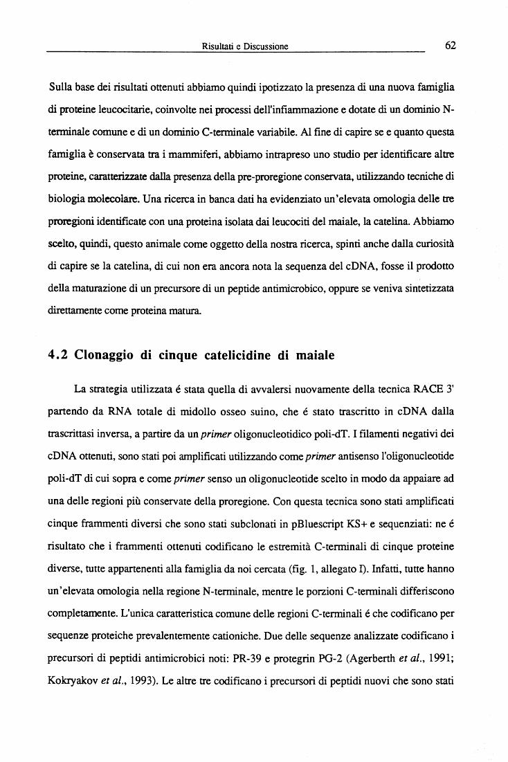
Risultati e Discussione 62
Sulla base dei risultati ottenuti abbiamo quindi ipotizzato la presenza di una nuova famiglia
di proteine leucocitarie, coinvolte nei processi dell'infiammazione e dotate di un dominio N-
tenninale comune e di un dominio C-terminale variabile. Al fine di capire se e quanto questa
famiglia è conservata tra i mammiferi, abbiamo intrapreso uno studio per identificare altre
proteine, caratterizzate dalla presenza della pre-proregione conservata, utilizzando tecniche di
biologia molecolare. Una ricerca in banca dati ha evidenziato un'elevata omologia delle tre
proregioni identificate con una proteina isolata dai leucociti del maiale, la catelina. Abbiamo
scelto, quindi, questo animale come oggetto della nostra ricerca, spinti anche dalla curiosità
di capire se la catelina, di cui non era ancora nota la sequenza del cD N A, fosse il prodotto
della maturazione di un precursore di un peptide antimicrobico, oppure se veniva sintetizzata
direttamente come proteina matura.
4. 2 Clonaggio di cinque catelicidine di maiale
La strategia utilizzata é stata quella di avvalersi nuovamente della tecnica RACE 3'
partendo da RNA totale di midollo osseo suino, che é stato trascritto in cDNA dalla
trascrittasi inversa, a partire da un primer oligonucleotidico poli-dT. I filamenti negativi dei
cD NA ottenuti, sono stati poi amplificati utilizzando come primer antisenso l'oligonucleotide
poli-dT di cui sopra e come primer senso un oligonucleotide scelto in modo da appaiare ad
una delle regioni più conservate della proregione. Con questa tecnica sono stati amplificati
cinque frammenti diversi che sono stati subclonati in pBluescript KS+ e sequenziati: ne é
risultato che i frammenti ottenuti codificano le estremità C-terminali di cinque proteine
diverse, tutte appartenenti alla famiglia da noi cercata (fig. l, allegato n. Infatti, tutte hanno
un'elevata omologia nella regione N-terminale, mentre le porzioni C-terminali differiscono
completamente. L'unica caratteristica comune delle regioni C-terminali é che codificano per
sequenze proteiche prevalentemente cationiche. Due delle sequenze analizzate codificano i
precursori di peptidi antimicrobici noti: PR-39 e protegrin PG-2 (Agerberth et al., 1991;
Kokryakov et al., 1993). Le altre tre codificano i precursori di peptidi nuovi che sono stati
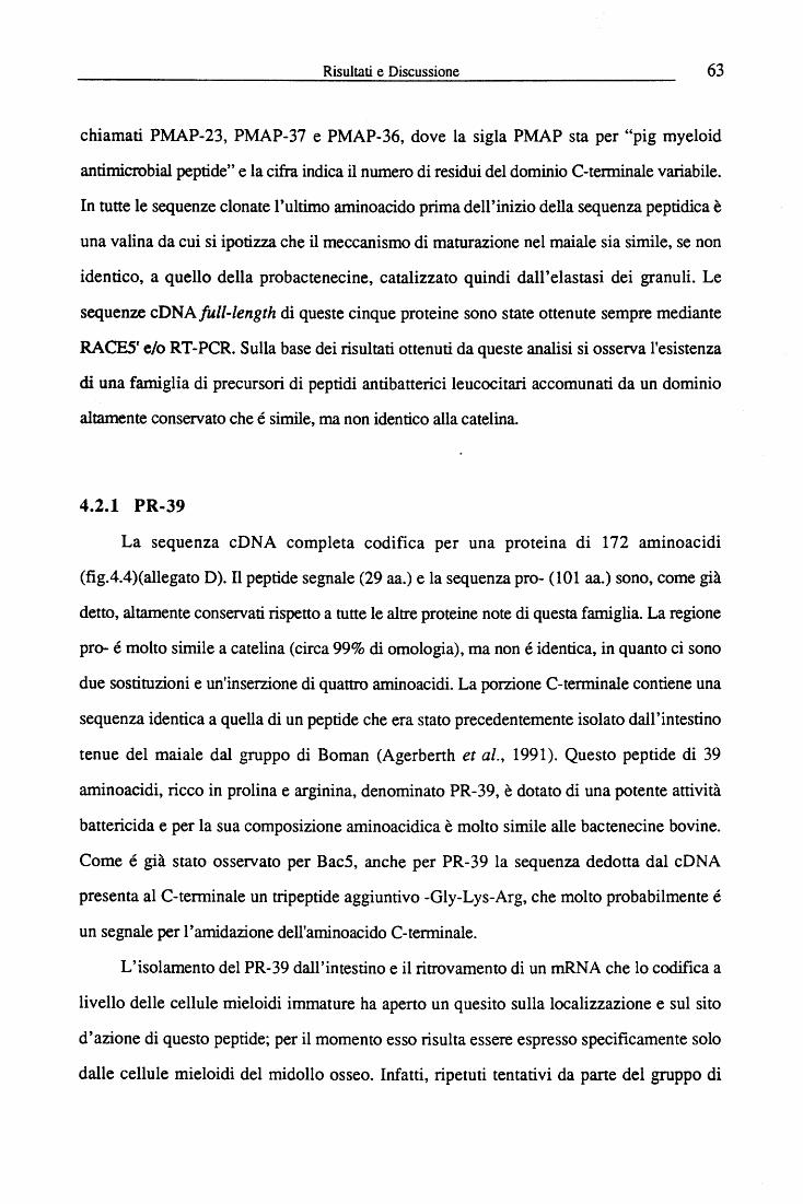
Risultati e Discussione 63
chiamati PMAP-23, PMAP-37 e PMAP-36, dove la sigla PMAP sta per "pig myeloid
antimicrobial peptide" e la cifra indica il numero di residui del dominio C-terminale variabile.
In tutte le sequenze clonate l'ultimo aminoacido prima dell'inizio della sequenza peptidica è
una valina da cui si ipotizza che il meccanismo di maturazione nel maiale sia simile, se non
identico, a quello della probactenecine, catalizzato quindi dall'elastasi dei granuli. Le
sequenze cDNAfull-length di queste cinque proteine sono state ottenute sempre mediante
RACE5' e/o RT-PCR. Sulla base dei risultati ottenuti da queste analisi si osserva l'esistenza
di una famiglia di precursori di peptidi antibatterici leucocitari accomunati da un dominio
altamente conservato che é simile, ma non identico alla catelina.
4.2.1 PR-39
La sequenza cDNA completa codifica per una proteina di 172 aminoacidi
(fig.4.4)(allegato D). Il peptide segnale (29 aa.) e la sequenza pro- (101 aa.) sono, come già
detto, altamente conservati rispetto a tutte le altre proteine note di questa famiglia. La regione
pro- é molto simile a catelina (circa 99% di omologia), ma non é identica, in quanto ci sono
due sostituzioni e un'inserzione di quattro aminoacidi. La porzione C-terminale contiene una
sequenza identica a quella di un peptide che era stato precedentemente isolato dall'intestino
tenue del maiale dal gruppo di Boman (Agerberth et al., 1991). Questo peptide di 39
aminoacidi, ricco in prolina e arginina, denominato PR-39, è dotato di una potente attività
battericida e per la sua composizione aminoacidica è molto simile alle bactenecine bovine.
Come é già stato osservato per Bac5, anche per PR-39 la sequenza dedotta dal cDNA
presenta al C-terminale un tripeptide aggiuntivo -Gly-Lys-Arg, che molto probabilmente é
un segnale per l'amidazione dell'aminoacido C-terminale.
L'isolamento del PR-39 dall'intestino e il ritrovamento di un mRNA che lo codifica a
livello delle cellule mieloidi immature ha aperto un quesito sulla localizzazione e sul sito
d'azione di questo peptide; per il momento esso risulta essere espresso specificamente solo
dalle cellule mieloidi del midollo osseo. Infatti, ripetuti tentativi da parte del gruppo di
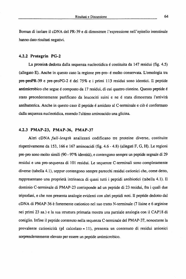
Risultati e Discussione 64
Boman di isolare il cONA del PR-39 e di dimostrare l'espressione nell'epitelio intestinale
hanno dato risultati negativi.
4.2.2 Protegrin PG-2
La proteina dedotta dalla sequenza nucleotidica é costituita da 147 residui (fig. 4.5)
(allegato E). Anche in questo caso la regione pre-pro- é molto conservata. L'omologia tra
pre-proPR-39 e pre-proPG-2 é del 75% e i primi 113 residui sono identici. Il peptide
antimicrobico che segue é composto da 17 residui, di cui quattro cisteine. Questo peptide é
stato precedentemente purificato da leucociti suini e ne é stata dimostrata l'attività
antibatterica. Anche in questo caso il peptide é amidato al C-terminale e ciò é confermato
dalla sequenza nucleotidica, essendo l'ultimo aminoacido una glicina
4.2.3 PMAP-23, PMAP-36, PMAP-37
Altri cDNA full-length analizzati codificano tre proteine diverse, costituite
rispettivamente da 153, 166 e 167 aminoacidi (fig. 4.6 - 4.8) (allegati F, G, H). Le regioni
pre-pro sono molto simili (90- 97% identità), e contengono sempre un peptide segnale di 29
residui e una pro-sequenza di 101 residui. Le sequenze C-terminali sono completamente
diverse (tabella 4.1), seppur contengono sempre parecchi residui cationici che, come detto,
rappresentano una proprietà intrinseca di quasi tutti i peptidi antibiotici (tabella 4.1). Il
dominio C-terminale di PMAP-23 corrisponde ad un peptide di 23 residui, fra i quali due
triptofani, e che non presenta analogie evidenti con altri peptidi noti. Il peptide dedotto dal
cDNA di PMAP-36 è fortemente cationico nel suo tratto N-terminale (7lisine e 6 arginine
nei primi 23 aa.) e la sua struttura primaria mostra una parziale analogia con il CAP18 di
coniglio. Infine il peptide contenuto nella sequenza C-terminale del PMAP-37, nonostante la
prevalente cationicità (pl calcolato= 11), presenta un contenuto di residui anionici
sorprendentemente elevato per essere un peptide antimicrobico.
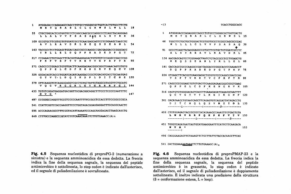
l ATGGAGACCCAGAGAGCCAGCCTGTGCCTGGGGCGCTGGTCACTGTGGCTTCTG H E T Q R A S L C L G R W S L W L L
55 CTGCTGGCACTCGTGGTGCCCTCGGCCAGCGCCCAGGCCCTCAGCTACAGGGAG L L A L V V P S A S AfQ A L S Y R E
109 GCCGTGCTTCGTGCTGTGGATCGCCTCAACGAGCAGTCCTCGGAAGCTAATCTC A V L R A V D R L N E Q S S E A N L
163 TACCGCCTCCTGGAGCTGGACCAGCCGCCCAAGGCCGACGAGGACCCGGGCACC Y R L L E L D Q P P K A D E D P G T
211 CCGAAACCTGTGAGCTTCACGGTGAAGGAGACTGTGTGTCCCAGGCCGACCCGG P K P V S F T V K E T V C P R P T R
271 CAGCCCCCGGAGCTGTGTGACTTCAAGGAGAACGGGCGGGTGAAACAGTGTGTG 0 P P E L C D F K E N G R V K Q C V
325 GGGACAGTCACCCTGGATCAGATCAAGGACCCGCTCGACATCACCTGCAATGAG G T V T• L D Q I K D P L D I T C N E
379 GTTCAAGGTGTCAGGGGAGGTCGCCTGTGCTATTGTAGGCGTAGGTTCTGCATC V 0 G V R G G R L C Y C R R R F C I
433 TGTGTCGGATGAGGATGACGGTTGCGACGGCAGGCTTTCCCTCCCCCAATTTTC C V ·G *
481 CCGGGGCCAGGTTTCCGTCCCCCAATTTTTCCGCCTCCACCTTTCCGGCCCGCA
541 CCATTCGGTCCACCAAGGTTCCCTGGTAGACGGAGGAGGATTTGCAGGCAACTC
595 ACCCAGAAGGCCTTTCGGTACATTAAAATCCCAGCAAGGAGACCTAAGCATCTG
649 CTTTGCCCAGGCCCGCATCTGTCAAATAAATTCTTGTGAAACC(A)n
18
36
54
72
90
108
120
144
147
Fig. 4.5 Sequenza nucleotidica di preproPG-2 (numerazione a sinistra) e la sequenza amminoacidica da essa dedotta. La freccia indica la fine della sequenza segnale, la sequenza del peptide antimicrobico è sottolineata, lo stop codon è indicato dall'asterisco, ed il segnale di poliadenilazione è sovralineato.
-13 TCACCTGGGCACC
1 ATGGAGACCCAGAGGGCCAGCCTGTGCCTGGGGCGCTGGTCACTG M E T o R A s L c L G R w s L 15
46 TGGCTTCTGCTGCTGGGACTCGTGGTGCCCTCGGCCAGCGCCCAG w L L L L G L v v p s A s A o 30
~ 91 GCCCTCAGCTACAGGGAGGCCGTGCTTCGTGCTGTGGATCGCCTC
A L s 'f. R E A v L R A v D R L 45
136 AACGAGCAGTCCTCGGAAGCTAATCTCTACCGCCTCCTGGAGCTG N E o s s E A N L 'f. R L L E L 60
181 GACCAGCCGCCCAAGGCCGACGAGGACCCGGGCACCCCAAAACCT D o p p K A D E D p G T p K p 75
226 GTGAGCTTCACGGTGAAGGAGACTGTGTGTCCCAGGCCGACCCGG v s F T v K E T v c p R p T R 90
271 CAGCCCCCGGAGCTGTGTGACTTCAAGGAGAACGGGCGGGTGAAG o p p E L c D F K E N G R v K 105
316 CAGTGTGTGGGGACAGTCACCTTGAAAGAGATCAGAGGCAACTTT o c v G T v T L K E I R G N F 120
361 GACATAACCTGTAATCAGCTTCAGAGTGTCAGGATTATAGACCTG D I T c N Q L o s v R I I D L 135 --------L------------1----------B---------~--
406 CTGTGGAGAGTACGTCGGCCACAGAAACCCAAATTTGTGACTGTA L w R v R R p Q lt p lt r v T v 150
---------1-------L-----------r--------B------451 TGGGTCAGATAATTAGTGCATGAAGGAATTTCATTCTTCAAGACA
" v R * 153 --------1 496 CAGGGAAGAGTTCTAAAGTTCTGCTTATTCTACCACAAGCTTCAG
541 GACTGG~TTCTTGTGAAAGC(A)n
Fig. 4.6 Sequenza nucleotidica di preproPMAP-23 e la sequenza amminoacidica da essa dedotta. La freccia indica la fine della sequenza segnale, la sequenza del peptide antimicrobico è in grassetto, lo stop codon è indicato dall'asterisco, ed il segnale di poliadenilazione è doppiamente sottolineato. È inoltre indicata una predizione della struttura (B = conformazione estesa, L = loop ).
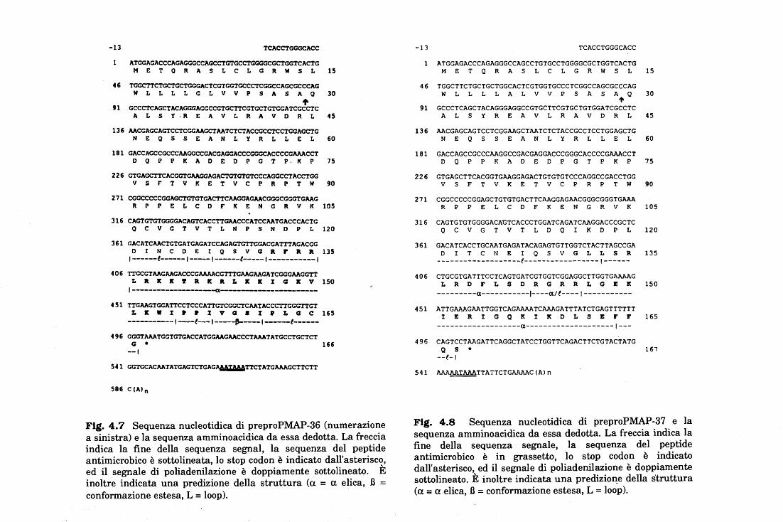
-13 TCACC'l'GGGCACC
l ATGGACACCCACAGGCCCACCCTGTGCCTCGCGCGC'l'GGTCACTG M E T Q R A S L C L C R W S L 15
46 TGGCTTCTGCTGCTCGCACTCGTCGTGCCCTCGGCCACCGCCCAC W L L L L C L V V P S A S A Q 30 • 91 CCCCTCAGCTACAGGGAGGCCGTGCTTCGTGCTGTGGATCGCCTC A L S Y · R E A V L R A V D R L 45
136 AACGAGCAGTCCTCGGAAGCTAATCTCTACCGCCTCCTGGAGCTG N E Q S S E A N L Y R L L E L 60
181 GACCAGCCGCCCAAGGCCGACGAGGACCCGGGCACCCCGAAACCT D Q P P K A D E D P G T P. K P 75
226 GTGAGCTTCACGGTGAAGGAGACTGTGTGTCCCAGGCCTACCTGG V S r T V K E T V C P R P T W 90
271 CGGCCCCCGGAGC'l'GTGTGACTTCAAGGAGAACGGGCGGGTGAAG R P P E L C D F K E N G R V K 105
316 CAGTGTGTCGGGACAGTCACCTTGAACCCATCCAATGACCCACTG Q C V G T V T L N P S N D P L 120
361 GACATCAACTGTGATGAGATCCACAGTGTTGGACGATTTAGACGG D I N C D E I Q S V Q R F R R 135 l------t------1-----1------t-----1-----------l
406 TTGCGTAAGAAGACCCGAAAACGTTTGAAGAAGATCGGCAAGGTT L R K K ~ R K R L K K I Q K V 150
1-------------------a---------------451 TTCAAGTGGATTCCTCCCATTGTCGGCTCAATACCCTTGGGTTGT
L K W I P • I V Q 8 I P L Q C 165 -----------1---t---1-----~---1-----t------
496 GGGTAAATGGTGTGACCATCGAAGAACCCTAAATATGCCTGCTCT Q • 166
--l
5.1 GGTCCACAATATGAGTCTCAC~TTCTATGAAAGCTTCTT
586 C(A)n
Fig. 4. 7 Sequenza nucleotidica di preproPMAP-36 (numerazione a sinistra) e la sequenza amminoacidica da essa dedotta. La freccia indica la fine della sequenza segnai, la sequenza del peptide antimicrobico è sottolineata, lo stop codon è indicato dall'asterisco, ed il segnale di poliadenilazione è doppiamente sottolineato. È inoltre indicata una predizione della struttura (a = a elica, J3 = conformazione estesa, L= loop).
-13 TCACCTGGGCACC
ATGGAGACCCAGAGGGCCAGCCTGTGCCTGGGGCGCTGGTCACTG M E T Q R A S L C L G R W S L 15
46 TGGCTTCTGCTGCTGGCACTCGTGGTGCCCTCGGCCAGCGCCCAG W L L L L A L V V P S A S A Q 30
't 91 GCCCTCAGCTACAGGGAGGCCGTGCTTCGTGCTGTGGATCGCCTC
A L S Y R E A V L R A V D R L 45
136 AACGAGCAGTCCTCGGAAGCTAATCTCTACCGCCTCCTGGAGCTG N E Q S S E A N L Y R L L E L 60
181 GACCAGCCGCCCAAGGCCGACGAGGACCCGGGCACCCCGAAACCT D Q P P K A D E D P G T P K P 75
226 GTGAGCTTCACGGTGAAGGAGACTGTGTGTCCCAGGCCGACCTGG V S F T V K E T V C P R P T W 90
271 CGGCCCCCGGAGCTGTGTGACTTCAAGGAGAACGGGCGGGTGAAA R P P E L C D F K E N G R V K 105
316 CAGTGTGTGGGGACA~TCACCCTGGATCAGATCAAGGACCCGCTC Q C V G T V T L D Q I K D P L 120
361 GACATCACCTGCAATGAGATACAGAGTGTTGGTCTACTTAGCCGA D I T C N E I Q S V G L L S R 135
-------------------e-----------------1------406 CTGCGTGATTTCCTCAGTGATCGTGGTCGGAGGCTTGGTGAAAAG
L R D F L S D R G R R L G E K 150 ---------a-----------J----a/f----1-----------
451 ATTGAAAGAATTGGTCAGAAAATCAAAGATTTATCTGAGTTTTTT I E R I G Q K I K D L S E F F 165
-------------------a--------------------1---496 CAGTCCTAAGATTCAGGCTATCCTGGTTCAGACTTCTGTACTATG
Q s • 167 --f-1
541 AAAAAIAAATTATTCTGAAAAC(Ain
Fig. 4.8 Sequenza nucleotidica di preproPMAP-37 e la sequenza amminoacidica da essa dedotta. La freccia indica la fine della sequenza segnale, la sequenza del peptide antimicrobico è in grassetto, lo stop codon è indicato dall'asterisco, ed il segnale di poliadenilazione è doppiamente sottolineato. È inoltre indicata una predizio11:e della struttura (a= a elica, J3 =conformazione estesa, L= loop).
Risultati e Discussione 67
Un'altra peculiarità di questo peptide di mammifero è data dalla sorprendente omologia (67-
53% identità) che si osserva tra una parte centrale della sequenza del peptide e il tratto N-
terminale delle cecropine di insetto (cecropina B'di D. melanogaster e cecropina A di H.
cecropia) (fig. 3, allegato H).
Studi di predizione di struttura secondaria, effettuati mediante il programma PhD,
indicano che questi peptidi possono assumere ~onformazioni antipatiche. In particolare
PMAP-23 sembra adottare una struttura a forcina ("hairpin") con un "loop" a metà sequenza
in corrispondenza di due proline (fig. 4.6). Le predizioni strutturali degli altri due peptidi
indicano una forte tendenza ad assumere conformazioni ad a-elica: nel PMAP-36 l'elica si
estende dal residuo 2 al20 (fig. 4.7), mentre nel PMAI?-37 si distinguono due eliche dal3 al
12 e dal 17 al 34 con il tratto intermedio che ha equiprobabilità di formare un "loop" o di
strutturarsi ad a-elica (fig. 4.8). Inoltre, visualizzando le eliche secondo una proiezione a
ruota elicale (helical-wheel), si nota una netta separazione dei residui carichi e idrofobici,
indicativa di una struttura anfipatica (fig. 4.9).
4.2.4 Proprietà dei peptidi sintetici PMAP-23, PMAP-36(1-20) e PMAP-37
In generale, tutte queste proprietà suggerivano che i peptidi corrispondenti alle
sequenze dedotte dai cD NA esercitassero un'attività antibiotica. Per verificare questa ipotesi
sono stati sintetizzati chimicamente i tre peptidi: PMAP-23, PMAP-37 e il frammento 1-20 di
PMAP-36 (per i dettagli delle sintesi vedi allegati F, G, H ed M). Un primo controllo dei
peptidi sintetizzati è stato effettuato mediante analisi degli spettri di dicroismo circolare,
ottenuti sia in ambiente acquoso che idrofobico per aggiunte successive di trifluoroetanolo
(fig. 3, allegato I). Lo spettro del PMAP-23 indica che il peptide in ambiente acquoso ha una
struttura disordinata e che la presenza di trifl uoroetanolo al 30% promuove una
considerevole variazione conformazionale. L'interpretazione di questo spettro è piuttosto
difficile vista la presenza di due triptofani che interferiscono con lo spettro conformazionale.
Il risultato che si evince da questa analisi indica soprattutto l'assenza di una conformazione
Risultati e Discussione 68
omogenea, dovuta forse al ripiegamento imposto dalle proline. Anche per PMAP-36(1-20) e
PMAP-37 si osserva una variazione strutturale per l'aggiunta di trifluoroetanolo che indica la
prevalenza di una conformazione ad a-elica .
L'attività battericida dei peptidi sintetici é stata testata sia contro batteri Gram negativi
(Escherichia coli, Sa/monella typhimurium, Pseudomonas aeruginosa) che Gram positivi
(Bacillw megaterium e Staphilococcw aureus.). I risultati di questi lavori sono stati ottenuti
da altri ricercatori nel nostro laboratorio ed hanno dimostrato che, seppure i peptidi sono tutti
attivi contro batteri Gram positivi e Gram negativi, la suscettibilità dei batteri varia molto da
peptide a peptide (tabella I, allegato I). In generale, PMAP-23 è ugualmente attivo contro
entrambi i gruppi di peptidi, mostrando minime concentrazioni inibenti (MIC) in un
intervallo compreso tra 2 J.J.M (E. coli e B. megaterium) e 16 J.!M (P. aeruginosa). Il
ventimero corrispondente alla sequenza N-terminale di PMAP-36 è attivo contro specie
Gram positive come Bacillus megaterium e Staphilococcus aureus a valori di MIC pari a 3-
6 ~' mentre inibisce scarsamente la crescita di Sa/monella typhimurium, un batterio Gram
negativo che è in genere molto suscettibile all'azione di altri peptidi antimicrobici. PMAP-37,
viceversa, è molto attivo nei confronti dei batteri Gram negativi (MIC di 1-4 J.!M) e poco
efficace nell'inattivazione di due diversi ceppi di Staphilococcus aureus ..
La natura anfipatica dei PMAP suggerisce che la loro attività sia subordinata alla
permeabilizzazione delle membrane batteriche. Infatti, esperimenti di permeabilizzazione
delle membrane (per la metodica vedi allegato M) hanno mostrato che PMAP-23 e -37 sono
attivi a concentrazioni molto basse (0.2-1 J.J.M), mentre PMAP-36(1-20) richiede
concentrazioni circa cinquanta volte superiori (fig. 4, allegato I). Dei tre peptidi, infine,
PMAP-37 mostra una certa capacità di lisare gli eritrociti umani a concentrazioni di 50 J.!M.
Questo valore è decisamente elevato se confrontato con le concentrazioni usare da peptidi
altamente emolitici come la mellitina, ma è comunque ragguardevole nei confronti di altri
peptidi antibiotici.
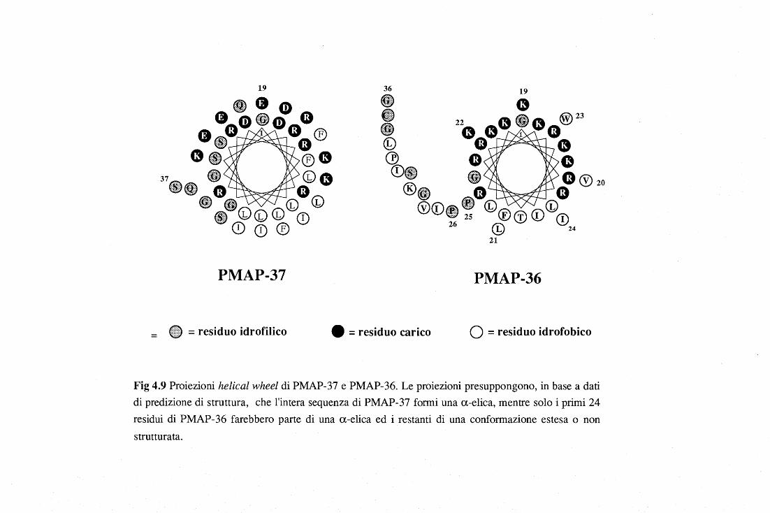
19
PMAP-37 PMAP-36
= @9 = residuo idrofilico e = residuo carico Q = residuo idrofobico
Fig 4.9 Proiezioni helical wheel di PMAP-37 e PMAP-36. Le proiezioni presuppongono, in base a dati di predizione di struttura, che l'intera sequenza di PMAP-37 formi una a-elica, mentre solo i primi 24 residui di PMAP-36 farebbero parte di una a-elica ed i restanti di una conformazione estesa o non strutturata.
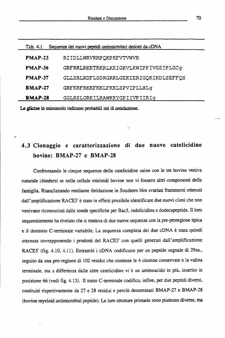
Risuitlti e Discussione 70
T ab. 4.1 Sequenze dei nuovi peptidi antimicrobici dedotti da cD NA
PMAP-23 RIIDLLWRVRRPQKPKFVTVWVR
PMAP-36 GRFRRLRKKTRKRLKKIGKVLKWIPP IVGS IPLGCg
PMAP-37 GLLSRLRDFLSDRGRRLGEKIERIGQKIKDLSEFFQS
BMAP-27 GRFKRFRKKFKKLFKKLSPVIPLLHLg
BMAP-28 GGLRSLGRKILRAWKKYGPIIVPIIRig
Le glicine in minuscolo indicano probabili siti di amidazione.
4.3 Clonaggio e caratterizzazione di due nuove catelicidine
bovine: BMAP-27 e BMAP-28
Confrontando le cinque sequenze delle catelicidine suine con le tre bovine veniva
naturale chiedersi se nelle cellule mieloidi bovine non vi fossero altri componenti della
famiglia. Rianalizzando mediante ibridazione in Southern blot svariati frammenti ottenuti
dall'amplificazione RACE3' è stato in effetti possibile identificare due nuovi cloni che non
venivano riconosciuti dalle sonde specifiche per Bac5, indolicidina e dodecapepride. Il loro
sequenziamento ha rivelato che si trattava di due nuove sequenze con la pre-proregione tipica
e il dominio C-terminale variabile. La sequenza completa dei due cDNA è stata quindi
ottenuta sovrapponendo i prodotti del RACE3' con quelli generati dall'amplificazione
RACE5' (fig. 4.10, 4.11). Entrambi i cDNA codificano per un pepride segnale di 29aa.,
seguito da una pro-regione di 102 residui che contiene le 4 cisteine conservate e la valina
terminale, ma a differenza dalle altre catelicidine vi è un aminoacido in più, inserito in
posizione 66 (vedi fig. 4.13). ll tratto C-terminale codifica, infine, per due peptidi diversi,
costituiti rispettivamente da 27 e 28 residui e perciò denominati BMAP-27 e BMAP-28
(bovine myeloid antimicrobial pepride). Le loro strutture primarie sono piuttosto diverse, ma
Risultati e Discussione 71
entrambe hanno un tratto N-terminale piuttosto cationico e una coda C-terminale più
idrofobica, e con una glicina come ultimo aminoacido (tabella 4.1). Quest'ultimo residuo
viene probabilmente rimosso durante la maturazione dei peptidi, la cui struttura finale
risulterebbe quindi composta da 26 e 27 residui con il C-terminale amidato.
4.3.1 Proprietà dei peptidi sintetici: BMAP-27 e BMAP-28
I peptidi corrispondenti alle due sequenze cationiche sono stati sintetizzati
chimicamente col metodo in fase solida Fmoc e purificati mediante RP-HPLC. Questo
lavoro, eseguito da altri ricercatori del nostro laboratorio, ha consentito di analizzare la
struttura secondaria e l'attività antimicrobica di BMAP-27 e BMAP-28. Studi di previsione
di struttura, eseguiti come descritto precedentemente per i PMAP, indicano che entrambi i
peptidi possono assumere una conformazione ad a-elica anfipatica, che si interrompe in
corrispondenza del residuo in posizione 19 corrispondente ad una prolina, formando quindi
una specie di "coda" idrofobica, non strutturata (fig. 4.12). Spettri di dicroismo circolare
mostrano che entrambi i peptidi hanno in soluzione acquosa una struttura "random coil", che
assume caratteristiche di un'a-elica in seguito all'aggiunta di trifluoroetanolo.
L'attività antibatterica dei peptidi sintetici è stata valutata mediante saggi di minima
concentrazione inibente (MIC). In tabella 4.2 sono riportati i dati ottenuti, da cui si deduce
che BMAP-27 e BMAP-28 sono entrambi efficacemente attivi contro batteri Gram positivi
(S. marcescens, B. megaterium, S aureus ), e Gram negativi (E.coli, S. typhimurium e P.
areruginosa), nonché contro funghi quali C. albicans, C. neoformans e R. rubra. Oltre ad
essere batteriostatici, questi due peptidi sono anche battericidi, in quanto da esperimenti di
minima concentrazione battericida (MBC) si ottengono risultati analoghi. La loro attività
antimicrobica è parzialmente legata alla capacità di permeabilizzare la membrana batterica E'
stato infatti dimostrato che a concentrazioni di 0.2 e 0.5 J..Lg/ml sono in grado di
permeabilizzare rapidamente la membrana interna di E.coli (per i dettagli tecnici vedi allegato
M).
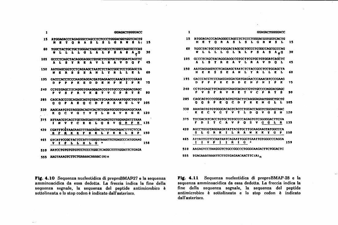
1
15 AtGGAGACCCAGAGGGCCAGCCtC1'CCCTGGGACGOTtCACtG ·a B T Q R A S L S L G R W l L!
IO !GGCTAC!GC!GCTGGGACT~CCAG W L L L L G L A L P l A l AfQ
105 GCCCtCAGCTACAGGGAGGCCOTGCttcGTGCTGTGGAtCAGT'JC A L S Y R E A V L R A V D Q F
t 50 llATGAGCGGTCCTCAGA.AGCtAATCtCTACCGGC1CCTGGAGCt A N l R S S E A N L Y R L L E L
t 95 GACCCACCTCCCAAGGAGGACGAtGAGAACCCAMCAtCCCGAA<J D P P P K E D D E N P N l P k
2 40 CCTGTGAGCTTCAGGGTGAAGOAGACCGTGTGCCCCAOGACGAGC P V S F R V K l t V C P ft t S
215 CAGCAGCCCGCAGAGCAGtGTOACttCMGGAAMTGGGCtGGtO
t5
30
45
IO
l5
t O
a Q P l B Q C D F k E N G L V 105
330 AAGCAATGTGtAGGGACAGTCACTCtGGATGCGGtGAAAGGCA.M K Q C V G t V T L D A V k G K 120
315 AtTAAOGTCACCTGCGAGGAGtTGCAGAGtGttGGAAOATttA.M l N V T C E E L a S V G R F k 135
420 CGATttcGTAAGAAGTtTAAGAiActCttTAAGAAACTTTCTCCA R F R K K F K K L F K K L S P 150
415 GTCATTCCGCTACTCCATTTGGGGTAAATTGTGAGCCCATGGAAG V l P L L H L G • 158
510 AATCCTGTGTGTGTCCtCCCTGGCTCAGGCttTTGGATTCTGAGA
555 AAGTAAAGTCTTCTCAMACAAAAC C Al n
Fig. 4.10 Sequenza nucleotidica di preproBMA.Il27 e la sequenza amminoacidica da essa dedotta. La freccia indica la fine della sequenza segnale, la sequenza del peptide antimicrobico è sottolineata e lo stop codon è indicato dall'asterisco.
1 GGAGACTGGGGACC
15 ATGGAGACCCAGAGGGCCAGCCTCTCCCTGGGACGGTGGTCACTG H E T Q R A S L S L G R W S L 15
IO tGGCTACtGCtGCTGCGACTAGCGCtGCCCTCGGCCAGCGCCCAG N L L L L G L "A L P S A S A f Q 30
105 GCCCTCAGCTACAGGGAGGCCGTGCtTCGTGCTGTGGATCAGCTC A L S Y R E A V L R A V D Q L 45
150 AATGAGAAGTCCTCAGAAGCTAATCTCTACCGCCtCCTGGAGCTA N E k S S E A N L Y R L L E L 60
195 GACCCACCTCCCAAGGAGGACGATGAGAACCCAAACATCCCGAAG D P P P K E D O E N P N l P K 15
240 CCTGTGAGCTTCAGGGTGAAGGAGACCGTGTGCCCCAGGACGAGC P V S F R V k E T V C P R T S 90
285 CAGCAGTCCCCGGAGCAGTGTGACTTCAAGGAGAATGGGCTGCTG O Q S P E Q C O F K E N G L L 105
330 AAAGAGTGTGTGGGGACAGTCACCCTGGACCAGGTCGGGAGTAAC K E C V G T V t L D Q V G S N 120
375 TTCGACATCACCTGTGCTGTGCCCCAGAGTGTCGGGGGACTTCGA F D I T C A V P O S V G G L R 135
420 AGCCTGGGlAGGAAGATATTACGTGCTTGGAAGAAGTATGGCCCA S L G R k l L R A N K K Y G P 150
465 ATTATTGTTCCAATAATCAGAATTGGGTGAATTGTGGGCCCAGGA I I V P l l R I G • 159
510 AAGAGTCCTAAGGGTCTGCCGGCCCTGGGGAAGACTTCTGGACTC
555 TGAGAAATAAATTCTTGTGAGAACAACTTC(Ain
Fig. 4.11 Sequenza nucleotidica di preproBMAP-28 e la sequenza amminoacidica da essa dedotta. La freccia indica la fine della sequenza segnale, la sequenza del peptide antimicrobico è sottolineato e lo stop codon è indicato dall'asterisco.
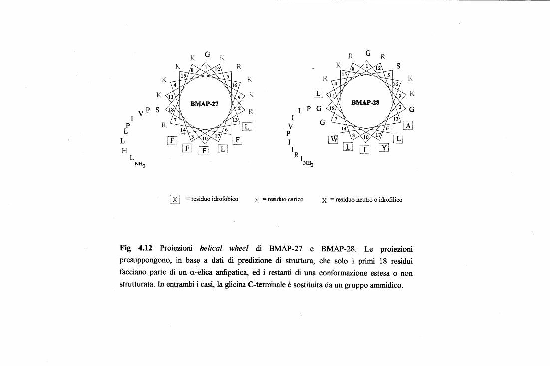
K G K R G R
K K
iL: ~J K
p I p G I
v I p R !Li v G
L l .. _.~ p L
!fl I ,---------, ;---: I i I i LYJ H ~ L!J RI
c_:_::__,
L NH2 NH2
[RJ = residuo idrofobico = residuo carico X = residuo neutro o idrofilico
Fig 4.12 Proi~zioni helical wheel di BMAP-27 e BMAP-28. Le proteztom presuppongono, in base a dati di predizione di struttura, che solo i primi 18 residui facciano parte di un q-elica antipatica, ed i ·restanti di una conformazione estesa o non strutturata. In en~nunbi i cas~ la glicina C-terminale è sostituita da un gruppo ammidico.
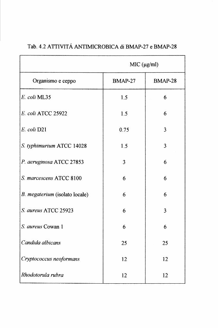
Tab. 4.2 ATTIVITA ANTIMICROBICA di BMAP-27 e BMAP-28
MIC (J.tg/ml)
Organismo e ceppo BMAP-27 BMAP-28
E. coli ML35 1.5 6
E. coli ATCC 25922 1.5 6
E. coli D21 0.75 3
S. typhimurium ATCC 14028 1.5 3
P. aeruginosa ATCC 27853 3 6
S. marcescens ATCC 8100 6 6
B. megaterium (isolato locale) 6 6
S. aureus ATCC 25923 6 3
S. aureus Cowan l 6 6
Candida albicans 25 25
Cryptococcus neoformans 12 12
Rhodotorula rubra 12 12
Risultaù e Discussione 75
Inoltre, entrambi i peptidi risultano avere un'attività emolitica a concentrazioni simili o
di poco superiori a quelle battericide (circa 5-10 Jlg/ml). Questa attività emolitica è piuttosto
inusuale per i peptidi antimicrobici. Nel caso della mellitina è stato dimostrato che la sua
coda idrofobica C-terminale è essenziale per questa attività (Merrifield et al., 1994 ). Si può
quindi ipotizzare che anche nei BMAP la coda C-terminale possa giocare un ruolo
n eli' emolisi.
4.4 Studio a livello proteico di quattro catelicidine bovine
Il lavoro di clonaggio, facilitato dalla strategia RACE3', mi ha permesso di
caratterizzare dieci diversi componenti di questa nuova famiglia di peptidi antimicrobici
(fig.4.13). Le informazioni ottenute dalle sequenze dei cDNA infatti indicano chiaramente
che nelle cellule della serie mieloide dei mammiferi vengono prodotti peptidi antimicrobici
anche molto diversi tra loro, ma che hanno sempre una pre-proregione altamente conseiVata.
Successivamente, infatti, sfruttando questa tecnica e modificando leggermente gli
oligonucleotidi innesco specifici per la proregione, sono state isolate altre catelicidine da
questi e da altri organismi. Nel nostro laboratorio, un collega è riuscito ad isolare la
sequenza codificante per Bac7, dimostrando che anche questo peptide fa parte a pieno titolo
della famiglia delle catelicidine.
La necessità di spiegare meglio le caratteristiche delle proforme mi ha spinto verso lo
studio delle catelicidine a livello proteico. Dopo una serie di tentativi infruttuosi per
esprimerle come proteine ricombinanti, si è deciso di intraprendere la via della purificazione
delle proteine naturali a partire da estratti acidi dei neutrofili bovini o alternativamente dei
loro granuli citoplasmatici. A tal fine sono state messe a punto diverse metodiche,
generalmente rapide e tecnicamente semplici, per ottenere quattro delle sei catelicidine
scoperte nei neutrofili bovini. Il limite è legato alla disponibilità degli anticorpi, in quanto i
vari stadi si purificazione venivano seguiti con tecniche immunoenzimatiche quali Western
blot e dot blot.
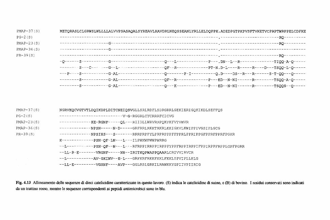
PMAP-37(S) PG-2(S) PMAP-23(S) PMAP-36(S) PR-39(S)
PMAP-37(S) PG-2(S) PMAP-23(S) PJ:V'"AP-36 (S) PR-39(S)
METQRASLCLGRWSLWLLLLA1VVPSASAQALSYREAVLRAVDR1NEQSSEANLYRLLE1DQPPK.ADEDPGTPKPVSFTVKETVCPRPTWRPPELCDFKE ----- ------ -------------- _,-- --- ------- ·---------------- ------------- • ---- -------·--------- -·----RQ----- - ----
--------------------G--------------------------------------------.------------------------RQ-----------------------------G--------------------------------------------.-------------------------------------- ---- ----- ------------------ ---·---------------·--------- ·----- ---- • - -·-------·-·-·-------------- RQ-- ·~-------Q~-----s-----------G----------------------Q---L-------------p---.DN--L--R--------------TIQQ-A-Q-----
--------S---C-------G--L-------------------QF--R-------------PT-N.D-L----R-----R----D---TSQQ-1-Q-------- P-----S--- ---------G---AL--------------- ----Q-------P- I-----·-----Q. D----- DS--R--- R------ S-T-QQ---Q- -----· --------S-----------G-AL----------·---------QF--R--- ----------P--- ED--N-NI------R------- -TSQQ-A-Q--------------5------------G-AL--------------------Q---K--------------P---ED--N-NI------R--------TSQQS--Q-----
NGRVKQCVGTVTLDQlKDPLDITCNEIQSVGLLSRLRDF1SDRGRRLGEKIERIGQKIKDLSEFFQS -------------------------- -V-G- RGGRLCYCRRRFCICVG ---·------·--------KE-RGNF-------QL----RIIDL1WRVRRPQKPKFVTVWVR -- ---- -----------'N P SN-----N- D-----GRFRR1RKKTRKR1KKI GKVLKWI P P IVGS I P1GCG ------------~NPSIHS---5-------RRRPRPPYLPRPRPPPFFPPRLPPRIPPGFPPRFPPRFPGKR
K-------------PSN-QF-1N---1---I1PWKWPWWPWRRG --L-----------PSN-QF--N---L---RFRPPIRRPPIRPPFYPPFRPPIRPPIFPPIRPPFRPP1GPFPGRR --tL-R-E-------VRGNF-----NH--IRITKQPWAPPQAARLCRIVVIRVCR --L-----------AV-GKINV--E-1---GRFKRFRKKFKKLFKKLSPVIPLLHLG --tL-E---------VGSNF----AVP---GG1RSLGRKI1RAWKKYGPIIVPIIRIG
Fig. 4.13 Allineamento d(!,Jle sequenze di dieci catelicidine caratterizzate in questo lavoro. (S) indica le catelicidine di suino, e (B) di bovino. I residui conservati sono indicati da un trattino rosso, mentre le s~uenze corrispondenti ai peptidi antimicrobici sono in blu.
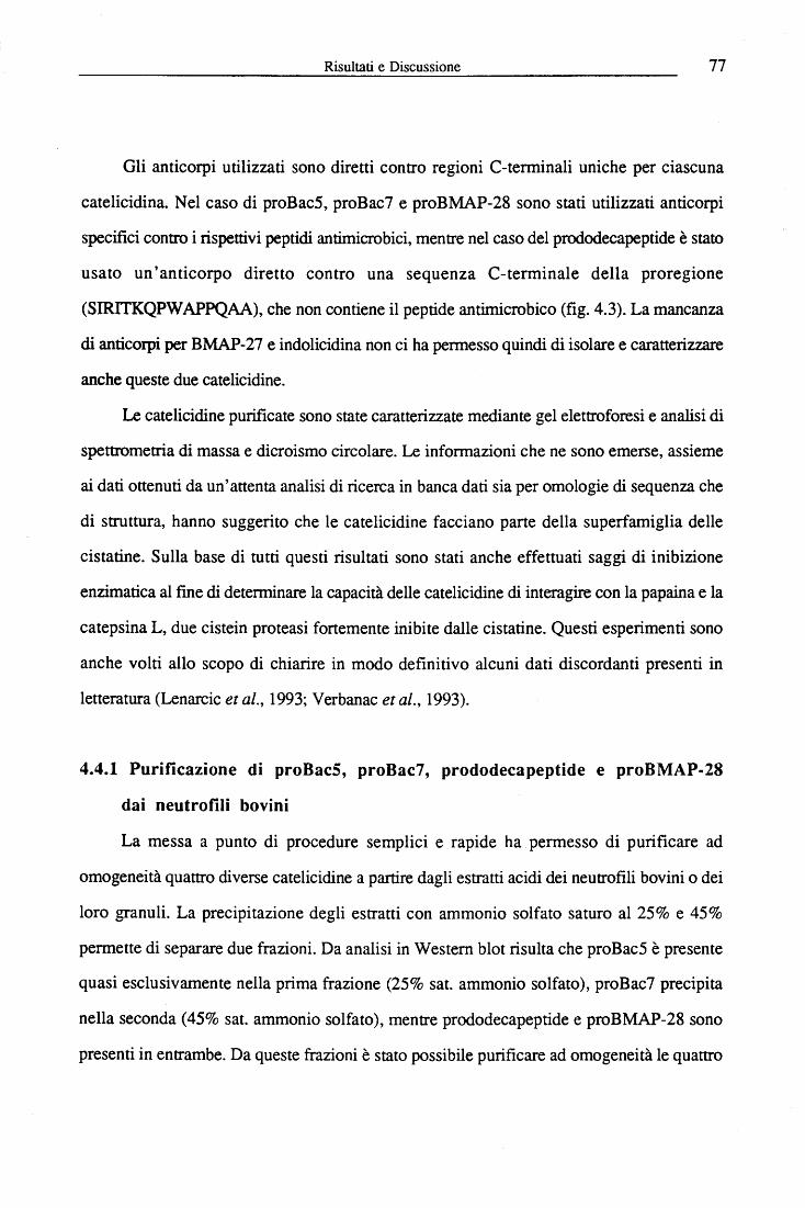
Risultati e Discussione 77
Gli anticorpi utilizzati sono diretti contro regioni C-terminali uniche per ciascuna
catelicidina. Nel caso di proBac5, proBac7 e proBMAP-28 sono stati utilizzati anticorpi
specifici contro i rispettivi peptidi antimicrobici, mentre nel caso del prododecapeptide è stato
usato un'anticorpo diretto contro una sequenza C-terminale della proregione
(SIRITKQPW APPQAA), che non contiene il peptide antimicrobico (fig. 4.3). La mancanza
di anticorpi per BMAP-27 e indolicidina non ci ha permesso quindi di isolare e caratterizzare
anche queste due catelicidine.
Le catelicidine purificate sono state caratterizzate mediante gel elettroforesi e analisi di
spettrometria di massa e dicroismo circolare. Le informazioni che ne sono emerse, assieme
ai dati ottenuti da un'attenta analisi di ricerca in banca dati sia per omologie di sequenza che
di struttura, hanno suggerito che le catelicidine facciano parte della superfamiglia delle
cistatine. Sulla base di tutti questi risultati sono stati anche effettuati saggi di inibizione
enzimatica al fine di determinare la capacità delle catelicidine di interagire con la papaina e la
catepsina L, due cistein proteasi fortemente inibite dalle cistatine. Questi esperimenti sono
anche volti allo scopo di chiarire in modo definitivo alcuni dati discordanti presenti in
letteratura (Lenarcic et al., 1993; Verbanac et al., 1993).
4.4.1 Purificazione di proBacS, proBac7, prododecapeptide e proBMAP-28
dai neutrofili bovini
La messa a punto di procedure semplici e rapide ha permesso di purificare ad
omogeneità quattro diverse catelicidine a partire dagli estratti acidi dei neutrofili bovini o dei
loro granuli. La precipitazione degli estratti con ammonio solfato saturo al 25% e 45%
permette di separare due frazioni. Da analisi in Western blot risulta che proBac5 è presente
quasi esclusivamente nella prima frazione (25% sat. ammonio solfato), proBac7 precipita
nella seconda (45% sat. ammonio solfato), mentre prododecapeptide e proBMAP-28 sono
presenti in entrambe. Da queste frazioni è stato possibile purificare ad omogeneità le quattro
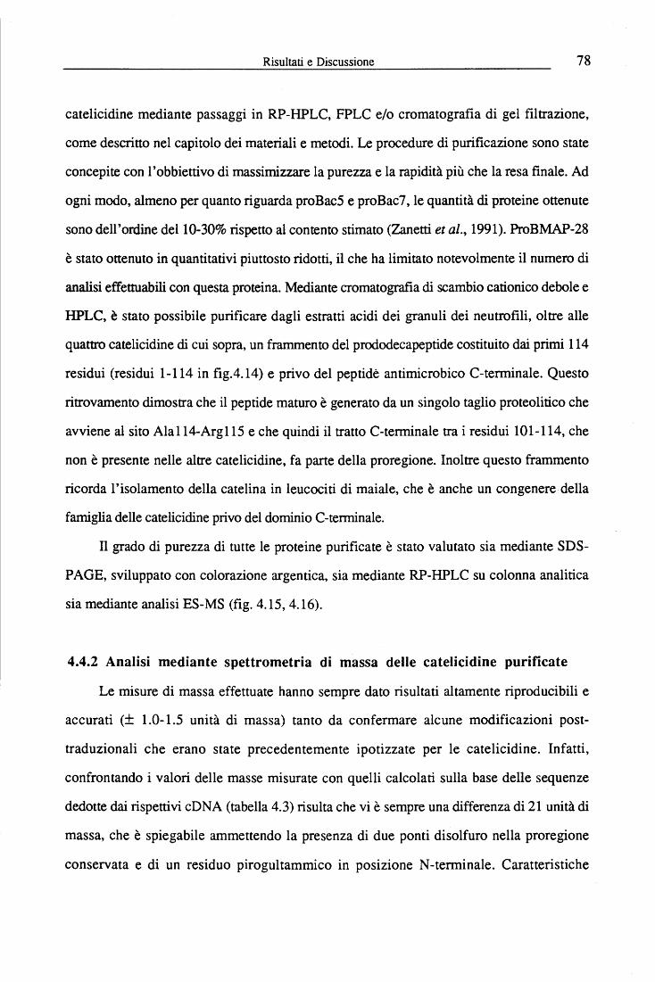
Risultati e Discussione 78
catelicidine mediante passaggi in RP-HPLC, FPLC e/o cromatografia di gel filtrazione,
come descritto nel capitolo dei materiali e metodi. Le procedure di purificazione sono state
concepite con l'obbiettivo di massimizzare la purezza e la rapidità più che la resa finale. Ad
ogni modo, almeno per quanto riguarda proBac5 e proBac7, le quantità di proteine ottenute
sono dell'ordine dell0-30% rispetto al contento stimato (Zanetti et al., 1991). ProBMAP-28
è stato ottenuto in quantitativi piuttosto ridotti, il che ha limitato notevolmente il numero di
analisi effettuabili con questa proteina. Mediante cromatografia di scambio cationico debole e
HPLC, è stato possibile purificare dagli estratti acidi dei granuli dei neutrofili, oltre alle
quattro catelicidine di cui sopra, un frammento del prododecapeptide costituito dai primi 114
residui (residui 1-114 in fig.4.14) e privo del peptide antimicrobico C-terminale. Questo
ritrovamento dimostra che il peptide maturo è generato da un singolo taglio proteolitico che
avviene al sito Ala 114-Arg 115 e che quindi il tratto C-terminale tra i residui l O 1-114, che
non è presente nelle altre catelicidine, fa parte della proregione. Inoltre questo frammento
ricorda l'isolamento della catelina in leucociti di maiale, che è anche un congenere della
famiglia delle catelicidine privo del dominio C-terminale.
ll grado di purezza di tutte le proteine purificate è stato valutato sia mediante SDS-
PAGE, sviluppato con colorazione argentica, sia mediante RP-HPLC su colonna analitica
sia mediante analisi ES-MS (fig. 4.15, 4.16).
4.4.2 Analisi mediante spettrometria di massa delle catelicidine purificate
Le misure di massa effettuate hanno sempre dato risultati altamente riproducibili e
accurati (± 1.0-1.5 unità di massa) tanto da confermare alcune modificazioni post-
traduzionali che erano state precedentemente ipotizzate per le catelicidine. Infatti,
confrontando i valori delle masse misurate con quelli calcolati sulla base delle sequenze
dedotte dai rispettivi cD NA (tabella 4.3) risulta che vi è sempre una differenza di 21 unità di
massa, che è spiegabile ammettendo la presenza di due ponti disolfuro nella proregione
conservata e di un residuo pirogultammico in posizione N -terminale. Caratteristiche
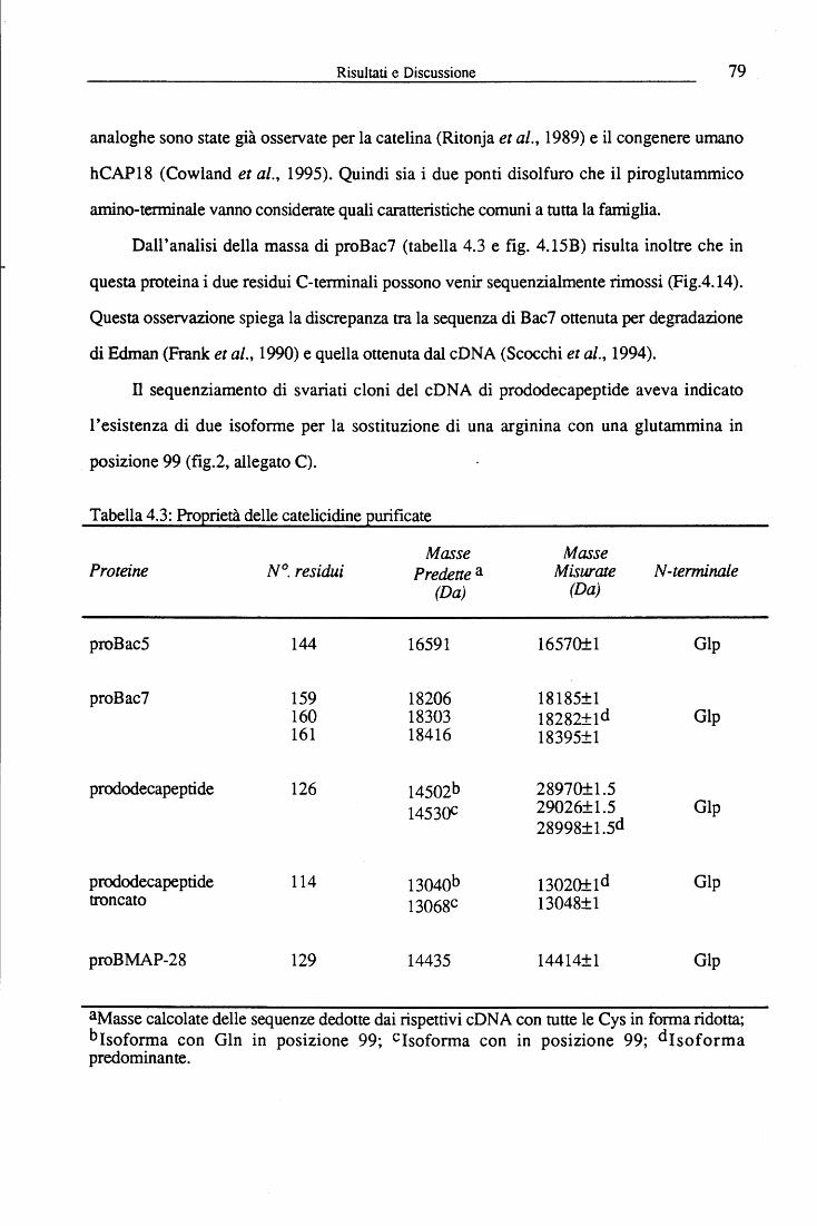
Risultati e Discussione 79
analoghe sono state già osservate per la catelina (Ritonja et al., 1989) e il congenere umano
hCAP18 (Cowland et al., 1995). Quindi sia i due ponti disolfuro che il piroglutammico
amino-terminale vanno considerate quali caratteristiche comuni a tutta la famiglia.
Dall'analisi della massa di proBac7 (tabella 4.3 e fig. 4.15B) risulta inoltre che in
questa proteina i due residui C-terminali possono venir sequenzialmente rimossi (Fig.4.14).
Questa osservazione spiega la discrepanza tra la sequenza di Bac7 ottenuta per degradazione
di Edman (Frank et al., 1990) e quella ottenuta dal cDNA (Scocchi et al., 1994).
Il sequenziamento di svariati cloni del cONA di prododecapeptide aveva indicato
l'esistenza di due iso forme per la sostituzione di una arginina con una glutammina in
posizione 99 (fig.2, allegato C).
Tabella 4.3: Proprietà delle catelicidine purificate
Proteine
proBac5
proBac7
prododecapeptide
prododecapeptide troncato
proBMAP-28
N°. residui
144
159 160 161
126
114
129
Masse Predette a
(Da)
16591
18206 18303 18416
14502b 14530C
13040b 13068C
14435
Masse Miswate N-terminale
(Da)
16570±1 Gip
18185±1 18282±1d Gip 18395±1
28970±1.5 29026±1.5 Gip 28998±1.5d
13020±1d Gip 13048±1
14414±1 Gip
aMasse calcolate delle sequenze dedotte dai rispettivi cD NA con tutte le Cys in forma ridotta; bJsoforma con Gin in posizione 99; CJsoforma con in posizione 99; dJsoforma predominante.
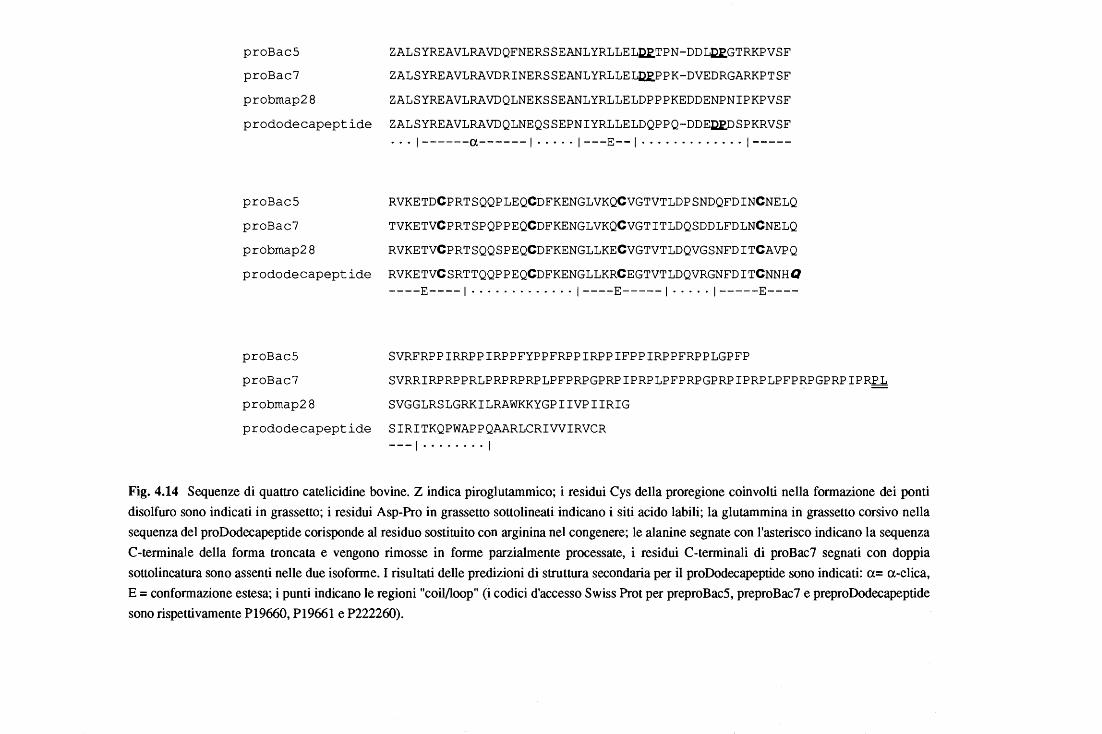
proBacS ZALSYREAVLRAVDQFNERSSEANLYRLLEL~TPN-DDL~GTRKPVSF
proBac7 ZALSYREAVLRAVDRINERSSEANLYRLLEL~PPK-DVEDRGARKPTSF
probmap28 ZALSYREAVLRAVDQLNEKSSEANLYRLLELDPPPKEDDENPNIPKPVSF
prododecapeptide ZALSYREAVLRAVDQLNEQSSEPNIYRLLELDQPPQ-DDE~SPKRVSF
· · · 1------a------1 · ·· ·· 1---E--1 · ·· · ·· · · ····· 1-----
proBacS RVKETDCPRTSQQPLEQCDFKENGLVKQCVGTVTLDPSNDQFDINCNELQ
proBac7 TVKETVCPRTSPQPPEQCDFKENGLVKQCVGTITLDQSDDLFDLNCNELQ
probmap28 RVKETVCPRTSQQSPEQCDFKENGLLKECVGTVTLDQVGSNFDITCAVPQ
prododecapeptide RVKETVCSRTTQQPPEQCDFKENGLLKRCEGTVTLDQVRGNFDITCNNHQ ----E----1 · ·· · · · ····· ·· 1----E-----1 · · · ·· 1-----E----
proBacS SVRFRPPIRRPPIRPPFYPPFRPPIRPPIFPPIRPPFRPPLGPFP
proBac7 SVRRIRPRPPRLPRPRPRPLPFPRPGPRPIPRPLPFPRPGPRPIPRPLPFPRPGPRPIPR~
probmap28 SVGGLRSLGRKILRAWKKYGPIIVPIIRIG
prododecapeptide SIRITKQPWAPPQAARLCRIVVIRVCR ---1 ........ l
Fig. 4.14 Sequenze di quattro catelicidine bovine. Z indica piroglutammico; i residui Cys della proregione coinvolti nella formazione dei ponti disolfuro sono indicati in grassetto; i residui Asp-Pro in grassetto sottolineati indicano i siti acido labili; la glutammina in grassetto corsivo nella sequenza del proDodecapeptide corisponde al residuo sostituito con arginina nel congenere; le alanine segnate con l'asterisco indicano la sequenza C-terminale della forma troncata e vengono rimosse in forme parzialmente processate, i residui C-terminali di proBac7 segnati con doppia sottolineatura sono assenti nelle due isoforme. I risultati delle predizioni di struttura secondaria per il proDodecapeptide sono indicati: a= a-elica, E= conformazione estesa; i punti indicano le regioni "coil/loop" (i codici d'accesso Swiss Prot per preproBac5, preproBac7 e preproDodecapeptide sono rispettivamente P19660, P19661 e P222260).
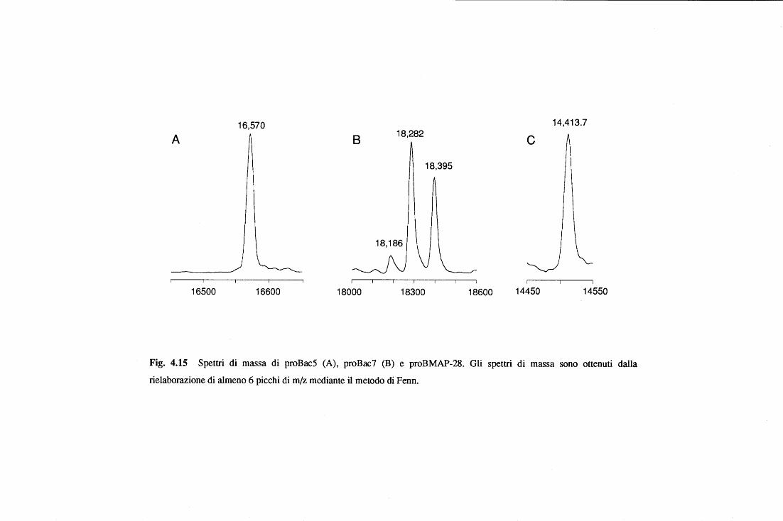
16,570 14,413.7
A 8 18,282 c 18,395
18,186
16500 16600 18000 18300 18600 14450 14550
Fig. 4.15 Spettri di massa di proBac5 (A), proBac7 (B) e proBMAP-28. Gli spettri di massa sono ottenuti dalla
rielaborazione di almeno 6 picchi di m/z mediante il metodo di Fenn.
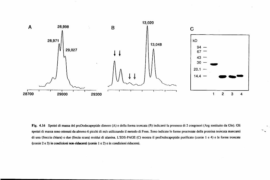
13,020 A 28,998 8 c
28,971 kD 13,048
94 29,027 ! ~ 67
43 30 ...
20.1
! ! 14.4 -----~
28700 29000 29300 1 2 3 4
Fig. 4.16 Spettri di massa del proDodecapeptide dimero (A) e della forma troncata (8) indicanti la presenza di 2 congeneri (Arg sostituito da Gin). Gli
spettri di massa sono ottenuti da almeno 6 picchi di m/z utilizzando il metodo di Fenn. Sono indicate le forme processate della proteina troncata mancanti
di una (freccia chiara) o due (frecia scura) residui di alanina. L'SDS-PAGE (C) mostra il proDodccapeptide purificato (corsie l e 4) e le forme troncate
(corsie 2 e 3) in condizioni non-riducenti (corsie l e 2) e in condizioni riducenti.
....
Risultati e Discussione 83
La spettrometria di massa ha confermato la presenza dell'isomorfismo ed ha inoltre
dimostrando che il prododecapeptide esiste come dimero, formato dalle tre possibili
combinazioni delle isoforme (fig. 4.16A, tabella 4.3). Diverso è invece il comportamento del
prododecapeptide troncato al C-terminale, che pur essendo sempre presente in due isoforme
(fig. 4.16B), non è mai presente come dimero. Questo comportamento è osservabile anche
confrontando il prododecapeptide intero e la forma troncata mediante SDS PAGE in
condizioni riducenti e non riducenti (fig. 4.16C). Ciò dimostra che la dimerizzazione è
dovuta alla formazione di un legame disolfuro e l'assenza di dimero nella forma troncata
indica che il o i ponti disolfuro si formano a livello del dominio antimicrobico C-terminale.
Questo risultato, del tutto inaspettato ci suggerisc·e che la struttura ciclica ipotizzata per
il dodecapeptide (Romeo et al., 1988) debba essere riconsiderata, nonostante questa sia
comunque attiva come antibatterico in quanto un peptide ciclico sintetico è stato prodotto e
risulta essere attivo, mentre l'analogo lineare non lo è (Gallis et al., 1989). I nostri dati
indicano che il peptide nativo potrebbe essere presente almeno in parte come dimero
covalente, che unisce due dodecapeptidi per mezzo di legami disolfuro intramolecolari. Se
ciò fosse vero, questo risulterebbe essere il primo esempio di una simile struttura tra i peptidi
antimicrobici. Sarebbe altresì interessante determinare l'arrangiamento dei ponti disolfuro tra
i due monomeri (parallelo o antiparallelo) e il meccanismo d'azione antibatterica, in relazione
soprattutto alla specie monomerica.
Tutte le quattro catelicidine contengono almeno un legame Asp-Pro nella loro
proregione; in particolare proBac5 contiene tre di questi legami in posizione 32, 40 e 85 della
proforma (fig. 4.16), mentre le altre catelicidine contengono solamente un legame Asp-Pro
in posizione 32 (proBac7 e proBMAP-28) o 40 (prododecapeptide). Questo legame è noto
per essere idrolizzabile in condizioni acide (Landon, 1977) e in genere la suscettibilità
all'idrolisi varia molto a seconda dalla sua disposizione spaziale nella struttura della proteina.
In tutte le preparazioni di proBac5 e proBac7 erano sempre presenti, in misura più o
meno rilevante frammenti a più basso peso molecolare, riconosciuti in Western blot dagli
Risultati e Discussione 84
anticorpi specifici per Bac5 e Bac7, rispettivamente. Questa stessa osservazione era stata
precedentemente riportata anche da Zanetti et al. (1990). Mediante analisi con ES-MS è stato
possibile attribuire l'origine di questi frammenti: nel caso di proBac5 essi sono dovuti
all'idrolisi dei primi due legami Asp-Pro (frammenti di massa 11967 e 12835 Da), mentre
nel caso di proBac7 si forma un frammento (massa 14664 Da), originato dall'idrolisi
dell'unico legame Asp-Pro presente. Analizzando, mediante ES-MS e SDS-PAGE, le
proteine incubate a temperatura ambiente a diverse concentrazioni di 1FA (0.05% a 0.5%)
per tempi più o meno lunghi (da un giorno ad una settimana), si è osservato che il grado di
idrolisi aumenta considerevolmente incrementando l'acidità del mezzo, mentre le proteine
sono perfettamente stabili se incubate per una settimana a temperatura ambiente in condizioni
neutre o leggermente basiche (pH 8). Sorprendentemente, invece, il legame Asp40-Pro41
presente nel prododecapeptide risulta praticamente stabile in condizioni acide, mentre quello
del proBMAP-28 subisce una blanda idrolisi nelle stesse condizioni, anche se non allivello
di proBac5 e proBac7. Infine, il terzo legame Asp85-Pro86 di proBac5, localizzato
all'interno della regione con il ponti disolfuro, sembra essere molto resistente, tanto da non
venir alterato da un trattamento di 16 ore a 37°C in acido formico al 37%. La peculiare
!abilità all'idrolisi acida dimostrata dai legami Asp-Pro che si trovano nella regione N-
terminale di proBac5 e proBac7, suggerisce la presenza di un'organizzazione spazi al e
specifica intorno a questi legami che potrebbe favorire la reazione di idrolisi. Inoltre, nella
regione compresa tra i residui 32 e 44, il livello d'identità tra le quattro catelicidine è solo del
25%, valore relativamente basso se confrontato con il62% che si ha per tutta la proregione
(fig. 4.14). Sembra valida, quindi, l'ipotesi che l'organizzazione spaziale dei legami Asp-
Pro sia diversa nelle diverse catelicidine, con un effetto sul grado di stabilità di tale legame in
ambiente acido.
Risultati e Discussione 85
4.4.3 Disposizione dei ponti disolfuro
Utilizzando delle procedure standard, basate sulla proteolisi limitata e analisi con ES-
MS dei frammenti è stato possibile attribuire la disposizione dei ponti disolfuro nella
proregione. Dopo un sondaggio della suscettibilità delle catelicidine ali 'idrolisi enzimatica, si
è scelto di usare il digerito triptico di proBac7 come campione di analisi. I frammenti generati
dalla digestione, protratta per 16 ore a 37°C, e successivamente separati in RP-HPLC, sono
stati analizzati mediante ES-MS e le loro masse sono state confrontate con i valori calcolati
dalla sequenza nota. Lo spettro di massa di uno dei frammenti isolati corrispondeva ad un
peptide di 2924 Da (fig. 4.17), che è consistente con la massa calcolata per il frammento di
sequenza Glu77-Arg102 di proBac7 (fig. 4.14), in cui è presente un ponte disolfuro. Per
verificare questa ipotesi, il frammento è stato sottoposto ad una serie di procedure per
determinare la presenza del legame -S-S-. Una parte del frammento è stato trattato con acido
performico e, dopo liofilizzazione, è stato analizzato con ES-MS, riscontrando un
incremento di 126 unità di massa, che corrispondono all'aumento di massa dovuto
ali' ossidazione della cistina con formazione di due acidi cisteici ed alla formilazione del
residuo N -terminale. Questo risultato, già da solo indicava la presenza di due cisteine nel
frammento analizzato, ma non permetteva di controllare l'integrità del ponte disolfuro. La
restante parte del frammento è stata quindi divisa in due aliquote, una è delle due stata ridotta
con ditiotreitolo e poi, entrambe, sono state trattate con iodoacetamide. Il prodotto della
reazione è stato infine analizzato mediante ES-MS dopo desalazione con RP-HPLC. Si è
potuto riscontrare che il frammento pretrattato con DTI aveva subito un incremento di massa
di 116 unità, in ottimo accordo con la S-alchilazione di entrambi i gruppi SH liberi, mentre il
frammento non ridotto non ha subito modificazioni, il che ci ha permesso di confermare la
presenza di un ponte disolfuro integro tra la Cys in posizione 78 e la Cys in posizione 95
della sequenza di proBac7. Per deduzione logica l'altro ponte disolfuro si forma tra Cys56 e
Cys67.
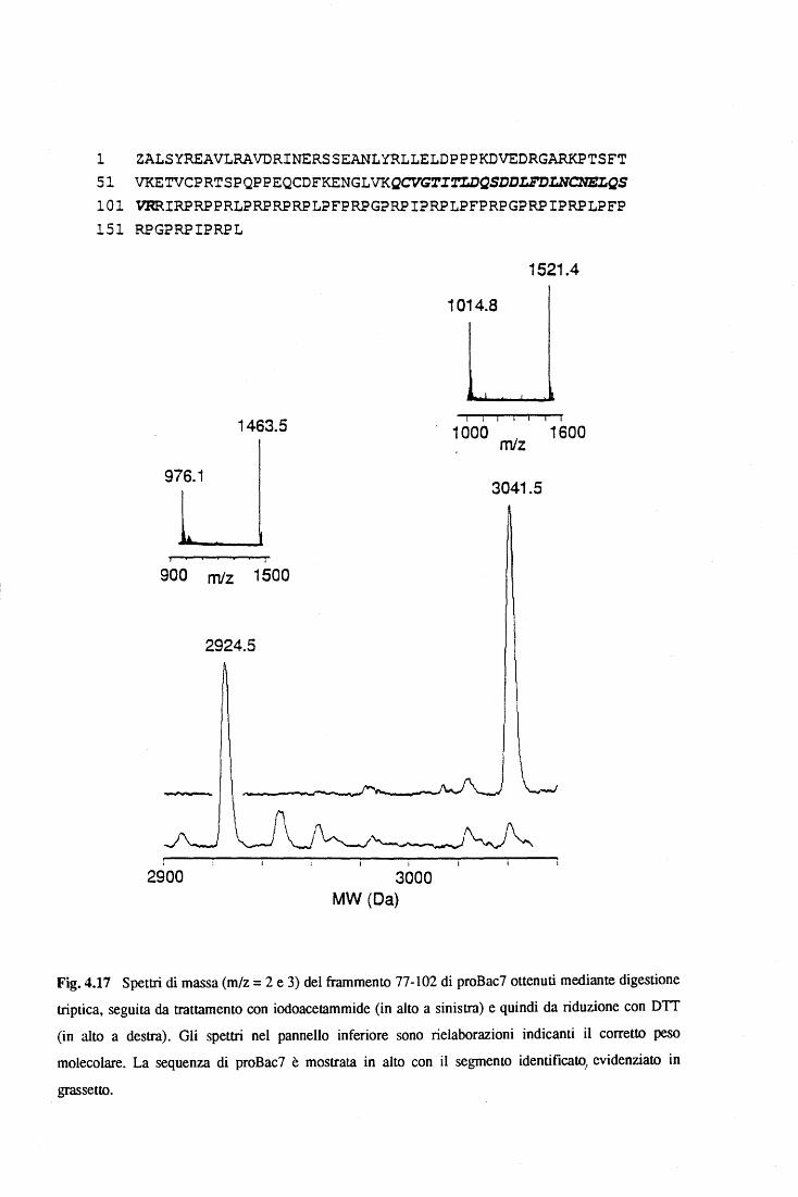
l ZALSYREAVLRAVDRINERSSEANLYRLLELDPPPKDVEDRGARKPTSFT 51 VKETVCPRTSPQPPEQCDFKENGLVKQCVGriT.LDQSDDLFDLNCNELQS 101 VRRIRPRPPRLPRPRPRPLPFPRPGPRPIPRPLPFPRPGPRPIPRPLPFP 151 RPGPRPIPRPL
1463.5
976.1
900 mfz 1500
2924.5
2900 3000 MW (Da)
1521.4
1014.8
l l i i i i i
~000 mlz 1600
3041.5
Fig. 4.17 Spettri di massa (m/z = 2 e 3) del frammento 77-102 di proBac7 ottenuti mediante digestione
triptica, seguita da trattamento con iodoacetammide (in alto a sinistra) e quindi da riduzione con DTI
(in alto a destra). Gli spettri nel pannello inferiore sono rielaborazioni indicanti il corretto peso
molecolare. La sequenza di proBac7 è mostrata in alto con il segmento identificato, evidenziato in
grassetto.
Risultati e Discussione 87
La presenza di un legame cisteico tra le prime due cisteine della prosequenza delle
catelicidine è in accordo anche con le analisi di sequenziamento di Edman effettuate sul
congenere umano (Cowland et al., 1995).
4.4.4 Predizioni di struttura delle catelicidine
Gli spettri di dicroismo circolare della forma troncata del prododecapeptide, di
proBac5 e di proBac7 sono mostrati in fig. 4.18. Nel primo caso, lo spettro può essere
assimilato ad uno spettro della sola proregione conservata, poiché manca del contributo
relativo al peptide C-terminale. Dali' analisi di questo spettro risulta che il contenuto di a-
elica è piuttosto basso, in quanto l' ellitticità misurata a 222 nm suggerisce un contenuto
inferiore al20%. L'elaborazione con il programma CONTIN conferma questa osservazione,
e indica la presenza di una conformazione ad a-elica per circa il 20-30% e di una
conformazione estesa per circa il 50%.
Predizioni della struttura secondaria del prododecapeptide sono state effettuate
utilizzando alcuni algoritmi matematici che tengono conto della sequenza primaria della
proteina. Uno dei metodi considerati più affidabili è quello a profilo neurale usato dal
programma PhD, messo a disposizione dali 'EMBL, che ha un livello di accuratezza del
72.1% per proteine globulari idrosolubili. Oltre a questo, sono stati usati altri due metodi che
utilizzano algoritmi che identificano il "folding" più probabile per una determinata sequenza
proteica: il metodo TOPITS ed il metodo TIIREADER. Le predizioni generate dai tre metodi
sono consistenti con i risultati dei CD, e indicano un basso ( <20%) contenuto di
conformazione in a-elica, e un più alto contenuto di conformazione estesa (30-40% ).
Inoltre, tutti i tre metodi prevedono che il tratto ad a-elica sia localizzato nel segmento N-
terminale (fig. 4.14 ). Risultati simili sono stati ottenuti per le proregioni delle altre tre
catelicidine.
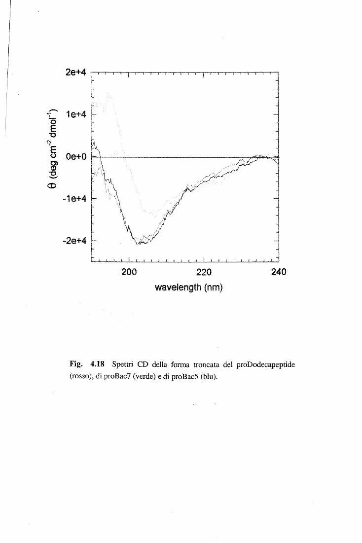
l-t-
1e+4 r
-1e+4
200 i ! l l l !
220 wavelength (nm)
J 240
Fig. 4.18 Spettri CD della forma troncata del proDodecapeptide (rosso), di proBac7 (verde) e di proBacS (blu).
Risultati e Discussione 89
La somiglianza degli spettri CD di proBac5 e proBac7 (fig. 4.18) indica che il
contributo attribuibile alle regione C-terminale , corrispondente a Bac5 e Bac7 (fig. 4.14)), è
confrontabile nelle due proteine. I domini antimicrobici, ricchi in Arg e Pro, portano ad un
aumentato contenuto di conformazione "coil". Assumendo che il contributo della proregione
conservata sia paragonabile tra le tre proteine, si è calcolato l'ipotetico spettro di Bac5 e Bac7
per sottrazione dei dati del prododecapeptide troncato ai dati di proBac5 o proBac7. Questi
spettri sono consistenti con una conformazione non strutturata o di una poliprolina di tipo n (Ronish & Krimm, 197 4 ).
4.4.5 Omologie di sequenza e di struttura delle catelicidine con i chininogeni
Da una ricerca di omologia di sequenza in banca dati (Swissprot) effettuata mediante il
programma BLITZ, a diversi valori di PAM ("point accepted mutation"), risulta che le
proregioni delle catelicidine si possono mettere in relazione ai membri della superfamiglia
delle cistatine. I dati più interessanti sono stati ottenuti con la proregione di Bac5 ad un PAM
di 120, che mostra un buon livello di allineamento, oltre che con le altre catelicidine, anche
con i domini cistatino-simili dei chininogeni. In particolare, si osserva una somiglianza del
50% ed una identità del25% con il primo dominio (residui 23- 132 del chininogeno bovino,
(fig. 4.19), e circa 48% di somiglianza e 20% di identità con il secondo e terzo dominio. Ad
ulteriore supporto di questo dato, si è osservato che la predizione di struttura mediante il
programma TOPITS, basato su elementi di topologia delle proteine, indica come miglior
struttura di riferimento quella della cistatina di pollo (Bode et al.,1988). Le considerazioni
che si possono fare, a questo punto, sono le seguenti: i) il segmento ad a-elica, predetto per
il tratto N-terminale delle catelicidine, si trova in una regione equivalente al segmento ad a-
elica presente nella struttura cristallografica della cistatina di pollo ; ii) i quattro residui
cisteinici della proregione mantengono pressoché la stessa posizione e lo stesso
arrangiamento dei ponti disolfuro della cistatina; iii) i tratti a conformazione estesa che sono
Risultati e Discussione 90
stati predetti nelle catelicidine sono distribuiti in modo analogo a quelli presenti nella
cistatina.
In generale, quindi, le catelicidine dimostrano un'interessante similarità di sequenza e
di struttura con le cistatine, almeno per quanto riguarda lo "scaffold" predetto. Questo
suggerisce la possibilità di analogie anche sul piano funzionale. Le cistatine sono note per
essere potentissimi inibitori endogeni delle catepsine, cistein-proteasi tra le più attive (per
rassegne sul tema vedi Barrett, 1987; Turk & Bode, 1991). Nei chininogeni, solo il secondo
e il terzo dominio cistatino-simile mostrano una potente attività inibitoria, mentre il primo
dominio risulta inattivo (Salvesen et al., 1986). Inoltre, nel nostro laboratorio era stata
precedentemente descritta una moderata inibizione di proBac5 nei confronti della catepsina L
(Verbanac et al., 1993); mentre il gruppo di Turk, che per primo aveva indicato un'attività
analoga per la catelina (Kopitar et al., 1989), ha ritrattato poi questi dati giustificandoli con la
presenza di una stefina contaminante nel preparato usato per i saggi (Lenarcic et al., 1993).
Viste le indicazioni suggerite dalle analogie con le cistatine e i dati contrastanti tra proBac5 e
catelina, abbiamo saggiato tutte le catelicidine purificate per la loro potenziale attività di
inibizione della catepsina L. Sia proBac5 che proBac7 mostrano una certa capacità inibitoria
dell'enzima, ma le costanti di inibizione misurate hanno valori dell'ordine di 0.6-3.0 ~che
sono quindi notevolmente più elevate che per le cistatine (0.1 nM). La possibilità della
presenza di contaminanti è esclusa dal fatto che le proteine non inibiscono la papaina, che è il
bersaglio d'elezione di tutte le cistatine. Un comportamento analogo a proBac5 e proBac7lo
si osserva anche per proBMAP-28 che mostra valori di inibizione (ICso) confrontabili. A
differenza dalle altre, il prododecapeptide e la sua forma troncata sono invece completamente
inattive nei confronti della catepsina L.
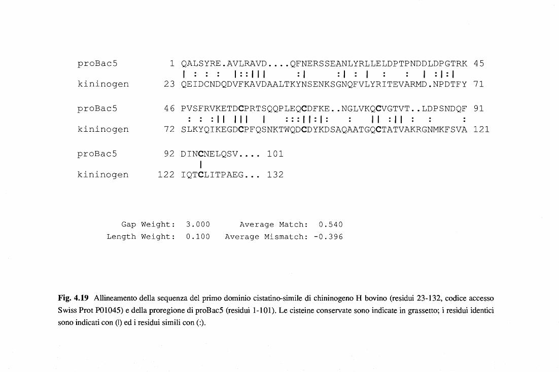
proBac5
kininogen
proBac5
kininogen
proBac5
kininogen
l QALSYRE.AVLRAVD .... QFNERSSEANLYRLLELDPTPNDDLDPGTRK 45 l::: 1::111 :1 :1 l l :1:1
23 QEIDCNDQDVFKAVDAALTKYNSENKSGNQFVLYRITEVARMD.NPDTFY 71
46 PVSFRVKETDCPRTSQQPLEQCDFKE .. NGLVKQCVGTVT .. LDPSNDQF 91 :::Il 111 l :::11:1: Il :Il
72 SLKYQIKEGDCPFQSNKTWQDCDYKDSAQAATGQCTATVAKRGNMKFSVA 121
92 DINCNELQSV .... 101 l
122 IQTCLITPAEG ... 132
Gap Weight: 3.000 Length Weight: 0.100
Average Match: 0.540 Average Mismatch: -0.396
Fig. 4.19 Allineamento della sequenza del primo dominio cistatino-simile di chininogeno H bovino (residui 23-132, codice accesso Swiss Prot P01045) e della proregione di proBac5 (residui 1-101). Le cisteine conservate sono indicate in grassetto; i residui identici sono indicati con (l) ed i residui simili con(:).
Risultati e Discussione 92
Questi dati sono quindi in accordo con i risultati precedenti (Verbanac et al., 1993) in
quanto confermano che alcune, ma non tutte le catelicidine interagiscono con la catepisina L.
Nelle cistatine attive vi sono alcuni residui altamente conservati quale la Gly in posizione 9 e
il tratto QXVXG, che sono strategici per il legame e la conseguente inibizione specifica
dell'enzima. Questi motivi non sono presenti nelle catelicidine ed inoltre il tratto QXVXG
corrisponde ai residui 38-42 all'interno della regione a bassa conservazione (fig. 4.14).
L'assenza del sito specifico di legame potrebbe spiegare la debole attività residua che si
osserva in alcune catelicidine. In ogni caso questa attività non sembra poter svolgere un
ruolo fisiologico.
Per concludere questa sezione è interessante valutare anche le analogie fra catelicidine e
chininogeni a livello genico. Di recente, infatti, sono state caratterizzate le strutture dei geni
di tre catelicidine suine (Gudmundsson et al., 1995; Zhao et al., 1995a e 1995b) e di due
catelicidine bovine (Scocchi et al., manoscritto in preparazione) che mostrano un'identica
organizzazione introne/esone. La sequenza dell'mRNA è codificata da quattro esoni che
sono interrotti da tre introni. Il primo esone contiene la sequenza del breve tratto 5' non
tradotto e i codoni codificanti i primi 66 residui della pre-proregione (29 aa. del peptide
segnale e i primi 37 della proregione); il secondo ed il terzo sono relativamente piccoli e
codificano per i successivi due tratti di 36 e 24 aminoacidi della prosequenza, ciascuno dei
quali porta le due cisteine coinvolte nel ponte disolfuro; il quarto esone, infine, contiene la
sequenza completa del peptide antimicrobico, preceduta dagli ultimi quattro residui della
parte "pro" e seguita dal tratto 3' non tradotto.
Confrontando questi geni con quelli dei chininogeni e delle cistatine si evince che
l'organizzazione introni/esoni della sequenza codificante per la proregione è praticamente
uguale all'organizzazione dei geni delle cistatine e dei chininogeni nei tratti codificanti per i
domini cistatino-simili (Rawling & Barret, 1990). Quindi, sulla base delle somiglianze
riscontrate a livello sequenziale, strutturale e genico viene confermata l'ipotesi che le
catelicidine rappresentino ·un sotto gruppo della superfamiglia delle cistatine e che, come altri
Risultati e Discussione 93
membri di questa superfamiglia (Rawlings e Barret, 1990; Turk e Bode, 1991; Hu et al.,
1995), siano precursori di peptidi biologicamente attivi, in cui il dominio cistatino-simile ha
sostanzialmente perso il ruolo di inibitore.
4.5 Conclusione
n lavoro presentato in questa tesi ha portato alla messa a punto di una tecnica semplice
e riproducibile che è diventata un vero e proprio strumento, ormai largamente usato, per il
clonaggio di catelicidine, precursori di peptidi antimicrobici. Infatti, oltre alle proteine
descritte in questa tesi, svariate altre catelicidine, codificanti per nuovi peptidi, sono state
recentemente caratterizzate in altri organismi quali l'uomo (Larrick et al., 1995; Agerberth et
al., 1995}, il topo e la capra (Bagella et al., 1995). L'elevata omologia di sequenza,
osservata per le pre-proregioni delle catelicidine finora trovate, è di notevole interesse al fine
di comprendere quale sia la spinta funzionale che ha determinato un così alto livello di
conservazione. La risposta deve derivare da svariati tasselli, quali la caratterizzazione della
maturazione e della funzione a livello cellulare; lo studio della regolazione dei geni che
codificano le catelicidine e della loro evoluzione; l'identificazione di nuove catelicidine anche
in organismi molto distanti dal punto di vista filogenetico; la caratterizzazione della struttura
molecolare ad alta risoluzione. A quest'ultimo proposito, è bene ricordare che sono
attualmente in corso tentativi di cristallizzazione di catelicidine, che si avvarranno anche di
prodotti ricombinanti, al fine di risolvere la loro struttura con metodi di diffrazione a raggi
x.
Bibliografia 94
Bibliografia
Agawa Y, Lee S, Ono S, Aoyagi H, Ohono M, Taniguchi T, Anzai K and Kirino Y. 1991 Interaction with phospholipid bilayers, ion channel fonnation, and antimicrobial activity of basic amphipatic a-helical mode! peptides of various chain lenght, J. Biol. Chem. 266, 20218- 20222.
Agerberth B, Lee J-Y, Bergman T, Carlquist M, Boman HG, Mutt V and Jornvall H (1991) Amino acid sequence of PR-39. isolation from pig intestine of a new member of the family of proline-arginine-rich antibacterial peptides. Eur J Biochem 202, 849-854
Agerberth B, Gunne H, Odeberg J, Kogner P, Boman HG, Gudmundsson GH (1995) FALL-39, a putative human peptide antibiotic, is a cysteine-free and expressed in hone marrow and testis. Proc Nati Acad Sci USA 92, 195-199
Amiche M, Oucancel F, Lajeunesse E, Boulain J-C, Menez A and Nicolas P (1993) Molecular cloning of a cONA encoding the precursor of adenoregulin from frog skin. Relationship with the vertebrate defensive peptides, dennaseptins. Biochem Biophys Res Commun 191:983-990.
Andersson M, Gunne H, Agerberth B, Boman A, Bergman T, Sillard R, Jornvall H, Mutt V, Olsson B, Wigzell H, Oagerlind A, Boman HG and Gudmundsson GH (1995) NK-lysin, a novel effector peptide of cytotoxic T and NK cells. Structure ans cONA cloning of the porcine fonn, induction by interleukin 2, antibacterial and antitumor activity, EMBO J 14, 1615-1625.
Bagella L, Scocchi M and Zanetti M (1995) cONA sequences of three sheep myeloid cathelicidins, FEBS Lett. 376, 225-228.
Baggiolini M, Horisberger U, Gennaro R and Oewald B (1985) Identification of three types of granules in neutrophils ofruminants, Lab. Invest. 52, 151-158.
Barrett AJ (1972) A new assay for the cathepsin B l and other thiol proteinases, Anal Biochem 47, 280-289.
Barrett AJ and Kirschke H (1981) Cathepsin B, Cathepsin H, and Cathepsin L, Methods Enzymol.80, 535-562.
Bibliografia 95
Barrett AJ (1987) The Cystatin: a new class peptidase inhibitors, Trends Biochem Sci 12, 193-196.
Bechinger B, Zasloff M and Opella SJ (1993) Structure and orientation of the antibiotic peptide magainin in membranes by solid-state nuclear magnetic resonance spectroscopy, Protein Sci 2, 2077-2084
Berkowitz BA, Bevins CL, Zasloff M ( 1990) Magainins: a new family of membrane-active host defense peptides. Biochem Pharmaco/39:625-629
Bevins CL and Zasloff M (1990) Peptides from frog skin, Annu. Rev. Biochem. 59, 395-414.
Bevins CL (1994) Antimicrobial peptides as agents of mucosal immunity, in: Marsh J, Goode JA (eds) Antimicrobial peptides. Ciba Foundation Symposium No. 186. John Wiley & Sons, Chichester, pp 250-269.
Bieth JG ( 1984) In vivo significance of kinetic constants of protein proteinase inhibitors, Biochem Med. 32, 387-397.
Bode, W., Engh, R., Musil, D., Thiele, U., Huber, R., Karshikov, Brzin, A.J., Kos, J. and Turk, V. (1988) The 2.0 angstroms x-ray crystal structure of chicken egg white cystatin and its possible mode of interaction with cysteine proteinases, EMBO J. 7, 2593-2599.
Boman HG (1991) Antibacterial peptides: key components needed in immunity, Cell65, 205-207.
Boman HG, Faye I, Gudmundsson GH, Lee J-Y and Lidholm D-A (1991) Celi-free immunity in Cecropia, Eur. J. Biochem. 20 l, 23-31.
Boman HG, Agerberth Band Boman A (1993) Mechanism of action on Escherichia coli of cecropin P1 and PR-39, two antibacterial peptides from pig intestine. lnfect Immun 61, 2978-2984
Borregaard N, Hieple JM, Simons ER and Clark RA (1983) Subcellular localization of the b-Cytochrome component of the human neutrophil microbicidal oxidase: translocation during activation, J. Celi. Biol. 97, 56-61.
Bibliografia 96
Boxer L and Smolen J (1988) Neutrophil granule constituents and their release in health and disease, Hematol.!Oncol.Clinics. of N. Am. 2, 101-134.
Cabiaux V, Agerberth B, Johansson J, Homblé F, Goormaghtigh ande Ruysschaert J-M (1994) Secondary structure and membrane interaction of PR-39, a Pro+Arg-rich antibacterial peptide. Eur J Biochem 224, l O 19-1027
Casteels P and Tempst P ( 1994) Apidaecin-type peptide antibiotics function through a non-porefonning mechanism involving stereospecificity. Biochem Biophys Res Commun 199,339-345
Chen Y-H, Yang JT and Chau KH (1974) Determination ofthe helix and B form ofproteins in acqueous solution by circular dichroism, Biochemistry 13, 3350-3359.
Chen C, Brock R, Luh F, Chou PJ, Larrick JW, Huang RF and Huang (1995) The solution structure of the active domain of CAP18 - a lipopolyshaccaride binding protein from rabbit leukocytes FEBS Lett. 370,46-52.
Chomczynski P and Sacchi N (1987) Single-step method of RNA isolation by acid guanidinium thiocyanate-phenol-chloroform extraction. Anal. Biochem. 162, 156-159.
Chomczynski P (1992) Downward alkaline capillary method, Anal. Biochem. 201, 134-139.
Cohn ZA, Hirsch JG (1960a) The isolation and properties of the specific cytoplasmic granules of rabbi t polymorphonuclear leukocytes. J Exp M ed 112:983-994
Cohn ZA, Hirsch JG (1960b) The influence ofphagocytosis on the intracellular distribution of granule-associated components of polymorphonuclear leukocytes. J Exp Med 112:1015-1022
Church GM and GilbertW (1984) Genomic sequencing. Proc. Nati. Acad. Sci. USA 81, 1991-1995.
Christensen B, Fink J, Merrifield RB, and Mauzerall D (1988) Channel-forming properties of cecropins and related model compounds incorporated into planar lipid membranes Proc. Nat. Acad. Sci. USA 85, 5072-5076.
Bibliografia 97
Clark DP, Durell S, Maloy WL and Zasloff M (1994) Ranalexin. A nove! antimicrobial peptide from bullfrog (Rana catesbeiana) skin, structurally related to the bacterial antibiotic, polymixin. J Biol Chem 269:10849-10855
Cociancich S, Bulet P, Hetru C and Hoffmann JA (1994) The inducible antibacterial peptides of insects, Parasitol. Today 10, 132-139.
Cociancich S, Ghazi A, Hetru C, Hoffmann JA and Letellier L (1993) Insect defensin, an inducible antibacterial peptide, fonns voltage-dependent channels in Micrococcus luteus, J. Biol. Chem. 268, 19239-19245.
Cowland JB, Johnsen AH and Borregaard N. (1995) hCAP-18, a cathelin/pro-bactenecin-like protein of human neutrophil specific granules. FEBS Lett. 368, 173-176.
Daher KA, Selsted ME and Lehrer RI ( 1986) Direct inactivation of viruses by human granulocyte defensins, l Viro/60,1068-1074.
Diamond G, Zasloff M, Eck H, Brasseur M, Maloy WL and Bevins C (1991) Trachea! antimicrobial peptide, a cysteine-rich peptide from mammalian trachea! mucosa: peptide isolation and cloning of cONA. Proc Nati Acad Sci USA 88,3952-3956
Diamond G, Jones DE, BevinsC (1993) Airway epithelial cells are the site of expression of a mammalian antimicrobial gene. Proc Nati Acad Sci USA 90:4596-4600
Diamond G and Bevins CL (1994) Endotoxin upregulates expression of an antimicrobial peptide gene in mammalian airway epitheliar cells, Chest 105, 51S-52S.
Eisenhauer PB, Harwig SSL, Szklarek D, Ganz T, Selsted ME and Lehrer RI (1989) Purification and antimicrobial properties of three defensins from rat neutrophils. I nfect Immun 57, 2021-2027
Eisenhauer PB, Harwig SSL and Lehrer RI (1992) Cryptdins: antimicrobial defensins of the small intestine. lnfect Immun 60, 3556-3565
Elsbach P, Weiss J, Franson R, Beckerdite-Quagliata S, Schneider Sand Harris L (1979) Separation and purification of a potent bactericidaVpenneability-increasing protein and a closelyassociated phospholipase A2 from rabbit polymorphonuclear leukocytes, l. Biol. Chem. 254, 11000-11009.
Bibliografia 98
Elsbach P and Weiss J (1993) Bactericidal/penneability increasing protein and host defense against Gram-negative bacteria and endotoxin, Curr. Opin. Immunol. 5, 103-107.
Evans EW, Beach GG, Wunderwlich J and Hannon BG (1994) Isolation of antimicrobial peptides from avian heterophils, J. Leukocyte Biol. 56, 661-665.
Fleischmann J, Selsted ME and Lehrer R ( 1985) Opsonic activity of MCP-1 and MCP-2, cationic peptides from rabbitt alveolar macrophages, Diagn Microbiol Infect Dis 3, 233-242.
Frank R, Gennaro R, Schneider K, Przybylski M, Romeo D ( 1990) Amino acid sequences of two proline-rich bactenecins, antimicrobial peptides of bovine neutrophils, J Biol Chem 265, 18871-18874
Frohman MA, Dush MK and Martin GL (1988) Rapid production of full-length cDNAs from rare transcrips: Amplification using a single gene-specific oligonucleotide primer. Proc. Nati. Acad. Sci. (USA) 85, 8998-9002.
Fujii G, Selsted ME and Eisenberg D (1993) Defensin promote fusion and lysis of negatively charged membranes Protein Sci 2, 1301-1306.
Gabay JE and Almeida RP (1993) Antibiotic peptides and serine protease homologs in human polymorphonuclear leukocytes: defensins and azurocidin, Curr. Opin. Immunol. 5, 97-102.
Gabay JE ( 1994) Antimicrobial proteins with homology to serine proteases, in Antimicrobial Peptides: Ciba Foundation Symposium No. 186. John Wiley and Sons, Chichester, pp. 237-249.
Gallis B, Mehl J, Prickett KS, Martin JA, Merriam J, March CJ and Cerretti DP (1989) Antimicrobial activity of synthetic bactenecin, Biotechnol Ther l, 335-346.
Gallo RL, Ono M, Povsic T, Page C, Eriksson E, Klagsbrun M and Bernfield M (1994) Syndecans, celi surface heparan sulfate proteoglycans, are induced by a proline-rich antimicrobial petide from wounds. Proc Nati Acad Sci USA 91, 11035-11039.
Ganz T, Selsted ME, Szklarek D, Harwig SSL, Daher K, Bainton DF and Lehrer RI (1985) defensins. Natural peptide antibiotics from human neutrophils, J Clin Invest 76, 1427-1435.
Bibliografia 99
Ganz T and Lehrer RI (1994) Defensins, Curr. Opin. Immunol. 6, 584-589.
Ganz T (1994) Biosynthesis of defensins and other antimicrobial peptides, in Antimicrobial peptides. Ciba Found. Symp. 186 (Marsh, J. and Goode, J.A. eds.) pp. 62-76, John
. Wiley and Sons, Chichester
Gennaro R, Dewald B, Horisberger U, Gubler HU and Baggiolini M (1983a) A novel type of cytoplasmic granule in bovine neutrophils, J. Celi. Biol. 96, 1651-1661.
Gennaro R, Dolzani L and Romeo D (1983b) Potency of bactericidal proteins purified from the large granules of bovine neutrophils, lnfect. Immun. 40, 684-690
Gennaro R, Skerlavaj Band Romeo D (1989) Purification, composition, and activity of two bactenecins, antibacterial peptides ofbovine neutrophils, lnfect. Immun. 51,3142-3146.
Gennaro R, Romeo D, Skerlavaj B and Zanetti M (1991) Neutrophil and eosinophil granules as storage of "defense" proteins, in Blood Celi Biochemistry (Harris, J.R, ed) Vol. 3, pp. 335-368, Plenum Publ. Co, New York.
Gibson BW, Tang D, Mandrell R, Kelly M and Spindel ER (1991) Bombinin-like peptides with antimicrobial activity from skin secretions of the asian toad, Bombina orientalis, J. Biol.Chem. 266, 23103-23111
Grimaldi G, Manfioletti Gand Schneider C (1987) A l vector for directional cONA cloning and in vitro transcription. Nucleic Acids Res. 15, 9608
Gudmunsson GH, Lindholm D-A, Astling B, Gan R and Boman H (1991) The cecropin locus. Cloning and expression of gene cluster coding three antibacterial peptides in Hyalophora cecropia, J Biol Chem 266, 11510-11517.
Gudmundsson GH, Magnusson KP, Chowdhary BP, Johansson M, Andersson L and Boman H (1995) Structure of the gene for porcine peptide antibiotic PR-39, a cathelin gene family member: Comparative mapping of the locus for the human peptide antibiotic FALL-39, Proc. Nati. Acad. Sci. USA 92, 7085-7089.
Harwig SSL, Swiderek KM, Kokryakov VN, Tan L, Lee TD, Panyutich EA, Aleshina GM, Shamova OV and Lehrer RI (1994) Gallinacins: cysteine-rich antimicrobial peptides of chicken leukocytes. FEBS Lett 342, 281-285
Bibliografia 100
Harwig SSL, Kokryakov VN, Swiderek KM, Aleshina GM, Zhao C and Lehrer RI (1995) Prophenin-1, an exceptionally proline-rich antimicrobial peptide from porcine leukocytes. FEBS Lett 362, 65-69
Hill CP, Yee J, selsted ME and Eisenberg D (1991) Crystal structure of defensin HNP-3, an amphipathic dimer: mechanisms of membrane penneabilization, Science 251,1481-1485
Hirsch JG (1956) Phagocytin: a bactericidal substance from polymorphonuclear leukocytes. J Exp M ed 103:589-611
Hoffmann JA, Hetru C (1992) Insect defensins:inducible antibacterial peptides, Immunol. Today 13, 411-415
Hoffmann JA (1995) Innate immunity of insects, Curr Opin Immunol 7, 4-10
Holak TA, Engstrom A, Kraulis PJ, Lindeberg G, Bennich H, Jones TA, Gronenborn AM and Clore GM (1988) The solution confonnation of the antibacterial peptide cecropin A: a nuclear magnetic resonance and dynamical simulated annealing study, Biochemistry 27, 7620-7629
Hu B, Coulson L, Moyer Band Price P (1995) Isolation and molecular cloning of a novel bone phsphoprotein related in sequence to the cystatin family of thiol protease inhibitors, J. Biol. Chem. 270, 431-436.
Hultmark D, Steiner H, Rasmuson T, Boman HG (1980) Insect immunity: purification and properties of three inducible bactericidal proteins from hemolymph of immunized pupae of Hyalophora cecropia. Eur J Biochem 106:7-16
Hultmark D (1993) Immune reactions in Drosophila and other insects: a model for innate immunity, Trends Genet 9:178-183.
Hultmark D (1994) Ancient relationships, Nature 367: 116-117
Iwanaga S, Muta T, Shigenaga T, Seki N, Kawano K, Katsu T and Kawabata S-I (1994) Structure-function relationships of tachyplesins and their analogues, in: Marsh J, Goode JA (eds) Antimicrobial peptides. Ciba Foundation Symposium No. 186. John Wiley & Sons, Chichester, pp 160-175
Bibliografia 101
J acob L and Zasloff M ( 1994) Potential therapeutic applications of magainins and other antimicrobial agents of animai origin. In: Marsh J, Goode JA (eds) Antimicrobial peptides, Ciba Foundation Symposium No. 186. John Wiley and Sons, Chichester, pp 197-223.
Jones DE and Bevins CL (1992) Paneth cells of the human small intestine express an antimicrobial peptide gene, J Biol Chem 267:23216-23225.
Jones DE and Bevins CL (1993) Defensin-6 mRNA in human Paneth cells: implications for antimicrobial peptides in host defense of the human bowel, FEBS Lett 315:187-192.
Jones DT, Taylor WR and Thomton JN (1992) A new approach to protein fold recognition, Nature 358, 86-89.
Kania A and Natori S (1989) Cloning of a gene cluster for sarcotoxin l, antibacterial proteins from Sarcophaga peregrina, FEBS Lett. 258, 199-202.
Kjeldsen L, Bjerrum OW, Askaa J and Borregaard N (1992) Subcellular localization and release of human neutrophil gelatinase, confirming the existence of separate gelatinase-containing granules, Biochem. J. 287, 603-610
Kopitar M, Ritonja A, Popovic T, Gabrijelcic D, Krizaj I and Turk V (1989) A new type of low-molecular mass cysteine proteinase inhibitor from pig leukocytes, Biol. Chem. Hoppe-Seyler 370, 1145-1151.
Komeva HA and Lehrer RI ( 1993) Protegrins: leukocyte antimicrobial peptides that combine features of corticostatic defensins and tachyplesins, FEBS Lett 327,231-236
Kreil G (1994) Peptides containing a D-amino acid from frogs and molluscs, J Bio[ Chem 269, 10967-10970
Kokryakov VN, Harwig SSL, Panyutich EA, Shevchenko AA, Aleshina GM, Shamova O V, Komeva HA and Lehrer RI (1993) Protegrins: leukocyte antimicrobial peptides that combine features of corticostatic defensins and tachyplesins, FEBS Lett. 327, 231-236.
Landon M (1977) Cleavage at aspartyl-prolyl bonds, Methods Enzymol. 47, 145-149.
Larrick JW, Morgan JG, Palings I, Hirata M, Yen MH (1991) Complementary DNA sequence of rabbit CAP18, a unique lipopolysaccharide binding protein. Biochem Biophys Res Commun 179,170-175
Bibliografia 102
Lanick JW, Hirata M, Shimomura Y, Yoshida M, Zheng H, Zhong J and Wright SC, (1993) Antimicrobial activity of rabbit CAP18-derived peptides. Antimicrob Agents Chemother 37,2534-2539.
Lee J-Y, Boman A, Sun C, Angersson M, Jornvall H, Mutt V and Boman H (1989) Antibacterial peptide from pig intestine: isolation of a mammalian cecropin, Proc. Nas. Acad. Sci. USA, 86, 9159-9162.
Lehrach H, Diamon D, Wozney JM and Boedtker H (1977) RNA molecular weight determinations by gel electrophoresis under denaturing conditions, a criticai reexamination, Biochemistry 16, 4743-4750.
Lehrer R and Ganz T (1990) Antimicrobial polypeptides of human neutrophils. Blood 76:2169-2181.
Lehrer R, Lichtenstein AK and Ganz T (1993) Defensins: antimicrobial and cytotoxic peptides ofmammalian cells, Annu. Rev.lmmunol. 11, 105-128.
Lenarcic B, Ritonja A, Dolenc I, Stoka V, Berbic S, Pungercar J, Strukely Band Turk V (1993) Pig leukocyte cysteine proteinase inhibitor (PLCPI), a new member of the stefin family, FEBS Lett. 336, 289-292.
Lichtenstein AK, Ganz T, N guyen T-M, Selsted ME and Lehrer RI ( 1988) Mechanism of target cytolysis by peptide defensins. Target celi metabolic activities, possibly involving endocytosis, are crucial for expression of cytotoxicity, l /mmuno/140, 2686-2694
Lichtenstein A (1991) Mechanism of mammalian celllysis mediated by peptide defensins. Evidence for an initial alteration of the plasma membrane, l C !in Invest 88,93-100
Litteri L an d Rome o D ( 1993) Characterization of bovine neutrophil antibacterial polypeptides which bind to Escherichia coli, Infect Immun 61, 966-969.
Liu L and Ganz T (1995) The pro region of human neutrophil defensin contains a motif that is essential for nonna! subcellular sorting, Blood 85, 1095-1103.
Marchini D, Giordano PC, Amons R, Bernini LF and Dallai R (1993) Purification and primary structure of ceratotoxin-a and ceratotoxin-b, 2 antibacterial peptides from the female reproductive accessory glands of the medfly Ceratitis capitata (Insecta, Diptera), Insect Biochem. Molec. Biol. 5, 591-598.
Bibliografia 103
Merrifield RB, Merrifield EL, Juvvadi P, Andreu D and Boman HG (1994) Design and synthesis of antimicrobial peptides. In: Marsh J, Goode JA (eds) Antimicrobial peptides, Ciba Foundation Symposiwn No. 186. John Wiley and Sons, Chichester, pp 5-26.
Mignogna G, Simmaco M, Kreil G and Barra D (1993) Antibacterial and haemolytic peptides containing D-alloisoleucine from the skin of Bombina variegata, EMBO J 12, 4829-4832.
MorA, Nguyen VH, Delfour A, Migliore-Samour and Nicolas P (1991) Isolation, amino acid sequence and synthesis of dennaseptins, a novel antimicrobial peptide of amphibian skin, Biochemistry 30, 8824-8830
MorA and Nicolas P (1994a) Isolation and structure of novel defensive peptides from frog skin, Eur J Biochem 219, 145-154
MorA and Nicolas N (1994b) The NH2-tenninal a.-helical domain 1-18 of dennaseptin is responsible for antimicrobial activity, J Bio l C hem 269, 1934-1939
Morikawa N, Hagiwara K and Hakajima T (1992) Brevinin-1 and -2, unique antimicronbial peptides from the skin of the frog, Rana brevipoda porsa, Biochem. Biophys. Res. Commun. 189, 184-190
Mottola C, Gennaro R, Marzullo A and Romeo D (1980) Isolation and partial characteriazation of the plasma membrane of purified bovine neutrophils, Eur. J. Biochem. 111,341-346.
Murphy CJ, Foster BA, Mannis MJ, Selsted ME and Reid TW (1993) Defensins are mitogenic for epitheliar cells and fibroblasts, J Cell Physiol 155, 408-413.
Nakajima Y, Qu XMand Natori S (1987) lnteraction between liposomes and sarcotoxin lA, a potent antibaterial protein of Sarcophaga peregrina (flesh fly) J Biol Chem 262, 1665-1669
Ohara O, Dori t RL an d Gilbert W ( 1989) One-sided polymerase chain reaction: the amplification of cDNA. Proc. Nati. Acad. Sci. (USA) 86, 5673-5677.
Ouellette AJ, Greco RM, James M, Frederick D, Naftilan J, Fallon JT (1989) Developmental regulation of cryptdin, a corticostatin/defensin mRNA in mouse small intestina! crypt epithelium. l Celi Biol108:1687-1695
Bibliografia 104
Ouellette AJ, Miller SI, Henschen AH and Selsted ME (1992) Purification and primary structure of m urine cryptidin-1, a Paneth celi defensin. F EBS Lett 304, 146-148
Ouellette AJ, Hsieh MM, Nosek MT, Cano-Gauci DF, Huttner KM, Buick RN and Selsted ME (1994) Mouse Paneth cell defensins: primary structures and antibacterial activities of numerous cryptdin isoforms. lnfect Immun 62, 5040-5047
Pardi A, Hare DR, Selsted ME, Morrison RD, Bassolino DA and Bach AC (1988) Solution structures of the rabbit neutrophil defensin NP-5. l Mol Bio/201:625-636
Pardi A, Zhang XL, Selsted ME, Skalicky JJ and Yip PF ( 1992) NMR studi es of defensin antimicrobial peptides. 2. Three dimensionai structures of rabbit NP-2 and human HNP-1. Biochemistry 29:8720-8728
Patterson-Delafield J, Martinez RJ and Lehrer RI (1980) Microbicidal cationic proteins in rabbit alveolar macrophages: a potential host defense mechanism. lnfect Immun 30, 180-192
Pouny, Y, Rapoport D, MorA, Nicolas P and Shai Y (1992) Interaction of antimicrobial dermaseptin and its fluorescently labeled analogues with phospholipid membranes. Biochemistry 31:12416-12423.
Provencher SW and Glockner J ( 1981) Estimation of globular protein secondary structure from cicular dichroism, Biochemistry 20, 33-37.
Pungercar J, Strukelj B, Kopitar G, Renko M, Lenarcic B, Gubensek F, Turk V. (1993) Molecular cloning of a putative homolog of proline/arginine-rich antibacterial peptides from porcine bone marrow. FEBS Lett 336:284-288
Rawlings ND and Barrett AJ (1990) Evolution of proteins of the cystatin superfamily, l. Mol. Evol. 30, 60-71.
Radermacher SW, Schoop VM and Schluesener HJ (1993) Bactenecin, a leukocytic antimicrobial peptide, is cytotoxic to neuronal and glial cells. l Neurosci Res 36, 657-662
Rice WG, Ganz T, Kinkade JM Jr, Selsted ME, Lehrer RI and Parmley RT (1987) Defensin-rich dense granules of human neutrophils, Blood 10, 757-765.
Bibliografia 105
Ritonja A, Kopitar M, Jerala R and Turk V (1989) Primary structure of a new cysteine proteinase inhibitor from pig leukocytes. FEBS Lett 255:211-214
Romeo D, Skerlavaj B, Bolognesi M and Gennaro R (1988) Structure and bactericidal activity of an antibiotic dodecapeptide purified from bovine neutrophils, J. Biol. Chem. 263, 9573-9575.
Ronish EW and Krimm S ( 197 4) The calculated circular dichroism of polyproline II in the polarizability approximation, Biopolymers 13, 1635-1651.
Rost Band Sanders C (1994) Combining evolutionary information and neural networks to predict protein secondary structure. Proteins 19, 55-72.
Rost B (1995) Fitting ID predictions into 3D structures, in "Protein folds. A distance-based approach (Bohr, H. and Brunak, S. eds.) CRC Press, Boca Raton, pp. 132-151.
Salvesen G, Farley D, Shuman J, Przybyla A, Reilly C and Travis J (1987) Molecular cloning of human cathepsin G: structural similarity to mast cell and cytotoxic T lymphocyte proteinases, Biochemistry 26, 2289-2293.
Samakovlis C, Kylsten P, Kimbrell DA, Engstrom A and Hultmark, D (1991) The andropin gene and its product, a male specific antibacterial peptide in Drosophila melanogaster, EMBO J 10, 163-169
Sambrook J, Fritsh EF and Maniatis T (1989) Molecular Cloning: A Laboratory Manual (2nd ed.), Cold Spring Harbor Laboratory Press, Cold Spring Harbor, NY.
Sanger F, Nicklen S and Coulson A R (1977) DNA sequencing with chain-terminating inhibitors, Proc. Nati. Acad. Sci. U. S. A. 74, 5463-5467
Schaefer BC (1995) Revolutions in rapid amplification of cDNA ends: new strategies for polymerase chain reaction cloning of full-length cD NA ends. Anal. Biochem. 221, 255-273.
Schluesener HJ, Radermacher SW, Melms A and Jung S (1993) Leukocytic antimicrobial peptides kill autoimmune T cells. J Neuroimmunol47, 199-202
Schonwetter BS, Stolzenberg ED and Zasloff MA (1995) Epithelial antibiotics induced at sites of infiammati o n. S cience 267, 1645-1648
Bibliografia 106
Scocchi M, Skerlavaj B, Romeo D and Gennaro R (1992) Proteolytic cleavage by neutrophil elastase converts inactive storage proforms to antibacterial bactenecins, Eur J Biochem 209, 589-595.
Scocchi M, Romeo D, and Cinco M ( 1993) Antimicrobial activity of two bactenecins against spirochetes. Infectlmmun 61, 3081-3083
Scocchi M, Romeo D and Zanetti M (1994) Molecular cloning of Bac7, a proline- and arginine-rich antimicrobial peptide from bovine neutrophils, FEBS Lett 352, 197-200
Selsted ME, Szklarek D and Lehrer RI (1984) Purification and antibacterial activity of antimicrobial peptides ofrabbit granulocytes, lnfect lmmun 45, 150-154
Selsted ME, Harwig SSL, Ganz T, Shilling JW and.Lehrer RI (1985) Primary structures of three human neutrophil defensins. J Clin Invest 16, 1436-1439
Selsted ME and Harwig SSL (1987) Purification, primary structure and antimicrobial activities of a guinea pig neutrophil defensin. lnfect Immun 55, 2181-2186
Selsted ME, Miller SI, Henschen AH and Ouellette AJ ( 1992a) Enteric defensins: antibiotic peptide components of intestine host defense, J Celi Bio/118, 929-936
Selsted ME, Novotny MJ, Morris WL, Tang Y-Q, Smith W and Cullor JS (1992b) Indolicidin, a novel bactericidal tridecapeptide amide from neutrophils, J Biol Chem 261, 4292-4295.
Selsted ME, Tang Y-Q, Morris WL, McGuire PA, Novotny MJ, Smith W, Henschen AH and Cullor JS (1993) Purification, primary structures, and antibacterial activities of B-defensins, a new family of antimicrobial peptides from bovine neutrophils. J Biol Chem 268, 6641-6648
Selsted ME andOuellette AJ (1995) Defensins in granules of phagocytic and non-phagocytic cells. Trends Celi Bio15: 114-119
Shigenaga T, Muta T, Toh Y, Tokunaga F and lwanaga S (1990) Antimicrobial tachyplesin peptide precursor: cDNA cloning and cellular localization in horseshoe crab (Tachypleus tridentatus). J Bio/ Chem 265, 21350-21354
Bibliografia 107
Skerlavaj B, Romeo D and Gennaro R (1990) Rapid membrane permeabilization and inhibition of vital functions of Gram-negative bacteria by bactenecins, lnfect. Immun. 58, 3724-3730.
Simmaco M, Barra D, Chiarini F, Noviello L, Melchiorri P, Kreil Gand Richter K (1991) A family of bombinin-related peptides from the skin of Bombina variegata, Eur l Biochem 199, 217-222
Simmaco M, Mignogna G, Barra D and Bossa F (1993) Novel antimicrobial peptides from s.kin secretion of the European frog Rana esculenta, FEBS Lett 324, 159-161
Simmaco M, Mignogna G, Barra D and Bossa F (1994) Antimicrobial peptides from skin secretions of Rana esculenta. Molecular cloning of cDNAs encoding esculentin and brevinins and isolation of new active peptides, l. Biol. Chem._269, 11956-11961
Smith PK, Krohn RI, Hermanson GT, Mallia AK, Gartner FH, Provenzano MD, Fujimoto EK, Goeke NM, Olson BJ and Klenk DC ( 1985) Measurement of protein using bicinchoninic acid, Anal. Biochem. 150, 76-85.
Southern EM (1975) Detection of specific sequences among DNA fragments separated by gel electrophoresis, l Mol Bio/98, 503-517.
Spitznagel JK (1990) Antibiotic proteins of human neutrophils, l. Clin. Invest. 86, 1381-1386.
Steiner H, Hultmark D, Engstrorm A, Bennich H, Boman HG (1981) Sequence and specificity of two antibacterial proteins involved in insect immunity, Nature 292, 246-248.
Steiner H (1982) Secondary structure of the cecropins: antibacterial pepetides from the moth Hyalophora cecropia, FEBS Lett 137, 283-287.
Steiner H, Andreu D and Merrifield RB (1988) Binding and action of cecropin and cecropin analogues: antibacterial peptide from insects Bioch Biophys Acta 939, 260-266.
Sturrock SS and Collins JF (1993) MPsearch V1.3, Biocomputing Research Unit, U niversity of Edimburgh, UK.
Tang YQ and Selsted ME (1993) Characterization of the disulfide motif in BNBD-12, an antimicrobial 8-defensin peptide from bovine neutrophils. J Biol Chem 268, 6649-6653.
Bibliografia 108
Territo MC, Ganz T, Selsted ME and Lehrer R (1989) Monocyte-chemotactic activity of defensins from human neutrphils, l Clin Invest 84, 2017-2020.
Terry AS, Poulter L, Williams DH, Nutkins JC, Giovannini MG, Moore CH and Gibson BW (1988) The cDNA sequence coding for prepro-PGS (prepro-magainins) and aspects of the processing of this prepro-polypeptide. l Biol Chem 263, 5745-5751
Tobias PS, Mathios JC and Ulevitch RJ (1988) A family of lipopolysaccharide binding proteins involved in responses to gram-negative seotis, l. Biol. Chem. 263, 13479-13481.
Tossi A, Scocchi M, Skerlavaj Band Gennaro R (1994) Identification and characterization of a primary antibacterial domain in CAP18, a lipopolysaccharide binding protein from rabbit leukocytes. FEBS Lett 339:108-112
Tryselius Y, Samakovlis C, Kimbrell DA and Hultmark D (1992) CecC, a cecropin gene expressed during metamorphosis in Drosophila pupae, Eur l Biochem 204, 395-399.
Turk V and Bode W (1991) The cystatins: protein inhibitors of cysteine proteinases, FEBS Lett. 285,213-219.
Turk B, Dolenc I, Turk V and Bieth JG (1993) Kinetics of the pH-induced inactivation of human cathepsin L, Biochemistry 32,375-380.
Turner RJ (1994) Immunology: a comparative approach, J. Wiley and Sons, Chichester, UK.
Valore EV and Ganz T (1992) Posttranslational processing of defensin precursors in immature human myeloid cells, Blood 79, 1538-1544.
Van Abel RJ, Tang Y-Q, Dobbs CH, Tran D, Barany G, Selsted ME (1995) Synthesis and characterization of indolicidin, a tryptophan-rich antimicrobial peptide from bovine neutrophils. Int J Peptide Protein Res 45:401-409
Verbanac D, Zanetti M and Romeo D (1993) Chemotactic and protease-inhibiting activities of antibiotic peptide precursors, FEBS Lett. 317, 255-258.
W ade D, Boman A, Wahlin B, Drain CM, Andreu D, Boman HG and Merrifield RB (1990) All-D amino-acid containing channel-forming antibiotic peptides, Proc Nati Acad Sci USA 87, 4761-4765
Bibliografia 109
Waddell WJ (1956) A simple ultraviolet spectrophotometric method for the determination of protein, J. Lab. Clin. Med. 48, 311-314.
Weersink AAUL, et al, (1993) J. Immunol. 150, 253-263
Weiss J, Elsbach P, Olsson I and Odeberg O (1978) Purification and characterization of a potent bactericidal and membrane active protein from the granules of human polymorphonuclear leukocytes, J. Biol. Chem, 253, 2664-2672.
Weiss J (1993) Leukocyte-derived antimicrobial proteins, Curr. Opin. Hematol. l, 78-84.
White SH, Wimley WC, Selsted ME (1995) Structure, function, and membrane integration of defensin, Curr Op Struct Bio/5, 521-527.
Wimley WC, Selsted ME and White SH (1994) Interactions between human defensins and lipid bilayers: evidence for formation of multimeric pores, Protein Sci 3, 1362-1373
Xanthopoulus KG, Lee JY, Gan R, Kockum K, Faye I and Boman H (1988) The strucure of the gene for cecropin B, an antibaterial immune protein from Hyalophora cecropia, Eur J Biochem 172, 371-376.
Zanetti M, Litteri L, Gennaro R, Horstmann H and Romeo D ( 1990) Bactenecins, defense polypeptides of bovine neutrophils, are generated from precursor molecules stored in the large granules, J. C eli Bio l. 111, 1363-13 71.
Zanetti M, Litteri L, Griffiths G, Gennaro R an d Romeo D (1991) Stimulus induced maturation of probactenecins, precursors of neutrophil antimicrobial polypeptides, J. Immunol. 146, 4295-4300.
Zanetti M, Gennaro R and Romeo D (1995) Cathelicidins: a novel family of antimicrobial peptide precursors, FEBS Lett. 374,1-5.
Zasloff M ( 1987) Magainins, a class of antimicrobial peptides from Xenopus skin: isolation, characterization of two acri ve forms, and partial cD NA sequence of precursor, Proc Nati Acad Sci USA 84, 5449-5453.
Zasloff M (1992) Antibiotic peptides as mediators of innate immunity. Curr Opin Immunol 4, 3-7.
Zerial A, Skerlavaj B, Gennaro R and Romeo D (1987) Inactivation of herpes virus by protein components ofbovine neutrophil granules. Antivir Res 7:341-352
Bibliografia 110
Zeya Hl and Spitznagel JK (1966) Cationic proteins of polymorphonuclear leukocyte lysosomes l. Resolution of antibacterial and enzymatic activities. J Bacteriol91, 750-754
Zhao C, Liu L andLehrer RI (1994) Identification of a new member of the protegrin family by cONA cloning. FEBS Lett 346:258-288
Zhao C, Ganz T and Lehrer RI (1995a) The structure of porcine protegrin genes, FEBS Lett. 368, 197-202.
Zhao C, Ganz T and Lehrer RI (1995b) Structures of genes for two cathelin associated antimicrobial peptides: prophenin-2 and PR-39, FEBS Lett. 376, 130-134.
Zimmennann GR, Legault P, Selsted ME and Pardi A (1995) Solution structure of bovine neutrophil B-defensin-12: the peptide fold of B-defensin is identica! to that of classica! defensins, Biochem in press.
Allegati 111
Appendice
La maggior parte del lavoro descritto in questa tesi di dottorato è stata pubblicata in
diverse riviste internazionali di biochimicaed è stato presentato a congressi sia nazionali che
internazionali. Di seguito sono elencate le pubblicazioni più significative che sono state
allegate a questa tesi:
Lista delle pubblicazioni allegate:
A. Zanetti M, Del Sal G, Storici P, Schneider C and Romeo D (1993) The cD NA of the
neutrophil antibiotic Bac5 predicts a pro-sequence homologous to a cysteine proteinase inhibitor that is common to other neutrophil antibiotics. J Biol Chem 268:522-526
B. Del Sal G, Storici P, Schneider C, Romeo D and Zanetti M (1992) cDNA cloning of
the neutrophil bactericidal peptide indolicidin. Biochem Biophys Res Commun 187:467-472
C. Storici P and Del Sal G, Schneider C, Zanetti M (1992) cDNA sequence analysis of an antibiotic dodecapeptidefrom neutrophils. FEBS Lett 314:187-190
D. Storici P and Zanetti M (1993) A cDNA derivedfrom pig bone marrow cells predicts a
sequence identica/ to the intestina/ antibacterial peptide PR-39. Biochem Biophys Res Commun 196:1058-1065
E. Storici P and Zanetti M (1993) A nove/ cDNA sequence encoding a pig leukocyte
antimicrobial peptide with a cathelin-like pro-sequence. Biochem Biophys Res Commun 196: 1363-1368
Allegati 112
F. Zanetti M, Storici P, Tossi A, Scocchi M and Gennaro R (1994) Molecular cloning and chemical synthesis of a nove/ antibacterial peptide derivedfrom pig myeloid cells. J Biol Chem 269:7855-7858
G. Storici P, Scocchi M, Tossi A, Gennaro R and Zanetti M (1994) Chemical synthesis and biologica[ activity of a nove l antibacterial peptide deduce d from a pig myeloid cDNA. FEBS Lett 337:303-307
H. Tossi A, Scocchi M, Zanetti M, Storici P and Gennaro R (1995) PMAP-37, a nove/ antibacterial peptide from pig myeloid cells. cDNA cloning, chemical synthesis and activity. Eur J Biochem 228:941-946
l. Gennaro R, Scocchi M, Storici P, Tossi A, Skerlavaj B, Bagella L, Risso A, Romeo D and Zanetti M (1995) Chemical synthesis, structuralfeatures and biologica/ acivity of nove l mammalian antibiotic peptides identified through ampli.fication of homologous cDNA sequences, Minerva Biotec 7:67-73.
L. Romeo D, Gennaro R, Zanetti M, Tossi A, Skerlavaj B, Storici P, Scocchi M, Litteri L, Verbanac D (1995) Bovine neutrophil antibiotic peptides and their precursors: structure and role in innate immunity, Croat. Chem. Acta 68:607-614.
M. Tossi A, Scocchi M, Zanetti M, Gennaro R, Storici P and Romeo D (1996) An approach combining rapid cDNA amplification of nove/, cathelinecidin-derived, antimicrobial peptides. In Current Protocols in Antimicrobial Peptide Research -Methods Mol. Biol., in press
fHE JOL:Rl'AL OF BIOLOGICAL CHEMISTRY @ 1993 by The American Society for Biochemistry and Molecular Biology, !ne.
AllEGATO A
Vol. 268, ~o. l, lssue of January 5. pp. 522-526. 1993 Printed in U.S.A.
The cDNA of the Neutrophil Antibiotic Bac5 Predicts a Pro-sequence Homologous to a Cysteine Proteinase Inhibitor That Is Common to Other N eutrophil Antibiotics*
(Received for publication, August 7, 1992)
Margherita Zanettit§, Giannino Del Salt~. Paola Storici+, Claudio Schneider~. and Domenico Romeot From the +Dipartimento di Biochimica, Biofisica e Chimica delle Macromolecole, Università di Trieste, 1-34127 Trieste and the ~Laboratorio Nazionale Consorzio lnteruniversitario Biotecnologie, AREA Science Park, Padriciano, l-34012 Trieste, ltaly
Bac5 is a 5-kDa proline- and arginine-rich antibi-otic, stored as inactive precursor (proBac5) in the large granules of bovine neutrophils. A full-length cDNA encoding the precursor form of Bac5 has been cloned. The encoded protein (pre-proBac5) has a calculated mass of 20,031 Da and a pl of 9.21. This comprises a putative signal peptide of 29 amino acid residues and a 101-residue pro-sequence that precede the mature antibiotic. The pro-sequence is acidic and may neu-tralize the highly cationic Bac5, thus accounting for the inactivation of the antibiotic activity observed in in vitro experiments. The structure of mature Bac5 agrees closely with the amino acid sequence previously determined, with an additional tripeptide tail predict-ing carboxyl-terminal amidation. A valyl residue is deduced at the cleavage site for the proteolytic matu-ration of proBac5, consistent with a previous obser-vation showing elastase as the enzyme involved in this processing step. The region upstream of Bac5 reveals high identity to corresponding regions of two neutro-phil antimicrobial polypeptides, CAP18 from rabbit and bovine indolicidin. The COOH-terminal sequences of these antibiotics are completely unrelated. The pro-region also exhibits remarkable similarity to pig cath-elin, an inhibitor of cathepsin L, indicating a common evolutionary origin.
NèutrophllS are primary effector ~ells in the efferent limb of the immune response .. They are rapidly attracted to infec-tion sites, wbere tbey ingest noxious microbes into pbagocytic vaéuoles. Corièoinitantly, tbey release into tbe vacuoles higbly reactive oxygen derivatives as well as a variety of a11tibiotic polyj>eptides (1:...:9).· The. effective concentrati.ons a11d broad sp~ctrà' of~aètiVity'.of these -alltibiotics, 'as' test.ed iiì .in' vitro exJ)èririientS~"'suggest tliat tbey IIÌay' play r a félevant role . m: . bòsi defe'nse .. réactions. The . nf:!Utrophil. antibiotici of knÒwn seq\ieiice ·,'include· tbe bàctericidal/perineabilitY-increasing"'
" - ., • ::C',.•. • ,·~ .,.,. ~ -'" '- - !'.. - . ' - . . ; ; . • • ,- ~ ":- •. : _: - • : • • ~ . • . •· '
. : .~ ThiS work WaS supported by ~ts froin the Natiòn81 Rèsearch : Council (ProgettO Finalizzato Biotècnologie e BiOstrumentazione}. and from th~ Italiail Ministry for University and Research. The costs of publication of this article were defrayed in part by the payment of page charg8s. Thi.S'&rticlé Ìnust therefore be héreby marked "advèr-tisement" in àéCordaDce with 18 u.s.c. Seetiori 1734 8oiely to indicate this fact. · ., , , , , . ·
: The riucleòtide 8equence(s) reported in this pape~ has been sUbmitÌed to the GenBank~/EMBL Data Bank with acce8sion number(s) L0265~~:' ~ ;~:;; '!1..~ '· . . . ; . . . .·.;i \ . . .
§.To whom correspondence should be addressed: Dipartimento di Biochii:D.ic8, Biofisica e Chimica delle Macromolecole, via A. Valerio, 38, I-34127 Trieste; Italy. Fax: 39~40-6763691. - · ·.· ·<:~:-. .: ··;·: ·:!o;·_ .. ~.";.~i}_.:'' ' .
protein, the defensins, and a family of proteins collectively named serprocidins, all contained in the azurophil granules (5, 10, 11).
Two pòlypeptides belonging to a new family of proline- and arginine-rich antibiotica, associated to the large granules of bovine neutrophils (12), have been purified a11d assayed for antimicrobial activity (13). The two polypeptides, named bac-tenecins or, in an abbreviated form, Bac5 a11d Bac7 (13), are synthesized in immature myeloid cells of the hone marrow and stored as inactive probactenecins in the large granules (12). Purified probactenecins do not display any a11tibiotic activity in vitro on organisms that are susceptible to the mature forms (14). Removal of the pro-portion, wbich results in appearance of the bactericidal activity, has been sbown to be triggered by neutrophil stimulation witb bacteria (8).
In this report we describe the cloning a11d sequence of a cDNA that encodes the precursor form of Bac5, a11d we compare the deduced amino acid sequence witb tbat of otber known proteins. The pro-portion ofproBac5 basa high degree of homology to corresponding regions of two recently de-scribed neutrophil a11timicrobial polypeptides, indolicidin (15, 16) a11d CAP18 (17). We also observe extensive identity of these pro-portions to catbelin, a11 inbibitor of the cysteine proteinase cathepsin L (18, 19), wbich may prevent pro~olytic tissue damage at inflammatory sites. Thus, a common evolu-tionary origin may be sbared by molecules tbat appear to exert complementary functions in bost defense. Tbe combi-nation of a11tibiotic activity a11d tissue protection from inap-propriate proteolysis may satisfy the requirements for a11 effective bost response to botb microbial infection a11d tiSsue injury. ~ - . . ' ]...,~;
..,_. ~ ; ... _ .~·--.-..... '.· .... -~~-.;:,...f.;~;.'!_--:: ... ):... .. ·-i-~-; ... ,1~-.'-"d~ EX.PERIMENTAL PROCEDl!RES. ;. .,.< ,,
A1olecular Cloning of .. Veutrophil A.ntibiotic Peptide Bac5 523 by plaque hybridization with a degenerate oligonucleotide deduced from the Bac5 amino acid sequence. Filters were hybridized as de-scribed (23). Positive clones were isolated and cDI\A fragments subcloned in Bluescript KS"' (Stratagene).
Primer Extension Analysis and PCR Amplification of 3' and 5' Ends-For primer extension analysis, a 23-nt oligomer was annealed with lO ~g of total RNA. The extension was performed with Moloney murine leukemia virus (M-MLV) reverse transcriptase (BRL) for l h at 43 ·c. Pinal products were analyzed on a 6% polyacrylamide sequencing gel.
PCR amplification of the 3' end was performed by the rapid amplification of cDNA ends RACE protocol (24). Ten J.l.g of total RNA was used to synthesize first strand cDNA with M-MLV reverse transcriptase. The reaction was performed in 20 J.Ll using as a primer the oligonucleotide/adaptor 5'-TCGGA TCCCTCGAGAAGC(Ths-3' at 40 ·c with the buffer supplied. After l h, the mixture was heated to 95 ·c and 80 J.Ll of PCR buffer (10 mM Tris-HCI, pH 8.3, 50 mM KCI, 1.5 mM MgCb), containing 50 pmol ofthe upstream oligonucle-otide 5'-CGCGAAITCAAAGCCTGTGAGCTTC-3' (corresponding to nt 233-248 of the cDNA sequence) and of the downstream oligo-nucleotide/adaptor 5' -CGAGCTCGGA TCCCTCGAGAAGCTT -3', was added. Amplification was performed with 2.5 units of Taqi polymerase (Perkin Elmer-Cetus) for 30 cycles: 2 min at 94 ·c, l min at 55 ·c, and 3 min at 72 ·c. To amplify the 5' end, 10 J.l.g of total RNA was used to synthesize the first strand using as a primer the oligonucleotide 5' -AGGCGGT AGAGA TT AGCITCTGA-3', comple-mentary to nt 162-184 of the cDNA sequence. Reverse transcription was performed with the M-MLV reverse transcriptase as above. The single-stranded cDNA obtained was tailed with terminai deoxynu-cleotidyl transferase (BRL) in a 30-J.Ll reaction mixture uSing the supplied buffer containing l mM dGTP. Tailed cDNA was purified as described (25) and amplified as above, with an upstream primer complementary to the 20-dGTP tail and a downstream primer (5'-AGGAAITCGCTCAITGAACTGATCCAC-3') located 4 nucleo-tides upstream of the 3' end fust strand primer. To assemble the full-length c DNA, l J.Lg of total RNA was reverse-transcribed using the 3' end oligonucleotide 5' -GAGAAGCTTCACAAGATTIT A TTTT-TTGG-3', complementary to nt 637-658. The product was amplified with the same 3' end primer and the 5' end primer 5' -CAAGA-ATTCGGAGACTGGGGACCATGGAGA-3' that corresponds to nt 1-21. Polymerast: chain reaction conàitions were as described above.
DNA Sequencing-cDNA was f1rst sequenced directly in À and then subcloned in pBluescript KS+ vectors (Stratagene). The se-quence was determined on double-stranded DNA (26) by the dideoxy chain termination method (27) using the T7 sequencing kit (Phar-macia LKB Biotechnology Inc.). Both strands were sequenced using universal and reverse primers and synthetic oligonucleotides deduced from the cDNA sequence. Regions with high G+C content were also sequenced in parallel with deazaguanosine (Pharmacia LKB Biotech-nology lnc.) using an automated fluorescent DNA sequencer (EMBL Fluorescent DNA Sequencer). Compilation and analysis were per-formed using the IntelliGenetics Suite version 5.4 (lntelliGenetics I ne.)
Homology searches were carried out using the Swiss Prot data base.
The hydropathy plot was obtained according to Kyte and Doolittle (28).
Antibodies-The IgG fraction of rabbit antiserum to Bac5 was obtained as described (12).
In Vitro Transcription-Translation-The pBluescript KS+ vector containing the full-length clone was linearized downstream with Hindiii, and l J.l.g of DNA was transcribed with T7 RNA polymerase in a 20-J.Ll reaction volume containing 40 mM Tris-HCl, pH 8, 8 mM MgCl2, 50 mM NaCl, l mM ATP, CTP, and UTP, 0.2 mM GTP, l mM cap analogue (Pharmacia LKB Biotechnology Inc.), 10 mM dithiothreitol, 35 units of RNasin (Pharmacia LKB Biotechnology Inc.), 20 units of T7 RNA polymerase (Stratagene) at 37 ·c. Trans-lation reactions were performed using rabbit reticulocyte lysate (Nov-agene) according to the manufacturer's instructions, with a fmal RNA concentration of 40 ~tg/ml. For membrane translocation analy-sis, the translation reaction was performed in the presenc~ of 0.5 J.Ll of undiluted dog pancreatic microsomes (Du Pont-New England Nuclear) for l h a t 34 ·c. Immunoprecipitation of translated products was performed as described. (29). Translation products were analyzed on a 15% SDS-polyacrylamide gel electrophoresis.
RESULTS cD1\~A Cloning-A random hexamer-primed bovine hone
marrow cDNA library was constructed in À T7-T3/E-H vec-tor. To develop a pro be with which to screen the cD NA library, two 33-mer sets of antisense degenerate oligonucleotides were synthesized, based on the amino acid sequence of Bac5 deter-mined by plasma desorption mass spectrometry (PD-MS) (30). The encoded sequence corresponds to residues 9-19 of mature Bac5 (amino acid residues 139-149 in Fig. 3). The two sets of oligonucleotides differ at a position corresponding to Arg (amino acid residue 142 in Fig. 3), resulting in a 16-fold degeneracy for OlBac5A and a 32-fold degeneracy for OlBac5B (Table I). By Northern analysis only OlBac5A was shown to recognize a 0.7-kb mRNA from bovine hone marrow (not shown) and was therefore used as a probe to screen the cDNA library by plaque hybridization. Five clones that gave dupli-cate signals after the primary screening were plaque-purified. The corresponding insert size was found to vary from 0.2 to 0.4 kb.
The clone containing the largest insert (393 bp, correspond-ing to nt 69-461 of the sequence in Fig. 3), incomplete at its 51 and 31 ends, was completely sequenced in both directions. This fragment encodes the first 19 amino acid residues of Bac5 present at its 31 end. Northern analysis with the 32P-labeled fragment reveals a transcript of about O. 7 kb in bovine hone marrow cells (Fig. lA). No significant hybridization was observed in different bovine tissues, including heart, kidney, liver, lung, small intestine, spleen, and stomach (not shown).
The 51 end of the mRNA was determined by primer exten-sion analysis (Fig. 1B ). Reverse transcription of bovine hone marrow cells mRNA from an oligonucleotide primer comple-mentary to n t 162-184 shows that the start si te of transcrip-tion occurs 184 nucleotides upstream of the primer sequence. This was later confirmed by the isolation and sequencing of the full-length cDNA, generated by the RACE strategy to the 31 an d the 5 1 ends as in Fig. 2.
Features of the Predicted Structure-The complete cD NA sequence of 661 bp and its deduced amino acid sequence are shown in Fig. 3. A 531-nucleotide-long open reading frame starts at nucleotide 15. The predicted start codon is preceded by a short 51 -untranslated region of 14 nucleotides. The sequence flanking the putative translation start site at posi-tion 15 shows homology to the consensus sequence described by Kozak (31). A stop codon is found at position 543, followed by 116 bp of 3' -untranslated region. A typical polyadenylation signa! is found at position 643, 19 nucleotides upstream of the poly(A) tail. .
The mRNA encodes a protein of 176 residues (pre-proBac5} with a calculated mass of 20031 and an overall calculated pl of 9.21. The start methionine is followed by a 28-amino acid hydrophobic region (see the hydropathy plot in Fig. 4) corre-sponding to a putative signa! peptide, as expected for a protein stored in cytoplasmic granules. The likely cleavage site for the signa! peptidase is followed by a pro-portion including 11 positively and 19 negatively charged residues distributed over 101 residues, with a calculated pl of 4.14. Four Cys residues, clustered in the carboxyl-terminal moiety of the pro-sequence, indicate that there may be two intramolecular disulfide bonds in this region.
The cDNA sequence corresponding to mature Bac5 is at the 3' end of the open reading frame (underlined in Fig. 3). Its deduced sequence matches the amino acid sequence pre-viously determined (30), with the exception of a discrepant -Gly-Pro dipeptide predicted at positions 170-171 in piace of a previously determined Arg residue. A -Gly-Arg-Arg tripep-tide tail, not identified by amino acid analysis and protein
524 .Molecular Cloning of .:\'eutroplnf Antibiotic Peptide Bac5
TABLE I Degenerate oligonucleotide probes for Bac5
Both oligonucleotide sequences are antisense and are complementary to amino acid residues 139-149 of the deduced sequence. I*, inosine; R, A an d G; Y, C an d T.
P robe Sequence 3'--+ 5' Degeneracy
OlBac5A OlBac5B
Pro GGI* GGI*
Pro GGI* GGI*
Ile TAR TAR
Arg GCI* YCT
Pro GGI* GGI*
Pro GGI* GGI*
Phe AAR AAR
Tyr ATR ATR
Pro GGI* GGI*
Pro GGI* GGI*
Phe AAR AAR
16 32
A B
-28 s -18 s - -2fJ7
• -234
- -213
- 192 - 184
FIG. l. Northern analysis of total RNA from bovine hone marrow cells an d transcription initiation si te of Bac5 mRN A. A, a Northern blot containing total RNA from bovine hone marrow cells was hybridized to the 32P-labeled 393-bp fragment included between nt 69 and 461 of Bac5 cD N A. B, the start site for initiation of Bac5 mRNA was determined by primer extension analysis using a synthetic oligonucleotide complementary to nucleotides 162-184 of Bac5 cDNA. Total RNA from bovine hone marrow cells was hvbrid-ized to the primer and extended using reverse transcriptas~:~The extended product (left) was electrophoresed adjacent to size markers (right).
o i~ ~ S'
~ VlQ = '* ?* ?%2W
olC~ RACE 5' (15)0-.....;.;--.;;;..;; __
clone Bac5
<.> < W/d@fff#~
3' A( n)
+-~l olB olB2~ RACE 3' ----.-.,;,------,.._-al Tu
-ol** o!B4_.
---------------,.._-olB3
50bp
FIG. 2. Partial restriction endonuclease map and sequenc-ing strategy for cDNA encoding Bac5. A schematic representa-tion ofthe full-length cONA ofBac5 is shown at top. Clone Bac5 was isolated by screening a bovine hone marrow cONA library. RACE 5 1
:
(15)G indicates a tail of dGTP; olC, 5 1 -GCGGAATTC(Chs-31; olB, 51-AGGCGGTAGAGATTAGCTTCTGA-3';olBI,5'-AGGAATT-CGCTCATTGAACTGATCCAC-31. RACE 3': olT** is the 5'-TCGGA TCCCTCGAGAAGC(Ths-3 1 oligonucleotide/adaptor; olB2, 5 1 -CGCGAATTCAAAGCCTGTGAGCTTC-31, ol** is the oligonu-cleotide/adaptor 5' -CGAGCTCGGATCCCTCGAGAAGCTT -3 1
•
The full-length cONA was generated using olB3 51 -GAGAAGC-TTCACAAGATTTTAI I I I I"l'GG-31,andolB451-CAAGAATTC-GGAGACTGGGGACCATGGAGA-31.
sequencing of Bac5, is also predicted at the carboxyl end. In Vitro Transcription and Translation-T o verify the de-
duced open reading frame encoding the gene ofBac5, the full-length cDNA cloned in Bluescript vector was transcribed and translated in vitro. The translation products were analyzed by h:nmunoprecipitation with antibodies to purified Bac5.
The results indicate that the mRNA directs the synthesis of a polypeptide with an apparent Mr of 21000 (Fig. 5, lane a), not present in the controllanes (Fig. 5,lanes band c). The apparent masses of the products obtained either in the pres-ence (Fig. 5, lane e) or absence (Fig. 5, lane d) of microsomal membranes are in good agreement with previously reported values (12).
GGAGACTGGGGACC
15 ATGGAGACCCAGAGGGCCAGCCTCTCCCTGGGACGGTGTTCACTG M E T Q R A 5 L 5 L G R C 5 L 15
60 TGGCTACTGCTGCTGGGACTAGTGCTGCCCTCGGCCAGCGCCCAG W L L L L G L V L P 5 A 5 A f Q 30
105 GCCCTCAGCTACAGGGAGGCCGTGCTTCGTGCCGTGGATCAGTTC A L 5 'i R E A V L R A V D Q F 45
150 AATGAGCGGTCCTCAGAAGCTAATCTCTACCGCCTCCTGGAGCTA N E R S S E A N L 'i R L L E L 60
195 GACCCTACACCCAATGATGACTTGGACCCAGGCACCAGAAAGCCT D P T P N D D L D P G T R K P 75
240 GTGAGCTTCAGGGTGAAGGAGACCGATTGCCCCAGGACAAGCCAG V S F R V K E T D C P R T 5 Q 90
285 CAGCCCCTGGAGCAGTGTGACTTCAAGGAGAATGGGCTGGTGAAA Q P L E Q C D F K E N G L V K 105
330 CAGTGTGTGGGGACAGTCACCCTGGACCCATCAAATGACCAATTT Q C V G T V T L D P S N D Q F 120
375 GACATAAACTGTAATGAGCTTCAGAGTGTCAGATTTCGTCCACCA D I N C N E L Q S V R F R P P 135
420 ATCCGTCGTCCACCAATCCGTCCGCCGTTCTATCCACCGTTCCGC I R R P P I R P P F 'i P P F R 150
465 CCGCCGATCCGCCCACCGATCTTCCCACCGATCCGGCCACCGTTC P P I R P P I F P P I R P P F 165
510 CGTCCACCCTTAGGACCGTTTCCTGGTAGACGGTGAACAATAGGC R P P L G P F P G R R * 176
555 AGATAACTCCCTGATAAGGGCTTCTGATGAATCAGAAGCCCAGGG
600 AAGACCTCTTGGGATCTCTTTTGCCCTGAGTCAGCATCCAAAAAA
645 TAAAATCTTGTGAAAAC (A) n
FIG. 3. Nucleotide and predicted amino acid sequence of Bac5. The nucléotide sequence is num.bered on tbe left. The first methionine is num.bered on tbe right as amino acid l. The arrow indicates the end of the putative signa! sequence, including the first 29 amino acid residues. Tbe sequence of the mature Bac5 is under-lined. The amidation signa! is indicated by a double underline, the stop codon is marked with an asterisk, and the polyadenylation signa! is overlined. Cysteine residues are boldface type.
Sequence Comparisons-Homology searches through the Swiss-Prot Data Bank identified two known proteins that share 71 and 52% identity, respectively, with pre-proBac5: a specific inhibitor of cathepsin L, isolated from pig leukocytes and named cathelin (18, 19), and a lipopolysaccharide-binding protein from rabbit granulocytes, named CAP18 (17). The homology is confmed to the region upstream of mature Bac5 (Fig. 6), and includes four invariant cysteine residues. Re-markable similarity (77% identity) is also exhibited by the same region of the recently sequenced indolicidin (16), an-other member of the repertoire of bactericidal peptides of bovine neutrophils (15).
DISCUSSION
Bac5 belongs to a proline- and arginine-rich family of antimicrobial peptides (13, 32). This polypeptide is stored in the large granules of bovine neutrophils as inactive proform or proBac5 (12). The mature Bac5 is generateci by proteolytic cleavage catalyzed by elastase (14). Additional information is provided bere on the structure and function of proBac5
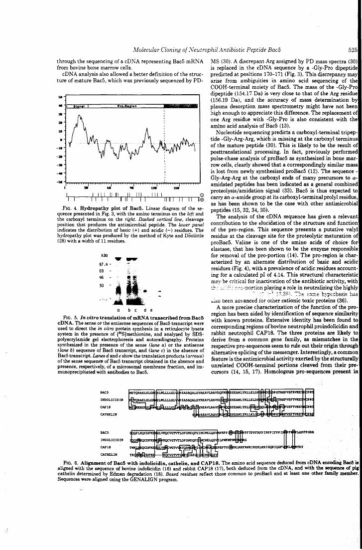
.lvfolecular Cloning of Neutrophil Antibiotic Peptide Bac5 525 through the sequencing of a cDNA representing Bac5 mRNA from bovine hone marrow cells.
cD NA analysis also allowed a better definition of the struc-ture of mature Bac5, which was previously sequenced by PD-
se .. Slqnel Pro·Aeglon &E 311
ZII
18
• -18
-ZII
-311 -· -se
l l Ili Il Il Il Ili Ili l 0 l Il l l Ili l [l 11111 Il Il le
FIG. 4. Hydropathy plot of Bac5. Linear diagram of the se-quence presented in Fig. 3, with the amino terminus on the left and the carboxyl terminus on the right. Dashed vertìcal fine, cleavage position that produces the antimicrobial peptide. The lower panel indicates the distribution of basic ( +) an d acidic (-) residues. The hydropathy plot was produced by the method of Kyte and Doolittle (28) with a width of 11 residues.
leDo
97.4-69 -
.... o b c d e
FIG. 5. In vitro translation of mRN A transcribed from Bac5 cD N A. The sense or the antisense sequences of Bac5 transcript were used to direct the in vitro protein synthesis in a reticulocyte Jysate system in the presence of [35S)methionine, and analyzed by SDS-polyacrylamide gel electrophoresis and autoradiography. Proteins synthesized in the presence of the sense (lane a) or the antisense (lane b) sequence of Bac5 transcript, an d (lane c) in the absence of Bac5 transcript. Lanes dande show the translation products (arrows) of the sense sequence of Bac5 transcript obtained in the absence and presence, respectively, of a microsomal membrane fraction, and im-munoprecipitated with antibodies to Bac5.
BACS
INOOLICIDIN
CAP18
CATHELIN
BACS
INOOLICIDIN
CAP18
CATHELIN
MS (30). A discrepant Arg assigned by PD mass spectra (30) is replaced in the cDNA sequence by a -Gly-Pro dipeptide predicted at positions 170-171 (Fig. 3). This discrepancy may arise from ambiguities in amino acid sequencing of the COOH-terminal moiety of Bac5. The mass of the -Gly-Pro dipeptide (154.17 Da) is very close to that of the Arg residue (156.19 Da), and the accuracy of mass determination by plasma desorption mass spectrometry might have not been high enough to appreciate this difference. The replacement of o ne Arg residue with -Gly-Pro is also consistent with the amino acid analysis of Bac5 (13).
Nucleotide sequencing predicts a carboxyl-terminai tripep-tide -Gly-Arg-Arg, which is missing at the carboxyl terminus of the mature peptide (30). This is likely to be the result of posttranslational processing. In fact, previously performed pulse-chase analysis of proBac5 as synthesized in hone mar-row cells, clearly showed that a correspondingly similar mass is lost from newly synthesized proBac5 (12). The sequence-Gly-Arg-Arg at the carboxyl ends of many precursors to a-amidated peptides has been indicated as a generai combined proteolysis/amidation signai (33). Bac5 is thus expected to carry an a-amide group at its carboxyl-terminaiprolyl residue, as has been shown to be the case with other antimicrobiai peptides (15, 32, 34, 35).
The analysis of the cDNA sequence has given a relevant contribution to the elucidation of the structure and function of the pro-region. This sequence presents a putative vaiyl residue at the cleavage site for the proteolytic maturation of proBac5. V aline is o ne of the amino acids of c ho ice for elastase, that has been shown to be the enzyme responsible for removal of the pro-portion (14). The pro-region is char-acterized by an alternate distribution of basic and acidic residues (Fig. 4), with a prevalence of acidic residues account-ing for a calculated pi of 4.14. This structural characteristic may be criticai for inactivation of the antibiotic activity, with t~: :,:: ;,::: n:o-portion playing a role in neutralizing the highly
- .• ~ ;~ ~ l?.3Sì. :':v~ ssme hypc:thesis h&.s a.lso been aàvanced ior other cationic toxic proteins (36).
A more precise characterization of the function of the pro-region has been aided by identification of sequence similarity with known proteins. Extensive identity has been found to corresponding regions of bovine neutrophil proindolicidin and rabbit neutrophil CAP18. The three proteins are likely to derive from a common gene family, as mismatches in the respective pro-sequences seem to rule out their origin through alternative splicing ofthe messenger. Interestingly, a common feature is the antimicrobiai activity exerted by the structurally unrelated COO H-terminai portions cleaved from their pre-cursors (14, 15, 17). Homologous pro-sequences present in
FIG. 6. Alignment of Bac5 with indolicidin, cathelin, and CAP18. The amino acid sequence deduced from cDNA encoding Bac5 is aligned with the sequence of bovine indolicidin (16) and rabbit CAP18 (17), both deduced from the cONA, and with the sequence of pig cathelin determined by Edman degradation (18). Boxed residues reflect those common to proBac5 and at least one other family member. Sequences were aligned using the GENALIGN program.
526 Molecular Cloning of Neutrophil Antibiotic Peptide Bac5
different mammalian species may thus represent a common carrier for these, and perhaps other, defense peptides. How-ever, this may be a hallmark of only a limited number of peptides, since other neutrophil antimicrobial proteins exhibit totally unrelated pro-sequences (11, 37-39).
Indirect evidence that the pro-sequence itself may play an active role independent of the antibiotic activity is given by the extensive identity shared with cathelin, a specific inhibitor of the cysteine proteinase cathepsin L isolated from pig leu-kocytes (19). Catbelin, wbich markedly differs from otber families of cysteine proteinase inhibitors, appears to be a bomologous protein (18) truncated at a position correspond-ing to the cleavage site of proBac5. Reasonably, sequences tbat are divergent copies of cathelin may also bave retained tbe same function. Consistent witb tbis bypotbesis, purified proBac5 bas proved to inbibit tbe in vitro activity of buman cathepsin L, with a K of about 10-8 M.2
Tbe fast extracellular release of proBac5 observed upon neutrophil stimulation with a soluble stimulus (8) may furtber support its contribution in limiting tbe elastinolytic activity of cathepsin L (40), witb positive effects for tbe integrity of inflamed tissues. Althougb a similar inbibitory function bas not been reported for proindolicidin and CAP18, we are tempted to speculate that ali these pro-sequences are members of the same family of cysteine proteinase inbibitors. Their association with COOH-terminal sequences carrying antibi-otic activity might bave occurred througb convergent evolu-tion, as additional specificities, to serve new physiological functions.
Acknowledgments-W e thank Licio Collavin for assistance in com-puter analysis and Francisco Baralle for critically reading the man-uscript.
REFERENCES l. Klebanoff, S. J. (1988) in lnflo.mmation: Basic Principles and Clinical
Correlates (Gallin, J. l., Goldstein, l. M., and Snydermann, R., eds) pp. 391-444, Raven Press, New York
2. Lehrer, R. 1., Ganz, T., Selsted, M. E., Babior, B. M., and Curnutte, J. T. (1988) Ann. Intern. Med. 109, 127
3. Joiner, K. A., Ganz, T., Albert, J., and Rotrosen, D. (1989) J. CeU BioL 109 2771-2782
4. Elsbach, P. (1990) Trends BiotechnoL 8, 26-30 5. Lehrer, R. l., and Ganz, T. (1990) Blood 76, 2169-2181 6. Spitznagel, J. K. (1990) J. Clin. lrwest. 86, 1381-1386
2 D. Verbanac and M. Zanetti, manuscript in preparation.
7. Gennaro, R., Romeo, D., Skerlavaj, B., and Zanetti, M. (1991) in Blood Celi Biochemistry (Harris, J. R., ed) pp. 335-368, Plenum Publishing Co., New York
8. Zanetti, M., Litteri, L., Griffiths, G., Gennaro, R., and Romeo, D. (1991) J. ImmunoL 146, 4295-4300
9. Zasloff, M. (1992) Curr. Opin. lmmunoL 4, 3-7 10. Elsbach, P., and Weiss, J. (1988) in lnflo.mmation: Basic Principles and
Clinical Correlates (Gallin, J. 1., Goldstein, l. M., and Snydermann, R., eds) pp. 445-470, Raven Press, New York
11. Almeida, R., P., Melchior, M., Campanelli, D., Nathan, C., and Gabay, J. (1991) Biochem. Biophys. Res. Commun. 177, 688-695
12. Zanetti, M., Litteri, L., Gennaro, R., Horstmann, H., and Romeo, D. (1990) J. CeU Biol. 111, 1363-1371
13. Gennaro, R, Skerlavaj, B., and Romeo, D. (1989) lnfect. lmmun. 57, 3142-3146
14. Scocchi, M., Skerlavaj, B., Romeo, D., and Gennaro, R. (1992) Eur. J. Biochem., in press
15. Selsted, M. E., Novotny, M. J., Morris, W. L., Tang, Y.·Q., Smith, W., and Cullor, J. S. (1992) J. BioL Chem. 267, 4292-4295
16. Del Sal, G., Storici, P., Schneider, C., Romeo, D., and Zanetti, M. (1992) Biochem. Biophys. Res. Commun. 187,467-472
17. Larrick, J. W., Morgan, J. G., Palings, L, Hirata, M., and Yen, M. H. (1991) Biochem. Biophys. Res. Commun. 179, 170-175
18. Ritonja, A., Kopitar, M., Jerala, R, and Turk, V. (1989) FEBS Lett. 255, 211-214
19. Kopitar, M., Ritor;ja, A., Popovic, T., Gabrijelcic, D., Krizaj, 1., and Turk, V. (1989) BioL Chem. Hoppe-Seyler 370, 1145-1151
20. Chomczinski, P., and Sacchi, N. (1987) Anal. Biochem. 162, 156-159 21. Lehrach, H., Diamond, D., Wozney, J. M., and Boedtker, H. (1977) Bio-
chemistry 16, 4743-4750 22. Grimaldi, G., Manfioletti, G., and Schneider, C. (1987) Nucleic Acids Res.
15,9608 23. Church, G. M., and Gilbert, W. (1984) Proc. NatL Acad. Sci. U. S. A. 81,
1991-1995 24. Frohman, A. M., Dush, K. M., and Martin, G. R. (1988) Proc. NatL Acad.
Sci. U. S. A. 85, 8998-9002 25. Del Sal, G., Ruaro, E., Philipson, L., and Schneider, C. (1992) CeU 70, 595-
607 26. Del Sal, G., Manfioletti, G., and Schneider, C. (1989) BioTechniques 7,
514-520 27. Sanger, F., Nicklen, S., and Coulson, A., R. (1977) Proc. Natl. Acad. Sci.
U. S. A. 7 4, 5463-5467 28. Kyte, J., and Doolittle, R. F. (1982) J. Mol. Biol. 157, 105-132 29. Dasso, M. C., and Jackson, R. J. (1989) Nucleic Acids Res. 17, 3129-3144 30. Frank, R. W., Gennaro, R., Schneider, K., Przybylski, M., and Romeo, D.
(1990) J. BioL Chem. 265 18871-18874 31 Kozak, M. (1986) CeU 44, 283-292 32. Agerberth, B., Lee, J., Bergman, T., Carlquist, M., Boman, H., G., Mutt, V.,
and Jornvall, H. (1991) Eur. J. Biochem. 202, 849-854 33. Mains, R. E., Eipper, B. A., Glembotski, C. C., and Dores, R. M. (1983)
Trends l'.·eurosci. 6, 229-235 34. Boman, H. G., Boman, l. A., Andreu, D., Li, Z.-q., Merrifield, R. B.,
Schlenstedt, G., and Zimmermann, R. (1989) J. BioL Chem. 264,5852-5860
35. Gibson, B. W., Tang, D., Mandrell, R., Kelly, M., and Spindel, E. R. (1991) J. BioL Chem. 266, 23103-23111
36. Barker, R. L., Gleich, G. J., and Pease, L. R. (1988) J. Er.p. Med. 168, 1493-1498
37. Gray, P. W., Flaggs, G., Leong, S. R, Gumina, R. J., Weiss, J., Ooi, C. E., and Elsbach, P. (1989) J. BioL Chem. 264, 9505-9509
38. Daher, K., Lehrer, R. I., Ganz, T., and Kronenberg, M. (1988) Proc. NatL Acad. Sci. U. S. A. 85, 7327-7331
39. Ganz, T., Rayner, J. R., Valore, E. V., Tumolo, A., Talmadge, K, and Fuller, F. (1989) J. ImmunoL 143, 1358-1365
40. Mason, R. W., Johnson, D. A., Barrett, A. J., and Chapman, H. A. (1986) Biochem. J. 233, 925-927
Vol. 187, No. 1, 1992
August 31, 1992
ALLEGATO 8-
BIOCHEMICAL ANO BIOPHYSICAL RESEARCH COMMUNICATIONS Pages 467-472
cDHA CLONrNG or TBZ NBOTROPHIL BACTERICIDAL PZPTIDB INDOLICIDIN
Giannino Del Sall, 2 , Paola Storicil, Claudio Schneider,2 Domenico Romeol, and Margherita Zanetti~*
loepartment of Biochemistry, Biophysics & Macromolecular Chemistry, University of Trieste, 34127 Trieste, Italy
2National Laboratory of the Interuniversity Consortium for Biotechnology (CIB), AREA Science Park, Padriciano,
34012 Trieste, Italy
Received July 13, 1992 ·.
SOMMARY: A structurally novel, tryptophan-rich antimicrobial tridecapeptide amide, named indolicidin, has recently been purified from bovine neutrophils (Selsted et al. (1992) J. Biol. Chem. 267, 4292-4295). Here we describe the molecular cloning of this endoantibiotic, which is synthesised in bone marrow cells as a 144 amino acid residue precursor. The encoded protein has a predicted mass of 16479 Da and a pi of 6.51. A putative signal peptide of 29 amino acids precedes a 101 residue pro-reqion. The mature peptide is at the 3' end of the open reading frame. A glycine, not found in purified indolicidin, is present at the carboxyl terminus of the deduced sequence and is very likely invol ved in post -translational peptide amidation. o 1992 Acadeatc Prass, rnc.
A major function of cytoplasmic granules of neutrophils is the delivery of several antimicrobial oligo- and poly-peptides to phagocytic vacuoles (1,2). The neutrophil "endoantibiotics" so far characterized in various animal species ( 3-6) do not exhibit common structural features, but are all very cationic. Furthermore, they show diverse antimicrobial spectra. The treatment of ingested microbes with a combination of granule endoantibiotics is thus likely to generate a situation full of potentialities far pathogen inactivation.
We have started the characterization of the repertoire of endoantibiotics of bovine neutrophils by resolving granule extracts into several active fractions (7) . From these we h-ave purified t o homogenei ty and sequenced three antimicrobial
* To whom correspondence should be addressed.
0006-291 X/92 $4.00 Copyright 1.0 1992 by Academic Press. /ne.
467 Ali rights of reproduction in any jorm reserved.
Vol. 187, No.1, 1992 BIOCHEMICAL ANO BIOPHYSICAL RESEARCH COMMUNICATIONS
peptides: an arginine-rich cyclic dodecapeptide, capable of killing both E.coli and S.aureus (8) and two proline- and arginine-rich polypeptides, BacS and Bac7, which appear to be active mainly, but not exclusively, on Gram-negative organisms (9,10).
More recently, Selsted and coworkers have purified an antibacterial tridecapeptide amide from bovine neutrophils, which is characterized by an unusual high proportion of tryptophan residues and therefore named indolicidin (11). In this report we describe the cloning and characterization of the full length cONA that codes for indolicidin, which appears to be synthesised in bone marrow cells as pre-proforrn.
MATERIALS ANO METHODS
cONA generation. Total RNA was extracted from bovine bone marrow cells with
guanidiniurn thiocyanate (12) . A primer derived from a previously characterized incomplete cDNA of BacSt, was used to obtain the 3' end by the RACE protocol (13). Ten ~g of total RNA was used to synthesise the first strand cDNA with 1.5 ng of primer adaptor 3'-(dTla)CGAAGAGCTCCCTAGGCT-5' and Moloney murine leukernia virus (MMLV) reverse transcriptase (BRL) (lh at 40°C). The reaction rnixture (20 ~l) was then heated to 95°C and 80 ~l of the PCR buffer (10 mM Tris.HCl, pH 8.3, 50 rnM KCl, 1.5 rnM MgC12) were added with 50 pmoles of both the upstrearn oligonucleotide 5'-CGCGAATTCAAAGCCTGTGAGCTTC-3' (240-255 bp of BacS cONA sequence) and the downstrearn prirner adaptor 3'-TTCGAAGAGCTCCCTAGGCTCGAGC-5'. PCR was carried out with 2.5 units of Taq polyrnerase for 30 cycles of denaturation at 94°C for 2 min, annealing at 55°C for l min and polyrnerization at 72°C for 2 min.
The RACE to the 5' end, was carried out using the oligonucleotide 5'-TCTGAACAAATCAGACACTTA-3' derived from the 3' end sequence of indolicidin. Single stranded cONA was tailed using terminal deoxynucleotidyl transferase (BRL) and dGTP. Arnplification was performed using the upstream primer complernentary to the dGTP tail (dCis), and a downstream primer 5'-ACGAATTCCGAGGCTCAGGATCTGACA-3'. Amplification conditions were as above. cONA seguencing.
The amplified cONA was cloned in Bluescript sK+ vector (Stratagene) . Sequencing was performed on both strands with the dideoxy chain termination method (14) . Regions with high G+C content were also sequenced in parallel with deazaguanosine and automated fluorescent DNA sequencing (EMBL fluorescent DNA sequencer) . DNA sequence analysis was conducted with the aid of the IntelliGenetics Suite version 5.4, and homology search was carried out on the Swiss-Prot database. No;thern and primer extension analyses.
Total RNA (5 ~g) was separated by electrophoresis on 6.7% formaldehyde/1% agarose gels, transferred to a nylon membrane
tzanetti, M., Del Sal, G., Storici, P., Schneider, c., and Romeo, D., rnanuscript in preparation.
468
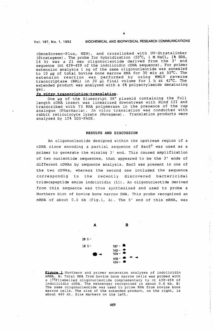
• Vol. 187, No.1, 1992 BIOCHEMICAL ANO BIOPHYSICAL RESEARCH COMMUNICATIONS
(GeneScreen-Plus, NEN), and crosslinked with UV-Stratalinker (Stratagene) . The probe for hybridization (55°C, l M NaCl, 1% SDS, 16 h) was a 21 mer oligonucleotide derived from the 3' end sequence (nt 439-459 of the indolicidin cDNA sequence) . For primer extension analysis l ng of the same oligonucleotide was annealed to 10 ~g of total bovine bone marrow RNA for 30 min at 55°C. The extension reaction was performed by using MMLV reverse transcriptase (BRL) in 30 ~l final volume for l h at 42°C. The extended product was analyzed with a 6% polyacrylamide denaturing gel. In yitro transcription-translation.
One ~g of the Bluescript SK+ plasmid containing the full length cDNA insert was linearized downstream with Hind III and transcribed with T3 RNA polymerase in the presence of the cap analogue (Pharmacia) . In vitro translation was conducted with rabbit reticulocyte lysate (Novagene) . Translation products were analysed by 15% SDS-PAGE.
RESULTS ANO DISCUSSION
An oligonucleotide designed within the upstream region of a cONA clone encoding a partial sequence of Bac5t was used as a primer to generate the missing 3' end. This caused amplification of two nucleotide sequences, that appeared to be the 3' ends of different cDNAs by sequence analysis. Bac5 was present in one of the two cDNAs, whereas the second one included the sequence correspondig to the recently discovered bactericidal tridecapeptide amide indolicidin (11). An oligonucleotide derived from this sequence was thus synthesised and used to probe a Northern blot of bovine bone marrow RNA. This probe recognized an mRNA of about 0.6 kb (Fig.l, A). The 5' end of this mRNA, was
28 s-18 s-
A
-~
B
587- • 540- • 504- • 458- • - ~ 434- •
ligure l.Northern and primer extension analyses of indolicidin mRNA. A: Total RNA from bovine bone marrow cells was probed with a [3 2P]labelled oligonucleotide complementary to nt 439-459 of indolicidin cONA. The messenger recognized is about 0.6 kb. B: The same oligonucleotide was used to prime RNA from bovine bone marrow cells. The size of the extended product, on the right, is about 460 nt. Size markers on the left.
469
Vol. 187, No.1, 1992 BIOCHEMICAL ANO BIOPHYSJCAL RESEARCH COMMUNICATIONS
l AGACTGGGGACCATGCAGACCCAGAGGGCCAGCCTCTCACTGGGG M Q T Q R A s L S L G 11
46 CGGTGGTCGCTGTGGCTACTGCTGCTGGGACTAGTGGTGCCCTCG R W S L W L L L L G L v V P s 26
91 GCCAGCGCCCAAGCCCTCAGCTACAGGGAGGCCGTGCTTCGTGCT A S AtQ A L S Y R E A V L R A 41
136 GTGGATCAGCTCAATGAGCTGTCCTCAGAAGCTAATCTCTACCGC V D Q L N E L S S E A N L Y R 56
181 CTCCTGGAGCTAGACCCACCTCCCAAGGATAATGAAGATCTGGGC L L E L D P P P K D N E D L G 71
226 ACTCGAAAGCCTGTGAGCTTCACGGTGAAGGAGACTGTGTGCCCC T R K P V S F T V K E T V C P 86
271 AGGACGATTCAGCAGCCCGCGGAGCAGTGTGACTTCAAGGAGAAA R T I Q Q P A E Q C D F K E K 101
316 GGGCGGGTGAAACAGTGTGTGGGGACAGTCACCCTGGACCCATCC G R V K Q C V G T V. T L D P S 116
361 AATGACCAGTTTGACCTAAACTGTAATGAGCTCCAGAGTGTCATC N D Q F D L N C N E L Q S V L 131
406 CTACCCTGGAAATGGCCATGGTGGCCTTGGCGCAGAGGTTGATGG L P W K W P W W P W R R G * 144
451 AGAAGAGCTGTCAGATCCTGAGCCTCGGGAAGAGTCTTAAGTGTC
496 TGATTTGTTCAGATTCGGGCTTCTGGACAGTG~TTCTT
541 GTGAAAACGGAAAAAAAAAAAAAAA
Figura 2. Nucleotide and deduced amino acid sequence of indolicidin. The nucleotide sequence is numbered on the left, from the 5' end of the mRNA. The amino acid sequence is numbered from the first methionine on the right. The signal peptide is overlined, and the arrow indicates the putative cleavage site for signal peptidase. The sequence of mature indolicidin is underlined, and the stop codon is marked with an asterisk. The polyadenylation signal is double underlined.
determined by primer extension using the same synthetic oligonucleotide, and shown to be about 440 nt upstream of the primer (Fig. l, B) . The 550 bp full length cDNA encoding indolicidin, shown in Fig.2, was then obtained by the RACE protocol to the 5' end.
Sequence analysis revealed that indolicidin is synthesised as 144 amino acid precursor that shares 51% and 74% identity, respectively, with CAP18 from rabbit (15) and cathelin from pig (16). The predicted mass of the precursor is 16479 Da, as confirmed by in vitro translation of the transcript (not shown), with a calculated pi of 6.51. The sequence predicts a hydrophobic 29 residue signal peptide, overlined in Fig.2, that is followed by a pro-region of 101 residues.
470
l
Vol. 187, No.1, 1992 BIOCHEMICAL ANO BIOPHYSICAL RESEARCH COMMUNICATIONS
The deduced sequence of mature indolicidin, located at the carboxyl end of the precursor (underlined in Fig. 2), is comprised of 14 amino acid residues, including a carboxy-terminal glycine. Indolicidin as purified from neutrophils is a tridecapeptide ending with an amidated arginine (11). The C-terminal glycine thus appears to be lost by posttranslational processing that results in aamidation of the carboxyl end of indolicidin, with the nitrogen atom of the amide deriving from the missing residue (17).
Selsted et al. ( 11) di d no t previde evidence for the existence of a p~ecursor of indolicidin, since they isolated only the mature tridecapeptide amide from granule extracts of bovine neutrophils. In contrast, BacS and Bac7, other bactericidal polypeptides of bovine neutrophils, have been found to be present as proforms in the neutrophil large granules (18), wherefrom they can be isolated as such. Their proteolytic maturation is carried out by elastase (19) that is contained in neutrophil primary granules, as an early event during phagocytosis (2}. In this respect, it is interesting to note that valine, a specific cleavage site for elastase, is present at the C-terminus of the pro-sequence of indol icidin (resi due 130 in Fig. 2} , thus suggesting that elastase might also be responsible for the maturation of proindolicidin. Whether this maturational step is carried out in the resting neutrophil is not clear. Since Selsted at al. (11) suggest that indolicidin is stored as mature peptide in a still unknown organellar location, it is possible that indolicidin and elastase co-localize to the primary granules, allowing for proteolytic maturation being carried out in this compartment. However, the possibility that the pro-region is retained and proindolicidin represents the neutrophil storage form, as in the case of probactenecins, cannot be ruled out. Antibodies to indolicidin will be useful in identification of its intracellular localization as well as investigations on its processing.
ACKNOWLEDGMENTS. We thank P. Edemi for performing the automated nucleotide sequence analysis. This work was supported by grants from the National Research Council (Progetto Finalizzato "Biotecnologie e Biostrumentazione") and from the Italian Ministry for University and Research.
REFERENCES 1.Joiner, K.A., Ganz, T., Albert, J., and Rotrosen, D. (1989} J.
Cell Biol. 109, 2771-2782.
471
Vol. 187, No.1, 1992 BIOCHEMICAL ANO. BIOPHYSICAL RESEARCH COMMUNICATIONS
2.Zanetti, M., Litteri, L., Griffiths, G., Gennaro, R., and Romeo, D. (1991) J. Immunol. 146, 4295-4300.
3.Lehrer, R.I., and Ganz, T. (1990) Blood 76, 2169-2181. 4.Spitznagel, J.K. (1990) J. Clin Invest. 86, 1381-1386. S.Gennaro, R., Romeo, D., Skerlavaj, B., and Zanetti, M. (1991)
In Blood Cell Biochemistry (J .R. Harris Ed.), pp. 335-368, Plenurn Publ.Co., New York.
6.Boman, H.G. (1991) Cell 65, 205-207. ?.Gennaro, R., Dolzani, L, and Romeo, D. (1983) Infect. Immun.
40, 684-690. 8.Romeo, D.,Skerlavaj, B., Bolognesi, M., and Gennaro, R. (1988)
J. Biol. Chern. 263; 9573-9575. 9.Gennaro, R., Skerlavaj, B., and Romeo, D. (1989) Infect. Irnmun.
57, 3142-3146. 10.Frank, R.W., Gennaro, R., Schneider, K., Przybylski, M., and
Rorneo, D. (1990) J. Biol. Chem. 265, 18871-18874. 11.Selsted, M.E., Novotnj, M.J., Morris, W.L., Tang, Y-Q.,Smith,
w., and Cullor, J.S. (1992) J. Biol. Chem. 267, 4292-4295. 12.Chomczinski, P., and Sacchi, N. (1987).Anal. Biochem. 162, 156-
159. 13.Frohman, A. M., Dush, K. M., and Martin, G. R. (1988) Proc
Natl. Acad. Sci. USA 85, 8998-9002. 14.Sanger, F., Nicklen, s., and Coulson, A., R. (1977) Proc. Natl.
Acad. Sci. u. s. A. 74, 5463-5467. 15.Larrick, J. W., Morgan, J. G., Palings, I., Hirata, M., and
Yen, M., H. (1991) Biochem. Biophys. Res. Commun. 179, 170-175. 16.Ritonja, A., Kopitar, M., Jerala, R., and Turk, V. (1989) FEBS
Lettr. 255, 211-214. 17.Bradbury, A., F., and Srnyth, D., G. (1991) Trends Biochern. Sci.
16, 112-115. 18.Zanetti, M., Litteri, L., Gennaro, R., Horstmann, H.,and Romeo,
D. (1990) J. Cell Biol. 111, 1363-1371. 19.Scocchi, M., Skerlavaj, B., Romeo, D., and Gennaro, R., Eur.
J. Biochem., accepted for publication.
Reprinted from Biochemical and Biophysical Research Communications 187, 467-472, 1992 Copyright© 1992 Academic Press, Inc. Printed in U.S.A.
472
AllEGATO C
Volume 314. number 2, 187-190 FEBS 11857 December 1992 'r; 1992 Federation of European Biochemical Societies 00145793'92/S5.00
cD NA sequence analysis of an antibiotic dodecapeptide from neutrophils
Paola Storicia, Giannino Del Sala,b, Claudio Schneiderb and Margherita Zanettia
aDepartment of Biochemistry, Biophysics and Macromolecular Chemistry, University o/Trieste, 34127 Trieste, ltaly and bNational Laboratory of the lnteruniversity Consortium for Biotechnology ( CIB), AREA Science Park, Padri ciano, 34012 Trieste, Italy
Received 20 October 1992
The full-1ength cDNA of a neutrophi1 antibiotic dodecapeptide has been cloned by reverse transcription!PCR from bovine bone marrow RNA. This peptide was originally iso1ated from bovine neutrophils, and shown to exert a potent antimicrobial activity in vitro on both Escherichia coli and Staphy/ococcus aureus. The cDNA codes fora polypeptide of 155 amino acid residues with a predicted mass of 17,629 Da and api of 8.03. The deduced sequence comprises a putative signa] peptide of 29 amino acids, a 114 residue pro-region, and a carboxy-termina1 dodecapeptide corresponding to the mature antibiotic. The pro-sequence disp1ays extensive identity to corresponding regions of other structurally unre1ated
antibiotic peptides of bovine neutrophils recently cloned.
Neutrophil; Antibiotic; Cyclic dodecapeptide; cDNA
l. INTRODUCTION
A centrai role of neutrophils in the initial host re-spense to microbial challenge is testified by a massive recruitment of these cells at the infection si te, followed by rapid and effective pathogen inactivation. Killing is ensured by the production of highly reactive oxygen derivatives [l] combined with the release of granule-associated cytotoxic proteins into phagocytic vacuoles [2.3]. Oligo- and polypeptides with diverse antimicro-bial spectra ha ve been isolated from neutrophils of var-ious animai species [4-7]. In particular, from granule extracts of bovine neutrophils, four cationic peptides have been purified and characterized. Two of them, associated as inactive proforms to the Iarge granules [8,9], are known as Bac5 an d Bac7, an d exhibit bacteri-cidal activity mainly, but not exclusiveiy, on Gram-negative organisms [10,11]. In addition to these, a cyclic dodecapeptide, previous1y named bactenecin [12], and a Trp-rich tridecapeptide amide named indolicidin [13], exert a potent antibacterial action on both Escherichia coli and Staphylococcus aureus. We report here the nu-cleotide sequence of a full-length cDNA encoding the precursor form of the cyclic dodecapeptide. The cD NA cloning of Bac5 [14] and indolicidin [15] has recently shown __ !~~!-!~e~ ~ti1Jigtics_s1l~re_ highly. similar pro-sequences. A homoiogous pro-sequence is also pre-dicted to precede the sequence· of the dodecapeptide. Althoùgh the maturè forms of ali these peptides appear structurally unrelated, they are likely to derive from a
Correspondence address: M. Zanetti. DipartimentÒ di Biochimica, Bi-ofisica and Chimica delle Macromolecole, via A. Valerio 38, Univer-sità di Trieste, 34127 Trieste, ltaly. Fax: (39) (40) 676 3691.
Published by Elsevier Science Publishers B. V.
common gene family also including members isolated from different animai species [16, 17].
2. EXPERIMENT AL 2.1. cDNA c/oning
T o tal RNA was extracted from bovine bo ne marrow cells with guanidinium thiocyanate [18). The rapid amplification of cD NA ends, or RACE, [19) was used to obtain both the 3' and 5' ends of the dodecapeptide cDNA. To generate the 3' end, the first strand cDNA wa~ synthesized usir1g l j.lg oftotal RNA and 1.5 ng ofprimer adaptor 5'-TCGGA TCCCTCGAGAAGC(dT18)-3' and Moloney m urine leu-kemia virus (MML V) reverse transcriptase (Bethesda Research Labo-ratori es, Gaithersburg, MD) (l h at 40°C}. The reaction mixture (20 J.ll) was tben beated to 95°C and 80 ,.ul of the PCR buffer (10 mM Tris-HQ, pH 8.3, 50 mM KG, 1.5 mM MgCl:J were added with 50 pmol of both the upstream oligonucleotide 5' -CGCGAA TT-CAAAGCCTGTGAGCTTC-3' and the downstream primer adaptor 5'-CGAGCTCGGATCCCTCGAGAAGCTT-3'. PCR was carried out with 2.5 U of Taqi polymerase (Perkin-Elmer-Cetus, Norwalk, CT) for 30cycles of denaturation at 94°C for 2 min, annealingat 55°C for l min and polymerization at 72°C for 2 min.
The RACE to the 5' end was carried out using the oligonucleotide 5'-CAGATCCAGTAGCITGAGGC-3' derived from the 3' end se-quence of the dodecapeptide. Single-stranded cD NA was tailed using terminai deoxynucleotidyl transferase (Bethesda Research Laborato-ries) and dGTP. Amp1ification conditions were as above, using an upstream primer complementary to the dGTP tail (dCI5), and a downstream primer 5'-GACGAATICGAGT AAGAAAACCCTT A-3'.
2.2. cDNA sequencing and sequence aruzlysis The amplified cDNA was cloned in Bluescript KS+ vector (Strat-
agene, San Diego, CA). Sequencing was performed on both strands with the dideoxy chain-termination method [20}. Regions with high G+C content were also sequenced in parallel with deazaguanosine and automated ftuorescent DNA sequencing (EMBL ftuorescent DNA sequencer, Hcridelberg, Germany). Analysis ofthe DNA sequenèe was conducted witk the aid ofthe lntelliGenetics Suite version 5.4 (lntelli-Genetics Ine., Mountain View, CA), and the homology search was
187
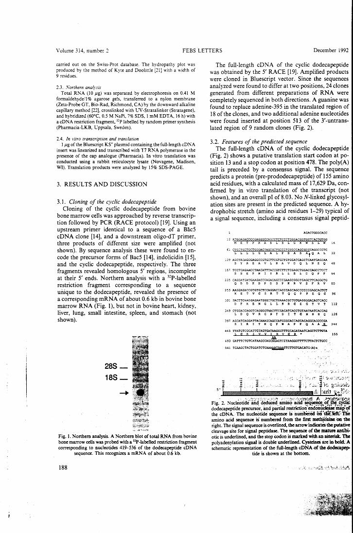
Volume 314, number 2 FEBS LETTERS December 1992
carried out on the Swiss-Prot database. The hydropathy plot was produced by the method of Kyte and Doolittle [21] with a width of 9 residues.
2.3 . .Vorthern analysis Total RNA (10 jig) was separated by electrophoresis on 0.41 M
forma1dehyde/1% agarose ge!s, transferred to a nylon membrane (Zeta-Probe GT, Bio-Rad, Richmond, CA) by the do.,..nward alkaline capillary method [22], crosslinked with UV-Stratalinker (Stratagene), and hybridized (60°C, 0.5 M NaPi, 7% SDS, l mM EDTA, 16 h) with a cD NA restriction fragment, 32P labelled by random prirner synthesis (Pharmacia-LKB, Uppsala, Sweden).
2.4. In vitro transcription and translation l Jig of the Bluescript KS+ plasmid containing the full-length cD NA
insert was linearized and transcribed with T7 RNA polymerase in the presence of the cap analogue (Pharmacia). In vitro trans1ation was conducted using a rabbit reticulocyte lysate (Novagene, Madison, Wl). Translation products were analyzed by 15% SDS-PAGE.
3. RESULTS AND DISCUSSION
3.1. Cloning of the cyclic dodecapeptide Cloning of the cyclic dodecapeptide from bovine
bone marrow cells was approached by reverse transcrip-tion followed by PCR (RACE protocol) [19]. Using an upstream primer identica! to a sequence of a Bàc5 cDNA clone [14], and a downstream oligo-dT primer, three products of different size were amplified (not shown). By sequence analysis these were found to en-code the precursor forms of Bac5 [14], indolicidin [15], and the cyclic dodecapeptide, respectively. The three fragments revealed homologous 5' regions, incomplete at their 5' ends. Northern analysis with a 31P-labelled restriction fragment corresponding to a sequence unique to the dodecapeptide, revealed the presence of a corresponding mRNA of about 0.6 kb in bovine bone marrow RNA (Fig. l), but no t in bovine heart, kidney, liver, lung, small intestine, spleen, and stomach (not shown).
288-188-
Fig. l. Northem analysis. A Northem blot oftotal RNA from bovine bone marrow cells was probed with a 32P-labelled restriction fragment corresponding to nucleotides 419-536 of the dodecapeptide cONA
sequence. This recognizes a rnRNA of about 0.6 kb.
188
The full-length cDNA of the cyclic dodecapeptide was obtained by the 5' RACE [19]. Amplified products were cloned in Bluescript vector. Since the sequences analyzed were found to differ a t two positions, 24 clones generated from different preparations of RNA were completely sequenced in both directions. A guanine was found to replace adenine-395 in the translated region of 18 ofthe clones, and two additional adenine nucleotides were found inserted at position 513 of the 3'-untrans-lated region of 9 random clones (Fig. 2).
3.2. Features of the predicted sequence The full-length cDNA of the cyclic dodecapeptide
(Fig. 2) shows a putative translation start codon at po-sition 13 and a stop codon at position 478. The poly(A) tail is preceded by a consensus signal. The sequence predicts a protein (pre-prododecapeptide) of 155 amino acid residues, with a calculated mass of 17,629 Da, con-firmed by in vitro translation of the transcript (not shown), and an overall p l of 8.03. No N-linked glycosyl-ation sites are present in the predicted sequence. A hy-drophobic stretch (amino acid residues 1-29) typical of a signal sequence, including a consensus signal peptid-
o o "' 5' l
AGACTGGGCACC
l; ATGGAGACCCCGAGGGCCAGCCTCTCCCTGGGACGGTGGTCACTGTGG METPRASLSLGRWSLW
€1 CTGCTGCTGCTGGGACTAGCGCTGCCCTCGGCCAGCGCCCAGGCCCTC L L L L G L A L P S A S At0 A L
l C 9 AGCTACAGGGAGGCCGTGCTTCGTGCTGTGGATCAGCTCAATGAGCAA S Y R E A V L R A V D 0 L N E 0
15 7 TCCTCAGAACCTAACATT':'ACCGTCTTCTCGAGCTGGACCAGCCTCCT SSEPNIYRLLELDQPP
2 C 5 CAGGATGATGAAGACCCAGACAGCCCGAAGCGGGTGAGCTTCAGGGTG ODDEDPDSPKRVSFRV
2 53 AAGGAGACCGTGTGCTCCAGGACCACCCAGCAGCCCCCCGAGCAGTGT K E T V C S R T T 0 0 P P E 0 C
3 O l GACTTCAAGGAGAATGGGCTGCTGAAACGCTGTGAGGGGACAGTCACC D F K E N G L L K R C E G T V T
3 ~ 9 CTGGACCAGGTCAGGGGTAACTTCGACATCACCTGTAATAATCACCAG L D O V R G N F D I T C N N H O
397 AGCATCAGGATTACAAAGCAGCCATGGGCACCACCACAGGCAGCCCGA
32
48
64
80
96
112
128
S I R I T K 0 P W A P P 0 A A _B., 144
4 4 5 TTATGTCGCATTGTAGTGATAAGGGTTTGCAGATAATCAGCTCTTGTA L C R I V V I R V C R " 155
4 93 GATTTCTGTCATAAGCCAG~GTCCTAAGGGTTTTC'l'TACTCTGCC 541 TCAAGCTACTGGATCTG~TTCTTGTGACATG (A) D
.... ~--~!.~ (~""' E; ···h:'"-.~~··:.~!~~~-~!-i .
. i.. ~ ·l ,. ..,;;, :~·le ~pi:.iot:· ' :t !;&rh Air.~;,''.i;·
Fig.' 2. Nucleotidé'~ld~~ ~g;Ji~;p~~ ~Ji~ dodecapeptide pr~r. andpartial reStriction~endon~ mapof the cONA.· The. nucleòtide' sequence is nùnibCna on"~féftr: 'Ì1Ìe amino acid sequence is nwnbered from the fiist mcth!ODine 011 1he right. The signal sequence is overlined, the arrow indicates the putative cleavage si te for signal peptidase. The sequence of the mature antlòi· otic is underlined, and the stop éodon is marked with an aSterisk. 1be polyadenylation signal is double underlined. Cysteineì are ·m bokl A schema tic representatiori of the full-length cONA of the dodccilpep-
tide is shown at the bottom.

Volume 314. number 2 FEBS LETTERS December 1992
58
alual -- .,,.;:'\.2dc-
l l
~ '
) \ ~fl • ! w
,v \vlt vvr\t\v . . . : l l
28
111
-111
-28
-31 l
-41 l l l l
-58 l l l l l
128 1SII
l l l Il Il DIII l 111 l l l 0 l Il l 1111 l l Il l Ili Ili lE>
Fig. 3. Hydropathy plot [21] of the cyclic dodecapeptide precursor. Linear diagram of the deduced amino acid sequence, with the amino-terminai on the left and the carboxyl-terminal on the right. A dashed vertical line shows the cleavage site for the maturation of the cycli(} dodecapeptide. The lower pane! indicates the distribution of basic ( .... )
and acidic (-) residues.
ase cleavage site (Ala-X-Ala) [23] is present at the amino-terminus of the protein (Fig. 3). The putative signal peptide is followed by a 114 amino acid long hydrophilic pro-sequence, corresponding to residues 30-143, which is characterized by a cluster ofnegatively charged amino acids in the region between residues 59 and 71. The carboxy-terminal dodecapeptide (residues 1~155) corresponds to the mature antibiotic isolated from granule extracts of neutrophils, and exactly matches the sequence determined by protein sequencing
Dodecapeptide
Indolicidin
Bac5
Dodecapeptide L
Indolicidin
Bac5
[ 12]. The two cysteine residues a t positions 146 an d 154 are known to maintain the mature antibiotic in a cyclic structure by a disulfìde bond [12]. In the conformation proposed [12), the peptide chain forms a bend at residue 7, with the hydrophobic residues clustered around the bend. This compact structure is reminiscent of defen-sins, a family of numerous small antimicrobial peptides with a characteristic cysteine motif forming three intra-molecular disulfìde bonds [24).
Unlike defensins, which are expressed by various cell types of mammals and insects [24], the bovine neutro-phil has so far been the only source ofthe dodecapeptide [12). Recently, another basic, cysteine-rich peptide, with functional similarities to both the dodecapeptide and the defensins, has been iso1ated from bovine trachea! mucosa [25]. The sequences of all these cysteine-rich peptides, as deduced from their mRNAs, lack the car-boxy-terminal glycine required for amidation, a charac-teristic feature of several other antibacterial peptides [14, 15,26,27], that may be required to increase their biostability. The amino-terminus of the dodecapeptide is preceded by an Ala residue at position 143. This is a likely site of proteolytic cleavage for elastase, which may be responsible for the remo val of the pro-sequence from the precursor [9]. Due to base substitution, the clones analyzed predicted the alternative presence of Gin and Arg at position 128 ofthe pro-sequence, point-ing to the existence of either a polymorphic, or an addi-tional gene. Genomic cloning will resolve this issue, since neither possibility can be excluded at present.
3.3. Observed similarity of the dodecapeptide precursor with other proteins
The sequence of the deduced precursor is compared in Fig. 4 with that of Bac5 [14] and indolicidin [15), both isolated from bovine neutrophils. As expected from the cloning strategy, the pro-sequence of the dodecapeptide displays extensive identity to corresponding regions of
PSASAQALSYREAVLRAVDQLN
SASAQALSYREAVLRAVDQLN
59
59
59
118
118
118
Dodecapeptide RITKQPwi*rlcrivvirvcr--------------------- 155
'1pwkwp g-------------------------------- 144
~-=~=~frppir · pfyppfrppirppifppirppfrpp1gpfpgrr 17 6
Indolicidin
Bac5
Fig. 4. Alignment of the predicted sequence of the cyclic dodecapeptide with other antibiotic peptides of bovine neutrophil. The amino acid sequence deduced from the cD NA encoding the dodecapeptide is aligned with the deduced sequences ofindolicidin [15], and Bac5 [14]. Boxed residues reftect those common to the dodecapeptide and a t least one other peptide. Aligned cysteines are shaded. Smallletters indicate the sequences corresponding to the mature antibiotics, and asterisks show the carboxy-terminal residues of the pro-sequences. Sequences were aligned using the GENALIGN
program (lntelliGenetics).
189
Volume 314, number 2 FEBS LETTERS De ce m ber 199 2
both Bac5 and indolicidin, including 4 Ìn\'ariant cyste-ine residues, that may offer possible structural con-straints. Both structural and functional implications have been previously suggested [14] by the presence of homologous pro-regions in the sequences of proBac5 and proindolicidin. The evidence that the cyclic dode-capeptide also carries a homologous region further strengthens the hypothesis of a common pro-sequence acting as a carrier for defense peptides. In fact, in addi-tion to the bovine antibiotics mentioned above, 2 ho-mologous proteins from different animai species bave been identified, providing evidence for a more wide-spread presence of this sequence: CAP18, an anti-bacterial protein from rabbit [17], and cathelin, an in-hibitor of cathepsin L isolated from pig neutrophils [16]. These exhibit about 57% and 66% identity, respectively, to the pro-region of the dodecapeptide (not shown).
Both proBac 5 and proindolicidin carry a valyl resi-due (marked with an asterisk in Fig. 4) at the cleavage si te for the proteolytic maturation of the respective an-tibiotics. The pro-sequence of the dodecapeptide as-signes Ile at the same position, followed by a stretch of 13 additional residues (131-143 ofthe sequence) extend-ing its carboxy-terminus. l t is not known whether these residues are cleaved from the precursor during the intra-cellular maturation of the antibiotic. Immunochemical investigations with antibodies to the dodecapeptide will clarify this point. In particular, since the cyclic dode-capeptide has been the only proteolytic product so far isolated from neutrophils [12], it will be interesting to know whether the same precursor may produce more biologically relevant fragments.
Acknowledgements: We thank Dr. D. Romeo for helpful discussion. This work was supported by grants from the National Research Coun-cil (Progetto Finalizzato 'Biotecnologie e Biostrumentazione') and from the :. •:.:~ Ministry for University and Research.
REFERENCES [l] K.lebanoff, S.J. (1988) in: Infiammation: Basic Principles and
Clinical Correlates (J.I. Gallin, I.M. Goldstein and R. Snvder-mann eds.) pp. 391-444, Raven Press, New York. -
190
[2] Joiner, K.A., Ganz. T .. Albert. J. and Rotrosen, D. (1989) J. Celi Bio!. 109. 2771-2782.
[3] Zanetti. M .. Litteri. L.. Griffiths, G., Gennaro, R. and Romeo, D. (1991) J. Immunol. 146, 4295--4300.
[4] Elsbach, P. and Weiss, J. (1988) in: lnflammation: Basic Princi-ples and Clinica! Correlates (J.I. Gallin, I.M. Goldstein and R. Snydermann eds.) pp. 391-444 Raven Press, New York.
[5] Lehrer, R.I. and Ganz, T. (1990) Blood 76, 2169-2181. (6] Gennaro, R., Romeo, D., Skerlavaj, B. and Zanetti, M. (1991)
in: Blood Celi Biochemistry (J.R. Harris ed.) pp. 335-368, Ple-num, New York.
[7] Zasloff, M. (1992) Curr. Opin. Immunol. 4, 3-7. [8] Zanetti, M., Litteri, L., Gennaro, R., Horstmann, H. and Romeo,
D. (1990) J. Celi Biol. 111, 1363-1371. [9] Scocchi, M., Skerlavaj, B., Romeo, D. and Gennaro, R., Eur. J.
Biochem. (in press). [10] Gennaro, R., Skerlavaj, B. and Romeo, D. (1989) Infect. Immun.
57, 3142-3146. [Il] Frank, R.W .• Gennaro, R., Schneider, K., Przybylski, M. and
Romeo, D. (1990) J. Biol. Chem. 265, 18871-18874. [12] Romeo, D., Skerlavaj, B., Bolognesi, M. and Gennaro, R. (1988)
J. Biol. Chem. 263, 9573-9575. (13] Selsted, M.E., Novotnj, M.J., Morris, W.L., Tang, Y.-Q., Srnith,
W. and Cullor, J.S. (1992) J. Biol. Chem. 267, 4292--4295. (14] Zanetti, M., Del Sal, G., Storici, P., Schneider, C. and Romeo,
D. (1993) J. Bio!. Chem. (in press). [15] Del Sal, G., Storici, P., Schneider, C., Romeo, D. and Zanetti,
M. (1992) Biochem. Biophys. Res. Commun. 187, 467--472. (16] Ritonja, A., Kopitar, M., Jerala, R. and Turk, V. (1989) FEBS
Lett. 255, 211-214. [lì] Larrick, J.W., Morgan, J.G., Palings, I., Hirata, M. and Yen, M.
H. (1991) Biochem. Biophys. Res. Commun. 179, 170-1 7 5. [18] Chomczinski, P. and Sacchi, N. (1987) Anal. Biochem. 162, 156-
159. [19] Frohman, A.M., Dush, K.M. and Martin, G.R. (1988) Proc Nati.
Acad. Sci. USA 85, 8998-9002. [20] Sanger, F., Nicklen, S. and Coulson, A.R. (1977) Proc. Nati.
Acad. Sci. USA 74, 5463-5467. [21] Kyte, J. and Doo1itt1e, R.I. (1982) J. Mol. Biol. 157, 105-132. {22] Chomczynski, P. (1992) Anal. Biochem. 201, 134-139. (23] von Heijne, G. (1986) Nucleic Acids Res. 14, 4683. [24] Lehrer, R.I., Ganz, T. and Selsted, M.E. (1991) CeU 64,229-230. [25] Diamond, G., Zasloff, M., Eck, H., Brasseur, M., Maloy, W.L.
and Bevins, C.L. (1991) Proc. Nati. Acad. Sci. USA 88, 3952-3956.
[26] Agerberth, B., Lee, J., Bergrnan, T., Carlquist, M., Boman, H.G., Mutt, V. and Jornvall, H. (1991) Eur. J. Biochem. 202, 849-854.
[27] Boman, H.G., Boman, I.A., Andreu, D., Zong-qu Li, Merrifie1d, R.B., Sch1enstedt, G. and Zimmermann, R. (1989) J. Biol. Chem. 264, 5852-5860.
ALLEGATO D Vol. 196, No. 3, 1993
November 15, 1993
BIOCHEMICAL ANO BIOPHYSICAL RESEARCH COMMUNICATIONS Pages l 058-l 065
A cONA DERIVED FROM PIG BONE MARROW CELLS PREDICTS A SEQUENCE IDENTICAL TO THE INTESATINAL
ANTIBACTERIAL PEPTIDE PR-39 1
Paola Storici 1 • 2 and Margherita Zanetti 1 • 37
lNational Laboratory of the Interuniversity Consortium for Biotechnology, (CIB), AREA Science Park, Padriciano,
34012 Trieste, Italy 2oepartment of Biochemistry, Biophysics and
Macrornolecular Chemistry, University of Trieste, 34127 Trieste, Italy
3oepartment of Science and Medical Technologies, University of Udine, 33100 Udine, Italy
Received September 6, 1993
SUMMARY: We have recently shown that a group of antimicrobial peptides of bovine neutrophils share highly identica! pro-sequences. In this paper we report the cONA sequence of a 172 amino acid residue pig myeloid protein showing a sirnilar pro-sequence of 101 residues. The carboxyl-moiety of the predicted protein is identical to the mature form of the proline- and arginine-rich antibacterial peptide named PR-39, isolated from pig intestine. t: 1993 Academi= P::ess, Inc.
Peptide-based antimicrobial defenses have been described in a
variety of species and tissues and may represent an important
component of anima l host defense ( 1-7) Of the antimicrobial
peptides identified so far, some are thought to be unique i.n
primary structure, others can be classified into families, based
on common sequences and secondary structure. Three distinct groups
have been identified in mammaJian neutrophils, namely defensins
and ~-defens ins ( 8-11), serpi'ocidins ( 11) and a proline- and
arginine- rich group of peptid~s (3), which may contribute to the
oxygen-independent defense mechanisms of these cells. The proline-
and arginine- rich family of antibacterial peptides includes Bac5
and Bac7, identified in bovine neutrophils ( 12) . Both peptides are
active against gram-negative <12) and gram-positive (unpublished)
organisms. Their amino acid sequences are highly repetitive, being
characterized by severa! Pro-Arg-Pro and Arg-Pro-Pro repeats,
lsequence data from this article have been deposited with the EMBL/GenBank Libraries under Accession No. L23825. * To whom correspondence should be addressed.
0006-291X/93 $4.00 Copyright© 1993 by Academic Press, lnc. All rights of reproduction in any form reserved. l 058
Vol. 196, No. 3, 1993 BIOCHEMICAL AND BIOPHYSICAL RESEARCH COMMUNICATIONS
respectively (13). t-:c;re ::-ec;er:::.:y, a:-: additi::::nal rr.ember of the proline- and arginine- r:c~ group of antibiotic peptides named PR-39 has been isole.ted frorr: the small intestine of pig, possibly contributing to the defense of the mucosal surfaces of this tissue (14). Although no obvious overall homology can be observed among the three peptides, their amino acid compositicn and the spectra of activity are very similar (14). No data are available on the intracellular localization and storage form of the intestinal PR-39. Conversely, Bac5 and Bac7 ha ve been shown to be stored as inactive precursors in the large cytoplasmic granules of neutrophils (15). The primary structure of the precursor of Bac5 has recently been defined (16) and reveals a pro-sequence highly identical to cathelin/PLCPI, an inhibitor of the cysteine proteinase cathepsin L (17-19). A number of structurally unrelated antimicrobial and/or lipopolysaccharide-binding proteins from various animal species have been found to share a similar pro-region ( 20-23), which may thus represent a common carrier for these peptides. We then decided to undertake a study to identify proteins characterized by the presence of this carrier, in myeloid
cells other than bovine. In this paper we report the cDNA cloning of one such protein from pig bone marrow cells. Sequencing of the full length cONA reveals that the N-terminal domain homologous to cathelin/PLCPI is associated to a C-terminal sequence identical to the amino acid sequence of the intestinal PR-39. Our data thus support the existence of a myeloid-derived PR-39, characterized by a pro-region cowmon to other neutrophil antimicrobial peptides.
MATERIALS ANO METHOOS
cONA cloning Total RNA was extract.ed from pig bone marrow cells with
guanidinium thiocyanate (24). Amplification :Jf the 5 1 and 3 1 ends was performed by the rapid amplification of cONA ends (RACE) method (25) The conditions used were as in (21). For the 3 1 end RACE, a downstream primer adaptor 5 1 -TCGGATCCCTCGAGAAGC (T18) -3' was used to reverse transcribe l ~g of total RNA. Amplification was done with the do·wnstream primer adaptor 5 1-CGAGCTCGGATCCCTCGAGAAGCTT-3' and an upstream oligonucleotide 5'-CGCGAATTCTGTGAGCTTCAGGGTG-3'. In the RACE to the 5 1 end, the oligonucleotide primer 5'-CGGAGGTGGCCTTGGCCT-3', derived from the 3 1 end sequence of PR-39, was used for reverse transcription. Amplification was done with an oligonucleotide 5 1-ATGGAATTCGGTCGGGGACGTCTC-31 as downstream primer, with an upstream primer complementary to the dGTP tail (21)
cONA sequencing The amplified cONA
(Stratagene, San Diego, was
CA). cloned in Sequencing
1059
Bluescript SK+ vector was performed on both
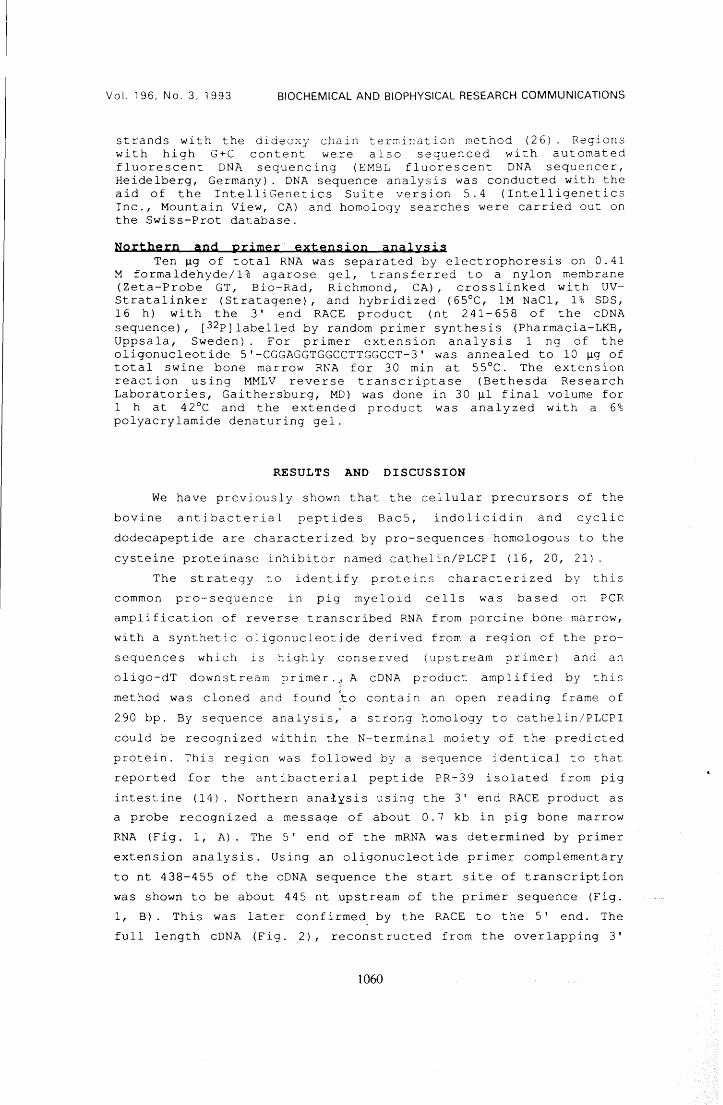
Vol. 196, No.3, 1993 BIOCHEMICAL AND BIOPHYSICAL RESEARCH COMMUNICATIONS
strands with the diàeoxy chain termination method (26). Regions with high G+C content were also sequenced with automateà fluorescent DNA sequencing (EMBL fluorescent DNA sequencer, Heidelberg, Germany}. DNA sequence analysis was conducted with the a id of the IntelliGenetics Sui te version 5. 4 ( Intelligenetics Inc., Mountain View, CA) and homology searches were carried out on the Swiss-Prot database.
Northern and primer · extension analysis Ten ~g of total RNA was separated by electrophoresis on 0.41
M formaldehyde/1% agarose gel, transferred to a nylon membrane (Zeta-Probe GT, Bio-Rad, Richmond, CA), crosslinked with UV-Stratalinker (Stratagene}, and hybridized (65°C, 1M NaCl, 1% SDS, 16 h) with the 3' end RACE product (nt 241-658 of the cONA sequence), [32p]labelled by random primer synthesis (Pharmacia-LKB, Uppsala, Sweden}. For primer extension analysis l ng of the oligonucleotide 5'-CGGAGGTGGCCTTGGCCT-3' was annealed to 10 ~g of total swine bone marrow RNA for 30 min a t 55°C. The extension reaction using MMLV reverse transcriptase (Bethesda Research Laboratories, Gaithersburg, MD) was done in 30 ~l final volume for l h at 42°C and the extended product was analyzed with a 6% polyacrylamide denaturing gel.
RESULTS ANO DISCUSSION
We have previously shown that the cellular precursors of the bovine antibacterial peptides Bac5, indolicidin and cyclic dodecapeptide are characterized by pro-sequences homologous to the cysteine proteinase inhibitor named cathelin/PLCPI (16, 20, 21).
The strategy to identify proteins characterized by this common pro-sequence in pig myeloid cells was based on PCR amplification of reverse transcribed RNA from porcine bone marrow, with a synthetic oligonucleotide derived from a region of the pro-sequences which is highly conserved (upstream primer) and an oligo-dT downstream primer ..• A cDNA product amplified by this method was cloned and found to contain an open reading frame of 290 bp. By sequence analysis, a strong homology to cathelin/PLCPI could be recognized within the N-terminal moiety of the predicted protein. This region was followed by a sequence identical to that reported for the antibacterial peptide PR-39 isolated from pig intestine ( 14} . Northern ana:l:tsis using the 3' end RACE product as a probe recognized a message of about 0.7 kb in pig bone marrow RNA (Fig. l, A). The 5' end of the mRNA was determined by primer extension analysis. Using an oligonucleotide primer complementary to nt 438-455 of the cDNA sequence the start site of transcription was shown to be about 445 nt upstream of the primer sequence (Fig. l, B). This was later confirme~ by the RACE to the 5' end. The full length cDNA (Fig. 2}, reconstructed from the overlapping 3'
1060
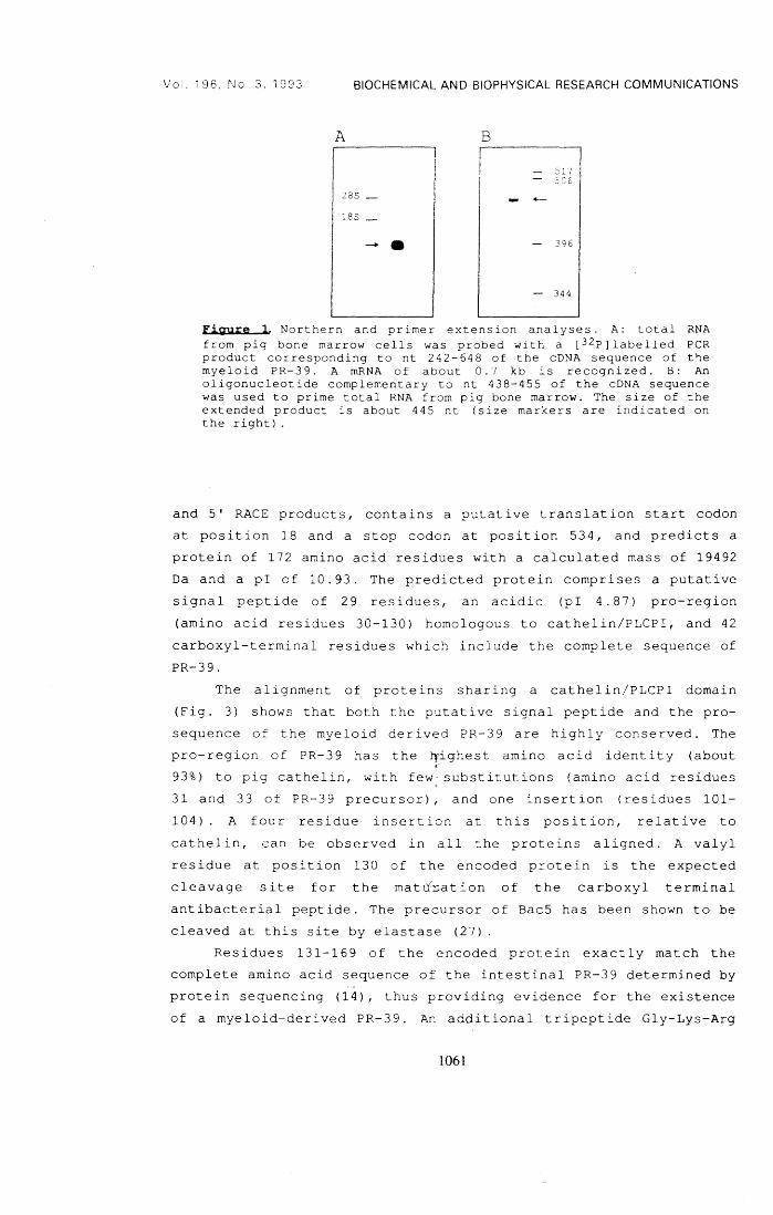
Vol. i 96, No. 3, 1993 BIOCHEMICAL ANO BIOPHYSICAL RESEARCH COMMUNICATIONS
A B
28S -
lSS _ -· - 396
- 344
Figure l Northern and pr imer extens ion analyses. A: total RNA from pig bo ne marrow ce lls was p robe d w i t h a [ 32p] la be l led PCR product corresponding to nt 242-648 of the cDNA sequence of t~e
myeloid PR-39. A mRNA of about O. 7 kb is recognized. B: An oligonucleotide complementary to nt 438-455 of the cDNA sequence was used to prime total RNA from pig bone marrow. The size of the extended product is about 445 nt (size markers are indicated on the right).
and 5' RACE products, contains a putative translation start codon at position 18 and a stop codon at position 534, and predicts a protein of 172 amino acid residues with a calculated mass of 19492 Da and a pi of 10.93. The predicted protein comprises a putative signal peptide of 29 residues, an acidic (pl 4. 87) pro-region (amino acid residues 30-130) homologous to cathelin/PLCPI, and 42
carboxyl-terminal residues which include the complete sequence of PR-39.
The alignment of proteins sharing a cathelin/PLCPI domain (Fig. 3) shows that both the putative signal peptide and the pro-sequence of the myeloid derived PR-39 are highly conserved. The pro-region of PR-39 has the 11ighest amino acid identity (about 93%) to pig cathelin, with few substitutions (amino acid residues 31 and 33 of PR-39 precursor), and one insertion (residues 101-104). A four residue insertion at this position, relative to cathelin, can be observed in all the proteins aligned. A valyl residue at position 130 of the encoded protein is the expected cleavage site for the matd:r:ation of the carboxyl terminal antibacterial peptide. The precursor of Bac5 has been shown to be cleaved at this site by elastase (27)
Residues 131-169 of the encoded protein exactly match the complete amino acid sequence of the intestinal PR-39 determined by protein sequencing (14), thus providing evidence for the existence of a myeloid-derived PR-39. An additional tripeptide Gly-Lys-Arg
1061
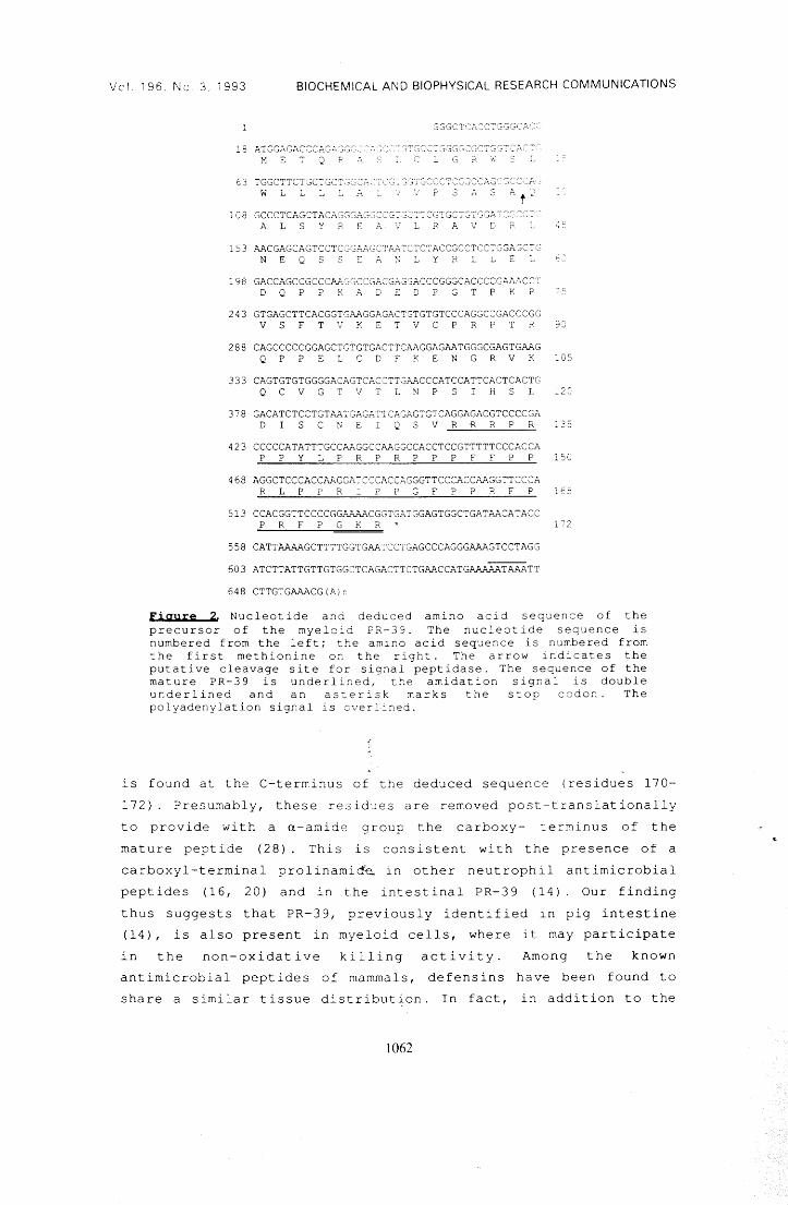
Vol. l96 No.3 ì993 BIOCHEMICAL ANO BIOPHYSICAL RESEARCH COMMUNICATIONS
SSGCTCACC~SGGCAC~
~1E~QP
63 TGGCTTCTGCTGCT:;c;:::,-,,·". :-;··:·:·'";··;-:-·:~('<~''"':~:~.~~~ .. ~;,.~· w T ~ L ~ A s .~. s
108 GCCCTCAGCTACAGGGAGGCCGTGCTTCGTGCTGTGGATCGCCT~ A L S Y ? "' A ::., R P.. V C ?. ~:'
15 3 AACGAGCAGTCCTCC;C~P.J..GC"'.'l~_::C7CTACCGCCTCCTGGAGCTC~ N E Q S S _ A ~ : Y R r ~ :
l98 GACCAGCCGCCCAJ.. GG·CCGP..CGAGGP..CCCGGGCACCCCGAA.I..CC':· D Q p p K K D 2 D p G T p K p
243 GTGAGCTTCACGGTGAAGGAGACTGTGTGTCCCAGGCCGACCCGC V S F T V K E V C P R P T R 90
288 CAGCCCCCGGAGCTGTGTGACTTCAAGGAGAATGGGCGAGTGAAG Q P P E L C D F K E N G R V K ~05
3 3 3 CAGTGTGTGGGGACAGTCACCTT,GAACCCATCCATTCACTCACTG Q C V G T V T L N P S H S L l2C
378 GACATCTCCTGTAATGAGATTCAGAGTGTCAGGAGACGTCCCCGA D I S C N E Q S V R ~ R P R :35
423 CCCCCATATTTGCCAAGGCCAAGGCCACCTCCGTTTTTCCCACCA P P Y L P R P R P P P F F P P 150
468 AGGCTCCCACCAAGGATCC:CACCAGGGTTCCCACCAAGGTTCCCA R L P P R I P P G F P P R F P 165
513 CCACGGTTCCCCGGP.-r..AACGGTGATGGAGTGGCTGATAACATACC P R F P G K R 172
558 CATTAAAAGCTTTTGGTGAATCCTGAGCCCAGGGAA.~GTCCTAGG
603 ATCTTATTGTTGTGGCTCAGACTTCTGAACCATGAAAAATAA.~TT
648 CTTGTGAAACG(A)r.
Figure 2 Nucleotide anà deàuced amino acid sequence of the precursor of the myeloi.d PR-39. The nucleotiàe sequence is numbered from the left; the amino acid sequence is numbered from the first methionine an the right. The arrow indicates the putative cleavage site far signal peptidase. The sequence of the mature PR-39 is underlined, the amidation signal is double underlined and an as~erisk marks the stop coàon. The polyadenylation signal is overlined.
is found at the C-terminus of the deduced sequence (residues 170-
172). Presumably, these residues are removed post-translationally
to provide with a a-amide group the carboxy- terminus of the
mature peptide (28). This is consistent with the presence of a
carboxyl-terminal prolinamida in other neutrophil antimicrobial
peptides (16, 20) and in the intestinal PR-39 (14) Our finding
thus suggests that PR-39, previously identified in pig intestine
( 14), is al so present in myeloid cells, where i t may participate
in the non-oxidative killing activity. Among the known
antimicrobial peptides of mammals, defensins have been found to
share a similar tissue distribution. In fact, in addition to the
1062
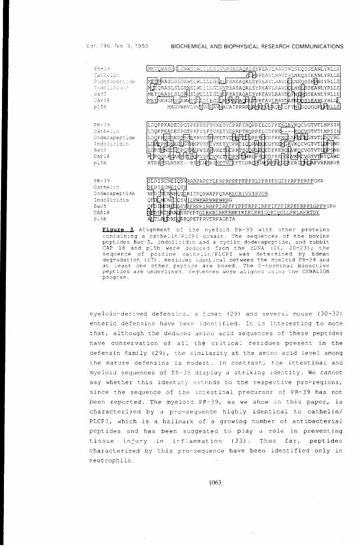
Vo!. 196, No 3, 1993 BIOCHEMICAL ANO BIOPHYSICAL RESEARCH COMMUNICATIONS
l >1ETQKASIJ ~"c;r:z:;.;s =-.vr::...:.=-.:L.:..::./F/P SASA ~,ALSYR2A'.J:LF;A'J;::;' RLNEQSSEANLYRLLE
plSh
?R-39
Dcdecapep-::de Inàolìcid::_:; Bac5 CAP18 pl5~
PR-39 Cathelin 2odecapeptide Indolicidin Bac5 C.Z'I.Pl8 pl5h
r-----=zr-r..rREAVLRAVDRLNEQSSEANLYRLLE NEQSS~IYRLLE NE SEANLYRLLE
FRPPIRRPPIRPPFYPPFRPPIRPPIFPPIRPPFRPPLGPFPGRR L~l.~~~SPEPTGLRKRLRKFRNKIKEKLKKIGQKIQGLLPKLAPRTDY
RRQPEFPRVTRPAGPTA
Figure 3. Alignment of the myeloid PR-39 with other proteins containing a cathelin/PLCPI domain. The sequences of the bovine peptides Bac 5, indolicidin and a cyclic dodecapep~ide, and rabbit CAP 18 and pl5h were deduced from the cDN.~ (lE, 20-23); the sequence of porcine cathelin/PLCPI was determined by Edman degradation (17). Residues identical between the myeloid PR-39 and a t least one other pept i de are boxed. The C-terrr.inal bioactive peptides are underlined. Sequences were aligned using ~he GENALIGN prograr.,.
myeloid-derived defensi~s, a human (29) and several mouse (30-32) enteric defensins have been identified. It is interesting to note
that, although the deduced amino acid sequences of these peptides
have conservation of all the critical residues present in the
defensin family (29), the similarity at the amino acid level among the mature defensins is rnodest. In contrast, the intestinal and
myeloid sequences of PR-39 display a striking identity. We cannot say whether this identity extends to the respective pro-regions,
since the sequence of the intestinal precursor of PR-39 has not been reported. The myeloid P~-39, as we show i:n this paper, is characterized by a pro-seque:nce highly identical to cathelin/ PLCPI, which is a hallmark of a growing number of antibacterial pept ides and has been suggested to play a role in preventing
tissue injury in inflammation ( 33) Thus far, peptides characterized by this pro-sequence have been identified .only in neutrophils.
1063
Vo!. 196, No. 3, 1993 BIOCHEMICAL ANO BIOPHYSICAL RESEARCH COMMUNICATIONS
ACKNOWLEDGMENTS: We thaiìk ? rof. Romec fo~ helpfu: disc~ssio~.
This work was supported by gra:1ts from :::.:r~e National Research
Council (Progetto Finalizzato "Biotecnoloqie e Biostrumentazione")
and from the Italian Ministry for University and Research.
REFERENCES
1.Elsbach, P., and Weiss, J. {1993) Curr. Opin. Immunol. 5, 103-107.
2.Zasloff, M. (1992) Curr. Opin. Immunol. 4, 3-7. 3.Gennaro, R., Romeo, D., Skerlavaj, B., and Zanetti, M. (1991)
in Blood Cell Biochemistry, vol. 3, (J. R. Harris Ed.), pp. 335-368, Plenum Publ. Co., New York.
4 . Boman, H . G . ( 19 91) Ce l l 6 5, 2 O 5-2 O 7 . 5.Lehrer, R. I. and Ganz, T. (1990) Blood 76, 2169-2181. 6.Bevins, C. L. and Zasloff, M. (1990) Annu. Rev. Biochem. 59,
395-414. 7.Boman, H. G., and Hultmark, D. (1987) Annu. Rev. Microbiol. 41,
103-126. 8.Lehrer, R.· I., Ganz, T., and Selsted, M. E. (1991) Cell 64,
229-230. 9.Selsted, M. E., Tang, Y., Morris, W. L., McGuire, P. A.,
Novotny, M. J., Smith, W., Henschen, A. H., and Cullor, J. S. (1993) J. Biol. Chem. 268, 6641-6648.
10.Tang, Yi-Q. and Selsted, M. E. (1993) J. Biol. Chem. 268, 6649-6653.
1l.Gabay, J. E. and Almeida, R. P. (1993) Curr. Opin. Immunol. 5, 97-102.
12.Gennaro, R., Skerlavaj, B. and Romeo, D. (1989) Infect. Immun. 57' 3142-314 6.
13.Frank, R. W., Gennaro, R., Schneider, K., Przybylski, M., and Romeo, D. (1990) J. Biol. Chem. 265, 18871-18874.
14.Agerberth, B., Lee, J., Bergman, T., Carlquist, M., Boman, H., Mutt, V., and Jornvall, H. (1991) Eur. J. Biochem. 202, 849-854.
15.Zanetti, M., Litteri, L., Gennaro, R., Horstmann, H., and Romeo, D. (1990) J. Cell Biol. 111, 1363-1371.
16.Zanetti, M. Del Sal, G., Storici, P., Schneider, C., and Romeo, D. (1993) J. Biol. Chem. 268, 522-526.
17.Ritonja, A., Kopitar, M., ~erala, R., and Turk, V. (1989) FEBS Lett. 255, 211-214.
18.Kopitar, M., Ritonja, A., ~opovic, T., Gabrijelcic, I. K. and Turk, V. (1989) Biol. Chem. Hoppe-Seyler 370, 1145-1151.
19.Kakegawa, H., Nikawa, T., Tagami, K., Kamioka, H., Sumitani, K., Kawata, T., Drobnic-Kosorok, M., Lenarcic, B., Turk, V. and Katunuma, N. (1993) FEBS Lett. 321, 247-250.
20.Del Sal, G., Storici, P., Schneider, C., Romeo, D. and Zanetti, M. (1992) Biochem. Biophys, Res. Commun. 187, 467-472.
21.Storici, P., Del Sal, G., ·schneider, C., and Zanetti, M. (1992) FEBS Lett. 314,187-190.
22.Larrick, J. W., Morgan, J. G., Palings, I., Hirata, M., and Yen, M. H. (1991) Biochem. Biophys. Res. Commun. 179, 170-175.
23.Levy, O., Weiss, J., Zarember, K., Ooi, C. E., and Elsbach, P. (1993) J. Biol. Chem. 268, 6058-6063.
24.Chomczinski, P., and Sacchi, N. (1987) Anal. Biochem. 162, 156-159.
25.Frohman, A. M., Dush, K. M. and Martin, G. R. (1988) Proc. Natl. Acad. Sci. USA 85, 8998-9002.
1064
Vol. 196, No. 3, 1993 BIOCHEMICAL AND BIOPHYSICAL RESEARCH COMMUNICATIONS
26.Sanger, F., Nicklen, S., and Coulson, A. R. (1977) Proc. Natl. Acad. Sci. USA 74, 5463-5467.
27.Scocchi, M., Skerlavaj, B., Romeo, G., and Gennaro, R. (1992) Eur. J. Biochem. 209, 589-595.
28.Bradbury, P. •. F. and Smyth, D. '0. (1991) Trends Biochem. Sci. 16, 112-115.
29.Jones, D. E. and Bevins, \~. L. (1992) J. Biol. Chem. 267, 23216-23225.
30.0uellette, A. J., Greco, R. Naftilan, J. and Fallon, J. T. 1695.
M., James, M., Frederick, D., (1989) J. Cell Biol. 108, 1687-
31.0uellette, A. J., Miller, S. I., Henschen, A. H., and Selsted, M. E. (1992) FEBS Lett. 304, 146-148.
32.Selsted, M. E., Miller, S. I., Henschen, A. H., and Oulette, A. J. (1992) J. Cell Biol. 118, 929-936.
33.Verbanac, D., Zanetti, M., and Romeo, D. (1993) FEBS Lett. 3, 255-258.
1065
l
, ALLEGATO E
Vol. 196, No. 3, 1993
Nowmber 15, 1993
BIOCHEMICAL ANO BIOPHYSICAL RESEARCH COMMUNICATIONS Pages 1363-1368
A NOVEL cDNA SEQUENCE ENCODING A PIG LEUKOCYTE ANTIMIC~OBIAL PEPTIDE WITH A CATHELIN-LIKE PRO-SEQUENCE+
Paola Storici 1.2 and Margherita Zanetti 1.3/f
lNational Laboratory ofthe Interuniversity Consortium for Biotechnology, (CIB), AREA Science Park, Padriciano, 34012 Trieste, ltaly
2Department of Biochemistry, Biophysics and Macromolecular Chemistry, University ofTrieste, 34127 Trieste, ltaly
3Department of Science and Medicai Technologies, U niversity of Udine, 33100 Udine, ltaly
Received October l, 1993
SUMMARY: It has recently been shown that the precursors of various structurally unrelated leuk:ocyte antimicrobial peptides share similar pro-regions. These, in turn, are highly identical to a cysteine proteinase inhibitor named cathelin, or PLCPI. In this paper we report a novel cONA sequence of porcine bone marrow origin, encoding a protein characterized by a cathelin-like domain. The putative protein is 147 amino acid residue long, with a calculated mass of 16479 Da and appears to be the precursor of a recently isolated antimicrobial peptide named protegrin PG-2. The unique sequence of the mature PG-2 is located at the C-tenninus of the precursor. Similar to the previously reported precursors, both the signal peptide and the pro-sequence of pre-proPG-2 appear highly CODsetVed. O 1993 Academic Press, Inc.
The antimicrobial peptides of phagocytes are believed to play a relevant role in the non-
oxidative defense mechanisms of these cells (1-4). These peptides show significant diversity in
-strocture, spectrum of activity and-distribution arnong species (3-8). Based on structural features.
many of the leuk:ocyte antimicrobial peptides bave been grouped into severa! families (5-8). ··· ·Sequence identity within families may vary considerably. In the case of defensins, a family of
small antimicrobial peptides which are derived from larger precursors (9), the mRNA sequences
show a limited similarity in the regions encoding the mature peptides and a high sequence conservation in the 5' regions (10, 11). More recently, highly identica! 5' regions bave been observed within the mRNA sequences encoding the precursors of various antimicrobial peptides
with diverse antimicrobial spectra and no obvious common structural features (12-16). These
include Bac5 (6), indolicidin (17) and a cyclic dodecapeptide (18) from bovine, and CAP18 (12)
from rabbit leukocytes, which originate from the respective precursors by proteolytic maturation. In generai, the sequence similarity of the precursors of these peptides is remarkably high in the
+ . Sequence data from this article bave been deposited w1th the EMBL/GenBank Data Libraries under Accession No. L24745. #To whom correspondence should be addressed.
1363
0006-29lX/93 $4.00 Copyright C 1993 by Academic Press, lnc.
Ali rights of reproduction in any form reserved.
Vol. 196, No.3, 1993 BIOCHEMICAL ANO BIOPHYSICAL RESEARCH COMMUNICATIONS
signal peptide and pro-sequence coding regions and is virtually absent in the regions encoding the
mature antimicrobial peptides. An interesting feature of these precursors is the extensive similarity
of the common pro-region to the sequence of a porcine inhibitor of the cysteine proteinase cathepsin L termed cathelin (20) or PLCPI (pig leukocyte cysteine proteinase inhibitor, 21). The
structural similarity of the N-terminai regions of ali these proteins suggests a common evolutionary
origin and identifies a nove! protein family characterized by a cathelin-like N-terminai domain (15,
16). To determine the complexity of this protein family in mammalian myeloid cells, we bave
undertaken an investigation aimed at the identification of new members in porcine bone marrow cells.
In this paper we report the sequence of a pig myeloid cDNA encoding a previously
unrecognized precursor characterized by a cathelin-like domain. The C-terminai sequence of the putative precursor corresponds to the sequence of the antimicrobial peptide named protegrin-PG-2, recently isolated from porcine leukocytes (8).
MATERIALS AND METHODS
cDNA cloojp~ Total RNA was extracted from pig bone marrow cells with guanidinium thiocyanate (22).
One Jlg of total RNA was reverse transcribed using the same experimental conditions and the antisense primer as in (15), and the 3' end of the cDNA was amplified (15) with an antisense primer adaptor 5'-CGAGCfCGGATcccrCGAGAAGCIT-3' and a sense oligonucleotide primer 5'-CGCGAA TICTGTGAGCITCAGGGTG-3'. T o obtain the 5' end of the cD N A, 20 pmol of the antisense oligonucleotide primer 5'-CCAGAATICGATGCAGAACCTACGCCTACA-3', derived from a unique 3' end sequence of PG-2 cDNA was used to reverse transcribe l J.Lg of porcine total RNA. PCR amplification was done by addition of 30 pmol of the same antisense oligonucleotide primer and 50 pmol of a sense oligonucleotide primer 5'-ACCGAATICACCTGGGCACCATG-3', using 2.5 U/J.Ll Taq I polymerase (Perkin Elmer-Cetus), for 30 cycles: l min at 94° C, l min at 55° C and 2 mio at 72° C.
cDNA seguepcjpc apd seguepçe analysjs The amplified cD NA was cloned in Bluescript SK+ vector (Stratagene, San Diego, CA).
Sequencing was performed on both strands with the dideoxy chain termination method (23). Regions with high G+C content were also sequenced by automatèd tluorescent DNA sequencing (EMBL fluorescent DNA sequencer, Heidelberg, Germany) with deazaguanosine mixes. DNA sequence analysis was conducted with the aid of the IntelliGenetics Suite version 5.4 (lntelligenetics lnc., Mountain View, CA) and homology searches were carried out on the Swiss-Prot database.
Nortbem and prjmer exteosjoo analyses Ten JJ.g of total RNA was separated by electrophoresis on 0.41 M formaldehyde/1%
agarose gel, transferred to a nylon membrane (Zeta-Probe GT, Bio-Rad, Richmond, CA), crosslinked with UV-Stratalinker (Stratagene), and hybridized (65°C, 0.5 M NaPi pH 7.2, 7% SDS, l mM EDT A) with the 3' end amplification product [32P]labelled by random primer synthesis (Pharmacia-LKB, Uppsala, Sweden). For primer extension analysis l ng of the oligonucleotide 5'-CCAGAATICGATGCAGAACCTACGCCTACA-3' was annealed to 5 J.Lg of total swine bone marrow RNA for 30 min at 55°C. The extension reaction using MML V reverse transcriptase (Bethesda Research Laboratories, Gaithersburg, MD) was done in 30 tù final volume for l h at 42°C and the extended product was analyzed with a 6% polyacrylamide denaturing gel.
RESULTS AND DISCUSSION
Based on cDNA sequence analysis. the precursors of a number of different neutrophil antimicrobial peptides bave been found to share highly identical pro-sequences (12-16). To identify
1364
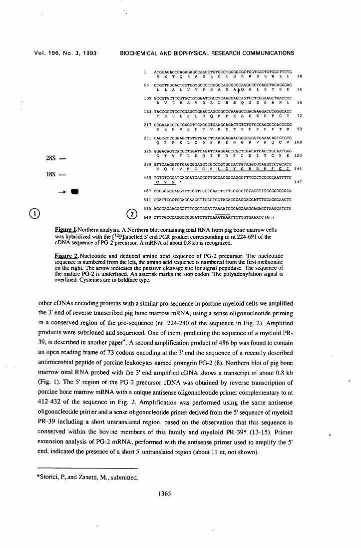
Vol. 196, No. 3, 1993 BIOCHEMICAL ANO BIOPHYSICAL RESEARCH COMMUNICATIONS
ATGGAGACCCAGAGAGCCAGCCTGTGCCTGGGGCGCTGGTCACTGTGGCTTCTG M E T Q R A S L C L G R W S L W L L
55 CTGCTGGCACTCGTGGTGCCCTCGGCCAGCGCCCAGGCCCTCAGCTACAGGGAG L L A L V V P S A S AfQ A L S Y R E
109 GCCGTGCTTCGTGCTGTGGATCGCCTCAACGAGCAGTCCTCGGAAGCTAATCTC A V L R A V D R L N E Q S S E A N L
163 TACCGCCTCCTGGAGCTGGACCAGCCGCCCAAGGCCGACGAGGACCCGGGCACC Y R L L E L D Q P P K A D E D P G T
217 CCGAAACCTGTGAGCTTCACGGTGAAGGAGACTGTGTGTCCCAGGCCGACCCGG P K P V S F T V K E T V C P R P T R
271 CAGCCCCCGGAGCTGTGTGACTTCAAGGAGAACGGGCGGGTGAAACAGTGTGTG
18
36
54
72
90
Q p P E L C D F K E N G R V K Q C V 108
28S
18S
325 GGGACAGTCACCCTGGATCAGATCAAGGACCCGCTCGACATCACCTGCAATGAG G T V T L D Q I K D P L D I T C N E 120
379 GTTCAAGGTGTCAGGGGAGGTCGCCTGTGCTATTGTAGGCGTAGGTTCTGCATC V Q G V R G G R L C Y C R R R F C I 144
433 TGTGTCGGATGAGGATGACGGTTGCGACGGCAGGCTTTCCCTCCCCCAATTTTC C V G * 147
487 CCGGGGCCAGGTTTCCGTCCCCCAATTTTTCCGCCTCCACCTTTCCGGCCCGCA
541 CCATTCGGTCCACCAAGGTTCCCTGGTAGACGGAGGAGGATTTGCAGGCAACTC
595 ACCCAGAAGGCCTTTCGGTACATTAAAATCCCAGCAAGGAGACCTAAGCATCTG
649 CTTTGCCCAGGCCCGCATCTGTCAAATAAATTCTTGTGAAACC(A)n
Fjgure J.Northem analysis. A Northem blot containing total RNA from pig bone marrow cells was hybridized with the [32P]labelled 3' end PCR product corresponding to nt 224-691 of the cD NA sequence of PG-2 precursor. A mRNA of about 0.8 kb is recognized.
Fjgure 2. Nucleotide and deduced amino acid sequence of PG-2 precursor. The nucleotide sequence is numbered from the left, the amino acid sequence is numbered from the frrst methionine on the right. The arrow indicates the putative cleavage site for signal peptidase. The sequence of the mature PG-2 is underlined. An asterisk marks the stop codon. The polyadenylation signal is overlined. Cysteines are in boldface type.
other cONAs encoding proteins with a similar pro-sequence in porcine myeloid cells we amplified the 3' end of reverse transcribed pig bone marrow mRNA, using a sense oligonucleotide priming in a conserved region of the pro-sequence (nt 224-240 of the sequence in Fig. 2). Amplified products were subcloned and sequenced. O ne of them, predicting the sequence of a myeloid PR-39, is described in another paper*. A second amplification product of 486 bp was found to contain an open reading frame of 73 codons encoding at the 3' end the sequence of a recently described antimicrobial peptide of porcine leukocytes named protegrin PG-2 (8). Northem blot of pig bone marrow total RNA probed with the 3' end amplified cONA shows a transcript of about 0.8 kb (Fig. 1). The 5' region of the PG-2 precursor cONA was obtained by reverse transcription of porcine bone marrow mRNA with a unique antisense oligonucleotide primer complementary to nt 412-432 of the sequence in Fig. 2. Amplification was performed using the same antisense oligonucleotide primer and a sense oligonucleotide primer derived from the 5' sequence of myeloid PR-39 including a short untranslated region, based on the. observation that this sequence is conserved within the bovine members of this family and myeloid PR-39* (13-15). Primer extension analysis of PG-2 mRNA, performed with the antisense primer used to amplify the 5' end, indicated the presence of a short 5' untranslated region (about 11 nt, not shown).
*Storici, P., and Zanetti, M., submitted.
1365
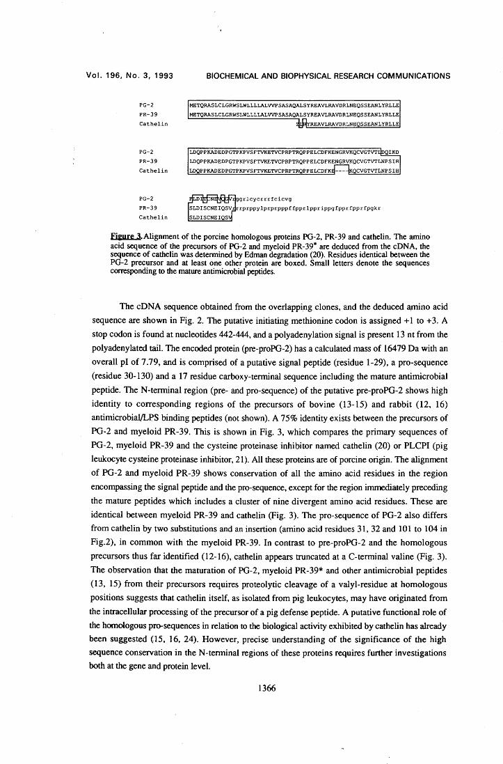
Vol. 196, No. 3, 1993
PG-2
PR-39 Cathelin
PG-2 PR-39 Cathelin
PG-2 PR-39 Cathelin
BIOCHEMICAL ANO BIOPHYSICAL RESEARCH COMMUNICATIONS
METQRASLCLGRWSLWLLLLALVVPSASAQALSYREAVLRAVDRLNEQSSEANLYRLLE METQRASLCLGRWSLWLLLLALVVPSASAQALSYREAVLRAVDRLNEQSSEANLYRLLE
REAVLRAVDRLNEQSSEANLYRLLE
LDQPPKADEDPGTPKPVSFTVKETVCPRPTRQPPELCDFKENGRVKQCVGTVT QIKD
Fieure 3.Alignment of the porcine homologous proteins PG-2, PR-39 and cathelin. The amino acid sequence of the precursors of PG-2 and myeloid PR-39* are deduced from the cONA, the sequence of cathelin was determined by Edman degradation (20). Residues identical between the PG-2 precursor and at least one other protein are boxed. Small letters denote the sequences corresponding to the mature antimicrobial peptides.
The cDNA sequence obtained from the overlapping clones, and the deduced amino acid sequence are shown in Fig. 2. The putative initiating methionine codon is assigned +l to + 3. A stop codon is found at nucleotides 442-444, and a polyadenylation signa! is present 13 n t from the polyadenylated tail. The encoded protein (pre-proPG-2) has a calculated mass of 16479 Da with an overall p l of 7 .79, and is comprised of a putative signa! peptide (residue 1-29), a pro-sequence (residue 30-130) and a 17 residue carboxy-terminal sequence including the mature antimicrobial peptide. The N-terminai region (pre- and pro-sequence) of the putative pre-proPG-2 shows high identity to corresponding regions of the precursors of bovine (13-15) and rabbit (12, 16) antimicrobial/LPS binding peptides (not shown). A 75% identity exists between the precursors of PG-2 and myeloid PR-39. This is shown in Fig. 3, which compares the primary sequences of PG-2, myeloid PR-39 and the cysteine proteinase inhibitor named cathelin (20) or PLCPI (pig leukocyte cysteine proteinase inhibitor, 21). Ali these proteins are of porcine origin. The alignment of PG-2 and myeloid PR-39 shows conservation of ali the amino acid residues in the region encompassing the signa! peptide and the pro-sequence, except for the region immediately preceding the mature peptides which includes a cluster of nine divergent amino acid residues. These are identica! between myeloid PR-39 and cathelin (Fig. 3). The pro-sequence of PG-2 also differs from cathelin by two substitutions and an insertion (amino acid residues 31, 32 and 101 to 104 in Fig.2), in common with the myeloid PR-39. In contrast to pre-proPG-2 and the homologous precursors thus far identified (12-16), cathelin appears truncated at a C-terminai valine (Fig. 3). The observation that the maturation of PG-2, myeloid PR-39* and other antimicrobial peptides (13, 15) from their precursors requires proteolytic cleavage of a valyl-residue at homologous positions suggests that cathelin itself, as isolated from pig leukocytes, may bave originated from the intracellular processing of the precursor of a pig defense peptide. A putative functional role of the homologous pro-sequences in relation to the biological activity exhibited by cathelin has already been suggested (J5, 16, 24). However, precise understanding of the significance of the high sequence conservation in the N-terminai regions of these proteins requires further investigations both at the gene and protein leve!.
1366
Vol. 196, No. 3, 1993 BIOCHEMICAL ANO BIOPHYSICAL RESEARCH COMMUNICATIONS
The 17 residue carboxy-terminal sequence of the predicted protein includes the complete sequence ofthe antimicrobial peptide protegrin PG-2 determined by Edman degradation (8). PG-2, as purified from pig leukocytes, consists of 16 residues. An additional glycine as the carboxy-terminal residue of the sequence deduced from the cD NA suggests that the mature peptide has a carboxy-terminal amidated residue (25). Although not experimentally determined, this post-translational modification is not contradicted by FAB-MS data (8). The sequence of this peptide is arginine-rich and is characterized by four cysteine residues. Among the known neutrophil antimicrobial peptides, a small arginine-rich peptide with an intramolecular disulfide bond, termed cyclic dodec.apeptide, has previously been recognized as a component of the protein family characterized by the cathelin-like domain (14). About 66% identity is shared by the amino acid sequences of the precursors of the dodecapeptide and PG-2. However, the conservation is confined to a region including pre- and prosequences (not shown), since the alignment of the C-terminai sequences corresponding to the mature antimicrobial peptides does not show any significant resemblance. As reported (8), the structure of the mature protegrin PG-2 is more related to the defensin family of antimicrobial peptides.
Two additional protegrins, each of 18 amino acid residues, have been purified from pig leukocytes (8). Most of the residues of these congeners, designated PG-1 and PG-3, are identica! to the mature PG-2 (8). This structural similarity strongly suggests that the precursor forms of PG-1 and PG-3, whose sequences have not been reported yet, may also carry a cathelin-like domain.
ACKNOWLEDGMENTS: This work was supported by grants from the National Research Council (Progetto Finalizzato "Biotecnologie e Biostrumentazione") and from the Italian Ministry for University and Research. W e thank Dr. D. Romeo for critically reviewing the manuscript.
REFERENCES
l. Lehrer, R. l. and Ganz, T. (1990) Blood 76, 2169-2181. 2. Boman, H. G. (1991) Cell65, 205-207. 3. Gennaro, R., Romeo, D., Skerlavaj, B., and Zanetti, M. (1991) in Blood Celi Biochemistry,
vol. 3, (J. R. Harris Ed.), pp. 335-368, Plenum Publ. Co., New York. 4. Elsbach, P., and Weiss, J. (1993) Curr. Opin. Immunol. 5, 103-107. 5. Gabay, J. E. and Almeida, R. P. (1993) Curr. Opin. Immunol. 5, 97-102. 6. Gennaro, R., Skerlavaj, B. and Romeo, D. (1989) Infect. Immun. 57, 3142-3146. 7. Selsted, M. E., Tang, Y., Morris, W. L., McGuire, P. A., Novotny, M. J., Smith,
W.Henschen, A. H., and Cullor, J. S. (1993) J. Biol. Chem. 268, 6641-6648. 8. Kokryakov, V. N., Harwig, S. S. L., Panyutich, E. A., Shevchenko, A. A., Aleshina, G.
M.,Shamova, O. V., Korneva, H. A. and Lehrer, R. I. (1993) FEBS Lett. 327, 231-236. 9. Lehrer, R. 1., Ganz, T., and Selsted, M. E. (1991) Cell64, 229-230.
10. Ganz, T., Rayner, J. R., Valore, E. V., Tumolo, A., Talmadge, K., and Fuller, F. (1989) J.Immunol. 143, 1358-1365.
11. Jones, D. E. and Bevins, C. L. (1992) J. Biol. Chem. 267, 23216-23225. 12. Larrick, J. W., Morgan, J. G., Palings, I., Hirata, M., and Yen, M. H. (1991) Biochem.
Biophys. Res. Commun. 179, 170-175. 13. Del Sal, G., Storici, P., Schneider, C., Romeo, D. and Zanetti, M. (1992) Biochem.
Biophys. Res. Commun. 187, 467-472. 14. Storici, P., Del Sal, G., Schneider, C., and Zanetti, M. (1992) FEBS Lett. 314,187-190. 15. Zanetti, M., Del Sal, G., Storici, P., Schneider, C., and Romeo, D. (1993) J. Biol. Chem.
268, 522-526. 16. Levy, 0., Weiss, J., Zarember, K., Ooi, C. E., and Elsbach, P. (1993) J. Biol. Chem. 268,
6058-6063.
1367
.l.
Vol. 196, No. 3, 1993 BIOCHEMICAL ANO BIOPHYSICAL RESEARCH COMMUNICATIONS
17. Selsted, M. E., Novotnj, M. J., Morris, W. L., Tang, Y-Q., Smith, W. and Cullor, J. S. (1992) J. Biol. Chem. 267, 4292-4295.
18. Romeo, D., Skerlavaj, B., Bolognesi, M. and Gennaro, R. (1988) J. Biol. Chem. 263, 9573-9575.
19. Ooi, C. E., Weiss, J., Levy, O. and Elsbach, P. (1990) J. Biol. Chem. 265, 15956-15962. 20. Ritonja, A., Kopitar, M., Jerala, R., and Turk, V. (1989) FEBS Lett. 255, 211-214. 21. Kakegawa, H., Nikawa, T., Tagami, K., Kamioka, H., Sumitani, K., Kawata, T., Drobnic-
Kosorok, M., Lenarcic, B., Turk, V. and Katunuma, N. (1993) FEBS Lett. 321, 247-250. 22. Chomczinski, P., and Sacchi, N. (1987) Anal. Biochem. 162, 156-159. 23. Sanger, F., Nicklen, S., and Coulson, A. R. (1977) Proc. Nati. Acad. Sci. USA 74, 5463-
5467. 24. Verbanac, D., Zanetti, M., and Romeo, D. (1993) FEBS Lett. 317, 255-258. 25. Bradbury, A. F. and Smyth, D. G. (1991) Trends Biochem. Sci. 16, 112-115.
Reprinted frorn Biochernical and Biophysical Research Cornrnunications 1%, 1363-1368 (1993) Copyright© 1993 Acadernic Press, Inc. Printed in U.S.A.
1368
.l
l
Communication
Molecular Cloning and Chemical Synthesis of a Novel Antibacterial Peptide Derived from Pig Myeloid Cells*
(Received for publication, November 10, 1993, and in revised form, January 5, 1994)
Margherita ZanettiH'Il, Paola Storici:J:Ii, Alessandro Tossill, Marco Scocchill, and Renato Gennaro§ From the '};Laboratorio Nazionale Consorzio Interuniversitario Biotecnologie, AREA Science Park, Padriciano, I-34012 Trieste, §Dipartimento di Scienze e Tecnologie Biomediche, Università di Udine, I-33100 Udine, and liDipartimento di Biochimica, Biofisica e Chimica delle Macromolecole, Università di Trieste, I-34127 Trieste, Italy
A group of myeloid precursors of defense peptides has recently been shown to bave highly homologous N-ter-minai regions. Using a strategy based on this homology, a nove! cDNA was cloned from pig hone marrow RNA and found to encode a 153-residue polypeptide. This comprises a highly conserved region encompassing a 29-residue signa! peptide and a 101-residue prose-quence, followed by a unique, 23-residue, cationic, C-terminai sequence. A peptide corresponding to this C-terminai sequence was chemically synthesized and shown to exert antimicrobial activity against both Gram positive and negative bacteria at concentrations of 2-16 J.Dl· The activity of this potent and structurally novel antibacterial peptide appears to be mediated by its abil-ity to damage bacterial membranes, as shown by the rapid permeabilization of the inner membrane of Escherichia coli.
Leukocytes are a key element in host defense against micro-biai infections. They act by killing microorganisms intracellu-Iariy, through both oxidative and nonoxidative pathways. The oxygen-independent system reiies on an arsenai of cationic, granuie-associated antimicrobial peptides and proteins, which are discharged into the phagocytic vacuoies concomitantiy with the production of toxic oxygen derivatives {1-3). A variety of these peptides have been isolated and shown to exert potent in vitro cidai activity (4-8). Among these are the bactericidall permeability-increasing protein (4), a cyclic dodecapeptide (9), indoiicidin (10), a number of small, cysteine-rich peptides clas-sified as defensins (5), /3-defensins (11) and protegrins (12), the proiine- and arginine-rich peptides Bac5 and Bac7 (13, 14), and
* This work was supported by grants from the National Research Council (Progetto Finalizzato Biotecnologie e Biostrumentazione) and from the ltalian Ministry for University and Research (MURST 60% an d 40% ). The costs of publication of this artici e w ere defrayed in part by the payment of page charges. This article must therefore be hereby marked "advertisernent" in accordance with 18 U.S.C. Section 1734 solely to indicate this fact.
The nucleotide sequence(s) reported in this paper has been submitted to the GenBank™ l EMBL Data Bank with accession number(s) L26053.
11 To whom correspondence should be addressed: Laboratorio Nazio-nale Consorzio Interuniversitario Biotecnologie (C!B), AREA Science Park, Padriciano, 99, I-34012 Trieste, Italy. Fax: 39-40-398985.
ALLEGATO F
THE Jot'R:-<AL m BJO~CJGICA.:. CHE?>IIl'TR) Yol. 269. ~o 11. lssue of :\Iarch 18. pp. 7855- i858, 1994
l[ 1994 by The American Society for Biochemistry and Molecular Biolog:~.·. !ne. Printed in CS.A.
the serprocidins ( 15 ). These peptides show significant diversity in structure, spectrum of activity, and species distribution.
cD NA an d genomi c sequences, when known, ha ve shown that these peptides are synthesized in hone marrow cells as precur-sors (preproproteins) from which the mature peptides are de-rived by proteolytic processing (5, 15-23).
W e ha ve recently reported that the precursors of a number of structurally unreiated antimicrobiai peptides share highly ho-mologous N-terminai regions encompassing the signai peptide and the prosequence (17-21). The common prosequence is ho-mologous to a polypeptide from porcine leukocytes, termed cathelin (24). A group of defense peptide precursors thus exists, in which a structurally highly varied C-terminai region, dis-playing antimicrobial (9, 10, 12, 13) andlor lipopoiysaccharide binding activity (25) after processing, is attached to a highiy conserved N-terminai region.
mRNA sequences encoding proteins with these features ha ve been identified in bovine (17-19), porcine (20, 21), and rabbit (25, 26) hone marrow cells, suggesting that further unknown defense peptide precursors of this type might exist. In view of the high conservation of the mRNA 5'-sequence, a study was thus undertaken to identify mRNA sequences encoding the pre-cursors of such novei antimicrobiai peptides in pig myeloid cells. A similar approach was followed by Jones and Bevins (27) to obtain the cD NA of human defensin-5 from Paneth cells by exploiting the high conservation of mRNA sequences in the 5' -region of rabbit and human defensin precursors.
By using a molecular biological approach based on amplifi-cation of cDNA ends containing the conserved proregion ho-mologous to cathelin, we were abie to clone several porcine myeloid cDNAs coding for the precursors of potential antimi-crobial peptides.
In this paper we report the cloning of one such cDNA encod-ing a putative antimicrobial peptide precursor with a unique, highiy cationic, 23-residue-long C-terminai sequence. A peptide corresponding to this sequence was chemically synthesized and shown to possess a potent in vitro antimicrobial activity against Gram-positive and -negative bacteria. This peptide was termed PMAP-23, from "porcine myeioid antibacterial peptide" of 23 residues.
EXPERIME11-"TAL PROCEDURES cDNA Cloning and Sequencing-Total RNA was extracted from pig
hone marrow cells with guanidinium thìocyanate (28). The reagents and generai methodology used to obtain the 3 '-end cDNA were as previously described (18). In particular, reverse transcription was performed using the primer adaptor 5'-TCGGATCCCTCGAGAAGC(TlS)-3'. Amplifica-. tion was done with the antisense primer adaptor 5' -CGAGCTCGGATC-CCTCGAGAAGCTT-3' and a sense oligonucleotide 5'-CGCGAATTCT-GTGAGCTTCAGGGTG-3'. The latter oligonucleotide was derived from a highly conserved sequence of the proregion of the Bac5 precursor cDNA (18), also present in other antimicrobial peptide precursors (17, 19-21). The experimental conditions to obtaìn the 5'-end cDNAwere as described (21). Briefly, the 5'-region of PMAP-23 precursor cDNA was obtaìned by reverse transcription of porcine hone marrow mRNA with the antisense olìgonucleotide primer 5'-TCTGACCCATACAGTCA-CAAAT-3' complementary to nucleotides 438-459 ofthe 3'-end (Fig. 1), internai to the unique antibiotic domain of the PMAP-23 precursor. Amplification was performed using the sense oligonucleotide primer 5'-ACCGAATTCACCTGGGCACCATG-3' from the highly conserved 5'· end (including nucleotides -13 to +3, Fig. l) and an antisense oligo-nucleotide 5' -CCGAATTCCGACGTACTCTCCACAGCAG-3' derived from nucleotides 403-423 in the antibiotic domaìn. The amplified cDNA was cloned in Bluescript SK+ vector (Stratagene, San Diego, CA) and sequenced as previously described ( 18).
7855
7856 A Not·el Pig Myeloid Antibacterial Peptide
Sequence Analyses-D::\'A sequence analysis was conducted v.ith the ai d ofthe IG suite version 5.4 i IntelliGenetics Inc .. l\1ountain View, CA1. Homology searches were carried out on the Swiss-Prot data base using the FastDB an d Genalign programs. Prediction of the seconda!")· struc-ture ofthe peptide was obtained using the "PeptideStructure~ and '·Pep" programs in the GCG version 7 <Genetics Computer Group Inc .. .Madi-son, WIJ and IG suites, respectively.
.Vorthern and Primer Extension Analyses-Northern and primer ex-tension analyses were performed as described (17 J. A synthetic anti-sense oligonucleotide 5'-TCTGACCCATACAGTCACAAAT-3', 32P-Ja-beled using standard protocols, was used to hybridize a ~orthern blot of pig hone marrow total R.l\JA and for primer extension analysis of PMAP-23 mRNA.
Peptide Synthesis-A Milligen 9050 synthesizer loaded with Fmoc-Arg1 substituted PEG-PS resin W. l mmol) (Milligen, Bedford, MAl was used for solid-phase synthesis. For each coupling step, 0.8 mmol each of Fmoc-protected amino acid (l\iilligen or Novabiochem, Laufelfingen, Sv.;tzerlandJ, N-hydroxybenzotriazole (AJdrich-Chemie, Steinheim, Germany), and 2-(lH-benzotriazole-I-y!)-1,1,3,3-tetramethyluronium tetrafluoroborate (Novabiochem) were used. Side chains were protected as follows: trityl (GJn). t-butyl (Glu, Asp, Thr), t-butyloxycarbonyl (Lys, TrpJ, and 2.2,5,7,8-pentamethylchroman-6-sulfonyl <Argl. Deprotection and cleavage from the resin were carried out using a mixture of 90'ìc trifluoroacetic acid, 29é each of phenoL thioanisole. ethanedithiol, an d triisopropylsilane, and l '7c each ofmethanol and water. The peptide was then repeatedly extracted with ether and purified by reverse phase HPLC on a C18 colurnn <Delta-Pak, Waters. Bedford. MAl, using a 0-609é acetonitrile gradient in 0.059é trifluoroacetic acid.
Analytical Assays-The peptide concentration was determined from tryptophan absorbance <29), the amino acid analysis was performed using the Pico-Tag system <Waters) 130), and the molecular mass was determined v.;th an API III io n spray mass spectrometer ( PE SCIEX. Toronto, Canada). Circular dichroism was carried out on a Jasco J-600 spectropolarimeter with a celi path length of 2 mm. Peptide samples <0.1 mg/m!l in 5 ID.\1 sodium phosphate buffer. pH 7.0, containing 0-45'ìc (v/v 1 trifluoroethanol were used.
Antibacterial and Membrane Permeabilization Actù:ities-The mini-mal inhibitory concentration of the purified peptide was determined as pre\;ously described ( 13) against Escherichia coli l\!L-35 and ATCC 25922, Salmonella typhimurium ATCC 14028, Pseudomonas aerugi-nosa ATCC 27853, Bacillus megaterium ']oca! isolate), and Staphylo-coccus aureus ATCC 25923 strains. Permeabilization of the inner mem-brane of the Jactose permease-deficient. 13-galactosidase-constitutive E. coli ML-35 strain was evaluated as reported <31) by following the un-masking of 13-galactosidase. Total 13-galactosidase activity was deter-mined v.;th bacteria lysed by ultrasonication. Hemolytic acti\;ty was determined as described (32J by using human er)"throcytes prepared from fresh, anticoagulated blood. Zero Cblank) and 100'/c hemolysis were evaluated in the absence of additives and the presence of 0.29é Triton X-100, respectively, while melittin iSigmaJ was used as a positive con-tro!.
RESULTS A.~D DISCUSSION
A search was carried out for porcine hone marrow transcripts with sequence homology with those previously described for bovine (17-19) and rabbit (25, 26) defense peptide precursors. An approach based on polymerase chain reaction amplification of cDNA ends, using oligonucleotide primers derived from the
. 5'-region that is common to ali these precursors, allowed the identification of severa} novel cDNA sequences. Two of these were found to encode the precursors of the antibacterial pep-tides PR-39 (20) and protegrin PG-2 (21), respectively, whereas a third encoded the putative precursor of a novel peptide, termed PMAP-23, with potential antibacterial activity. The se-quence of this cDNA, shown in Fig. l, was obtained from two polymerase chain reaction-generated, overlapping clones ex-tending from nucleotide -13 to +423 (5'-end cDNA) and from nucleotide +224 to the polyadenylated tail (3'-end cDNA). A corresponding transcript of approximately O. 7 kilobase (not shown) was detected by probing a Northern blot of pig hone marrow total RNA with an antisense oligonucleotide derived
1 The abbreviations used are: Fmoc, N-(9-fluorenyl)methoxycarbonyl; HPLC, high pressure liquid chromatography.
A:'GGAGA:::AGAGSG::AGCC':'G':G:CTGGGGC:GC:'G~':':AC':'G
M E ':' ': F. h S !.. C : G R \>i S :_ --
~E TGGC':'TCTGCTGC~GGGAC~CGTGG':'GCCCTCGGCCAG:~:CCA.G
li L L L L G L V V P S A S A ;; 30 .,. c· G':C:':'CAGC:'ACAGGGhGGCCGTGCTTCGTGCTG'l'GGJ..7C:;::-:-c
A :., S Y R E A V L R A V ~ R l ~5
: 3 é AACGAGCAGTCCTCGGAAGCTAATCTC':' ACCGCC'!'CC:'GGJ..GC':G N E Q S S E A N L Y R L L E L 60
: 8 l GACCAGCC:;CCCAAGGCCGACGAGGACCCGGGCACCCCAAhACCT D Q P P K A D E D P G T P K P 75
:2 6 GTGAGCTTCAC:GGTGAAGGAGACTGTGTGTCCCAGGCCGAC:CCGG V S F T V K E T V C P R P ':' F. 90
CAGCCCCCGGAGCTGTG'!'GACTTCAAGGAGAACGGGCGGG':'GAAG Q P P E L C D F K E N G R V K l 05
3:6 CAGTG':'G7GGGGA:::AGT:::ACC':'TGAAAGAGA':'CAGAGGCAAC'!":'7 C C V G T V ':' L K E I R G N F :2C
3 E l GA':A':' AACC'!'G':' AA T::AGC'I"!'CAGAG7G7ChGGATTATAGACCTG D I T C N Q L Q S V R I I D L :::5 --------L--~--~--~--- i--~-- --~--13---~--------
4 8 6 C7G7GGAGAG7ACG7CGGCCACAGAAACCCAAAT':'TG':'GA:::':!'G':"A L W R V R R P Q lt P JC F V 'l' V :se
---------i -------l.-----------1--------/3------~51 TGGG':'CAGA ':' AA T':'AGTGCATGAAGGAA '!'':"TCATTCT'!'C:AAGACA
W V R * 153 --------l
~?E CAGGGAAGAG':'TCT~t.AAG~TCTGC'!'':'ATTCTACCACAA3C:"':'C:AG
Frc. l. Nucleotide and deduced amino acid sequences and sec· ondary structure prediction for PMAP-23. Numbering is on the left for nucleotide::; and on the right for amino acids. The end ofthe putative signa! sequence is indicated by an arrou·, the stop codon by an asterisk, and the polyadenylation signa! by a double underline. The sequence of the putative antibacterial peptide is shown in boldface. The position of the predicted 13-sheet ((3) and loop !Lì conformations is also shovm.
from a unique 3 '-end sequence complementary to nucleotides 438-459 in Fig. l. A short 5 '-untranslated regio n of approxi-mately 16 nucleotides was indicated by primer extension anal-ysis of the mRNA.
Nucleotide sequence analysis predicted an open reading frame of 153 codons corresponding to a protein of 17,487 Da, with a calculated pl of 9.62. An extended N-terminai region of this putative protein (Fig. l) was found to have ali the struc-tural features common to previously characterized porcine (20, 21) and bovine (17-19) defense peptide precursors with cathe-lin-like proregions. A comparison of this protein with the ho-mologous porcine preproPG-2, preproPR-39, and cathelin is shown in Fig. 2. The conserved region includes a 29-residue-long putative signa} peptide and a 101-residue-long prose-quence that is 83% homologous to cathelin (24). A 23-residue C-terminai sequence follows this domain immediately after a valyl residue at position 130. This residue is conserved in most of the previously characterized members of this precursor group (Fig. 2 and Refs. 17, 18, 20, 21) and is like]y to be the proteolytic cleavage site responsible for the release of active peptide (17, 18, 20, 21, 23). The 23-residue C-terminai sequence did not reveal any significant similarity to catalogued se-quences in the Swiss Prot data base.
This sequence is highly cationic, containing 5 arginines and 2 lysines and only l negatively charged residue. The sequence also contains 11 highly hydrophobic residues, including 2 tryp-tophans. This suggests some form of amphipathic structure, which is typical of antibacterial peptides, but the presence of 2 prolines in the centrai region of the sequence precludes an extended a-helical conformation. Secondary structure predic-tion analysis indicated that PMAP-23 may consist of an anti-parallel t3~sheet connected by a loop in the region of residues 9-15, thus adopting a hairpin-like structure. This does not
A .Vocel Pig Afyeloid Antibacterial Pcptide 7857
?G-2
FIG. 2. Alignment ofPMAP-23 with PG-2, PR-39, and cathelin. The amino acid sequence ofpreproP:\1.-\.P-23. deduced from cD~A. is aligned v.ith the sequences of preproPG-2 121 l and preproPR-39 120), also deduced from pig myeloid cDKA, and that of cathelin determined by Edman degradation ( 241. Boxe d residues reflect those common to preproPI\1.A.P-23 an d a t Jeast one other polypeptide.
resemble the a-helical <33, 34J or disulfide bond-stabilized 13-sheet <35) structures already found for antimicrobial pep-tides.
The peptide PMAP-23 (RIIDLL\VRVRRPQKPKFVTY\\'VR) corresponding to the C-terminai residues 131-153 in Fig. l was synthesized by the automated solid-phase method and purified to homogeneity by HPLC. The peptide was shown to be correct by mass determination (2962.5 Da versus the calculated mass of 2962.7 Da) and amino acid analysis (not shown).
To test the predicted structure of synthetic PMA.P-23. circu-lar dichroism spectra were recorded in an aqueous em·iron-ment and in the presence oftrifluoroethanol, an organic solvent that mimics the membrane environment. The spectrum in aqueous buffer resembled that of a random coil peptide, but a considerable change was observed at 309c or more trifluoro-ethanol (not shown). The interpretation of these spectra and structural analysis were complicateci by the presence ofthe two tryptophan residues, and no predominant conformation was evident. The spectra, however, did not preclude the presence of the predicted 13-sheet and coil conformations. Attempts will be made to obtain the solution structure of P!\IAP-23 by ~MR spectroscopy, as the synthetic approach allows the facile pro-duction of pure peptide in the amounts required for this tech-nique.
PMAP-23 displays a remarkable in vitro antibacterial activ-ity against severa} species of bacteria when tested by the mini-mal inhibitory concentration assay <Table l). Gram-negative CE. coli and S. typhimurium) and Gram-positive (S. aureus and B. megaterium) bacteria were equally susceptible to PMAP-23 a t concentrations ranging from 2 to 8 P,.\1. Even the growth of P aeruginosa, a Gram-negative species often resistant to antimi-crobial peptides, was suppressed at 16 J.lM.
The predicted amphipathic nature of PMAP-23 suggested that its activity might be related to bacterial membrane dam-age. Accordingly, permeabilization experiments were per-formed on the E. coli ML-35 strain, and a rapid, PMAP-23-induced permeabilization of the bacteri al inner membrane was observed (Fig. 3). The kinetics of permeabilization was meas-ured after addition of l and 10 p..\1 peptide, and a steady state was reached at 9 and 7 min, respectively, after a lag time of about 2 min. This kinetics is comparable with that of the pro-line- and arginine-rich Bac5 and Bac7 (31) and considerably faster than that observed for human defensins (36), measured on similar systems. Conversely, the peptide failed to lyse hu-man erythrocytes even at a concentration of 100 J.l.\t, suggesting a certain degree of target membrane specificity.
Our results thus clearly show that synthetic PMAP-23 is a potent, membrane-active antibacterial agent and suggest that a natura! PMAP-23 derived from the precursor here described is a component of the leukocyte defense system.
The combined molecular biological'chemical synthesis ap-proach we have devised has proved a powerful tool in rapidly identifying this new antibacterial peptide precursor "rith a cathelin-like prosequence (17-21, 25, 26). The usefulness of this approach is further supported by the identification of two additional cDNAs belonging to this group of precursors and
TABLE I Antibacterial activity of PMAP-23
The minima! inhibitory concentration CMICJ was defined as the low-est PMA.P-23 concentration that prevented visible bacterial growth af-ter 18 h of incubation with approximately 1.5 x 10⁵ colony forming units/ml at 3i cc in Mueller-Hinton broth (13). These results are the mean of at least five independent determinations with a divergence of not more than one minima! inhibitory concentration value.
Organisrn and strain
E. coli ML35 E. coli ATCC 25922 S. typhimurium ATCC 14028 P aeruginosa ATCC 27853 B. megaterium <local isolate) S. aureus ATCC 25923
... I ....
-'~ IO min. 20
~ne
UM
2 4 8
16 2 4
FIG. 3. Permeabilization of E. coli ML-35 inner membrane by PMAP-23. Permeabilization is determined by following the unmasking of cytoplasmic j3-galactosidase activity spectrophotometrically at 405 nm. Bacteria labout 107/ml) were suspended in 10 mM sodium phos-phate buffer. pH i.5, containing 100 mM NaCl and 1.5 mM substrate 130). Trace a, untreated bacteria; traces band c, l and 10 JlM PMAP-23, respectively. The arrow indicates the addition of PMAP-23. Results are representative of three independent experiments.
encoding two novel antibacterial peptides.2 The same strategy may thus be applied in determining how extensively this pro-tein family is represented in different animai species.
Acknowledgments-We are indebted to Prof. Mario Furlanut, Direc-tor ofthe Farmacologia Clinica e Tossicologica Service ofthe University ofUdine, for mass determination and to Prof. Domenico Romeo from the Dipartimento di Biochimica, Biofisica e Chimica delle Macromolecole, Università di Trieste, for critically reading the manuscript.
REFERENCES l. Joiner, K. A., Ganz, T., Albert, J ., and Rotrosen, D. (1989) J. Celi Biol. 109,
2771-2782 2. Zanetti, M., Lìtteri, L., Griffiths, G., Gennaro, R., and Rorneo, D. (1991) J.
Immunol. 146, 4295-4300 3. Klebanotr, S. J. (1988) in Inflammation: Basic Principles and Clinical Corre-
lates tGallin, J. L, Goldstein, I. M .. and Snyderrnann, R., edsl pp. 391--444, Raven Press, New York
4. Elsbach. P., and Weiss, J. (19931 Curr. Opin. Immunol. 5, 103-107 5. Le ber, R. I.. Lichtenstein, A. K., and Ganz, T. (1993) Annu. ReL·. Immunol. 11,
105-128
2 M. Zanetti, manuscript in preparation.
7858 A Xovel Pig lvfyeloid A.ntibacterial Pcptide
6. Gennaro. R., Romeo. D .. Skerlavaj. B .. and Zanetti. ;\L '1391' in Blood Ccii Bìochemistrv < Harris. J. R.. ed' Vol 3. pp. 335-368, Plenum Pubhshing Corp .. :\'ew -York
7. Boman, H. G. <1991 J Celi 65, 205-20';' 8. Zasloff, M. 11992.> Curr. Opin. Immunol. 4, 3-': 9. Romeo. D., Skerlavaj. B .. Bolognesi. M .. an d Gennaro. R. <1988: J. Bio!. Che m.
263, 9573-9575 10. Selsted. M. E., Novotny. M. J .. ~lorris. W. L.. Tang. Y-Q .. Smith. W .. and Culi or.
J. S. 119921 J. Biol. C.'-:.em. 267, 4292-4295 11. Selsted. M. E., Tang, Y-Q., Morris, W. L., McGuire. P. A., !\ovotny. :'>1. J .. Smith,
W, Henschen, A. H., and Cullor, J. S. i1993ì J. Biol. Che m. 268, 6641-6648 12. Kokryakov, V. N., Harwig, S. S. L., Panyutich. E. A.. Shevchenko, A. A.,
Aleshina, G. M .. Shamova, O. V., Korne,·a. H. A .. and Leher. R. I. (19931 FEBS Lett. 327, 231-236
13. Gennaro, R., Skerlavaj, B., and Romeo, D. l1989.' Jnfect. lmmun. 57, 3142-3146
14. Frank. R. W, Gennaro, R.. Schneìder, K, Przybylski M., an d Romeo, D. 1 19901 J. Biol. Chem. 265, 18871-18874
15. Gabay. J. E .. and Almeida, R. P. 11993) Curr. Opin. lmmunol. 5, 97-102 16. Daher, K A., Lehrer, R. I., Ganz, T., and Kronenberg, M. i1988) Proc. Natl.
Acad. Sci. U. S. A. 85, 7327-7331 17. Del Sal, G., Storici, P., Schneider, C., Rorneo. D., and Zanetti, M. (19921 Bio·
chem. Biophys. Res. Commun. 187, 467-472 18. Zanetti, M., Del Sal, G .. Storici, P., Schneider, C., an d Romeo, D. ( 19931 J. Bio/.
Chem. 268, 522-526 19. Storici, P., Del Sal, G., Schneider. C .. and Zanetti. M. 11992' FEBS Lett. 314,
187-190 20. Storici. P., and Zanetti, M. 11993: Biochem. Biophys. Res. Com m un. 196, 1058-
1065 21. Storici. P.. and Zanetti. :--.1.1 1993,Biochcm. Biophyc;. Reo. CommuTi. 196, 1363-
1368 22. Zanetti. M .. Litteri, L.. Gennaro. R .. Horstmann. H .. and Romeo. D. <1990< J.
Celi Bio/. 111, 1363-13':1 23. Scocchi, ;\1., Skerlavaj, B., Romeo. D., an d Gennaro. R.' 1992. Eu r. J. Bwchem.
209, 589-595 24 Rìtonja, A., Kopitar, M .. Jerala. R., and Turk. Y. 11989, FEBS Lctt 255, 211-
214 25. Larrick. J. W, Morgan, J. G., Palings, I., Hirata, M., and Yen, :'>1. H. <1991t
Biochem. Biophys. Res. Commun. 179, 17(}-.175 26. Levy. 0., Weiss, J., Zarember, K, Ooi, C. E .. and Elsbach. P. 1 1993.• J. Biol.
Chem. 268, 6058-6063 27. Jones, D. E., and Bevins, C. L. 11992) J. Biol. Chem. 267, 23216-23225 28. Chomczinski, P .. and Sacchi, N. (1987) Anal. Biochem. 162, 156-159 29. Edelhoch, H. !19671 Biochemistry 6, 1948-1954 30. Cohen, S. A., and Strydom, D. J. (1988)Anal. Biochem. 174, 1-16 31. Skerlavaj, B., Romeo, D., and Gennaro, R. <1990\lnfect. lmmun. 58, 3724-
3730 32. Zasloff, M. (19871 Proc. Nati. Acad. Sci. U. S. A 84, 5449-5453 33. Marion, D., Zasloff, M .. and Bax, A. (1988) FEBS Lett. 227, 21-26 34. Holak, T. A., Engstrom, A, Kraulis, P. J., Lindeberg, G., Bennich, H., Jones, T
A., Gronenbom.A. M., and Clore, G. M. (1988) Biochemistry· 27, 762(}-.7629 35. Hill, C. P., Yee, J., Selsted, M. E., and Eisenberg, D. 11991 l Science 251,
1481-1485 36. Lehrer, R. l., Barton, A., Daher, K A., Harv•ig, S. S. L., Ganz, T.. and Selsted.
M. E. <1989' J. Clin. lnvest. 84, 553-561
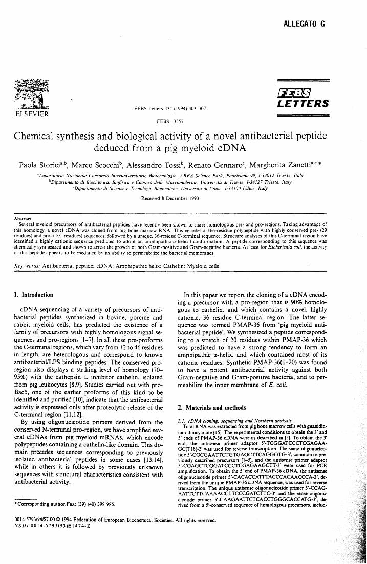
ALLEGATO G
FEBS Letters 33'7 (1994) 303-307
[i[j/]]fJ LETTERS
FEBS 13557
Chemical synthesis and biologica! activity of a novel antibacterial peptide deduced from a pig myeloid cDNA
Paola Storicia,b, Marco Scocchib, Alessandro Tossib, Renato Gennaroc, Margherita Zanettia,c.* 'Laboratorio l'·la=ionale Consor=io 1nteruniversiwno Biotecnologie, AREA Science Park. Padriciano 99, J-34012 Trieste. Italy
b Dipartimento di Biochimica. Biofisica e Chimzca delle Afacromolecole. Università di Trieste, 1-34127 Trieste. 1taf.v 'Dipartimento di Scien=e e Tecnologie Biomediche. Università di L'dine. 1-33100 Udine. ltal_v
Received 8 December 1993
Abstract Severa! myeloid precursors of antibacterial peptides have recently been shown to share homologous pre- and pro-regions. Taking advantage of
this homology, a nove! cONA was cloned from pig bone marrow RNA. This encodes a 166-residue polypeptide with highly conserved pre- (29 residues) and pro- (101 residues) sequences, followed by a unique. 36-residue C-terminai sequence. Structure analyses ofthis C-terminai region have identified a highly cationic sequence predicted to adopt an amphipathic :z-helical conformation. A peptide corresponding to this sequence was chemically synthesized and shown t o arrest the growth of both Gram-positive and Gram-negative bacteria. A t least for Escherichia coli. the activity of this peptide appears to be mediated by its ability to permeabilize the bacterial membranes.
Key words: Antibacterial peptide; cDl\'A; Amphipathic helix: Cathelin; Myeloid cells
l. lntroduction
cDNA sequencing of a variety of precursors of anti-bacterial peptides synthesized in bovine, porcine and rabbit myeloid cells, has predicted the existence of a family of precursors with highly homologous signa} se-quences and pro-regions [1-7]. In ali these pre-profonns the C-terminai regions, which vary from 12 to 46 residues in length, are heterologous and correspond to known antibacterial/LPS binding peptides. The conserved pro-region also displays a striking level of homology (70-95%) with the cathepsin L inhibitor cathelin, isolated from pig leukocytes [8,9]. Studies carried out with pro-Bac5, one of the earlier profonns of this kind to be identified and purified [10], indicate that the antibacterial activity is expressed only after proteolytic release of the C-terminai region [11,12].
By using oligonucleotide primers derived from the conserved N-terminai pro-region, we bave amplified sev-era! cDNAs from pig myeloid mRNAs, which encode polypeptides containing a cathelin-like domain. This do-main precedes sequences corresponding to previously isolated antibacterial peptides in some cases [13.14], while in others it is followed by previously unknown sequences with structural characteristics consistent with antibacterial activity.
• Corresponding author.Fax: (39) (40) 398 985.
In this paper we report the cloning of a cD NA encod-ing a precursor with a pro-region that is 90% homolo-gous to cathelin, and which contains a novel, highly cationic. 36 residue C-terminai region. The latter se-quence was termed PMAP-36 from 'pig myeloid anti-bacterial peptide'. We synthesized a peptide correspond-ing to a stretch of 20 residues within PMAP-36 which was predicted to have a strong tendency to fonn an amphipathic a-helix, and which contained most of its cationic residues. Synthetic PMAP-36(1-20) was found to have a potent antibacterial activity against both Gram-negative and Gram-positive bacteria, and to per-meabilize the inner membrane of E. coli.
2. Materials and methods
2.1. cDNA c/oning, sequencing and Northern analysis Total RNA was extracted from pig bone marrow cells with guanidin-
ium thiocyanate [15]. The experimental conditions to obtain tbe 3' and 5' ends of PMAP-36 cONA were as described in [5]. To obtain the 3' end, the antisense primer adaptor 5'-TCGGATCCCfCGAGAA-GC(TlS)-3' was used for reverse transcription. The sense oligonucleo-tide 5' -CGCGAATICfGTGAGCITCAGGGTG-3', common to pre-viously described precursors [1-5), and the antisense primer adaptor 5'-CGAGCfCGGATCCCfCGAGAAGCIT-3' were used for PCR amplification. T o obtain the 5' end of PMAP-36 cONA, tbe antìsense oligonucleotide primer 5'-CACACCATTTACCCACAACCCA-3', de-rived from the unique PMAP-36 cONA sequence, was used for reverse transcription. The unique antisense oligonucleotide primer 5'-CCAG-AAITCITCAAAACCITCCCGATCITC-3' and the sense oligonu-cleotide primer 5'-CAAGAAITCfCACCfGGGCACCATG-3', de-rived from a 5' -conserved sequence of homologous precursors, includ-
0014-5793/94/$7.00 O 1994 Federation of European Biochemical Societies. Ali rights reserved. SSD1 0014-5793(93)E 1474-Z
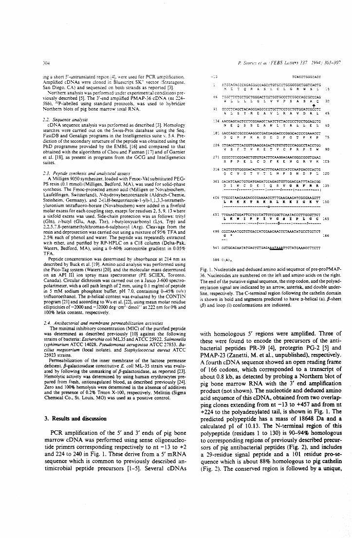
304
mg a short 5'-untranslated reg10n [4]. were used for PCR ampìification. Amplified cD~As were cloned in Bluesc:ipt sK· vector fStratagene. San Diego. CA) and sequenced on both strands as reported (3].
~orthern analysis was performed un der ex peri menta! conditions pre-viously described [5]. The 3'-end amplified PMAP-36 cD~A lnt 224--586 ), 3"P-labelled using standard protocols. was used t o hybridize Northern blots of pig bone marrow total RNA.
2.2. Sequence anafysis cDNA sequence analysis was performed as described [3]. Homology
searches were carried out on the Swiss-Prot database using the Seq. FastDB and Genalign programs in the lntelligenetics suite v. 5.4. Pre-diction of the secondary structure of the peptide was obtained using the PhD programme provided by the EMBL [ 16] an d compared t o that obtained with the algorithms of Chou and Fasman [17] and of Garnier et al. [ 18], as present in programs from the GCG an d lntelligenetics suites.
2.3. Pepride synchesis and analytical assays A Milligen 9050 synthesizer. loaded with Fmoc-Val substituted PEG-
PS resin (0.1 mmol) (Milligen. Bedford. MA), was used for solid-phase synthesis. The Fmoc-protected amino acid (Milligen or ;-.;ovabiochem. Laufelfingen. Switzerland), N-hydroxybenzotriazole (Aldrich-Chemie. Steinheim. Germany). and 2-(1 H-benzotriazole-1-yl)-1, 1.3.3-tetrameth-yluronium tetrafluoro-borate (Novabiochem) were added in a fivefold molar excess for each couplìng step, except for residues 2, 5, 9. 13 where a sixfold excess was used. Side-chain protection was as follows: trityl (Gin), r-butyl (G1u, Asp, Thr), c-butyloxycarbonyl (Lys, Trp) and 2.2.5. 7 .8-pentamethylchroman-6-sulphonyl (Arg). C1eavage from the resin and deprotection was carried out using a mixture of95% TFA and 2.5% each of phenol and water. The peptide was repeatedly extracted with ether. and purified by RP-HPLC on a CIS column (Delta-Pak. Waters. Bedford, MA), using a 0--60% acetonitrile gradient in 0.05% TFA.
Peptide concentration was determined by absorbance at 214 nm as described by Buck et al. [ 19]. Amino aci d analysis was perforrned using the Pico-Tag system (Waters) [20]. and the molecular mass deterrnined on an API III ion spray mass spectrometer (PE SCIEX, Toronto. Canada). Circular dichroism was carried out on a Jasco J-600 spectro-polarimeter. with a celi path length of 2 mm. using 0.1 mg/ml of peptide in 5 mM sodium phosphate buffer. pH 7.0. containing ~5% (v/v) trifluoroethanol. The a-helical content was evaluated bv the CONTIN program [21] and according to Wu et al. [22]. using mea~ molar residue ellipticities of -2000 and -32000 deg· cm:· dmol- 1 at 222 nm forO% and 100% helix content, respectively.
2.4. Antibacterial and membrane permeabili=ation acrivities The minima! inhibitory concentration (MI C) of the purified peptide
was determined as described previously [10) against the following strains ofbacteria: Escherichia coli ML35 and ATCC 25922. Sa/monella typhimurium ATCC 14028. Pseudomonas aeruginosa ATCC 27853. Ba-cil/us megaterium (local isolate). and Staphylococcus aureus ATCC 25923 strains.
Permeabilization of the inner membrane of the \actose perrnease deficient. P-galactosidase constitutive E. coli ML-35 strain was evalu-ated by following the unmasking of ft-galactosidase. as reponed [23]. Hemolytic activity was determined by using human erythrocytes pre-pared from fresh, anticoagulated blood, as described previously [24]. Zero an d l 00% hemolysis were determined in the absence of additi ves and the presence of 0.2% Triton X-100, respectively. Melittin (Sigma Chemical Co., St. Louis, MO) was used as a positive control.
3. Results and discussion
PCR amplification of the 5' and 3' ends of pig hone marrow cDNA was performed using sense oligonucleo-tide primers corresponding respectively t o n t -13 t o + 2 and 224 to 240 in Fig. l. These derive from a 5' mRNA sequence which is com.mon to previously described an-timicrobial peptide precursors [1-5]. Several cDNAs
P Sumn ci ai FEBS LL'ltcrs 337 ]YY4,: 303-307
-:3
J..:-:::;::;A.GACCCAGAGGGCCAGCCTGTG:c:-:::;GGGCGC':"GG'ICACTG ME':'QRhSLCLGRWSL
< 6 :::;::;C":"':'CTGC':GCTGGGACTC~"l'GG':"GCCCTCGGCCAGO:GCC:::A~
loiLLLLGLVVPSAShQ 't
91 GCCCTCAGCTACAGGGAGGCCGTGC':TCGTGCTGTGGATCGCCTC ALSYREAVLRAVDRL
13 6 MCGAGCAGTCCTCGGAAGC':: AATC'l'C'!'ACCGCCTCCTGGAGC':'G NEQSSEANLYRLLEL
:81 GACCAGCCGCCCAAGGCCGACGAGGACCCGGGCACCCCGAAACC':: ::lQPPKADEDPGTPKP
2 2 6 GTGAGC'::TCACGG'l'GAAGGAGACTGTGTGTCCCAGGCCTACC'!'GG VSFTVKETVCPRPTW
2 71 CGGCCCCCGGAGCTGTGTGACTTCAAGGAGAACGGGCGGG'!'GAAG RPPE:LCDFKENGRVK
316 CAGTGTGTGGGGACAGTCACC1'TGAACCCATCCAATGACCCACTG
30
60
"75
90
:es
Q C V G T V "; L N P S N D P L :20
3 61 GACATCAACTGTGATGAGATCCAGAGTGTTGGACGAT':'TAGACGG ::l l N C D E l Q S V G Il F Il Il :35 ------(------1-----; ------t-----1-----------1
4 O 6 ':'TGCGTAAGMGACCCGAAAACG'l'':TGAAGAAGATCGGGAAGGT'!' I. P. lt IC 1' P. IC P. I. Jt 1: I G Jt V :so
i --------------------«----------------------4 51 ";':'GAAGTGGATTCC'l'CCCATTGTCGGC'l'CAATACCCT'I'GGG':"l'GT
I. IC W I P P I V G 8 I P I. G C 165 ----------- l ----t--- l -----r.----- 1------t------
4 9 6 GGCT AAATGGTGTG~CCATGGAAGAACCCT AAATATGCCTGC'!'C'!' c; • :66
--!
541 GGTGC~CAA'!'ATGAGTC'!'GAG~T'l'C'!'ATGAAAGCTTCTT
586 C(A)n
Fig. l. !'o.'ucleotide and deduced amino aci d sequence of pre-proPMAP-36. ~ucleotides are numbered on the left and amino acids on the right. The end of the putative signa! sequence, the stop codon, and the polyad-enylation signa! are indicated by an arrow, asterisk, and double under-line. respective1y. The C-terminai region following the cathelin domain is shown in bold and segments predicted to have a-helical (a), P-sheet (/3) and loop (\) conforrnations are indicated.
with homologous 5' regions were amplified. Three of these were found to encode the precursors of the anti-bacterial peptides PR-39 [4], protegrin PG-2 [5] and PMAP-23 (Zanetti, M. et al., unpublished), respectively. A fourth cD NA sequence showed an open reading frame of 166 codons, which corresponded to a transcript of about 0.8 kb, as detected by probing a Northem blot of pig hone marrow RNA with the 3' end amplification product (not shown). The nucleotide and deduced amino aci d sequence of this cD N A, obtained from two overlap-ping clones extending from nt -13 to +457 and from nt +224 to the polyadenylated tail, is shown in Fig. l. The predicted polypeptide has a mass of 18648 Da and a calculated pl of 10.13. The N-terminai region of this polypeptide (residues l to 130) is 90-94% homologous to corresponding regions of previously described precur-sors of pig antibacterial peptides (Fig. 2), and includes a 29-residue signal peptide and a 101 residue pro-se-quence which is about 88% homologous to pig cathelin (Fig. 2). The conserved region is followed by a unique,
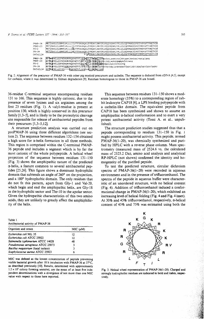
P Storici et aL. i FEBS Letters 337 · 19Y4i 3(;3-307 305
?!".AP-36 ?!".AP-23 PG-2
~~~ ~ ~~~ ~~~~~: ~ ~~ ~ ~~~ ~~ ~~:: ~~ !~2~~~ ~~~ ~ ~~ ~~~~~~;~ ~ ~~~ ~ ~ :~~~~~ ~~: ~~~~~~~~ ~:~ ~~~~~~~~~~~ ME':'(;F.AS LCLGRW S LWL LL:.JAI:. ',-.;p SASAC,'-i..SYRE:.A VLF.A VDRLNEQS S EANL YRLLELDQPPKADE::PG':'P K?VSF:VKE':'VCP\ ME':'Q;:.As LCLGRW S LWL:. L :l.rJ:, \'V? SA SAQALE._ '!REA VLR.A VDRLI\EC S S EANL YRLLELDC:P P KA:: E::PG':'? K?V S F':VKE':'VC ~
Z -8~ REA VLF.A VDRLNECS S EANL YRLLE:..DQP P KADEDPG':'P KPV S F:VKE':'VCry
Fig. 2. Alignment of the precursor of PMAP-36 with other pig myeloid precursors and cathelin. The sequence is deduced from cONA [4,5], except for cathelin, where it was determined by Edman degradation [9). Residues homologous to those in PMAP-36 are boxed.
36-residue C-terminai sequence encompassing residues 131 to 166. This sequence is highly cationic. due to the presence of seven lysines and six arginines among the first 23 residues (Fig. l). A valyl-residue is present a t position 130, which is highly conserved in this precursor family [ 1.3-5], an d is likely t o be the proteolytic cleavage si te responsible for release of antibacterial peptides from their precursors [l ,3-5, 12].
A structure prediction analysis was carried out on proPMAP-36 using three different algorithms (see sec-tion 2). The sequence between residues 132-154 obtained a high score for ~-helix formation in ali three methods. This region is comprised within the C-terminai PMAP-36 peptide and includes a segment which is by far the most cationic of the whole polypeptide. A helical wheel projection of the sequence between residues 131-150 (Fig. 3) shows the amphipathic nature of the predicted ~-helix, a feature common to several antibacterial pep-tides [25.26]. This figure shows a dominant hydrophilic domain that subtends an angle of 260° o n the projection. and a l 00° hydrophobic domain. The only residues that do not fit this pattern, apart from Gly-1 and Val-20, which begin and end the amphipathic helix, are Gly-18 in the hydrophilic sector and Thr-1 O in the apolar sector. Given the hydropathic characteristics of this two amino acids, they are unlikely to greatly affect the amphiphilic-ity of the helix.
Table l Antibacterial activity of PMAP-36
Organism and strain
Escherichia coli ML 35 Escherichia coli ATCC 25922 Salmone/la typhimurium ATCC 14028 Pseudomonas aeruginosa ATCC 25873 Baci/lus megaterium (1ocal isolate) Staphylococcus aureus ATCC 25923
MIC (.uM)
12 12 48
3 3 6
MIC was defined as the lowest concentration of peptide preventing visible bacterial growth after 18 h incubation with PMAP-36 at 37°C, as described previously [l 0]. Results, determined with approximately 1.5 x l 05 colony forming units/ml, are the t:nean of a t least five inde-pendent determinations with a divergence of not more than one MIC value with respect to those here reported.
This sequence between residues 131-150 shows a mod-erate homology (35%) to a corresponding region of rab-bi t leukocyte CAP18 [6], a LPS binding polypeptide with a cathelin-like domain. The equivalent peptide from CAP18 has been synthesized and shown to assume an amphipathic a:-helical conformation and to exert a very potent antibacterial activity (Tossi A. et al.. unpub-lished).
The structure prediction studies suggested thus that a peptide corresponding to residues 131-150 in Fig. l might possess antibacterial activity. This peptide, termed PMAP-36(1-20), was chemically synthesized and puri-fied by HPLC with a reverse phase column. Mass spec-trometry (measured mass of 2524.6 vs. the calcu1ated mass of 2525.2 Da), amino acid analysis and analytical RP-HPLC (not shown) confirmed the identity and ho-mogeneity of the purified peptide.
To test the predicted structure, circular dichroism spectra of PMAP-36(1-20) were recorded in aqueous environment and in the presence oftrifluoroethanol. The spectra of the peptide in aqueous buffer were character-istic of an unordered structure, with no helical content (Fig. 4). Addition of trifluoroethanol induced a confor-mational change in PMAP-36(1-20), which exhibited an increasing level ofhelical folding (Fig. 4 and Fig. 4 inset). At 30% and 45% trifluoroethanol, respectively, a:-helical content of 45% and 75% was estimated using both the
K
R
R
G
R
F
K G
T
K
K
K
RV
Fig. 3. Helical whee1 representation of PMAP-36(1-20). Charged and strongly hydrophobic residues are indicated in bold and italics, respec-tively.
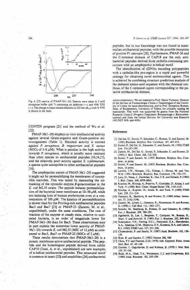
306
2 1-
2 ~,t 9 ~
o "'-----,--.,-..,....--i o 20 40
%TFE
>< o -2
200 nm 220 240
Fig. 4. CD spectra of PMAP-36(1-20). Spectra were taken in 5 mM phosphate buffer (pH 7) containing no additives (-) and 45% TFE ( ---). The change in mean residue ellipticity a t 222 nm ( e:~0 with % TFE is shown in the inset.
CONTIN program [21] and the method of Wu et al. [22}.
PMAP-36{1-20) displays in vitro antibacterial activity against severa! Gram-negative and Gram-positive mi-croorganisms (Table l). Maximal activity is exerted against P. aeruginosa, B. megarerium and S. aureus (MICs of 3-6 ,UM). What is peculiar is the high activity towards P. aeruginosa, which is usually more resistant than other species t o antibacterial peptides [l 0,24,27], and the relatively poor activity against S. typhimurium. a species qui te susceptible to other antibacterial peptides [lO].
The amphipathic nature of PMAP-36(1-20) suggested i t might ·act by permeabilizing the membranes of suscep-tible microbes. This was tested by measuring the un-masking of the cytosolic enzyme ,8-galactosidase in the E. coli ML35 strain. The peptide induces permeabiliza-tion ofthe bacterial inner membrane at 10-50 ,UM, while not inducing lysis of human erythrocytes even at a con-centration of 100 ,UM. The kinetics of permeabilization is slower than for the Pro/Arg-rich antibacterial peptides BacS and Bac7 [23] or PMAP-23 (Zanetti, M. et al., unpublished), under the same conditions. The rate of reaction of the enzyme at steady state, relative to soni-cated bacteria, is an order of magnitude lower for PMAP-36(1-20) than for Bac5. These observations may in part explain the somewhat lower activity of PMAP-36(1-20) towards E. coli ML35 (MI C of 12 ,UM) as com-pared to Bac5, Bac7 or PMAP-23 (MICs of 2 ,UM).
These results demonstrate that PMAP-36(1-20) is a potent. membrane-active antibacterial peptide. This pep-tide and the homologous peptide derived from rabbit CAP18 (Tossi, A. et al., unpublished) are new examples of a-helical antibacterial peptides. This structural motif is common in ìnsect [25] and amphibian [26] antibacterial
P Storici er al./ FEBS Lctters 337 r 1994) 303-307
peptides. but to our knowledge was not found in mam-malian antibacterial peptides. with the possible exception ofporcine P l cecropin [28]. Furthermore, PMAP-36 and the. C-terminai domain of CAP18 are the only anti-bacterial peptides derived from cathelin-containing pre-cursors with an amphipathic a-helical motif.
The identification · of cDNAs encoding polypeptides with a cathelin-like pro-region is a rapid and powerful strategy for obtaining nove! antimicrobial agents. This is achieved by combining structure prediction analysis of the deduced amino acid sequence with the chemical syn-thesis of the C-terminai region corresponding to the pu-tative antibacterial domain.
Acknowledgements: We are indebted to Prof. Mario Furlanut, Director of the Servizio di Farmacologia Clinica e Tossico logica of the Univer-sity of Udine, for mass determination. and to Prof. Domenico Romeo, Dept. of Biochemistry, University of Trieste, for critically reading the manuscript. This work was supported by grants from the National Research Council (Progetto Finalizzato Biotecnologie e Biostrumen-tazione) and from the Italian MiPistry for University and Research (MURST 60% and 40%).
References
[l] Del Sal. G .. Storici. P .. Schneider, C., Romeo. D. and Zanetti, M. (1992) Biochem. Biophys. Res. Commun. 187, 467-472.
[2) Storici, P., Del Sal, G .. Schneider, C. and Zanetti. M. (1992) FEBS Lett. 314,187-190.
[3] Zanetti, M., Del Sal, G .. Storici, P., Schneider. C. and Romeo, D. (1993) J. Biol. Chem. 268. 522-526.
[4) Storici. P. and Zanetti, M. (1993) Biochem. Biophys. Res. Com-mun., in press.
[5] Storici, P. and Zanetti, M. (1993) Biochem. Biopbys. Res. Com-mun .. in press.
[6] Larrick. J.W., Morgan. J.G .. Palings, 1., Hirata, M. and Yen. M.H. (1991) Biochem. Biophys. Res. Commun. 179. 170-175.
[7] Levy, 0 .. Weiss. J .• Zarember. K .. Ooi, C.E. and Elsbach. P. (1993} J. Bio l. Chem. 268, 6058-6063.
[8] Kopitar, M., Ritonja, A., Popovic, T .• Gabrijelic. D., Krizaj, l. and Turk, V. (1989) Biol. Chem. Hoppe-Seyler 370, 1145-1151.
[9] Ritonja. A .. Kopitar. M., Jerala, R. and Turk. V. (1989) FEBS Lett. 255, 211-214.
[IO] Gennaro, R .. Skerlavaj, B. and Romeo. D. (1989) Infect. lmmu-nol. 57, 3142-3146.
[11] Zanetti, M., Litteri, L., Gennaro, R., Horstmann, H. and Romeo, D. (1990) J. Celi Biol. 111. 1363-1371.
[12] Scocchi, M., Skerlavaj. B., Romeo, D. and Gennaro, R. (1992) Eur. J. Biochem. 209. 589-595.
[13] Agerberth, B., Lee, J .• Bergman, T., Carlquist, M., Boman, H., Mutt, V. and Jomvall. H. (1991) Eur. J. Biochem. 202, 849-854.
[14} Kokryakov, V.N., Harwig, S.S.L., Panyutich, E.A., Shevchenko, A.A., Aleshina, G. M., Shamova, O. V., Komeva. H.A. and Lehrer, R.I. (1993) FEBS Lett. 327, 231-236.
[15] Chomczinski. P. and Sacchi, N. (1987) Anal. Biochem. 162, 156-159.
[16] Rost, B. and Sander. C. (1992) Nature 360. 540. [17] Chou, P.Y. and Fasman, G.D. (1978) Adv. Enzymol. Relat. Areas
Mol. Biol. 47, 45-148. [18] Gamier, J .. Osguthorpe. D. and Robson, B. (1978) J. Mol. Biol.
120. 97-106. [19] Buck, M.A., 01ah, T.A., Weit.mtann, C.J. and Cooperman, B.S.
(1989) Anal. Biochem. 182. 295-299.
P Storici et al. i FEBS Leuers 337 · 1994; 303-307
[20] Cohen, S.A. and Strydorn. D.J. (1988) Anal. Biochern. 174. 1-16. [21] Provencher, S.W. (1984) EMBL Techn. Rep. DA07. [22] Wu, C-S. C., Ikeda, K. and Yang, J.T. (1981) Biochernistry, 20,
566-570. [23] Skerlavaj, B., Rorneo, D. and Gennaro. R. (1990) Infect. Irnrnu-
nol. 58. 3i24-3730. [24) Zaslotf. M. (1987) Proc. Nati. Acad. Sci. USA 84. 5449-5453. [25] Holak. T.A., Engstrorn. A, Kraulis. P.J .. Lindeberg, G .. Bennich,
H .. Jones, T.A., Gronenbom. A.M. and Clore, G.M. (1988) Bio-chernistry 27, 7620-7629.
307
[26) Marion. D .. Zaslotf, M. and Bax, A. (1988) FEBS Lett. 227,21-26. [27] Moore, K.S., Bevins. C.L., Brasseur. M.M., Tornassini. V.
Tumer, K., Eck, H. and Zaslotf, M (1991) J. Biol. Chern. 266. 19851-19857.
[28] Lee. J.-Y.. Bornan, A .. Chuanxin. S., Andersson. M .. Jemvall, H .• Mutt. V. and Bornan, H.G. (1989) Proc. ~ati. Acad. Sci. USA 86, 9159-9162.
Eur. J. Biochem. 228. 9-+1-<;1-+6 ill!Y:'! iD FEBS 1995
AllEGATO H
PMAP-37, a no v el antibacterial peptide from pig myeloid cells cDNA cloning, chemical synthesis and activity
:\lessandro 1DSSI 1• Marco SCOCCHI". ~1argherita ZA .. '\'ETTF', Paola STORICI' and Renato GEl\1';\'ARO'
1 Dipartimento di Biochirruca. Biofisica e Chirruca delle Macromolecole. Università di Trieste, Italy 2 Laboratorio Nazionale Consorzio Interuni,•ersitario Biotecnolo2:ie. AREA Science Park. Padriciano, Trieste. ltalv 3 Dipartimento di Scienze e Tecnologie Biomediche. Università di Udine, Italy •
(Received 11 ~ovember 1994)- EJB 94 1739/3
A molecular biologica! approach, based on preproregion homology in the precursors of severa! diverse antibacterial peptides, was used to clone a pig bone marrow cDNA encoding a novel 167-residue polypep-tide. The preproregion of this polypeptide is highly sirnilar to corresponding regions in congeners from pig, cattle and rabbit. It is followed by a unique, cationic, 37-residue sequence, which was predicted to have a high propensity for an a-helical conformation. A peptide, terrned PMAP-37, corresponding to this sequence, was chernically synthesized and shown to undergo a transition from a random coil to an or-dered, mainly helical, conforrnation on addition of trifluoroethanol. This behaviour is typical of an amphi-pathic a helix, a structure common to several membrane-active, antirnicrobial peptides. In vitro experi-ments showed that PMAP-37 strongly inhibits the growth of severa! strains of Gram-negative and Gram-positive bacteria, with minimal inhibitory conce~trations ranging over 1-4 11M, and permeabilizes the inner membrane of Escherichia coli. Interestingly, the 15-32 stretch of PMAP-37 show a remarkable sirnilarity to N-terminai stretches in cecropins B and A from Drosophila melanogaster and Cecropia hyalophora, respectively. This affords an uncommon example of sequence convergence.
Keywords. Antibacterial peptide: PMAP-37: amphipathic helix; myeloid cells.
Innate defense mechanisms are crucial for the prevention of infection in animals, and, in particular, play a centrai role as a frrst line of defense in vertebrates, giving antigen-driven im-mune strategies time to mobilize.
Among the severa} components of the innate defense system are peptides endowed with antimicrobial activity. Those most extensively investigated include (a) the inducible antibiotic pep-tides of insects, (b) the tachyplesin farnily of peptides found in horseshoe crab species, (c) the antimicrobial peptides secreted by specialized cells in the skin and stomach of amphibia, and (d) the peptides contained in circulating white cells and mucosal epithelial cells of mammals [1-6).
Although all these peptides show wide diversity in sequence, some common features appear to be required for their activity. They are, in generai, highly cationic and tend to adopt an amphi-pathic conformation that seems mandatory for their functioning as membrane-active agents.
In mammals, the cytoplasmic granules of neutrophils are an abundant source of a number of antimicrobial peptides, and those identified so far show a significant diversity in structure, spectrum of activity and species distribution [1, 3-6]. Several of these peptides have been cloned and their cONA sequences
Correspondence to R. Gennaro, Dipartimento di Scienze e Tecnolo-gie Biomediche, Università di Udine, Via Gervasutta 48, 1-33100 Udine, ltaly
Fax: + 39 40 6763691. Abbreviations. PMAP, porcine myeloid antibacterial peptide; Fmoc,
fluorenylmethyloxycarbonyl, MIC, minima! inhibitory concentration. Note. The novel nucleotide sequence data published bere has been
deposited with the EMBL sequence data bank is available under acces-sion number(s) 39641.
indicate that they are generally synthesized as precursors that release the mature, biologically active peptide after proteolytic cleavage.
Recently, we [7-14) and others (15-18] have shown, through cONA cloning, that several bovine, porcine and rabbit leukocyte antimicrobial peptides derive from precursors with highly conserved preproregions, although the mature peptides themselves are diverse. In fact, these include Arg-rich and Pro-rich peptides such as Bac5 and Bac7 [9, 14], PR-39 [11] and C12 [17), a-helical peptides such as CAP18 [15] and PMAP-36 [12], Cys-containing peptides such as the cyclic dodecapeptide [8] and the protegrins [10, 18], and other peptides such as the Trp-rich indolicidin [7) and PMAP-23 [13]. The more loosely related rabbit p15s also belong to this farnily (16).
Several of the precursors of the above mentioned peptides were cloned through cONA amplification, using primers derived from the conserved region [7-14, 18). This strategy led to the identification of the precursors of both previously isolated [7-11, 14, 18] and of novel [12, 13, 18] peptides. In this paper, we describe the cloning of the precursor of one such novel peptide that has been named PMAP-37, from 'porcine myeloid antibac-terial peptide' of 37 residues. The cONA-deduced sequence has been used to obtain this peptide by chemical synthesis. PMAP-37 has been shown to adopt an amphipathic a-helical confonna-tion and to exert a potent antibacterial activity in vitro, most likely by damaging bacterial membranes, as shown by an inner membrane permeabilization assay with Escherichia coli. More-over, this peptide affords an interesting example of convergence, as a centrai stretch of 18 residues within its sequence shows a high degree of similarity to part of cecropin B from Drosophila melanogaster [19].
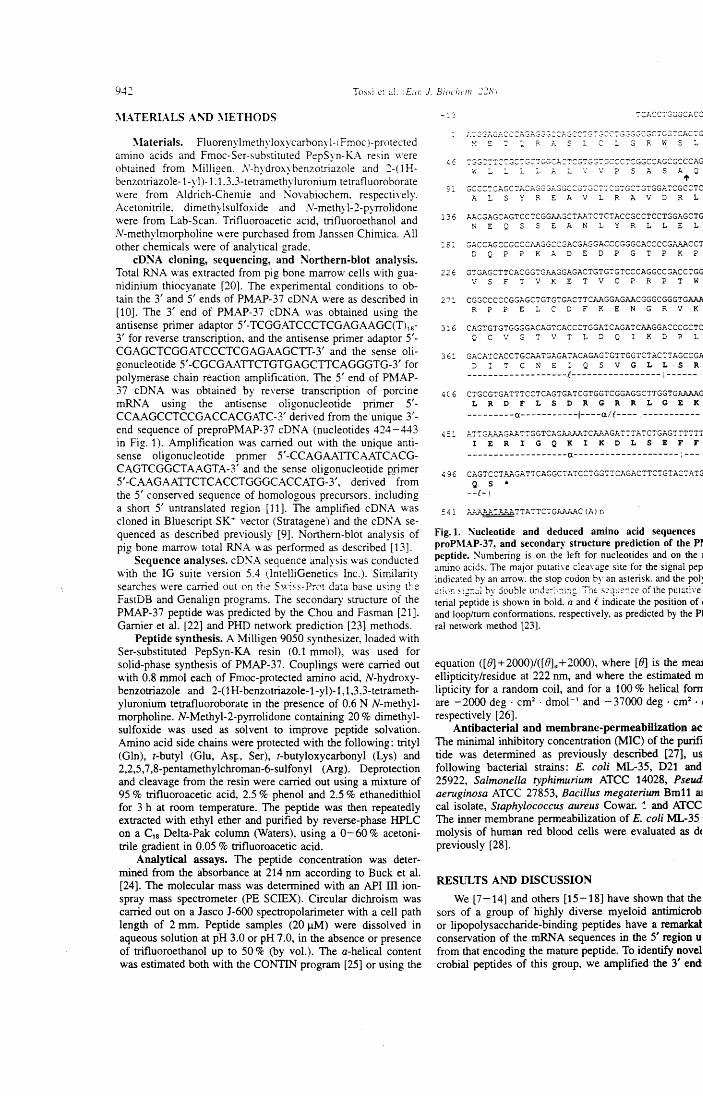
9-C Tms: è: :.JI. iLo: J. Biochcm :::;8)
:\L\TERIALS A!\'D l\IETHODS
~laterials. Fluorenylmethyloxycarbonyl-(fmoc)-protected amino acids and Fmoc-Ser-substituted PepSyn-K.I\ resin were obtained from Milligen. ,\'-hydroxybenzotriazole and 2-( 1 H-benzotriazole-1-yl)-1.1.3.3-tetramethyluronium tetrafluoroborate were from Aldrich-Chemie and I\ovabiochem. respectively. Acetonitrile, dimethylsulfoxide and N-methyl-2-pyrrolidone were from Lab-Scan. Trifluoroacetic acid, trifluoroethanol and N-methylmorpholine were purchased from Janssen Chimica. All other chemicals were of analytical grade.
cD:SA cloning, sequencing, and Northern-blot analysis. Total RNA was extracted from pig bone marrow cells with gua-nidinium thiocyanate [20]. The experimental conditions to ab-taio the 3' and 5' ends of PMAP-37 cDNA were as described in [10]. The 3' end of PMAP-37 cDNA was obtained using the antisense primer adaptor 5'-TCGGATCCCTCGAGAAGC(T)18-
3' for reverse transcription, and the antisense primer adaptor 5'-CGAGCTCGGATCCCTCGAGAAGCIT-3' and the sense oli-gonucleotide 5'-CGCGAATTCTGTGAGCITCAGGGTG-3' for polymerase chain reaction amplification. The 5' end of PMAP-37 cDNA was obtained by reverse transcription of porcine mRNA using the antisense oligonucleotide primer 5'-CCAAGCCTCCGACCACGATC-3' derived from the unique 3'-end sequence of preproPMAP-37 cDNA (nucleotides 424-443 in Fig. 1 ). Amplificati an was carri ed aut with the unique anti-sense oligonucleotide primer 5' -CCAGAATTCAATCACG-CAGTCGGCTAAGTA-3' and the sense oligonucleotide mimer 5'-CAAGAAITCTCACCTGGGCACCATG-3', derived from the 5' conserved sequence of homologous precursors. including a short 5' untranslated region (11]. The amplified cDNA v.·as cloned in Bluescript SK" vector (Stratagene) and the cDNA se-quenced as described previously (9]. Northern-blot analysis of pig bone marrow total RNA was performed as described [13].
Sequence analyses. cD~A sequence analysis was conducted with the IG suite version 5.4 (lntelliGenetics Inc.). Similarity searches were carried aut on the Swis5-PrC't data base usin!! t~'e
FastDB and Genalign programs. The secondary structure of the PMAP-37 peptide was predicted by the Chou and Fasman [21], Garnier et al. [22] and PHD network prediction [23] methods.
Peptide synthesis. A Milligen 9050 synthesizer, loaded with Ser-substituted PepSyn-K.A resin (0.1 mmol), was used for solid-phase synthesis of PMAP-37. Couplings were carried aut with 0.8 mrnol each of Fmoc-protected amino acid, N-hydroxy-benzotriazole and 2-(lH-benzotriazole-1-yl)-1, 1,3,3-tetrameth-yluronium tetrafluoroborate in the presence of 0.6 N N-methyl-morpholine. N-Methyl-2-pyrrolidone containing 20% dimethyl-sulfoxide was used as solvent to irnprove peptide solvation. Amino acid side chains were protected with the following: trityl (Gln), t-butyl (Glu, Asr;. Ser), t-butyloxycarbonyl (Lys) and 2,2,5,7 ,8-pentarnethylchroman-6-sulfonyl (Arg). Deprotection and cleavage frorn the resin were carried aut using a mixture of 95 % trifluoroacetic acid, 2.5 % phenol and 2.5 % ethanedithiol for 3 h at room temperature. The peptide was then repeatedly extracted with ethyl ether and purified by reverse-phase HPLC on a C18 Delta-Pak colurnn (Waters), using a 0-60% acetoni-trile gradient in 0.05 % trifluoroacetic acid.
Analytical assays. The peptide concentration was deter-rnined from the absorbance at 214 nrn according to Buck et al. [24]. The rnolecular rnass was determined with an API ili ion-spray mass spectrometer (PE SCIEX). Circular dichroism was carried out on a Jasco J-600 spectropolarirneter with a celi path length of 2 mrn. Peptide samples (20 J..LM) were dissolved in aqueous solution at pH 3.0 or pH 7 .0, in the absence or presence of trifluoroethanol up to 50% (by vol.). The a-belical content was estimated both with the CONTIN prograrn [25] or using the
v ~ : : R ~ S L C L G R W S :
91 GCCC~CAGC~ACAGG~~SG:CG~GCT7:G7GC:GTGGATCGCCTC
A L S Y R E A V L R A V D R L
136 AACGAGCAGTCCTCGGAAGCTAATCTCTACCGCCTCCTGGAGCTG N E Q S S E A N L Y R L L E L
:8: GACCAGCCGCCCAAGGCC~ACGAGGACCCGGGCACCCCGAAACC~ D Q P P K A D E D P G T P K P
226 GTGAGCTTCACGGTGAAGGAGACTGTGTGTCCCAGGCCGACCTGG V 5 F T V K E T V C P R P T W
271 CGGCCCCCGGAGCTGTGTGACTTCAAGGAGAACGGGCGGGTGAAA R P P E L C D F K E N G R V K
3:6 CAGTGTGTGGGGACAGTCACCCTGGATCAGATCAAGGACCCGCTC Q C V G T V T L D Q I K D P L
361 GACATCACCTGCAATGAGATACAGAGTGTTGGTCTACTTAGCCGA D I T C N E : Q 5 V G L L S R
-------------------(-----------------:------406 CTGCGTGATTTCCTCAGTGATCGTGGTCGGAGGCTTGGTGAAAAG
L R D F L S D R G R R L G E K ---------a-----------1----all----. -----------
~51 ATTG?~GAATTGGTCAGAAAP.TCAAAGATTTATCTGAGTTTTTT
I E R I G Q K I K D L S E F F -------------------a--------------------:---
4 9 6 CAGTCCT.x..AGATTCAGGC:ATCCTGGTTCAGACTTCTGTAC':'ATG Q s *
--(-1
541 ~TTATTCTG~~C(AJn
Fig. l. Nucleotide and deduced amino acid sequences proPMAP-37, and seconda11· structure prediction of the P~ peptide. Numbering is on the Jeft for nucleotides and on the 1
amino acids. The major putati\e clea\'age site for the signal pep indicated by an arrow. the stop codon by an asterisk. and the poi~ <J:it•J; ~<;:::.Ji by double und:::-:';:ins. "T'ht s:.>q'J:-::;:e of the put:>tive terial peptide is shown in bold. a and t indicate the position of j and loop/tum conformations. respectively, as predicted by the Pl ral network method [23].
equation ([8]+2000)/([B]a +2000), where [8] is the meru ellipticity/residue at 222 nrn, and where the estimated m lipticity far a random coil, an d far a 100% helical fom are -2000 deg · cm2 • dmol- 1 and -37000 deg · cm2
• 1
respectively [26]. Antibacterial and membrane-penneabllization ac
The minima! inhibitory concentration (MI C) of the purifi tide was determined as previously described [27], us following bacterial strains: E. coli ML-35, D21 and 25922, Salmonella typhimurium ATCC 14028, Pseud aeruginosa ATCC 27853, Bacillus megaterium Bm11 aJ cal isolate, Staphylococcus aureus Cowar. ~ and ATCC The inner membrane perrneabilization of E. coli ML-35 molysis of human red blood cells were evaluated as dc previously [28].
RESlJLTS AND DISCUSSION
We [7-14] and others [15-18] bave sbown that the sors of a group of highly diverse myeloid antirnicrob or lipopolysaccharide-binding peptides bave a remarkal conservation of the rnRNA sequences in the 5' region u from that encoding the mature peptide. To identify novel crobial peptides of this group, we arnplified the 3' end
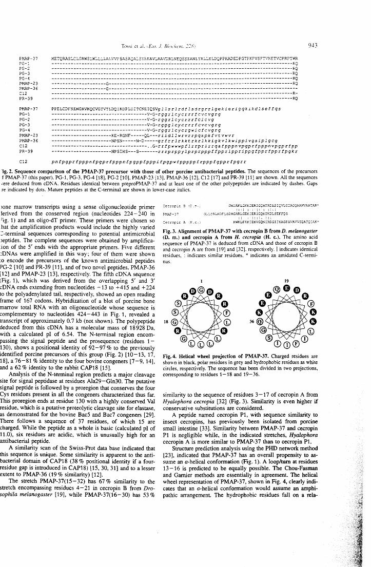
To~si è! al. 1D11: ./. Biochnn. 228.1 9-+3
H'..AP-37 PG-1 ?G-2 ?G-3 PG-4 PMAP-23 P.ti.AP-36 C12 PR-39
l''.E':'QRASLCLGRWSLWLLl.LAL\",'PSASA~AI..SYr.EAVLRAVDRLNEQSSEA."JLYRLLELD·QPPKADEDPG'l'?K?VSFTVKETVCPRP':'WR
-------------------------------------------~---------------------------------------------RQ -----------------------------------------------------------------------------------------RQ -----------------------------------------------------------------------------------------RQ -----------------------------------------------------------------------------------------RQ --------------------G--------------------------------------------------------------------RQ --------------------G---------------------------------------------------------------------------------------------------------------------------------------------------------------R------------------------------------------------------------------------------------------RQ
PMAP-37 PG-1 PG-2 PG-3 PG-4 Pt-'..AP-23 ?.ti..AP-36 Cl2 PR-39
PPELCDFKENGRVKQCVGTVTLDQIKDPLDITCNEIQSVg 11 s r l rdf lsd rgr r l gek ieri gqk i kdl se! fqs -----------------------------------V-G-rgg r l c y c r r r f c ve vg rg -----------------------------------V-G-rgg r l c y c r r r f c i c vg -----------------------------------V-G-rggg l cy c r r r f cv c vg rg -----------------------------------V-G-rgg r l c y c rg w i cf c vg rg ----------------------KE-RGNF-----01---r i i d 11 w rv r rpq kpk f v t vwv r ----------------------NPSN-----N-D-----g r f r r l r k k t r k r l k k i g k v l k w ippi vg si p l g cg ----------------------------------- .. G-r r fpwwwp f l r rpr l r rqa fpppn vpgpr fpppn vpgpr fpp ----------------------NPSIHS---5-------r r rprppy lprprpppf fppr lppr ippg fppr fppr fpgk r
Cl2 pnfpgprfpppnfpgprfpppnfpgpptpppifpgpwfpppppfrpppfgpprtpgrr
'ig. 2. Sequence comparison of tbe PMAP-37 precursor '\\itb those of other porcine antibacterial peptides. The sequences of the precursors f PMAP-37 (this paper). PG-1, PG-3, PG-4 [18], PG-2 [10], PMAP-23 [13]. PMAP-36 [12], C12 [17] and PR-39 [11] are shown. All the sequences •ere deduced from cDNA. Residues identical between preproPMAP-37 and at least one of the other polypeptides are indicated by dashes. Gaps re indicated by dots. Mature peptides at the C-terminai are shown in lower-case italics.
>one marrow transcripts using a sense oligonucleotide primer lerived from the conserved regio n (nucleotides 224-240 in =ig. 1) and an oligo-dT primer. These primers were chosen so hat the amplification products would include the highly varied ::-terminai sequences corresponding to potential antimicrobial Jeptides. The complete sequences were obtained by amplifica-:ion of the 5' ends with the appropriate primers. Five different :DNAs were amplified in this way; four of them w ere shown :o encode the precursors of the known antimicrobial peptides PG-2 [10] and PR-39 [11], and of two novel peptides, PMAP-36 [12] and PMAP-23 [13], respectively. The fifth cDNA sequence (Fig. 1), which was derived from the overlapping 5' and 3' cDNA ends extending from nucleotides -13 to +415 and +224 to the polyadenylated tail, respectively, showed an open reading frame of 167 codons. Hybridization of a blot of porcine bone marrow total RNA with an oligonucleotide whose sequence is complementary to nucleotides 424-443 in Fig. 1, reveaied a transcript of approximately 0.7 kb (not shown). The polypeptide deduced from this cD NA has a molecular mass of 18 928 Da, with a calculated pl of 6.54. The N-terminai region encom-passing the signai peptide and the prosequence (residues 1-130), shows a positionai identity of 92-97% to the previously identified porcine precursors of this group (Fig. 2) [ 1 O -13, 17, 18], a 76-81% identity to the four bovine congeners [7 -9, 14], and a 62% identity to the rabbit CAP18 [15].
Anaiysis of the N-terminai region predicts a major cleavage site for signai peptidase at residues Ala29-Gln30. The putative signai peptide is followed by a proregion that conserves the four Cys residues present in ail the congeners characterized thus far. This proregion ends at residue 130 with a highly conserved Val residue, which is a putative proteolytic cleavage site for elastase, as demonstrated for the bovine Bac5 and Bac7 congeners [29]. There follows a sequence of 37 residues, of which 15 are charged. While the peptide as a w ho le is basic ( calculated p l of 11.0), six residues are acidic, which is unusually high for an antibacteriai peptide.
A similarity scan of the Swiss-Prot data base indicated that this sequence is unique. Some similarity is apparent to the anti-bacteriai domain of CAP18 (38 % positionai identity if a four-residue gap is introduced in CAP18) [15, 30, 31] and to a lesser extent to PMAP-36 (19% similarity) [12].
The stretch PMAP-37(15-32) has 67% similarity to the stretch encompassing residucs 4-21 in cecropin B from Dro-sophila melanogaster [19], while PMAP-37(16-30) has 53%
:ec~c.p:.r. g (~·.!!ì.; GWLR..l<LGKKIER!GQH':"RDA.SIQVLGIAQQAA."'ì'VAATAR-;:!1 l! li l!1: :l:i
P!'.AP- 3 7 .:;:.:.~.R:.R:)!:'LSDRGRRLGEK!ERIGQKIKDLSEFFQS
: l ' ~ l : : ; i ! : l Se::ropir. A tE. c.) KWKLFKKIEKVGQNIRDGI 1KAGPAVA\t'VGQA':'Q:h."'i.~
Fig. 3. Alignment of PMAP-37 '\\ith cecropin B from D. melanogaster (D. m.) and cecropin A from H. cecropia (H. c.). The amino acid sequence of PM.AP-37 is deduced from cDNA and those of cecropin B and cecropin A are from [19] and [32}. respectively. l indicates identica! residues. : indicates similar residues, * indicates an amidated C-tenni-nus.
Fig. 4. Helical wbeel projection of PMAP-37. Charged residues are shown in black. polar residues in grey and hydrophobic residues as white circles, respectively. The sequence has been divided in two projections, corresponding t o residues 1 -18 an d 19-36.
similarity to the sequence of residues 3-17 of cecropin A from Hyalophora cecropia [32] (Fig. 3). Similarity is even higher if conservative substitutions are considered.
A peptide named cecropin P1, with sequence similarity to insect cecropins, has previously been isolated from porcine small intestine [33]. Similarity between PMAP-37 and cecropin P1 is negligible while, in the indicated stretches, Hyalophora cecropin A is more sirnilar to PMAP-37 than to cecropin Pl.
Structure prediction anaiysis using the PHD network method [23], indicated that PMAP-37 has an overall propensity to as-sume an a-helical conformation (Fig. 1). A loop/turn at residues 13-16 is predicted to be equally possible. The Chou-Fasman and Garnier methods are essentiaily in agreement. The helical wheel representation of PMAP-37, shown in Fig. 4, clearly indi-cates that an a-helicai conformation would assume an amphi-pathic arrangement. The hydrophobic residues fall on a rela-
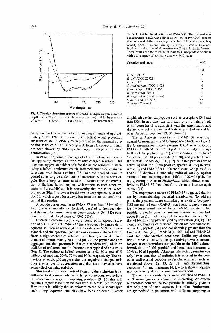
944 To~~i et al. tEur. J. Biochcm. 2:281
\
... o 4 \
200 210 220 230 240
Wavelength (nm)
Fig. S. Circular dichroism spectra ofPMAP-37. Spectra were recorded at pH 3 with 20 )lM peptide in the absence (· · · · · ·) and in the presence of 15 Ck (---). 30% (- ·-·-) and 45 % (--) trifluoroethanol.
tively narrow face of the helix, subtending an angle of approxi-mately 1 ooo -120°. Furthermore, the helical wheel projection for residues 18-30 closely resembles that for the segment com-prising residues 5 -17 in cecropin A from H. cecropia, which has been shown, by NMR spectroscopy, to adopt an a-helical conformation [34].
In PMAP-37, residue spacings of i+3 or i+4 are as frequent for oppositely charged as for similarly charged residues. This does not suggest an evident role for the acidic residues in stabi-lizing a helical conformation via intramolecular side chain in-teractions with basic residues [35], nor are charged residues placed so as to give a favourable interaction with the helix di-pole. How a loop/tum after residue 13 would affect the orienta-tion of flanking helical regions with respect to each other, re-mains to be established. It is noteworthy that the helical wheel projection (Fig. 4) shows a breakdown in amphipathicity at resi-due 13, which argues for a deviation from the helical conforma-tion at this residue.
A peptide corresponding to PMAP-3 7 (residues 131-167 in Fig. 1) was chemically synthesized, purified to homogeneity, and shown to be correct by mass determination (4364.4 Da com-pared to the calculated mass of 4365.0 Da).
Circular dichroism spectra were measured in aqueous solu-tion at pH 3.0 and 7.0. PMAP-37 has a tendency to aggregate in aqueous solution at neutral p H but dissolves in 50% trifluoro-ethanol, and the spectrum (not shown) assumes a shape that re-flects a high content of a-helical structure (estirnated helical content of approximately 80% ). At p H 3.0, the peptide does not aggregate and the spectrum is that of a random coil, while on addition of trifluoroethanol it becomes that typical of an a helix (Fig. 5). The estimated helical content at 15 %, 30% and 45% trifluoroethanol was 30 %, 70 %, and 80 %, respectively. The be-haviour at acidic pH suggests that the negatively charged resi-dues play a role in aggregation and that they may also bave some effect on helix stability.
Structural information derived from circular dichroism is in-sufficient to determine whether a hinge connecting two helices is present in the region comprising residues 13-16; this will require a higher resolution method such as NMR spectroscopy. However, it is unlikely that an uninterrupted a helix should span such a long sequence, and kinks bave been observed in other
Table l. Antibacterial activity of PMAP-37. The minima) inhil concentration (MICJ was defined as the lowest PMAP-37 concent that preYented visible bacterial growth after 18 h incubation \vith ap imately 1.5 X 10⁵ colony-forming units/ml, at 37 °C in Mueller-f broth or, in the case of B. megaterium Bm11, in Luria-Bertani · These results are the mean of at least four independent determin: with a divergence of not more than one MIC value.
Organism and strain
E. coli ML35 E. coli ATCC 25922 E. coli D21 S. typhimurium ATCC 14028 P. aeruginosa ATCC 27853 B. megaterium Bm11 B. megaterium (!oca! isolate) S. aureus ATCC 25923 S. aureus Cowan 1
MIC
11M 2 1 2 4 2 4 4
32 64
amphipathic a-helical peptides such as cecropin A [34] and ittin [36]. In any case, the formation of an a helix on add of trifluoroethanol is consistent with the amphipathic natu the helix, which is a structural feature typical of severa} fan of antibacterial peptides [32, 34, 36-40]
The antibacterial activity of PMAP- 37 was evah against Gram-negative and Gram-positive bacteria (Table 1: the Gram-negative microorganisms tested were susceptib PMAP-37 with MICs of 1-4 J.lM. This activity is compa to that of the peptide Cs [31], corresponding to residues 1 125 of the CAP18 polypeptide [15, 30], and greater than th the peptide PMAP-36(1-20) [12]. Ali three peptides are eq active against the Gram-positive species B. megaterium, while C, 8 and PMAP-36(1- 20) are also active against S. au PMAP-37 displays a markedly reduced activity against strains of this microorganism (MICs of 32-64J1M). lnt~ ingly, cecropin A from Hyalophora, which shows some larity to PMAP-37 (see above), is virtually inactive agair aureus [41].
The amphipathic nature of PMAP-37 suggested that it' act by permeabilizing bacterial membranes. To address point, the fi-galactosidase unmasking assay describèd previ1 [28] was carried out. PMAP-37 was found to rapidly permc ize the inner membrane of the E. coli ML-35 strain. At peptide, a steady state for enzyme activity was reached about 8 min from addition, and the reaction rate was 60-that of bacteria completely lysed by sonication (Fig. 6). Thc ciency and kinetics of permeabilization are comparable to of the C, 8 peptide [31] and considerably greater than tho Bac5 and Bac7 [28], PMAP-36(1-20) (12] and PMAP-23 evaluated under identica! conditions. Unlike any of these tides, PMAP-37 shows some lytic activity towards human c rocytes at concentrations comparable to the MIC values ~ hemolysis at 10 J1M peptide) and hemolysis increases to 30 % at 50 11M peptide. Although this activity remains con ably lower than that of melittin, it is unusual in the conte other antibacterial peptides so far characterized, such as mentioned above [12, 13, 28, 31], and dermaseptins cecropins [41] and magainins [42], which show a negligib molytic activity at antibacterial concentrations.
The sequence similarity between stretches of PMAP-3 of D. melanogaster cecropin B is interesting. An evoluti relationship between the two peptides is unlikely, given th that only part of their sequence is similar. Furthermore. are synthesized as precursors with different preproregion
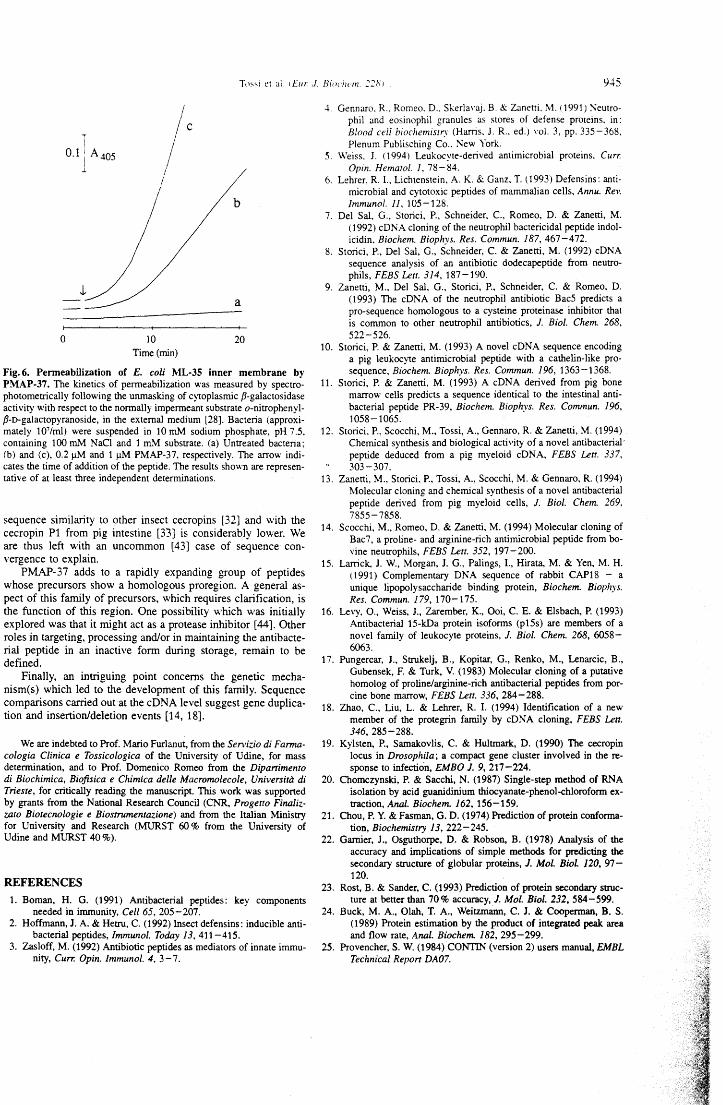
0.1 A405 le l
b
a
o lO 20 Time(min)
Fig. 6. Penneabilization of E. coli ML-35 inner membrane by PMAP-37. The kinetics of permeabilization was measured by spectro-photometrically following the unmasking of cytoplasmic p-galactosidase activity with respect to the normally impermeant substrate o-nitrophenyl-P-o-galactopyranoside, in the external medium [28]. Bacteria (approxi-mately 107/ml) were suspended in 10 mM sodium phosphate, pH 7.5, containing 100 mM NaCl and 1 m.\1 substrate. (a) Untreated bacteria; (b) and (c), 0.2 11M and 111M PMAP-37, respectively. The arrow indi-cates the time of addition of the peptide. The results shown are represen-tative of at least three independent determinations.
sequence similarity to other insect cecropìns [32] and wìth the cecropin P1 from pìg intestine [33] is considerably lower. We are thus left with an uncommon [43] case of sequence con-vergence to explaìn.
PMAP-37 adds to a rapidly expanding group of peptides whose precursors show a homologous proregion. A generai as-pect of this family of precursors, which requires clarification, is the function of this region. One possibility which was initially explored was that it rnight act as a protease inhibitor [44]. Other roles in targeting, processing and./or in maintaining the antibacte-rial peptide in an inactive form during storage, remain to be defined.
Finally, an intriguing point concems the genetic mecha-nism(s) which led to the development of this farnily. Sequence comparisons carried out at the cDNA level suggest gene duplica-tion and insertionldeletion events [14, 18].
We are indebted to Prof. Mario Furlanut, from the Servizio di Farma-cologia Clinica e Tossicologica of the University of Udine, for mass determination, and to Prof. Domenico Romeo from the Dipartimento di Biochimica, Biofisica e Chimica delle Macromolecole, Università di Trieste, for critically reading the manuscript. This work was supported by grants from tbe National Research Council (CNR, Progetto Finaliz-zato Biotecnologie e Biostrumentazione) and from tbe Italian Ministry for University and Research (MURST 60% from tbe University of Udine and MURST 40%).
REFERENCES 1. Boman, H. G. (1991) Antibacterial peptides: key components
needed in immunity, Celi 65, 205-207. 2. Hoffmann, J. A. & Hetru, C. (1992) lnsect defensins: inducible anti-
bacterial peptides, lmmunol. Today 13, 411-415. 3. Zasloff, M. (1992) Antibiotic peptides as mediators of innate immu-
nity, Cun: Opin. lmmunol. 4, 3-7.
4.
5.
6.
7.
8.
9.
9.:+5
Gennaro. R .. Romeo. D .. SkerlaYaj. B. & Zanetti. \1. 11991) Seutro-phil and eosinophil granules as stores of defense proteins. in: Blood ce/l biochemisrry (Harris. J. R., ed.) vol. 3, pp. 335-368. Plenum Publisching Co .. ;-..;ew York.
\\'eiss. J. (1994) Leukocyte-derived antimicrobial proteins. Curr. Opin. Hematol. l, 78-84.
Lehrer. R. L. Lichtenstein, A K. & Ganz. T. ( 1993) Defensins: anti-microbial and cytotoxic peptides of mammalian cells, Annu. Rev. Immunol. 11, 105-128.
Del Sal, G., Storici, P., Schneider. C., Romeo, D. & Zanetti, M. (1992) cD NA cloning of the neutrophil bacteri ci dal peptide indol-icidin. Biochem. Biophys. Res. Commun. 187, 467-472.
Storici, P., Del Sal, G., Schneider, C. & Zanetti, M. (1992) cDNA sequence analysis of an antibiotic dodecapeptide from neutro-phils, FEBS Lett. 314, 187-190.
Zanetti, M., Del Sal, G., Storici, P., Schneider, C. & Romeo. D. (1993) The cDNA of the neutrophil antibiotic Bac5 predicts a pro-sequence homologous to a cysteine proteinase inhibitor that is common to other neutrophil antibiotics, J. Biol. Chem. 268, 522-526.
10. Storici, P. & Zanetti, M. (1993) A novel cDNA sequence encoding a pig leukocyte antimicrobial peptide with a cathelin-like pro-sequence, Biochem. Biophys. Res. Commun. 196, 1363-1368.
11. Storici, P. & Zanetti, M. (1993) A cDNA derived from pig hone marrow cells predicts a sequence identica! to the intestina! anti-bacterial peptide PR-39, Biochem. Biophys. Res. Commun. 196, 1058-1065.
12. Storici. P., Scocchi, M., Tossi, A., Gennaro, R. & Zanetti, M. (1994) Chemical synthesis and biologica] activity of a nove! antibacteriaJ· peptide deduced from a pig myeloid cDNA, FEBS Lett. 337, 303-307.
13. Zanetti, M., Storici. P., Tossi, A., Scocchi, M. & Gennaro, R. (1994) Molecular cloning and chem1cal synthesis of a nove! antibacterial peptide derived from pig myeloid cells, J. Biol. Chem. 269, 7855-7858.
14. Scocchi, M., Romeo, D. & Zanetti, M. (1994) Molecular cloning of Bac7, a proline- and arginine-rich antimicrobial peptide from bo-vine neutrophils, FEBS Lett. 352, 197-200.
15. Larrick, J. W., Morgan, J. G., Palings, 1., Hirata, M. & Yen, M. H. (1991) Complementary DNA sequence of rabbit CAP18 - a unique lipopolysaccharide binding protein, Biochem. Biophys. Res. Commun. 179, 170-175.
16. Levy. 0., Weiss, J., Zarember, K., Ooi, C. E. & Elsbach, P. (1993) Antibacterial 15-kDa protein isoforms (p15s) are members of a novel farnily of leukocyte proteins, J. Bio/. Chem. 268, 6058-6063.
17. Pungercar, J., Strukelj, B., Kopitar, G., Renko, M., Lenarcic, B., Gubensek, F. & Turk, V. (1983) Molecular cloning of a putative homolog of proline/arginine-rich antibacterial peptides from por-cine hone marrow, FEBS Lett. 336, 284-288.
18. Zhao, C., Liu, L. & Lehrer, R. l. (1994) Identification of a new member of tbe protegrin farnily by cDNA cloning, FEBS Lett. 346, 285-288.
19. Kylsten, P., Samakovlis, C. & Hultmark, D. (1990) The cecropin locus in Drosophila; a compact gene cluster involved in tbe re-sponse to infection, EMBO J. 9, 217-224.
20. Chomczynski, P. & Sacchi, N. (1987) Single-step metbod of RNA isolation by acid guanidinium tbiocyanate-phenol-chloroform ex-traction, Anal. Biochem. 162, 156-159.
21. Chou, P. Y. & Fasman, G. D. (1974) Prediction ofprotein conforma-tion, Biochemistry 13, 222-245.
22. Garnier, J., Osgutborpe, D. & Robson, B. (1978) Analysis of tbe accuracy and implications of simple metbods for predicting tbe secondary structure of globular proteins, J. Mol. BioL 120, 97-120.
23. Rost, B. & Sander, C. (1993) Prediction of protein secondary struc-ture at better tban 70% accuracy, J. Mol. Biol. 232, 584-599.
24. Buck, M. A., Olah, T. A., Weitzmann, C. J. & Cooperman, B. S. (1989) Protein estimation by the product of integrated peak area and flow rate, Anal. Biochem. 182, 295-299.
25. Provencher, S. W. (1984) CONTIN (version 2) users manual, EMBL Technical Report DA07.
946 To~~i et al. rEu1: J. Biochcm. ::::,'!1
26. Wu. C.-S. C.. Ikeda. K. & Yang. J. T. (J 981) Orde re è conformation of polypeptides and proteins in acidic dodecyl sulfate solution. Biochemistry 20, 566-570.
27. Gennaro, R., Skerlavaj, B. & Romeo. D. ( 1989) Purification. composition. and activity of two bactenecins. antibacterial pep-tides of bovine neutrophils. Infect. Immun. 57, 3142-3146.
28. Skerla\'aj, B., Romeo, D. & Gennaro. R. (1990) Rapid membrane permeabilization and inhibition of vita! functions of gram-nega-tive bacteria by bactenecins. Infect. lmmun. 58, 3724-3730.
29. Scocchi, M .. Skerlavaj, B., Romeo, D. & Gennaro, R. (1992) Proteo-lytic cleavage by neutrophil elastase converts inactive storage proforms to antibacterial bactenecins, Eur. l. Biochem. 209, 589-595.
30. Larrick, J. W., Hirata, M., Shimomoura, Y., Yoshida, M., Zheng. H., Zhong, J. & Wright S. C. (1993) Antimicrobial activity of rabbit CAP18-derived peptides, Antimicrob. Agents Chemother. 37. 2534-2539.
31. Tossi, A., Scocchi, M., Skerla,·aj, B. & Gennaro, R. (1994) Identifi-cation and characterization of a primary antibacterial domain in CAP18, a lipopolysaccharide binding protein from rabbit leuko-cytes, FEBS Lett. 339, 108-112.
32. Boman, H. G., Faye, 1., Gudmundsson, G. H., Lee, J.-Y. & Lidholm, D.-A. (1991) Celi-free immunity in Cecropia. A mode! system for antibacterial proteins, Eur. l. Biochem. 201, 23-31.
33. Lee, J.-Y., Boman, A., Sun, C., Andersson, M., J~rn\'all, H .. Mutt, V. & Boman, H. G. (1989) Antibacterial peptides from pig intes-tine: Isolation of a marnmalian cecropin, P roe. Nati A c ad. Sci. USA 86, 9159-9162.
34. Holak, T. A., Engstrom, A.. Kraulis, P. J.. Lindeberg, G., Bennich, H., Jones, T. A., Gronenborn, A. M. & Clore, G. M. (1988) The solution conformation of the antibacterial peptide cecropin A~ a nuclear magnetic resonance and dynamical simulated annealing study, Biochemistry 27, 7620-7629.
35. Marqu!-ee. S. & Baldwin L. R. <1987l Helix stabilization by C · · · Ly:-.- salt bridges in shot1 peptides of de nora design. P l\'atl Acad. Sci. USA 84, 8898-8902.
36. Bazzo. R .. Tappin. M. J., Pastore. A .. Harvey, T. S., Carver, J. P Campbell, I. D. (1988) The structure of melittin, Eur. l. Bioc/ 173. 139-146.
37. Marion. D .. Zasloff, M. & Bax. A. (1988) A two-dimensional N study of the antimicrobial peptide magainin 2, FEBS Lett. ; 21-26.
38. Williams. R. W., Starman, R., Taylor, K. M. P., Gable, K., Beele1 Zasloff, M. & CO\·ell. D. (1990) Raman spectroscopy of syntt antirnicrobial frog peptides magainin 2a and PGLa, Biochem1 29. 4490-4496.
39. Mor. A., Nguyen, V. H., Delfour, A., Migliore-Samour, D. & Ni< las, P. ( 1991) Isolation, amino aci d sequence, an d synthesi: dermaseptin, a nove! antirnicrobial peptide of amphibian s Biochemistry 30, 8824-8830.
40. Sipos, D., Andersson, M. & Ehrenberg, A. (1992) The struc of the marnmalian antibacterial peptide cecropin Pt in solut deterrnined by proton-NMR, Eur. l. Biochem. 209, 163-169
41. Boman, H. G., Wade, D., Boman, l. A., Wàhlin, B. & Merrifi B. (1989) Antibacterial and antimalarial properties of pept that are cecropin-melinin hybrids FEBS Lett. 259, 103-106.
42. Zasloff, M. (1987) Magainins, a class of antimicrobial peptides f Xenopus skin: Isolation, characterization of two active forms, partial cONA sequence of a precursor, Proc. Nati Acad. Sci. l 84. 5449-5453.
43. Doolittle, R. F. (1994) Convergent evolution: the need to be expl Trends Biochem. Sci. 19, 15-18.
44. Verbanac, D., Zanetti, M. & Romeo, D. (1993) Chemotactic protease-inhibiting activities of antibiotic peptide precun FEBS Lett. 317, 255-258.
ALLEGATO I
\11'\ì:J{\A lllOTf'C l'J'l'i_-,,-_-,
Chemical synthesis, structural features and biological activity of novel mammalian antibiotic
peptides identified through amplification of homologous cDNA sequences
RE::\ATO GE~::\ARO*, I\1.ARCO SCOCCHI**. PAOL-\ STORICI***, ALESS.-\....~DRO TOSSI***. BARBARA SKERLAVA.J* LuiGI BAGELL-\**, .-\...'\GEL-\ RISSO*. DOJvlE~ICO ROMEO*** and MARGHERITA ZA .. '\'ETTI* **
During the last decade a variety of potent antimicro-bial peptides bave been isolated from both animals and plants. In contrast to the better known anti-biotics, that are enzymatically synthesized in-a com-plex fashion, the antimicrobial peptides are directly encoded by genes. Although ali these peptides display a wide diversity in sequence, they show some common features such as a high content of basic residues and the tendency to adopt an amphi-pathic conformation. Most of the antibacterial pepti-des derive from proteolytic processing of larger pre-cursor and can be grouped into families, based on their amino acid sequences and structural motifs. Recently, we bave shown that in several antimicro-bial peptide precursors from bovine neutrophils, a structurally varied C-terminai region, which displays antimicrobial activity after processing, is preceded by a highly conserved preproregion. In view of the presence of the highly conserved region, a study was undertaken to identify homologous pre-cursors of novel antimicrobial peptides in porcine myeloid cells. By using a molecular biologica[ approach based on amplificatiori of cDNA.ends con-taining the conserved nucleotide sequence, we bave cloned the cDNAs of twò known and three novel porcine members of this family. The. novel polypep-tides contain C-terminai regions which show diverse and unique sequence5~ rimging froò:ì 23 to 3 7 .. resi-dues, ·with features · consistènt with' àri.timièrobial activity. Peptides correspondifig;to these sequences bave thus been' chemicany synthe5i.Zed and shown to exert a· potent in vitro antibacterial activity against gram:negative and gram-positive microorganisms. KEY woRDS: Antibiotic peptides - ~CE,. ' . : ·
Address reprint request to: R. Gennaro, Dipartimento di Scienze e Tecnologie Biomediche, Università di Udinè, Via Gervasutta 48, 33100 Udine.
*Università degli Studi di Udine Dzpmtimellfo di Scienze e Tecnologie Biomediche
**AREA Science Park di Tn'este, Laboratorio .\azionale Consorzio 111ferunit•ersitario Biotecnologie
---c~Hiversità di Trieste Dipmtimento di Biochimica. Biqfi:,ica
e Chimica delle Macromolecole
D uring the last decade a varìety of potent ami-microbial peptides ha,Te been isolated from
both animals and plants. In the animai kingdom these peptides are found in insects and other arth-ropods as well as in vertebrates, including amphi-bia, birds and mammals.l-6
These peptides are part of the innate defense system and represent a surprisingly simple tool by which 'non-self microbial cells can be selectively recognized and inactivated by animals. They con-stitute the main defense system in the lower phyla and have been retained in vertebrates, where they play a centrai role in host defense, giving the orga-nism time to mobilize antigen-driven immune • stra-tegies. i. ' < -· -
In contrast to the better known antibiotics, that are enzyri:laticaliy synthesized ii1 a complex fashion; thé antimiciobial peptides. are directly. encoded by genes: In.· some settings they are. elaborniect. ·ai site5 which are exposed tO bacterial· invasiori Ce.·g: muc~ sal surfaces), in · omers. mey- provi de· an' imRòrtant tool for the · norì-oxidative killirìg · activity in profe~ sional phagocytes.3-6: ~ ·' ..... · ;...·,:i. ;;_':L ~-~f~ :.
Although all these 'peptidès' display a· wide' 'divèr~ sity in sequence, they show some common feàni~s
Vol. 7- No.1 MINERVA BIOTECNOLOGICA
/
,;E'\'\ARO CHE\1IC:\L 'Y'\THE-.;b ~TJU CTUU\L FL\TL RE':\ '\D l\lOLOGlC.-\L .\CT1\Tlì OF '\0\TL \L\ \1\1-\LlA'\ :\'\TllllOTIC PEI'TIDES
such as a high content of basic residues and the tendency to adopt an amphipathic conformation. These characteristics appear to be mandatory for their functicning as membrane-active agents.
~1ost of the antibacterial peptides derive from proteolytic processing of larger precursor and can be grouped into families, based on their amino acid sequences and structural motifs.
Recently, we have shown that in several antimi-crobial peptide precursors from boYine neutrophils, a structurally varied C-terminai region, \vhich displays antimicrobial activity after processing, is preceded by a highly conserved preproregion.-:--1o ~1embers of this family of precursors have also been found in rabbit 11 12 and pig myeloid cells.13 14
In view of the presence of the highly conserved region, a study v.'as undertaken to identify homolo-gous precursors of novel antimicrobial peptides in porcine myeloid cells.
By using a molecular biologica! approach b?sed on amplification of cD:'\A ends containing the con-served nucleotide sequence, \\·e have cloned the cDNAs of two known 15 16 and three novel porcine members of this family. The novel polypeptides contain C-terminai regions which show diverse and unique sequences. r~lngi::g frorn 23 :o 37 residues, with features consistent with antimicrobial activitv.
Peptides cnrresponding to the.;;.e o;;equences h~ve :rr~:-
exert a potent in vitro amibacterial actìùty against gram-negative and gram-positive microorganisms.
Materials and methods
cDNA cloning and sequencing Toial RNA was extracted from pig bone marrow
cells with guanidinium thiocyanate.17 Amplification of the 5' and 3'. ends was performed by the rapid amplification of cDNA ends (RACE) protocol. The reagents_and the experimental conditions were as previously described.IS Briefly, to obtain the 3' end the antisense primer adaptor 5'-TCGGATCCCTCGA-GAAGC(flS)-3' was used for reverse transcription. The sense oligonucleotide 5'-CGCGAATTCTG-TAGCTTCAGGGTG-3', common to previously described congeners of this. family of precursors, and the antisense primer. adaptor 5'-CGAGCTCGGATCCCTCGAGAAGCTI-3' were used
for PCR amplification. The 5· end was then ampli-fied by using a sense oligonucleotide primer deri-\·ed from the highly consetYed 5' end. an d inclu-ding a 5' untranslated sequence. and a specific antisense oligonucleotide primer derived from the unique antibacterial domain of each precursor. The am_flified cDNAs were then cloned in Bluescript SK \·ector (Stratagene) and sequenced as previou-sly described. 9
Sequence analyses The IG suite (lntelliGenetics Inc.) was used for
cD~A sequence analysis. Homology searches v.·ere carried out on the Swiss-Prot database using the FastDB and the Genalign programs. The secondary structure of each peptide was predicted using the PHD network prediction methods.1s
Peptide syntbesis an d anaZvtical assays The peptides corrisponding to the putative anti-
bacterial domain of the precursors bere described were synthesized \Vith a continuous flow Milligen 9050 symhesizer (Millipore), using the Fmoc che-mistry. Appropriately substituted PEG-PS resins ~'·p~·,:. l]SP,-1 fnr so:j--1 '::'ly.lç;e cy·,rhe.;;;._ F'1ch C'>1'!)1;na
~-;~~ ~e;f~;1;ed. ~iri~·; S-8· ·f~ld~·~ol;r ex~~~~- of Fm0:: :1rot~cred ami:1o :1cid. in the flreo;;ence of
2-ÙH-benzotriazole-1-yl)-1, l ,3,3-tetra~ethyluronium tetrafluoro-borate. Cleavage from the resin and deprotection were carried out with trifluoroa-cetic acid in the presence of the appropriate sca-vangers. The synthesized peptides were repeatedly extracted with diethyl ether and purified by RP-HPLC on a C18 column (Delta-Pak, Waters) using a 0-60% water/acetonitrile gradient in the presence of 0.05o/o TFA. . .
Analytical assays '
.. ~è·; conà~nçraticm~ ~f ·.tJie P~P!i~~~:y;~:~.4~!i~ffii:: ned fròm tryptophan absorbance,tfyvh~n present, or. by absorbance -at214 ~.20.The molecular mass was measured with an API: I ion spray mass spec.:. trometer (Perkiir Elmer SCIEX} •- Circular· dichroism spectra were ·reéorded ·with ·à JàscO J-6oo .. spéctr0:: polarimeter in the absence and the presence of tri~ fluoroethanol, and the secondary structure <:ontent evaluated according to Wu et a/:21
68 MINERVA BIOTECNOWGICA March l~
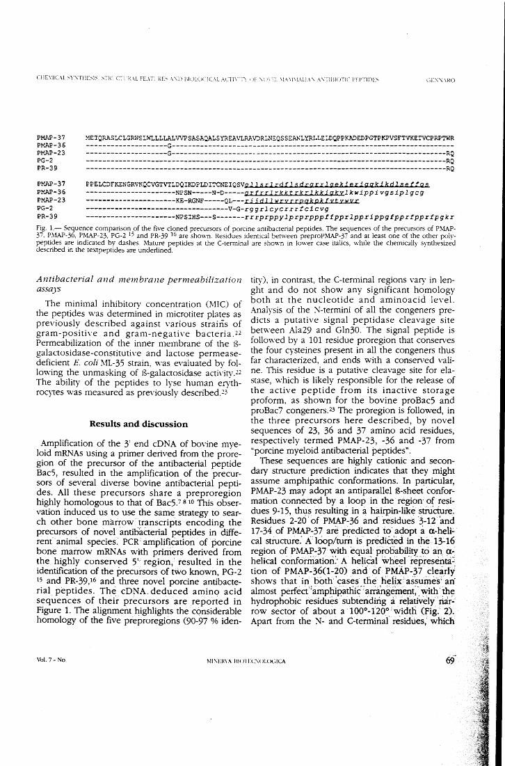
CE'\'\.·\ HO
PMAP-37 PMAP-36 PMAP-23 PG-2 PR-39
METQRASLCLGRWSLWLLLLALVVPSASAQALSYRSA\~RAVJR~EQSSEANLYRLLE~DQPP~EDPGTPKPVSFTVKETVCPRPTWR
--------------------G------------------------------------------------------------------------------------------G--------------------------------------------------------------------RQ -----------------------------------------------------------------------------------------RQ -----------------------------------------------------------------------------------------RQ
PMAP-37 PMAP-36 PMAP-23 PG-2 PR-39
PPELCDFKENGRVKQCVGTVTLDQIKDPLDITCNEIQSVg 11 s r1 ~d t l s drgrrl geki eri gqki kdl se tra s ----------------------NPSN-----N-0-----qrf rr 1 rkkt rk r l kki qkylkwippi vgsipl gcg ----------------------KE-RGNF-----QL---ri i d l l wrvrrpqkpktyt vwvr -----------------------------------V-G-rgg r l cycr r r t ci cvg ----------------------NPSIHS---5-------r r rprppy lp rprpppf tppr lppr ippg tppr fppr tpg k r
F~. 1.- Sequence comparison _of the five don ed precursors of porcine antibacterial peptides. The sequences of the precursors of P.\LA.P-3.t. ~.\1AP-36, .r.rv~-23, PG-2 1' and PR-39 l6 are shown. Residues identica! berv-.•een preproP!vfAP-37 and at Ieast one of the other poly-pept!?es are mdJCated by dashes. Mature peptides at the C-terminai are shown in Iower case italics, while the chemically synthesized descnbed m the textpeptides are underlined.
Antibacterial and membrane permeabilization assays
The minimal inhibitory concentration (MIC) of the peptides was determined in microtiter plates as previously described against various strains of gram-positive and gram-negative bacteria.22 Permeabilization of the inner membrane of the B-galactosidase-constitutive and lactose permease-deficient E. coli !\11-35 strain, was evaluated by fol-lowing the unmasking of B-galactosidase activity.22 The abilit)' of the peptides to lyse human eryth-rocytes was measured as previously described.23
Results and discussion
Amplification of the 3' end cDNA of bovine mye-loid mRNAs using a primer derived from the prore-gian of the precursor of the antibacterial peptide Bac5, resulted in the amplification of the precur-sors of several diverse bovine antibacterial pepti-des. Ali these precursors share a preproregion highly homologous to that of Bac5.7 s1o This obser-vation induced us to use the same strategy to sear-ch other bone mar~ow transcripts encoding the precursors of novel antib'acterial · peptides in diffe-rent'·animal species.' PCR· amplification of ·porcine bone marrow mRNAs with primers derived from the highly conserved S'· region,-resulted in the identification of the precursors of two known, PG-2 IS and PR-39,16 and three novel porcine antibacte-rial peptides. Thè cD N A_ deduced amino acid sequences of their precursors are reported in Figure l. The alignment highlights the considerable homology of the five preproregìòns (90-97 % iden-
tity), in contrast, the C-terminai regions vary in len-ght and do not show any significant homology both at the nucleotide and aminoacid level. Analysis of the N-termini of all the congeners pre-dicts a putative signal peptidase cleavage site between Ala29 and Gln30. The signal peptide is followed by a 101 residue proregion that conserves the four cysteines present in all the congeners thus far characterized, and ends with a conserved vali-ne. This residue is a putative cleavage site for ela-stase, which is likely responsible for the release of the active peptide from its inactive storage proform, as shown for the bovine proBacS and proBac7 congeners.23 The proregion is followed, in the three precursors here described, by novel sequences of 23, 36 and 37 amino acid residues, respectively termed PMAP-23, -36 and -37 from "porcine myeloid antibacterial peptides".
These sequences are highly cationic and secon-dary structure prediction indicates that they might assume amphipathic conformations. In particular, PMAP-23 may adopt an antiparallel B~sheet confor-mation connected by a loop in the. region· of resi-dues 9-15, thus resulting in a hairpin-likè stri.iciure. Residues 2-20. of PMAP-36 and- residues -3-12 and 17-34 of PMAP-37 arè predicted to adopt a. a-heli~ cal structure: A.: lÒOp/tum is prediéted in the 1~16 region of PMAP-37 with equal probability tò an, a-helical conformatiorì> A helical wheel -rèp-resenia: tion of PMAP.:.36(1-20) and- Òf PMAP-37 dearly shows that in bòrh· 'cases the heH:x> àssuines; an almost perfect ''amphipathic: arringemént; · with' me hydrophobic residues . subteridirig a ie1a~veiy" riai-· row sector of about a 100°-120°~width (Fig.· 2). Apart from the N- and C-terminai' residues," which
Vol. 7- No. MI'\EH\A BI( HEC\ULOCICA
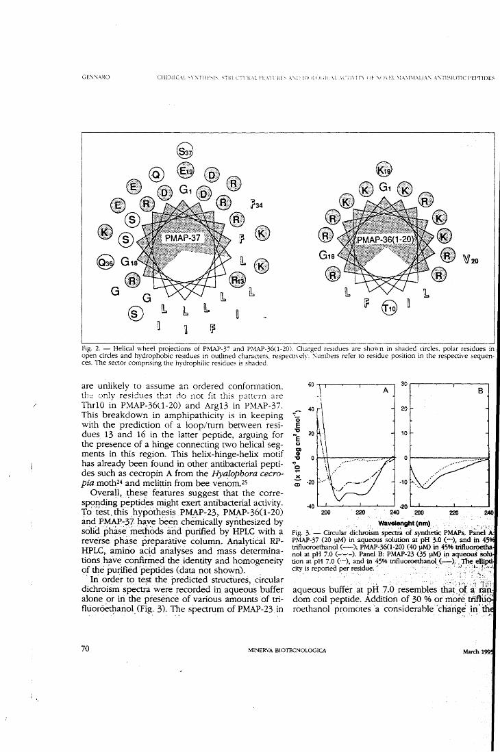
/
GE:\:\ARO CHL\llC\l. '-Y:\TllE~l~. <;Tin. (T\ l\.\ L! L\l l ì\L~ \\.l l l\i' 1l <i( d(. \L .\C.i l\ lTì f !!- V)\ EL \L-\\l\1.\LL\ '\ .\ \.-TllliOTJC i'El'TlDE~
~ G1 •• G .-.. G
®
~e [b - \b
u
Fig. 2. - Helical \vheel projections of Pt\~-37 and P:'>tAP-360-20). Charged residues are shown in shaded circles, polar residues in open circles and hydrophobic residues in outlined characters, respecti\ely. :\umbers referto residue position in the respectiYe sequen-ces. The sector comprising the hydrophilic residues is shaded.
are unlikelv to assume an ordered conformation. th·~ only r~~idues th::n do not fit this pattern are Thr10 in PMAP-360-20) and Arg13 in Pl\1AP-37. This breakdown in amphipathicity is in keeping with the prediction of a loop/turn between resi-dues 13 and 16 in the latter peptide, arguing for the presence of a hinge connecting two helical seg-ments in this region. This helix-hinge-helix motif has already been found in other antibacterial pepti-des such as cecropin A from the Hyalophora cecro-pia moth24 an d melitti~ from bee. venom. 25
Overa~l, .· ~hese features suggest that the corre-sp()rding peptides might exert antibacterial activity. 'fo /est. this ,hypo:thesi~ P~-23,. P~P-36(1-20) and PMAP-~7 ha,ve been chemically synthesized by solid phasè ·metho~ irìd purified by HPLC with a reveise phase preparative column. Analytical R.P-HPLC, amino acid analyses and mass _determina-tioris bave confmlied the .1dentity and homogeneity of the purifiéd peptides (data not shown). ·In· or~er to_ te~t thè. predicted siructures, _circular
dichroism spectra wère recorded in aqtieous buffer alone .or iri me presénce of various amount5 of tri-flùor6et~anol, (Fig: 3). The spectrum of PMAP-23. in
60 l\ l A ] 30
B ~ l
\
40 20
o E "C 20 10 "'e C.)
a o "' o o 'b ,... ~ -10 <D
:40 200 220 200 220 240
Wavelenght (nm) Fig. 3 .. - Circular. dichroiSm spe~ of synthetiC: PMAPs,. P:Ìn~l A: PMAP-37 (20 lJM) in aqueous solution at pH 3.0 (---), and in 45 trifluoroethanol (-); PMAP-36(1-20) (40 p.M) in _45% trifluoro.etfla nol at pH 7.0 (-·-·-). Panel B: PMAP-23 (35 l}M) in aqueou$ Solu tion at pH 7.0 (····), and in 45% trifluoroethanol_(~): , The ellipti, city is reported per residue.· · · '1
·- -~· · !-""" _;> i: ; .~ic '
_' . . . ·.. : . . . ... :; . . , .. -· .• "?·;.~ _: :·; ~fi-~1·
aqueous buffer at pH 7.0resembles ma~_~(~; ~n do m co il peptide. Addition of 30 % or more. tiiflu ,'. roethanol proinotes ·a considerahle_'charige _i~· m,_
70 MINERVA BIOTECNOLOGICA
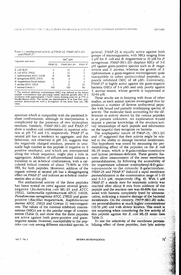
Cl ìL\iiC \L 'Y'\Till>l'-. 'THl ':·;·\ .1-L\L FL\TU\E~ A '\i) BIOLCX,JC\L .\CT!\Tìì. uf :\U\TL .\L\\1\1\Ll:\:\ A:\TIBIOTJC J'El'TIDE~ GE:\:\ARO
T>.BLE 1.-Antibacteriai actir·izr cif P.l!AP-23. P.lL4.P-36f 1-20} and P.l!AP-3 7.
Organism and strain !\ne· (J..L~1!
P!\IAP-23 PMAP-36<1-20) P!\IAP-3'7
E coli ~vlL35 2 12 2 E coli ATCC 25922 4 12 S. (Jpbimurium ATCC 1ct028 8 48 4 P aemginosa ATCC 27853 16 3 2 B. megate1ium Oocal isolate) 2 3 4 S. aureus ATCe 25923 4 6 32 S. aureus Cowan l ND !\TI 64
•) The minima! inhibitorv concentration (MIC) was defined as the lowest peptide concentration tliat prevemed_ \'isible bacterial growth after 18 h incubation with approximately 1.5xlO' colony forming unitsiml. at y:-oe in !\lueller-Himon hroth. The results are representative of at least four inde-pendent determinations with a divergence of not more than one MIC ,·alue.
spectrum which is compatible with the predicted B-sheet conformation, although its interpretation is complicated by the presence of two tryptophan residues. Similarly, PMAP-36(1-20) and PMAP-37 show a random coil conformation in aqueous solu-tion at pH 7.0 and 3.0, respectively. P.M.i\P-37 at neutra! pH has a tendency to aggregate, and this behaviour, not observed at pH 3.0, suggests that the negatively charged residues, present in unu-sually high number in this peptide (6 negative vs. 9 positive residues), and which are interspersed along the whole sequence, might play a role in aggregation. Addition of trifluoroethanol induces a transition to an a.-helical conformation, with a cal-culated helical content of about 75-80% at 45% TFE, for both peptides. Moreover, addition of the organic solvent at neutral pH has a disaggregating effect on PMAP-37 and induces an a.-helical confor-mation also at this pH.
The antibacterial activity of the three peptides has been tested in vitro against several gram-negative (Escherichia coli ML-35 and ATCC 25922, Sa/monella typhimurium ATCC 14028, Pseudomonas aeruginosa ATCC 27853) and gram-positive (Bacillus megaterium, Staphilococcus aureus ATCC 25923 and Cowan l) microorgani-sms. The values of the. minimal inhibitory concen-trations (MICs) are in the pmolar range of concen-tration (Table I), and show that the .three peptides are active against both gram-positive and gram-negative strains. However, susceptibility to the pep-tides can vary among different microbial species. In
generai, P:\iAP-23 is equally active against both groups of microorganisms, v..·ith MICs ranging from 2 }JIVI for E. coli and B. megaterium to 16 pM for P. aeruginosa. PMAP-36(1-20) displays MICs of 3-6 }J!\1 against gram-positive species such as B. mega-terium and S. aureus, whereas the gro~vth of S. ~1pbimuriunz, a gram-negative microorganism quite susceptible to other antimicrobial peptides, is poorly inhibited (MIC of 48 pM). Conversely, PMAP-37 is highly active against the gram-negative bacteria (MICs of 1-4 pM) and only poorly against S. aureus strains, whose gro~vth is suppressed at 32-64 }JM.
These resuits are in keeping with those of other studies, as each animai species investigated thus far produces a number of diverse antibacterial pepti-des with broad and partially overlapping spectra of acti\·ity. The molecular basis accounting for the dif-ferences in acth·ity shown by the various peptides is at present unknown. An explanation would require a precise knowledge of the structure/acti-vity relationships of each peptide and information on the target(s) they recognize on bacteria.
The amphipathic nature of PMAP-23, -36(1-20) and -37 suggested that their activity might be rela-ted to the ability to damage bacterial membranes. This hypothesis was tested by measuring the per-meabilizing effect of the peptides on the E. coli ML-35 strain, which is B-galactosidase-constitutive and lactose permease-deficient. These genetic fea-tures allow measurement of the inner membrane permeabilization, by following the accessibility of the impermeant substrate o-nitrophenyl-B-D-galac-topyranoside to the cytosolic B-galactosidase. PMAP-23 and PMAP-37 induced a rapid membrane permeabilization in the concentration range of 1-1 O and 0.2-1 pM, respectively (Fig. 4). With l pM PMAP-37 a steady state for enzymatic activity was. reached after about 8 min from addition of the peptide an d the reaction rate. was 60-80% that mea~ · sured with bacteria . completely lysed by ultrasoni-cation, indicating a massive damage of the bacterial: membranes. On the contrary, P.f-.1l.J:>-36<1-20) indu~: ces permeabilization at much higper concentration51
(l 0-50 pM) an d with slower kinetics. ~ This result. is not surprising. when considering the low activity. of, this peptide against the. E .. coli ML-35. strain (sèe~ Table I). . . . . -· _ . :. :'!
To test the selectivity of the membrane permea• bilizing effect of these peptides, .· their lytic activity,
Vol. 7- No. l MINERVA BIOTECNOLOGICA
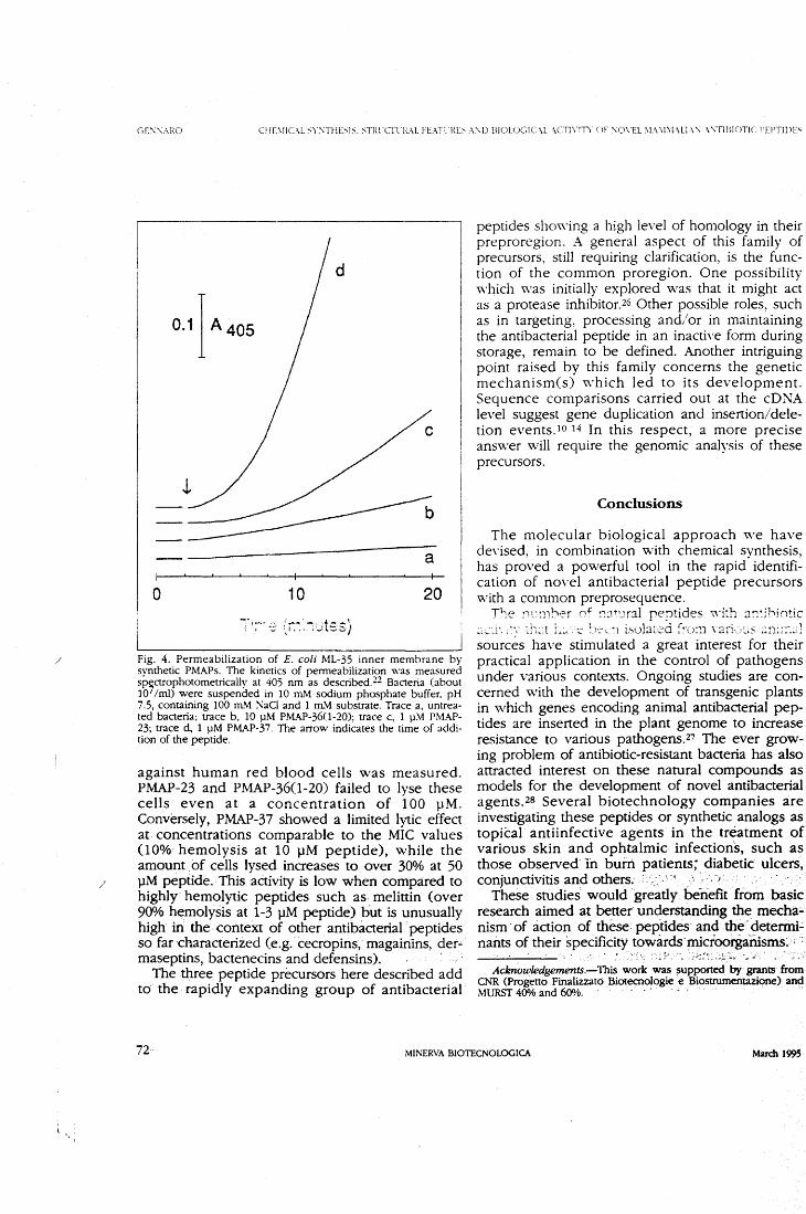
/
/
GE:\.'\.\1\0
0.1] A 405
a
o 10 20
Fig. 4. Permeabilization of E. coli ML-35 inner membrane by synthetic PMAPs. The kinetics of permeabilization was measured sp,ctrophotometrically a t 405 nm as described. 22 Bacteria (about l O /mi) were suspended in 10 ITLl\.1 sodium phosphate buffer. p H 7.5, containing 100 ITLl\.1 KaCI and l mM substrate. Trace a, untrea-ted bacteria; trace b, 10 pM PM.AJ>-360-20); trace c, l pM PMAP-23; trace d, l pM PMAP-37. The arrow indicates the time of addi-tion of the peptide.
against human red blood cells was measured. PMAP-23 and P.MAP-36(1-20) failed to lyse these cells even a t a concentration of l 00 p M. Conversely, PMAP-37 showed a limited lytic effect at concentrations comparable to the MIC values (lOo/o hemolysis at 10 pM peptide), while the amount . of cells lysed increases t o over 30% a t 50 pM pèptide. · This activity is low when compared to highiy· hemolytic peptides such as melittin (over 900h hemolysis at 1-3 pM peptide) but is unusually high iri the context of other antibàcterial peptides so far characterized (e.g .. cecropins, magainins; der-maseptins, bactenecins and defensins).
The three peptide precursors here described add tO the rapidly expanding group of antibacterial
peptides showing a high level of homology in their preproregion. A generai aspect of this family of precursors, stili requiring clarification, is the func-tion of the common proregion. One possibility which was initially explored was that it might act as a protease inhibitor.26 Other possible roles, such as in targeting, processing and/or in maintaining the antibacterial peptide in an inactive form during storage, remain to be defined. Another intriguing point raised by this family concerns the genetic mechanism(s) which led to its development. Sequence comparisons carried out at the cDNA level suggest gene duplication and insertion/dele-tion events.1o 14 In this respect, a more precise answer will require the genomic analysis of these precursors.
Conclusions
The molecular biologica! approach \Ve have devised, in combination with chemical synthesis, has proved a powerful tool in the rapid identifi-cation of novel antibacterial peptide precursors V..'ith a common preprosequence.
Tl:e !''1':ìlber c;+ ::Jt•Jral reptides '\Yith a:;~;J,in:ic ~>:... .. _i~-.;-~~· :1-::~t l-~~.-~ :~)·:\.._ ~-ì istJlar~~~à :~(J:11 \-2ri·:_>~~,s ~-:!!it~-:~:!
sourcés have stimulated a great interest for their practical application in the control of pathogens under various conte}..'tS. Ongoing studies are con-cerned with the development of transgenic plants in which genes encoding animai antibacterial pep-tides are inserted in the plant genome to increase resistance to various pathogens.27 The ever grow-ing problem of antibiotic-resistant bacteria has also attracted interest on these natural compounds as models for the development of novel antibacterial agents.zs Several biotechnology companies are investigating these peptides or synthetic analogs as topkal antiinfective agents in the trèatment of various skin and ophtalmic, infections, such as those observed· ìn burn patients; diaberic· ulcers, conjunctivitis an d others. · ·:' ·' ' . ~ · , ·, ·,
These stùdies would · greatly benefit from basic research aimed àt bettet undeistanding the mecha:.. nism · of àction of these pep"rides· and the ·detenni.;. nants of their specificity towiuds. m,icroorgarusms~ ; :
Acknowledgements.-This. work was $Upported by· grants from CNR (Progetto Finalizzato Biotecnologie e Biostrumentazìone) and MURST 400A> and 600A>. - '
72 MINERVA BIOTECNOLOGICA March 1995
CHE\liC\L ~ Y:\THE:-.1:--. :--THt CTL.H.:\L IT:\Tl-1\E:-. :\'\l! BIUL<-Jl;JC\L :\CTI \ ITY c li '\U\U. \L\ \l\L-\1.1\ '\ .\ '\TIBIOTIC l'l:J'TIDE:-. GE:\'\AHO
References 1. Boman HG. Antibacteri:il peptide~: key components needt·J in
immunitv. Celi 1991:65:205--:-. 2. Hoffmarin ]A. Hetru C. Insect defensins: inducible antibacterial
peptides lrnmunol Today 1992:13:--111-5. 3. Zasloff ;\1. Antibiotic peptides as mediators of innate immunit\·.
Curr Opin lmmunol 1992:4:3--:-. · 4. Gennaro R. Romeo D. Skerlavaj B. Zanetti \1. :\'eutrophil and
eosinophil granules as stores of defense proteins. In: Harris JR. ed. Blood celi biochemistrY. \'OL 3. :\'ew York:Plenum Press. 1991: 335-68. .
5. w·eiss J. Leukocyte-deriYed antimicrobial proteins. Curr Opin Hemato\1994:1:78-84.
6. Leher RL Lichtenstein AK. Ganz T. Defensins: antimicrobial and 01otoxic peptides of mammalian cells. Annu. Re\·. Immunol. 1993;11:105-28.
- Del Sal G. Storici P. Schneider C, Romeo D. Zanetti . .\1. cD:\A cloning of the neutrophil bactericidal peptide indolicidin. Biochem Biophys Res Commun 1992:187:467--2.
8. Storici P, Del Sal G. Schneider C. Zanetti .\1. cD:\'A sequence analysis of an antibiotic dodecapeptide from neutrophils. FEBS Lett 1992;314:18'7-90.
9. Zanetti M. Del Sal G. Storici P. Schneider C. Romeo D. The cD\:A of the neutrophil antibiotic Bac5 predicts a pro-sequence homologous to a cysteine proteinase inhibitor that is common to other neutrophil amibiotics. J Bio! Chem 1993:268:522-6.
10. Scocchi M. Romeo D. Zanetti .\1. Molecular cloning of Bac-. a proline- and arginine-rich antimicrobial peptide from beh ule neutrophils. FEBS Lett 1994;.352: 19--200.
11. Larrick j\X'. ;\1organ JG. Palings l. Hirata .\1. Yen .\lH. Complementary D:'\A sequence of rabbit CAP18 - a unique lipopolysaccharide binding protein. Biochem Biophys Res Commun 1991;179:170-5.
12. Levy O, W'eiss j. Zarember K. Ooi CE. Elsbach P. Antibacterial 15-kDa prmein isoform~ (o15<-) are members of a nove! bmilv of leuko~--"::~~· prut~ins. j ~~d~t. Che!~·~ l S·'~)j:2~1~·::,~~~;:-~~""-:>.1• ~
13. Pungercar J Strukelj B, Kopitar G et al. Molecuìar cìoning of a putative homolog of proline 1arginine-rich antibacterial peptides from porcine bone marrow. FEBS Lett 1993;336:284-8.
1--1. Zh:.to C. Liu L. Lebrer RL Jdentification of a new member of the protegrin family by cD\:A cloning. FEBS Lett 1994;346:285-8.
15. Storici P. Z:metti :'\1. A nove! cD:\A sequence encoding a pig leukocyte antimicrobial peptide with a cathelin-like pro-sequence. Biochem Biophys Res Commun 1993;196:1363-8.
16. Storici P. Zanetti .\1. A cD:\A derived from pig bone marrow cells predicts a sequence identica! to the intestina! antibaaerial pepti-de PR-39. Biochem Biophys Res Commun 1993:196:1058-65.
F. Chomczynski P. Sacchi :\'. Single-step method of R.."\A isolation by acid guanidinium thioqanate-phenol-chloroform e:\.'traction. Anal Biochem 1987:162:156-9.
18. Rost B. Sander. C. Prediction of protein secondary structure at better than '0% accura0··] :'\1ol Bio! 1993:232:584-99.
19. Edelhoch H. Spectroscopic determination of tryptophan and tyrosine in proteins. Biochemisuy 1967:6:1948-54.
20. Buck !\1A, Olah TA, Weitzmann CJ Cooperman BS. Protein esti-mation by the product of integrated peak area and flow rate. Anal Biochem 1989:182:295-9.
21. \\'u C-SC, Ikeda K, Yang JT. Ordered conformation of polypep-tides and proteins in acidic dodecyl sulfate solution. Biochemistrv 1981:20:566-ìO.
22. Skerlavaj B,-Romeo D, Gennaro R. Rapid membrane permeabi-lization and inhibition of vita! functions of gram-negative baae-ria lw bactenecins. Infect Immun 1990:58:3'724-30.
23. Scocchi .\1. Skerlavaj B. Romeo D, Gennaro R. Proteol)1ic clea-\·age by neutrophil elastase converts inactive storage proforms to antibacterial bactenecins. Eur J Biochem 1992:209:589-95.
24. Holak TA. Engstrom A. Kraulis PJ et al. The solution conforma-tion of the antibacterial peptide cecropin A: a nuclear magnetic resonance and dynamical simulated annealing study. Biochemistry 1988:2'7:1620-9.
25. Bazza R, Tappin .\l_j, Pastore A. Harvey TS, Carver JA. Campbell ID. The structure of melittin. Eur J Biochem 1988; 173:139-46.
26. \'erbanac D, Zanetti M. Romeo b. Chemotactic and protease-inhibiting acti\'ities of antibiotic peptide precursors. FEBS Len 1993: 317:255-8. :.:~ ,:'l~~!~~; .. ·:li C: ~-.:~~~:~~~ri G. ::-.·:-.-)j:.lction c,f ar, a;"1t~~Jacte:i11 ,gene in potato (Solanum tuberosum L.) using a binary vector in Agrobacterium rbizogenes. J Genet & Breed 1991;45:307-16.
28. Gabay JE. l.Jbiquitous natura! antibiotics. Science 1994:263:373-4.
Vol. 7- No. l MIKERVA BIOTECNOLOGICA
/
Tip. S.A.E.M.M. - Saluzzo
AllEGATO l
CROATICA CHEMICA ACTA CCACAA 68 (3) 607-614 (1995!
ISSN 0011-1643 CCA-2259 Conference Paper
Bovine Neutrophil Antibiotic Peptides and Their Precursors: Structure and Role in Innate Immunity*
Domenico Romeo, Renato Gennaro, Margherita Zanetti, Alessandro Tossi, Barbara Skerlavaj, Paola Storici, Marco Scocchi, Laura Litteri
and Donatella Verbanac#
Department of Biochemistry, Biopyhsics and Macromolecular Chemistry, University of 'lheste and Department of Medical Sciences and Technology,
University of Udine, Italy
Received March 7, 1995; revised March 20, 1995; accepted March 20, 1995
Four peptides were characterized in extracts of bovine neutrophil granules: an Arg-rich dodecapeptide, maintained in a cyclic struc-ture by a disulfide bridge; a Trp-rich tridecapeptide named indo-licidin; and two 43- and 59 amino acids long peptides, named Bac5 and Bac7, with frequent repeats of the triplets Arg-Pro-Pro and Pro-Arg-Pro, respectively. The full length cDNA of the first three of these peptides was char-acterized recently. Sequence analysis showed that the prose-quences of the predicted precursors of all the three peptides are highly identica! and exhibited a\'So a remarkable similarity to cathelin, a porcine inhibitor of cath~psin L. Purified proBac5 actu-ally proved in in vitro assays to inbibit cathepsin L, but not other cysteine proteinases such as cathepsin B. Unlike proBac5, proBac7 is selectively chemotactic to monocytes. Severa! fragments of Bac5 and Bac7 (from 6 to 35 residues) were synthesized by the Fmoc method. The results of antibacterial as-says show that the N -terminai portio n, the most cationi c o ne in both Bac5 and Bac7, is essential for the antimicrobial activity and that the minima} length necessary to arrest the growth of suscep-tible bacteria is 18-20 residues.
* Presented at the Meeting of Croatian Biochemists, Zagreb 1993.
# Permanent address: Department of Chemistry and Biochemistry, Medicai Faculty, Univer-sity of Rijeka, Braée Branchetta 22, 51000 Rijeka, Croatia.
608 D. ROM EO /:J Al_
INTRODUCTION
Cationic antimicrobial polypeptides are considered to play a significant role in host defense, both at the level of respiratory and intestina! epithelia an d in phagocytic cells such as macrophages and neutrophils. Hirsch 1 was the first to describe a crude protein fraction of neutrophil granule extracts with bactericidal properties. A few years later, Zeya and Spitznagel2·3 at-tempted a partial purification of bactericidal peptides from other cationic proteins of neutrophil granules, showing their selective activity on gram-positive and gram-negative organisms and Candida albicans.
Later on, a variety of these polypeptides w ere purified from neutrophils, macrophages, epithelia and body fluids and their antimicrobial properties were characterized (for reviews, see Refs. 4-10). This led to the coinage of definitions such as »endoantibiotics« or »antibiotics from within«, 4 as well as to the recognition of their importance as effectors in innate immunity. They ha ve thus become a very challenging field of research a t the crossroad of cell and molecular biology, immunology and, in generai, modern biotechnology.
In particular, the role of the neutrophil antimicrobial peptides has been extended beyond that of simply combatting invading microorganisms. In fact, recent applications of recombinant DNA techniques, sequence compari-sons and imaginative experiments have unravelled new functions for at least some of these ))antibiotics« and their precursors. These roles include the regulation of LPS toxicity, 11·12 recruitment of monocycles to inflar1una-tory sites13- 15 and inhibition of cathepsin L, 15 a major factor in inflammatory tissue degradation.
Antinlicrobial Peptides of Bovine "-Veutrophils
Azurophil granules of neutrophils contain an arsenal of antimicrobial peptides. 4-6,8,10 ,
Bovine neutrophils also possess these classica! azurophil antibiotic pep-tides.16.17 However, in ruminant neutrophils, the azurophil granules are pre-sent in relatively small number. 18 Conversely, these cells contain a novel, abundant type of subcellular granule of a large size and high density, which appears to contain most of the antimicr_pbial factors. 19
Thus far, in addition to azurocidin, 16 and defel_lsins, 17 four novel cationic antimicrobial peptides of unique structure have been purified from extracts of bovine neutrophil granules: i) a dodecapeptide with four arginyl residues, maintained in a cyclic structure by a disulfide bond between the two cyste-ine residues;20 ii) a tridecapeptide with five triptophanyl residues and a car-boxyamidation at the C-terminai arginine, named indolicidin;21 iii) a proline- and argininine-rich peptide, named Bac5, with 43 amino acid resi-dues, a repeated motif Arg-Pro-Pro-large hydrophobic residue22·23 and very

BOVINE NElJTROPHIL A..'<TJBIOTlC ì'EPTIDES A.,'\[) PRECURSORS
TABLE I
Antibiotic polypeptides of azurophil granules
BPI or CAP57 (Bactericidal/ Permeability Increasing Protein, 50-60 kDa) Serprocidins (cathepsin G, elastase, proteinase 3, azurocidin or CAP37, 25-29 kDa) Defensins (ca. 4 kDa)
609
likely a carboxyamidation at the C-terminai proline;24 iv) a second, struc-turally different, proline- and arginine-rich peptide, named Bac7, with 59 amino acid residues and repeated Pro-Arg-Pro tripiets (from position 10 to the C-terminus proiine is present every second residue) and three tandemiy repeated tetradecamers in the region between residues 15 and 56. 22•23
Although with marked selectivity and different potency, all the four pep-tides are active on gram-negative and gram-positive organisms. 16·20- 22 In generai, Bac5 and Bac7 are much more active on enteric bacteria, such as Escherichia coli, Sal monella typhimurium, Klebsiella pneumoniae. In addi-tion, both peptides immobiiize and kill Leptospira interrogans and biflexa, whereas they are inactive on another genus of spirochetes, Borrelia burgdoferi.25 Finally, Bac7 neutralizes human herpes simplex virus (HSV1 and HSV2), but not a picornavirus (rhinovirus 1B).26
In gram-negative bacteria and Ieptospire, Bac5 and Bac7 very likeiy bind to LPS in the outer cell membrane. 16•25 •27 Then, they rapidiy move to the in-ner membrane, whose permeability is dramatically increased with a con-comitant drop in the ATP content, loss of ability of macromolecuiar biosyn-
TABLE Il
Novel antimicrobial peptides of bovine neutrophils ----------------------------- -- ---------- .. - ---------------- -------
Dodecapeptide RLCRIVVIRVCR
I ndolicidin ILPWKWPW\VPWRR Bac5 RFRPPIRRPPIRPPFYPPFRPPIRPPI FPPIRPPFRPLGPFP Bac7 RRIRPRPPRLPRPRPRPLPFPRPGPRPI PRPLPFPRPGPRPIPRPLPFPRPGPRPIPRP
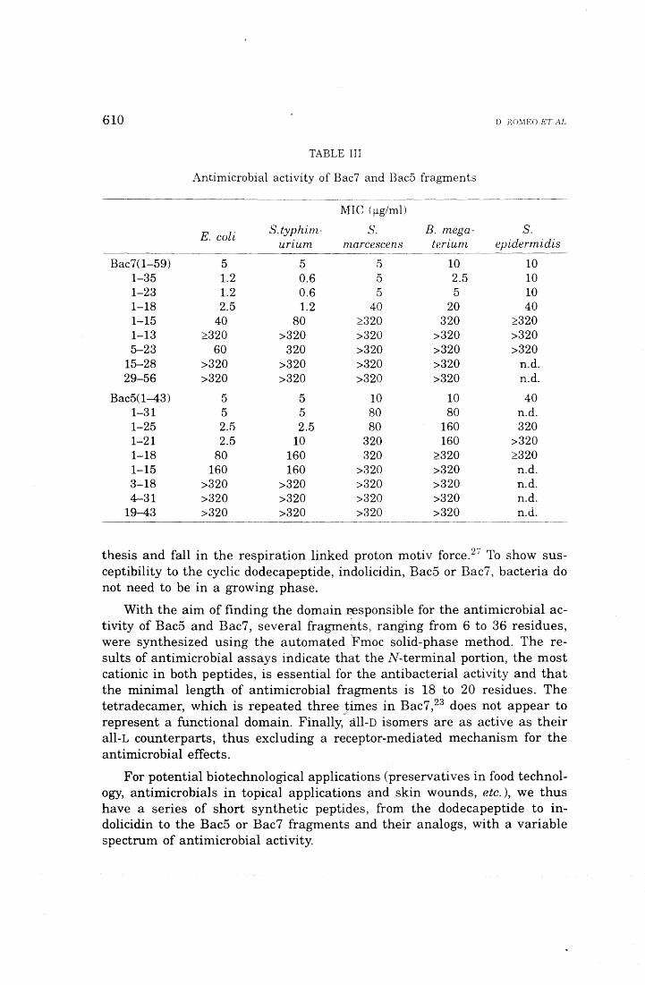
610 D. RO:.lEO ET AL
TABLE III
Antimicrobial activity of Bac7 and Bac5 fragments ------~-------·---~---·--------------···-·-----
~n c ( 1-1g/ml)
E. coli S.typhim- S. B. mega- S. urzum marcescens terium epidermidis
Bac7(1-59) 5 5 5 10 10 1-35 1.2 0.6 5 2.5 10 1-23 1.2 0.6 5 5 10 1-18 2.5 1.2 40 20 40 1-15 40 80 ~320 320 ~320
1-13 ~320 >320 >320 >320 >320 5-23 60 320 >320 >320 >320
15-28 >320 >320 >320 >320 n.d. 29-56 >320 >320 >320 >320 n.d.
Bac5(1-43) 5 5 lO 10 40 1-31 5 5 80 80 n.d. 1-25 2.5 2.5 80 160 320 1-21 2.5 10 320 160 >320 1-18 80 160 320 ~320 ~320
1-15 160 160 >320 >320 n.d. 3-18 >320 >320 >320 >320 n.d. 4-31 >320 >320 >320 >320 n.d.
19-43 >320 >320 >320 >320 n.d.
thesis and fall in the respiration linked proton motiv force. 21 To show sus-ceptibility to the cyclic dodecapeptide, indolicidin, Bac5 or Bac7, bacteri a do not need to be in a growing phase.
With the aim of finding the domain responsible for the antimicrobial ac-tivity of Bac5 and Bac7, several fragments, ranging from 6 to 36 residues, were synthesized using the automated 'Fmoc solid-phase method. The re-sults of antimicrobial assays indicate that the N-terminai portion, the most cationic in both peptides, is essential for the antibacterial activity and that the minimal length of antimicrobial fragments is 18 to 20 residues. The tetradecamer, which is repeated three times in Bac7, 23 does not appear to represent a functional domain. Finally,,.· all-D isomers are as active as their all-L counterparts, thus excluding a receptor-mediated mechanism for the antimicrobial effects.
For potential biotechnological applications (preservatives in food technol-ogy, antimicrobials in topical applications and skin wounds, etc.), we thus have a series of short synthetic peptides, from the dodecapeptide to in-dolicidin to the Bac5 or Bac7 fragments and their analogs, with a variable spectrum of antimicrobial activity.
BOVINE NEUTI\OPHIL A."<T!BIOTIC PEPT!DE::; A>iD l'l·: H \ T;-;uE;-; 611
Biosynthesis and Processing ol Bac5 and Bac7 Prccursors
Investigations utilizing the classica! tools of cell biology have shown that Bac5 and Bac7 are synthesized in hone marrow cells, presumably at the myelocyte stage of cell maturation, as preproforms of 23.5 and 21 kDa, re-spectively.28 PreproBac7 is then processed to a protein of 20 kDa, which is the granule storage, precursor form of Bac7, as suggested by \Vestern blot analysis. 28 Conversely, preproBac5 is first converted to a polypeptide of 15.8 kDa. This is then processed (very likely in a post-ER compartment) to a 15 kDa polypeptide, which is the granule storage, precursor form of Bac5,28 presumably by removal ofthe C-terminai tripeptide and concomitant amidation of the C-terminus. 24
As shown by immunogold electron microscopy, the 20 kDa proBac7 and the 15 kDa proBac5 are specifically targeted a t the matrix of the large granules. 28
In uitro experiments conducted with purified proBac7 and proBac5 have indicated that these precursors can be stepv.rise converted to the correspond-ing mature antibiotic peptides by elastase. 30 This points to the ne ed of co-operation between the large granules and the azurophils. Experiments car-ried out with phagocytosing neutrophils have indeed shown that such a cooperation occurs in the phagocytic vacuoles or extracellularly, under con-ditions favouring a concomitant release of proBac7/proBac5 and elastase. 31
Incidentally, detection of Bac7 and Bac5 antigens in phagocytic vacuoles by immunogold electron microscopy31 provides experimental evidence that the precursor molecules are actually n1obilized from the large granules and dis-charged into the vacuoles.
The proteolytic conversion of proBac7 and proBac5 to mature peptides is a requirement for manifestation of the antibiotic activity. In fact, purified proBac7 and proBac5 do not affect the viability of microorganisms suscep-tible to the corresponding mature peptides;30 This might be due to the mask-ing of the N-termini moiety, which is required for full expression of the an-tibacterial activity (see above).
Molecular Cl o n ing of Bouine J.Veu t roph i l Antilnicrobial P ept id es
Molecular cloning and sequencing of the full length cDNA of Bac5, in-dolicidin, and the dodecapeptide25·29 ,32 have provided the following informa-tion: i) the predicted signal sequence comprises 29 amino acids and is prac-tically identica! in the three prepropeptides; ii) the three antimicrobial peptides are located at the carboxyl end of the corresponding precursors; iii) at the C-terminus of Bac5 there is a Gly-Arg-Arg sequence, which is gener-ally considered a proteolysis/a-amidation signal;. the indolicidin precursor also terminates with a Gly, which is lost post-translationally for a-amidation of the tenninal Arg; iv) the hydrophylic pro-region in the three precursors has a length of 101 residues, is highly identica! and contains four invariant Cys.
612 D. ROMEO ET M,
Furthermore, it is characterized by a net negative charge. which n1ay neu-tralize the highly cationic N-terminal domain of the antimicrobial peptides, thereby masking their activity.
The pn~~dicted pro-region shows a high identity with the proregion of two rabbit neutrophil proteins, CAP18 and p15, described as LPS-binding pro-teins. 33·34 In addition, the pro- region sequence is homologous with a pig neutrophil protein, which is a potent and specific inhibitor of the cysteine protease cathepsin L and was thus named cathelin.35 Purified proBac5 was actually shown to inhibit in vitro cathepsin L, but not cathepsin B, with a Ki of 6 x Io-8 M. 15
Suitable stimulants may cause release of proBac5 from the large gran-ules into the extracellular fluid. 31 Since cathepsin L is a major factor in the degradation of inflamed tissue and may cause hone resorption as well as marked damage to the glomerular basement membrane, its potential inhi-bition by the precursors of large granule peptides is of great significance an d points to a wider role of these proteins in the modulation of the inflamma-tory response.
Acknowledgements. - The work in the laboratory of the Authors was supported by grants from the Italian National Research Council (CNR Progetto Finalizzato »Biotechnologie e Biostrumentazione«) and of the 1-finistry for University and Research.
REFERENCES
l. J. G. Hirsch, J. Exp. Med. 103 (1956) 589-592. 2. H. I. Zeya and J. K. Spitznagel, Science 142 (1963) 1085-1087. 3. H. I. Zeya and J. K. Spitznagel, J. Babteriol. 91 (1966) 750- 754 4. P. Elsbach, TIBTECH 8 (1990) 26-30. 5. R. L Lehrer and T. Ganz, Blood 76 (1990}:'2169-2181. 6. J. K. Spitznagel, J. Clin. Invest. 86 (1990~ 1381-1386. 7. H. B. Boman, Cell 65 (1991) 205-207. 8. R. Gennaro, D. Romeo, B. Skerlavaj, and l\1. Zanetti, in Blood Cell Biochemistry,
J. R. Harris (Ed.), Plenum Pubi. Co., (1991) 335-368. 9. :rvL Zasloff, Curr. Opinion Immunol. 4 (1991) 3-7.
10. J. E. Gabay and R. P. Almeida, Curr. Opinion Immunol. 5 (1993) 97-102. 11. C. E. Ooi, J. Weiss, l\1. E. Doerfler, and.P. Elsbach, J. Exp. Med. 174 (1991)
649-655. 12. M. N. Marra, C. G. Wilde, M. S. Collins, J. L. Snable, l\f. B. Thornton, and R. \V.
Scott, J. Immunol. 148 (1992) 532-537. 13. M. C. Territo, T. Ganz, M. E. Selsted, and R. I. Lehrer, J. Clin. Invest. 84 (1989)
2017-2020. 14. H. A. Pereira, W. M. Shafer, J. Pohl, L. E. l\fartin, and J. K. Spitznagel, J. Clin.
Invest. 85 (1990) 1468-1476. 15. D. Verbanac, M. Zanetti, and D. Romeo, FEBS Lettr. 317 (1993) 255-258. 16. L. Litteri and D. Romeo, lnfec. Immun. 61 ( 1993) 966-969.
BOVINE NEUTROPlfiL ,\.:"iTJBIOTIC PEPTIDES A~D PnECUR.SOHS 613
17. M. E. Selsted, Y.-Q. Tang, \V. L .. Morris, P. A. McGuire, l\1. J. Novotny, \V. Sn1ith, A. H. Henschen, and J. S. Cullor, J. Biol. Chem. 268 (1993) 6641-6648.
18. M. Baggiolini, U. Horisberger, R. Gennaro, and B. De\vald, Lab. Invest. 52 (1985) 151-158.
19. R. Gennaro, B. Dewald, U. Horisberger, H. U. Gubler, and M. Baggiolini, J. Cel! Biol. 96 (1983) 1651-1661.
20. D. Romeo, B. Skerlavaj, M. Bolognesi, and R. Gennaro, J. Biol. Chern. 263 (1988) 9573-9575.
21. M. E. Selsted, M. J. Novotny, W. L. Morris, Y. Q. Tang, \V. Smith, and J. S. Cullor, J. Biol. Chem. 267 ( 1992) 4292-4295.
22. R. Gennaro, B. Skerlavaj, and D. Romeo, lnfec. Immun. 57 (1989) 3142-3146. 23. R. W. Frank, R. Gennaro, K. Schneider, M. Przybylski, and D. Romeo, J. Biol.
Chem. 265 (1990) 18871-18874. 24. M. Zanetti, G. Del Sal, P. Storici, C. Schneider, and D. Romeo, J. Biol. Chem. 268
(1993) 522-526. 25. M. Scocchi, D. Romeo, and M. Cinco, Infec. Immun. 61 (1993) in press. 26. A. Zerial, B. Skerlavaj, R. Gennaro, and D. Romeo,Antivir. Res. 7 (1987) 341-352. 27. B. Skerlavaj, D. Romeo, and R. Gennaro, lnfec. Immun. 58 (1990) 3724-3730. 28. M. Zanetti, L. Litteri, R. Gennaro, H. Horstmann, and D. Romeo, J. Cell Biol. 111
(1990) 1363-1371. 29. G. Del Sal, P. Storici, C. Schneider, D. Romeo, and M. Zanetti, Biochem. Biophys.
Res. Commun. 187 (1992) 467-472. 30. M. Scocchi, B. Skerlavaj, D. Romeo, and D. Gennaro, Eur. J. Biochem. 209 (1992)
589-595. 31. M. Zanetti, L. Litteri, G. Griffiths, R. Gennaro, and D. Romeo, J. Immunol. 146
(1991) 4295-4300. 32. P. Storici, G. Del Sal, C. Schneider, and lVL Zanetti, FEBS Lettr. 314 ( 1992!
187-190. 33. J. \V. Larrick, J. G. Morgan, L Palings, M. Hirata, and M. H. Yen, Biochem. Bio-
pyhs. Res. Commun. 179 (1991) 170-175. 34. O. Levy, J. Weiss, K. Zarember, C. E. Ooi, and P. Elsbach, J. Biol. Chem. 268
(1993) 6058-6063. 35. M. Kopitar, A. Ritonja, T. Popovié, GabrijetCic, I. Krizaj, and V. Turk, Biol. Chen~.
Hoppe-Seyler 370 (1989) 1145-1151. .
SAZETAK
Antibioticki peptidi i njihovi prekursori izolirani iz neutrofila goveda: struktura i uloga u pri{odenoj imunosti
Domenico Romeo, Renato Gennaro, Margherita Zanetti, Alessandro Tossi, Barbara Skerlavaj, Paola Storici, Marco Scocchi, Laura Litteri i Donatella Verbanac
Iz neutrofilnih govedih granulocita do sada su odredena cetiri peptida: Arg-bo-gati dodekapeptid, s jednom disulfidnom vezom; Trp-bogati tridekapeptid, nazvan indolicidin; te dva polipeptida koja se sastoje od 43 odnosno 59 aminokiselina, naz-vani Bac5 i Bac7, u kojima nailazimo na pravilno ponavljanje slijeda Arg-Pro-Pro odnosno Pro-Arg-Pro.
. l
614 D_ RO:\IEO ET AL
Nedavno je odredena cjelokupna cDNA za prva od trì spomenuta peptida. Sek-vencijska analiza pokazuje da je slijed pretpostavljenog prekursora za sva tri peptida visoko identican i pokazuje znacajnu slicnost s katelinom - inhibitorom katepsina L izoliranim iz svinja.
ProCiséeni proBac5 upotrebljen u in vitro eksperimentima, pokazuje inhibiciju katepsina L, ali ne i katepsina B druge poznate cisteinske proteaze, dok je proBacì selektivno kemotaktican za monocite.
Pojedini dijelovi peptida Bac5 i Bac7 (od 6 do 35 aminokiselinskih ostataka), sin-tetizirani su metodom Fmoc. Rezultati pokusa na bakterijama pokazuju da je upravo N-terminalni kraj najjaCi kationski dio kod Bac5 i Bac7, bitan za antibakterijsku ak-tivnost, a minimalna duljina lanca nuzna za suzbijanje rasta bakterija iznosi 18 do 20 aminokiselinskih ostataka.
ALLEGATO M
An Approach Combining Rapid cDNA Amplification and Chemical Synthesis for the
ldentification of Novel, Cathelicidin-Derived, Antimicrobial Peptides
Alessandro Tossi,3 Marco Scocchi,a,b Margherita Zanetti,b,c Renato Gennaro,c Paola
Storici3 and Domenico Romeoa
aneparttnent of Biochemistry, Biophysics and Macromolecular Chemistry, University of
Trieste, Trieste; hNational Laboratory of the Interuniversity Biotechnology Consortium,
AREA Science Park, Padri ciano 99, Trieste; cnepartment of Biomedica! Science and
Technology, University ofUdine, Udine, Italy.
Current Protocols in Antimicrobial Peptide Research - Methods in Molecular Biology
l. INTRODUCTION
Host defence in mammals involves a cooperative effort by a variety of antimicrobial
peptides (l), which are produced by both phagocyte precursors and some epithelia. They are
called on to combat invading pathogens at sites of infection and to help protect mucosa!
surfaces. Although sharing some common properties, such as a high content of basic residues,
these peptides vary widely in both size and primary structure.
The neutrophil antimicrobial peptides are synthesized in myeloid precursor cells as
prepropeptides, and are stored in cytoplasmic granules as either fully processed products (2) or
propetides (3 ). Molecular cloning of cONA from bovine, porcine, rabbit, human and ovine
myeloid mRNA has revealed tbe presence of a family of antimicrobial peptide precursors, that
we bave named cathelicidins and whicb currently counts over twenty members ( 4,5)
The cathelicidins bave highly conserved preproregions while the C-terminai domains,
which correspond to the antimicrobial peptides, are highly varied (Fig. l). Some of these
peptides had already been isolated as components of the antimicrobial arsenal of neutrophils
(6-10), whereas others were deduced from the cONA of their precursors (5,11-17) and sbown
to be antimicrobial after cbemical synthesis (l 3'-16, 18 ). The C-terminai peptides in cathelicidins
include Cys-ricb peptides, namely the cyclic dodecapeptide and five protegrins ( 5,19-21 ), the
Pro- and Arg-ricb Bac5, Bac7, Bac7.5, prophenin and PR-39 (5,12,22-24), the Trp-rich
indolicidin and PMAP-23 (14,25), and tbe a.-belical PMAP-36, PMAP-37, SMAP-29,
CAP18(106-142) and FALL-39/human CAP18 (5,11,13,15-17).
These peptides exert a broad spectrum antimicrobial activity at micromolar
concentrations, with a wide overlap in specificity but also with significant differences in
specific potencies among eacb otber (4,6-10,13-16,18). Although severa! approaches have
been used to investigate their mode of action, their killing mecbanism(s) is stili incompletely
understood. Some bave been shown to rapidly permeabilize the membranes of susceptible
bacteria (13-15,18), witb leakage of cellular metabolites and marked reduction of
macromolecular biosyntbesis (26 ).
Extending the search for cathelicidins to various animai species might lead to an
expansion of the family, thereby allowing for species correlations. W e ha ve thus devised a
strategy which pennits the discovery of new cathelicidins and a better characterization of the
defense system in different animals species. This strategy consists in i) identificarlo n of nove!
cathelicidins as deduced from their cDNA, ii) identification of their putative antimicrobial
domain and iii) chemical synthesis and biologica! characterization of the corresponding
peptides. A similar strategy can be applied to other precursors of antimicrobial peptides with a
conserved domain (27).
The molecular biologica! approach for the amplification of nove! cathelicidin cDNA
sequences is based on the high consetvation of their preproregions at the nucleotide leve!. This
strategy is a modified RACE (Rapid Amplification of cDNA Ends) cloning protocol. RACE is
a PCR-based technique which was initially developed to facilitate cloning of full-length cDNA
ends after production of a partial sequence from a cDNA library (28-30). We have modified
this protocol by using sets of cathelicidin-specific and non specific primers to reverse transcribe
the 5' and the 3' ends of myeloid mRNAs containing consetved preproregions, and to amplify
the cDNA ends by PCR. By using the appropriate primers, overlapping 5' and 3' ends are
generated, from which the full-length cDNA sequence is deduced. This method is not only
much faster than screening a cDNA library, but is also faster and simpler than the originai
RACE (28).
Once a new member of the cathelicidin family has been identified, the putative
antimicrobial activity of the C-terminai, non-consetved region needs to be confrrmed. We
provide advice for the identification of the antimicrobial domain and for the chemical synthesis
of the corresponding peptide, including some suggestions for predicting possible difficulties
and overcoming them. Fmally, we describe assays for determining antimicrobial and
membrane-permeabilizing activity of the synthetic peptides.
2. MATERIALS
2.1. Preparation of myeloid cell-enriched bo ne man-o w cells
The phosphate buffered saline (PBS) is 137 mM NaCl, 1.7 mM KCI, 8.1 mM
Na2HP04, 1,5 mM KH2P04, pH 7.4. Lysis buffer is 155 mM ~Cl, 10 mM KHC03 and
0.1 mM EDTA, pH 7.4 at 4°C. Ficoll-Paque is a product of Pharmacia Biotech (Uppsala,
Sweden).
2.2. Reverse transcription, and amplijication of cDNA
The reverse transcriptase buffer (buffer l) is 250 mM Tris-HCI, 375 mM KCI, 15 mM
MgCI2, pH 8.3. The PCR reaction buffer (buffer 2) is 100 mM Tris-HCI, 500 mM KCI, 15
mM MgCI2 and 0.1% gelatin (w/v), pH 8.3. The proteinase K reaction buffer (buffer 3) is
200 mM Tris-HCI, 100 mM EDTA and 10% SDS (w/v), pH 8.0. The dNTP mixture contains
10 mM of each of the four deoxyribonucleoside triphosphates, as obtained from Sigma (St ..
Louis, MO). Proteinase K (l mg/ml) can be obtained from Merck (Darmstadt, Germany). W e
use Moloney · Murine Leukemia Virus (M-ML V) reverse transcriptase (200 U/Jll) from BRL
(Gaithersburg, MD). Primers l to 4 (see Table 1), and sequence specific primers, were
synthesized using an Applied Biosystem 394 DNA/RNA synthesizer (Foster City, CA). The
RNAse inhibitor RNAguard (34 U/J.Il) can be obtained from Pharmacia Biotech. Taq DNA
polymerase (2.5 U/Jll) is from Perkin Elmer/Cetus (Norwalk, CT). Ali other reagents are of
molecular biology grade.
2.3. Peptide synthesis, purijication and characterization
The reagents to be employed and the suppliers wherefrom they can be obtained are the
following: Fmoc-protected amino acids and 2-( 1H-benzotriazole-1-yl-)-1 ,1,3,3-
tetramethyluronium tetrafluoroborate (TBTU) from Novabiochem (Laufelfingen, Switzerland);
N-hydroxybenzotriazole (HOBt) from Sigma (St. Louis, MO); 1-hydroxy-7-azabenzotriazole
(HOAt), 0-(7-azabenzotriazol-1-yl)-1,1,3,3-tetramethyluronium hexafluorophosphate
(HATU) and PEG-PS resins from Perseptive BioSystems (Framingham, MA);
triisopropylsilane (TIPS), phenol, 1,8-diazabicyclo(5,4,0)undec-7-en (DBU) and acetic
anhydride from Aldrich-Chemie (Steinheim, Germany); ethanedithiol (EDT, stench),
trifluoroacetic acid (TFA, corrosive), piperidine (stench) and N-methylmorpholine (NMM)
from Fluka Chemie (Buchs, Switzerland); synthesis grade N,N-dimethylfonnamide (DMF,
toxic), N-methylpyrrolidone (NMP), dichloromethane (DCM), dimethylsulphoxide (DMSO),
and HPLC grade acetonitrile (toxic) from Romil Chemicals (Loughborough, UK). Ali other
reagents and solvents are of analytical grade.
2.4. Biologica/ activity assays
The buffer used to dilute bacteria is 10 mM sodium phosphate, 137 mM NaCI, pH 7.4
(buffer 4) in the case of MIC and MBC assays, and 10 mM sodium phosphate, 100 mM NaCl,
p H 7.4 (buffer 5) for the membrane permeabilization assay. Mueller-Hinton medium and
Sabouraud liquid medium can be obtained from Difco Laboratories (Detroit, MI), U-shaped,
96-well microdilution plates from Sarsted, (Nlimbrecht, Germany), and o-Nitrophenyl-(3-D-
galactopyranoside (ONPG) from Sigma.
3.METHODS
3.1. Preparation of myeloid cell-enriched bo ne marrow cells
Bone marrow cells are collected from ribs soon after slaughtering. Ribs are cut open
with a cast cutter and the marrow quickly transferred with a spatula to a tube containing l
mM EDTA in PBS, prewarmed to 37°C. Coarse debris and clots are sedimented and lipids
aspirated with a Pasteur pipette. The cell suspension is then passed through a screen of filter
cloth to remove small clumps and quickly diluted in PBS containing 10 mM glucose.
Erythrocytes and erythroid cells are lysed with three volumes of lysis buffer for 2 to 4 min at
4 °C, and intact cells collected by centrifugation at 200g for l O min. The pellet is resuspended
in PBS containing 0.5 mM MgCI2 and l O mM glucose, an d the mature granulocytes are then
removed by centrifugation through Ficoll-Paque at 800g for 40 min. The layer at the Ficoll-
Paque/PBS interface, enriched in immature cells of the myeloid lineage, is drawn off and
used for RNA extraction after washing twice with PBS.
3.2. Amplification of cathelicidin cDNAs
The method used to obtain cathelicidin cDNAs is schematically illustrateci in Fig. 2.
3.2 .1. 3'-end amplijication of cathelicidin cDNAs
The cONA 3'-ends of cathelicidins are obtained by reverse transcription of a myeloid
total RNA population. W e recommend a single-step extraction method ( 31) for obtaining
high-quality, undegraded RNA. In our experience, eliminati o n of nonpolyadenylated species
is not required (see Notes l and 2).
The primer used for the annealing step is a 36-base oligonucleotide (primer l, see
Table l) with a stretch of 18 dT residues and an adaptor sequence with restriction sites
required for the cloning step.
l. Prepare total RNA at a concentration >l pg/pl and piace l p1 in a 1.5 m1 tube.
2. Add l p l primer l (l pmol/pl), 4 p l of buffer l and 8 p l H20.
3. lncubate 5 min at 68°C to denature the RNA.
4. Incubate 15 min at 55°C to anneal the mRNA to the primer.
5. Add 2 p1 dithiothreitol (100 mM), 2 p1 of the lO mM dNTP mixture, 1.5 p1 of M-ML V
reverse transcriptase and 0.5 p1 of RNAguard.
6. Mix gently and incubate for 60 min at 42°C.
7. Heat 5 min at 95°C to denature the reverse transcriptase.
The cD NA is then amplified by PCR using a specific primer corresponding to a highly
conserved cathelicidin preproregion sequence as sense primer (primers 2A or 28 or 2C), and
the antisense adaptor primer 3. The nucleotide sequences of the three different sense primers
used to amplify cathelicidins from different mammals are listed in Table l. All these primers
correspond to the most highly conserved sequences in the proregion and include a 5'-end
restriction site. Primer 2A is specific for bovine 3' ends. Primers 2B and 2C amplify ali the
known 3'-ends belonging to the bovine, porcine, and ovine c~~elicidins. Primer 2C may also
be used to obtain the cDNA sequence of rabbit cathelicidin CAP18. As the number of
cathelicidins is continuously growing, new primers may be designed so as to amplify a larger
number of cDNAs.
The amplification cycles are carried out on an automated Perkin Elmer Cetus Gene
Amp 9600 PCR thermal cycler. Different instruments may require modification of some of
the parameters listed below, to optimize the system (see Note 3):
8. Prepare tbe following reaction mixture onice (100 J1l final volume): lO J1l of buffer 2, l J1l of
SO pmol/pl primer 2, l J1l of 50 pmol/Jll primer 3, l J1l of Taq DNA polymernse, 20 J1l of
cONA template from step 7, 67 pl H20.
9. Carry out 30 cycles of amplification using the following program: l min denaturation at 94°C;
l min annealing at 55°C (see Note 4), 2 min elongation at 72°C. This is followed by a final
elongation step of 5 min at 72°C.
10. Check for the presence of amplified products on 10 J1l of the reaction mixture by agarose gel
electrophoresis ( 32)
It is useful to include a proteinase K digestion step to remove T aq polymerase before
digesting the PCR products with restriction enzymes, since the polymerase may otherwise
remain bound to the DNA and inhibit restriction endonuclease activity (33).
11. Add 10 pl of buffer 3 and 10 pl of proteinase K to the PCR products.
12. Incubate for 30 min at 37°C and then 10 min at 68°C.
The reaction mixture is then extracted once with phenol-chloroform and once with
chloroform before ethanol precipitation (32). The amplified products are then digested with
the appropriate restriction enzymes, the fragments are purified using agarose gel
electrophoresis, cloned in the appropriate plasmid vector and then sequenced according to
standard methods (32).
322. 5'-end amplification of cathelicidin cDNAs
To obtain the 5'-end of cathelicidin cDNAs, reverse transcription specificity is
achieved by using a sequence-specific primer deri,v_ed from a low homology region (mature
peptide-coding region and 3'-untranslated region) of the previously determined 3'-end. The
experimental conditions used for reverse transcription of the 5'-end are similar to those used
for the 3'-end amplification, except that in this case the specific primer is used at a
concentration of 0.1 pmoVpl. A low primer concentration increases the specificity.
The 5' cDNA can then be amplified by using primer 4 which corresponds to the
conserved 5'-untranslated region of cathelicidins (Fig. l), and a sequence-specific antisense
primer that anneals upstream of the primer used for reverse transcription (this "nested" primer
increases the specificity and efficiency of the amplification reaction). We have designed two
slightly different sense primers 4 based on the highly conserved 5'-untranslated region of
cathelicidins: primer 4A amplifies bovine 5' cDNA ends and primer 4B amplifies porcine 5'
cD NA ends (see Note 5). The PCR products are then processed as in step 11 of section 3.2.1.
The full-length cD NA sequence of the identified cathelicidin is then obtained from the
overlapping sequences of the 5'- and 3'-end amplification products.
3.3. Synthesis of Antimicrobial Peptides
3 3 .l. Sequence analysis
The cathelicidins that have so far been identified carry a C-terminai domain with
antimicrobial activity. This strongly suggests that an antimicrobial peptide should be present in
the C-terminai non-conserved region of any new member of this family. However, the mature
peptide need not correspond to the entire non-conserved region. Thus, an analysis of the C-
terminai region is necessary so as to identify the putative antimicrobial domain, before
synthesis is attempted. The following criteria can be of help:
l. A sequence with a high nurnber of positively charged residues; this is very likely to be present
in the antirnicrobial domain and to be essential for its activity.
2. Similarity to previously characterized antimicrobial peptides derived from cathelicidins. To date
these have fallen into the following broad classes: long peptides (> 39 residues) which show an
altemating, and sometimes repetitive, distribution of Pro, Arg and hydrophobic residues;
mediurn sized peptides (29-39 residues) with an amphipatic a.-helical stretch at the N-tenninus,
and often a hydrophobic C-tenninal tail with one or more Pro; short peptides ( < 20 residues)
with one or two disulfide bridges; short peptides (13-23 residue) with two or more Trp residues.
3. 1be presence of a proteolytic processing site after the consexved region (see Fig. 1). 1bis may
help identify the beginning ofthe antimicrobial peptide, which need not necessarily beat the end
of the consexved region (19). Studies with Bac5 and Bac7 indicate that elastase may be the
responsible enzyme (34) so that Val-Xaa or Ala-Xaa sites should be looked for.
4. The prediction of an amphipatic confonnation. This is indicated by a sequence of roughly
altemating hydrophilic (mainly cationic) and hydrophobic residues. It can easily be identified
for a.-helical type peptides by the use of a helical wheel projection (see Note 6).
3.3.2. Synthesis, cleavage and characterization
The synthesis of antimicrobial peptides essentially passes through the following stages:
l. Plaruùng the synthesis, including prediction of difficult stretches, so as to maximize yield (see
Note 6).
2. Carrying out the synthesis, choosing appropriate coupling cycles.
3. Cleavage of the peptide from the resin and deprotection, work-up and purification.
Predicting difficult sequences requires consideration of severa! factors (e. g. solvation of
the polymeric support and growing peptide, inter- and intramolecular interactions in the
growing peptide that may lead to steric hindrance, the chemical characteristics of individuai
Fmoc- and side-chain protected amino acids) and a certain amo un t of guesswork, as peptide
synthesis is a largely unpredictable process. Long peptides, and those which can assume a
regular secondary structure during synthesis may lead to problems. The rates and efficiencies
of single coupling reactions may depend on the amino acids involved and on the presence of
side-chain protecting groups which lead to steric hindrance at the N-terminai amine. These
problems and their solution are covered extensively in volume 35 of this series and other
excellent books (35-37).
With special regards to the antimicrobial peptides derived from cathelicidins, we have
generally found that those belonging to the Pro/ Arg rich class can be synthesized to lengths of
over 30 residues quite easily and with good yields. On the other band, the a.-helical peptides
often present difficuit couplings in the middle of the sequence, leading to .collapse of the
synthesis and at best a formidable purification problem.
Syntheses are carried out on a Milligen 9050 continuous flow instrument, and are
nonnally perfonned under the following conditions:
l. A 0.05-0.1 mmol scale using PEG-PS resins (Fmoc-PAL-PEG-PS for C-tenninal amides,
prelaoded Fmoc-AA-PEG-PS resins for C-tenninal carboxylic acids, where AA is the required
amino acid).
2. A four fold excess of Fmoc protected amino acid, HOBt and TBTU (1:1:1) for each coupling,
and a 1.7 fold excess ofNMM with respect to amino acid.
3. DMF as solvent.
4. 45 min coupling cycles at a flow rate of 3 mVmin.
S. 12 min Fmoc deprotection cycles, using 20% piperidine in DMF, at a flow rate of3 mVmin.
6. When present, Gln, Asn, and His side chains are protected with the trityl (Trt) group; Lys and
Trp side chains with the t-butoxycarbonyl (Boe) group; Asp, Glu, Ser, Thr, and Tyr side chains
with the t-butyl (tBu) group and Arg is protected with the 2,2,5,7,8-pentamethylchroman-6-
sulphonyl (Pmc) group. Cys may be protected with either the Trt or S-acetamidomethyl (Acm)
groups depending on the method which will eventually be chosen for disulphide bridge
fonnation (35).
lf potentially difficult stretches are predicted (see Note 6), one or more of the following
options may help avoid poor couplings:
7. Increasing the coupling time (e. g. to 60 min).
8. Using a different solvent, such as DMSO/NMP (1:4) or the so called "magie mixture" (see Note
7).
9. Increasing the Fmoc deprotection time or adding the more powerful base DBU (at 0.7%), to the
20% piperidine cleavage mixture (see Note 8).
10. Heating to 40- 48°C (see Note 9).
11. Increasing the excess of amino acid and activators.
12. Using different activators, such as the recently available HOAt and HATU instead of HOBt and
TBTU.
Any or a combination of options 7 to 12 may easily be included in synthesis protocols.
Options 7 to 9 may be frequently included if there is any doubt as to coupling difficulty. Option
10 should be used with caution (see Note 9). Options 11 and 12 increase the cost of a ·'
synthesis; they may be included if a stretch of difficult couplings is predicted, or in a repeat
synthesis where the sites of difficult couplings are already known.
Cleavage of peptides from the resin and removal of side-chain protecting groups is
carried out using 1F A with the addition of appropriate scavengers that remove reactive
intermediates. These scavengers are chosen in relation to the types of residues and side-chain
protecting groups that are present, as is extensively described in the literature (35-37). W e use
92.5% 1FA, 2.5% EDT, 2.5% phenol and 2.5% water as a default mixture. We add 1-2%
TIPS if severa! Pmc protected Arg residues are present and 2.5% thioanisole if Met, Trp or
Tyr are present Generally, 10 to 20 mi of T'FA mixture per gram of resin are adequate. The
mixture is added to resin which has been previously extensively washed (methanol followed by
ether for PEG-PS resins) and vacuum dried, and the reaction left for at least 2 hours. The resin
is agitated either by slow N2 bubbling or placing the reaction vessel on an oscillating tray. The
former method has the advantage of removing oxygen, which reduces oxidation of susceptible
residues such as Trp. T o this end, it is recommended that contact of the reaction mixture with
iron containing objects _or dust be avoided. The cleavage mixture is then removed from the
resin by vacuum filtration on a syntered funnel, which is washed with a small amount of fresh
T'FA. Depending on the volume of the mixture, the crude peptide can then be directly
precipitated with severa! volumes of cold ether, isopropyl ether or tbutyl ether (see Note 10),
or the mixture previously reduced in volume by rotary evaporation. Care should be taken not
to reduce the volume excessively, as reattachment of protecting groups may occur in some
cases. Copious precipitation is normally observed immediately on addition of ether; if in doubt
as to complete precipitation, the suspension may be left overnight in a freezer before
proceeding further. The suspension is then centrifuged, the ether removed, fresh ether is added
and the pellet broken up and ground or sonicated to remove traces of scavenger. Mter
repeating the process severa! times, the crude peptide is vacuum dried and ready for
purification by RP-HPLC (see Chapter 16, M. Selsted).
To confmn the correct nature of the purified peptide, severa! methods can be
employed. W e bave used amino acid analysis, but this frequenùy gives rise to uncertain results,

especially as certain residues (e.g. Arg, Lys, Leu, Pro) are often overrepresented in
antimicrobial peptides, and other residues (e.g. Trp) are damaged during hydrolysis.
Automated Edman degradation gives more accurate results, but is often inaccessible. W e tind
that mass spectroscopic analysis using electrospray ionization/quadropole (ES-MS) or laser
desorption/time of flight instruments gives accurate results rapidly and easily. ES-MS is
compatible with the eluent from RP-HPLC columns, and indeed the HPLC system can be
coupled directly to the mass spectrometer. Apart from confuming the presence of the correct
peptide structure, this method can give a good estimate of its purity (see Note 11).
3 3.3. Examples of syntheses
3.3.3.1. SYNTHESIS OF BOVINE Bac7(1-35)
• Sequence: H-Arg-Arg-De-Arg-Pro-Arg-Pro-Pro-Arg-Leu-Pro-Arg-Pro-Arg-Pro-Arg-Pro-
Leu-Pro-Phe-Pro-Arg-Pro-Gl y-Pro-Arg-Pro-Ile-Pro-Arg-Pro-Leu-Pro-Phe-Pro-OH
• Characteristics: despite the length, considered to be an easy synthesis. The proline in
position 35 could give rise to diketopiperazine formation (36,37) so that a fast Fmoc-
cleavage cycle (7 mVmin, 4 min) was introduced after coupling of Phe-34.
• Resin: 0.8 g of Fmoc-Pro-Pepsyn-KA (polyacrylamide/Kieselghur), with a substitution of
0.1 mmol/g (see Note 12).
• Synthesis conditions: 0.08 mmol scale, 45 min couplings (except prolines before
hydrophobic residues, and residues 1-5, l hr) with 5 fold excess of Fmoc-AA (except
resìdues 1-15 which were in 8 fold excess). Solvent DMF.
• Cleavage conditions: 95% 1FA, 2.5% EDT, 2.5% phenol, 2 hrs.
• Crude: 0.33 g ( -100% yield, may contain scavengers) with purity of 75% (integration of
HPLCpeaks)
• Remarks: ES-MS after purification showed one component. The synthesis was satisfactory
considering the length of the peptide.
3.3.3.2. SYNTHESIS OF BOVINE PMAP-23
• Sequence: H-Arg-lle-lle-Asp-Leu-Leu-Trp-Arg-V al-Arg-Arg-Pro-Gln-Lys-Pro-Lys-Phe-
V al-Thr-V al-Trp-V al-Arg-OH
• Characteristics: predicted difficult couplings between residues Arg-1 to Leu-5 and
between residues Lys-16 to Trp-21.
• Resin: 0.82 g ofFmoc-Arg(Pmc)-PEG-PS with a substitution of 0.11 mmol/g.
• Synthesis conditions: 0.09 mmol scale, 30 min couplings (except lle-2, Leu-5, ali valines,
45 min) with 8 fold excess of Fmoc-AA. Capping (see Note 13) after coupling of lle-2,
Leu-5 andali valines. Solvent DMF; precoupling column wash with DMSOINMP (1:4).
• aeavage conditions: 90% TFA, 2% each of EDT, phenol, thioanisole, TIPS and H20, 2
hrs
• Crude: 0.16g (60% yield, may contain scavengers) with purity of about 50%.
• Remarks: Spectroscopic monitoring of Fmoc cleavage indicates that incomplete couplings
may ha ve occurred at Arg-1 O, Pro-15 and Phe-17. The mass of the purified peptide, as
measured by ES-MS (2962 Da), is in accordance with theory, and is accompanied by
weak shoulders with masses greater by 16 and 32 Da, probably due to oxidation of Trp
residues (see Note 11 ).
3.3.3.3. SYNTHESIS OF PORCINE PMAP-36(1-20)
• Sequence: H-Gly-Arg-Phe-Arg-Arg-Leu-Arg-Lys-Lys-Thr-Arg-Lys-Arg-Leu-Lys-Lys-
lle-Gly-Lys-Val-OH
• Characteristics: Peptide corresponding to the predicted amphipathic helical region of
PMAP-36.
• Resin: 0.53 g ofFmoc-Val-PEG-PS with a substitution of0.19 mmol/g.
• Synthesis conditions: 0.1 mmol scale, 45 min couplings with 5 fold excess ofFmoc-AA
except residues Arg-2, Arg-5, Arg-13, Lys-16, 6 fold excess and Lys-19, 8 fold excess.
• Cleavage conditions: 95 TFA, 2.5% each ofphenol and H20, 2 hrs
• Crude: 0.2 g (80% yield, may contain scavengers). HPLC indicates 2 main peaks.
• Remarks: ES-MS indicates the first HPLC peak corresponds to the correct peptide ( 60%,
2524.6 Da) and the second one to a deletion product missing a glycine (40%, 2468.2).
Coupling of the glycine residues in this peptide had not been expected to be difficult. The
correct peptide, consisting of only the predicted a-helical portion of PMAP-36, shows a
good antimicrobial activity (13).
3.4. Biologica/ activity
Once pure synthetic peptides bave been obtained, we essentially carry out four types of
assays to characterize their biologica! activity:
1. Minimal inhibitory concentration (MIC) assay to detennine if the peptide displays antimicrobial
activity and against which microorganisms.
2. Minimal bactericidal concentration (MBC) assay to detennine if the peptide's activity is
bactericidal or bacteriostatic.
3. Bacterial membrane penneabilization assay to investigate the possible mecharùsms of action
4. Hemolytic activity assay, to test potential activity on animai cells.
3.4 .1. M/C detenni natio n
Bacterial strains are grown ovemight to stationary phase in Mueller-Hinton broth a t
37°C. 0.5-1 ml of stationary phase culture is then added to 50 ml of fresh broth and
subcultured with stirring for 2 to 4 h at 37°C, depending on bacterial growth rate, to obtain
mid-Iogarithmic phase bacteria. The bacteria are subsequendy sedimented at l,OOOg for 10 mio
and washed once by centrifugation with buffer 4. The bacteria are then resuspended in buffer 4
and their density is determined by measuring the absorbance at 600 nm and referring to
previously determined standards. MICs are determined by a microdilution susceptibility test in
sterile 96-well microtitre plates. Microorganisms (1-2 x 10⁴ CFU/50 pl) are pipetted into the
wells, which contain seria! l: l dilutions (50 p l in Mueller-Hinton broth) of the peptide to be
tested. The highest peptide concentration tested is usually 200-400 pg/ml. The plates are then
incubated overnight at 37°C and the MIC corresponds to the lowest peptide concentration
which completely inhibits bacterial growth, as determined by visual inspection. Controls lack
the peptide but contain TFA at a maximal fina! concentration of 0.01 %, as this may be present
.f" •
after peptide purification by RP-HPLC. The MIC assay of funga! species is carried in the same
way except that Sabouraud liquid medium is used throughout, the number of fungi added to
the wells is 4-8 x 10³ CFU/50 pl and incubation is for24-36 h.
3.4 2. MBC determination
T o evaluate the bactericidal activity of the synthetic peptides, aliquots of medium taken
from the MIC well and the previous l or 2 wells showing no visible bacterial growth at the end
of the MIC assay are appropriately diluted in buffer 4, plated on Mueller-Hinton agar and
incubated for 16-18 h at 37°C to allow colony counts. The MBC value is defined as the lowest
peptide concentration causing an a t least 99.9% reduction in the number of microorganisms,
evaluated as colony fonning units, added at the beginning of the MIC determination.
3.4.3. Membrane penneabilization
The effect of synthetic peptides on the permeability of bacterial inner membrane is
evaluated by following the unmasking of cytoplasmic ~-galactosidase activity, using the
nonnally inpermeant ONPG substrate in the medium. These experiments are carried out with
the lactose permease deficient, ~-galactosidase constitutive Escherichia coli ML-35 strain.
Briefly, logarithmic phase bacteria (about l x 107 CFU) are incubated at 37°C in l ml of buffer
5 containing 1.5 mM ONPG, in a plastic spectrophotometer eu vette. A t O time a known
amount of the synthetic peptide is added and the rate of o-nitrophenol production is recorded
at 405 nm. The blank, which is performed in the absence of peptide, nonnally shows no
increase in absorbance. The total ~-galactosidase activity is measured with bacteria lysed by
ultrasonication, and is carried out both in the presence and absence of peptides to exclude any
potential interference of these cationic molecules with enzyme activity. The rate of
permeabilization is obtained by determining the slope of the curve, once a steady state has been
reached, and comparing it to that obtained after sonication of bacteria. Other methods for
investigating the interactions of peptides with membranes are described in the Chapter by S.
Fidai et al.
3.4.4. Hemolytic activity
Erythrocytes are prepared from freshly drawn blood. This is centrifuged, the
erythrocyte pellet is washed three times with cold PBS, and a 20% suspension (v/v) is prepared
in PBS. The suspension is brought to 10% by addition of PBS and increasing amounts (up to
200-400 pg/ml) of synthetic peptide, and incubated at 37°C for 15 min in an Eppendorf tube.
The reaction is stopped by addition of an equal volume of cold PBS rapidly followed by
centrifugation at lOOOOg for l to 2 min. The supernatant is carefully removed, appropriately
diluted if neccessary, and the absorbance (Apep) measured at 350 nm. The Ablank is evaluated
in the absence of additives and 100% hemolysis (Atot) in the presence of 0.2% Triton X-100.
Melittin is used as a positive contro! at concentrations between l and 30 pg/ml. The
percentage hemolysis is determined as (Apep- Ablank)/(Atot-Abtank) x 100.
4. NOTES
l. To minimize the activity of ribonucleases, which may be accidentally introduced from many
potential sources in the laboratory, wear gloves and ensure that all solutions, glassware and
plasticware are RNAse free (32).
2. Due to the sensitivity of the PCR method, RNA to be used for amplification should be
pretreated with DNAse, to avoid unwanted amplification of contaminating genomic DNA.
3. It is criticai to avoid contamination from exogenous nucleic acids. Avoid preparing DNA or
PCR reagents or carrying out the PCR in a place where large amounts of plasmids, DNA or
PCR products are handled, and always use gloves and sterile plasticware and reagents (32, 38).
4. Sequence specific antisense primers should have a similar length and GC content to primers 2,
3 and 4 (optimal annealing at 55°C). Suggestions for the design of PCR primers are included in
volume 15 ofthis series (39) and in othermanuals (32,38).
S. This protocol provides a faster and simpler amplification method compared to the originai 5'
RACE (28). However, difficulties in amplification of cathelicidins may be experienced in some
cases, using these two primers, due to mismatches in the sequence. If this is the case, the
originai method of Frohman (28) may be used. Briefly, the 3'-end of the first cONA strand is
modified by the addition of a homopolymer tail using tenninal deoxynucleotidyl transferase.
This strand is then amplified by PCR using a sense primer which contains a 3' sequence
complementary to the homopolymer tail and a 5' cloning adaptor site sequence, and the
sequence specific antisense primer.
6. We bave found the PC-based program ''Peptide Companion" by V. Lebl, G. Lebl, and V.
Krchnak (CoshiSoft/PeptiSearch, Tucson, AZ) useful in this respect
7. This consists of a DMF/NMP/DCM (1:1:1) mixture with 2M ethylene carbonate and 0.2%
Triton-X 100. It can be used in a precoupling column washing cycle, rather than as the solvent
used during coupling. This mixture must however not come into contact with the 20%
piperidine Fmoc-cleavage mixture, as a precipitate occurs.
8. This Fmoc-cleavage mixture should not be used after introduction of an Asp residue, as DBU
causes aspartimide fonnation.
9. W e use a thennostated column and beat the mixtures used for the precoupling wash (see Note 7)
and Fmoc-cleavage with a recirculating bath. Use this option sparingly as dehydration of residues
such as Gin and Asn may result (se e ref. 35, p. 5).
10. t-butyl etheris the most efficient Ethers should be free ofperoxides.
11. Peaks with lowermolecular weight are nonnally due to easily identifiable amino acid deletions, or
dehydration especially when Asp and Ser residues are present. Peaks with higher molecular
weights can be due to non-covalent adducts (commonly Na+, +23; K+, +39; 1FA, +114) or
covalent modifications (e.g. oxidation, +16). In the fonner case, these peaks are eliminated by
increasing the orifice voltage, as the more energetic collision with buffer gas strips these ions
away, although peptide fragmentation may also occur. Oxidation is commonly a problem if Trp
or Met residues are present, and may someòmes occur as an anefact of iooization in the
spectrometer.
12. This type of resin was used before PEG-PS resins became available
13. Capping can be carried out with 20% acetic anhydride in DMF. This ensures that if incomplete
coupling occurs, the incorrect peptide is blocked.

Acknowledgements
The fmancial assistance of the Italian National Research Council (CNR), the Italian Ministty of
University and Research and the Universities ofTrieste and Udine is gratefully acknowledged.
References
l. Boman, H.G. (1995) Peptide antibiotics and their role in innate immunity. Annu. Rev. lmmunol.
13, 61-92.
2. Ganz, T. (1994) Biosynthesis of defensins and other antimicrobial peptides, in Antimicrobial
Peptides, CibaFoundalion Symposium 186 (H.G. Boman, J. Marsh, and J.L. Good eds.), J. Wùey
& Sons, Clùchester, U.K, pp. 77-90.
3. Zaneui, M., Utteri, L., Gennaro, R., Horsnnarm, H., and Romeo, D. (1990) Bactenecins, defense
polypeptides of bovine neutrophil.s, are genernted from precursor molecules stored in the large
grcmules. J. Celi Biol. 111, 1363-1371.
4. Zaneui, M., Germaro, R., and Romeo D. (1995) Cathelicidins: a novel protein family with a
common proregion and a variable C-terminai antimicrobial domain. FEBS Lett. 374, 1-5.
S. Bagella, L., Scocchi, M., and Zanetti, M. (1995) cONA sequences of three sheep myeloid
cathelicidins. FEBS Lett. 376, 225-228.
6. Romeo, D., Skerlavaj, B., Bolognesi, M., and Germaro, R. (1988) Structure and bactercidal
activity of an antibiotic dodecapeptide purified from bovine neutrophils. J. Biol. Chem. 263, 9573-
9575.
7. Gennaro, R., Skerlavaj, B., and Romeo, D. (1989) Purification, composition, and activity of two
bactenecins, antimicrobial peptides ofbovine neutrophils.Infect.lmmun. 57, 3142-3146.
8. Agerbenh B., Lee J-Y., Bergman T., Carlquist M., Boman H. G., Mutt V., and Jomvall H. (1991)
Amino acid sequence of PR-39. Isolation from pig intestine of a new member of the family of
proline-arginine-rich antibacterial peptides. Eur. J Biochem. 202, 849-854.
9. Selsted ~~ :Sovany M.J., Morris W.L., Tang Y -Q., Smith W., and Cullor J.S. (1992)
lndolicidin, a novel bactericidal tridecapeptide amide from neutrophils. J. Bio l. Chem. 267, 4292-
4295.
10. Kokryakov V.N., Harwig S.S.L., Panyutich E.A., Shevchenko A.A., Aleshina G.M., Shamova
O.V., Komeva H.A., and Lehrer R.I. (1993) Protegrins: leukocyte antimicrobial peptides that
combine features of corticostatic defensins and tachyplesins. FEBS Lett. 327, 231-236.
11. Larrick, J.W., Morgan, J.G., Palings, l., Hirata, M., and Yen, M.H. (1991) Complementary DNA
sequence of rabbit CAP18, a unique lipopolysaccharide binding protein. Biochem. Biophys. Res.
Conunun. 179, 170-175.
12. Pungercar, J., Strukelj, B., Kopitar, G., Renko, M., Lenarcic, B., Gubensek, F., and Turk, V.
(1993) Molecular cloning of a putative homolog of proline/arginine-rich antimicrobial peptides
from porcine hone manow. FEBS Lett. 336, 284-288.
13. Storici, P., Scocchi, M., Tossi, A. Gennaro, R., and Zanetti M. (1994) Chemical synthesis and
biological activity of a novel antimicrobial peptide deduced from a pig myeloid cONA. FEBS Lett.
337' 303-307.
14. Zanetti, M., Storici, P., Tossi, A., Scocchi, M., and Gennaro R. (1994) Molecular cloning and
chemical synthesis of a novel antimicrobial peptide derived from pig myeloid cells. J. Biol. Chem.
269, 7855-7858.
15. Tossi, A., Scocchi, M., Zanetti, M., Storici, P., and Gennaro, R. (1995) Porcine myeloid
antimicrobial peptide, PMAP-37, a novel antimicrobial peptide from pig myeloid cells: cONA
cloning, chemical synthesis and activity. Eur. J. Biochem. 228,941-946.
16. Agerberth, B., Gunne, H., Odeberg, J., Kogner, P., Boman, H.G., and Gudmundsson, G.H. (1995)
FALL-39, a putative human peptide antibiotic, is cysteine-free and expressed in hone marrow and
testis. Proc. Nati. Acad. Sci. (USA) 92, 195-199.
17. Larric~ I. W., ~ M., B~ R.F., Lee, J., Zhong, J., and Wright, S.C. (1995) Human
CAP18: a nove! antimicrobiallipopolysaccharide-binding protein. Infect. Immun. 63, 1291-1297.
18. Tossi, A., Scocchi, M., Skerlavaj, B., and Gennaro, R. (1994) Identification and characterization
of a primary anàmicrobial .bna:in in C.APl 8, a lipopolysaccharide bùxfing protein from rabbit
leukocytes, FEBS Lett. 339, l 08-112.
19. Storici, P., Del Sal G., Schneider, C., and Zanetti, M. (1992) cDNA sequence analysis of an
antibiotic dodecapeptide from neutrophils. FEBS Lett. 314, 187-190.
20. Storici P., and Zanetti M. (1993) A novel cDNA sequence encoding a pig antimicrobial peptide
with a cathelin-like pro-sequence. Biochem. Biophys. Res. Commun. 196, 1363-1368.
21. Zhao, C., Liu, L., and Lehrer, R.I. (1994) Identification of a new member of the protegrin family
by cD NA cloning. FEBS Lett. 346, 285-288.
22. Zanetti, M., Del Sal, G., Storici, P., Schneider, C., and Romeo, D. (1993) The cONA of the
neutrophil antibiotic Bac5 predicts a pro-sequence homologous to a cysteine proteinase inhibitor,
that is common to other neutrophil antibiotics. J. Biol. Chem. 268, 522-526.
23. Scocchi, M., Romeo D., and Zanetti M. (1994) Molecular cloning ofBac7, a proline- and arginine-
rich antimicrobial peptide from bovine neutrophils. FEBS Lett. 352, 197-200.
24. Storici P., and Zanetti M. (1993) A cONA derived from pig bone marrow cells contains a sequence
identical to the intestina! antimicrobial peptide PR-39. Biochem. Biophys. Res. Commun. 196,
1058-1065.
25. Del Sal, G., Storici, P., Schneider, C., Romeo, D., and Zanetti, M. (1992) cONA cloning of the
neutrophil bactericidal peptide indolicidin Biochem. Biophys. Res. Commun. 187,467-472.
26. Skerlavaj, B., Romeo, D., and Gennaro, R. (1990) Rapid membrane penneabilization and
inhibition of vital functions of Gram-negative bacteria by bactenecins./nfect. Immun. 58, 3724-
3730.
27. Jones, D.E., and Bevins, C.L. (1992) Paneth cells of the human small intestine express an
antimicrobial peptide gene. J. Biol. Chem. 267, 23216-23225.
28. Frohman, M.A., Dush, M.K., and Martin, G.L. (1988) Rapid production of full-length cDNAs
from rare transcrips: Amplification using a single gene-specific oligonucleotide primer. Proc. Natl.
Acad. Sci. (USA) 85, 8998-9002.
29. Ohara, 0., Dorit, R.L., and Gilbert. W. (1989) One-sided polymerase chain reaction: the
amplification of cONA. Proc. Natl. Acad. Sci. (USA) 86, 5673-5677.
30. Schaefer, B.C. (1995) Revolutions in rapid amplification of cONA ends: new strategies for
polymerase chain reactioo clooing offuil-lengthcDNA ends. .. 4naL Biochem. 227, 255-T73.
31. Chomczynski, P., and Sacchi, N. (1987) Single-step method of RNA isolation by acid guanidinium
thiocyanate-phenol-chlorofonn extraction. Anal. Biochem. 162, 156-159.
32. Sambrook, J., Fritsh., E.F., and Maniatis, T. (1989) Molecular Cloning: A Laboratory Manual
(2nd ed.), Cold Spring Harbor Laboratory Press, Cold Spring Harbor, NY.
33. Crowe, J.S., Cooper, H.J., Smith, M.A., Sims, M.J., Parker, D., and Gewert, D. (1991) Improved
cloning efficienty of polymerase chain reaction (PCR) products after proteinase K digestion.
Nucleic Acids Res. 19, 184.
34. Scocchi, M., Skerlavaj, B., Romeo, D., and Gennaro, D. (1992) Proteolytic cleavage by neutrophil
elastase converts inactive storage profonns to antimicrobial bactenecins. Eur. J. Biochem. 209,
589-595.
35. Pennington, M.W. and Dunn, B.M. (eds.) (1994) Peptide Synthesis Protocols - Methods in
Molecular Biology vol. 35, Hwnana Press, Totowa, NJ.
36. Grant, G.A. (1992) Synthetic Peptides. A User Guide, W.H. Freeman and Co., New York.
37. Atherton, E. and Sheppard, R. D. (1989) Solid Phase Synthesis - A Practical Approach. IRL
Oxford University Press, Oxford.
38. Imis, M.A, and Gelfand, D.H. (1990) Optimization of PCRs, in PCR Protocols: A Guide to
Methods and Applications (M. A. Innis, D.H. Gelfand, J.J. Sninsky, and T.J. White, eds.),
Academic Press, San Diego, pp. 3-12.
39. White B.A., (ed) (1993) PCR Protocols - Methods in Molecular Biology val 15, Hwnana Press,
Totowa, NJ.
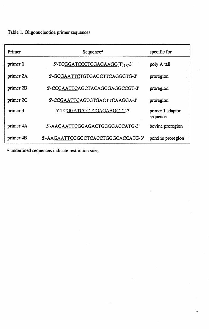
Table l. Oligonucleotide primer sequences
Primer Sequenc& specific for
primer l 5'-TCGGAICCCfCGAGAAGC(T)18-3' poly A tai1
primer2A 5'-GCGAATICTGTGAGCTTCAGGGTG-3' proregion
primer 28 5'-CCGAA TICAGCT ACAGGGAGGCCGT-3' proregion
primer 2C 5'-CCGAATICAGTGTGACITCAAGGA-3' proregion
primer 3 5'-ICQGAICCCfCGAGAAGCTI-3' primer l adaptor sequence
primer4A 5'-AAGAA TICGGAGACfGGGGACCATG-3' bovine proregion
primer4B 5'-AAGAAIICGGGCTCACCTGGGCACCATG-3' porcine proregion
a underlined sequences indicate restriction sites
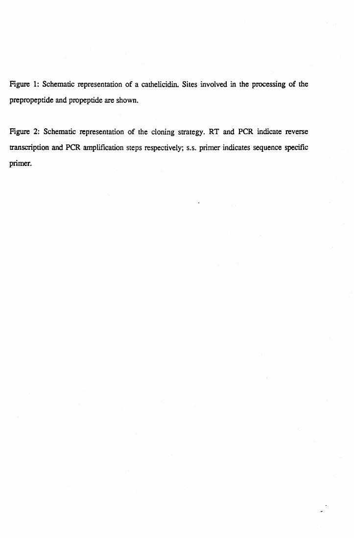
Figure l: Schematic representation of a cathelicidin. Sites involved in the processing of the
prepropeptide and propeptide are shown.
Figure 2: Schematic representation of the cloning strategy. RT and PCR indicate reverse
transcription and PCR amplification steps respectively; s.s. primer indicates sequence specific
primer.
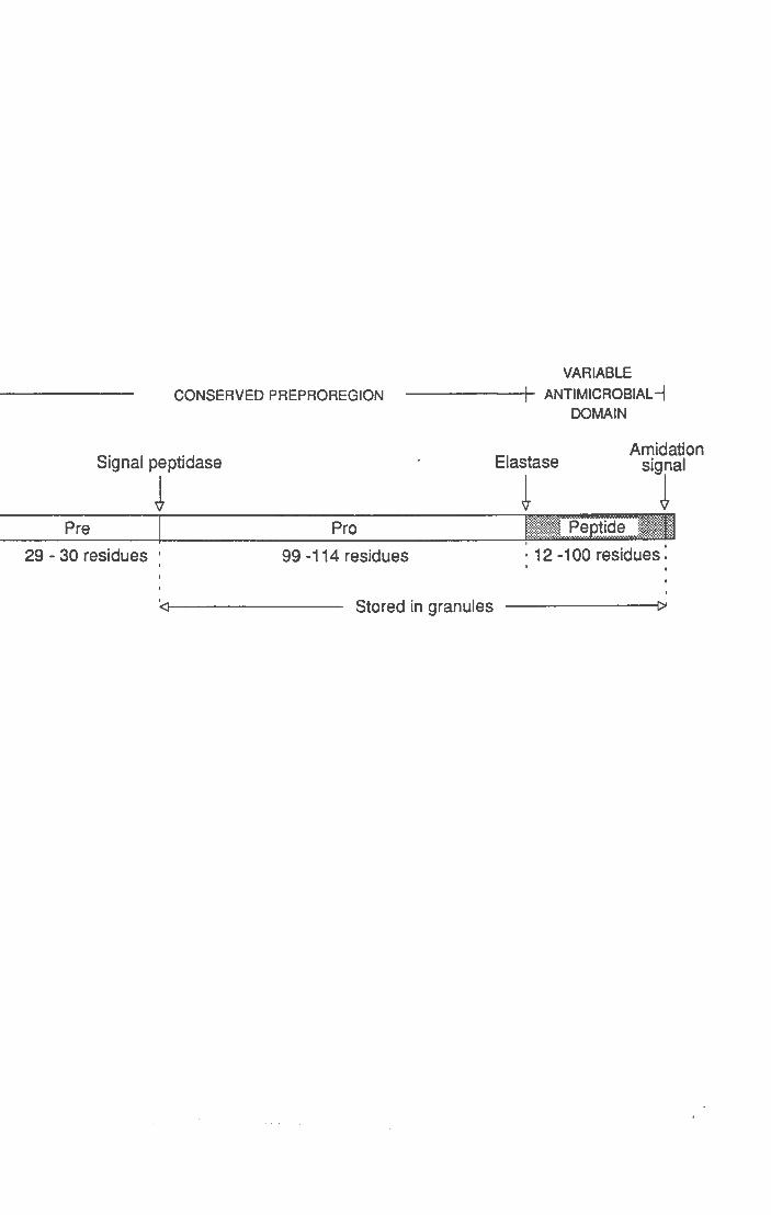
CONSERVED PREPROREGION VARIABLE
------+- ANTIMICROBIAL -j DOMA IN
Signal peptidase
J Amidation
Elastase signal
~ ~ Pre j Pro
' ' · 12-100 residues, '
29 - 30 residues : 99 -114 residues
Stored in granules ------f)ol
RT
PCR
RT
PCR
3'-end amplification
mRNA preproregion antimicrobial
domain 5' _ __. ________ ~~m~Zi!ZiZl~Zi!ZiZl:rtAL.-AAAAA 3'
- - - - - - - - - - - - - - - - - - - - - - - - - - - - -< Pril#~:L::::::l cONA(-)
! cONA(-)
5' - - - - - - - - - - - - - - - - - - - - - - - - - - - - - - - - - ~ : /)/ m:l 3'
lprum~2)~--------------------------------------------fi~~l
! 3'-end
5' --------------
5' -end amplification
mRNA preproregion antinùcrobial
domain 5' ------------E:Zl~!':Zmli~iZL--AAAAA 3'
_ - - - - - - - - - - - - - - - - - - - - - -(s.s.primer l cONA(-)
! cONA(-)
5' - - - - - - - - - - - - - - - - - - - - - - - - - - - 3'
l prum~ 4 >~-----------------------------------~.s.prumerl
3.
3'
Indice i
INDICE
Capitolol
Immunità innata e peptidi antimicrobici l
l. l Introduzione l 1.2 I distretti fisilogici che producono i peptidi antimicrobici negli animali 4 l. 3 I fagociti professionali 5 l. 4 I granulociti neutro fili 7 l. 5 I peptidi antimicrobici animali l O l. 6 Peptidi antimicrobici degli insetti 1.6.1 Cecropine l. 6.2 Defensine l. 7 Peptidi antimicrobici degli anfibi l. 7 .l Magainine l. 7.2 Bombinine e H-bombinine l. 7. 3 Dermaseptine l. 7.4 Brevinine e Esculentine l. 8 Peptidi antimicrobici dei mammiferi l. 8 .l Peptidi con ponti di solfuro
Defensine e Criptidinr fl-defensine Protegrine Dodecapeptide
l. 8. 2 Peptidi lineari con elevata percentuale di particolari aminoacidi
14 15 17 18 18 19 21 21 22 23 23 25 26 27 27
Peptidi ricchi in Prolina e Arginina: Bactenecine, PR-39 e Profenine 27 Un peptide ricco in Triptofano: I ndolicidina 30
l. 8. 3 Peptidi ad a-elica 30 CAP18 30
l. 8. 4 Proteine antibatteriche dei neutrofili Bactericidal!permeability-increasing protein ( BP I) Serprocidine
l. 9 Meccanismi d'azione dei peptidi antibiotici animali 1.1 O Biosintesi e maturazione di peptidi antibiotici animali
31 31 32 34 36
Indice ii
Capitolo2
Scopo e sviluppo della tesi 40
Capitolo3
Metodiche utilizzate 42
3. l Clonaggio e caratterizzazione delle sequenze dedotte 42 3 .1.1 Preparazione di cellule mieloidi immature da midollo osseo ed estrazione
di RNA totale 42 3 .1.2 Northem blot e ibridazione con sonde radioattive 43 3.1.3 Costruzione della genoteca di cD NA e vaglio con sonde oligonucleotidiche
degenerate 44 3.1.4 Amplificazione del cDNA di catelicidine 44
3.1.5 3.1.6 3.1.7 3.1.8 3.2 3.2.1
3.2.2 3.2.3 3.2.4 3.2.5 3.2.6 3.2.7 3.2.8
Amplificazione specifica dei terminali 3' di cDNA codificanti catelicidine: RACE 3' 45 Amplificazione specifica dei terminali 5' di cDNA codificanti catelicidine: RACE 5' 46 Sequenziamento del DNA e analisi delle sequenze Primer extension Trascrizione e traduzione in vitto Southem blot Purificazione e caratterizzazione strutturale Isolamento di neutrofili bovini, estrazione proteica e iniziale frazionamento mediante salting out. Purificazione di catelicidine bovine Saggi analitici per proteine Digestione con tripsina e modificazione chimica dei ponti disolfuro Dicroismo circolare Idrolisi acida limitata Ricerche in banche dati e predizioni di struttura Saggi di inibizione enzimatica
48 49 49 50 50
50 51 52 53 54 54 55 55
Indice iii
Capitolo 4
Risultati e Discussione 56
4.1 Clonaggio molecolare di cDNA e analisi della sequenza aminoacidica dedotta 56 4.1.1 Bac5 56 4.1.2 4.2 4.2.1 4.2.2 4.2.3 4.2.4
4.3
4.3.1 4.4 4.4.1
4.4.2 4.4.3 4.4.4 4.4.5
Indolicidina e dodecapeptide Clonaggio di cinque catelicidine di maiale PR-39 Protegrin PG-2 PMAP-23, PMAP-36, PMAP-37 Proprietà dei peptidi sintetici PMAP-23, PMAP-36(1-20) e PMAP-37
Clonaggio e caratterizzazione di due nuove catelicidine bovine: BMAP-27 e BMAP-28 Proprietà dei peptidi sintetici: BMAP-27 e BMAP-28 Studio a livello proteico di quattro catelicidine bovine Purificazione di proBac5, proBac7, prododecapeptide e proBMAP-28 dai neutrofili bovini Analisi mediante spettrometria di massa delle catelicidine purificate Disposizione dei ponti disolfuro Predizioni di struttura delle catelicidine Omologie di sequenza e di struttura delle catelicidine con i chininogeni
4.5 Conclusione
Bibliografia
Appendice
Lista delle pubblicazioni allegate
58 62 63 64
64 67
70 71 75
77 78 85 87 89
93
94
111
111


![3HU TXHVWH VXH SHFXOLDULWj WURYD LQGLFD]LRQH … · (Sistema Reticolo Endoteliale) ed il potere di proteolisi dei macrofagi. La cumarina è in grado di aumentare l'ossigenazione tissutale](https://img.pdfslide.tips/doc/110x75/5c66809009d3f20f218c59ce/3hu-txhvwh-vxh-shfxoldulwj-wuryd-lqglfdlrqh-sistema-reticolo-endoteliale.jpg)