Embed Size (px)
Citation preview

AVERTISSEMENT
Ce document est le fruit d'un long travail approuvé par le jury de soutenance et mis à disposition de l'ensemble de la communauté universitaire élargie. Il est soumis à la propriété intellectuelle de l'auteur. Ceci implique une obligation de citation et de référencement lors de l’utilisation de ce document. D'autre part, toute contrefaçon, plagiat, reproduction illicite encourt une poursuite pénale. Contact : [email protected]
LIENS Code de la Propriété Intellectuelle. articles L 122. 4 Code de la Propriété Intellectuelle. articles L 335.2- L 335.10 http://www.cfcopies.com/V2/leg/leg_droi.php http://www.culture.gouv.fr/culture/infos-pratiques/droits/protection.htm

FACULTE DES SCIENCES
V.F.R. Sciences et Techniques BiologiquesÉcole Doctorale Ressources Procédés Produits EnvironnementDépartement de Formation Doctorale Biologie - Agronomie - EnvironnementSecteur Biologie Forestière
Thèse
présentée pour l'obtention du titre de
Docteur de l'Vniversité Henri Poincaré, Nancy 1
en Biologie Végétale et Forestière
par Sébastien DUPLESSIS
Caractérisation par ingénierie génomique des profilsd'expression génique de Pisolithus tinctorius et
d'Eucalyptus globulus au cours du développement de lasymbiose ectomycorhizienne
Soutenue publiquement le 18 Avril 2001
Membres du jury :
Président: M. Jean-Pierre Jacquot
Rapporteurs : M. Christophe Plomi on
M. Patrick Saindrenan
Examinateurs: M. Jean-Claude Debaud
M. Francis Martin
Professeur, Université Henri Poincaré, Nancy l
Chargé de Recherche, LN.R.A.-Bordeaux
Chargé de Recherche, C.N.R.S.-Strasbourg
Professeur, Université Claude Bernard, Lyon l
Directeur de Recherche, LN.R.A.-Nancy
VMR 1136 INRA-VRP, « Interactions Arbres/Micro-Organismes »Centre INRA de Nancy, 54280 Champenoux
A Mesdames Roche, Truchot et Barrière
Respectivement enseignantes en biologie
en collège, lycée et université

Remerciements
Je voudrais remercier en premier lieu Francis Martin, mon directeur de recherche, pour
m'avoir accueilli au sein de son équipe, il y a quelques années de cela et aussi pour m'avoir soutenu et
toujours conseillé dans les moments où j'en avais le plus besoin. Je le remercie pour sa très grande
disponibilité, pour ses précieux conseils et pour la patience dont il a su faire preuve à mon égard dans
bien des occasions. Je te remercie surtout Francis pour la façon dont tu as conçu notre relation dans le
travail que nous avons réalisé ensemble ... Notamment, pour la très grande confiance que tu m'as
accordée et pour cette capacité que tu as à me motiver autant dans mes activités de recherche!
Je tiens à remercier Messieurs Patrick Saindrenan et Christophe Plomion pour avoir accepté d'être
rapporteurs de cette thèse de Doctorat. L'avènement de ce manuscrit aura été plein de péripéties pour
moi et agrémenté de "quelques" retards pour eux. Je tiens à les remercier pour leur indulgence et leur
compréhension. Je tiens à remercier l'ensemble des membres de mon jury de thèse pour avoir accepté
de juger ce travail et pour leurs encouragements.
Je voudrais aussi remercier les deux personnes qui m'ont formé aux techniques de biologie
moléculaire et de génomique: Denis Tagu et Catherine Voiblet (viviane de la BM !!) ... Je te remercie
pour ta sympathie, Denis, et surtout pour ta patience lors de la dernière ligne droite de la rédaction de
la thèse. Tu m'as été d'un précieux secours et d'une très grande disponibilité. Je tiens aussi à remercier
les stagiaires que j'ai eu plaisir à encadrer au cours de cette thèse, même si parfois, j'aurais voulu me
montrer plus présent. Plus particulièrement, Marine et Natacha. Les travaux que vous avez réalisés
étaient d'une grande qualité, et j'ai eu plaisir à partager ces moments avec vous!
Je tiens aussi à remercier Pascale Frey-Klett, Jean Garbaye, Frédéric Lapeyrie et François Le Tacon
pour l'amabilité et la disponibilité dont ils ont fait preuve au cours de ces quatre années de "microbes"
en commun. Je les remercie tous, ainsi que Francis et Denis pour la manière dont fonctionne leur
équipe ... Cette synergie se ressent fortement de la part des étudiants-chercheurs et elle est
extrêmement bénéfique à la réalisation de nos projets.
Je remercie aussi l'ensemble des Microbes et tout ce qu'ils ont pu m'apporter: JLJ (My Copilote
Enhancer !!), Sofia, Béou, Cricrou, Dominique, Daniel, Patrice, Jean-Louis, Hubert, Chantal, Franck,
Anne, tous les post-doctorants et DEA, et tous ceux qui passent et repartent (Tarikoto il piloto, Aneta
Maria Rincàn Heranz etc, etc ... , Mirco con questo caz*** di telefonino).
Enfin, je remercie toutes les personnes extérieures à l'UMR et qui ont contribué à la réussite de mon
travail. Je les remercie pour leur soutien au cours de ces derniers mois difficiles. Mes pensées vont à
mon italienne, à ma mère et à toute la familly connection de Montpellier. ET AUSSI à Nico et Laurent
(+ Zombie Eaters) ... Vivement des passages plus fréquents sur Bordeaux!! Mais aussi à tous ces amis
qui m'ont, sans le savoir, énormément soutenu par leur amitié dans cette ville qui était l'autre bout du
monde pour moi il y a encore quelques années (Stéphan et la famille Cornevaux, Yves, Cé, Mario,
Tonny, Fanfan, Big Jim, Frédo et tous les "branques" de Zajedni).

Sommaire
Abbréviations
INTRODUCTION
Introduction générale 1
Chapitre 1 : Living Together Underground: a Molecular Glimpse of the Ectomycorrhizal Symbiosis. DansMolecular Biology of Fungal Development (H Osiewacz, ed .), "Mycology Series", Dekker & Dekker, NewYork. 2001, sous presse 6
RESULTATS
Chapitre 2 : Identification des gènes régulés par la symbiose ectomycorhizienne Eucalyptus globulus-Pisolithus tinctorius par hybridation différentielle de filtres d'ADNe 23
Identification of symbiosis-regulated genes in Eucalyptus globulus - Pisolithus tinctorius ectomycorrhizaby differential hybridization of arrayed cDNAs.Plant Journal 25:181-192 25
cDNA array analysis of ectomycorrhiza development. Manuscrit en préparation pour la revue PlantPhysiology 36
Chapitre 3 : Clonage et analyse de l'expression de l'ADNc HydPt-3codant une nouvelle hydraphobine dePisolithus tinctorius '" 52
Cloning and expression analysis of a new hydrophobin cDNA from the ectomycorrhizalbasidiomycete Pisolithus. Current Genetics, soumis. Cloning and expression analysis of a newhydrophobin cDNA fram the ectomycorrhizal basidiomycete Pisolithus. Current Genetics(sous presse) 53
Chapitre 4 : Caractérisation et analyse de l'expression de gènes codant des protéines de voies detransduction chez Pisolithus tinctorius 69
DISCUSSION GENERALE 90
CONCLUSIONS ET PERSPECTIVES 105
REFERENCES BIBLIOGRAPHIQUES 109

Abbréviations
2D-PAGE
ADN
ADNc
AIA
ARN
ARNm
EST
Gène SR
ITS
kb
kDa
ORF
pb
PCR
Protéine SR
RT-PCR
SOM
SRAP
SSC
SSH
Electrophorèse bidimensionnelle
Acide désoxyribonucléique
Acide désoxyribonucléique complémentaire
Acide Indole Acétique
Acide ribonucléique
Acide ribonucléique messager
Expressed Sequence Tag (Etiquette de gène
exprimé)
Gène Régulé parla Symbiose
InternaI Transcribed Spacer
Kilo paire de bases
Kilo Dalton
Open Reading Frame
Paire de bases
Polymerase Chain Reaction (Réaction de
Polymérisation en chaine)
Protéine régulée par la Symbiose
Reverse tanscription-PCR
Self Organizing Maps
Symbiosis Regulated Acidic Polypeptide
Sodium Sodium Citrate
Suppressive Subtractive hybridization
IntEadactiDn géaéFale

A l'échelle de la planète, les forêts recouvrent environ 32% des terres émergées. Elles
sont principalement localisées dans la zone intertropicale (environ 40%) mais les niches
écologiques exploitées sont beaucoup plus vastes et les écosystèmes forestiers couvrent ainsi
une partie des zones tempérées, boréales et montagneuses. Leur présence dans des milieux
parfois défavorables s'explique par la capacité des arbres à établir des associations mutualistes
avec d'autres organismes. Notamment au sein de la rhizosphère, de nombreux champignons
(> 5000 espèces) sont capables d'établir des symbioses avec le système racinaire des arbres. Ces
associations symbiotiques impliquant un végétal et un champignon se caractérisent par la mise
en place d'organes mixtes différenciés: les ectomycorhizes (Smith & Read 1997).
Une telle association mutualiste permet aux deux partenaires d'en tirer des bénéfices
essentiellement trophiques. Ainsi, la croissance de la plante sur des sols aux teneurs
généralement limitées en éléments minéraux est favorisée. De même, le champignon assure sa
croissance végétative dans le sol sous la forme d'un réseau dense d'hyphes mycéliens et la
symbiose lui permet d'effectuer son cycle biologique (production de sporophores). Les
symbioses entre plantes et champignons sont apparues au cours des temps géologiques sous
différentes formes et impliquent différentes espèces. La symbiose ectomycorhizienne (LePage et
al., 1997) est rencontrée dans les écosystèmes forestiers des régions tempérées, boréales et
montagneuses et concerne la plupart des arbres de ces écosystèmes (Fagacées, Salicacées,
Pinacées,,,.) et certains champignons filamenteux du sol qui appartiennent aux ascomycètes et
basidiomycètes. Cette symbiose se caractérise par une morphologie particulière développée sur
le système racinaire des arbres lor~ de la mise en place d'un organe mixte et différencié:
l'ectomycorhize. Au sein de cet organe, sont généralement distingués i) les hyphes
extramatriciels qui s'étendent dans le sol environnant le système racinaire et ayant
essentiellement un rôle de prospection et d'absorption, ii) les hyphes différenciés formant un
manchon plus ou moins épais autour de l'organe racinaire et enfin iii) les hyphes ayant pénétré
le cortex racinaire et mis en place avec les cellules racinaires une interface symbiotique appellée
réseau de Hartig. C'est à ce niveau que sont généralement localisés les échanges les plus
importants entre les deux partenaires de la symbiose: squelettes carbonés, eau, acides aminés et
autres éléments minéraux. Les champignons améliorent ainsi la nutrition azotée et phosphatée
du symbiote végétal mais aussi son statut hydrique (Hampp & Schaeffer 1999 ; Botton & Chalot
1999). D'autre part, l'arbre est une source majeure en squelettes carbonés pour le champignon,
lui permettant ainsi d'assurer sa croissance végétative mais aussi d'assurer son cycle biologique
en réalisant sa fructification et la dissémination de spores assurant ainsi la continuité de
l'espèce. La maîtrise de la mycorhization en pépinière et en forêt permet d'améliorer la
1

croissance et la vigueur de plants forestiers (Grove & LeTacon, 1993), ainsi que la production de
certains champignons comestibles (Danel & Camacho 1997; Guérin-Laguette et al., 2000).
Les associations ectomycorhiziennes ont donc une importance autant écologique
qu'économique, de par leur statut majeur au sein des écosystèmes forestiers. Dans le cadre
d'une meilleure compréhension de leur écologie, de leur développement et de leur
fonctionnement, l'étude de ces systèmes symbiotiques a intéressé de nombreux laboratoires.
Ainsi, en quelques dizaines d'années, les connaissances concernant la physiologie et le
fonctionnement des ectomycorhizes se sont considérablement accrues (Smith & Read 1997). Les
mécanismes d'absorption, d'accumulation et de transports des différents métabolites échangés
au sein de ces symbioses sont abordés par des équipes de plus en plus nombreuses (Chalot &
Brun 1998 ; Nehls et al., 1999). Au cours de la dernière décennie, à travers les avancées
spectaculaires qu'a connu la biologie moléculaire, les efforts de recherche ont porté sur le
contrôle génétique du développement et du fonctionnement de cette symbiose (Martin & Tagu
1999). Ce contrôle se place probablement à différentes étapes du processus développemental,
dans les différents tissus, chez chacun des symbiotes et s'initie précocément lors des processus
de reconnaissance entre les deux partenaires (Martin et al., 1997; Kim et al., 1999). Il se poursuit
lors des premiers contacts, puis plus tardivement lorsque les structures différenciées se mettent
en place, alors qu'une réorganisation des schémas métaboliques chez la plante et chez le
champignon est observée (Botton & Chalot, 1999 ; Hampp & Schaeffer 1999).
L'objectif du projet de recherche, dans lequel cette thèse s'incrit, est la compréhension
des mécanismes moléculaires induisant, contrôlant et accompagnant l'établissement de la
symbiose ectomycorhizienne. Il s'agit notamment de caractériser les signaux, les protéines et
les gènes affectant et affectés par ces processus de différenciation. Au delà des informations
pouvant être acquises dans le cadre de l'étude des ectomycorhizes, ce sont les relations entre la
plante et son cortège de microorganismes commensaux, symbiotiques et pathogènes qui sont
ainsi appréhendées.
Depuis 1986, le laboratoire de Microbiologie Forestière du Centre INRA de Nancy
(Unité Mixte de Recherche INRA/UHP "Interactions Arbres/Micro-Organismes" depuis Janvier
2001) a dévéloppé différentes stratégies de recherche afin d'identifier et de caractériser les
signaux échangés entre les deux partenaires (Béguiristain et al., 1995 ; Béguiristain & Lapeyrie
1997 ; Ditengou & Lapeyrie 2000 ; Ditengou et al., 2000 ; Lagrange et al., 2001) et de caractériser
les fonctions cellulaires (au travers de leurs gènes et protéines) activées ou réprimées lors de la
symbiose ectomycorhizienne (Hilbert et al., 1991 ; Burgess et al., 1995 ; Tagu et al., 1996 ; Martin
et al., 1997). Le modèle expérimental étudié - l'ectomycorhize Eucalyptus spp. - Pisolithus
tinctorius - a été choisi car il est facilement manipulable in vitro, permet l'obtention de
mycorhizes en quelques jours et en quantité suffisante pour des analyses biochimiques et
moléculaires. Le genre Eucalyptus est très vaste (recouvre plus de 600 espèces) et est l'essence la
plus plantée dans le monde. Cependant, son intérêt dans nos pays tempérés est limité au sud de
2

l'Europe. Le basidiomycète ectomycorhizien Pisolithus recouvre un complexe d'espèces
associées à l'eucalyptus, aux pins et à d'autres essences forestières (Chambers & Caimey 1999).
Sa distribution est mondiale et il est très utilisé dans l'hémisphère sud pour le reboisement.
Sur ce modèle expérimental, l'équipe a acquis un grand nombre de données notamment
sur les signaux moléculaires échangées et sur les protéines et les gènes régulés par le processus
symbiotique (Martin et al., 1997 ; Martin & Tagu 1999). La comparaison de cartes
polypeptidiques des partenaires libres et associés a permis notamment d'identifier des protéines
réprimées (Hilbert & Martin 1988 ; Hilbert et al. 1991) ou stimulées (Hilbert et al., 1991 ; Burgess
et al., 1995) lors de l'interaction. Parmi ces protéines se trouvent des polypeptides fongiques
acides de 30-32 kDa régulés par la symbiose (SRAP32, pour Symbiosis Regulated Acidic
Polypeptides), de fonction inconnue et très abondants dans les parois de Pisolithus (Laurent et
al., 1999). Des criblages différentiels de banques d'ADNe d'ectomycorhizes (Tagu et al., 1993) ou
des clonages d'ADNe en utilisant des sondes hétérologues ont permis d'identifier chez la racine
d'eucalyptus des transcrits régulés par la symbiose : il s'agit de ceux codant l'a-tubuline
(EgTubAl, protéine du cytosquelette) (Camero Diaz et al., 1996), une glutathion-S-transférase
(EgHypar, régulée par l'hypaphorine) (Nehls et al., 1998) et de l'isocitrate déshydrogénase
(Eglcdh) (Boiffin et al., 1998). De même, chez le partenaire fongique, plusieurs gènes stimulés par
la symbiose et codant des protéines pariétales (les hydrophobines) (Tagu et al., 1996), la
glutamate déshydrogénase à NADP (Lorillou 1995) et des protéines du protéasome (Mourer
1998) ont été décrits.
Cette dizaine de gènes et de protéines sont autant de marqueurs du développement de
l'ectomycorhize entre Eucalyptus globulus et Pisolithus tinctorius. Ils ne représentent cependant
que quelques éléments dans le réseau très complexe d'interactions génétiques et épigénétiques
accompagnant la mise en place des structures différenciées de l'ectomycorhize. Afin d'obtenir
une vision plus étendue des fonctions cellulaires en activité dans la symbiose et de proposer
une image générale et intégrée de son fonctionnement, la mise en place d'une approche globale
d'identification des gènes exprimés au sein de l'ectomycorhize E. globulus-P. tinctorius a été
envisagée (Tagu et al., 1993; Tagu & Martin 1995 ; Martin & Voiblet 1998). L'initiation d'un tel
projet a pu voir le jour grâce au développement conséquent des techniques utilisées en
génomique. Les différentes approches pouvant être retenues sont l'analyse complète des
composantes protéiques des deux organismes dans la symbiose (le protéome), l'analyse de la
partie transcrite du génome (le transcriptome) ou encore l'obtention de banques de mutants des
deux partenaires altérés dans différentes fonctions cellulaires. L'utilisation de la mutagénèse
reste encore à ce jour difficile sur ce système. Le Pisolithe est réfractaire jusqu'à présent aux
différentes techniques de transformation génétique testées (électroporation, biolistique,
transformation par Agrobacterium tumefasciens actuellement en cours d'étude ... ). Quant à
l'eucalyptus, aucun protocole d'obtention en routine de plantes transgéniques n'est disponible.
La stratégie la plus appropriée en 1995, au début de ce projet de génomique fonctionnelle, était
3
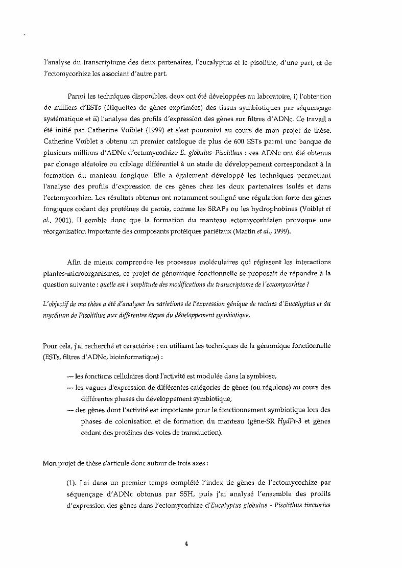
l'analyse du transcriptome des deux partenaires, l'eucalyptus et le pisolithe, d'une part, et de
l'ectomycorhize les associant d'autre part.
Parmi les techniques disponibles, deux ont été développées au laboratoire, i) l'obtention
de milliers d'ESTs (étiquettes de gènes exprimées) des tissus symbiotiques par séquençage
systématique et ii) l'analyse des profils d'expression des gènes sur filtres d'ADNe. Ce travail a
été initié par Catherine Voiblet (1999) et s'est poursuivi au cours de mon projet de thèse.
Catherine Voiblet a obtenu un premier catalogue de plus de 600 ESTs parmi une banque de
plusieurs millions d'ADNe d'ectomycorhize E. globulus-Pisolithus : ces ADNe ont été obtenus
par clonage aléatoire ou criblage différentiel à un stade de développement correspondant à la
formation du manteau fongique. Elle a également développé les techniques permettant
l'analyse des profils d'expression de ces gènes chez les deux partenaires isolés et dans
l'ectomycorhize. Les résultats obtenus ont notamment souligné une régulation forte des gènes
fongiques codant des protéines de parois, comme les SRAPs ou les hydrophobines (Voiblet et
al., 2001). Il semble donc que la formation du manteau ectomycorhizien provoque une
réorganisation importante des composants protéiques pariétaux (Martin et al., 1999).
Afin de mieux comprendre les processus moléculaires qui régissent les interactions
plantes-microorganismes, ce projet de génomique fonctionnelle se proposait de répondre à la
question suivante: quelle est l'amplitude des modifications du transcriptome de ['ectomycorhize ?
L'objectif de ma thèse a été d'analyser les variations de l'expression génique de racines d'Eucalyptus et du
mycélium de Pisolithus aux différentes étapes du développement symbiotique.
Pour cela, j'ai recherché et caractérisé; en utilisant les techniques de la génomique fonctionnelle
(ESTs, filtres d'ADNc, bioinformatique) :
-les fonctions cellulaires dont l'activité est modulée dans la symbiose,
-les vagues d'expression de différentes catégories de gènes (ou régulons) au cours des
différentes phases du développement symbiotique,
- des gènes dont l'activité est importante pour le fonctionnement symbiotique lors des
phases de colonisation et de formation du manteau (gène-SR HydPt-3 et gènes
codant des protéines des voies de transduction).
Mon projet de thèse s'articule donc autour de trois axes:
(1). J'ai dans un premier temps complété l'index de gènes de l'ectomycorhize par
séquençage d'ADNe obtenus par SSH, puis j'ai analysé l'ensemble des profils
d'expression des gènes dans l'ectomycorhize d'Eucalyptus globulus - Pisolithus tinctorius
4

au stade de formation du manteau fongique. Cette étude a été effectuée en collaboration
avec C. Voiblet et N. Encelot; elle a fait l'objet de la publication n° 2 (Voiblet et al., 2001).
(2). Afin de comprendre la régulation de l'expression des gènes au cours de la formation
de la mycorhize d'Eucalyptus - Pisolithus, j'ai analysé à l'aide des filtres à haute densité
en ADNc, la régulation temporelle de 715 gènes de l'ectomycorhize, en comparant les
niveaux des transcrits aux différents stades de différenciation de l'ectomycorhize
(colonisation, formation du manteau, formation du réseau de Hartig, mise en place du
métabolisme symbiotique). Afin d'affiner cette analyse, j'ai introduit au laboratoire les
outils de statistiques bioinformatiques nécessaires à la mise en évidence des régulons
(manuscrit en préparation, Chapitre 2).
(3). Au delà de l'analyse globale du transcriptome symbiotique, j'ai effectué une série
d'études détaillées sur quelques gènes dont l'expression est cruciale pour la
morphogenèse de l'ectomycorhize. Il s'agit d'un membre de la famille multigénique
codant une hydrophobine (publication nO 3, Duplessis et al., 2001b), et de plusieurs
gènes impliqués dans les voies de transduction de signaux.
Ce manuscrit comporte:
- Une analyse bibliographique (Chapitre 1 ; Duplessis et al., 2001a), qui se propose de
faire le point sur nos connaissances actuelles dans le domaine du développement de
l'ectomycorhize,
- La présentation des résultats (Chapitres 2, 3 et 4),
- Une discussion générale qui s'efforcera de replacer dans le contexte plus général des
interactions plantes/micro-organismes nos résultats et enfin les conclusions et perspectives
ouvertes par ce travail.
5
ehapitlie ·1
Synthèse bibliographique
Living Together Underground: a Molecular Glimpse ofthe Ectomycorrhizal Symbiosis
Public.ation n° 1
Mo/ecu/ar Bi%gy ofFUlIga/ Deve/opmellt (H Osiewacz, ed .) In Mycology Series, Dekker & Dekker, New York
Living Together Underground: a Molecular Glimpse of
the Ectomycorrhizal Symbiosis
Sébastien Duplessis, Denis Tagu and Francis Martin"
Unité Mixte de Recherche INRA-UHP "Interactions Arbres/Micro-organismes", Centre deRecherches de Nancy, 54280 Champenoux, France
SUMMARY
Ectomycorrhiza is the result of a series of complex interactions leading to a finely tuned mutualisticsymbiosis between a plant and a compatible soil fungus. Ultrastructural observations combined withcytochemical and biochemical studies revealed that morphological and metabolic changes in thesymbiont cells lead to the final phenotype of the active ectomycorrhizal roots. What could be themolecular basis of such a progressive, highly organized ontogenic process? What is the role ofrhizospheric chemicals in symbiosis development? How many genes control ectomycorrhizadevelopment-as distinct from providing the housekeeping functions of the fungal and plant cells? After abrief overview of the evolution, biology and anatomy of ectomycorrhiza, these are sorne of the mostimportant questions that will be discussed in the present chapter. Proteomics and genomics havedemonstrated that symbiosis formation induces major changes in gene expression. The expression ofabout 17% of the genes analyzed arrays in the EucalyptuslPisolithus ectomycorrhiza by cDNA areregulated upon symbiosis development. Those symbiosis-regulated genes include several fungalmultigene families encoding for cell wall and membrane proteins. In colonised roots, expression of genescoding for metabolic, stress and defence functions is enhanced. As a result, alterations in gene expressionis associated with extensive changes in protein biosynthesis. These initial results presage a wealth ofinformation that will be obtained from the application of genomics to various ectomycorrhizalassociations.
Key words: cel! wall proteins; ectomycorrhiza development; genomics; protein patterns; rhizosphericsignaIs; symbiosis; symbiosis-regulated gene.
• For correspondence: Fax +33383394069; e-mail: [email protected]
6

1. INTRODUCTION
Within the rhizosphere which hosts a large anddiverse community of prokaryotic andeukaryotic microbes that compete and interactwith each other and with plant roots,mycorrhizal fUllgi are almost ubiquitous. Theectomycorrhizal hyphae and the root tips form anovel composite organ, so-called mycorrhiza,which is the site of nutrient and carbon transferbetween the two symbionts. This associationallows terrestrial plants to grow efficiently insuboptimal environments (1). Among thevarious types of mycorrhizal symbioses, theendomycorrhizal, ectomycorrhizal and ericoidassociations are found on most annual andperennial plants (probably > 90%). About twothirds of these plants are symbiotic witharbuscu1ar mycorrhizal glomalean fungi (2).Ericoid mycorrhizas are ecologically important,but mainly restricted to heath1ands (3). While arelatively small number of plants, ca SOOO,form ectomycorrhiza, their global importance isamplified by their wide occupancy of terrestrialecosystems. Trees of Betulaceae, Cistaceae,Dipterocarpaceae, Fagaceae, Pinaceae,Myrtaceae, Salicaceae and several tribes inFabaceae are ectomycorrhizal plants (Fig. lA),dominating boreal, temperate, mediterraneanand sorne subtropical forest ecosystems (1).Within days after their emergence in the upper10 cm of the soil profiles (e.g. organic humusand mor layer), most of the short roots oftheseectomycorrhizal shrubs and trees are colonisedby ectomycorrhizal fungi, and in most casessymbiotic colonization is close to 100% (4).The fun gal mycelium and the root tips form anovel composite organ, so-calledectomycorrhiza, which is the site of nutrient andcarbon transfer between the two symbionts (5,6).
Ectomycorrhiza is structurally characterized by(i) the presence of an extensive extramatrica1mycelia1 web prospecting the soil and gatheringnutrients, (H) a mantle of funga1 hypha:ensheating the root and mainly acting as astorage compartment, and (iii) a network ofhypha: growing in the apop1astic space of therhizodermis (in angiosperms) and cortex (inconifers) root cells (Fig. lB). The fungus gainsaccess to sugars from the plant while plantnutrient and water uptake is mediated via thefungus (Fig. 2). In addition, the establishmentof the symbiosis is requested for the completionof the fungal life cycle (Le., formation offruiting bodies) (Fig. lA). The formation of thesymbiosis requires several days and inducesmajor morphological changes including a novelspatial tissue organization, changes in cell
7
shape, and the generation of different cell types(7, S). Ectomycorrhiza formation thereforeinvolves a series of complex and overlappingontogenic processes in the mycobiont and thehost plant: increased rhizogenesis, enhancedhyphae branching, aggregation of theproliferating hyphae, arrest of meristematicactivity in roots surrounded by the fungalmantle, radial elongation of epidermal cells.These morphological changes are accompaniedby the onset of novel protein patterns (9) andmetabolic organizations (10, Il) in fungal andplant cells leading to the functioningsymbiosis. What could be the molecu1ar basisof such a progressive, high1y organizedontogenic process? What is the role of cell-tocell signaling in symbiosis development? Howmany genes control ectomycorrhizadevelopment-as distinct from providing thehousekeeping functions of the fungal and plantcells? After a brief overview of the evolution,bio10gy and anatomy of ectomycorrhiza, theseare sorne of the most important questions thatwill be tackled in the present chapter.
Il. MYCORRHIZAS AREANCESTRAL SYMBIOTICINTERACTIONS
The first mycorrhiza1 associations must havebeen derived from earlier types ofplant-fungusinteractions, such as the fungus Geosiphonpyriforme forming endocytobiosis with Nostoc(Cyanobacteria) (12) and endophytic fungifound in the bryophite-like precursors ofvascu1ar plants (13). Structures similar toarbuscular mycorrhiza have been observed inplant fossils from the Early Devonian (14),whereas fossil ectomycorrhiza have been foundin the middle Eocene (15). Based onphylogenetic analysis of the rRNA gene, it hasbeen suggested that ectomycorrhizalbasidiomycetes evolved convergently fromsaprophytic ancestors (16). The switch betweensaprophytic and mycorrhiza1 1ifestyles like1yhappened many time during evolution of fungallineages as revealed by recent molecularphylogenetic analyses (17). This may havefacilitated evolution of ectomycorrhiza11ineageswith a broad range of physiologica1 andecologica1 functions reflecting partly theactivities of their disparate saprotrophicancestors. These symbioses have had majorconsequences for the diversification ofboth themycobiont and their hosts (1S). It remains to bedetermined whether the development of differentlatera1 root structures (actinorhiza, mycorrhiza,mycorrhizal nodules) are govemed by the sameset of genes (19). Current ectomycorrhizalfungal species (ca 6000) inain1y belong to


homobasidiomycetes (agarics, bolets), althoughmany species are found within the ascomycetes(truffles, terfez) and zygomycetes.
Fig. 1. The ectomycorrhizal symbiosis. A. A seedling ofDouglas tir (Pselldolsllga mentiesii) colonised by theectomycorrhizal basidiomycete Laccaria bic%r. Thefungal mycelium has developed ectomycorrhizas on theroot system and has produced a fruting body above ground(Photograph courtesy of P. Frey-Klell). B. Transversesection of a Elica/ypills/Pisolilhus ectomycorrhiza showingthe extramatrical hyphae (EM), the mande (M); the fungalhyphae have begun to penetrate between the epidermalcells (E) of the root cortex (C) to form the Hartig net(HN). Ep~dermal cells (E) are radially enlarged. CC,central cyltnder (Photograph courtesy of B. Dell).
111. A BENEFICIAL SYMBIOSIS
Ectomycorrhizal communities are taxonomicallydiverse (20, 21) and are likely able to maintaina large degree of functional diversity (22).Although a few tree/fungus combinations areunique, a great many different fungi cancombine with a great many different trees. Asingle host tree could simultaneously interactwith dozens offungal species (4) and this highsymbiont diversity likely allowsectomycorrhizal associations to use most N andP forms present in forest soils (23). Thesymbiosis between trees and soilbomeectomycorrhizal fungi results in an intimaterelationship between the plant and its symbioticpartner (Fig. lB). It provides several benefits toboth the host plant and its fungal associate(s).The prospecting and absorbing extraradicalhyphal web (1000 m of hyphae/m of root)captures soil minerais (phosphate, nitrogen,water, micronutrients) (1) and organic nitrogen(24, 25), assimilates and translocates a largeproportion ofthem to the growing plant (1,24)(Fig. 2A). Ectomycorrhizal fungi affect not onlyminerai and water uptake, but also adaptation toadverse soil chemical conditions (5) andsusceptibility to diseases (l) and contributesubstantially to plant productivity (6). On the
8
other hand, the fungus within the root isprotected from competition with other soilmicrobes and, therefore, is a preferential user ofthe plant carbon (ca 20% of the hostphotoassimilates) (Fig. 2B). Mycorrhizal fungirepresent an interface in the soil-plant systemand have the ability to regulate plantmetabolism. In addition, they constitute linksin the chain of transfers by which carbon andnitrogen move between plant and soilcompartments (26, 27) and can thus influencecarbon and nitrogen cycling rates in host plantsand forest ecosystems (23, 28, 29).
IV. ECTOMYCORRHIZAONTOGENESIS: THE DANCE IS THESAME, THE COUPLES ARE DIFFERENT
Morphological and anatomical changes thataccompany ectomycorrhiza development havebeen studied and described in great detail invarious associations [e.g. Picea abieslAmanitamuscaria (7, 30); P. abieslHebelomacrustuliniforme (31); EucalyptuslPisolithus (8,32- 34); Alnus rubralAlpova diplophloeus (35);and Betula pendulalPaxillus involutus (36)].The mature organization of ectomycorrhizavaries with the host and fungal species (37). Inaddition, a survey of almost any natural fungalpopulation will reveal a considerable range inphenotypes (38). However, although sorne ofthe details vary, early stages of ectomycorrhizadevelopment have well-characterized similarmorphological transitions. In an effort toprovide a useful framework in which tocategorise existing data on gene expression andaccomodate future efforts to categorize existingnatural fungal variants and future experimentalmutants, we have subdivided ectomycorrhizaformation in discrete stages from thepreinfection (rhizospheric) phase to themorphogenesis per se (Fig. 3) (39). Sporegermination and saprophytic growth of thehyphae is initiated in the rhizosphere (Fig. 4A).In most natural situations, hyphae are howeverinvading a newly emerging root frompreviously established ectomycorrhiza.Extensive preinfection branching of hyphaerequires the presence of host plant roots (Fig.4C). After contact, hyphal growth on the rootsurface initiates swelling of the hyphal tips(pads) and formation of dense finger-likestructures (Fig. 4D). Hyphae aggregate initiallyto form wefts and then ensheath the lateral roots(Fig. 4E). After root penetration, intraradicalhyphae proliferate and form a coenocyticstructure in the root apoplastic space (i.e., theHartig net) (Fig. 4F). This intraradical fungalweb is active in nutrient transfer and an activetraffic of carbohydrates promotes extensive
3


Fig. 2. Ectomycorrhiza: a mutualistic symbiosis. (A)Extramatrical hyphae prospect the soil, gather nutrients (N,P, H20) and translocate them to the mantle. Mineral andorganic N are assimilated and the synthesized amino acids(Gin, Ala, and Arg), together with inorganic P polymers(PolyP, polyphosphates) are accumulated. Amino acids andPi are then transferred through the intraradicular hyphalnet (the Hartig net) to the root ceUs and the other hosttissues. (B),On the other hand, sucrose (suc) downloadedto the root ceUs is degraded by apoplastic invertases inglucose (gluc) and fructose (fruc). Glucose is thentranslocated to the fungal compartmentthrough the Hartignet hyphae. The carbohydrate is then stored in the mantleas glycogen and lipids, or transported to the extramatricalhyphae.
growth of extemal hyphal web that gathersnutrient in soil. These morphological changesare concomitant with accelerated nucleardivision, cytoskeletal rearrangements andsynthesis of differentiation-related genes andproteins (see below). Growth and differentiationof the plant root and fungal hyphae must betightly coordinated. This multistepdevelopment therefore implies the existence ofadevelopmental strategy for building up anectomycorrhiza that early on imposes a basicscheme, on top of which subsequent speciesspecifie customizations occur. Behind everyaspect of ectomycorrhiza development theremust likewise be genetic control from theearliest proliferation ofhyphae to the builded upof the complicated symbiotic structure.
Since fungal mutants affected in theirability to form ectomycorrhiza are not yetavailable, one approach to identify the geneticprocesses that trigger and regulateectomycorrhiza development is to look fornatural variation in symbiosis structure. It hasbeen shown that natural populations of sibmonokaryotic and dikaryotic strains of Laccariabicolor (40), H. cylindrosporum (41) andPisolithus (42) vary greatly in their ability toform mycorrhizas. Sorne L. bicolor variantsundergo morphological changes that signal theonset of mycorrhiza formation but fail tocomplete the development process and do notmove on to the next stage (43). They have beenclassified into different basic categories:intraradical hyphal network not formed, hyphalnetwork formed but do not develop further,Hartig net development normal but failure ofmantle to form. This suggests that themorphogenetic programmes for thedifferentiation of the mantle and the Hartig netare partly independent and they likely involvedifferent set of genes. This has been recentlyconfirmed by the differential effect of auxintransport inhibitors on the formation of themantle and the Hartig net (44). Variation inmycorrhizal structures appears to be geneticallydetermined, which should make it possible toidentify the loci that contribute to thisvariation.
Ar.
9
4

Phenotypic stages
~IMer Il Elon 1.---1Come! II N-me! Il P·me!
1 Germ Il Pif Il Bran Il Adh 1 1Pen liMan IlHat 1
~ • • •Preinleelion C%nizalion Differenlialion Funclioning
1 ! 1 1 1 ...
dey 0 dey 2 dey 4 dey 6 dey 8
lime course of development of Euca/yplus-Piso/ilhus ectomycorrhlza
Fig. 3. Main phenotypic stages of ectomycorrhiza development. Abbreviations: Germ, Spore germination; Pif, Preinfectiongrowth ofhyphae; Bran, Hyphae branching on to root surface; Adh, Attachment ofhyphae on root surface and formation of theadhe.sion pads; Pen, Penetration ?etween epidermal cells; Man, Hyphae aggregation to form mantle; Hal', Differentiation of theHarllg net; LaI, Increased formatIOn of la~eral roots; Aler, Changes in meristematic activity; Elon, Radial elongation of epidermalcells; C-mel, N-mel, and P-mel, Changes 10 carbon, mtrogen, and phosphate metabolisms, including transfer between symbionts.
V. SIGNALLING CHEMICALS INTHE RHIZOSPHERE AND IN SYMBIOTICTISSUES
In almost ail plant-microbe interactionsexchange of signaIs between the partners is theearliest step in a series of interaction events,leading to contact at the host surface andsubsequent development of the microbialstructures in the host-plant tissues (45) (Fig. 3).Signalling processes must exist to bring themycobiont into the vicinity of susceptible hostroots. This mechanism has been poorlyinvestigated in ectomycorrhizal symbioses and alimited set of chemical signaIs produced byeither the host or symbiont have been identifiedso far (46-49). Gnly the broad outlines of thesignalling processes have been defined, but thelittle that is known suggests that someintriguing similarities exist betweenectomycorrhizal associations and other plantmicrobe symbioses (45, 50, 51).
No specifie chemicals able to attractectomycorrhizal fungi toward the root surfacehave been identified yet, although theiroccurrence has been suggested (52). However,there is evidence that host root exudates containmore than one kind of metabolites that canstimulate hyphal growth and/or morphologicalfeatures of the colonising ectomycorrhizalhyphae and several factors likely help thepartners match each other. Host plants secretecontinuously a spectrum of chemicals able toattract rhizospheric microbes. Within these
10
compounds, C20 diterpene abietic acid is ableto stimulate spore germination ofectomycorrhizal bolets, such as Suillus species(46). Among the secreted compounds arephenolic substances, especially flavonoids. Theectomycorrhizal Pisolithus spp. respond totraces of eucalypt flavonoids [e.g. the flavonolrotin (quercetin-3-rotinoside) (49)] byenhancedgrowth (Fig. 4A). Conjugates of flavonoids,such as rutin, are more soluble in water thantheir aglycones; thus, they diffuse readily andcan be hydrolyzed to more-active metabolites(53). The presence of such compounds probablyincreases the possibility of the interaction.Interestingly, increased branching of theendomycorrhizal Gigaspora rosea in thepresence of root exudates is enhanced by therutin aglycone, quercetin (54). Cytokinin, suchas zeatin, presents in the rhizosphere can alterhyphal branching of Pisolithus and mimickssome of the earliest step of the ectomycorrhizalinteraction (Fig. 4A) (49). The branching ismore numerous and compact in the presence ofthe phytohormone and this bushy type ofhyphal branching pattern likely increases thechance for the hyphae to enter in contact withthe root surface. In addition to altering fungalmorphology, zeatin interacts with themetabolism of alkaloid in the hyphae. Thepresence of zeatin results in the increasedaccumulation and secretion of hypaphorine, atryptophan betaine (55) able to triggermorphological changes in eucalyptus roots(e.g., arrest ofroot hair growth) (47, 48).

Although the colonization of emergingroot tips by ectomycorrhizal hyphae is ofteninitiated from older mycorrhizal parts of theroot system, ectomycorrhizal fungi may bewidely dispersed in the different soil horizons.It is tempting to speculate that extensivegradients of mixed chemicals in the soil providethe mechanisms by which the mycobionts areinitially recruited to the root from the generalrhizosphere populations of fungi. Then, after thehyphae accumulate in the mucigel that isadjacent to the root surface, the symbiosis canfurther proceed through the hyphae attachmentand colonization (Fig. 4B). It is not knownhow these rhizospheric chemicals may play arole in setting up the ectomycorrhizalassociations. In addition to attracting andstimulating ectomycorrhizal mycelium (andother symbiotic microbes) these plantmetabolites, such as fIavonoids, have numerousother activities (e.g. antimicrobial activities,modification of plant growth) (56) confirming alack of specificity. The presence of multiplenon-specifie signais is ecologically consistentwith the lack of specificity of theectomycorrhizal symbiosis. It is highlyimprobable that the wide range ofectomycorrhizal trees species secrete a singleuniversal signalling chemical to which ailectomycorrhizal fungi respond. Individualfl!ngal species may sense one or a set of specifiesignales) within a complex cocktail of plantchemicals and would respond according to whatis secreted by any given host plant. Thecomposition and concentration of the signallingcompounds mixture that is secreted in the hosttree rhizosphere is probably crucial. Furtherinvestigation is currently underway to identifyadditional root chemicals involved in thealteration of hyphal morphology to fullyunderstand signalling and recognition processesin ectomycorrhizas.
On the other hand, ectomycorrhizalfungi have the potential to morphologically alterthe host root through refined intervention in thedevelopmental programme of the host plant.Hyphae enter the root preferentially at theelongation or differentiation zone and thenmigrate intercellularly toward the exodermis (inmost Angiosperms) (Fig. lB) and to theendodermis in conifers. Intraradicularproliferation of the Hartig net hyphae implieshost cell wall openings away from the hyphaltips (57). Cell wailloosening and breakdownare likely involved in this apoplasticprogression of the syncitial mycobiont.Multiple evidences have been provided thatauxins, such as indole-3-acetic acid (IAA), playa raie in this process and in additional earlystage processes of ectomycorrhiza development(50, 58, 59). The hypothesis that
11
ectomycorrhizal fungi disturb root tissues bysecreting IAA (and, in its wake, ethylene) isrelativey old (60, 61). IAA-released byectomycorrhizal fungi elicits similar rootresponses as those induced by ectomycorrhizaformation including an enhanced rhizogenesisand dichotomous branching ofpine raots (62,63). It has been suggested that the intraspecificvariations in symbiotic structures of 1.bie%r-Pinus banksiana mycorrhizas are relatedto the differences in IAA-synthesizing activityamong the various fungal isolates (43).Tryptophan released in root exudates could besufficient to trigger the increased biosynthesis ofIAA or a homologue ofthis phytohormone inectomycorrhizal fungi (64). Pine inoculated withmutant of H. eylindrosporum strainsoverproducing lAA produced an increasednumber of ectomycorrhizal roots (65) whichpresented a strikingly altered morphology (Le.hyphae proliferation leading to a multiseriateHartig net) (57). It has been suggested that thisHartig net hypertrophy was the result of anincreased cell wall loosening resulting fromlocal increase in lAA concentration (57). Thepresence of increased concentration of IAA as aresult of fungal colonization of root tissues hasnot yet been experimentally demonstrated.
The local increase in the auxinconcentration could also be realized bystimulating the influx or inhibiting the efflux ofauxins in the colonised root zone (66). Toinvestigate the raie of polar auxin transport inectomycorrhiza development, Douglas fir(Pseudotsuga mensiesii) seedlings were exposedto the phytotropin triiodobenzoic acid (TIBA)(44, Rincon & Le Tacon, unpublished results).Subsequently roots were inoculated with theectomycorrhizal basidiomycete L. bie%r. Inlateral roots treated with TIBA, cross-sectionsrevealed that TIBA inhibited the formation ofthe fungal mantle (44). Alternatively, the auxintransport inhibitor N-( l-naphthyl)phtalamic acic(NPA) induced similar alteration of themycorrhiza development (67). The failure of 1.bie%r to aggregate to form the mantle in thephytotropin-treated seedlings points at aprominent role ofpolar auxin transport in earlystages of mycorrhiza development. TIBA andNPA are known to block the basipetal transportof IAA in the outer cortex and epidermisthrough the inactivation of the auxin effluxcarrier PIN (pin-formed) complexes (66). Thepleiotropic effects of auxin-secreted byectomycorrhizal fungi, the negative impact ofphytotropins on the formation of symbiosistissues and the data obtained with Hebelomamutants overproducing IAA are strongindications for a crucial role of auxin (andethylene) in ectomycorrhiza morphogenesis. Themost parsimonious explanation of this set of
6

downstream genes including components of thesignalling patways, such as the ras GTPase andserine/threonine kinases (Duplessis & Martin,unpublished results). Once the fungal hyphae arewithin the root, other trophic and developmentalinputs, from both symbionts, are likelynecessary for successful symbiosis. In enteringits novel niche, the colonizing hyphae need toadjust to its new environment. One essentialmodification is the alteration of the cell surfaceleading to the insulation of the mycobiontand/or changes in the permeability of the cellsurface allowing the symbiotic trafflc.
Fig. 4. The different interactions between the host root andthe ectomycorrhizal fungus and the main morphogeneticstages observed during ectomycorrhiza development. A.The preinfection stage: Al. Host root releases chemicals(e.g.• flavonoids, cytokinines) in the rhizosphere able toalter the morphology of the compatible ectomycorrhizalfungus (e.g., enhanced hyphal branching). A2. Conversely,the hyphae releases various compounds (auxins, alkaloids)eliciting changes in the root morphology (e.g., increasedrhizogenesis, decay of root hairs). B. The colonizationstage: running hyphae allach to the root surface and then,experienced drastic morphological changes, such as tipswelling, leading to a finger·lïke structure on the rootepidermal cclI. Hyphae initiate their aggregation betweenhost cells to forrn hyphal webs. C. Morphogenesis per se :Massive and rapid aggregation of hyphae around the rootlead to the formation of a pseudoparenchyma, the mantle;penetration between epidermal cells and cortical cells;andfonnation of the Hartig net with concomitantcoordinated alteration in the root structure.
";- 8~lX1dIU
(8.1/_ lIuxlne. h~papllorin81
@"~.~o::>
Al
__-~O>
A2
Rhizosphere
highly branched" hyphae
h~··;;·.':·'·",,-K~ ~ .. '..~' """""--"-,,,,,,-,,,---:JI (e.g.rulin.ZllBIiIl)
data obtained through different approaches isthat hyphae proliferation to form the mantle andthe growth of hyphae through plant walls isaccompanied by a fungus-induced, local increaseof the auxin concentration. This enhanced auxinconcentration appears to be reached both throughhyphae secretion and alteration of plantsynthesized auxin transport. A localaccumulation of auxin during the early stages ofectomycorrhiza development is consistent withthe expression of the auxin down-regulatedtranscripts adr-6 and upregulation of the auxininduced glutathion-S-transferase, EgHypar, inectomycorrhizal tissues (68, 69). Whethertryptophan or/and other components of the plantexudates induces an IAA amplification loop inthe rhizospheric mycelium, the IAA synthesismust be tightly control1ed or compensated byother factors since above a certain level,exogenously supplied IAA inhibits rootdevelopment. The latter may explain theobserved arrest of root meristematic activity inmature mycorrhiza (33).
The fungal alkaloid, hypaphorine, abetaine of tryptophan, is the major indoliccompound isolated from the ectomycorrhizalfungus Pisolithus (55). It is produced in largeramount by this fungus during mycorrhizadevelopment (47) and upon triggering by rootexudates and zeatin (47, Lagrange and Lapeyrie,unpublished results). Hypaphorine acts as anIAA antagonist (48) and it affects root hairs ofEucalyptus seedlings by reducing theirelongation rate (Fig. 4A), while it has noactivity on root elongation and development(70). This indolic alkaloid induces drasticchanges in the tubulin and actin cytoskeletons.For example, actin microfilament, which extendas long cables in untreated eucalypt root hairs,are markedly induced to form thicker bundles(Ditengou et al., unpublished results) fol1owingthe application ofhypaphorine.
It thus seems as if auxins, itsderivatives and antagonists were master keys inectomycorrhiza development. Although theabove summarizes the scarce current knowledgeon signal1ing processes in plant-ectomycorrhizalfungi associations, it does not explain why aparticular tree establishes a symbiosis with acertain type of mycobiont, or why most hostplants can interact with hundreds ofectomycorrhizal fungi. Most probably thesolutions to these puzzles lie in the nature ofsignais and receptors themselves. Plants andfungi excrete a wide range of more- or lessattractive compounds (e.g., flavanoids,alkaloids). Both partners possess one to manytypes of signal receptors/sensors that may bindwith several (or a number of) rhizosphericexcreted signais. In tum, signals/sensorscomplexes activate/repress expression of
7
12
VI. CHANGING THE NATURE OFTHE FUNGAL AND PLANT SURFACE
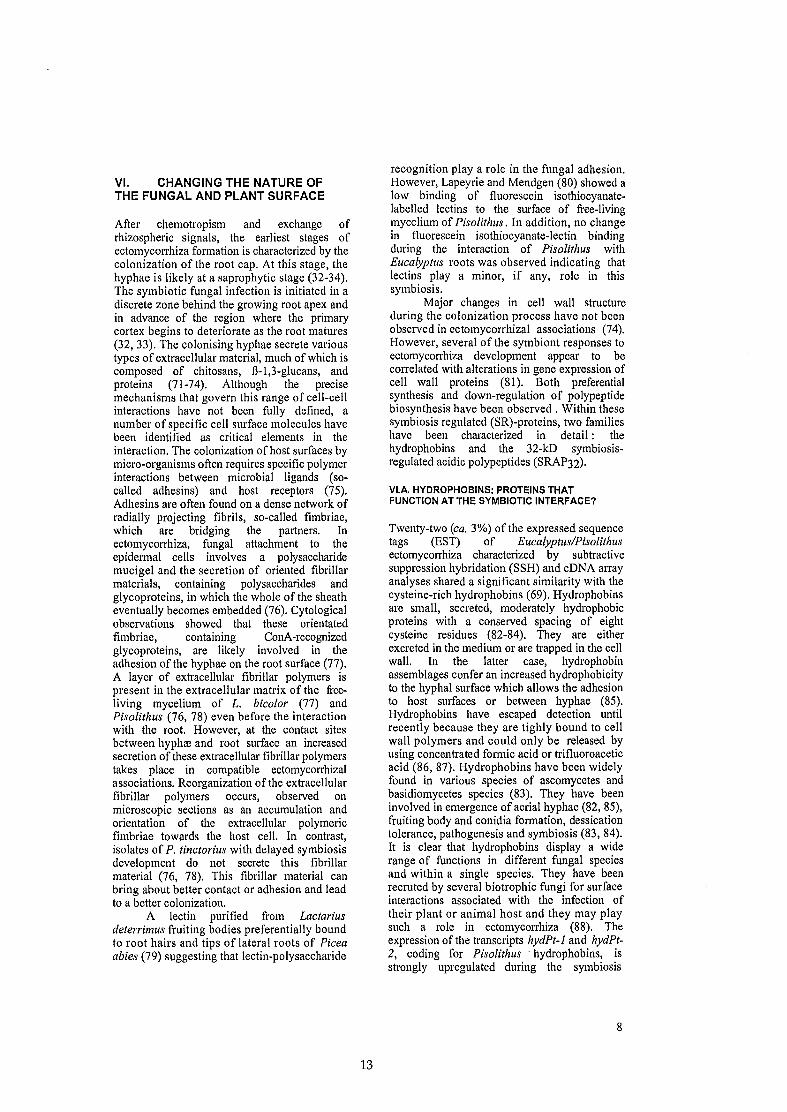
After chemotropism and exchange ofrhizospheric signais, the earliest stages ofectomycorrhiza formation is characterized by thecolonization of the root cap. At this stage, thehyphae is likely at a saprophytic stage (32-34).The symbiotic fungal infection is initiated in adiscrete zone behind the growing root apex andin advance of the region where the primarycortex begins to deteriorate as the root matures(32, 33). The colonising hyphae secrete varioustypes of extracellular material, much ofwhich iscomposed of chitosans, B-I,3-glucans, andproteins (71-74). Although the precisemechanisms that govern this range of cell-cellinteractions have not been fully defined, anumber of specifie cell surface molecules havebeen identified as critical elements in theinteraction. The colonization ofhost surfaces bymicro-organisms often requires specific polymerinteractions between microbial ligands (socalled adhesins) and host receptors (75).Adhesins are often found on a dense network ofradially projecting fibrils, so-called fimbriae,which are bridging the partners. Inectomycorrhiza, fungal attachment to theepidermal cells involves a polysaccharidemucigel and the secretion of oriented fibrillarmaterials, containing polysaccharides andglycoproteins, in which the whole of the sheatheventually becomes embedded (76). Cytologicalobservations showed that these orientatedfimbriae, containing ConA-recognizedglycoproteins, are likely involved in theadhesion of the hyphae on the root surface (77).A layer of extracellular fibrillar polymers ispresent in the extracellular matrix of the freeliving mycelium of L. bieolor (77) andPisolithus (76, 78) even before the interactionwith the root. However, at the contact sitesbetween hyphre and root surface an increasedsecretion of these extracellular fibrillar polymerstakes place in compatible ectomycorrhizalassociations. Reorganization of the extracellularfibrillar polymers occurs, observed onmicroscopie sections as an accumulation andorientation of the extracellular polymericfimbriae towards the host cell. In contrast,isolates of P. tinetorius with delayed symbiosisdevelopment do not secrete this fibrillarmaterial (76, 78). This fibrillar material canbring about better contact or adhesion and leadto a better colonization.
A lectin purified from Laetariusdeterrimus fruiting bodies preferentially boundto fOot hairs and tips of lateral fOots of Pieeaabies (79) suggesting that lectin-polysaccharide
13
recognition play a role in the fungal adhesion.However, Lapeyrie and Mendgen (80) showed alow binding of fluorescein isothiocyanatelabelled lectins to the surface of free-livingmycelium of Pisolithus. In addition, no changein fluorescein isothiocyanate-Iectin bindingduring the interaction of Pisolithus withEucalyptus fOots was observed indicating thatlectins play a minor, if any, role in thissymbiosis.
Major changes in cell wall structureduring the colonization process have not beenobserved in ectomycorrhizal associations (74).However, several of the symbiont responses toectomycorrhiza development appear to becorrelated with alterations in gene expression ofcell wall proteins (81). Both preferentialsynthesis and down-regulation of polypeptidebiosynthesis have been observed . Within thesesymbiosis regulated (SR)-proteins, two familieshave been characterized in detail: thehydrophobins and the 32-kD symbiosisregulated acidic polypeptides (SRAP32).
VI.A. HYDROPHOBINS: PROTEINS THATFUNCTION AT THE SYMBIOTIC INTERFACE?
Twenty-two (ca. 3%) of the expressed sequencetags (EST) of Eucalyptus/Pisolithusectomycorrhiza characterized by subtractivesuppression hybridation (SSH) and cDNA arrayanalyses shared a significant similarity with thecysteine-rich hydrophobins (69). Hydrophobinsare small, secreted, moderately hydrophobieproteins with a conserved spacing of eightcysteine residues (82-84). They are eitherexcreted in the medium or are trapped in the cellwall. In the latter case, hydrophobinassemblages confer an increased hydrophobicityto the hyphal surface which allows the adhesionto host surfaces or between hyphae (85).Hydrophobins have escaped detection untilrecently because they are tighly bound to cellwall polymers and could only be released byusing concentrated formic acid or trifluoroaceticacid (86, 87). Hydrophobins have been widelyfound in various species of ascomycetes andbasidiomycetes species (83). They have beeninvolved in emergence of aerial hyphae (82, 85),fruiting body and conidia formation, dessicationtolerance, pathogenesis and symbiosis (83, 84).It is clear that hydrophobins display a widerange of functions in different fungal speciesand within a single species. They have beenrecruted by several biotrophic fungi for surfaceinteractions associated with the infection oftheir plant or animal host and they may playsuch a role in ectomycorrhiza (88). Theexpression of the transcripts hydPt-1 and hydPt2, coding for Pisolithus . hydrophobins, isstrongly upregulated during the symbiosis
8

formation (89). Additional hydrophobin genes,hydPt-3 to hydPt-8, which shows 47 to 52 %homology with other Pisolithus hydrophobins(69) have been identified inEucalyptuslPisolithus mycorrhiza. Theexpression of their transcripts is increased 6 to8-fold in the symbiotic tissues. Nonwettableand water-repellent mycorrhizas of Eucalyptusare often found in air pockets in soil (90). Themost likely explanation for this lies in theobserved deposition of hydrophobins in fungalcell walls (87). However, Pisolithus hyphaesimultaneously expresse several types ofhydrophobins in its wall during the formationof the symbiosis. Whether these differenthydrophobins are involved in the adhesion ofhyphae on the root surface, the mechanicalpenetration of hyphae between root cells, or theaggregation of hyphae to form the mantle (81,88) remain to be determined by selective geneinactivation.
VI.B. SYMBIOSIS·REGULATED POLYPEPTIDESWITH ADHESIN·TYPE MOTIF
Many SRAPs, observed in soluble proteinextracts of Eucalyptus/Pisolithusectomycorrhizas (9, 91), are also abundantlyaccumulated in Pisolithus cell walls. A familyof cell wall acidic polypeptides (so-calledSRAP32) composed of at least six isoforrnswith different charge and/or molecular mass hasbeen isolated (92). These polypeptides areencoded by a multigene family and a dozen ofslightly different sequences have been identified(Sorin, Voiblet & Tagu, unpublished results).The SRAP32 proteins showed no significanthomology with known proteins, but the centralpart of the deduced protein contains an Arg-GlyAsp (RGD) motif. The RGD motif was firstdiscovered in fibronectin as a cell attachmentsite (93) and was subsequently found to be therecognition sequence for a number of integrinreceptors (94). The presence of the RGD motifsuggests that SRAP32 are coding for adhesionproteins (92). Comparing the upregulation ofSRAP 32 transcript levels and the increasedconcentration of SRAP 32 polypeptides inectomycorrhiza (9, 91, 92) showed that therewas a good correlation between changes inprotein synthesis and transcript levels (69, 92).Immunogold labelling confirmed that SRAP32and immunocross-reacting SRAP 31 arelocalised in cell walls of the free-living andsymbiotic hyphae. These proteins could befound mainly associated to the flocculentmaterial covering the hyphal surface, but theyare never observed in the thin, delicate filaments
14
or fimbriae bridging hyphae or the fungal cellsto the surface of the root.
Cell wall proteins are known to forrncross-linked networks with other proteins andpolysaccharides in fungal walls (75), but thestructural properties and the functionalsignificance of such networks are not yetknown. It is tempting to speculate that highlevels of SRAP32, degradation ofmannoproteins (72), and increased levels ofhydrophobins (89) take place simultaneously tomodify the molecular architecture of proteinnetworks in a manner that allows newdevelopmental fates for both fungal celladhesion and root colonization by the fungus(75,81). Further investigation of the structureand regulation of SR wall proteins will providea more complete picture of their role indeveloping ectomycorrhizal tissues.
VII. ESTs AND cDNA ARRAYS FORGENE EXPRESSION ANALYSIS
It is increasingly clear that developmentalpathways leading to the ectomycorrhizalsymbiosis can be considered as modular (Fig.2), and that developmental transitions areaccompanied by global changes in theexpression of specifie complements of genesunder the control of rhizospheric andintracellular signais (see above) (39, 50). Todate, it is not possible to predict the number ofsymbiosis-specific fungal and plant genes.However, owing to the fact that ectomycorrhizasare widespread, a significant number ofmycorrhiza-specific genes must exist. A goal ofprimary importance is to achieve acomprehensive description of the mechanismsinduced in both symbionts at each stage of thesymbiosis development. This molecularsketching of the ectomycorrhiza developmentshould be carried out simultaneously ondifferent associations to identify common'molecular signatures' typical ofthis symbiosis.Over the last decade, changes in gene expressionhave mainly been studied by using twodimensional gel electrophoresis. Up- and downregulated proteins have been found inPisolithuslEucalyptus (9, 91), AmanitamuscarialPicea abies (95), PaxillusinvolutuslBetula pendula (96) and SuillusbovinuslPinus sylvestris (97) associations.These investigations confirrned thatectomycorrhizal development leads to analteration of gene expression in both symbiontsand to the synthesis of ectomycorrhiza-specificproteins (ectomycorrhizins) (9, 91). However,this approach was limited because only arestricted set of proteins can be visualized on2D gels. While such studies have been fruitful
9

extramatrlc:~-' hypha'! .,
••
....• r • 4:'
• <>. .

in the past, their potential use in developing acomplete and accurate understanding of thesymbiotic factors during the course of themycorrhiza development is limited. Althoughmodulation of the symbiont interactions clearlyimplies post-translational modifications,regulation of transport mechanisms, and proteindegradation, key mechanisms in fungal generegulation take place at the transcriptionallevel.Consequently, detection of even subtle geneexpression modulations will provide acomprehensive framework for studying eventswhich affect cellular differentiation andmetabolism, and regulation on a genomic scale.The fact that ectomycorrhizal fungi are not yetamenable to gene inactivation has prevented theapplication of forward genetics to decipherectomycorrhiza development. Therefore,alternative molecular techniques for theidentification of SR-genes have been developed.Subtractive cDNA hybridization and differentialmRNA display were used to identify plant andfungal genes that are induced upon symbiosisdevelopment in ectomycorrhizal associationsinvolving Pisolithus/Eucalyptus (6S, 69),L. bieoloriPinus resinosa (9S, 99), and A.musearia/P. abies (100). These investigationsconfirmed that ectomycorrhiza development isaccompanied by striking changes in geneexpression at the transcriptional level andallowed the identification of a dozen of SRgenes (Fig. 5). For example, hexose transportersof the symbionts in the A. musearia/P. abies(101) and P. involutus/B. pendula (102)symbioses are regulated. The fungal genecoding for an hexose transporter, AmMstl, wasupregulated (101, 104), whereas the Piceahexose transporter was slightly down-regulated(104). Similarly, the expression of B.pendulahexose- and sucrose-transporters, BpSUC1,BpHEXl and BpHEX2, had been downregulated in mycorrhizal roots (102). Asstressed by the authors, the down-regulation ofexpression of these transporters is notcompatible with the increased carbon fluxestaking place in the roots as a result of thecarbon drain imposed by the mycobiont. Othertransporters are likely involved in the symbiotictraffic. A. musearia phenylalanine ammoniumIyase gene, AmPAL, is likely regulated inectomycorrhiza through changes in nitrogen andsugar levels (103). Whether gene expression ofthese metabolic genes is controlled by sugardependent regulation or by symbiosis-relateddevelopmental signais is not known. Nehls etal. (104) have suggested that the expression ofhexose-transporter gene, AmMstl, is onlyregulated by the hexose concentration of thesymbiotic apoplastic space of Amanita/Populusmycorrhiza. These findings illustrated thedrastic molecular changes experienced by the
15
partners during the mycorrhiza development andfunctioning (105-107).
"1I.1:rôtmM"ç;t.hyphn~ •
Fig. 5. Regulation of gene expression in theectomycorrhizal symbiosis. This figure compiles the knownupregulated genes in various types of ectomycorrhizas[Pisolithus/E. globulus (68, 69, 87, 89, 92, 119, 120);Paxillus involutuslBetula pendula (121); L. bicolorlPinusresinosa (98,99); and A. muscarialP. abies (100)]. ARF,ADP-ribosylation factor; A UT7, vesicular transport andautophagocytosis; CaM, calmodulin; COP9, constitutivephotomorphogenic subunit (related to proteasome); cpc2,cross-pathway control WD-repeat protein; eIF4A,elongation initiation factor 4A (dead-box helicase); erg6,0-(24)-sterol c-methyltransferase; erg11, sterol-14-alphademethylase; FUN34, transmembrane protein; hyd,hydrophobins; Hypar, hypaphorine- and auxin-regulatedglutathion-S-transferase; Icdh, NADP-isocitratedehydrogenase; LT6B, Salt-stress induced LTI6B protein;Mdh, mitochondrial malate dehydrogenase; Mstl,monsaccharide transporter; OMT, O-methyltransferase;PAL, phenylammonia lyase; SEND32, Senescence downregulated protein; tefl, Translation elongation factor 1a;TubAI, Cl.-tubulin; Ubc2, Ubiquitin-conjugating enzymeE2.
To identify cellular functions expressedin the symbiosis on a wider scale, ESTprogrammes have been developed on severalectomycorrhizal fungi (A. muscaria,H. cylindrosporum, P. tinetorius, Tuberborehii) (69, lOS, 109; P Bonfante, U Nehls,H Sentenac, pers. comm.) and ectomycorrhizalassociations (Euealyptus/Pisolithus, 69;B. pendula/P. involutus, A Tunlid & BS6derstr6m, pers. comm.). As the number ofESTs increases, comparisons across genera,species, ecotypes, and strains of symbioticfungi will become possible through 'digitalnorthern' (110). With multiple ESTprogrammes dealing with pathogenic (111-114)and mutualistic fungi, we will have in a nearfuture an unparalleled opportunity to ask whichgenetic features are responsible forcommon/divergent traits involved in
10

pathogenesis and symbiosis. A few of the manypossible breakthroughts will be incharacterisation of common transductionnetworks, identification of nove! surfaceproteins that play critical roles in plant-fungusinteractions and new insights into uniquemetabolic routes critical for mycorrhizafunctioning.
Quantitative analysis of thetranscriptome has become possible through'hybridization signature' methods which alIowlarge scale measurement of gene expression(115-117). cDNA array analyses are currentlyproviding efficient means of acquiring largeamounts of biological information foridentifying processes involved in plant-microbeinteractions (114,118). To take advantage ofthe available ESTs from theEucalyptus/Pisolithus ectomycorrhiza, we haveconstructed miniarrays of fungal and plant ESTs(69). These miniarray analyses provided a toolto broadly analyze the expression of severalhundreds genes during the symbiosisdevelopment, to identify SR genes, and toidentify candidate genes for further moredetailed analysis. About 80 SR genes (17%)were identified by differential screening of 480arrayed cDNAs between free-living partners andsymbiotic tissues (69). Even this modestcollection of genes begins to provide anindication of symbiosis environment asperceived by the symbionts (Fig. 5). Within thecellular functions which are strikingly regulatedby symbiosis development, we have identified:cell wall and membrane synthesis, stress anddefence responses, protein degradation (in plantcells), and protein synthesis (in hyphae) (Fig.5). EST/cDNA array analyses confirmed thatmost members of the hydrophobin and SRAP32
gene families are dramatically up-regulated (upto 8-fold) during fungal mantIe formation (seesection VI.B). Egubc2, which encodes aubiquitin-conjugating (E2) enzyme, andEgCops7a, coding for a subunit of theproteasome-related complex, are highly upregulated in ectomycorrhizal tips confirmingthat symbiosis development induces drasticplant protein degradation (9). Proteindegradation may be a result of stress conditionsexperienced by the fOots colonized by massiveamount of hyphae.
This data suggests a highly dynamicenvironment in which symbionts are sendingand receiving signais, exposed to high levels ofstress conditions and remodeling tissues. Astriking result of these study is the fact that allgenes investigated are common to thenonsymbiotic and symbiotic stages. At thedevelopmental stage studied, symbiosisdevelopment does not induced the expression of
16
ectomycorrhiza-specijlc genes, but a markedchange in the gene expression in the partners.
VIII. CONCLUSIONS
As outlined in this review, ectomycorrhizadevelopment influences both plant and fungusgene expression in a pleiotropic manuer. Arange of fungal tissues differentiates that can bedistinguished by a combination of anatomical,cytological and molecular features. On the otherhand, root tips proliferate and root cellsexperience major alteration in their shape andgene expression. Advances of recent years haveprovided insights on the molecular basis ofectomycorrhiza morphogenesis. With theidentification of several developmentallyregulated proteins and genes and a descriptionof their expression and activities, the ground isnow set for recasting earlier models ofsymbiosis development in molecular terms. Ilis apparent from this briefreview, however, thatthere is a vast complexity of geneticprogrammes with overlapping expressionpatterns. This include: induction of plantdefence/stress reactions, the down-expression ofplant protein biosynthesis, the initiation oflateral roots by fungal auxins, themorphogenetic switches of the fungal hyphae,and the establishment of novel cell walls andextracellular matrices. Among the manyremaining challenges is the elucidation ofmechanisms and inducer molecules thatintegrate the actions of these multipleprogrammes of gene expression in generating amature symbiotic organ. Studies in areas suchthe identification of chemicals andgenes/proteins involved in cell-cell interactions,control of cell expression at the level of signaltransduction will be the source for manyanswers. A comparative study of geneexpression in different types of ectomycorrhizasusing the molecular approaches includinggenomics and gene inactivation might reveal towhat extent similarities and differences in thevarious types of ectomycorrhizas are the resultof variation in the basic mechanisms underlyingthe respective developmental programmes andthe effects of the different trophic andenvironmental eues.
ACKNOWLEDGEMENTSSD was supported by a Doctoral Scholarship from theMinistère de l'Education Nationale, de la Recherche et dela Technologie. We also appreciated partial support fromthe Groupement de Recherches et d'Etude des Génomes,the INRA Collaborative Research Programme inMicrobiology, and the INRA GenoPop research gran!. Wethank Ors Frank Oitengou, Hubert Lagrange, FrédéricLapeyrie and Catherine Voiblet forstimulating discussions.
II

REFERENCES
1. S. E. Smith and D. 1. Read. Mycorrhizal
.. 2nd Ed" Ad'SymblOsls. ltlon. ca emlePress, London. 1997.
2. A. H. Fitter and B. Moyersoen.Evolutionary trends in root-microbesymbioses. Philo. Trans. Royal Soc. ofLondon B. 351: 1367-1375 (1996).
3. D. J. Read. Mycorrhizas in ecosystems.Experientia47: 376-390 (1991).
4. A. F. S. Taylor, F. Martin and D. J. Read.Fungal diversity in ectomycorrhizalcommunities of Norway spruce [Piceaabies (L.) Karst.] and beech (Fagussylvatica L.) along North-South transectsin Europe. In: Carbon and NitrogenCycling in European Forest Ecosystems,Ecological Studies 142, ED Schulze(ed.), Springer Verlag Berlin Heidelberg,2000,pp.343-365.
5. A. A. Meharg and 1. W. G. Cairney. Coevolution ofmycorrhizal symbionts andtheir hosts to metal-contaminatedenvironments. Adv. Ecol. Res. 30: 69112 (2000).
6. T. S. Grove and F. Le Tacon. Mycorrhizain plantation forestry. Adv. Plant Pathol.9: 191-227 (1993).
7. I. K6ttke and F. Oberwinkler. The cellularstructure of the Hartig net: coenocyticand transfer cell-like organization. Nord.J. Bot. 7: 85-95 (1987).
8. H. B. Massicotte, R. L. Peterson, C. A.Ackerley and A. E. Ashford. Ontogenyof Eucalyptus pilularis-Pisolithustinctorius ectomycorrhizae. II.Transmission electron microscopy. CanoJ. Bot. 65: 1940-1947 (1987).
9. J. L. Hilbert, G. Costa and F. Martin.Ectomycorrhizin synthesis . andpolypeptide changes during the earlystage of eucalypt mycorrhizadevelopment. Plant Physiol. 97: 977984 (1991).
10. B. Botton and M. Chalot. Nitrogenassimilation: enzymology inectomycorrhizas. Mycorrhiza: Structure,Molecular Biology and Function 2nd
Edition (A. K. Varrna and B. Hock,eds), Springer-Verlag, Berlin Heidelberg,New-York, 1999, pp. 333-372.
II. R. Hampp and C. Schaeffer. Mycorrhiza Carbohydrate and energy metabolism.Mycorrhiza: Structure, MolecularBiology and Function 2nd Edition (A. K.Varrna and B. Hock, eds), SpringerVerlag, Berlin Heidelberg, New-York,1999, pp. 273-303.
17
12. H. Gehrig, A. Schül3ler and M. Kluge.Geosiphon pyriforme, a fungus formingendocytobiosis with Nostoc(Cyanobacteria), is an ancestral memberof the G1omales: evidence by SSU rRNAanalysis. J. Mol. Evol. 43: 71-81 (1996).
13. M-A. Selosse and F. Le Tacon. The landflora: a phototroph-fungus partnership?Trends Ecol. Evol. 13: 15-20 (1998).
14. T. N. Taylor, W. Remy, H. Hass and HKerp. Fossil arbuscular mycorrhizaefrom the Early Devonian. Mycologia 87:560-573 (1995).
15. B. A. LePage, R. S. Currah, R. A.Stockey and G. W. Rothwell. Fossilectomycorrhizae from the middle Eocene.Am. J. Bot. 84: 410-412 (1997).
16. T. D. Bruns, T. M. Szaro, M. Gardes, K.W. Cullings, 1. 1. Pan, D. L. Taylor, T.R. Horton, A. Kretzer, M. Garbelottoand Y. Li. A sequence database for theidentification of ectomycorrhizalbasidiomycetes by phylogeneticanalysis. Mol. Ecol. 7,257-272 (1998).
17. D. S. Hibbett, L. B. Gilbert and M. J.Donoghue. Evolutionary instability ofectomycorrhizal symbioses inbasidiomycetes. Nature 407: 506-508(2000).
18. M. Brundrett The co-evolution ofmycorrhizas and roots of land plants:evaluation of the structure and functionof ancient and modem associations. NewPhytol. in press (2001).
19. E. Duhoux, G. Rinaudo, H. G. Diem, F.Auguy, D. Femandez, D. Bogusz, C.Franche, Y. Dommergues and B.Huguenin. Angiosperrn Gymnostomatrees produce raot nodules colonized byarbuscular mycorrhizal fungi related toGlomus. New Phytol. 149: 115-125(2001).
20. M. Gardes and T. D. Bruns. Communitystructure of ectomycorrhizal fungi in aPinus muricata forest: above- and belowground views. Cano J. Bot. 74, 15721583 (1996).
21. L. Jonsson, A. Dahlberg, M. C. Nilsson,O. Zackrisson and O. Karen.Ectomycorrhizal fungal communities inlate-successional Swedish boreal forests,and their composition followingwildfire. Mol. Eco 1. 8: 205-215 (1999).
22. 1. W. G. Caimey. Intraspecificphysiological variation: implications forunderstanding functional diversity inectomycorrhizal fungi. Mycorrhiza 9:125-135 (1999).
23. T. Wallenda, C. Strober, H. Hiigbom, H.Schinke1, E. George, P. Hiigberg and D.1. Read. Nitrogen uptake processes in
12

roots and mycorrhizas. Carbon andNitrogen Cyc!ing in European ForestEcosystems Ecological Studies 142, EDSchulze (ed.), Springer Verlag BerlinHeidelberg, 2000, pp. 122-143.
24. M. Chalot and A. Brun. Physiology oforganic nitrogen acquisition byectomycorrhizal fungi andectomycorrhizas. FEMS Mierobiol. Rev.22: 21-44 (1998).
25. F. Martin and S. Lorillou. Nitrogenacquisition and assimilation inectomycorrhizal systems. TreesContributions to Modern TreePhysiology H Rennenberg, W Eschrich,H Ziegler (eds), Backhuys Pub!.,Leiden, The Netherlands, 1997, pp. 423429.
26. M. W. Simard, D. A. Perry, M. D.Jones, D. D. Myrold, D. M. Durallandand R. Molina. Net transfer of carbonbetween ectomycorrhizal tree species inthe field. Nature 388: 579-582 (1997).
27. B. Wu, K. Nara and H. Taizo. Can 14C_labeled photosynthetic products movebetween Pinus densiflora seedlingslinked by ectomycorrhizal mycelia? NewPhytol. 149: 137-146 (2001).
28. P. Vivin, F. Martin and J-M. Guehl.Acsuisition and within-plant allocationof 1 C and lSN in CO2- emiched Quereusrobur plants. Physiol. Plant. 98: 89-96(1996).
29. A. H. Fitter, A. Heinemeyer and P. L.Staddon. The impact of elevated C02and global climate change on arbuscularmycorrhizas: a mycocentric approach.NewPhytol. 147: 179-187 (2000).
30. 1. Kottke and F. Oberwinkler. Rootfungus interactions observed on initialstages of mantle formation and Hartignet establishment in mycorrhizas ofAmanita musearia on Pieea abies inpure culture. Cano J. Bot. 64: 2348-2354(1986).
31. I. Brunner and C. Scheidegger. Effects ofhigh nitrogen concentrations onectomycorrhizal structure and growth ofseedlings of Picea abies (L.) Karst. NewPhytol. 129: 83-95 (1995).
32. G. A. Chilvers. Low power electronmicroscopy of the root cap region ofeucalypt mycorrhizas. New Phytol. 67:663-668 (1968).
33. D. P. Horan, G. A. Chilvers and F. F.Lapeyrie. Time sequence of the infectionprocess in eucalypt ectomycorrhizas.New Phytol. 109: 451-458 (1988).
34. J. Dexheimer, J. Gerard and P. Genet.Study of transformations of the root
18
system of Eucalyptus globulusassociated with Pisolithus tinetorius. 1.Aptitude to mycorrhization of differentkinds of roots. Phytomorphology 44:235-245 (1994).
35. H. B. Massicotte, C. A. Ackerley and R.L. Peterson. Ontogeny of Alnus rubraAlpova diplophloeus ectomycorrhizae. II.Transmission electron microscopy. CanoJ. Bot. 67: 201-210 (1989).
36. A. Brun, M. Chalot, R. D. Finlay and B.Soderstrom. Structure and function ofthe ectomycorrhizal association betweenPaxillus involutus (Batsch) Fr. andBetula pendula (Roth.). 1. Dynamics ofmycorrhiza formation. New Phytol. 129:487-493 (1995).
37. R. Agerer. Colour Atlas of
Ectomycorrhizae. 7th
edition. EinhomVerlag, Schwiibisch Bmünd, Germany.1993-1998.
38. G. Gay, R. Marmeisse, P. Fouillet, M.Bouletreau and J. C. Debaud.Genotype/nutrition interactions in theectomycorrhizal fungus Hebelomacylindrosporum Romagnesi. NewPhytol. 123: 335-343 (1993).
39. F. Martin and D. Tagu. Developmentalbiology of a plant-fungus symbiosis: theectomycorrhiza. Myeorrhiza: Structure,Molecular Biology and Function 2nd
Edition (A. K. Varma and B. Hock,eds), Springer-Verlag, Berlin Heidelberg,New-York, 1999, pp. 51-73.
40. B. R. Kropp, B. J. McAfee and J. A.Fortin. Variable loss of ectomycorrhizalability in monokaryotic and dikaryoticcultures of Laccaria bicolor. Cano J.Bot. 65: 500-504 (1987).
41. J. C. Debaud, G. Gay, A. Prevost, J. Leiand J. Dexheimer. Ectomycorrhizalability of genetically differenthomokaryotic and dikaryotic mycelia ofHebeloma cylindrosporum. New Phytol.108: 323-328 (1988).
42. M. S. Lamhamedi, A. 1. Fortin, H. H.Kope and B. R. Kropp. Geneticvariation in ectomycorrhiza formation byPisolithus arhizus on Pinus pinasterand Pinus banksiana. New Phytol. 115:689-697 (1990).
43. K. K. Wong, Y. Piché, D. Montpetit andB. R. Kropp. Differences in thecolonization of Pinus banksiana rootsby sib-monokaryotic and dikaryoticstrains of ectomycorrhizal Laeeariabicolor. Cano J. Bot. 67: 1717-1726(1989).
44. C. Karabaghli-Degron, B. Sotta, M.Bonnet, G. Gay and F. Le Tacon. The
13
auxin transport inhibitor 2,3,5·triiodobenzoic acid (TIBA) inhibits thestimulation of in vitro lateral rootformation and the colonization of thetap-root cortex ofNorway spruce (Piceaabies) seedlings by the ectomycorrhizalfungus Laccaria bicolor. New Phytol.140: 723-733 (1998).
45. A. M. Hirsch and M. J. McFall-Ngai.Fundamental concepts in symbioticinteractions: light and dark, day andnight, squid and legume. J. PlantGrowth Regul. 19: 113-130 (2000).
46. N. Fries, K. Serck-Hanssen, L. HallDimberg and O. Theander. Abietic acid,an activator ofbasidiospore germinationin ectomycorrhizal species of the genusSuillus (Boletaceae). Exp. Mycol. Il:360-363 (1987).
47. T. Béguiristain and F. Lapeyrie. Hostplant stimulates hypaphorineaccumulation in Pisolithus tinctoriushyphae during ectomycorrhizal infectionwhile excreted fungal hypaphorinecontrols root hair development. NewPhytol. 136: 525-532 (1997).
48. F. A. Ditengou and F. Lapeyrie.Hypaphorine from the ectomycorrhizalfungus Pisolithus tinctorius counteractsactivities of indole-3-acetic acid andethylene but not synthetic auxins ineucalypt seedlings. Mol. Plant-MicrobeInteract 13: 151-158 (2000).
49. H. Lagrange, C. Jay-Allemand and F.Lapeyrie. Rutin, the phenolglycosidefrom eucalyptus root exudates,stimulates Pisolithus hyphal growth atpicomolar concentrations. New Phytol.in press (2001).
50. F. Martin, F. Lapeyrie and D. Tagu.Altered gene expression duringectomycorrhizal development. TheMycota, Vol. V, Part A, PlantRelationships, G Caroll, P Tudzynski(eds), Springer-Verlag, Berlin,Heidelberg, 1997, pp. 223-242.
51. S. 1. Barker and D. Tagu. The roles ofauxins and cytokinins in mycorrhizalsymbioses. J. Plant Growth Regul. 19:144-154 (2000).
52. D. P. Horan and G.A. Chilvers.Chemotropism; the key toectomycorrhizal formation? New Phytol.116: 297-301 (1990).
53. H. A. Hartwig and D. A. Phillips.Release and modification of nod-geneinducing flavonoids from alfalfa seeds.Plant Physiol. 95: 804-807 (1991).
54. G. Nagahashi and D. D. Douds. Partialseparation of root exudate componentsand their effects upon the growth of
19
germinated spores of AM fungi. Mycol.Res. 104: 1453-1464 (2000).
55. T. Béguiristain, R. Côté, P. Rubini, C.Jay-Allemand and F. Lapeyrie.Hypaphorine accumulation in hyphae ofthe ectomycorrhizal fungus, Pisolithustinctorius. Phytochem. 40: 1089-1091(1995).
56. N. L. Paiva. An introduction to thebiosynthesis of chemicals used in plantmicrobe communication. 1. PlantGrowth Regul. 19: 131-143 (2000).
57. L. Gea, L. Normand, B. Vian and G.Gay. Structural aspects ofectomycorrhiza of Pin us pinaster (Ait.)Sol. formed by an IAA-overproducermutant of Hebeloma cylindrosporumRomagnési. New Phytol. 128: 659-670(1994).
58. N. Gogala. Regulation of mycorrhizalinfection by hormonal factors producedby hosts and fungi. Experientia 47: 331340 (1991).
59. H. Beyrie. The role of phytohormones inthe function and biology of mycorrhizas.Mycorrhiza: Structure, molecularbiology and function, Varma AK, HockB (eds), Springer Berlin Heidelberg NewYork, 1995, pp. 365-390.
60. V. Slankis. Effect ofnapthaleneacetic acidon dichotomous branching of isolatedraots of Pinus sylvestris. Physiol. Plant.3,40-43 (1950).
61. J. M. Ulrich. Auxin production bymycorrhizal fungi. Physiol. Plant. 13:429-443 (1960).
62. L. A. Rupp and K. W. Mudge. Ethephonand auxin induce mycorrhiza-likechanges in the morphology of root organcultures of Mugo pine. Physiol. Plant.64: 316-322 (1985).
63. L. A. Rupp, K. W. Mudge and F. B.Negm. Involvement of ethylene inectomycorrhiza formation anddichotomous branching of roots ofMugo pine seedlings. Cano J. Bot. 67:477-482 (1989).
64. W. T. Frankenberger and M. Poth.Biosynthesis of indole-3-acetic acid bythe pine ectomycorrhizal fungusPisolithus tinctorius. Appl. Environ.Microbiol. 53: 2908-2913 (1987).
65. G. Gay, L. Normand, R. Marmeisse, B.Sotta and J. C. Debaud. Auxinoverproducer mutants of Hebelomacylindrosp0nlm Romagnési haveincreased mycorrhizal activity. NewPhytol. 128: 645-657 (1994).
66. K. Palme and L. Galweiler. PIN-pointingthe molecular basis of auxin transport.
14

Curr. Op. Plant Biol. 2: 375-381(1999).
67. H. Tranvan, Y. Habricot, E. Jeannette, G.Gay and B. Sotta. Dynamics ofsymbiotic establislunent between anIAA-overproducing mutant of theectomycorrhizal fungus Hebelomacylindrosporum and Pinus pinaster.Tree Physiol. 20: 123-129 (2000).
68. U. Nehls, T. Béguiristain, F. Ditengou,F. Lapeyrie and F. Martin. Theexpression of a symbiosis-regulated genein eucalypt roots is regulated by auxinsand hypaphorine, the tryptophan betaineof the ectomycorrhizal basidiomycetePisolithus tinctorius. Planta 207: 296302 (1998).
69. C. Voiblet, S. Duplessis, N. Encelot andF. Martin. Identification of symbiosisregulated genes in Eucalyptus globulusPisolithus tinctorius ectomycorrhiza bydifferential hybridization of arrayedcDNAs. Plant J. In press (2001).
70. F. A. Ditengou, T. Béguiristain and F.Lapeyrie. Root hair elongation isinhibited by hypaphorine, the indolealkaloid from the ectomycorrhizal fungusPisolithus tinctorius, and restored byIAA. Planta 211: 722-728 (2000).
71. J. Dexheimer and J. C. Pargney.Comparative anatomy of the host-fungusinterface in mycorrhizas. Experientia 47:312-320 (1991).
72. D. Tagu and F. Martin. Molecularanalysis of cell wall proteins expressedduring the early steps of ectomycorrhizadevelopment. New Phytol. 133: 73-85(1996).
73. R. Balestrini, M. G. Hahn and P.Bonfante. Location of cell-wallcomponents in ectomycorrhizae ofCorylus avellana and Tuber magnatum.Protoplasma 191: 55-69 (1996).
74. P. Bonfante. At the interface betweenmycorrhizal fungi and plants: thestructural organization of cell wall,plasma membrane and cytoskeleton.Fungal associations, Mycota lX, BHock, (ed), Springer-Verlag, BerlinHeidelberg, 2001, pp. 000-000.
75. W. L. Chaffin, J. L. Lopez-Ribot, M.Casanova, D. Gozalbo and J. P.Martinez. Cell wall and secreted proteinsof Candida albicans: identification,function, and expression. Microbiol.Mol. Biol. Rev. 62: 130-180 (1998).
76. F. Lapeyrie, J. Lei, N. Malajczuk and J.Dexheimer. Ultrastructural andbiochemical changes at the pre-infectionstage of mycorrhizal formation by two
20
isolates of Pisolithus tinctorius. Ann.Sei. For. 46s: 754s-757s (1989).
77. 1. Lei, K. K. Y. Wong and Y. Piché.Extracellular concanavalin A-bindingsites during early interactions betweenPinus banksiana and two closely relatedgenotypes of the ectomycorrhizalbasidiomycete Laccaria bicolor. Mycol.Res. 95: 357-363 (1991).
78. 1. Lei, F. Lapeyrie, N. Malajczuk and J.Dexheimer. Infectivity of pine andeucalypt isolates of Pisolithus tinctorius(Pers.) Coker & Couch on roots ofEucalyptus urophylla S. T. Blake invitro. II. Ultrastructural and biochemicalchanges at the early stage of mycorrhizaformation. New Phytol. 116: 115-122(1990).
79. M. Giollant, J. Guillot, M. Damez, M.Dusser, P. Didier and E. Didier.Characterization of a lectin fromLactarius deterrimus. Research on thepossible involvement of the fungal lectinin recognition between mushroom andspruce during the early stages ofmycorrhizae formation. Plant Physiol.101: 513-522 (1993).
80. F. Lapeyrie and K. Mendgen. Quantitativeestimation of surface carbohydrates ofectomycorrhizal fungi in pure culture andduring Eucalyptus root infection. Mycol.Res. 97: 603-609 (1993).
81. F. Martin, P. Laurent, D. De Carvalho,C. Voiblet, R. Balestrini, P. Bonfanteand D. Tagu. Cell wall proteins of theectomycorrhizal basidiomycetePisolithus tinctorius: Identification,function, and expression in symbiosis.Fungal Genet. Biol. 27: 161-174 (1999).
82. 1. G. H. Wessels. Tansley Review N°45.Wall growth, protein excretion andmorphogenesis in fungi. New Phytol.123: 397-413 (1993).
83. M. 1. Kershaw and N. J. Talbot.Hydrophobins and repellents: proteinswith fundamental roles in fungalmorphogenesis. Fungal Genet. Biol 23:18-33 (1998).
84. H. A. B. Wiisten, M. Richter and J. M.Willey. Structural proteins involved inemergence of microbial aerial hyphae.Fungal Genet. Biol 27: 153-160 (1999).
85. H. A. B. Wiisten and J. M. Willey.Surface-active proteins enable microbialaerial hyphae to grow into the air.Microbiology 146: 767-773 (2000).
86 O. M. H. De Vries, M. P. Fekkes, H. A.B. Wiisten and J. G. H. Wessels.Insoluble hydrophobin complexes in thewalls of Schizophyllum commune and
15

other filamentous fungi. Arch.Microbiol. 159: 330-335 (1993).
87. D. Tagu, R. De Bellis, R. Balestrini, O.M. H. De Vries, G. Piccoli, V. Stocchi,P. Bonfante and F. Martin. Immunolocalization of the hydrophobin HYDPt1 from the ectomycorrhizalbasidiomycete Pisolithus tinctoriusduring colonization of Eucalyptusglobulus roots. New Phytol. 149: 127135 (2001).
88. D. Tagu, 1. Kottke and F. Martin.Hydrophobins in ectomycorhizalsymbiosis: hypothesis. Symbiosis 25: 518 (1998).
89. D. Tagu, B. Nasse and F. Martin.Cloning and characterization ofhydrophobins-encoding cDNAs from theectomycorrhizal basidiomycetePisolithus tinctorius. Gene 168: 93-97(1996).
90. P. A. Vesk, A. E. Ashford, AL.Markovina and W. G. Allaway.Apoplasmic barriers and theirsignificance in the exodermis and sheathof Eucalyptus pilularis-Pisolithustinctorius ectomycorrhizas. New Phytol.145: 333-346 (2000).
91. T. Burgess, P. Laurent, B. Dell, N.Malajczuk and F. Martin. Effect of thefungal isolate aggressivity on thebiosynthesis of symbiosis-relatedpolypeptides in differentiating eucalyptectomycorrhiza. Planta 195: 408-417(1995).
92. P. Laurent, C. Voiblet, D. Tagu, D. DeCarvalho, U. Nehls, R. De Bellis, R.Balestrini, G. Bauw, P. Bonfante and F.Martin. A novel class of ectomycorrhizaregulated cell wall polypeptides inPisolithus tinctorius. Mol. PlantMicrobe /nteract. 12: 862-871 (1999).
93. M. D. Pierschbacher and E. Ruoslahti.Cell attachment activity of fibronectincan be duplicated by small syntheticfragments of the molecule. Nature 309:30-33 (1984).
94. E. Ruoslahti. RGD and other recognitionsequences for integrins. Annu. Rev. Cel!Dev. Biol. 12: 697-715 (1996).
95. M. Guttenberger and R. Hampp.Ectomycorrhizins - Symbiosis-specific orartifactual polypeptides fromectomycorrhizas? Planta 188: 129-136(1992).
96. P. Simoneau, J. D. Viemont, J. C.Moreau and D. G. Strullu. Symbiosisrelated polypeptides associated with theearly stages of ectomycorrhizaorganogenesis in birch (Betula pendula
21
Roth). New Phytol. 124: 495-504(1993).
97. M. Tarkka, S. S. Niini and M.Raudaskoski. Developmentally regulatedproteins during differentiation of rootsystem and ectomycorrhiza in Scots pine(Pin us sylvestris) with Suillus bovinus.Physiol. Plant. 104: 449-455 (1998).
98. S. J. Kim, J. Zheng, S. T. Hiremath andG. K. Podila. Cloning andcharacterization of a symbiosis-relatedgene from an ectomycorrhizal fungusLaccaria bieolor. Gene 222: 203-212(1998).
99. S. J. Kim, D. Bernreuther, M. Thummand G. K. Podila. LB-AUT7, a novelsymbiosis-regulated gene from anectomycorrhizal fungus, Laccariabicolor, is functionally related tovesicular transport and autophagocytosis.1. Bacteriol. 181: 1963-1967 (1999).
100. U. Nehls, S. Mikolajewski, M. Ecke andR. Hampp. Identification and expressionanalysis of two fungal cDNAs regulatedby ectomycorrhiza and fruit bodyformation. New Phytol. 144: 195-202(1999).
101. U. Nehls, J. Wiese, M. Guttenberger andR. Hampp. Carbon allocation inectomycorrhizas: identification andexpression analysis of an Amanitamuscaria monosaccharide transporter.Mol. Plant-Microbe /nteract. Il: 167176 (1998).
102. D. P. Wright, J. D. Scholes, D. J. Readand S. A. Rolfe. Changes in carbonallocation and expression of carbontransporter genes in Betula pendulaRoth. colonized by the ectomycorrhizalfungus Paxillus involutus (Batsch) Fr.Plant Cel! Environ. 23: 39-49 (2000).
103. U. Nehls, M. Ecke and R. Hampp.Sugar- and nitrogen-dependent regulationof an Amanita muscaria phenylalanineammonium Iyase gene. 1. Bacteriol.181: 1931-1933 (1999).
104. U. Nehls, A. Bock, M. Ecke and R.Hampp. Differentiai expression ofhexose-regulated fungal genes withinAmanita muscaria/Populus tremula xtremuloides ectomycorrhizas. NewPhytol., in press (2001).
105. D. Blaudez, M. Chalot, P. Dizengremeland B. Botton. Structure and function ofthe ectomycorrhizal association betweenPaxillus involutus and Betula pendula.II. Metabolic changes during mycorrhizaformation. New Phytol. 138: 543-552(1998).
106. J. W. G. Caimey and R. M. Burke.Physiological heterogeneity within
16

fungal mycelia: an important concept fora functional understanding of theectomycorrhizal symbiosis. New Phytol.134: 685-695 (1996).
107. R. Hampp, C. Schaeffer, T. Wallenda,C. Stülten, P. Johann and W. Einig.Changes in carbon partitioning orallocation due to ectomycorrhizaformation: biochemical evidence. Cano J.Bot. 73: s548-s556 (1995).
108. D. Tagu and F. Martin. Expressedsequence tags of randomly selectedcDNA clones from Eucalyptus globulusPisolithus tinctorius ectomycorrhiza.Mol. Plant-Microbe Interact. 8: 781-783(1995).
109. C. Voiblet and F. Martin. Identifyingsymbiosis-regulated genes in Eucalyptusglobulus-Pisolithus tinctoriusectomycorrhiza using suppressionsubtractive hybridization and cDNAarrays. Molecular Plant-MicrobeInteraction, 9th International Congress,Amsterdam July 25-30, P. J. G. M. deWit, T. Bisseling and W. J. StiekemaEds, 1999, pp. 208-213.
110. R. M. Ewing, A. Ben Kahla, O. Poirot,F. Lopez, S. Audic and J. M. Claverie.Large-scale statistical analyses of riceESTs reveal correlated patterns of geneexpression. Genome Res. 9: 950-959(1999).
Ill. M. Hahn and K. Mendgen.Characterization of in planta inducedrust genes isolated from a haustoriumspecifie cDNA library. Mol. PlantMicrobe Interact. 10: 427-437 (1997).
112. S. Kamoun, P. Hraber, B. Sobral, D.Nuss and F. Govers. Initial assessmentof gene diversity for the oomycetepathogen Phytophthora infestans basedon expressed sequences. Fungal Genet.Biol. 28: 94-106 (1999).
lB. D. Qutob, P. T. Hraber, B. W. S.Sobral and M. Gijzen. Comparativeanalysis of expressed sequences inPhytophthora sojae. Plant Physiol. 123:243-253 (2000).
114. P. M. Schenk, K. Kazan, 1. Wilson, J.P. Anderson, T. Richmond, S. C.Somerville and J. M. Manners.Coordinated plant defense responses inArabidopsis revealed by microarrayanalysis. Proc. Nat!. Acad Sei. USA 97:11655-11660 (2000).
115. L. Wodicka, H. L. Dong, M. Mittmann,M. H. Ho and D. J. Lockhart. Genomewide expression monitoring inSaccharomyces cerevisiae. Nat. Biotech.15: 1359-1367 (1997).
22
116. T. Desprez, J. Amselem, M. Cabocheand H. Hafle. Differentiai geneexpression in Arabidopsis monitoredusing cDNA arrays. Plant 1. 14: 643652 (1998).
117. T. Girke, J. Todd, S. Ruuska, J. White,C. Benning and J. Ohlrogge. Microarrayanalysis of developing Arabidopsisseeds. Plant Physiol. 124: 1570-1581(2000).
118. J. Gyorgyey, D. Vaubert, J. I. JiménezZurdo, C. Charon, L. Troussard, A.Kondorosi and E. Kondorosi. Analysisof Medicago truncatula nodule expressedsequence tags. Mol. Plant-MicrobeInteract. 13: 62-71 (2000).
119. E Camero Diaz, F Martin, D Tagu.Eucalypt Ct.-tubulin: cDNA c10ning andincreased level of transcripts inectomycorrhizal root system. Plant Mol.Biol. 31: 905-910 (1996)
120. V Boiffin, M Hodges, S Galvez, RBalestrini, P Bonfante, P Gadal, FMartin. Eucalypt NADP-dependentisocitrate dehydrogenase. cDNA c10ningand expression in ectomycorrhizae. PlantPhysiology 117: 939-948 (1998).
121. L Feugey, DG Strullu, P Poupard, PSimoneau . Induced defence responseslimit Hartig net formation inectomycorhizal birch roots. New Phytol.144: 541-547 (1999).
17

&lIapit~e 2
IdeR~[fÎ!(Catünl1 die gèRes régulés paf la sym~]ose
E1JtIic(dW1ptus g}éJbuZus-flis@Zithu5 M1Pbct€Jrim5 par hyh1)fiJda~~6n
di~~éreiAt]el]ede ffulltnes d'ADKe

Identification de gènes régulés par la symbiose
ectomycorhizienne Eucalyptus globulus-Pisolithus tinctorius
par hybridation différentielle de filtres d'ADNe.
Comme nous venons de le décrire dans le chapitre introductif, la mise en place d'un organe mixte au
niveau du système racinaire de l'hôte végétal, entraîne chez les deux symbiotes des modifications
profondes dans leur morphologie: arrêt de la croissance des poils absorbants sous l'influence de
l'hypaphorine fongique, allongement radial des cellules de l'épiderme racinaire chez la plante d'une
part et, modification de la morphologie des hyphes mycéliens sous l'influence des exsudats de la
plante et structure en palmette des hyphes fongiques au contact de la racine d'autre part. Cette gamme
de modifications qui aboutit à la mise en place de l'organe mycorhizien est accompagnée de
changements dans les profils de biosynthèse des protéines et de l'expression de plusieurs gènes chez
chacun des partenaires. Ces modifications suggèrent l'existence d'un programme génétique propre à
la mycorhize, présent dans les génomes des deux symbiotes et déclenché pendant cette
morphogénèse.
Afin de déterminer quelle était l'importance d'un tel programme, nous nous sommes attachés à
décrire le transcriptome de l'ectomycorhize E. globulus-P. tinctorius. Le modèle in vitro retenu pour
mener cette étude a été parfaitement décrit du point de vue morphologique et physiologique et une
étude partielle de ses composants protéiques et de plusieurs gènes régulés par la symbiose (gènes-SR)
en faisait un modèle de choix. Le système de synthèse de mycorhize à partir de radicules plagiotropes
d'Eucalyptus globulus permet d'obtenir des mycorhizes matures en l'espace de 8 jours et il est possible
de distinguer clairement les différentes étapes du processus ectomycorhizien. Pour débuter notre
étude du transcriptome, nous nous sommes intéressés à un stade médian de 4 jours de
développement. A ce stade, les hyphes fongiques recouvrent la racine et forment le manteau. Des
hyphes partant de ce manteau commencent aussi à pénétrer au sein du cortex racinaire pour mettre en
place le réseau de Hartig. Afin d'avoir une vision plus complète du processus développemental, nous
nous sommes tournés vers un autre système de culture d'ectomycorhize d'Eucalyptus-Pisolithus, qui est
vraisemblablement plus proche des conditions naturelles. En effet, les racines ont dans ce cas une
croissance orthotrope et l'échange de signaux rhizosphériques, occulté dans le premier système, a lieu
au cours des premiers jours de culture. Ce système de culture in vitro permet l'obtention d'organes
mixtes après 21 jours de culture.
Ce projet de génomique fonctionnelle se proposait dans un premier temps de mettre en place
un index des fonctions exprimées (séquençage d'ESTs) chez les deux symbiotes, l'Eucalyptus et le
Pisolithe, par séquençage d'ADNc prélevés aléatoirement ou de manière ciblée (par SSH ou
hybridation soustractive et suppressive) dans une banque d'une ectomycorhize de 4 jours. L'étude de
l'expression des différentes fonctions codées par 486 ESTs sélectionnées a été abordée à travers une
23
analyse du transcriptome de l'ectomycorhize de 4 jours à partir de filtres de moyenne densité en
ADNc (96 ADNc par filtres). Les résultats sont présentés dans l'article: Identification of symbiosis
regulated genes in Eucalyptus globulus-Pisolithus tinctorius ectomycorrhiza by differential
hybridization of arrayed cDNAs (Voiblet et al., 2001).
Dans un second temps, nous avons suivi l'expression de plus de 700 gènes fongiques et
racinaires dans l'organe ectomycorhizien, à travers une cinétique de développement, depuis les
premiers contacts entre les partenaires jusqu'au fonctionnement de l'organe mixte. L'étude de cette
chronoséquence a permis de suivre des vagues d'expression de gènes présentant des profils
d'expression similaires (ou régulons) au cours du développement. La technique des filtres de
moyenne densité en ADNc, utilisée lors de la précédente étude, aurait nécessité la confection de
plusieurs centaines de filtres pour mener à bien cette étude. Nous nous sommes donc orientés vers
une approche de dépôt d'ADNc par robotique sur filtre de nitrocellulose (cDNA arrays de 25
clones!cm2). La technique utilisant ces filtres portant près de 2000 ADNc a été développée et testée et
nous avons pu en apprécier la reproductibilité. A partir d'une analyse statistique bioinformatique
réalisée sur plus de 20 000 valeurs d'expression, nous avons obtenu les profils de régulation de plus de
700 E5Ts au cours du développement de l'ectomycorhize. Ces résultats sont présentés dans l'article:
cDNA array analysis of ectomycorrhiza development (Duplessis & Martin, 2001 ; article en
préparation pour la revue Plant Physiology).
24

~dentif.~cation of srmb]osis-Fegula~ed genes inEucalyptus globulus - Pisolithus tinctorius ectomycorrhiza
loy differential hybridization of arrayed cDNAs
Publication nO 2

The Plant Journal (2001) 25(2),181-191
Identification of symbiosis-regulated genes in Eucalyptusglobulus-Pisolithus tinctorius ectomycorrhiza by differentialhybridization of arrayed cDNAs
Catherine Voiblett , Sébastien Ouplessist , Nathalie Encelot and Francis Martin"Equipe de Microbiologie Forestière, Institut National de la Recherche Agronomique, Centre de Recherches de Nancy,54280 Champenoux, France
Received 18 September 2000; accepted 5 November 2000."For correspondence (fax +33 383 39 40 69; e-mail [email protected])'These authors contributed equally ta this work.
Summary
Ectomycorrhiza development alters gene expression in the fungal and plant symbionts. Theidentification of a large number of genes expressed exclusively or predominantly in the symbiosis willcontribute greatly to the understanding of the development of the ectomycorrhizal symbiosis. We haveconstructed a cONA library of 4-day-old Eucalyptus globulu~Pisolithus tinctorius ectomycorrhiza andsequenced 850 cONAs cloned randomly or obtained through suppression subtractive hybridization (SSH).Based on the absence of a database match, 43% of the ectomycorrhiza ESTs are coding for novel genes.At the developmental stage analysed (fungal sheath formation), the majority of the identified sequencesrepresented 'housekeeping' proteins, i.e. proteins involved in gene/protein expression, cell-wall proteins,metabolic enzymes, and components of signalling systems. We screened arrayed cONAs to identifysymbiosis-regulated genes by using differential hybridization. Comparisons of signais from free-Iivingpartners and symbiotic tissues revealed significant differences in expression levels (differentialexpression ratio >2.51 for 17% of the genes analysed. No ectomycorrhiza-specific gene was detected. Theresults successfully demonstrate the use of the cONA array and SSH systems as general approaches fordissecting symbiosis development, and provide the first global picture of the cellular functions operatingin ectomycorrhiza.
Keywords: cONA arrays, cell wall proteins, ectomycorrhiza, expressed sequence tags, suppressionsubtractive hybridization, symbiosis-regulated genes.
Introduction
Mycorrhizal associations are almost ubiquitous, and theeffects of this symbiosis on plant fitness and health, andon the ability of plants to grow productively in suboptimalenvironments, are profound. Ectomycorrhizal fungi arefound on most absorbing short roots of tree species inboreal and temperate forests where they affect growth andwater/nutrient absorption, and provide protection fromroot diseases (Smith and Read, 1997). The ectomycorrhizais characterized by the presence of three structuralcomponents: a sheath or mantle of fungal tissue whichencloses the root; a labyrinthine inward growth of hyphaebetween the epidermal and cortical cells; and an outwardlygrowing system of hyphal elements which form essentialconnections with both the soil and the fruit bodies of the
© 2001 Blackwell Science Ltd
25
fungi forming the ectomycorrhizas (Peterson andBonfante, 1994). The extramatrical hyphae, the ectomycorrhizal mantle and the intraradicular hyphal network areactive metabolic entities that provide essential nutrientresources (e.g. phosphate, amino acids) to the host plant.These nutrient contributions are reciprocated by theprovision of a stable carbohydrate-rich niche in the rootsfor the fungal partner, making the relationship a mutuaIistic symbiosis.
ln addition to metabolic changes, ectomycorrhiza development involves striking alterations in root and hyphaemorphology (Hardham and Mitchell, 1998; Martin andTagu, 1999; Peterson and Bonfante, 1994). Becauseectomycorrhizal fungi, belonging to phylogenetically dif-
181
182 Catherine Voiblet et al.
ferent taxa, can induce similar structures in several treespecies, they probably interact with some fundamental keysteps of the plant cell cycle, and a hypothetical set ofsymbiotic factors has been proposed (Martin and Hilbert,1991) and supported by various genetic and molecularstudies (Martin etai" 1997). Although conventional molecular approaches have identified several symbiosis-regulated (Sm genes (Camero Diaz etal., 1996; Kim etal., 1999;Nehls etai., 1998, 1999; Tagu etai., 1996; Wright etai.,2000), they are not convenient for tracking multiple genesduring the successive developmental stages leading tofunctioning symbiotic organs. In addition, the overallextent of these changes is unknown, and the functionalconnections as weil as the degree of co-ordination amongthe individual genetic components are poorly understood.Expressed sequence tag (ESTs) and cONA array analyseshave led to rapid identification of genes in a number oforganisms, and accelerated research by providing theground for large-scale functional analysis of thousands ofgenes (Desprez etai" 1998; Eisen etai., 1998; Hervé etai.,1996; Hofte etai., 1993; Newman etai., 1994; Reymondetai., 2000; Sasaki etai., 1998; Schena etai., 1995, Schenaetai., 1996; Wodicka etai., 1997). Because of the widespectrum of genes and signais involved, these approachesare weil suited for analysing the development and functioning of complex organs such as symbioses (Gyorgyeyetal., 2000).
This paper presents an analysis of 940 ectomycorrhizalESTs and a differential expression analysis of 486 selectedgenes from the ectomycorrhizal basidiomycete Pisolithustinctorius and its host plant Eucalyptus globulus inectomycorrhiza. This survey not only provides insightinto the cellular function operating in free-Iiving partnersand symbiotic tissues, but also allows us to identify SRgenes on a large scale.
Results
Overall distribution of sequence categories
We postulated that genes preferentially expressed in thedifferentiating ectomycorrhiza 4 days after contact mayinclude genes involved in processes related to the earlystages of symbiosis, together with genes of the novelsymbiotic metabolism. About 650 cDNA were randomlyselected from a cONA Iibrary of 4-day-old ectomycorrhizas,and another set of 200 mycorrhiza preferentially expressedsequences was generated by the suppression subtractivehybridization (SSH) technique (Diatchenko etaI., 1996).Upon assembling the 940 readable sequences obtainedfrom the 5' or 3' ends of 850 cDNAs, we were left with 634non-redundant ESTs (singletons) corresponding to different genes. These sequences have been deposited in theNCBI database (accession numbers AW600807-AW600908
and AW731605-AW731617). Among them, 324 species(51%) were identical or similar to known plant or fungalcharacterized genes, including genes of known function,putative open reading frames, and ESTs. These homologues have been Iisted in functional categories (see theEctomycorrhizaDB at http://mycor,nancy.inra.fr/pages/DNAdatabases/EctomycorrhizaDB.html). Finally, theremaining species showed no significant similarity to anyin the NCBI database, suggesting that these genes mightbe expressed only in E. globulus or P. tinctorius, or thatthey were very rare transcripts that have not been found inprevious EST projects.
The largest category (30%) of identified sequencescorresponded to genes coding for the gene/protein expression machinery, which includes transcripts such as thosecoding for ribosomal proteins, translational regulatoryproteins, elongation factors, and the ubiquitin/proteasomepathway components. For example, 41 ESTs coded forvarious plant and fungal ribosomal proteins. Cell-wallproteins such as hydrophobins (Tagu etal., 1996) andsymbiosis-regulated acidic polypeptides (SRAP32), thatare found at the surface of interacting fungal cells (Laurentetal., 1999), were also abundantly detected (10%). Asexpected in a symbiotic organ where bilateral transfers areintense and assimilative activity very high, a significantproportion of genes (13%) expressed in ectomycorrhizacode for enzymes of primary and secondary metabolism(e.g. ATP synthase; glyceraldehyde-3-P dehydrogenase;alanine aminotransferasel. Transcripts involved in cellsignalling and cell communication (e.g, ADP-ribosylationfactors; calmodulin, heterotrimeric GTPases; ras) werealso abundant (13%) in mycorrhizal tissues.
Nucleotide sequences were c1ustered by similarity todeduce ectomycorrhiza-specific redundancy. Table 1shows the most abundantly represented transcriptsobtained either by random cloning or SSH. The mostabundant transcript (contig EgPtdB57) corresponds to anovel gene which codes a small protein of 17 kDa (pl 4.6),so-called SRAP17. The other abundant transcripts arematching structural proteins (hydrophobins; types 1 and Ilof SRAP32; transmembrane FUN34 protein; extensinl. ametallothionein, components of the translation machineryand novel genes. The 8-(24)-sterol c-methyltransferase(ERG61 involved in ergosterol synthesis was cloned threetimes by SSH. A majority of the redundant clones havebeen obtained by SSH and correspond to SR genes,indicating that the subtraction helped to enrich formycorrhiza-induced genes.
Profiles of highly expressed genes in roots, free-livingmycelium and symbiotic tissues
To investigate changes in gene-expression profiles duringthe symbiosis development,' cDNA mini-arrays were
© Blackwell Science Lld, The Plant Journal, (2001), 25, 181-191
26
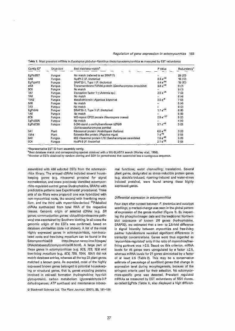
Regulation of gene expression in ectomycorrhiza 183
Table 1. Most prevalent mRNAs in Eucalyptus globulus-Pisolithus tinctorius ectomycorrhiza as measured by EST redundancy
Contig IDa Organism Best database matchb P value Redundancyc
EgPtdB57 Fungus No match (referred to as SRAP17) 29 (23)5A8 Fungus HydPt-2 (P. tinctorius) 8.6 e-50 19 (12)EgPtdA12 Fungus SRAP32-1, Type 1 (P. tinctorius) 4.4 e-94 18 (17)st54 Fungus Transmembrane FUN34 protein (Saccharomyces cerevisiae) 3.8 e-40 9(7)5C6 Fungus No match 9 (1)
7A7 Fungus Elongation factor 1-y (Artemia sp.) 2.5 e-2O 7 (3)7A6 Fungus No match 6 (4)11A2 Fungus Metallothionein (Agaricus bisporus) 3.5 e-5 7 (3)5B6 Fungus No match 5 (4)7A3 Fungus No match 6 (2)EgPtdA9 Fungus SRAP32-1, Type Il (P. tinctorius) 1.7 e-08 6 (6)1A6 Fungus No match 5 (0)6C8 Fungus WD-repeat CPC2 protein (Neurospora crassa) 2.8 e-127 5 (2)EgPtdB35 Fungus No match 4 (4)EgPtdC80 Fungus 0-(24)-sterol c-methyltransferase (ERG61 3.7 e-49 3 (3)
(Schizosaccharomyces pombe)5A1 Plant Ribosomal protein (Arabidopsis thaliana) 6.5 e-32 3 (0)13A4 Plant Extensin-Iike protein (Populus nigra) 7 e-42 3 (0)8A9 Fungus 60S ribosomal protein L10 (Saccharomyces cerevisiae) 7.5 e-1l3 3 (0)5C4 Fungus HydPt·3 (P. tlnctorius) 2.1 e-53 3 (0)
"Representative EST ID from assembly contig.b Best database match and corresponding species obtained with a WU-BLASTX search (Worley et al., 1998).cNumber of ESTs obtained by random c10ning and SSH (in parenthesesl that assembled into a contiguous sequence.
assembled with 486 selected ESTs from the ectomycorrhiza Iibrary. The arrayed cDNAs included several housekeeping genes (e.g. ribosomal proteins) for signalnormalization, and some previously identified ectomycorrhiza-regulated control genes (hydrophobins, SRAPs) withpredictable patterns (see Experimental procedures). Threesets of six filters were prepared: one was hybridized withnon-mycorrhizal roots, the second with free-Iiving myceIium, and the third with mycorrhiza-derived 32P-labelled
cDNAs synthesized from total RNA of the respectivetissues. Genomic origin of selected cDNAs (e.g. SR
genes; communication genes; ubiquitin/proteasome pathwayl was ascertained by Southern blotting. In ail cases thegenomic origin of the ESTs was confirmed, based ondatabase similarities (data not shown). A Iist of the mosthighly expressed genes in ectomycorrhizas, non-inoculated roots and free-Iiving mycelium can be found in theEctomycorrhizaDB (http://mycor.nancy.inra.fr/pages/DNAdatabases/EctomycorrhizaDB.html). A large part ofthese genes in ectomycorrhizas (e.g. 5C6, 7E9, 1E9) andfree-living mycelium (e.g. 5C6, 789, tOM, lOA 1) did not
match database entries, whereas ail the top 22 plant genesmatched a known gene. As expected, most of the highly
expressed known genes belonged to potential housekeeping or structural genes, that is, genes encoding proteinsinvolved in cell-wall formation (hydrophobins; hyp-richglycoprotein), carbon metabolism (glyceraldehyde-3-Pdehydrogenase; ATP synthase) and maintenance (riboso-
© Blackwell Science L1d, The Plant Journal, (2001), 25, 181-191
27
mal functions; water channelling; translation). Severalplant genes, designated as stress-inducible protein genes(e.g. abscisic-induced, ripening-induced and water-stressinduced proteins), were found among these highlyexpressed genes.
Differentiai expression in ectomycorrhiza
Four days after contact between P. tinctorius and eucalyptseedlings, a marked change was seen in the global patternof expression of the genes studied (Figure 1). By inspecting the phosphorimager data and the traditional Northernblot exposures of known SR genes (hydrophobins,SRAP32), we estimated that a two- to 2.5-fold differencein signal intensity between mycorrhiza and free-livingpartner hybridizations revealed significant differences intranscript concentrations. Genes were thus regarded as'mycorrhiza-regulated' only if the ratio of mycorrhiza/freeliving partners was ;;.2.5. Based on this criterion, mRNAlevels for 45 genes were upregulated by a factor ±2.5,whereas mRNA levels for 21 genes diminished by a factorof at least 2.5 (Table 2). This may be a conservativeestimate of percentage of symbiont genes that change inexpression level during morphogenesis, because of thestringent criteria used for their selection. No ectomycorrhiza-specific gene was detected. Prevalent regulatedmRNAs as measured by EST redundancy of SSH clones,so-called EgPtdx (Table 1), also displayed a high differen-

184 Catherine Vaiblet et al.
10' (a)
10'
10' 10' 10' ID'
Figure 1. Expression profiling of 400 genesbetween free-Iiving Pisofithus tinctor;us,non-mycorrhizal roots of Eucalyptusglobu/us and 4·day-old E. globu/us-P.tinctor/us ectomycorrhiza.For each gene, transcript levels (in arbitraryunits) were calculated for the free-livingmycelium (al: non-mycorrhizal roots lb):and ectomycorrhiza (x-axis in a and b) RNA,and displayed on a scatter plot. If the geneswere not affected by mycorrhizadevelopment, their transcript level wlli fallon the 45° line. Solid lines indicate 2.5·foldexpression differences between free-Iivingpartners and symbiotic tissues; dotted lines,10-fold expression differences. Clonescoding for the same gene are displayedonce.
<Il
13~> 0" 0- ....
ID'
Normalized mRNA levels in ectomycorrhiza
Normalized mRNA levels in ectomycorrhiza
10'
tial expression ratio on cDNA arrays (Table 2), confirmingthat SSH is an efficient procedure for c10ning differentiallyexpressed genes (Diatchenko etal., 1996). However, themost highly regulated genes, such as the fungal elF4A(clone 9C9) and the plant O-methyltransferase (OMT; clone8E9), were not found in the 200 SSH clones sequenced.
The highest differential expression (12-fold increase)detected was for a translation initiation factor elF4A gene('dead' box family helicases; Fischli etal., 1996) from P.tinctorius (clone 9C9). The translation elongation factor,tef1 (clone 6E1Dl. also showed an increased expression(3.4-fold) in the symbiotic mycelium. The known ectomycorrhiza-regulated genes, hydrophobins (Tagu etai., 1996)and SRAP32 (Laurent etaI., 1999), displayed the expected,mycorrhiza-enhanced expression pattern (between 2.5and 7.8-fold relative increases) (Figure 2). A strikingincrease (6.8-fold) was also observed for the transcripts
coding for the 14 a-methyl sterol demethylase (ERG11;clone 782). Interestingly, the 1).(24)-sterol c-methyltransferase (ERG6; contig EgPtdC80) was cloned three times bySSH, suggesting that ergosterol synthesis is increasedduring mycorrhiza formation. A strong increase (fourfold)was observed for the transcripts coding for a PisofithusWD-containing beta transducin (ud2B3), indicating thatsignalling pathway components might be transcriptionallyactivated in the ectomycorrhiza.
ln agreement with a previous study (Weiss etal., 1997)showing that ectomycorrhiza formation activates thephenylpropanoid pathway in short roots of larch, anelicitor-inducible isoflavone 7-G-methyltransferase (Heetai., 1998) (clone BE9) exhibited strong activation (6.8fold) in mycorrhizal eucalypt roots. The upregulation oftranscripts coding for components of the ubiquitin/proteasome pathway (clones 6E3 and 6C7) and stress proteins
© Blackwell Science Ltd, The Plant Journal, (2001). 25, 181-191
28
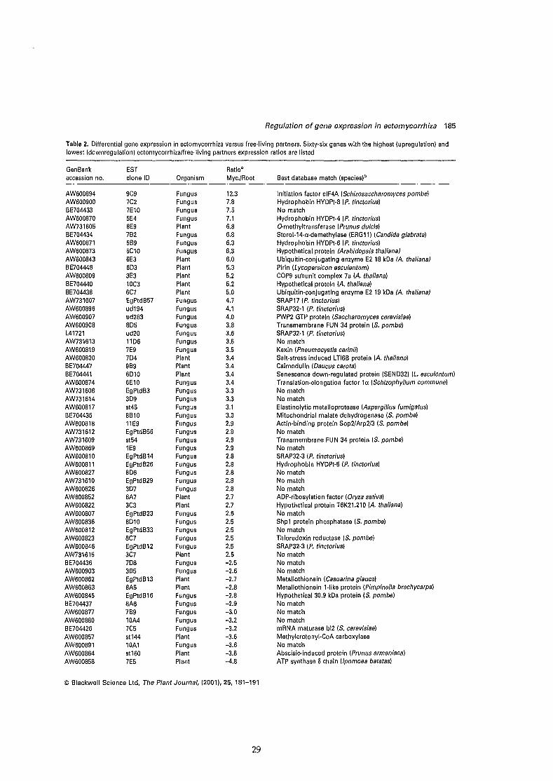
Regulation of gene expression in ectomycorrhiza 185
Table 2. Differentiai gene expression in ectomycorrhiza versus Iree-Iiving parlners. Sixty-six genes with the highest (upregulation) andlowest (downregulation) ectomycorrhiza/lree-Iiving parlners expression ratios are listed
GenBankaccession no.
AW600894AW600900BE704433AW600870AW731605BE704434AW600871AW600873AW600843BE704448AW600809BE704440BE704438AW731607AW600896AW600907AW600908L41721AW731613AW600819AW600820BE704447BE704441AW600874AW731606AW731614AW600817BE704435AW600818AW731612AW731609AW600869AW600810AW600811AW600827AW731610AW600826AW600852AW600822AW600807AW600836AW600812AW600823AW600846AW731615BE704436AW600903AW600862AW600863AW600845BE704437AW600877AW600880BE704426AW600857AW600891AW600864AW600858
ESTclone ID
9C97C27E105E48E97B25B95C106E36D33E310C36C7EgPtdB57ud194ud2838D5ud2011067E97D49B96D106E10EgPtdB33D9st458B1011E9EgPtdB56st541E9EgPtdB14EgPtdB268D6EgPtdB293D78A73C3EgPtdB238D10EgPtdB338C7EgPtdB123C77D83B5EgPtdB138A5EgPtdB168A67B910A47C5st14410A1st1607E5
Organism
FungusFungusFungusFungusPlantFungusFungusFungusPlantPlantPlantPlantPlantFungusFungusFungusFungusFungusFungusFungusPlantPlantPlantFungusFungusFungusFungusFungusFungusFungusFungusFungusFungusFungusFungusFungusFungusPlantPlantFungusFungusFungusFungusFungusPlantFungusFungusPlantPlantFungusFungusFungusFungusFungusPlantFungusPlantPlant
Ratio·Myc./Root
12.37.87.57.16.86.86.36.36.05.35.25.25.04.74.14.03.83.63.53.53.43.43.43.43.33.33.13.02.92.92.92.92.82.82.82.82.82.72.72.62.52.52.52.52.5-2.5-2.6-2.7-2.8-2.8-2.9-3.0-3.2-3.2-3.6-3.6-3.8-4.8
Best database match (species)b
Initiation factor elF4A (Schizosaccharomyces pombe)Hydrophobin HYDPt-8 (P. tinctorius)No matchHydrophobin HYDPt-4 (P. tinctorius)Q-methyltransferase (Prunus dulcis)Sterol-14-a-demethylase (ERG11) (Candida glabrata)Hydrophobin HYDPt-6 (P. tinctorius)Hypothetical protein (Arabidopsis thalianalUbiquitin'conjugating enzyme E2 18 kDa (A. thalianalPirin (Lycopersicon esculentum)COP9 subunit complex 7a (A. thaliana)Hypothetical protein (A. thaliana)Ubiquitin-conjugating enzyme E2 19 kDa lA. thalianalSRAP17 (P. tinctorius)SRAP32-1 (P. tinctorius)PWP2 GTP protein (Saccharomyces cerevisiae)Transmembrane FUN 34 protein (5. pombe)SRAP32-1 (P. tinctorius)No matchKexin (Pneumocystis carinil1Salt-stress induced LTI6B protein (A. thaliana)Calmodulin (Daucus carota)Senescence down-regulated protein (SEND32) (L. esculentum)Translation-elongation factor 1a (5chizophyllum commune)No matchNo matchElastinolytic metalloprotease (Aspergillus fumigatus)Mitochondrial malate dehydrogenase (S. pombe)Actin-binding protein Sop2/Arp2/3 (S. pombe)No matchTransmembrane FUN 34 protein (S. pombe)No matchSRAP32-3 IP. tinctorius)Hydrophobin HYDPt-5 (P. tinctorius)No matchNo matchNo matchADP-ribosylation factor (Oryza sat/va)Hypothetical protein T6K21.210 (A. thaliana)No matchShp1 protein phosphatase (S. pombe)No matchThioredoxin reductase (5. pombe)SRAP32-3 (P. tinctorius)No matchNo matchNo matchMetallothionein (Casuarina glauca)Metallothionein 1-like protein (Pimpinella brachycarpa)Hypothetical 30.9 kDa protein (5. pombe)No matchNo matchNo matchmRNA maturase bl2 (S. cerevisiae)Methylcrolonyl-CoA carboxylaseNo matchAbscisic·induced protein (Prunus armeniaca)ATP synthase Schain (lpomoea batatas)
© Blackvvell Science Lld, The Plant Journal, (2001), 25. 181-191
29

186 Catherine Voiblet et al.
Table 2. (continuedl
GenBank ESTaccession no. clone ID Organism
Ratioa
Myc./Root Best database match (species)b
AW600860BE704442BE704427AW731617BE704428BE704439BE704429
12C330212087A49A312045C9
PlantPlantFungusFungusFungusPlantFungus
-5.6-6.7-9.0-12.1-12.8-16.3-24.1
Polyubiquitin (A. thaliana)Cytochrome c oxidase subunit 6b-1 (O. sativa)Ring-box protein 1 (Homo sapiens)U6 snRNA-associated Sm-like protein LSm8 (S. pombe)NIPSNAP1 (Mus musculus)Zn-binding protein (A. thaliana)Rah1/Rad51 (Coprinus cinereusl
aRatio for normalized hybridization values of transcripts expressed in the symbiotic tissues and in the free-living partners.bBest database match (and corresponding species) obtained with a WU-BLASTX search at the Baylor College of Medecine Web server(Worley etaI., 1998).Only clones that produced a readable sequence are given.
(SEND32, LTI6B) is in agreement with the previouslyobserved degradation of plant proteins induced bymycorrhiza development (Burgess etal., 1995; Hilbertet al., 1991).
Within the known genes with a down-regulated expression, such as a Zn-binding protein (clone 12D4). polyubiquitine (clone 12C3). ATP synthase (0 chain) (clone 7E5)and metallothioneins (clones BAS and EgPtdB13), wereseveral plant genes. Remarkably, about one-third of thedifferentially expressed genes have no apparent homologyto any gene whose function is known.
Discussion
It is increasingly clear that developmental pathways leading to the ectomycorrhizal symbiosis can be considered asmodular, and that developmental transitions are accompanied by global changes in the expression of specificcomplements of genes (Martin and Tagu, 1999; Smith andRead, 1997). Hybridization to arrayed cDNA libraries permits simultaneous analysis of the level of thousands oftranscripts (Desprez etai., 1998; Eisen etai., 1998). In thepresent study, systematic sequencing of cDNA clones,SSH and cDNA mini-arrays were used to study the relativeabundance of transcripts corresponding to sets of clonesin the free-living P. tinctorius and E. glabulus, and in theectomycorrhizal symbiosis 4 days after contact. Thisinvestigation provided insights into the global gene activities involved in ectomycorrhiza development and function. About 17% of the genes represented by the arrayedESTs displayed ;;.2.5-fold differential expression duringsymbiosis morphogenesis. This may be a conservativeestimate of the percentage of E. globulus and P. tinctoriusgenes that change in expression level during early symbiosis development, because of the stringent criteria usedfor their selection. A striking result of the current data isthe fact that ail genes investigated are common to the nonsymbiotic and symbiotic stages. At the developmental
stage studied, symbiosis development does not induce theexpression of ectomycorrhiza-specific genes, but a markedchange in gene expression in the partners. The highestincreased expression detected was for the translation·initiation factor elF4A gene from the mycobiont. elF4A isboth a subunit of a high molecular-weight protein complexinvolved in m3G cap recognition, and is required as asingle polypeptide chain for mRNA binding to ribosome(Fischli etai., 199B}.lt is an ATP-dependent single-strandedRNA-binding protein with a sequence-independentunwinding activity (helicase), with similarity to other'dead' box family helicases. In yeast, elF4A is essentialfor translation and cell growth (Dominguez etal., 1999).Upregulation of elF4A and tef1 suggests that gene expression is significantly regulated at the level of proteinsynthesis in the symbiosis through translation initiation.This is in agreement with the observed high density oftranscripts (Camero Diaz etai., 1997) and ribosomes(Massicotte et al., 1987), and changes observed in proteinsynthesis (Burgess etai., 1995; Hilbert etai., 1991) inEucalyptus-Pisalithus ectomycorrhiza.
Within SR genes, a large set of sequences code forseveral members of the multi-gene SRAP32 (Laurent etai.,1999) and hydrophobin (Tagu et al., 1996) families. Itappears that the expression of these proteins is strikinglyupregulated (up to 4.7-fold) during mycorrhiza formation(Tables 1 and 2), confirming and extending our previousdata. The array data reflect the relative abundance of thetranscripts, which is influenced by both transcriptionalactivity and mRNA stability. It does not detect any regulation at the translational and post-translational levels.Comparing the upregulation of SRAP32 transcript levelsand the increased concentration of SRAP32 polypeptidesin ectomycorrhiza (Burgess etai., 1995; Hilbert etai., 1991;Laurent etai., 1999) showed that there was a goodcorrelation between changes in protein synthesis andtranscript levels. The cell wall-related clones included twodifferent members of the SRAPfamily (types 1having 80-
© Blackwell Science Lld, The Plant Journal, (2001),25,181-191
30
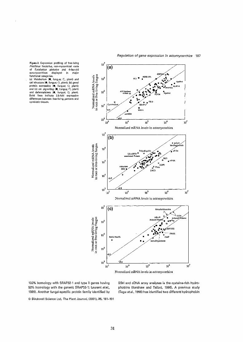
Regulation of gene expression in ectomvcorrhiza 187
7
la (a)
NIPSNAPt
•BC6 •
G3PD alb /., .--"..0/ HdPl"
A-'~ •• y• :.. ...... • .SRAP32., HydPl-6
ATP5ynt1w< • ~ ~\\~~( •dchain D là • FUN34 HydPI-8
~. ~'\: //. C 'j. /'
C ///1;I •• ?BIOCrt<7 .SRAPt7
7' ERGII
~~IMDH103 '--__-'x"'2.""5 -'
~ ~ ~ ~ ~
~ ~I)106
> "" "- ......«"'l&.~8'7 105.,,8~~
'asê~
o e 104
Z.E:
Figure 2. Expression profiling of free·livingPisolithus tinctorius, non·mycorrhizal rootsof Eucalyptus globulus and 4·day·oldectomycorrhizas displayed in majorfunctional categories.(al Metabolism 1., fungus; 0, plant) andcell structure (e, fungus; 0, plant); (b) genetprotein expression (e, fungus; 0, plant);and (c) cell signalling (., fungus; 0, plant)and defence/stress (e, fungus; 0, plantl.Solid Iines indicate 2.S·fold expressiondifferences between free'living partners andsymblotic tissues.
Nonnalized mRNA levels in eclomycorrhiza
107 r-::-....,....----------------;7~__,(b)
malurase(bI2)
~~10
6> "" "- ......« bOZ.E:cG>8'7
105]~N ......
~Sêë0:2 10
4Z.E:
,• polyA /
bin~Pt6teln/
Polyublquitin • / eF·laU6,nRNA· 0 00'" ~
assoclated Proleln __ •• .- ~ -.-.::li ./' tOC3
o .~..'f ~ 0 • elF4A
• ~04! COP9" .~. 00 Plrin• UBC2
Normalized mRNA levels in eclomycorrhiza
,,' .//".~
Iùhl/Rad51 • -
MelallOlhKnein/
"y --\ABAABA 0 • Induced teins
lndU~Pro.J -Z._. 1_ . SEND32
.-. 1 • .;"'" PWP2
! OM\"
Metalloprolease
Nonnalized mRNA levels in eclomycorrhiza
100% homology with SRAP32·1 and type II genes having
50% homology with the generic SRAP32-1; Laurent etaI.,1999), Another fungal-specific protein family identified by
SSH and cDNA array analyses is the cysteine-rich hydro
phobins (Kershaw and Talbot, 1998). A previous study(Tagu etaI., 1996) has identified two different hydrophobin
© Blackwell Science Ltd. The Plant Journal, (2001),25,181-191
31

188 Catherine Voiblet et al.
transcripts in ectomycorrhiza, HydPt-1 and HydPt-2. Herewe have identified additional hydrophobin genes, HydPt-3ta HydPt-B, which show 47-52% homology with other P.tinctorius hydrophobins (unpublished results). The expression of 5B9, 5E4 and 7C2, which are coding for variousparalogues of HydPt-2, is increased six- ta eightfold in thesymbiotic tissues (Table 2; Figure 2). Eucalypt ectomycorrhizas are often found in air pockets in sail in the wild.Such mycorrhizas formed in air are invariably nonwettable and water-repellent (Vesk etal., 2000). The mostlikely expianation for this lies in the observed deposition ofhydrophobins. Thus it appears that P. tinctorius hyphaepreferentially express some sets of structural proteins inthe wall during the formation of the symbiosis, confirmingthat for fungi interacting with plants the cell wall and itssurface are major players in the host-symbiont interface(Hardham and Mitchell, 1998; Martin etai., 1999). Theconcerted induction of these structural proteins impliestight control of expression among genes with potentiallyrelated functions, and opens the door for comparativestudies using the conserved elements in the regulatoryregions of these genes. Within the structural proteinscoded by SR genes are proteins involved in membranesynthesis and function: the transmembrane FUN34protein, ERG6 and ERG11. This supports an earlier report(Kottke and Oberwinkler, 1990) suggesting that symbiosisdevelopment leads ta an increased membrane formation.
Several genes with a putative function in signaltransduction are highly expressed and upregulated insymbiotic tissues; they represent ",13% of the c10nedgenes. These include genes with homology ta calmodulin,ras, heterotrimeric GTPases, and protein kinases(MAPKKK, MAPK). The expression of the P. tinctoriushomologue (ud283) of the PWP2 family of WD-repeatproteins involved in yeast cell separation (Shafaatian etal.,1996) is upregulated during mycorrhiza formation(Table 2).
The increased accumulation of an elicitor-inducibleisoflavone 7-0-methyltransferase (He etal., 1998) andstress-inducible proteins (LTI6B, SEND32) confirmed thata transient increase in plant defence/stress responses ismounted in eucalypt raots ta control fungal ingress(Feugey etaI., 1999). Several of the plant-regulated genesshowed a decreased expression in ectomycorrhizal tissues(e.g. metallothionein; ATP synthase; cytochrome c oxidase; Zn-binding protein), confirming previous results thatthe symbiosis formation induces downregulation ofprotein synthesis in colonized raots (Burgess et al., 1995;Hilbert etal., 1991). Degradation of these proteins probablyinvolved the ubiquitin/proteasome pathway, explainingthe increased expression of plant ubiquitin-conjugatingenzymes (clones 6E3 and 6C7).
Taken together, our results demonstrate the utility ofESTs, SSH and cDNA arrays in determining the major
changes taking place during symbiosis development. Thisglimpse into the complex ectomycorrhiza gene expressionwill provide the basis for a more precise moleculardissection of the complex genetic networks that controlsymbiosis development and function. The identified SRgenes might be especially interesting targets for ongoinggene disruption technology. Further studies are nowneeded ta delineate the functions of bath known andnovel genes that are differentially expressed duringectomycorrhiza development.
Experimental procedures
Biological materials and synthesis of ectomycorrhizas
Seeds of Eucalyptus globulus ssp. bicostata Kirkp. were sterilizedwith 20% calcium hypochlorite (v/v) for 20 min, rinsed with fourchanges of sterile water and plated onto low-sugar (5 mM)Pachlewski medium in 2.0% agar (Nehls etaI., 1998). After7 days, aseptically germinated seedlings were laid onto theedge of 21-day-old fungal mats of the ectomycorrhizal gasteromycete Pisolithus tinctorius Coker & Couch (synonym Pisolithusarhizus (Scop. per Pers.) Rauschert), isolate 441, grown on lowsugar Pachlewski medium in 2.0% agar, and left for 4 days in acontrolled enviranment growth cham ber with 16 h Iight (25°C,150 ilmol m-2 s-') and 8 h dark (Hilbert etai., 1991; Nehls etaI.,1998) for ectomycorrhiza formation. Non-inoculated and 4-dayold ectomycorrhizal seedlings, together with the edge of 25-dayold fungal mats, were then sampied, fixed in liquid N2 and storedat -so'e.
Construction of a cDNA Iibrary of ectomycorrhiza
Total RNA was isolated from 4-day-old ectomycorrhizas according to Boift!n et al. (1998). The RNA source material for the librarycontained the primary root and lateral root tips, inciuding themeristem and reot-cap cells. Most raots were ensheathed by theectomycorrhizal mantle with difterentiated inner and outer layers(Figure 1 of Martin etal., 1999). Formation of the Hartig net withhyphae growing anticlinally between rhizodermal cells wasinitiated. Thus ail the major plant and fungal cell types and
.processes related to the early stages of symbiosis were represented. At this stage, fungal transcripts represented ",65% of totalectomycorrhiza RNA (Camero Diaz etai., 1997).
The cDNA Iibrary was constructed using the CapFinder cDNAIibrary construction kit (Clontech, Palo Alto, CA, USA) accordingto the manufacturer's instructions. This PCR-based methodpraduces high-quality cDNA libraries containing a high proportion of full-Iength cDNA clones (Sasaki et al., 1998). cDNAs werecloned into EcoRI-digested pBluescript and À.gtl1 to generate1.6 x 104 and 2.5 x 10⁵ recombinants, respectively. Sequenceanalysis revealed that 85% of the clones contained the initiationcodon.
Suppression subtractive hybridization (SSH)
Doubled·stranded cDNAs corresponding to mRNAs expressed inE. globulus-P. tinctorius ectomycorrhizas after 4 days' contact(tester probe), and cDNAs from E. globulus raots and P. tinctoriusfree-Iiving mycelium, were separiltely obtained by using the
© Blackwell Science Ltd, The Plant Journal, (2001), 25, 181-191
32
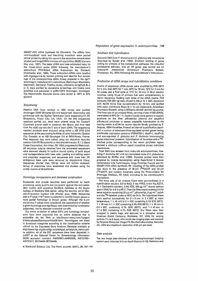
SMART-PCR cDNA Synthesis Kit (Clontech). The cDNAs fromnon-inoculated roots and free-Iiving mycelium were pooled(driver probe) by taking into account the respective concentrationof plant and fungal RNAs in 4-day-old mycorrhiza (35/65) (CarneroDiaz etal., 1997). The tester cDNA pool was subtracted twice bythe mixed-driver probe (SSH) following the manufacturer'sinstructions (PCR-Seiect cDNA Subtraction Kit, Clontech)(Diatchenko etal., 1996). These subtracted cDNAs were labelledwith digoxigenine by random priming and used for four screenings of the ectomycorrhiza cDNA Iibrary prepared in the )"gt11according to manufacturer's instructions (Boehringer-Mannheim,Mannheim, Germany). Positive clones, referred to as EgPtdA. B, Cor D, were purified by successive screenings, and inserts wereamplified and subcloned in pCR4-TOPO (Invitrogen, Groningen,The Netherlands). Bacterial clones were stored at -80°C in 35%glycerol.
Sequencing
Plasmid DNA from random or SSH clones was purified(Invitrogen SNAP Miniprep Kit) and sequenced. Sequencing wasperformed with the BigDye Terminator Cycle sequencing kit (PEBiosystems, Foster City, CA. USA). On the 940 sequencingreactions carried out, most were primed using the ClontechCapswitch primer used to construct the Iibrary, and thus readfrom the m3G cap into the 5' end of each cDNA. The sequencingreaction products were analysed using either a ABI 373S DNAsequencer at the sequencing facilities of Laval University (QuebecCity, Canada), or an ABI Genotyper 310 at INRA-Nancy, France.Raw sequence data were edited using the SEQUENCE ANALYSIS(version 3.3) (PE Biosystems) or SEQUENCHER (version 3.1.1) (GeneCodes Corporation, Ann Arbor, MI, USA) programs for Macintosh.Ali sequence outputs obtained from the automated sequencerswere scanned visually to confirm overall quality of peak shapeand correspondence with base calls. Leading and trailing vectorand polylinker sequences, and sequences with more than 3%ambiguous base calls were removed by SEQUENCHER filters.Sequences shorter than 100 bp were not further analysed.Group of sequences were assembled into clusters using thecontig routine of SEQUENCHER.
Hom%gy comparisons and database construction
Nucleotide and protein searches were performed by batchprocessing using aLASTN AND WU-aLASTX against the non-redun·dant nucleic acid sequence GenBank database at the BaylorCollege of Medicine Web server, using the MACPERL script Macsearch-Iauncher (version 2.6) (Worley etai., 1998). Sequenceswith a Pvalue ;;>10-6 were considered to identify known genes orhave partial homology to known genes. Although the aLASTscores and Pvalues were considered, the assessment of whethera given homology was significant was determined by investigatorjudgement, not by absolute numerical cut-offs.
The entire collection of edited ESTs and homology compari·sons have been organized into an online database that isaccessible via the Web at http://mycor.nancy.inra.fr/pages/DNAdatabases/EctomycorrhizaDB.html. Homologues are listedin functional categories according to the classification schemeutilized by the Expressed Gene Anatomy Database available athttp://www.tigr.org/docs/tigr-scripts/egad_scripts/role_report.spl).ln addition, ail of the EST sequences have been deposited indbEST at the National Center for Biotechnology Information(NCBI accession numbers AW600807-AW600908, AW731605AW731617, BE704426-BE704449).
© Blackwell Science Ud, The Plant Journal, (2001),25,181-191
33
Regulation of gene expression in ectomycorrhiza 189
Southern b/ot hybridization
Genomic DNA from P. tinctorius and E. globu/us was extracted asdescribed by Guidet etai. (1990). Southern blotting of genescoding for proteins of the transduction pathways, the ubiquitinlproteasome pathway, and ail SR genes was carried out onHybond-W membranes (Amersham Pharmacia Biotech,Piscataway, NJ, USA) following the manufacturer's instructions.
Production of cDNA arrays and hybridization conditions
Inserts of sequenced cDNA clones were amplified by PCR (95°Cfor 5 min, then 95°C for 1 min, 60°C for 30 sec, noc for 3 min for30 cycles, and a final cycle at noc for 15 min) in 50 J.l1 reactionvolumes, using 10 J.lM of primers that were complementary tovector sequences flanking both sides of the cDNA inserts. PCRproducts (150-300 ng) were diluted in 200 J.l110 X SSC, denaturedwith NaOH (0.2 M final concentration) for 10 min, and spottedonto nylon membrane filters (8 X 12 cm) (Hybond-N, AmershamPharmacia Biotech) using a Millipore 96-well dot·blot apparatus.The three sets of six arrayed filters, carrying a total of 560 cDNAs,were baked at 80°C for 1 h. Control clones were spotted at specificpositions on the fil ter: pBluescript plasmid and amplifiedpBluescript polylinker to assess unspecific hybridization to thecloning vector; a cONA for human desmin (kindly provided by T.Desprez, INRA·Versailles, France) as internai quantification standard; a number of ectomycorrhiza·regulated control genes havingpredictable expression patterns (PtSRAP32-1, HydPt-l, HydPt-2);and non-regulated E. globulus and P. tinctorius housekeepinggenes (ribosomal proteins). Hybridization of the arrays with a 32p.labelled probe corresponding to the vector polylinker regionshowed a relatively uniform signal intensities across individualmembranes.
Total RNA was isolated from 4-day-old ectomycorrhizas, free·living P. tinctorius 441 and non·inoculated roots of E. glabulus asdescribed by Boiffin etal. (1998). Complex probes were thenprepared by reverse transcription using SuperScript Il reversetranscriptase (Life Technologies, Cergy Pontoise, France) and theSMART-PCR cDNA Synthesis Kit. Labelling of the cONA probeswas done in the presence of 30 J.lCi 132PldCTP and 30 J.lCi[32PldATP, and randam hexamers using the Prime-a-Gene Kit(Promega, Madison, WI, USA) according to the manufacturer'sinstructions.
The three sets of six arrayed filters were pre-incubated in ahybridization solution (0.6 M NaCI, 2 mM PIPES, 5 mM Na2 EDTA,10 X Denhardt's solution, 0.5% SOS, 500 J.lg mr1 shared salmonsperm ONA) for 4-8 h at65°C. Then the filters were incubated in thesame solution containing 20 J.lg ml-1 pBluescript, 2 J.lg mr' polyAand the 32P-labelled probe at 65°C for 24 h. The hybridized filterswere washed successively for 3 x 5 min in 2 X SSC at roomtemperature, 1 X 40 min in 2 X SSC containing 0.1% SOS (65°C),1 x 40 min in 1 X SSC containing 0.1% SOS (65°C), 1 x 40 min in0.5 X SSC containing 0.1% SOS (65'C), and 1 X 40 min in0.1 X SSC containing 0.1% SOS (65°C). Dry filters were thenwrapped in plastic bags and exposed to a phosphor screen(Eastman Kodak Company, Rochester, NY, USA) for varyingperiods (1 h to 3 days), alter which the imaging plate was scannedby Personal Molecular Imager FX (Bio·Rad Laboratories, Hercules,CA. USA) at a maximum resolution of 50 J.lm per pixel.
Data ana/ysis
The raw image data obtained with the phosphorimager imagingsystem were imported into an Applè Macintosh G3. Detection and
190 Catherine Vo/blet et al.
quantification of the signais representing hybridized DNA wereperformed using the volume quantification method of theQUANTITY ONE software (Bio-Rad). Each spot was defined bymanual positioning of a grid of squares over the array image. Foreach image, the average pixel intensity within each square wasdetermined. Local background value for each membrane wascalculated on the basis of five positions with no DNA-spottedarea. Net signal was determined by subtraction of this meanbackground from the intensity for each spot. Spots deemedunsuitable for accurate quantification because of array artefactwere manually flagged and excluded from further analysis. Thedata table generated by QUANTITY ONE, containing the intensity ofeach of the spots, was then exported to the EXCEL 98 worksheetprogramme (Microsoft Corporation, Redmond, WA, USA) forfurther manipulation. The probe-to-probe variance was filteredout using signal intensities of the human desmin spotted at sixlocations on the filters (interfilter normalization). In addition, totake account of experimental vari"ations in specifie activity of thecDNA probe preparations or exposure time that might alter thesignal intensity, the data obtained from different hybridizationswere normalized by dividing the intensity for each (fungal orplant) dot by the average of the intensities of ail the (fungal orplant) dots present on the filter, to obtain a centred, normalizedvalue (Eisen et al., 1998). To determine the relative levels of plantand fungal transcripts in the intermingling mycorrhiza cDNAprobe, the total intensity was determined for ail the dotscorresponding to either fungal or plant cDNAs hybridized withthe mycorrhiza cDNA probe. Based on these values, the proportion of fungaltranscripts in 4-day-oid ectomycorrhiza was 65% ofthe total symbiosis transcripts, in agreement with our previousmeasurements carried out using the rDNA internai transcribedspacers (Carnero Diaz et al., 1997). Ali the expression data areavailable at the EctomycorrhizaDB web site (hllp:/lmycor.nancy.inra.fr/pages/DNAdatabases/EctomycorrhizaDB.html).
To determine the reproducibility of hybridizations in whichRNAs are present at a wide range of concentrations, wehybridized three copies of a tilter arrayed using cDNAs codingfor various components of the transduction pathways, SR genes(hydrophobins, SRAP32), and housekeeping genes (ribosomalproteins, elongation factors) with two labelled complex probesprepared from the same RNA fungal extract. The filter-to-filtervariation was relatively small (±10%) (data not shown). To assessthe variation due to sources other than the hybridization andreading steps, we hybridized samples independently preparedfrom two different fungal cultures grown in the same conditions.The sample-to-sample variation was slightly larger (±15%) thanobserved in the simple hybridization reproducibility experiments.
Acknowledgements
Catherine Voiblet and Sébastien Duplessis were supported byDoctoral Scholarships from the Ministère de l'EducationNationale, de la Recherche et de la Technologie. We would liketo thank Denis Tagu (INRA-Nancy) and Yves Brygoo (lNRAVersailles) for valuable discussions during the course of thisstudy. We are grateful to Uwe Nehls and Murielle Mourer (lNRANancy) for obtaining some of the ESTs in the preliminary stage ofthis project. The assistance of Murielle Mourer in the preparationof plants and isolation of RNA for the construction of the Iibrary isacknowledged. Finally, many thanks to Luc Simon (University ofLaval, Québec) and Christine Delaruelle (INRA-Nancy) for theirdedicated assistance in sequencing, and Béatrice Palin (lNRANancy) for assistance in hybridizations. The present investigationwas supported by grants from the Groupement de Recherches et
d'Etude des Génomes and the INRA (Action TransversaleMicrobiologie Fondamentale). The research utilized in part theDNA Sequencing Facilities at INRA-Nancy financed by the INRAand Région de Lorraine.
References
Boiffin, V., Hodges, M., Galvez, S., Balestrini, R., Bonfante, P.,Gadal, P. and Martin, F. (1998) Eucalypt NADP-dependentisocitrate dehydrogenase: cDNA c10ning and expression inectomycorrhiza. Plant Phys/ol. 117,939-948.
Burgess, T., Laurent, P., Dell, B., Malajczuk, N. and Martin, F.(1995) Effect of the fungal isolate aggressivity on thebiosynthesis of symbiosis-related polypeptides indifferentiating eucalypt ectomycorrhiza. Planta, 195,408-417.
Carnero Diaz, M.E., Martin, F. and Tagu, D. (1996) Eucalypt (1.
tubulin: cDNA cloning and increased level of transcripts inectomycorrhizal root system. Plant Mol. Biol. 31, 905-910.
Carnero Diaz, M.E., Tagu, D. and Martin, F. (1997) Ribosomal DNAinternai transcribed spacers ta estimate the proportion ofPisolithus tinctorius and Eucalyptus globulus RNAs inectomycorrhiza. Appl. Env. Microbiol. 63, 840-843.
Diatchenko, L., Lau, Y.F.C., Campbell, A.P. et al. (1996)Suppression subtractive hybridization: a method forgenerating differentially regulated or tissue-specifie cDNAprobes and libraries. Proc. Natl Acad. Sei. USA, 93, 6025-6030.
Desprez, T., Amselem, J., Caboche, M. and Hotte, H. (1998)Differentiai gene expression in Arabidopsis monitored usingcDNA arrays. Plant J. 14, 643-652.
Dominguez, O., Altmann, M., Benz, J., Baumann, U. and Trachsel,H. (1999) Interaction of translation initiation factor elF4G withelF4A in the yeast Saccharomyces cerevisiae. J. Biol. Chem.274, 26720-26726.
Eisen, M.B" Spellman, P.T., Brown, P.O. and Botstein, D. (1998)Cluster analysis and display of genome-wide expressionpatterns. Proc. Natl Acad. Sei. USA, 95, 14863-14868.
Feugey, L., Strullu, D.G., Poupard, P. and Simoneau, P. (1999)Induced defence responses limit Hartig net formation inectomycorhizal birch roots. New Phytol. 144, 541-547.
Fischll, A., Schmid, S.R., Coppolecchia, R. and Linder, P. (1996)The translation initiation factor elF4A fromSchizosaccharomyces pombe Is closely related to itsmammalian counterpart. Yeast, 12, 977-981.
Guidet, F., Rogowsky, P. and Langridge, P. (1990) A rapid methodof preparing megabase plant DNA. Nue!. Acids Res. 18,4955-4957.
Gyorgyey, J., Vaubert, O., Jiménez·Zurdo, J.I., Charon, C.,Troussard, L., Kondorosi, A. and Kondorosi, E. (2000) Analysisof Med/cago truncatula nodule expressed sequence tags. Mol.Plant-Microb. Interact. 13, 62-71.
Hardham, A.R. and Mitchell, H.J. (1998) Use of molecularcytologyto study the structure and biology of phytopathogenic andmycorrhizal fungi. Fungal Genet. Biol. 24, 252-284.
He, X.Z., Reddy, J.T. and Dixon, RA (1998) Stress responses inalfalfa (Medicago sativa L). XXII. cDNA c10ning andcharacterization of an elicitor-inducible isoflavone 7-Q.methyitransferase. Plant Mol. Biol. 36, 43-54.
Hervé, C., Perret, E., Trémousaygue, D. and Lescure, B. (1996)Differentiai screening using high density filters reveals adramatic change in gene expression at different growth phasein Arabidopsis cell suspension cultures. Plant Physiol.Biochem. 34, 425-430.
Hilbert, J.L., Costa, G. and Martin, F. (1991) Ectomycorrhizin
© Blackwell Science Ltd, The Plant Journal, (2001), 25, 181-191
34

synthesis and polypeptide changes during the early stage ofeucalypt mycorrhiza development. Plant Physiol. 97. 977-984.
Holte, H., Desprez, T., Amselem, J. et al. (1993) An inventory of1152 expressed sequence tags obtained by partial sequencingof cDNAs from Arabidopsis thaliana. Plant J. 4.1051-1061,
Kershaw, M.J. and Talbot. N.J. (1998) Hydrophobins andrepellents: proteins with fundamental roles in fungalmorphogenesis. Fungal Genet. Biol. 23. 18-33.
Kim. S.J., Bernreuther. O., Thumm. M. and Podila. G.K. (1999) LBAUT7. a novel symbiosis-regulated gene from anectomycorrhizal fungus, Laccaria bicolor, is functionallyrelated to vesicular transport and autophagocytosis. J. Bac.181. 1963-1967.
Kottke, 1. and Oberwinkler, F. (1990) Amplification of root-fungusinterface in ectomycorrhizae by Hartig net architecture. Ann.Sei. For. 46. 737s-740s.
Laurent, P., Voiblet, C., Tagu, D. et al. (1999) A novel class ofectomycorrhiza-regulated cell wall polypeptides in Pisolithustinctorius. Mol. Plant-Microb. Interact. 12. 862-871,
Martin, F. and Hilbert, J.L. (1991) Morphological, biochemical andmolecular changes during ectomycorrhiza development.Experientia. 47, 321-331.
Martin, F. and Tagu, D. (1999) Developmental biology of a plantfungus symbiosis: the ectomycorrhiza. In Mycorrhiza:Structure. Function. Molecular Biology and Biotechnology(Varma, A.K. and Hock. B., eds). Berlin: Springer-Verlag, pp.51-73.
Martin, F., Lapeyrie, F. and Tagu, D. (1997) Altered GeneExpression during Ectomycorrhiza Development ln theMycota. Vol. VI. Plant Relationships (Lemke. P. and Caroll, G.,eds). Berlin: Springer-Verlag, pp. 223-242.
Martin, F., Laurent, P., De Carvalho, O., Voiblet, C., Balestrini, R.,Bonfante, P. and Tagu, D. (1999) Cell wall proteins of theectomycorrhizal basidiomycete Pisolithus tinctorius:identification, function. and expression in symbiosis. FungalGenet. Biol. 27, 161-174.
Massicotte, H.B., Peterson, R.L., Ackerley, C.A. and Ashford, A.E.(1987) Ontogeny of Eucalyptus pilularis-Piso/lthus tinctoriusectomycorrhizae. II. Transmission electron microscopy. Cano J.Bot. 65, 1940-1947.
Nehls. U.• Béguiristain, T., Ditengou, F., Lapeyrie, F. and Martin, F.(1998) The expression of a symbiosis-regulated gene ineucalypt roots is regulated by auxins and hypaphorine. thetryptophan betaine of the ectomycorrhizal basidiomycetePisolithus tinctorius. Planta, 207, 296--302.
Nehls, U., Mikolajewski, S., Ecke, M. and Hampp, R. (1999)Identification and expression-analysis of two fungal cDNAsregulated by ectomycorrhiza and fruit body formation. NewPhytol. 144, 195-202.
Regulation of gene expression in ectomycorrhiza 191
Newman, T., De Bruijn, F.J., Green, P. et al. (1994) Genes galore: asummary of methods for accessing results from large-scalepartial sequencing of anonymous Arabidopsis cDNA clones.Plant Physio/. 106, 1241-1255.
Peterson, R.L. and Bonfante, P. (1994) Comparative structure ofvesicular-arbuscular mycorrhizas and ectomycorrhizas. PlantSail. 159, 79-88.
Reymond, P., Weber, H., Damond. M. and Farmer, E.E. (2000)Differentiai gene expression in response to mechanicalwounding and insect feeding. Plant Cell, 12. 707-719.
Sasaki, N., Nagaoka, S., Itoh, M. et al. (1998) Characterization ofgene expression in mouse blastocyst using single-passsequencing of 3995 clones. Genomics. 49, 167-179.
Shafaatian, R., Payton, MA and Reid, J.O. (1996) PWP2, amember of the WD-repeat family of proteins, is an essentialSaccharomyces cerevisiae gene involved in cell separation.Mol. Gen. Genet. 252.101-114.
Schena, M., Shalon, O., Davis, R.W. and Brown, P.O. (1995)Quantitative monitoring of gene expression patterns with acomplementary DNA microarray. Science, 270. 467-470.
Schena, M., Shalon, O., Helier, R., Chai, A., Brown, P.O. and Davis,R.W. (1996) Parallel human genome analysis: microarray-basedexpression monitoring of 1000 genes. Proc. Natl Acad. Sei.USA, 93. 10614-10619.
Smith, S.E. and Read, D.J. (1997) Mycorrhizal Symbiosis. 2nd edn.London: Academie Press.
Tagu, O., Nasse, B. and Martin, F. (1996) Cloning andcharacterization of hydrophobins encoding cDNAs from theectomycorrhizal basidiomycete Pisolithus tinctorius. Gene, 168,93-97.
Vesk, P.A., Ashford, A.E., Markovina, A.-L. and Allaway, W.G.(2000) Apoplasmic barriers and their significance in theexodermis and sheath of Eucalyptus pilularis-Pisolithustinctorius ectomycorrhizas. New Phyto/. 145. 333-346.
Weiss, M., Mikolajewski, S., Pelpp, H., Schmitt, U., Schmidt, J.,Wray, V. and Strack, D. (1997) Tissue-specifie anddevelopment-dependent accumulation of phenylpropanoids inlarch mycorrhizas. Plant Physiol. 114, 15-27.
Wodicka, L., Dong, H.L., Mittmann, M., Ho, M.H. and Lockhart,D.J. (1997) Genome-wide expression monitoring inSaccharomyces cerevisiae. Nature Biotech. 15, 1359-1367.
Worley, K.C., Culpepper, P.A., Wiese, B.A. and Smith, R.F. (1998)8EAUTY-X: enhanced BLAST searches for DNA queries.Bioinformatics, 14, 890-891.
Wright. D.P., Scholes, J.O., Read, D.J. and Rolfe, S.A. (2000)Changes in carbon ailocation and expression of carbontransporter genes in Betula pendula Roth. colonized by theectomycorrhizal fungus Paxillus involutus (Batsch) Fr. Plant CellEnv. 23, 39-49.
GenBank accession numbers AW600a07-AW60090a, AW73160S-AW731617 and BE704426-BE704449.
Supplementary Material
A table entitled: 'Highly abundant transcripts in Eucalyptus globulus-Pisolithus tinctorius ectomycorrhiza. free-livingmycelium and non-mycorrhizal raots', the EST sequences and the WU-BLASTX files are available at http://mycor.nancy.
inra.fr/pages/DNAdatabases/EctomycorrhizaDB.html/.
© Blackwell Science Lld, The Plant Journal, (2001),25,181-191
3S

cDNA array aRa~ysÏ!s ofectomycorrl1.iza deveIopment
Manuscrit en préparation

cDNA Array Analysis of Ectomycorrhiza Development
Sébastien DUPLESSIS & Francis MARTIN!
Unité Mixte de Recherche INRA/UHP 1136 "Interactions Arbres/Microorganismes", Institut National de la
Recherche Agronomique, Centre de Recherches de Nancy, 54280 Champenoux, France
Received 00 July 2000
Running title: Gene Expression during Ectomycorrhiza Development
Accession numbers:
* For correspondence (Fax +33 383 39 40 69; e-mail: [email protected]).
36

Summary
Development of ectomycorrhiza induces transcriptional reprogramming in the fungal and plant
symbionts. We monitored changes in gene expression in Eucalyptus globulus/Pisolithus tinctorius
ectomycorrhiza during the different stages of the symbiosis development using cDNAs
macroarrays. Comparisons of signaIs from free-living partners and symbiotic tissues revealed
significant differences in the expression levels (differential expression ratio> 2.5) for 17% to 24%
of the genes analyzed depending on the development stages. We derived groups of coordinately
expressed genes (i.e. regulons) using hierarchical clustering and Self organizing Maps. At least a
dozen of distinct temporal patterns of induction/repression were observed. The main fungal
regulons contained genes coding for ceIl-waIl and membrane proteins, communication genes, and
metallothionein-related poteins. In the host root, a major down-regulated regulon comprised genes
involved in water transport and stress suggestîng that mycorrhiza development improves water
uptake. Genes coding for mitochondrial respiration and protein translation were up-regulated. The
temporal expression pattern provided cIues to potential functions of several of previously
uncharacterized genes (e.g. SRAP17).
Key words: cDNA arrays; cell wall proteins; ectomycorrhiza; expressed sequence tags; suppression
subtractive hybridization; symbiosis-regulated genes
37

INTRODUCTION
Ectomycorrhizas are characterized structurally by (i) the presence of an extensive extramatrical
mycelial web prospecting the soil and gathering nutrients, (ii) a mantle of fungal hyphœ ensheating
the root and mainly acting as a storage compartment, and (iii) a network of hyphœ growing in the
apoplastic space of the rhizodermis (in angiosperms) and cortex (in conifers) root ceHs so-caHed Hartig
net. The fungus gains access to sugars from the plant while plant nutrient uptake is mediated via the
fungus. In addition, the establishment of the symbiosis is requested for the completion of the fungal
life cycle (Le., formation of fruiting bodies). The formation of the symbiosis requires several days and
induces major morphological changes including a novel spatial tissue organization, changes in ceH
shape, and the generation of different ceH types (Kottke & Oberwinkler, 1987; Massicote et al., 1987).
Differences in organogenetic programmes are reflected in variations in ectomycorrhizal root
morphology, but overal1 there are pronounced developmental similarities suggesting that key
developmental programmes are triggered in both symbionts. Symbiosis providing an additionallayer
of complexity to the chal1enge of designing experimental systems and framing questions, it was
relatively difficult to study these primary genetic determinants. For many years, most physiological
analyses were focused on limited sections of the complex metabolic networks taking place in the
symbionts (Martin and Botton, 1993; Hampp et al., 1995; Gianinazzi-Pearson, 1996; Harrison, 1998),
which limited the degree to which symbiosis behavior can be understood. The comprehensive
methods of analysis of functional genomics are now employed on a handful mycorrhizal associations
at several levels: those of the genome, transcriptome, proteome, and metabolome. A surge of
investigations based on these novel approaches have al10wed ta assess the development and
functioning of endomycorrhizal and ectomycorrhizal symbioses on a larger scale (Harrison, 1999;
Lapopin et al., 1999; Voiblet et al., 2001).
We have screened arrayed cDNAs to identify symbiosis-regulated genes in the Eucalyptus
globulus/Pisolithus tinctorius ectomycorrhiza during the mantle formation (Voiblet et al., 2001). At this
developmental stage, comparisons of gene expression in free-living partners and symbiotic tissues
revealed significant differences in the expression levels for 17% of the genes analyzed. This analysis
has identified roughly 65 symbiosis-regulated (SR) genes. No ectomycorrhiza-specific gene was
detected. Ectoycorrhiza formation is characterized by at least five main developmental stages: the
rhizospheric pre-infection, the root surface colonization and penetration, the fungal sheath
aggregation, the intraradicular Hartig net formation and the metabolical1y active symbiosis. In this
paper, we report on temporal patterns of gene regulation during the various stages of the Eucalyptus
globulus-Pisolithus tinctorius mycorrhiza organogenesis and demonstrate that groups of coordinately
expressed genes (Le. regulons) are expressed at the different stages.
38
RESULTS & DISCUSSION
Time sequence of the ectomycorrhizal development
The in vitro system used in this experiment for producing Eucalyptus-Pisolithus mycorrhizas (Fig. lA)
(Burgess et al, 1995; Burgess et al, 1996) was slightly different from the petri-dish techniques
(Malajczuk et al, 1990) previously used to study protein synthesis (Hilbert et al, 1991) and gene
expression (Voiblet et al, 2001). In particular, the seeds germinated and grew in the presence of the
fungal mycelium and exsudates. The in vitro system was designed to mimic events that occur in the
rhizosphere. Seedlings germinated in the presence of fungal exudates and the two partners grew
simultaneously with lateral tips being initiated in contact with mycelium. This system also avoided
any related mechanical stresses. Seeds germinated 3-4 days after sterilization. By day 7 the tap roots
were about 2 cm long and were in contact with the fungal mycelium. Both seedlings and fungus were
actively growing, so that within 18-20 days the whole root system was in contact with the mycelium.
After 4 days first emerging root tips (first order lateral) were observed. These root tips continued to
emerge until 21 days after contact between symbionts. Over the time sequence this results in samples
containing tips at different stages of development. By doing this we are in fact diminishing the chance
of observing symbiosis-related changes in gene expression. We distinguished four stages in E.
globulus-P. tinctorius mycorrhizal ontogenesis: preinfection (0 to 4 days), mantle formation (4 to 7
days), hartig net formation (7 to 15 days) and the mature mycorrhiza (15 to 28 days).
We sampled non-inoculated roots and ectomycorrhizal roots at 4,7, 12, 15,21 and 28 days after
contact between roots and mycelium (Fig. 1). Free-living mycelium was collected from the edges of 15
day-old colonies. The content of ergosterol in inoculated roots was determined to assess the
colonization of roots by the mycelium (Martin et al, 1990). Fungal content in colonized roots reached a
maximum of 25% of the inoculated root fresh weight 4 days after contact (Fig. lB).
Changes in gene expression during development
To examine gene-activity changes associated with the development of the E. globulus/P. tinctorius
symbiosis, we performed large-scale expression profiling using cDNA arrays. We used total RNA
isolated from six different symbiotic developmental stages in hybridizations with mRNA from
noninoculated control plants on a DNA microarray with 715 ectomycorrhiza expressed sequence tags
(ESTs). mRNA were derived from colonized roots collected 4, 7, 12, 15, 21 and 28 days after contact
between the growing root tips and the edges of the fungal colonies (Fig. 1). Thus, these time points for
RNA collection correspond to the various stages of ectomycorrhiza development: early hyphae-root
contacts, root surface colonisation, mantle formation, root penetration and subsequent Hartig net
39
formation, and mature symbiotic organ. We required that ESTs included for analysis show differential
expression equal to or greater than 2.0-fold in the two replicates. We found 79 (11%) to 164 (23%) ESTs
to meet these criteria depending on the developmental stage examined. A comparison of the
expression changes across these ESTs for each developmental step, based on the calculation of
distance matrices, is shown in Fig. 2.
Cluster analysis defines groups of coregulated genes. We applied two commonly used
algorithms to analyse gene expression profiles among the 715 ectomycorrhiza ESTs. A hierarchical
clustergram of genes was grouped by both related regulation patterns and expression amplitudes
(Eisen et al., 1998) (Fig. 2). 'Self-organizing maps' (SOMs) generate expression profiles organized by
shape, essentially independent of amplitude (Tamayo et al., 1999). Enhanced clustergrams and
enhanced SOMs listing aIl ESTs are available (Figs 2A and 2B, see http://mycor.nancy.inra.fr/
pages/DNAdatabases/EctomycorrhizaDB.html). We found that genes assigned to key SOM clusters
(Fig. 2 and Fig. 3) are also found in close proximity in the hierarchical clustergram, demonstrating that
both algorithms generate consistent overall patterns. This is noted for SOMs cl and c3, where SOM cl
corresponds in large part to the hierarchical cluster node highlighted in red in Fig. 2A. Similarly, ESTs
in SOM c3 group closely together in the clustergram (green in Fig. 2A).
Cluster afEucalyptus genes
As the upregulation of the genes coding for a-tubulin (EgTubAl) and EgHYPAR (hypaphorine- and
auxin-regulated glutathione-S-transferase) are known SR-genes (Carnero-Diaz et al., 1996; Nehls et al.,
1998), we analysed the cluster clade including these genes in detail (Figs 2A, top, SOM cl). It contains
genes expressed strongly during most of the mycorrhiza formation, except the last stage analyzed (28
days). It contains the EST coding the O-methyltransferase (clone 8E9) identified in our previous cDNA
array analysis of 4-day-old E. glabulus-P. tinctarius mycorrhiza. Cellular functions upregulated by
mycorrhiza formation includes: the mitochondrial respiration (ubiquitinol-cytochrome c reductase,
cytochrome c reductase, F(l)-F(O)ATPase), protein degradation and the ubiquitin/proteasome
pathway (ubc2, SEND32, CROCl, serine carboxypeptidase), and protein translation (phenylalanine
tRNA synthetase, rpS16, 60SrpX, tefl, rpL15, rpS29, rpS4, rpL39, rpS5, rpS15a).
Other nodes in the Eucalyptus clustergram define regulons with different properties. For
example, the node containing SOMc3 is composed of E5Ts strongly repressed during the early and
median stages of mycorrhiza formation. Among the 21 ESTs of SOM c3, aquaporins (TIP and MIP)
and water stress-associated genes, abscissic-stress ripening proteins and water-stress induced gene,
are abundantly represented (6 ESTs). A metallothionein (EgPtdC82) was also in this clade. The
downregulation of these genes suggests that mycorrhiza formation facilitates, as expected, water
transfer to the plant and thus relieves stresses experienced by the growing roots. Whether the water
stress experienced by control seedlings is related to their growth on agar plates remains to be
determined.
40

Cluster ofPisolithus genes
Because activation of the hydrophobin gene (hydPt-2) is a robust molecular marker for early symbiosis
development (Tagu et al., 1996, Duplessis et al., 2001), we analysed the cluster including this gene in
detail (Figs 2B, top). The hydPt2 regulon in this SOM contains 48 ESTs (from a maximum of 16
different genes). These are candidate markers for symbiosis-related changes in membrane and cell
wall and the encoded proteins likely function during symbiosis development. Several genes that
c1ustered with hydPt-2 (SRAP32 (32 kDa-symbiosis-regulated acidic polypeptides), SRAP17,
transmembrane FUN34 protein) were represented on the arrays by multiple spots. Their nearly
invariant clustering in the hydPt-2 regulon demonstrates the internaI consistency of our analysis.
HydPt-2 regulon members show a unique overall expression profile. They are strongly activated in the
early stages of mycorrhiza development (4 and 7 days after contact) when the mantle formation was
taking place. Moreover, these genes are moderately activated in the median stages of development
(stages 12 and lS days), and they are slightly downregulated in the mature mycorrhiza (stages 21 and
28 days). These regulon contains yet unknown genes coding for SR-cell wall proteins, such as proline
rieh proteins (clones 10A10, 9C7, and llA8), proteophosphoglycan (7Al and 8E10, glycosyltransferase
(8CS). Interestingly, it also contains other structural proteins: actin, tubulin and an homolog of the
yeast transmembrane FUN34 protein.
Other nodes in the clustergram (Fig 3B) define regulons with different properties. For example, the
node containing genes of the signalling pathways. Most of the genes presented a stable level of
transcript during mycorrhiza development (cpc2, Arf, Gpa). In contrast, genes encoding different
serine/threonine kinases (llCS, 9E6, 8A1) and a calcium-binding protein presented a stimulation
during Hartig net formation and further, indieating that specifie pathway could be switch-on at this
stage.
The other cluster presented in Figure 3 concerns essentially genes encoding metal-binding cystein rich
proteins (Fig. 3C). Alignments of the various sequences of 19 ESTs allowed us to distinguish 3
different groups (data not shown). One was closely related to a metallothionein gene described in
Agaricus bisporus, and the two others were related to cystein-rich protein with metal-binding domain.
It was quite interesting to notice that these 3 groups sharing a common biochemical function had
different regulation patterns. This suggests that they probably have distinct cellular roles or are
expressed in different fungal symbiotic tissues.
CONCLUSION
In conclusion, this investigation confirms and extends earlier results which found that changes in
morphology associated with mycorrhizal development were accompanied by changes in transcript
patterns (Voiblet et al., 2001) and that these changes commenced at the time of contact between the
two partners long before the formation of functional ectomycorrhiza. Quantitative changes in
41
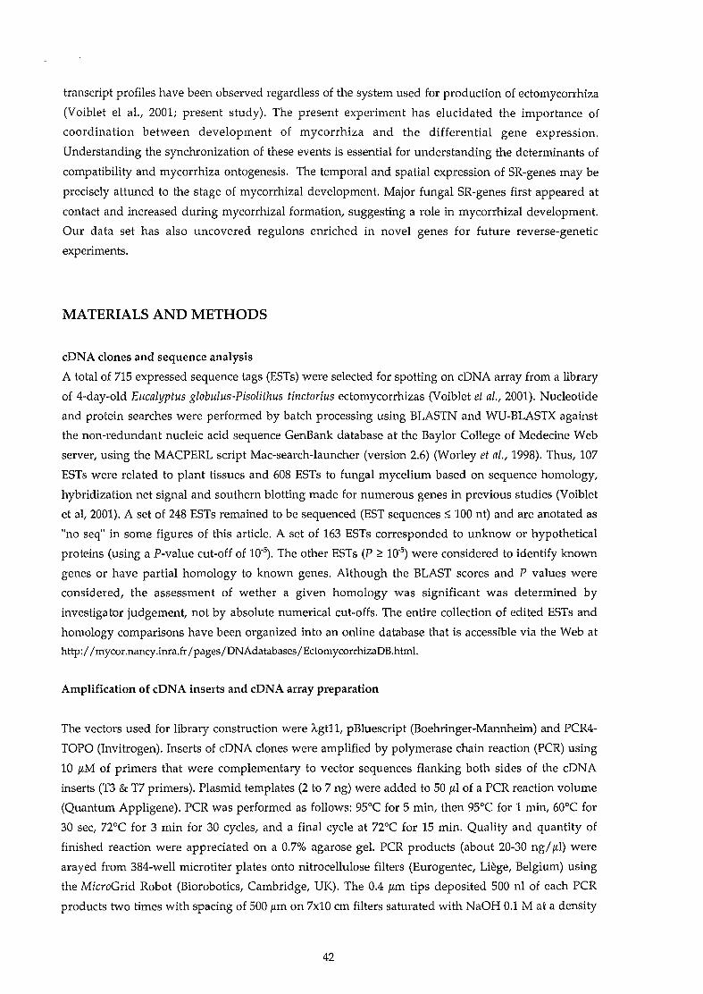
transcript profiles have been observed regardless of the system used for production of ectomycorrhiza
(Voiblet el al., 2001; present study). The present experiment has elucidated the importance of
coordination between development of mycorrhiza and the differential gene expression.
Understanding the synchronization of these events is essential for understanding the determinants of
compatibility and mycorrhiza ontogenesis. The temporal and spatial expression of SR-genes may be
precisely attuned to the stage of mycorrhizal development. Major fungal SR-genes first appeared at
contact and increased during mycorrhizal formation, suggesting a role in mycorrhizal development.
Our data set has also uncovered regulons enriched in novel genes for future reverse-genetic
experiments.
MATERIALS AND METHüDS
cDNA clones and sequence analysis
A total of 715 expressed sequence tags (ESTs) were selected for spotting on cDNA array from a library
of 4-day-old Eucalyptus globulus-Pisolithus tinctorius ectomycorrhizas (Voiblet et al., 2001). Nucleotide
and protein searches were performed by batch processing using BLASTN and WU-BLASTX against
the non-redundant nucleic acid sequence GenBank database at the Baylor College of Medecine Web
server, using the MACPERL script Mac-search-launcher (version 2.6) (Worley et al., 1998). Thus, 107
ESTs were related to plant tissues and 608 ESTs to fungal mycelium based on sequence homology,
hybridization net signal and southern blotting made for numerous genes in previous studies (Voiblet
et al, 2001). A set of 248 ESTs remained to be sequenced (EST sequences::; 100 nt) and are anotated as
"no seq" in some figures of this article. A set of 163 ESTs corresponded to unknow or hypothetical
proteins (using a P-value eut-off of 10'5). The other ESTs (P ~ 10'5) were considered to identify known
genes or have partial homology to known genes. Although the BLAST scores and P values were
considered, the assessment of wether a given homology was significant was determined by
investigator judgement, not by absolute numerical cut-offs. The entire collection of edited ESTs and
homology comparisons have been organized into an online database that is accessible via the Web at
http://mycor.nancy.inra.fr /pages/DNAdatabases/EctomycorrhizaDB.html.
Amplification of cDNA inserts and cDNA array preparation
The vectors used for library construction were "-gt11, pBluescript (Boehringer-Mannheim) and PCR4
TOPO (Invitrogen). Inserts of cDNA clones were amplified by polymerase chain reaction (PCR) using
10 !LM of primers that were complementary to vector sequences flanking both sides of the cDNA
inserts (T3 & T7 primers). Plasmid templates (2 to 7 ng) were added to 50 /lI of a PCR reaction volume
(Quantum Appligene). PCR was performed as follows: 95°C for 5 min, then 95°C for 1 min, 60°C for
30 sec, noc for 3 min for 30 cycles, and a final cycle at noc for 15 min. Quality and quantity of
finished reaction were appreciated on a 0.7% agarose gel. PCR products (about 20-30 ng/!LI) were
arayed from 384-well microtiter plates onto nitrocellulose filters (Eurogentec, Liège, Belgium) using
the MicroGrid Robot (Biorobotics, Cambridge, UK). The 0.4 /lm tips deposited 500 nI of each PCR
products two times with spacing of 500 !Lm on 7x10 cm filters saturated with NaOH 0.1 M at a density
42

of 25 clones per cm2• The nitrocellulose filters were washed and blocked according to the Eurogentec
protocol.
Biological materials and ectomycorrhizas synthesis
We used an in vitro system previously described by Burgess et al. (1996) for another Eucalyptus
Pisolithus ectomycorrhiza. Large round Petri dishes (140 mm diameter, 20 mm deep) were partially
filled with 50 ml of low-sugar (5 mM) Pachlewski medium in 2.0% agar. The agar was overlaid with
washed, autoclaved cellophane discs. Hyphal plugs of 14-day-old mycelium of the gasteromycete
Pisolithus tinctorius Cocker & Couch (synonym Pisolithus arhizus (Scop. Per Pers.) Rauschert), isolate
441, were placed 2.5 cm apart in 3 rows onto the cellophane. Fungal growth took place at 25°C in the
dark for la days. Seeds of Eucalyptus globulus spp. Bicostata Kirp. were sterilized with 20% calcium
hypochlorite (v/v) for 20 min, rinsed with four changes in sterile water and placed in a row 2 cm
above the fungal hyphae. The Petri dishes were then slanted at 70° for 3 days in the dark at 25°C
before being transfered to a controlled environment in growth chamber with 16 h light (25°C, 150
JLmol m'2 S_l) and Sh dark. Petri dishes of non-associated control mycelium and seedlings were
manipulated identically. The ectomycorrhiza time-course development began when the contact
between the germinating root of seedlings and the hyphae occurred (4 days in growth chamber). At 4,
7, 12, 15, 21 and 2S-days-post-contact, root systems with colonizing hyphae (mantle at early stages,
ectomycorrhizas at later stages) were sampled, fixed in liquid N2 and stored at -SO°e. About 50
colonized root systems (5 different Petri dishes) were sampled at each stages, and 15-day-old non
inoculated roots and fungal mats were sampled as control. Estimation of P. tinctorius mycelium
content in ectomycorrhiza during time-course development was estimated according to Martin et aL,
1990.
RNA isolation, probe synthesis and cDNA array hybridization
Total RNA was isolated from ectomycorrhizas at the different stages, free-living P. tinctorius 441 and
non-inoculated roots of E. globulus as described by Logemann et al. (19S7). Complex probes were then
prepared by reverse transcription using SuperScript II reverse transcriptase (Life Technologies) and
the SMART-PCR cDNA Synthesis Kit (Clontech, Palo alto, CA, USA). Labelling of the cDNA probes
was done in the presence of 30 mCi [33p]dCTP and 30 mCi [33p]dATP, and random hexamers using the
Prime-a-Gene Kit (Promega, Madison, WI, USA) according to the manufacturer's instructions. The
nitrocellulose filters were pre-incubated in 30 ml of a hybridization solution (5 X SSC solution, la X
Denhardt's solution, 0.5% SDS, 100 JLg/ml shared salmon sperm DNA) for 4 h at 65°e. Then the filters
were incubated in la ml of fresh hybridization solution containing 20 JLg/ml pBluescript, 2 JLg/ml
polyA oligonucleotide and the 33P-Iabelled probe at 65°C for 22 h. The hybridized filters were washed
successively for 3 x 5 min in 2 X SSC at room temperature, 2 x 20 min in 2 X SSC containing 0.5% SDS
(65°C), 2 x 20 min in 1 X SSC containing 0.1% SDS (65°C) and 2 x 20 min in 0.1 X SSC containing 0.1%
SDS (65°C). Filters were then wrapped in plastic bags and exposed to a phosphor screen (Eastman
Kodak Company, Rochester, NY, USA) for varying periods (24 h to 5 days), after which the imaging
43
plate was scanned by Personal Molecular Imager FX (Bio-Rad Laboratories, Hercules, CA, USA) at a
maximum resolution of 50 /lm per pixel.
Data analysis
The raw image data obtained with the phosphorimager imaging system were imported into an apple
Macintosh G3. Detection and quantification of the signaIs representing hybridized DNA were
performed using the volume quantification method of the QUANTITY ONE software (Bio-Rad). Each
spot was defined by 384-spots-grid positioning over the array image. For each image, the average
pixel intensity within each spot was determined. Spots deemed unsuitable for accurate quantification
because of array artefact were manually flagged and excluded from further analysis. The data table
generated by QUANTITY ONE, containing the intensity of each spot, was then exported to the EXCEL
98 worksheet programme (Microsoft Corporation, Redmond, WA, USA) for further manipulation.
Local background value for each membrane was calculated on the basis of 260 positions with no
DNA-spotted area. Net signal was determined by subtraction of this mean background from the
intensity for each spot. Spots that had an intensity of less than twofold that of the background had
their intensity raised ta the arbitrary value of O.l.The probe-ta-probe variance was filtered out using
signal intensities of the human desmin (kindly provided by T. Desprez, INRA Versailles, France)
spotted at eight locations on the filters and added ta the complex probes prior ta labelling procedure
(300 pg per probe as determined in previous manipulation) as internaI standard. In addition, to take
account of experimental variations in specific activity of the cDNA probe preparations or exposure
time that might alter the signal intensity, the data obtained from different hybridizations were
normalized by dividing the intensity for each (fungal or plant) spot by the average of the intensities of
aIl the (fungal or plant) spots present on the filter, ta obtain a centered, normalized value (Eisen et al.,
1998). AH the expression data are available at the EctomycorrhizaDB web site
(http://mycor.nancy.inra.fr /pages/DNAdatabases/EctomycorrhizaDB.html). Multiple experiments were
analyzed using Cluster and Treeview softwares (http://rana.lbl.gov). The Clusters and SOMs
generated with these softwares are available at the EctomycorrhizaDB web site.
Quality control of the array
A number of quality control of the array and hybridization procedure were performed. We selected 16
P. tinctorius genes and 7 E. globulus genes known to be Symbiosis-Regulated or ta be stable at early
stages of ectomycorrhiza formation (Voiblet et al., 2001) and placed them 2 to 4 times at different
locations on the filters. We also placed hybridization control as pBluescript, polylinker of this plasmid,
polyA and polyT oligonucleotides (Amersham Pharmacia Biotech, Piscataway, NJ, USA) and DNA
free PCR volume reaction at different locations. According ta the multiple net signaIs measured, we
estimated that the variations due ta the arrayer on filters were about 6 % and that the average
standard errors due to aspecific hybridization for SR- and house keeping-genes on each filter were
from 10 ta 20 % in the different treatments (16,8±3.7%).
44
ACKNOWLEDGMENTS
Sébastien Duplessis was supported by a Doctoral Scholarship from the Ministère de l'Education
Nationale, de la Recherche et de la Technologie. We would like to thank Denis Tagu (INRA-Nancy)
for valuable discussions during the course of this study. We are grateful to Catherine Voiblet for her
technical advices at the begining of this project. The assistance of Marine Wasniewski and Natacha
Steinwich in cDNA amplification and Jean-Luc Jany in ergosterol assays is acknowledged. We also
greatly appreciated the technical support of Sofia Kotowski. Finally, many thanks to Christine
Delaruelle (INRA-Nancy) for her dedicated assistance in sequencing and to Christian Herbé (INRA
Nancy) for Visual Basic Macros programing. The present investigation was supported by grants from
the Groupement de Recherches et d'Etude des Génomes and the INRA (Action Transversale
Microbiologie Fondamentale). The research utilized in part the DNA Sequencing Facilities at INRA
Nancy financed by the INRA and Région de Lorraine.
Figure 1: Photographs of the experimental system. (A) Pisolithus tinctorius free-living mycelium (top) ;
Eucalyptus globulus non-inoculated roots (bottom) and two intermediate stages in ectomycorrhiza
time-course development: mantle formation (7 days) and functionning symbiotic organ (28 days). (B)
Estimation of Pisolithus tinctorius mycelium content in ectomycorrhizas during time-course
development using ergosterol assay.
Figure 2: Hierarchical c1ustergrams of expression ratio corresponding to Eucalyptus globulus and
Pisolithus tinctorius genes during ectomycorrhiza development. Ratio of transcripts from 107 root
genes (A) and 608 fungal genes (B) were c1ustered with the Cluster and Treeview softwares. The
c1uster results are shown here along with the name of each ESTs and the protein showing the best
match. Time-course stages are given at the top of c1ustergrams. These c1usters can be consulted on the
web site http://mycor.nancy.inra.fr. ESTs anotated as 'no seq' were less than 100 bp. Ratio levels are
indicated (bottom left).
Figure 3: Selected fungal c1usters of genes involved in 3 different cellular function. P. tinctorius genes
encoding structural proteins (A), signalling pathways components (B) and stress-response elements
(C). In (C), the yellow, red and blue bars indicate ESTs sharing identical sequences. The Cluster results
are shown here along with the name of each ESTs and the protein showing the best match. Time
course stages are given at the top of c1ustergrams. Ratio levels are indicated (bottom left).
45

LITERATURE CITED
Burgess T, Laurent P, Dell B, Malajczuk N, Martin F (1995) Effect of fungal-isolate aggressivity on the
biosynthesis of symbiosis-related polypeptides in differentiating eucalypt ectomycorrhizas. Planta
195: 408-417.
Burgess T, Dell B, Malajczuk N (1996) In vitro synthesis of Pisolithus-Eucalyptus ectomycorrhiza:
synchronisation of lateral tip emergence and ectomycorrhizal development. Mycorrhiza 6: 189-196.
Carnero-Diaz E, Martin F, Tagu D (1996) Eucalypt a-tubulin: cDNA cloning and increased level of
transcripts in ectomycorrhizal root system. Plant Mol. Biol. 31, 905-910.
Duplessis S, Tagu D, Martin F (2001) Living together underground: a molecular glimpse of the
ectomycorrhizal symbiosis. In Molecular biology of fungal development (Osiewacz, ed.). Mycology
Series, Dekker & Dekker, New York. In Press.
Eisen MB, Spellman PT, Brown PO, Botstein D (1998) Cluster analysis and display of genome-wide
expression patterns. Proc. Natl. Acad. Sei. USA 95: 14863-14868.
Gianinazzi·Pearson V (1996) Plant cell responses to arbuscular mycorrhizal fungi: getting to the roots of
the symbiosis. Plant CellS, 1871-1883.
Hampp R, Schaeffer C (1995) Mycorrhiza-Carbohydrate and energy metabolism. In Mycorrhiza,
Structure, Function, Molecular Biology and Biotechnology (Varma A K and Hock B, eds). Berlin:
Springer-Verlag, pp 267-296.
Harrison MJ (1998) Development of the arbuscular mycorrhizal symbiosis. Curr. Opin. Plant Biol. 1,360
365.
Harrison MJ (1999) Molecular and cellular aspects of the arbuscular mycorhizal symbiosis. Annu. Rev.
Plant Physiol. Plant Mol. Biol. 50,361-389
Hilbert J-L, Costa G, Martin F (1991) Ectomycorrhizin synthesis and polypeptide changes during the early
stage of eucalypt mycorrhiza development. Plant Physiol. 97,977-984.
Kottke 1, Oberwinkler F (1987) The cellular structure of the Hartig net: coenocytic and transfer cell-like
organization. Nord. J. Bot. 7, 85-95.
Lapopin L, Gianinazzi·Pearson V, Franken P (1999) Comparative differential RNA display analysis of
arbuscular mycorrhiza in Pisum sativum wild type and a mutant defective in late stage development.
Plant Mol. Biol. 41:669-77.
46

Logemann J, Schell J, Willmitzer L (1987) Improved method for the isolation of RNA from plant tissues.
Anal. Biochem. 163: 16-20.
Malajczuk N, Lapeyrie F, Garbaye J (1990) Infectivity of pine and eucalypy isolates of Pisolithus tinctorius
on raots of Eucalyptus urophylla in vitro. New Phytol. 114, 627-631.
Martin F, Delaruelle C, Hilbert J-L (1990) An improved ergosterol assay to estimate fungal biomass in
ectomycorrhizas. Mycol. Res. 94: 1059-1064.
Martin F, Botton B (1993) Nitrogen metabolism of ectomycorrhizal fungi and ectomycorrhiza. Adv. Plant
Pathol. 9/ 83-102.
Massicote H B, Peterson R L, Ackerley C A, Ashford A E (1987) Ontogeny of Eucalyptus pilularis-Pisolithus
tinctorius ectomycorrhizae. II. Transmission electron microscopy. Can. J. Bot. 65, 1940-1947.
Nehls V, Béguiristain T, Ditengou F, Lapeyrie F, Martin F (1998) The expression of a symbiosis-regulated
gene in eucalypt raots is regulated by auxins and hypaphorine, the tryptophane betaine of the
ectomycorrhizal basidiomycete Pisolithus tinctorius. Planta 207, 296-302.
Tagu D, Nasse B, Martin F (1996) Cloning and characterization of hydrapobin-encoding cDNAs from the
ectomycorrhizal basidiomycete Pisolithus tinctorius. Gene 168, 93-97.
Tamayo P, Slonim D, Mesirov J, Zhu Q, Kitareewan S, Dmitrovsky E, Lander ES, Golub TR (1999)
Interpreting patterns of gene expression with self-organizing maps: methods and application to
hematopoietic differentiation. Prac. Natl. Acad. Sei. V S A 16:2907-12.
Voiblet C, Duplessis S, Encelot N, Martin F (2001) Identification of symbiosis-regulated genes in
Eucalyptus globulus-Pisolithus tinctorius ectomycorrhiza by differential hybridization of arrayed
cDNAs. Plant J. 25(3): 1-12.
Worley KC, Culpepper PA, Wiese BA, Smith RF (1998) BEAUTY-X: enhanced BLAST searches for DNA
queries. Bioinformatics 14: 890-891.
47

(A)
Pisolithus tinctorius
1100
(B)1.-
~--'"lU.!::l 75oS...0u
7-day-old1
>-eectoD1ycorrhizas .s
50 1 1 00u ~
QI.....0.wc::.,; -n "- 1 QI.wc::0u
25e •::s....Qiu>-~
a28-day-old
1 a~fioD1ycorrhizas 5 la 15 20 25 30
Days post-inoculation
Eucalyptus globulus
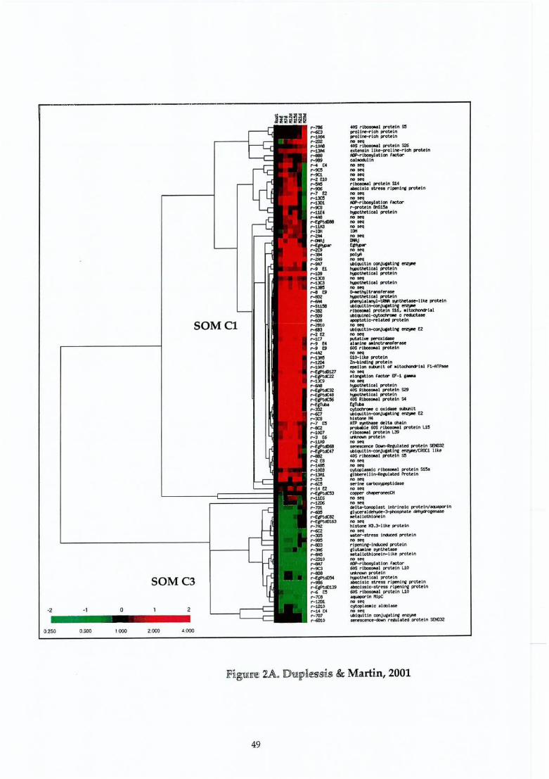
·2 ·1 o
SOM Cl
SOMC3
r-786~
r-1084r-2ll2r-10A8r-13A4r-eeer-ge!I
..... E4r-9alr-!lC1r-Z E10~
r-9OI5r-7 E2r-~r-1301r-9C8r-11E4...r-E1f'tdJ8Ilr-11Jl3r-11Ilr-2Mr---.Jr-E""""r-2C9r-3IMr-2I'Br-9ft7r-9 E1r-1D9r-13C8r-13C3r-1JllO,...., E9r'-lllIZ...r-SlJ!l8r-382.-.-r'-<iD8r-2810r-683r-2 E2r-1C7r-9 E4r-g E9r-4A2r-1:wlr-12I)4r-10A1r-ElIf't<Il127r-E~r-13ŒF'-ôIl8r-EjPt<02~48r-E~
r-EfT'*'"r-302r-(,I;lr-3C8r-7 El!r-«2r-1007r-3 E6r-11A!lr-ElII't<fl6llr-E1lf't<I:47r-882r-2 ESr-148llr-1003r-13A1r-2C:lr-«!Ir-14 E2r-E1lf't<I:53r-11E6r-1206r-701Miœr-Etpt«82r-Etptdl163r-7A2r-=r-3œlr-96Or-«l3r-3A6...r-2010r-8A7r-!lC3r-8Ollr-E~r-986r-EePt<fl139r-6 E!lr-7C8r-1201r-1010r-14 E4r-707r-6I!1o
40S ribosoMl protdn 5:'proltn f'lch protelnprolt lch protelnno ....40S rtbosoNl prout" S26.xtensln ltke-jlnllt...... lch protelnAlJ'-rlboo<jl.tlon r_cal.xlll1nno seqno seqno ....no seqrl_l proteln Si4MlIclslc s~ rlpenl~ protet"no ....no ....AlJ'-rlboo<jl.tlon r_..-prote1n 1lnS1llo,-thetlcol protelnno ....no seqno seqIlIlno ....IM\JE""""no seqpol!llno seqlbl~ltln cooJUfOtlnf~'-thetlcol proteln'-thetlcol protelnno ....'-thetlcol protelnno ....~ltr_F~~
~t:het1c.l protet"~1.11t1OJ1-~ ~thetose-ltke protelnlbl~ltlr>-<:onJUll.tlnt~
ribosOMl prote!" S16, .ltcx::t'loncrlallbl~lnol~ c reclJctosOapoptotJe-t"elaUd protetnno ....lbl~ltlr>-<:onJUllOtlnf~ E2no seqFUtotl.... peroxldOle.1.,lne .Irotransf«'ase60S rlbosoUl protetnno ....G1o-ltke protelnZn-l>lrdlnf protelnepsilon St.tu'llt oF' .Itoc:nonc*"lal Fl~TP_no ....~l~t1on Factor Ef-l gaMano seqh!:P>t:hetlcal proutn40'S RlboIoMl prout" S29'-thetlcol proteln40S Rl_1 proteln S4EfT'*'"cytoclrooe c oxldOle -..1tlbl~ltlr>-<:onJUllOtlnol ~ E2histone H4ATP ...,,- delta cholnpt'Obable 60S rlbosoNll protet" WrlbosoMl protet" l.39l.I'WIcul protetnno ....~ Dowrt-Rtfulated protet" SfN)32lbl~ltlr>-<:onJUll.t1nt~1tROC1 llk.40S ,..lbosoMl protet" 5'no ....no seqcytoplasalc rlbosoMl ~teln StSaelbilerellt~l.tcd Protelnno seqserine ~tldueno seq_a--~
no seqno seqœlt.a-tonoPlast Intrln:slc protelnlaquaporlnel__•
1__3-phoophote~
.etallothlcn:lnno ....hlstc:Jne HJ.3-11ke protet"no seqwat«'-str~ Il'lCiJc:td protet"no ....rtpenlnt-I~prote!"elutoolne ..,.,thetos<PIIttallothlcn:ln-llke protelnno seoACP"""I~lat1on Factor60S rlbosOMl pro~tn UOl..f'Mowl prote tn~~t1cal protetnabsclstc stre'ss rlpenlnQI protetnab:sctsstc-stre-ss rl~tni protetn60S rlbos.... l proteln UO_lnK1pCno seq~la.ic aldolaseno ....lJ)t~tt1n conJ~at1Olt~*'lt'sœtiCt"""dorll1 re0Jlated protetn seN)32
0.250 0.500 1000 2.000 4.000
49

50
Figure 2B. Duplessis & Martin, 2001

~-------._-----------------------------,
A...W13~-aœ
F-EllI't«43F-11Jl8F-3010F-5ClF-:lC4F-:lI)4F-EIC4F-5 EiF-E8f'td:~
F-7 E5F-' E4F-illlSIF-E8f'td:!lOF-E8f'td:54F-7A1F-E8f'td:14F-5C8F-EllI't«~F-5A8F-12 EiOF-~
F-11B3F-7C2F-5 E5F-~
F-8EiOF-Ellf't«44F-E8f'tcIJ81F-U E9F-'JC7F-aA10F-E8f'td:lF-E8f'tci:26F-ftI'tdl141F-Eel't<fl53F-EllI't«7F-1lC:lF-EllI't«45F-E8f'td:23F~
F-EQPtd:8F-E8f'td:84F-EllI't«'F-E8f'td:27F-10A10F-1B7
BF-104F-St16F-l1ll6F-l009F-~
F-11A5F-3 E4F-5C9F-1A6F-l1C!lF-9 E5F-5S2F-10010F-l1J10F-1101F-lIAlF~ E9F-St114N5C9F-St92F-3C7F-126ZF-l E10F-9C4F-5C6
G1Jl-llINlINl PIlOTEIN \1'T1w!jl: .....lno/tlr...,lno pro~ln kinasea'OSO-pathwa!! control I«l-f'epe.t proUln epc-2sloll'" to ncn-t'Tœ!no pro~!n kln...rasGTl'-I>lndlr>g pro~ln GPAlNIU nttrll...put.U.... l'ljrH"el.t.d kinase~theUcal t~.lne pIlopIlat...stf'lne/tt'r!onlne-pr'Ottln kl~put.U pro~ln ktnasecalet blndlnQ pro~ln
auxln-l.-...:.d pro~tn
s!l>1 pro~!n
calelne<Jrln reQUl.tory Stb>lltserlne/~lnepro~ln kinaseGTl'-I>tndlnQ proU!nprobable serlne/~ln<-pro~tn kinaserail!putative- histidine' kinaseQlutaoate receptorfO'-rlbosyl.tlon FactorJas"""le acld lndl.<:ed genocross-pathwa!! control ~epe.t protetn epc-2cross-pau-. control ijl)-r....t pro~tn cpc-2
IilIe'tal blndtne: ~in-f'tch proutn..ta 1 bindtn. ~Uln-rtch protetnflIiIe'tal blndlne ~ttln-rlch protein~tal btndini eystetn-rlch protetnflIiIe'tallothl~ln
flIiIe'tallothlOMtn~tallothloneln
Illrtallothl~tn
lIIetal blndlna' ~in-rlch proufnIIlet,,1 blndtni ~teln-rlch protetnlIlrtal btndtne Cl}Sttln-rtch proutnlIlIetal blndlni cysteln-rlch protetnlIlrtal blndtnc cystetn-rlch protetnIIrtal btndinc ~tetn-r"lch pr"Otein
C ~tal blndinc ~tdrH'lch proteln
1 Ile'tallothtonetnab1..hl blndlnl C\l$Uln-rlcll proteln
lllle'tai blndlni! ClJSUln....rfch protefnIIletal blndlni! cysteln-rlch pr-otelnlow telilQerature ancI salt responsl~ rrot.eln
F-EQl'td:&5 1~:~ bF-3 E9
F-EePtd:n 1;-5 ElF-11Jl2;-1381 af-12 E3F-~ E7F-l0 E7;-13 E9f'-12 EaF-71<JF-13 E2F-889F-9B3F'-10A4F-301F-71l4
c
-2 -, a
0.250 l.o.Xl
"------,_._----_.,-----, _________________________--1
Figure 3. Duplessis & Martin, 2001
51

Chapitre 3
Clonage et analyse dé l'expression de l'ADNe HydPt-3 codantune nouvelle hydrophobine de Pisolithus tinctorius.

Clonage et analyse de l'expression de l'ADNe HydPt-3 codant
une nouvelle hydrophobine de Pisolithus tinctorius.
L'approche de génomique fonctionnelle nous a permis d'identifier des fonctions cellulaires
particulièrement affectées par le processus symbiotique. L'une de ces fonctions correspond à la
synthèse et la structuration des parois fongiques. En effet, le passage d'hyphes libres dans le milieu, à
des hyphes différenciées formant le manteau et le réseau de Hartig, induit des modifications
morphologiques et biochimiques importantes des parois (Tagu & Martin, 1996; Martin et al., 1999).
Les hydrophobines - protéines fondamentales dans la biologie et la morphogénèse des hyphes des
champignons filamenteux - sont très abondantes et particulièrement régulées lors de la formation de
la symbiose ectomycorhizienne (Tagu et al., 1996). Nous présentons dans ce chapitre, la caractérisation
et l'étude de l'expression de l'ADNe HydPt-3 : Cloning and expression analysis of a new hydrophobin
cDNA from the ectomycorrhizal basidiomycete Pisolithus (Duplessis et al., 2üü1b ; article sous presse,
revue Current Genetics).
52
Cloning and expreSSÜlft analfsis oi a new 1i.y;dropbobincDNA from the ectomycorrhizal basidiomycete
Pisolithus
Publication n° 3
Sébastien Duplessis' Céline Sorin . Catherine Voiblet· Béatrice Palin'
Francis Martin' Denis Tagu
Cloning and expression analysis of a newhydrophobin cDNA from the ectomycorrhizalbasidiomycete Pisolithus
S. Duplessis' C. Sorin . C. Voiblet . B. Palin' F. Martin' D. Tagu (IBJ)
I.N.R.A.
UMR INRA/UHP 1136, Interactions Arbres/Micro-Organismes (laM)
Centre de Nancy
54280 Champenoux, France
email: [email protected]
Tel.: +33-383-394013
Fax: +33-383-394069
The GenBank accession number of the hydPt-3 sequence is AF097516
Current Genetics
(Submitted pt February 2001; Accepted 3 rd May 2001; in press)
53

AB5TRACT
Hydrophobins are fungal cell wall proteins whieh play a crucial role in cell adhesion and
aggregative processes. We have identified a new hydrophobin cDNA (hydPt-3) in the
symbiotie mycelium of Pisolithus tinctorius (putative P. albus) during the formation of
ectomycorrhizas with eucalypt roots. This sequence was highly divergent from two other
previously identified Pisolithus symbiosis-regulated hydrophobins, hydPt-l and hydPt-2.
Expression analyses demonstrated that hydPt-3 was also up-regulated during ectomycorrhiza
formation. In contrast to phytopathogenic fungi, changes in glucose or ammonium
concentrations in the growth medium did not influence the accumulation of any Pisolithus
hydrophobin mRNAs. This suggests that other factors act as regulators of hydrophobin gene
expression in ectomycorrhiza.
Keywords: Cell wall· Nutrients . Root . Symbiosis . Tree.
Introduction
Hydrophobins are morphogenetic cell wall or excreted proteins involved in several aspects of
fungal biological processes like aerial growth of the mycelium, aggregation of hyphae, host
surface adhesion or desiccation tolerance (Wôsten et al. 1999). Soluble hydrophobin monomers
have the capacity to self-assemble at hydrophilic/hydrophobie interfaces (Wessels and de
Vocht 2000). When self-assembly occurs at the surface of spores or hyphae, they become
covered with an hydrophobie layer capable of physical interactions with adjacent
hydrophobie substrates (for reviews see Wessels 1997, Kershaw and Talbot 1998). In several
pathogenic interactions, fungal hydrophobins have been shown to be up-regulated and in sorne
cases, even essential for plant surface colonization (Talbot et al. 1993). The regulation of
hydrophobin gene expression is complex and vary from one species to another (Kershaw and
Talbot 1998). In several pathogenie fungi, hydrophobin gene expression is however regulated
in vitro by nutrient source availability (Talbot et al. 1993, St Leger et al. 1992, Segers et al.
1999) and sensing the level of nutrients in the substrate could be an important signal for the
presence of a putative host, and could thus trigger expression of proteins involved in host
surface recognition (van den Ackerveken et al. 1994).
54

Ectomycorrhiza is a mutualistic symbiosis between filamentous soil-born fungi and tree
roots. In the ectomycorrhizal basidiomycete Pisolithus isolate 441 (putative P. albus), two
hydrophobin cDNAs (hydPt-l and hydPt-2) were previously characterized (Tagu et al.
1996). They correspond to transcripts which are up-regulated dming the early stages of
differentiation of eucalypt symbiotic tissues. Their mRNAs are abundant (Voiblet et al. 2001)
and the HYDPt-1 polypeptide is accumulated on the œIl walls of the hyphae (Tagu et al.
2001). The function of hydrophobins in ectomycorrhiza is still unknown and these
polypeptides may have several roles in mycorrhiza formation, such as binding the hyphae to
the root surface, aggregation of hyphae or root cell penetration by fungal hyphae (Tagu et al.
1995). Whether any of these functions is fulfilled byone or several types of hydrophobins is
unknown (Martin et al. 1999). Here we report the cloning of a new hydrophobin cDNA from
Pisolithus (hydPt-3). We show that its expression was up-regulated dming ectomycorrhiza
formation. Changes in glucose or ammonium concentrations did not affect the steady state
level of any of the three different hydrophobin transcripts in the mycelium. This indicates
that drastic changes in trophic conditions experienced by the colonizing hyphae are not the
main factors triggering the up-regulation of hydrophobin genes dming the process of root
colonization.
Materials and methods
Fungal strain and growth medium. The mycelium of Pisolithus isolate 441 (putative P. albus)
(Bougher and Syme 1998) was maintained at 25 oC on modified Pachlewski's medium
(containing 5.6mM glucose and 5mM NH4+, 2% w/v agar) (Hilbert et al. 1991). This
accession - 441 - was previously attributed to P. tinctorius, but recent phylogenetic analyses
indicated that this isolate corresponds to the Australian species P. albus (Bougher and Syme
1995). Mycorrhizas were synthesized as described in Nehls et al. (199Sa). Briefly, seedlings
of Eucalyptus globulus were deposited at the surface of Pisolithus mycelium on modified
Pachlewski's medium. Mycorrhiza were harvested after 1 or 6 days of contact, immediately
frozen in liquid nitrogen and kept at -SO oc. In order to test the effect of glucose or ammonium
concentrations, Pisolithus was grown on a cellophane membrane laid down agar plates on the
modified Pachlewski's medium (5.6 mM glucose; 5 mM NH4+) at 25 oC for 2 weeks. The
55

cellophane membrane carrying the mycelium was then transferred to a fresh modified
Pachlewski's medium for 2 additional days. Then, the cellophane membranes were tranferred
again on the different media depleted or enriched in glucose and ammonium for 6 days a t
25 oC, before harvesting. It is known from several independent studies performed on different
ectomycorrhizal species that several days on depleted media are necessary to starve the
mycelium (Littke et al. 1984; Kershaw and Stewart 1992; Nehls et al. 1998b). Four different
combinations were tested: 5.6 mM glucose and 5 mM NH4+ (control), 110 mM glucose and 5 mM
NH4+ (enriched in carbon), 0 mM glucose and 5 mM NH4+ (depleted in carbon), 5.6 mM glucose
and 0 mM NH4+ (depleted in nitrogen).
DNA manipulation. New hydrophobin sequences were identified in our expressed sequence
tags (ESTs) collection from P. tinctorius E. globulus ectomycorrhiza
(http://mycor.nancy.inra.frIpages/DNAdatabases/EctomycorrhizaDB.html) (Voiblet et al.
2001). Hydrophobin EST sequences were analysed with the Blast 2.1 (blastx) programme
(http://www.ncbLnlm.nih.gov/blast/blast.cgi) (Altschul et al. 1997). Double-strand DNA
sequencingwas performed on the new ESTs matching with known hydrophobins. Percentages
of identity were determined with Basic Blast (blastp) using the HYDPt-3 predicted amino
acid sequence as a reference. Sequence alignment and shading were performed with the
freeware programme SeqApp (D.G. Gilbert, version 1.9a169 for MacIntosh computers, 1992).
Total DNA was extracted from the mycelium of Pisolithus 441 as described in Camero Diaz et
al. (1997) by following the protocol of Guidet et al. (1991). About 10 /lg of total DNA was
digested by different restriction enzymes ovemight. Transfer to Nylon filters (Hybond N+,
Amersham-Pharmacia, Little Chalfont, UK) and hybridization were performed as described
by the manufacturer. Briefly, hybridization was done ovemight at 65 oC in 5x SSC, 5x
Denhardt and 0.5% (w Iv) SDS. Washes were performed at 65 oC for 1h in 2x SSC, 0.1% (wIv)
SDS and for 1h in lx SSC, 0.5% (w Iv) SDS. The hydPt-l, hydPt-2 and hydPt-3 cDNAs were
labelled with [a}2p] dCTP by random priming, and were used at 106 cpm per milliliter of
hybridization solution. Filters were then exposed to a Phosphor-Imager screen (Kodak) and
numerized in the Personal Molecular Imager fx by following manufacturer's instructions (Bio-
Rad, Hercules, CA, USA).
56

RNA manipulation. Total RNA from control mycelium, mycelia grown on different ammonium
and glucose concentrations, non-colonized roots, and roots in contact with the mycelium for 1 or
6 days were extracted using the guanidinium method described in Logemann et al. (1987).
About 5 to la /lg of RNA was separated on an agarose gel (1.5% w Iv) by electrophoresis in a
denaturing buffer containing formamide. The RNA was transferred to Hybond N+ filters and
the membranes were hybridized with the probes labelled as described above. A PCR
fragment containing the 5.85 rRNA from Pisolithus 441 was also used as a probe (Camero
Diaz et al. 1997). Hybridizations were performed at 42 oC ovemight in 50% (v Iv) formamide
(Ruiz-Avila et al. 1991). The first wash was performed at room temperature for 20 min (2x
SSPE, 0.1% (w Iv) SDS) foHowed by two washes at 65 oC for 30 min (2x SSPE, 0.1% (w Iv) SDS)
and one wash at 65°C for 30 min (lx SSPE, 0.1% (w Iv) SDS). Hybridization signaIs were
detected as described above and quantified by using the Quantity One programme (Bio-Rad).
The signaIs obtained with the hydrophobin probes were standardized to the signaIs of the
5.85 rRNA probe (Camero Diaz et al. 1997). Three independent repetitions were done and the
raw data submitted to one-factor analysis of variance (ANOVA): means were then compared
by Bonferroni test. AH the statistieal analysis were done at the probability threshold of 0.05
with Statview program 4.02 (Abascus Concepts).
Results and Discussion
About 850 cDNAs were cloned and partially sequenced (ESTs) from Pisolithus
Eucalyptus globulus ectomycorrhiza (Tagu and Martin 1995; Voiblet et al. 2001). Three of
these clones (5C4, 5D4 and 8C4) were identieal in sequence and showed a significant degree of
similarity with Pisolithus hydrophobin cDNAs. This new hydrophobin shared 52% and 54%
identity at the amino add level with hydPt-l and hydPt-2, respectively (Figure 1) and was
referred as hydPt-3. It presented the same distribution of hydrophobie domains as the other
Pisolithus hydrophobins (Martin et al. 1999). This sequence probably belongs to Class 1
hydrophobins (Wessels 1997). It encodes a 108 amino add polypeptide containing eight
cysteine residues (in bold in Fig. 1) at the conserved positions and a 17 amino add N-terminal
signal peptide (von Heijne 1983).
57
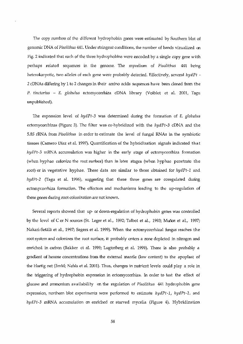
The copy nurnbers of the different hydrophobin genes were estimated by Southem blot of
genomic DNA of Pisolithus 441. Under stringent conditions, the number of bands vizualized m
Fig. 2 indicated that each of the three hydrophobins were encoded by a single copy gene wi th
perhaps related sequences in the genome. The mycelium of Pisolithus 441 being
heterokaryotic, two alleles of each gene were probably detected. Effectively, several hydPt
2 cDNAs differing by 1 to 2 changes in their amino acids sequences have been cloned from the
P. tinctorius - E. globulus ectomycorrhiza cDNA library (Voiblet et al. 2001, Tagu
unpublished).
The expression level of hydPt-3 was determined during the formation of E. globulus
ectomycorrhizas (Figure 3). The filter was co-hybridized with the hydPt-3 cDNA and the
5.85 rRNA from Pisolithus in order to estimate the level of fungal RNAs in the symbiotic
tissues (Camero Diaz et al. 1997). Quantification of the hybridization signaIs indicated tha t
hydPt-3 mRNA accumulation was higher in the early stage of ectomycorrhiza formation
(when hyphae colonize the root surface) than in later stages (when hyphae penetrate the
root) or in vegetative hyphae. These data are similar to those obtained for hydPt-l and
hydPt-2 (Tagu et al. 1996), suggesting that these three genes are coregulated during
ectomycorrhiza formation. The effectors and mechanisms leading to the up-regulation of
these genes during root colonization are not known.
Several reports showed that up- or down-regulation of hydrophobin genes was controlled
by the level of C or N sources (St. Leger et al., 1992; Talbot et al., 1993; Mufioz et al., 1997;
Nakari-SetaUi. et al., 1997; Segers et al. 1999). When the ectomycorrhizal fungus reaches the
root system and colonizes the root surface, it probably enters a zone depleted in nitrogen and
enriched in carbon (Bakker et al. 1999; Lugtenberg et al. 1999). There is also probably a
gradient of hexose concentrations from the external mantle (low content) to the apoplast of
the Hartig net (5mM; Nehls et al. 2001). Thus, changes in nutrient levels couId play a role in
the triggering of hydrophobin expression in ectomycorrhiza. In order to test the effect of
glucose and ammonium availability on the regulation of Pisolithus 441 hydrophobin gene
expression, northem blot experiments were performed to estimate hydPt-l, hydPt-2, and
hydPt-3 mRNA accumulation on enriched or starved mycelia (Figure 4). Hybridization
58

signaIs of the hydrophobin transcripts were normalized with the 5.85 rRNA signaIs. No
significant changes in hydPt-l, -2 and -3 transcripts were detected after ammonium or glucose
starvation, or after a 20-fold increase of glucose concentration.
The absence of changes in hydrophobin mRNA accumulation under our experimental
conditions indicates that ammonium and glucose concentrations are not essential regulators of
hydrophobin gene expression. Thus, ectomycorrhizal fungi colonizing root tissues might have
developed other strategies to sense the presence of a putative host surface. Other trophic or
developmental effectors (in combination or not with ammonium and glucose) may thus be
responsible for hydrophobin gene regulation in developing ectomycorrhizas.
Acknowledgements The authors would like to acknowledge Dr. Pascale Frey-Klett (INRA
Nancy, UMR IaM) for her help in the statistical analyses of the data. This work was
supported by «Groupement de Recherches et d'Etude des Génomes» and «Action
Transversale INRA Microbiologie Fondamentale ». SD and CV were supported by
scholarships from the French « Ministère de l'Education Nationale et de la Recherche et des
Technologies ». We greatly appreciated the technical support of Christine Delaruelle and
Sofia Kotowski. Dr. Phil Murphy (Wye College, University of London) is kindly
acknowledged for his expertise in Southern blot analysis.
59

REFERENCES
Altschul SF, Madden TL, Schaffer AA, Zhang J, Zhang Z, Miller W, Lipman DJ (1997)
Gapped BLAST and PSI-BLAST: a new generation of protein database search programs.
Nucl Acid Res 25:3398-3402
Bakker MR, Dieffenbach A, Ranger J (1999) Soil solution chemistry in the rhizosphere of roots
of sessile oak (Quercus petraea) as influenced by lime. Plant and Soil 209:209-216
Bougher NL, Syme K (1998) Fungi of Southeastern Australia. Perth, Australia: University of
Western Australia Press.
Carnero Diaz E,Tagu D, Martin F (1997) Ribosomal DNA internaI transcribed spacers to
estimate the proportion of Pisolithus tinctorius and Eucalyptus globulus RNAs in
ectomycorrhiza. Appl Environ Mierobiol 63:840-843
Guidet F, Rogowsky P, Taylor C, Weining S, Langridge P (1991) Cloning and characterisation
of a new rye-specifie repeat sequence. Genome 34: 81-87
Hilbert JL, Costa G, Martin F (1991) Ectomycorrhizin synthesis and polypeptide changes
during the early stage of eucalypt mycorrhiza development. Plant Physiol 97:977-984
Kershaw JL, Stewart GR (1992) Metabolism of lsN-labelled ammonium by the
ectomycorrhizal fungus Pisolithus tinctorius (Pers.) Coker & Couch. Mycorrhiza 1:71-77
Kershaw KJ, Talbot NJ (1998) Hydrophobins and repellents: proteins with fundamental roles
in fungal morphogenesis. Fungal Genet Biol 23:18-33
Littke WR, Bledsoe CS, Edmonds RL (1984) Nitrogen uptake and growth in vitro by Hebeloma
crustuliniforme and other Pacific Northwest mycorrhizal fungi. Can J Bot 62:647-652
Logemann J, Schell J, Willmitzer L (1987) Improved method for the isolation of RNA from
plant tissues. Anal Biochem 163:16-20
Lugtenberg BH, Kravchenko LV, Simons M (1999) Tomato seed and root exudate sugars:
composition, utilization by Pseudomonas biocontrol strains and role in rhizosphere
colonization. Environ Mierobiol1:439-446
Martin F, Laurent P, de Carvalho D, Voiblet C, Balestrini R, Bonfante P,Tagu D (1999) Cell
wall proteins of the ectomycorrhizal basidiomycete Pisolithus tinctorius: identification,
function, and expression in symbiosis. Fungal Genet Biol 27:161-174
60

Mufioz G, Nakari-SetaHi T, Agosin E, Penttila M (1997) Hydrophobin gene srhl , expressed
during sporulation of the biocontrol agent TricllOderma harzianum. Curr Genet 32:225-230
Nakari-Setala T, Aro N, Ilmén M, Mufioz G, Kalkkinen N, Penttila M (1997) Genetic and
biochemical characterization of the Trichoderma reesei hydrophobin HFBI. Eur JBiochem 248:415-323
Nehls D, Béguiristain T, Ditengou F, Lapeyrie F, Martin F (1998a) The expression of a
symbiosis-regulated gene in eucalypt roots is regulated by auxins and hypaphorine, the
tryptophan betaine of the ectomycorrhizal basidiomycete Pisolithus tinctorius. Planta
207:296-302
Nehls D, Mikolajewski S, Magel E, Hampp R (2001) The role of carbohydrates in
ectomycorrhizal functioning: gene expression and metabolic control. New Phytol in press
Nehls D, Wiese J, Guttenberger M, Hampp R (1998b) Carbon allocation in ectomycorrhizas:
identification and expression analysis of an Amanita muscaria monosaccharide
transporter. Mol Plant-Microbe Interact 11:167-176
Ruiz-Avila L, Ludevid MD Puigdomènech P (1991) DifferentiaI expression of a
hydroxyproline-rich cell-wall protein gene in embryonic tissues of Zea mays L. Planta
184:130-136
Segers GC, Hamada W, Oliver RP, Spanu PD (1999) Isolation and characterization of five
different hydrophobin-encoding cDNAs from the fungal tomato pathogen Cladosporium
fulvum. Mol Gen Genet 261:644-652
St. Leger R, Staples R, Roberts DW (1992) Cloning and regulatory analysis of starvation
stress gene, ssgA, encoding a hydrophobin-like protein from the entomopathogenic fungus,
Metarhizium anisopliae. Gene 120:119-124
Tagu D, De Bellis R, Balestrini R, de Vries OMH, Piccoli G, Stocchi V, Bonfante P, Martin F
(2001) Immunolocalization of hydrophobin HYDPt-l from the ectomycorrhizal
basidiomycete Pisolithus tinctorius during colonization of Eucalyptus globulus roots. New
Phytol 149:127-135
Tagu D, Kottke I, Martin F (1998) Hydrophobins in ectomycorrhizal symbiosis: hypothesis.
Symbiosis 25:5-18
61

Tagu D, Martin F (1995) Expressed sequence tags of randomly selected cDNA clones from
Eucalyptus globulus-Pisolithus tinctorius ectomycorrhiza. Mol Plant-Microbe Interact
8:781-783
Tagu D, Nasse B, Martin F (1996) Cloning and characterization of hydrophobin-encoding
cDNAs from the ectomycorrhizal basidiomycete Pisolithus tinctorius. Gene 168:93-97
Talbot NJ, Ebbole DJ, Hamer JE (1993) Identification and characterization of MPG1, a gene
involved in pathogenicity from the rice blast fungus Magnaporthe grisea. Plant Cell
5:1575-1590
van den Ackerveken GFJM, Dunn RM, Cozijnsen AJ, Vossen JPJ, van den Broek HWJ, de Wi tt
PJGM (1994) Nitrogen limitation induces expression of the avirulence gene avr9 in the
tomato pathogen Cladosporium fulvum. Mol Gen Genet 243:277-285
von Heijne G (1983) Patterns of amino acids near signal-sequence c1eavage sites. Eur. J.
Biochem. 133:17-21
Voiblet C, Duplessis S, Encelot N, Martin F (2001) Identification of symbiosis-regulated genes
in Eucalyptus globulus - Pisolithus tinctorius ectomycorrhiza by differential
hybridization of arrayed cDNAs. Plant J 25:181-192
Wessels JGH (1997) Hydrophobins: proteins that change the nature of the fungal surface.
Adv Microbial Physiol 38:1-45
Wessels JGH, de Vocht ML (2000) Hydrophobins, the fungal coat unravelled. Biochim.
Biophys. Acta 1469:79-86
Wosten HAB, Richter M, Willey JM (1999) Structural proteins involved in emergence of
microbial aerial hyphae. Fungal Genet Biol 27:153-160
62

Figure 1: Comparison of polypeptide sequences encoded by PisolithllS 441 hydrophobin
cDNAs. Amino acid sequences were deduced from the cDNA sequences hydPt-l, hydPt-2
(U29605, U29606) and the new hydPt-3 (AF097516). The hydPt-3 sequence corresponds to the
EST SC4 (Voiblet et al. 2001). Multiple sequence alignment was performed by the SeqApp
freeware programme. Dots indicate amino acids shared with HYDPt-3 sequence. Shading
shows identical amino acids in the three sequences, and hyphenations indicate gaps.
Conserved cysteines are in boldo
Figure 2: Southem blot analysis of hydrophobin genes in Pisolithlls 441. Total DNA (about 10
fJ..g) extracted from the mycelium of PisolithllS 441 was digested by EcoRI (E), Pst! (P), XhoI
(X), HindIII (H), or BamHI (B), separated by gel electrophoresis, transferred to a Nylon
fHter and probed with hydPt-l, hydPt-2 or hydPt-3 32P-radiolabelled cDNAs. After washes
under stringent conditions, hybridization signaIs were detected by the Personal Molecular
Imager fx (Bio-Rad).
Figure 3: Steady state level of hydPt-3 transcripts in ectomycorrhizas. Total RNAs (10 fJ..g)
extracted from non-colonized eucalypt roots (R), free-living mycelium of Pisolithlls 441 (F)
and roots in contact with the mycelium were probed with the hydPt-3 cDNA and the ITS
rDNA from PisolithllS 441. E: eady stage of colonization. L: late stage of colonization. A: the
gel was blotted onto a Nylon filter which was then hybridized with the hydPt-3 cDNA and
the ITS rDNA from PisolithllS 441. After several washes in stringent conditions,
autoradiogrammes were obtained. B: for each sample the ratio of the signaIs obtained for the
two probes was calculated and plotted (Camero Diaz et al. 1997).
Figure 4: Effect of changes in ammonium and glucose concentrations in the medium on the
accumulation ofhydrophobin mRNAs in Pisolithlls 441. Total RNA from free living hyphae
of Pisolithus 441 grown on agar plates were extracted after 6 days of contact with different
media enriched or depleted in glucose or ammonium. Northem blots were hybridized with the
hydPt-l, hydPt-2 or hydPt-3 radiolabelled cDNAs and the rDNA ITS from Pisolithus 441.
For each sample the ratio of the signaIs obtained for the hydPt and the ITS probes was
63

calculated and plotted (Camero Diaz et al. 1997). The multiplicative factors (compared to
the control condition) were calculated and are plotted.
64
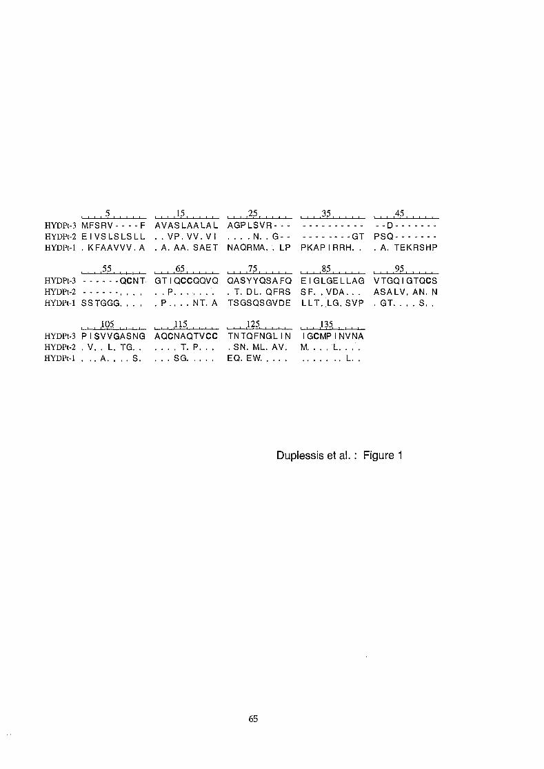
, , , ,5, , , , ,HYDPt-3 MFSRV - - - - FHYDPt-2 El VS LS LS L LHYDPt-l K FAAVVV. A
, , , ,55, , , , ,HYDPt-3 - - - - - - QCNTHYDPt-2 - - - - - HYDPt-l SS TGGG.
, , , ,105, , ! , ,
HYDPt-3 PI SVVGASNGHYDPt-2 V. L.TG.HYDPt-l .. A.•.. S,
, , , ,1;;, , , , ,AVASLAALAL
VP.VV.VIA. AA. SAET
, , , ,65, , , , ,GTIQCCQQVQ
. P.P.. NT. A
, , , ,115, , , , ,AQCNAQTVCC
T. P.SG...
, ! , ,25, , , , !
AGPLSVR- -. N. G - -
NAQRMA. LP
, , , ,75, , , , ,QASYYQSAFQ
T. DL. QFRSTSGSQSGVDE
, , , ,125, , , , ,TNTQFNGLIN. SN. ML. AV.EQ. EW.
, , , )5! ! ! , ,
--------GTPKAP 1RRH.
l , , ,85, , , , ,EIGLGELLAGSF.. VDA ..LLT. LG.SVP
, ! , ,135, l , , ,
IGCMPINVNAM. .L.... " L.
l , , ,45, , , , ,
--D-------PSQ------
A. TEKRSHP
, , , ,95, 1 l , ,
VTGQIGTQCSASALV.AN. N
GT. S.
Duplessis et al.: Figure 1
65
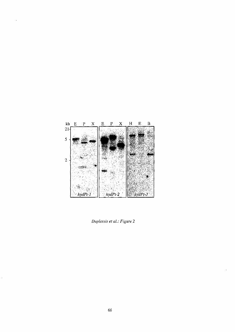
kb E21
5
2
Duplessis et al.: Figure 2
66

A
hydPt-3
5.8SrRNA
B
MycorrhizaF lE MI R
. fi l "'J '!>~ ~,~,~'... ?~{.~,! ~'1' " ! ':,: ;".... ~ 0<-,,'1, . ~
, \' !>- 'J" ,1 • ", ;.~ ,~,~'1,.., 1 ~. ' ! :J
ndF lE LI R
Mycorrhiza
580bp
. 170 bp
Duplessis et al. : Figure 3
67

D Control (5.6 mM glucose and 5 mM ammonium)
• Enriched in glucose (110 mM glucose: 5 mM ammonium)
2 [3 Depleted in glucose (0 mM glucose and 5 mM ammonium) 2
~ Depleted in ammonium (5.6 mM glucose and 0 mM ammonium)
ohydPt-l hydPt-2 hydPt-3
1
o
68
Duplessis et al. Figure 4

Ch~pitre 4
Cata€~éJisa1iion ei aRaillyse de j'e~ptess~on de gènescodant des proféines de \ÇoÏ!es de tr.ansduc~i@n chez
Pis@UfMtlS ti1flctari1!1J8

Caractérisation et étude de l'expression de gènes des voies de
transduction du champignon ectomycorhizien Pisolithus
tinctorius
Ce chapitre présente ma contribution à l'étude moléculaire de gènes des voies de transduction du
champignon ectomycorhizien Pisolithus tinctorius. Ce chapitre regroupe des résultats obtenus au cours
de mon travail de thèse et partiellement au cours de mon stage de DEA de Biologie Forestière
(Duplessis, 1998). Ce travail n'a pas encore donné lieu à des publications d'articles de recherche, mais
certains résultats apparaissent dans des chapitres de livre et des articles de synthèse (Duplessis et al.,
2001a i Martin et al., 2001).
Introduction
Contrairement à d'autres associations établies entre des plantes et des micro-organismes, telle que 1a
symbiose Légumineuses-Rhizobium (Hirsch & McFall-Ngai, 2000), la nature des médiateurs chimiques
symbiotiques, ainsi que leur mode d'action au sein des mycorhizes, sont encore mal définis. Toutefois, des
travaux ont montré que l'auxine induit des modifications morphologiques au sein de la racine (Rupp &
Mudge, 1985). Notamment, l'auxine qui est synthétisée et sécrétée par les champignons
ectomycorhiziens (Gea et al., 1994). Plus récemment, il a' été montré que cette auxine fongique régule
l'expression du gène iaa88 de l'hôte végétal de l'association Pinus pinaster-Hebeloma cylindrosporum
(Charvet-Candela, 2000) et ce dès les stades précoces de mycorhization. Ce gène appartient à une
famille de facteurs de transcription (Aux/IAA) et pourrait donc induire des réponses secondaires,
notamment lors des premières divisions cellulaires observées dans la racine. Il a aussi été montré que des
flavonoïdes (rutine) et des cytokinines (zéatine) des exsudats racinaires d'Eucalyptus globulus ont ID
effet positif sur la croissance des hyphes de P. tinctorius ou altèrent la morphologie des hyphes
mycéliens (Lagrange et al., 2001 i Martin et al., 2001). De plus, l'hypaphorine, le composé indolique
majeur du Pisolithe qui s'accumule sous l'influence de l'hôte dans les tissus mycéliens, agit comme ID
signal en régulant la croissance des poils absorbants de la racine (Béguiristain & Lapeyrie, 1997 i
Ditengou et al., 2000) et en stimulant l'expression du gène EgHypar dans les racines d'E. globulus (Nehls
et al., 1998). Des travaux récents ont permis de montrer que ce composé était un antagoniste de l'auxine,
perturbant la balance hormonale de la racine de l'Eucalyptus (Ditengou & Lapeyrie, 2000). Il a aussi
69
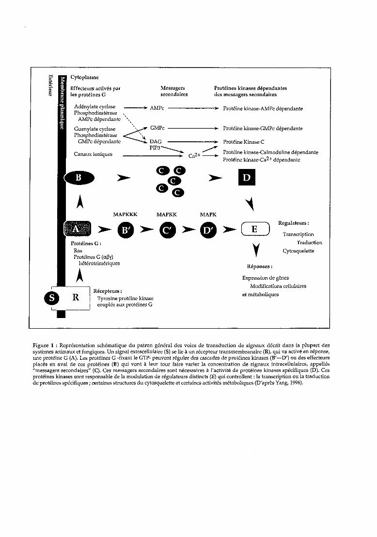
Réponses:
Expression de gènes
Modifications cellulaires
et métaboliques
Protéines kinases dépendantesdes messagers secondaires
--------:.0 Protéine kinase-GMPc dépendante
--------;:.o~ Protéine Kinase C
ca2+::::::: Protéine kinase-Calrnoduline dépendanteProtéine kinase-Ca2+ dépendante
--------:.0 Protéine kinase-AMPc dépendante
Messagerssecondaires
MAPKKK MAPKK MAPK
>- • >- <1 >- 0) >- ( E ) Regulateurs:
Transcription
y Traduction
Cytosquelette
Récepteurs :Tyrosine protéine kinasecouplée aux protéines G
A
Adénylate cyclase ---"':.o~ AMPcPhosphodiestérase ,
AMPc dépendante ""
Guanylate cyclase ~"" GMPcPhosphodiestérase '':4,
GMPc dépendante DAGPIP3----"
:.0Canaux ioniques
Effecteurs activés parles protéines G
Figure 1 : Représentation schématique du patron général des voies de transduction de signaux décrit dans la plupart dessystèmes animaux et fongiques. Un signal extracellulaire (5) se lie à un récepteur transmembranaire (R), qui va activé en réponse,une protéine G (A). Les protéines G -fixant le GTP- peuvent réguler des cascades de protéines kinases (B'-D') ou des effecteursplacés en aval de ces protéines (B) qui vont à leur tour faire varier la concentration de signaux intracellulaires, appellés"messagers secondaires" (C). Ces messagers secondaires sont nécessaires à l'activité de protéines kinases spécifiques (D). Cesprotéines kinases sont responsable de la modulation de régulateurs distincts (E) qui controllent : la transcription ou la traductionde protéines spécifiques; certaines structures du cytosquelette et certaines activités métaboliques (D'après Yang, 1996).
été rapporté que des fragments pariétaux polysaccharidiques, libérés par le champignon Hebeloma
crustuliniforme, déclenchent de nombreuses réponses cellulaires faisant intervenir 1a
phosphorylation/déphosphorylation de protéines dans les cellules racinaires de Picea abies (Salzer et
al., 1997).
Le contrôle morphogénétique s'exerce par des modifications dans l'expression génique au sein du
noyau de chacun des partenaires de la symbiose ectomycorhizienne. Nous savons qu'il existe au moins ID
signal rhizosphérique de Pisolithus tinctorius, l'hypaphorine, perçu par la racine d'Eucalyptus
globulus et y induisant des modifications dans l'expression génique (gène EgHypar). On peut supposer
que l'échange d'information entre la perception de l'hypaphorine et les autres médiateurs chimiques
(auxines, éliciteurs pariétaux) et la régulation transcriptionnelle de EgHypar passe par les voies de
transduction. Il en est vraisemblablement de même chez le champignon bien qu'aucune évidence directe
de stimulation de gènes par des signaux symbiotiques racinaires n'ait été rapportée.
Les voies de transduction de signaux sont connues à travers le règne vivant pour leur rôle clef dans
les processus de prolifération cellulaire, de différenciation, ou encore de réponse rapide d'acclimatation
des cellules face à lm facteur de leur environnement (Gadd, 1994; Yang, 1996). Cette transduction
intracellulaire consiste en plusieurs voies complexes, parallèles ou croisées, faisant intervenir de
nombreux médiateurs protéiques se régulant en cascade par phosphorylation/déphosphorylation ru
interactions protéine-protéine (Hirt, 1999). Les gènes qui codent les composantes de ces voies possèdent
des domaines très conservés d'un organisme à lm autre (Luan, 1998; Dickman & Yarden, 1999; Hunter,
2000).
D'une manière générale, un récepteur transmembranaire qui perçoit un signal extracellulaire
sollicite des GTPases ou protéines G intracellulaires (Figure 1). Cette sollicitation peut être directe ru
peut faire intervenir des protéines intermédiaires dont le rôle sera de rapprocher les protéines G du
récepteur (Boume et al., 1990; Scheffzek et al., 1998; Lorenz et al., 2000). Les récepteurs membranaires
sont susceptibles d'interagir de manière sélective avec plusieurs signaux à l'extérieur de la cellule, mais
aussi avec plusieurs modules de la machinerie intracellulaire. Cette combinatoire complexifie
considérablement les régulations obtenues in fine. Ce premier ensemble de protéines associées à 1a
membrane plasmalemmique, constitue le premier module des voies de transduction. C'est un point clef de
contrôle de la transduction et donc des processus morphogénétiques qui peuvent être déclenchés en aval,
puisqu'il détermine l'orientation de la suite des événements. Un second module implique des protéines
kinases et des protéines phosphatases. Il transmet l'information extracellulaire captée par le biais
d'une cascade de phosphorylation/déphosphorylation (Cohen, 1992) ou par l'activation d'effecteurs
qui modulent le taux de certains messagers secondaires cytosoliques tels que le calcium ou l'AMP cyclique
(Chardin, 1994). Les protéines qui se trouvent en fin de cette cascade constituent un dernier module. Elles
70

Tableau 1 : Liste des EST utilisées lors de cette étude, correpondant à des protéines des voix detransduction de signaux chez Pisolithus tinctorirls. Le pourcentage de similarité a été déterminé àl'aide des séquences en acides aminés déduites des différents ADNe. nt: nucléotides.
ADNc Protéine homologue Espèce % similarité Taille (nt)
6C8 G~ protein/Protein kinase C receptor Neurospora crassa 85% 1154 nt(Cross Pathway Control-CPC2)
785 Ras-like protein Suil/us bovinus 92% 792 nt
9C4 G~ protein/Protein kinase C receptor Neurospora crassa 85% 1076 nt(Cross Pathway Control-CPC2)
llA6 GTP-binding protein 0: subunit (GPA1) Coprinus congregatus 95% 1481 nt
1101 Calcineurine (PLe) regulatory subunit Filobasidiella neoformans 88% 903 nt
llC5 Serine/threonine protein kinase Homo sapiens 48% 1449 nt(raf related protein)

modifient l'état de phosphorylation de facteurs protéiques qui agissent sur la transcription de gènes
cibles, après leur transport dans le noyau (Karin & Smeal, 1992 ; Hunter, 2000).
Au cours des dernières années, plusieurs travaux ont mis en évidence le rôle clef des voies de
transduction dans la différenciation des structures pathogènes et la prolifération cellulaire de plusieurs
champignons filamenteux (Dean, 1997). Ainsi, chez Magnaporthe grisea, l'agent de la pyriculariose du
Riz, l'inactivation expérimentale des gènes codant des protéines des voies de transduction empêche le
développement des appressoria au contact de feuilles de riz (Mitchell & Dean, 1995 ; Liu & Dean,
1997). L'inactivation de gènes codant les sous-unités a et Bde protéines G, l'adénylate cyclase ou la sous
unité régulatrice de la calcineurine supprime la virulence de plusieurs agents pathogènes: Ustilago
maydis, un pathogène du maïs (Regenfelder et al., 1997), Cryphonectria parasitica, l'encre du
châtaignier (Kasahara & Nuss, 1997) Colletotrichum trifoUi, responsable de l'anthracnose de la
luzerne (Truesdell et al., 2000) ou encore Cryptococcus neoformans, l'agent de la méningite chez l'homme
( Li et al., 1997; Alspaugh et al., 1998).
La ressemblance des processus de préinfection chez ces champignons et lors des étapes précoces de
formation de la mycorhize suggère que les protéines des voies de transduction sont susceptibles de jouer
un rôle clef dans le déterminisme du développement de la symbiose. Afin de vérifier le rôle de telles
protéines, j'ai donc cloné et caractérisé plusieurs gènes des voies de transduction, potentiellement
impliqués dans la morphogénèse symbiotique, chez le champignon ectomycorhizien Pisolithus
tinctorius en vue d'une inactivation ultérieure par transgénèse.
Résultats
Afin d'établir le rôle éventuel des voies de transduction au cours du développement de l'ectomycorhize
Eucalyptus globulus-Pisolithus, j'ai envisagé de cloner et de caractériser plusieurs gènes codant des
protéines des voies de transduction du mycobionte. Trois stratégies sont envisageables pour cloner ces
gènes: i) l'utilisation de sondes hétérologues d'ADNe pour cribler une banque d'ADNe d'ectomycorhize,
ii) le clonage par PCR dirigée en utilisant des amorces complémentaires des sites conservés présents
dans la séquence nucléotidique et iii) la recherche des clones d'intérêt au sein d'une collection d'ESTs
(Expressed Sequence Tags ou étiquettes exprimées). C'est cette dernière approche que j'ai développé. En
effet, le séquençage systématique d'ADNe d'ectomycorhizes d'E.globulus-Pisolithus a permis de générer
près de 1000 ESTs de l'organe symbiotique (Tagu & Martin, 1995 ; Martin & Voiblet, 1998; Voiblet,
1999; Encelot, 2000; Voiblet et al., 2001). Parmi ces ESTs, 13 % correspondent à des gènes qui codent des
protéines des voies de transduction (Voiblet et al., 2001). Cette proportion élevée renforce l'idée de leur
rôle probable dans les processus de mise en place de l'organe symbiotique. Il est possible de déterminer
l'origine génomique des gènes des ESTs en se basant sur les similarités de séquence. Le Tableau 1 présente
71
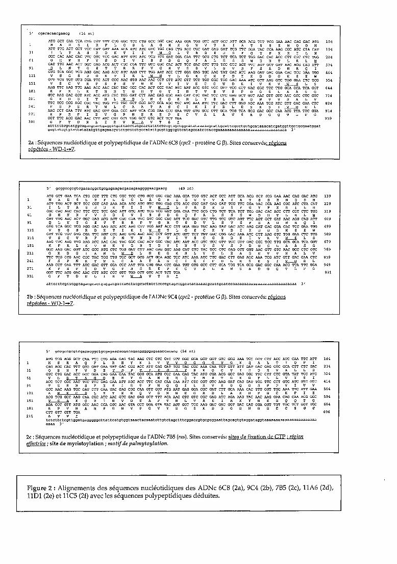
5 1 egaeaeaaegaaeg (14 nt)
~~~~~=~~=~~==~~~~=~=~~~=~~~~=~1~MAESLRFLGSLAGHKGWVTAIATSSENQDM~~~~~=~~~~~=~~~~=~~~~~~~~~=~~=lN
31 IL TAS R D K TIl V...IL.-.Q L T R D E D S F GYP K R l L HGGC CAC AAC CAC TTC GTC TCC GAC ATT GTC ATC TCG TCG GAT GGA CAA TTC GCG CTG TCT TCA TCT TGG GAC CAC ACC CTT CGT CTC ~ 284
61 G H N H F V S DIV l S S D G Q F A L S S S W D H T L R L I:!GAT TTG AAC ACT GGC GAG ACG ACT CGC CGA TTC GTC GGC CAC ACT TCC GAC GTC TTG TCC GTC AGT TTC AGT GCT GAT AAC AGG CAG ATT 374
91 ....llLNTGETTRRFVGHTSDVLSVSFSADNRQIGTG TCA GGC TCG AGG GAC AAG ACC ATC AAG CTT TGG AAT ACC CTT GGA GAG TGC AAG TAT GAC ATC MG GAT GAC GGA CAC TCC GAA ~ 464
ln V S G S R D K T l K L..1'L-.N T L G ECK y D l K D D G H S E W
=~~=~~-=~=~~==~==~=~=~~=~=~~=~ 5~151 V S C V R F S P N V N N P V l V S C G W D K l V K V...lL-.Il L S
AAG TTC AAG TTG MG ACC MC CAC TAC GGC CAC ACT GGC TAC ATC AAT ACC GTC TCC GTT TCC CCT GAC GGC TCC ~ GCA GCA TCA GGT 644181 K F K L K T N H Y G H T G YIN T V S V S PDG S L A A S G
GGC AAG GAC GGT ATC ACC ATG CTC TGG GAT CTT AAC GAG GGC AAG CAT CTC TAC TCC CTC GAG GCT GGT MC GTT GTC AAC GCC CTC GTC 734211 G K D G l T M L ....li-....ll L N E G K H LYS L E A G N V V N A L V
TTC TCG CCG AGC CGC TAC TGG TTG TGC GCT GCG ACT GCA AGC TGC ATC ~ ATC TTC GAC CTT GAG AGC ~ TCG ATC GTT GAC GAA CTC 824241 F S P S R y W L C A A TAS C l KIF D LES KSI...!l.....-.Il E L
AAG CCT GAA TTT ATC GAC GTT GGA CCC AAT TCA CGG GAA CCC GAA TGT GTG GCC CTT GCA TGG TCA GCG GAC GGC CM ACG TTA TTC GGA 914271 K P E F l 0 V G P N S R E P E C V A LAW SAD G Q T L F G
GGT TTC ACG GAC MC CTT ATC CGT GTT TGG GCT GTC ACT TCT TGA 959301 G F TON L l R V kL-..A v T S ~
a t t te t teg tatggeagaegcaetgaggatgat taeetatgeaetat t tcccegt age tggeatataaaaagea t tgca t tegcctgctgcataaacategatggt tge tgegaa tggatgagta tegtgtat tata tatgt tgagaaege tctgcc te tgeeatat tgegt tggcgt tcatageeatcccacegaaaaaaaaaaaaaaaaaaaaaaaaaaaaaa 3 1
2a :Séquences nucléotidique et polypeptidique de l'ADNe 6CS (cpc2 - protéine G 13). Sites eonservés~répétées - WD-l-+7.
5' gcggeegcgtegaeaeggetgegagaagaegacagaaggggaacgaacg (49 nt)
ATG GCT GAA TCA CTG CGT TTT CTG = TCC CTG GCC GGC CAC ~ GGA TGG GTC ACT GCC ATT GCA ACG TCT TCG GAA MC CAG GAC ATG 139MAESLRFLGSLAGHKGWVTAIATSSENQDM
ATT TTG ACT GCT TCC CGT GAT ~ ACA ATC ATC GTC ~ CAG CTG ACC CGC GAT GAG GAT TCG TTC GGA TAC CCA MG CGC ATC CTA CAT 22931 l L TAS R 0 K T l l V ïi..--.Q L T ROE 0 S F Gyp K R l L H
GGC CAC MC CAC TTC GTC TCC GAC ATT GTC ATC TCG TCG GAT GGA CAA TTC GCG CTG TCT TCA TCT TGG GAC CAC ACC CTT CGT CTC TGG 31961 G H N H F V S DIV l S S D G Q F A L S S S W D H T L R L I:!
GAT TTG AAC ACT GGC GAG ACG ACT CGC CGA TTC GTC GGC CAC ACT TCC GAC GTC TTG TCC GTC AGT TTC AGT GCT GAT AAC AGG CAG ATT 40991 Il L N T G E T T R R F V G H T S D V L S V S F SAD N R Q l
GTG TCA GGC TCG AGG GAC MG ACC ATC AAG CTT TGG AAT ACC CTT GGA GAG TGC MG TAT GAC ATC MG GAT GAC GGA CAC TCC GAA TGG 499121 V S G S R 0 K T l K L ~ T L G ECK y 0 l K 0 0 G H S E W
GTT TCG TGT GTG CGA TTC AGT CCC MC GTG MC MC CCT GTT ATC GTT TCT TGT GGC CGG GAC ~ ATC GTT MG GTC TGG GM CTC TCG 589151 V S C V R F S P N V N N P V l V S C G R D K l V K V l1....-.Jl L S
AAG TTC MG TTG MG ACC MC CAC TAC GGC CAC ACT GGC TAC ATC MT ACC GTC TCC GTT TCC CCT GAC GGC TCC TTG GCA GCA TCA GGT 679181 K F K L K T N H Y G H T G YIN T V S V S PDG S L A A S G
GGC MG GAC GGT ATC GCC ATG CTC TGG GAT CTT AAC GAG GGC AAG CAT CTC TAC TCC CTC GAG GCT GGT MC GTT GTC MC GCC CTC GTC 769211 G K D GIA M L ~ L N E G K H LYS L E A G N V V N A L V
~ TCG CCG AAC CGC TAC TGG ~ TGC GCT GCG ACT GCA AGC TGC ATC ~ ATC TTC GAC CTT GAG AGC ~ TCG ATC GTT GAC GAA CTC 859241 F S P N R Y W L C A A TAS C l KIF D LES KSI L-.ll E L
MG CCT GAA TTT ATC GAC GTT GGA CCC MT TCA CGG GAA CCC GAA TGT GTG GCC CTT GCA TGG TCA GCG GAC GGC CM ACG TTA TTC GGA 949271 K P E F l D V G P N S R E P E C V A LAW SAD G Q T L F G
GGT TTC ACG GAC AAC CTT ATC CGT GTT TGG GCT GTC ACT TCT TGA 991301 G F TON L l R V ~ V T S ~
a t t te t tegtatggcagacgcae tgaggatgat tacetatgcac ta t t tceccgtagctggcatataaaaagcat tgcatccaaaaaaaaaaaaaaaaaaaaaaaaaaaa 3 '
2b : Séquences nucléotidique et polypeptidique de l'ADNe 9C4 (cpc2 - protéine G 13). Sites conservés: régiQ!llirépétées - WO-l-+7.
5' gcggccgcgtcgaeaeggctgcgagaagacgacagaagggggagaaaccacacc (54 nt)
31
61
91
ln
151
181
211
ATG TCG AGG GCT CAA TTC CTG AGA GAG TAC MG CTC GTC GTC GTT GGC GGA GGT GGT GTC GGG ~ TCC GCT CTT ACC ATC CAA TTC ATT 144M S R A Q F L REY K L V V V G G G G V G K S A L T l Q F l
CAG AGC CAC TTT GTG GAT GM TAT GAC CCG ACC ATT GAG GAT TCG TAC CGC ~ CM TGT GTT ATT GAT GAC GAG GTC GCA CTT CTC GAC 234QSHFVDEYDPTIROSYRKQCVIDDEVALLD
GTC CTG GAC ACC GCC GGA CAG GAA GAA TAC GGG GCA ATG CGC GAA CAG TAC ATG CGA ACG GGC GAG GGC TTC CTT CTC GTA TAT TCC ATC 324VLpTAGOEEYGAMREQYMRTGEGFLLVYSI
ACC TCT CGC AAT TCC TTC GAG GAA ATT AGC ACC TTC CAT CAA CAA ATT CTC CGT GTC MG GAT CAG GAT TCG TTC CCT GTC ATC GTC GTC 414TSRNSFEEISTFHQQILRVKDQDSFPVIVV
GCC MC MG TGC GAT CTT GM TAC GAG CGC CM GTT GGT ATG AAT GAG GGA CGC GAT CTT GCA ~ CAC TTT GGT TGC ~ TTC ATT GAA 504A N K ç P LEY E R Q V G M N E G R 0 L A K H F G C K FIE
ACG TCG GCC MG CAA CGC ATC MC GTC GAC GAG GCT TTT ACA AAC CTC GTC CGC GAG ATC AGA MG TAC AAC MG GM CAG CAA ACG GGC 594~KQRINVDEAFTNLVREIRKYNKEQQTG
_==~=~~=~~=~~~_=~~=====~=~~==~6UR P V MAN P G N V P G V Y S G S K D D G D H G Gee sac
CTT GTT GTT TGA 696L v V if
tetette tcgt tggatgaggggge t tat tcatgtggtaaactaeaaate t tgtctagc t t tcggaccgtgcgcggaa t tagcagtgtagga tagg taaaaaaaaaaaaaaaaaaaaaaaaaaa 3'
2e : Séquences nucléotidique et polypeptidique de l'ADNe 7B5 (ras). Sites conservés: sites de fixation de GTP ;EgiQn~; site de myristoylation ; motif de palmytoylation.
Figure 2 : Alignements des séquences nuc1éotidiques des ADNe 6CS (2a), 9C4 (2b), 7B5 (2c), 11A6 (2d),1101 (2e) et 11C5 (2f) avec les séquences polypeptidiques déduites.
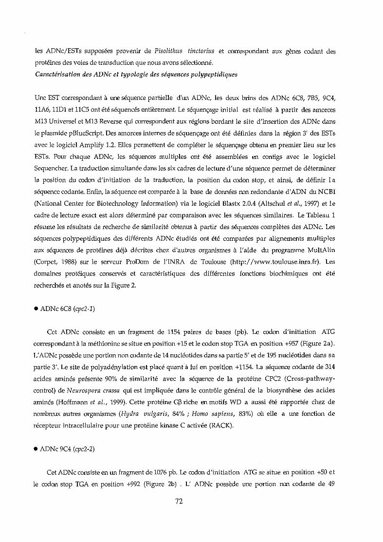
les ADNc/ESTs supposées provenir de Pisolithus tinctorius et correspondant aux gènes codant des
protéines des voies de transduction que nous avons sélectionné.
Caractérisation des ADNc et typologie des séquences polypeptidiques
Une EST correspondant à une séquence partielle d'un ADNc, les deux brins des ADNc 6C8, 7B5, 9C4,
11A6, 11D1 et lIC5 ont été séquencés entièrement. Le séquençage initial est réalisé à partir des amorces
M13 Universel et Ml3 Reverse qui correspondent aux régions bordant le site d'insertion des ADNc dans
le plasmide pBlueScript. Des amorces internes de séquençage ont été définies dans la région 3' des ESTs
avec le logiciel Amplify 1.2. Elles permettent de compléter le séquençage obtenu en premier lieu sur les
ESTs. Pour chaque ADNc, les séquences multiples ont été assemblées en contigs avec le logiciel
Sequencher. La traduction simultanée dans les six cadres de lecture d'une séquence permet de déterminer
la position du codon d'initiation de la traduction, la position du codon stop, et ainsi, de définir 1a
séquence codante. Enfin, la séquence est comparée à la base de données non redondante d'ADN du NCBI
(National Center for Biotechnology Information) via le logiciel Blastx 2.0.4 (Altschul et al., 1997) et le
cadre de lecture exact est alors déterminé par comparaison avec les séquences similaires. Le Tableau 1
résume les résultats de recherche de similarité obtenus à partir des séquences complètes des ADNc. Les
séquences polypeptidiques des différents ADNc étudiés ont été comparées par alignements multiples
aux séquences de protéines déjà décrites chez d'autres organismes à l'aide du programme MultAlin
(Corpet, 1988) sur le serveur ProDom de l'INRA de Toulouse (http://www.toulouse.inra.fr). Les
domaines protéiques conservés et caractéristiques des différentes fonctions biochimiques ont été
recherchés et anotés sur la Figure 2.
• ADNc 6C8 (cpc2-1)
Cet ADNe consiste en un fragment de 1154 paires de bases (pb). Le codon d'initiation ATG
correspondant à la méthionine se situe en position +15 et le codon stop TGA en position +957 (Figure 2a).
L'ADNc possède une portion non codante de 14 nucléotides dans sa partie S'et de 195 nucléotides dans sa
partie 3'. Le site de polyadénylation est placé quant à lui en position +1154. La séquence codante de 314
acides aminés présente 90% de similarité avec la séquence de la protéine CPC2 (Cross-pathway
control) de Neurospora crassa qui est impliquée dans le contrôle général de la biosynthèse des acides
aminés (Hoffmann et al., 1999). Cette protéine G~ riche en motifs WD a aussi été rapportée chez de
nombreux autres organismes (Hydra vulgaris, 84% ; Homo sapiens, 83%) où elle a une fonction de
récepteur intracellulaire pour une protéine kinase C activée (RACK).
• ADNc 9C4 (cpc2-2)
Cet ADNc consiste en un fragment de 1076 pb. Le codon d'initiation ATG se situe en position +50 et
le codon stop TGA en position +992 (Figure 2b) . L' ADNc possède une portion non codante de 49
72
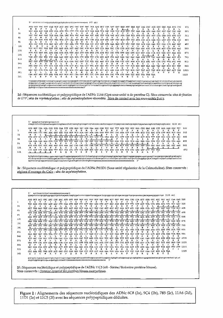
5' cttttttctcctcctctcttgctgtctecctteaeeeeaagag (43 nt)
ATG GGT TGC GTT CAG TeT ACT GGC GTC GAC GGC GAC GCA MG GCe CGA MT GAT GAG ATC GAG MC CAG CTC AGG CGC GAC MA ATG CTC 133H G C v Q S TGV 0 GOA K A R N 0 lL-l E N Q I.. R R Il K H L
GCC AM MC GAG ATC AM ATG TTA CTT CTC GGG GCT GGA GAG TCG GGC MG TCC ACG GTT CTC AM CM ATG AM CTC ATC CAC CAC GGC 22331 A K N E l K H L L L GAG E S G K S T V L K Q H K L l H H G
GGC TAC MC GAC TCG GM CGA GAT TCA TAC MG GM ATC ATC TTT TCG MT ACC ATA CAG TCA ATG CGC GCC ATT CTA GM GCG ATG CCT 31361 G Y NOS E ROS y K E l IFS N T l Q S H RAI L E A M P
CM CTT GAC ATC TCT TTG TCG CCT CM MT GAC GCT CGA CGA GCT GTC ATT CTC GCG CTT CCC ATA CAG ATA GM GGC GAT GTC TTA CCC 40391 QLDISLSPQNDARRAVILALPIQIEGDVLP
AGG GAC GTA GCA GAC GCC ATC CGC GGA CTC TGG AM GAC CCC GGT GTG AM GAG GCT GTC CGT CGG TCT CGG GAG TTC CAG CTG MC GAT 493121 R 0 V A 0 AIR G L W K 0 P G v KEA V R R S R E F Q L N 0
TCC GCT GTC TAT TAT TTC MT GCA ATT GAT CGC ATG GCA GCT CCA GGA TAC CTG CCG ACG GAC CM GAT ATC CTA AGG AGT CGA GTG MG 583151 S A V Y Y F N A l 0 R M A A P G Y L P T 0 Q 0 l L R S R V K
ACG ACC GGT ATC ACG GAG ACG ACA TTC MG GTT GGA GAG CTG ACG TAC CGC TTG TTT GAC GTC GGT GGC CAG CGA AGC GAG CGT MG MG 673181 T 1: G l T .Il T T F K V GEL TYR LED V G G--O.......-.R S .Il R K K
TGG ATC CAC TGC TTC GAG MC GTC ACA GCA CTC GTG TTC CTG GTC AGT TTG AGC GAG TAT GAC CAG ATG CTT TAC GAG GAC GM AGT GTG 763211 ....It l H CEE N V T A L V F L V S L S E y 0 Q M LYE 0 E S V
MT CGT ATG CM GAG GCT CTC ACA CTA TTC GAT TCC ATA TGC MT TCT CGA TGG TTC GTC MG ACA TCG ATC ATT CTG TTT TTG MC MG 853241 N R M Q E A L T L FOS l C N S R W F v K T S l l L FLN K
ATC GAT CTC TTT GCA GAG MG CTG CCT ACC TCA CCT TTG AGC GAC TAC TTT CCT GAC TAT ACG GGC GGT GM MC TAC GAT GCG GCC TGT 943271 I D L F A E K L PTS P L S D y F P D Y T G G E N Y D A A C
GAT TAC CTG CTA CAT CGC TTC GTA GCA CTC MC CAG MT GCT GCC ACG MG CM ATT TAC GCA CAT TAT ACC TGC GCG ACC GAT ACT CAG 1033301 0 Y L L H R F V A L N Q N A A T K Q l Y A H Y T C A TOT Q
CM ATC MG TTT GTG TTG AGT GCG ATC CM GAT ATC CTC CTA CAG ATA CAC TTA CGC GAG TGT GGT TTA TTG TM 1123331 Q l K F V L SAI Q 0 ILL Q l H L R E C G L L ~
tegaggcc tacaa t t tcegt tcacgt teac t tcgagge t t t t taaegcatcgt tcgt tecata tccaggt tcaggtc taggtactcact tgcacac tgt tgt t t t t t te t tatateaeata tggcac te tage tacgca te tcgtc tac tat t t tctcgt taa tacgagtc tacgtgate taec tatgtt t tccc t te tgcaagagg tee taa tgt tcaac tee tcttgccat t tcgtcggcgcgtacgtacgt t tgccecc tcat tee t t tgtgct te t tcacgtt t t tcteac t tcgccc t tgtgggag tca tccaaagcgtatgataeccat t te t tacga t tat tgtgtaatgaa tggacgtcttgaccaaaaaaaaaaaaaaaaaaaaaaaaaaaaaa3 '
2d : Séquences nucléotidique et polypeptidique de l'ADNe llA6 (Gpa sous-unité a de protéine G). Sites conservés: sites de fixationde GTP ; site de myristoylation ; site de palmitoylation réversible. Sites de contact avec les sous-unités ~ et y.
5' ggggtactgcgacgcaactcccaagtte te t tcgaaa tacgacc tcgtaacc te t taccgtgeeagategtgteeateae tccagcc t t tgagcaataatcgcagae tggaeaacag tccttagtcgtcacc (132 nt l
ATG GGT CM ACA CCG TCG CM TTT ATG GAG GAC ATG CM MG CGG TCC MC TTT ACG TCC GCC GM CTG GAG AGG CTT AM MG CGC TTC 222MGQTPSQFMEDMQKRSNFTSAELERLKKRF
ATG MG CTC GAC AGC GAT GGC TCG GGT TCG ATA GAT AGA GM GAG TTT CTC CAG ATA CCT CM ATA GCG TCC MC CCG CTT GCC TCC CGG 31231 M K L D S P G S G S T pRE E F L Q l P Q lAS N PLA S R
ATG ATT Gce ATA TTC GAC GM GAT GGT GGT GGC ACA GTC GAT TTC CAG GM TTC GTT GGT GGT CTT AGT GCA TTT AGC AGC CGC GGT GGC 40261 M lAI F D E D G G G T V P FOE F V G G L S A F S S R G G
CGA GM GAG MG CTA CGA TTT GCC TTC MG GTA TAC GAC GTC GAC CGT GAT GGG TTC ATC TCG MC GGG GM CTG TTC CTC GTG TTG MG 49291 R E E K L RFA F K V Y D V P R P G F T S N GEL F L V L K
ATG ATG GTC GGC MC MT CTT AM GAC CAG CAG CTC CM CM ATC GTG GAT MG ACT ATC ATG GAG GGT GAT CAC GAT CAG GAC GGC MG 582121 M M V G N N L K 0 Q Q L Q Q l V 0 K T l M E G 0 H 0 0 0 G K
CTC AGT TTC GM GAG TTT GCC CAG ATG GTT TCG MT ACG GAT ATC GTA AM CM ATG ACG CTC GAG GAT CTC TTC TM 672151 L S FEE F A Q M V S N T DIV K Q M T LED L F *
tgtgtccctgccaaagtggacagctggctaggacgagacatctcttctaatgtgtccctgecaaagtggacagctggctaggaegagacagatgaagttacgaegccgacgtgcatagtcatcacgcatgccccccataegatattaecccggccgcatggtgtatageetccacacgatttettccegatgaccaccagtgcctcgaggagcgtattaggtcaggatgtagcaatccccagt t tcatgtagaacacae taggttagcate tgcactgtt t t t te t tgtcccgaaaaaaaaaaaaaaaaaaaaaaaaaaaaaa 3 '
2e: Séquences nucléotidique et polypeptidique de l'ADNe Pt11Dl (Sous-unité régulatrice de la Calmoduline). Sites conservés:régions d'ancrage du Ca2± ; site de myristoylation.
5' aattaaccctactaaagggaacaaaagctggagetccecgcggtggcggccgc tctagaactagtggatcccccgggc tgcaggaa t tcgcggccgcgtcgacacggctgcgagaagacgacagaagggggetgc ( 13 5 nt)
ATG GCT ACC TTC ATC CGT MC ATT GTT GGT GGT GCC TTT TCT TAT GTG GCT GGT TTC ATA CTG CCA GCT GGG GCC ACC TCC ACC CM GAG 225MAT F l R N l V G G A F S y V A G FIL P A GAT S T Q E
TGC MG CCG GAC AGT MC CTC CM GTG GAT CTC ACT GAC GAG GTC ACT GAC MG GGG CCT TTC CCT AGT TCA CTA GGA ACG TTC TCT GAT 31531 C K P D S N L Q V D L T D E V T D K G P F P S S L G T F S D
GTT TGG MG TGT GTT AM CGC GCC AGT GGC CCT AM ACC TAT CAC GTT GCC GTC AM GCT ATC CGC ATA CCA ACT GAC GAC GAC TTG ATT 40561 V W K ç V KRA S G P K T Y li V A V K ArR T P T D D D L r
CCA CM AM GCC GM ATG TTG CGC AGC MG GTT CGG GAG TGG ATG AM TTG AM CAC GAC GCT GTA CAC CCT ATT CTG GGC TTT GTC TGC 49591 P 0 K A R M Cf R S K V R R W M K L K fi D A V li P r {, G F V ç
GGA TTT GGT CCT CTC CCA TCT CCT GTT TCA CCT TGG TTG GAT GGA GGG MC CTC ACA CGA TAC CTA AM GCA MT ACA GAG TTG GGC TCA 585121 G F G P L P S P V s P W {, D G G N L T R Y L KAN T R L G S
GAA CGA AGA CTC TAT CTG CTC CAO eGA ATC GCA GAA GGC CTT GAA TAC TTG CAT TCT CM AGG ATC CTC CAC GOT GAC TTG AAC GGG GGC 675151 R R R L Y L If 0 RrA E G LEY L H SOR T {, li G D {, N G G
MT ATC CTC TTT GAC GCT GM GGA AGC CCG TAC ATT GTG GAT TAT GGT TTG TTG CCG TTA ATC CTA GAG TTC AGG ATT TCG CCA TAC CTT 765181 NIL F D A E G S P y r V D Y G L L PLI L E F RIS P y L
TCT ACA CCC GTG GGG CGG GCT GTG AGT GGG TTG CGC GGA TCT CTT CCA GAC TCC ACC AGA TGG GGA TGG GAG AM GCG MT TTC ACA TTC 855211 S T P V G R A V S G L R G S L P 0 S T R W G W E KAN F T F
GM GGC GAC GTG TTA CTC GTT GGT AGT GTG ATG CTT CAG GTT CTG ACT GGC CAG GTT CCA TAC CAT TGT TCC GTT MT CTA GAG TCA GGA 945241 EGO V L L V G S V M L Q V L T G Q V P Y H CSV N LES G
TTT GGC CAT TCC TTT CTA CAC CCG TGG GCG GGC TGT MG GTG GGT TGC GCC GGA TTT TCC AGA CTC CAC CAG ATG GGG ATG GTG AM AGC 1035271 F G H S F L H P W A G C K V G C A G F S R L H Q M G M V K S
GM TTT CAC ATT CGA AGG CGA CGT GTA CTC GTT TGG AGT GTG ATG CTT CAG GTT CTG ACT GGC CAG GTT CCA TAC CAT TCT ATC GAG CGT 1125301 E F H l R R R R V L V W S V M L Q V L T G Q V P Y H SIE R
GAC GM CM GTC ATT TAC ACT ATC GCC CM GGC ATA CGA CCC CGT CGT CCA TCG TCA CAG GAT ATC ATG GAC GAG CAT TGG MC TTC ATT 1215331 0 E Q V l Y T l A Q G l RPR R P S S Q 0 l M 0 E H W N F l
CAG AGA TGC TGG GCT ACT GAT CCC CAG GAT CGT CCC TCG GCC GGG GM TTG GTA GCA TTT TTC CM CGT CAG CGC ATG ACA TM 1299361 Q R C W A T D P Q 0 R P S AGE L V AFF Q R Q R M T ~
qgtgate tgaac tagtt tg te tg t t t tg t teggtcagtat tacat te t tggt ta tee t tcagcat tgtgt tatcgaeagtgtcgtgtacctgagcataa tacgta tgtcac ta t tg taca tgt tggeatgceagaacga tggtccaa t t taaaaaaaaaaaaaaaaaaaaaaaaaaaaaa 3 '
2f: Séquences nucléotidique et polypeptidique de l'ADNe l1CS (stk - Sérine/thréonine protéine kinase).Sites conservés: Domaine conservé des protéines kinases eucarj/otiques.
Figure 2 : Alignements des séquences nucléotidiques des ADNe 6CS (2a), 9C4 (2b), 7B5 (2c), llA6 (2d),llDl (2e) et llC5 (2f) avec les séquences polypeptidiques déduites.

nucléotides dans sa partie 5' et de 85 nucléotides dans sa partie 3'. Le site de polyadénylation est placé
en position +1076. La séquence codante du clone ADNc 9C4 comprend 314 acides aminés et est similaire,
comme l'ADNc 6C8, à la protéine CPC2 de Neurospora crassa (85%). Les séquences de ces deux ADNc
sont très proches et ne possèdent que 3 nucléotides différents dans le cadre de lecture. Cela pourrait
signifier que les deux ADNc correspondent à deux allèles d'un même gène présent dans les deux noyaux
de la souche dicaryotique de Pisolithus. Toutefois, la séquence de l'ADNc 6C8 présente une séquence de
110 nucléotides dans la partie 3' qui n'a pas été détectée dans la séquence du clone 9C4. Ce point renforce
plutôt l'idée de la présence de deux gènes codant cette protéine G~ chez le Pisolithe.
Les séquences polypeptidiques déduites des ADNc 6C8 et de 9C4 (cpc2-1 & cpc2-2) ont été
confrontées à celles rapportées pour N. crassa et pour des protéines G~ d'autres organismes (Sondek et
al., 1996) permettant ainsi de replacer les sept motifs conservés WD (Trp-Asp) caractéristiques de ces
protéines (Figure 3).
• ADNc 7B5 (ras)
Cet ADNc a une taille de 792 pb. Le codon d'initiation ATG se situe en position +55 et le codon stop
TGA en position +694 (Figure 2c). L' ADNc possède une portion non codante de 54 nucléotides dans sa
partie 5' et de 99 nucléotides dans sa partie 3'. Le site de polyadénylation est placé en position +792. La
séquence code 213 acides aminés et présente 92% de similarité avec la séquence d'une protéine de 1a
famille RAS de Suillus bovinus. La séquence codante présente aussi de forts taux de similarité avec les
séquences de protéines RAS d'autres champignons (Coprinus cinereus, 91% ; Lentinula edodes, 87%).
L'alignement multiple de la séquence polypeptidique déduite a permis de déterminer la région
effectrice impliquée dans les interactions protéine-protéine et le motif CAAX (C : cystéine, A : acide
aminé aliphatique, X : acide aminé indéterminé) dans la partie C-terminale qui correspond à un signal
de palmitoylation pour l'ancrage à la membrane plasmique (Figure 2c). Les sites très conservés de
liaison au GTP nécessaires au passage d'un état inactif (liaison au GDP) à un état actif (liaison au GTP)
sont clairement identifiables. Ils sont nécessaires au déclenchement de la voie des MAP kinases par
RAS Ganoueix-Lerosay, 1993).
• ADNc llA6 (Gpa)
Cet ADNc consiste en un fragment de 1481 pb (Figure 2d). Le codon d'initiation ATG se situe en
position +44 et le codon stop TAA en position +1106. L' ADNc possède une portion non codante de 43
nucléotides dans sa partie S'et de 376 nucléotides dans sa partie 3'. Le site de polyadénylation est placé
en position +1481. La séquence code 354 acides aminés et présente 95% de similarité avec la séquence
d'une sous-unité ex. de protéine G hétérotrimérique de Coprinus congregatus. La séquence codante présente
aussi de forts taux de similarité avec des sous-unités ex. de protéine G de champignons filamenteux
73

WDl/6ca 1 MAESLRFLGSLA GH KGWVTAIAT SSENQ DM IL TASR D KTII VWQ L 45WDl/9C4 1 MAESLRFLGSLA GH KGWVTAIAT SSENQ DM IL TASR D KTII VWQ L 45
WD2/6Ca 46 TRDEDSFGYPKRILH GH NHFVSDIVI SS DG QFAL SSSW D HTLR LWD L 92WD2/9C4 46 TRDEDSFGYPKRILH GH NHFVSDIVI SS DG QFAL SSSW D HTLR LWD L 92
WD3/6Ca 93 NTGETTRRFV GH TSDVLSVSF SA DN RQIV SGSR D KTIK L WNT L 135WD3/9C4 93 NTGETTRRFV GH TSDVLSVSF SA DN RQIV SGSR D KTIK L WNT L 135
WD4/6ca 136 GECKYDIKDD GH SEWVSCVRF SPNV NN PVIV SCGW D KIVK V WE L 179WD4/9C4 136 GECKYDIKDD GH SEWVSCVRF SPNV NN PVIV SCGR D KIVK V WE L 179
WD5/6Ca 180 SKFKLKTNHY GH TGYINTVSV SP DG SLAA SGGK D GITM LWD L 221WD5/9C4 180 SKFKLKTNHY GH TGYINTV8V SP DG SLAA SGGK D GIAM LWD L 221
WD6/6Ca 222 NEGKHLYSLEA GN VVNALVFSP SRYWLC AA TASC IKIF D LESK8I VDE L 270WD6/9C4 222 NEGKHLYSLEA GN VVNALVFSP NRYWLC AA TASC IKIF D LESK8I VDE L 270
WD7/6Ca 271 KPEFIDV GI? NSREPECVALAWSA DG QTLF GGFT D NLIR V WA VT8 314WD7/9C4 271 KPEFIDV GI? NSREPECVALAWSA DG QTLF GGFT D NLIR V WA VTS 314
Consensus I?rotéine GI3* GH S DG .. aa .GS. D ..... a WD
Consensus I?rotéine GI3** GH D ... L 8GS D · .IK.L WD L
N ... l AAG · .VR.V.N T
.T. · .L .. I
Figure 3 : Détermination des motifs répétés WD, caractéristiques des protéines G~, au sein des séquencesprotéiques de cpc2 (6CS et 9C4).
*: séquence consensus de protéines G~ (d'après von Weizsacker et al., 1992)**: séquence consensus de protéines G~, déterminée par Müller et al., 1995.
pathogènes (Colletotrichum trifoUi, 84% ; Ustilago maydis, 84% ; Cryphonectria parasitica, 84% et
Magnaporthe grisea, 83%). La séquence polypeptidique déduite de llA6 est très proche de nombreuses
sous-unités lX de protéines G hétérotrimériques. On reconnaît, sur la Figure 2d, les sites de fixation du
GTP et de contact aux sous-unités ~ et y auxquelles elle est accrochée durant sa phase inactive
(Lambright et al., 1996). De plus deux sites d'ancrage à des lipides membranaires sont présents dans 1a
partie N-terminale: un site de myristoylation sur une glycine et un site de palmitoylation régulateur
sur une cystéine.
• ADNc llD1 (CnB)
Cet ADNc consiste en lm fragment de 903 pb (Figure 2e). Le codon d'initiation ATG se situe en
position +133 et le codon stop TAA en position +658. L'ADNc possède une portion non codante de 132
nucléotides dans sa partie S'et de 246 nucléotides dans sa partie 3'. Le site de polyadénylation est placé
en position +903. La phase de lecture code 213 acides aminés et présente 88% de similarité avec 1a
séquence d'une sous-unité régulatrice de la calcineurine (protéine phosphatase dépendante du calcium et
sensible à la calmoduline) de Filobasidiella neoformans. La séquence codante présente aussi de forts
taux de similarité avec la sous-unité régulatrice de la calcineurine d'autres organismes comme
Neurospora crassa (85%), Saccharomyces pombe (81%) et Saccharomyces cerevisiae (79%). La
comparaison de la séquence polypeptidique déduite de l'ADNc llD1 aux séquences décrites chez N.
crassa et Saccharomyces cerevisiae a permis d'identifier quatre sites d'ancrage au calcium très
conservés (Figure 2e) et lm site de myristoylation présent dans la partie N-terminale (glycine en
position 2). Cette sous-unité est assez proche dans sa structure primaire de la calmoduline et elle est
sensible aux flux de calcium dans la cellule (Cohen, 1992).
• ADNc llC5 (stk)
Cet ADNc a une taille de 1449 pb (Figure 2f). Le codon d'initiation ATG se trouve à la position +136
et le codon stop TAG en position +1299. Cet ADNc présente une partie non codante de 135 nucléotides
dans sa partie 5' et de 150 nucléotides dans sa partie 3'. Le site de polyadénylation se situe quant à lui
en position +1449. La séquence code 388 acides aminés et est similaire à des protéines Sérine/Thréonine
kinases (MAPKKK - Mitogen Activated Protein kinases, RAF) de différents organismes (Homo sapiens,
Rattus norvegicus, Fagus sylvtica, Brassica napus). Néanmoins,elle ne présente pas, comme dans les cas
précédents, de similarités très élevées avec les autres protéines « 50%). La confrontation de la séquence
polypeptidique déduite de l'ADNc 11C5, aux profils protéiques répertoriés dans les bases de données sur
le site web PROSITE (http://www.expasy.ch). confirme son appartenance au groupe des protéines
Sérine/Thréonine kinases et permet de replacer dans la séquence lm domaine conservé caractéristique
des protéines kinases eukaryotiques.
74

(A)
An·Aas
AI·Ras
Bt·Rsel
Ct-Ras
Mr-Asel
Mr·Ras3
r------DmII
Sb-Raalp
Ccl·Aas
Le·Ras
(B)
Cp·Cpg·2
Mg·MagA
Sp-Gpal
/Mg·MagC
Sc·Gpa2 KI.opa2
An·FadA
Ca-Cag1
Sc·Opal
Figure 4 : Analyse phylogénétique des GTPases de type Ras (A) et des protéines G hétérotrimériques (B) des champignonsfilamenteux et des levures. Les arbres (non enracinés) ont été construits à l'aide du programme PAUP 4.04 en utilisant lesalignements des séquences réalisés par le programme MultAlin (Corpet 1988). Dans l'arbre en (B), la séquence Um-Gpa a servie deréférence (Regenfelder et al. 1997). Les séquences de Pisolithus tinetorius (ce travail) sont indiquées dans un cadre noir. En (B), lessous-unités a des protéines G hétérotrimériques sont indiquées dans un cadre blanc. Les valeurs de "boostrap" pour (A) et pour (B)ont été obtenues pour 1000 répétitions. Abréviations: Pt, Pisolithus tinetorius ; Cc, Coprinus eongregatus ; Mg, Magnaporthe grisea ; Cp,CryphoHectria parasitiea ; Ne, Neurospora crassa; An, Aspergillus nidulans ; Um, Ustilago maydis ; Pc, Pneumocystis carinii f. sp. earinii; Sp,Schizosaecharomyces pombe ; Cn, Cryptococcus neoformans ; Sc, Saccharomyces cerevisiae ; KI, Kluyveromyces laetis ; Ca, CaHdida albieans ; Sb,suillus bovinus ; Cci, Coprinus ciHereus ; Le, LeHtinula edodes ; Fn, Filobasidiella neoformans var. neoformans ; Af, Aspergillus jumigatus ; Lb,Laeearia bieolor ; BE, Botryotinia fuekeliana ; Ct, Colletotriehum trifoIii ; Mr, Mucor raeemosus. Numéros d'accession(DDBJ/EMBL/GenBank) des séquences des sous-unités a des protéines G hétérotrimériques : Ce-Gpal, X68031 ; Mg-MagB,AF011341; Cp-Cpg-l, 132176; Ne-GHal, L11453; AH-FadA, U49917; Um-Gpal, U85775; Mg-MagC, AF011342; Ne-Gna2, L11452; PePegl, U30791; Um-Gpa2, U85776; Sp-Gpal, M64286; Mg-MagA, AFOl1340; Cp-Cpg-2, 132177; Cn-Gpal, U09372; Um-Gpa3, U85777;Se-Gpa2, U18778 ; KI-Gpa2, L47105 ; Sp-Gpa2, 013366; Ca-Cagl, M88113 ; Se-Gpal, M15867 ; Um-Gpa4, U85778. Numéros d'accession(DDBJ/EMBL/GenBank) des séquences des protéines G de type Ras: Sb-Raslp, AF250024; Cci-Ras, 013295; Le-Ras, D00742; Fn-Ras,AF294647 ; Af-Ras, L42299 ; An-Ras, U03025 ; Lb-Ras, AF034098 ; Bf-Rasl, U79558 ; Ct-Ras, AF044895 ; Mr-Rasl, M55175 ; Mr-Ras3,M55177.
Jld B.lmlll EalRI HII,i1l1 nd s"mlll E('(/RI 11/111111 IId &".m E(ClAI H/Ildlll IId &mlll EroRI IIIHdlIl .d 8Q'mHI EntRI J/l1l/UI IIII1d1I1 B.tmlll E""RI
1.Kb
8Kb
5Kb -
1,6Kh -
~ ...'~
-CnB Gpa stk epe2-1 epe2-2
18Kb
lOKb
5Kb
ras
Figure 5 : Détermination par empreinte électrophorétique selon Sourthern de la représentativité des gènes CnB, Gpa, Stk, ras etcpc2-11cpc2-2 dans le génome de Pisolithus tinetorius. Environ 5 /lg d'ADN total de P. tinctorius ont été digérés par les enzymesde restriction indiquées sur la figure. Les produits de digestion ont été séparés par électrophorèse sur gel d'agarose 1 % (plv),transférés sur membrane de Nylon puis hybridés à l'aide des ADNc radioactifs lIDl, lIA6, lIC5, 6C8 et 7B5 respectivement.Une échelle de taille des fragments d'ADN est indiquée à gauche des autoradiogrammes. nd : ADN génomique non digéré.

L'analyse phylogénétique des séquences polypeptidiques déduites des gènes ras (Figure 4A) et Gpa,
(Figure 4B) qui présentaient les pourcentages de similarités les plus élevés avec d'autres protéines
(respectivement 92% et 95%), a aussi été réalisée. Les cladogrammes ont été construits, à l'aide du
logiciel PAUP 4.üb4a, à partir des séquences des protéines RAS et GPA de levures et de champignons
filamenteux alignées avec les séquences de P. tinctorius en utilisant le programme MultAlin (Corpet,
1988). La protéine RAS est très proche dans sa séquence de celle de Ras1p du basidiomycète Suillus
bovinus. Chez ce champignon deux gènes codant deux protéines Ras distinctes ont été clonés (non publié,
séquences disponibles sur le site du NCBI) alors que les empreintes selon Southern révèlent dans le cas du
Pisolithe la présence d'un seul gène (Cf. Origine génomique des ADNc étudiés). La séquence de RAS
décrite ici est assez éloignée de celle décrite chez un autre champignon ectomycorhizien Laccaria
bicolor où la présence d'un seul gène codant RAS a aussi été détectée (S. Sundaram & G. Podila,
communication personnelle). La séquence déduite de Gpa quant à elle, est regroupée avec deux protéines
Ga décrites chez les champignons Magnaporthe grisea et Cryphonectria parasitica pour leur rôle
majeur dans la mise en place des structures pathogènes.
Origine génomique des ADNc étudiés
Sur la base du pourcentage de similarité de séquence, il apparaît que les différents ADNc étudiés sont
d'origine fongique (tableau 1). La banque d'ADNc d'ectomycorhize ayant été construite à partir de
transcrits d'origine à la fois fongique et végétal. Il était nécessaire de vérifier l'origine génomique des
clones ADNc sélectionnés par empreinte électrophorétique selon Southern (Figure 5) sur de l'ADNg de
Pisolithus, mais aussi d'Eucalyptus. Tous les ADNc - 7B5 (ras), 6C8 (cpc2-1), 9C4 (cpc2-2), llA6 (Gpa),
llDl (calcineurine, CnB) et llC5 (stk) - s'hybrident avec l'ADNg de Pisolithus tinctorius. Pour la
plupart des gènes, un faible nombre de bandes d'hybridation est observé sur les pistes d'ADN génomique
digéré par les enzymes BamHI, EcoRI et HindIII démontrant l'existence d'une seule ou d'un petit nombre
de copies de ces gènes.
Les gènes cpc2-1 et cpc2-2 présentent des signaux d'hybridation similaires dans chaque profil de
digestion, ce qui signifierai que les deux ADNc correspondraient à un seul et même gène. Sur ces
empreintes et sur celle de ras, une seule bande est observée par profil de digestion, ce qui suggère
l'existence d'un gène en copie unique au sein du génome du Pisolithe. L'hybridation du clone llDl (CnB)
en deux points sur l'ADNg fongique digéré par EcoRI s'explique par l'existence d'un site de restriction au
sein de la séquence nucléotidique de l'ADNc, suggérant là aussi l'existence d'un gène en copie unique. Les
empreintes des gènes Gpa et stk présentent plusieurs signaux d'hybridation .par piste d'ADN digéré.
Ces bandes surnuméraires peuvent s'expliquer par la présence d'une ou plusieurs séquences introniques
portées par les gènes qui possèderaient des sites de restriction pour ces enzymes. Nous pouvons aussi
75

9C4 6C8
bande surnuméraire
-epeZ
- ARNr5,SS
Figure 6 : Mise en évidence par empreinteélectrophorétique de type "northern" de deux transcritsepeZ de tailles différentes dans le mycélium de Pisolithustinetorills. Environ 3 /Lg d'ARN totaux extraits dumycélium ont été séparés par électrophorèse sur un geld'agarose en conditions dénaturantes, transférés sur unemembrane de Nylon puis hybridés avec des sondesd'ADNc radioactives correspondant à l'ADNc 9C4 ou 6C8.Une seconde hybridation avec une sonde correspondant àl'ARNr 5,85 a été réalisée afin d'apprécier la présence et laquantité d'ARN sur les pistes.
3,0
2,5 aa
S a~ 2,0<IlQI
'El0'0 1,5v<IlQI~ 1,0QI
./:l,QI
Ei 0,5l'<l
ël0,0
0 3 6
Temps (jours)
B 5 mM de glucose• 110 mM de glucose
a OmM d'ammonium
Figure 7 : Croissance des colonies fongiques de P. tinetorills sur milieu de Pachlewskimodifié (5 mM de glucose, 5 mM d'ammonium), enrichi en glucose (110 mM deglucose, 5 mM d'ammonium) ou appauvri en azote (5 mM de glucose, 0 mMd'ammonium). Les colonies sont cultivées pendant 2 semaines sur milieu dePachlewski modifié avant transfert sur le nouveau milieu de culture à tO. Lesdiamètres ont été mesurés aux temps tO, 3 et 6 jours sur 12 colonies fongiques. Test tde Student (P=0,5).

avoir affaire à des familles multigéniques. La recherche de gènes codant une sous-unité a de protéine C,
par PCR dirigée chez plusieurs champignons filamenteux pathogènes (Magnaporthe grisea,
Cryphonectria parasitica, Ustilago maydis ... ) a d'ailleurs permis la mise en évidence de deux à quatre
gènes de Gpa chez chacune de ces espèces. Il en est généralement de même avec les sérine/thréonine
proteine kinases (Andrews et al., 2000).
Analyse de l'expression de gènes des voies de transduction chez P. tinctorius
Concentration des transcrits chez P. tinctorius
Après avoir sélectionné et caractérisé les ADNc codant des gènes des voies de transduction de
P. tinctorius, l'expression de ces gènes et la régulation de cette expression chez le champignon cultivé
sous différentes conditions expérimentales a été étudiée. Dans un premier temps, la concentration en
ARNm de ces gènes a été estimée chez le Pisolithe. Des empreintes électrophorétiques ADNc/ARN,
réalisées avec 3 p.g d'ARN totaux extraits de colonies fongiques, permettent de détecter les transcrits des
gènes cpc2, ras, Gpa, stk et CnB chez le champignon cultivé sur milieu de Pachlewski modifié. Il est à
noter que plusieurs empreintes réalisées à partir du clone ADNc 6C8 (cpc2-1), ont montré une bande
d'hybridation surnuméraire d'un poids moléculaire supérieur à celui de la bande commune aux
empreintes des deux ADNc correspondant au gène cpc2 (Figure 6). Ce résultat suggère l'existence d'un
épissage alternatif d'un gène unique cpc2 chez le Pisolithe. Le clone 6C8 pourrait être un précurseur de
l'ARNm mature qui correspondrait donc au clone 9C4. Une observation identique a été rapportée lors de
l'étude du gène cpc2 chez la levure Saccharomyces cerevisiae (Hoffmann et al., 1999). De même, chez le
champignon phytopathogène Cryphonectria parasitica, un gène unique codant une sous-unité ~ de
protéine G a été identifié, alors que l'analyse des transcrits par Northern blot a révélé la présence de
deux ARNm de tailles différentes (Kasahara & Nuss, 1997).
Effet de la disponibilité en carbone et en azote et de la mycorhization sur le niveau des transcrits
La régulation des différents gènes dans le mycélium cultivé sous différentes conditions trophiques a été
appréciée. Pour cela, j'ai cultivé le champignon sur milieu de Pachlewski modifié (contrôle) et sur
milieu de Pachlewski supplémenté en glucose ou ne contenant pas d'azote. Des observations au
microscope photonique, menées sur plusieurs colonies fongiques, n'ont pas permis de détecter de
modifications dans la morphologie des hyphes mycéliens soumis à une modification trophique
(résultats non présentés). De plus, des mesures de la croissance mycélienne à partir de la taille des
colonies ont montré que les traitements expérimentaux ne modifiaient pas la croissance du champignon
(Figure 7). La technique des filtres à faible densité en ADNc a été retenue, afin de suivre simultanément
l'expression des différents gènes étudiés, en déposant les 6 ADNc sur des filtres de nylon (Voiblet et al.,
76

300
o 110 mM Glucose Do mM Ammonium • Ectomycorhize
l..r. 1-
J, ,
~ ....-- - _ :L cL-.' ...,...,- l,·. , t.
','" :,
';:,.',."
, "
c
'" l",,
,
'--
Figure 8 : Niveaux d'accumulation des transcrits Ras, CPa, cpc2, stk, et CnB dans l'ectomycorhized'E. globulus-P. tinctorius (en noir) et dans le mycélium de P. tinctorius enrichi en glucose (en gris)ou appauvri en ammonium (en blanc). Les niveaux d'expression sont exprimés par rapport à ceuxobtenus pour du mycélium se développant sur un milieu standard contenant 5 mM de glucose etd'ammonium (valeur 100 : ligne en pointillés). Les expériences ont été répétées deux fois pour lemycélium cultivé sur milieu enrichi en glucose ou sans azote (moyennes et écart-types rapportéssur le graphique).
0 ®
â) 100
fî2
,<l)
80rJlCfJ rJl~~
......~.g ,<l) u.......,p60 ~o cd
~::E .l:l<l) <l)
~~tJ"d 40;:l co "d~
CfJ S ~~.......co 20 ~<.I)~ Z
0 0A B C A B C
Milieux de culture Milieux de culture
Figure 9 : (A) Dosage des sucres solubles de P. tinctorius après 6 jours de croissance surdifférents milieux de culture par colorimétrie (dosage à l'anthrone). (B) Niveau des transcritsSRAP32 du mycélium de P. tinctorius cultivé sur différents milieux de cultures (A, 5 mM deglucose et 5 mM d'ammonium; B, 110 mM de glucose et 5 mM d'ammonium; C, 5 mM deglucose et a mM d'ammonium). Les mesures ont été effectuées sur 20 mg de mycéliumlyophilisé.

2001). En plus des gènes des voies de transduction étudiés, des ADNc fongiques correspondant à des gènes
fortement exprimés (HydPt2 codant une hydrophobine et SRAP32 codant une protéine pariétale régulée
par la symbiose et le glucose) et à des gènes de ménage cellulaire qui sont exprimés de façon stable (ATP
Synthase et rp60), ont été déposés sur les filtres,. La concentration relative des différents transcrits a
été mesurée à l'aide d'un phosphorimageur à partir des empreintes obtenues. Elle est exprimée par
rapport à la concentration des deux gènes de ménage dont l'expression ne varie pas chez le Pisolithe
cultivé sur les différents milieux (Figure 8).
Le mycélium de Pisolithe, transféré d'un milieu contenant 5mM, cultivé sur 110 mM de glucose,
présente une concentration intracellulaire en sucres solubles supérieure à celle du mycélium contrôle
(Figure 9A). Sachant que la protéine SRAP32 voit son expression régulée par la teneur extracellulaire en
glucose (Laurent, 1991), le niveau des transcrits du gène SRAP32 dans ces conditions expérimentales a été
vérifié. Il apparaît effectivement que la concentration des transcrits de SRAP32 est stimulée par l'ajout
de glucose dans le milieu de culture (Figure 9B). Le mycélium perçoit bien le changement de
concentrations extracellulaire et intracellulaire en glucose. Après normalisation des concentrations des
transcrits, il apparaît que le taux d'expression des gènes CnB et stk restent stables. Une légère
augmentation de la teneur en ARNm ras et GPa est mesurée dans le mycélium cultivé sur milieu enrichi
en glucose ou sans azote. Il semblerait donc que la concentration en glucose et en azote extracellulaire
n'affecte que modérément l'expression des gènes étudiés. Nous avons suivi par ailleurs l'expression de
ces gènes dans l'ectomycorhize d'E. globulus-P. tinctorius lors de la formation du manteau (Voiblet et
al., 2001, Chapitre 2). Les données expérimentales obtenues par filtres de faible densité en ADNc sont
similaires à ceux attendus (Figure 8) et seule l'expression des gènes cpc2 et Gpa est modifiée par le
processus symbiotique (150% pour cpc2 et 210% pour Gpa, par rapport au contrôle; Tableau 2).
Effet des exsudats racinaires d'Eucalyptus sur le niveau des transcrits
(étude réalisée en collaboration avec Hubert Lagrange)
Les régulations de faible ampleur observées pour les gènes de communication étudiés, lors de 1a
formation du manteau de l'ectomycorhize E. globulus-P. tinctorius, nous ont amené à nous interroger sur
la possibilité d'une régulation plus précoce des gènes des voies de transduction. Par exemple, lors du
stade de préinfection, lorsque les deux partenaires de la symbiose n'ont pas encore établi de contacts
physiques et qu'un dialogue moléculaire existe déjà par le biais de signaux moléculaires
rhizosphériques. Nous avons testé l'effet des exsudats racinaires d'Eucalyptus globulus et d'une
molécule présente parmi ces exsudats, la cytokinine zéatine, sur l'expression des gènes des voies de
transduction.
Nous avons suivi l'expression des gènes chez le Pisolithe après 9 jours de croissance sur un milieu de
culture contenant des exsudats racinaires d'E. globulus (selon Lagrange et al., 2001) ou de la zéatine (10-6
77

P20+MP20P20+MP20P20+MP20P20+MP20P20+MP20P20+MP20
450
400 D nO-Témoin
D nO-Exsudats
350 • nO-Zéatine2J'S
• Malt-TémoinVI1:: 300b D Malt-ExsudatsVI E:3 Malt-Zéatine<li
'1:l 250....~
~<li... 200::s..<li
~ 150
100
50
0
Figure 10 : Niveaux d'accumulation des transcrits Gpa, stk, cpc2 (6C8 & 9C4), ras et CnB dans le mycélium dePisolithus tinctorius cultivé sur un milieu de Pachlewski modifié (P20) ou un milieu complémenté en Malt (P20+M)contenant des exsudats racinaires d'Eucalyptus globulus (Lagrange et al., 2001) ou 10-6 M de zéatine, une moléculesde ces exsudats. Les niveaux d'expression sont exprimés par rapport à l'expression de gènes de ménage cellulaireprésent sur les filtres de nylon et dont l'expression ne varie pas. les expériences ont été répétées deux fois. Lesmoyennes et les écart-types pour ces deux répétitions sont reproduits sur la figure.

M). Des modifications morphologiques du mycélium, similaires à celles décrites lors des stades précoces
de mycorhization, ont été observées aux concentrations testées. Notamment, on note une modification de
l'ordre de ramification des hyphes mycéliens (Lagrange, 1997) ou encore une augmentation de 1a
synthèse d'hypaphorine (Lagrange, communication personnelle), le composé indolique majeur de P.
tinctorius qui régule par ailleurs le gène EgHypar dans la racine de la plante hôte (Nehls et al., 1998).
Plusieurs gènes des voies de transduction voient leur expression génique modifiée sous l'effet des
signaux testés (Figure 10). Ainsi, le gène stk de Pisolithus tinctorius est légèrement stimulé par les
exsudats de l'Eucalyptus (137%) et par la zéatine (143%), alors que les gènes Gpa et ras le sont plus
fortement (165 à 260% en fonction du traitement, Tableau 2). Par ailleurs, une légère répression de cpc2-1
et cpc2-2 est observée dans ces conditions (50 à 81%).
Tableau 2 : Concentrations relatives normalisées des transcrits ras, cpc2-1, cpc2-2, Gpa, Stk et CnB dePisolithus tinctorius mesurées dans les différents traitements expérimentaux.
Conditions expérimentales ras cpc2-1 cpc2-2 Gpa stk CnB
P. tinctorius témoin(milieu de Pachlewski, 5 mM 100% 100% 100% 100% 100% 100%Glucose et 5mM Ammonium)
Pachlewski supplémenté englucose (110 mM Glucose et 165% 130% 125% 150% 80% 100%mMAmmonium)
Pachlewski aS'pauvri enAmmonium ( mM Glucose et 140% 95% 105% 145% 80% 120%amMAmmonium)
Ectomycorhize E. globulus-P. tinctorius âgées ae 4 jours 125% 150% 130% 210% 85% 85%(Voiblet et al., 2001)
Pachlewski + exsudatsracinaires d'EUCal({6:tus globulus 165% 61% 81% 260% 137% 88%(Lagrange et al., 2 1)
Pachlewski + lO,6M de zéatine 215% 52% 68% 196% 143% 138%
78

Discussion et conclusions
La mise en place de l'organe symbiotique ectomycorhizien s'accompagne de la régulation de nombreux
gènes et de nombreuses protéines (Hilbert et al., 1991 ; Simoneau et al., 1993; Voiblet et al., 2001,
Chapitre 2). Les signaux moléculaires échangés entre les partenaires, durant le stade de préinfection,
affectent l'expression de certains de ces gènes (Nehls et al., 1998). Ces modifications dans l'expression
génique aboutissent en quelques jours au développement d'un organe caractéristique au niveau du système
racinaire (Horan et al., 1988 ; Brun et al., 1995) qui permet à la plante-hôte et au mycobionte
d'améliorer l'assimilation de métabolites nécessaires à leur croissance (Botton & Chalot, 1995; Vivin
et al., 1996 ; Hampp & Wingler, 1997 ; Wallenda et al., 2000).
Les voies de transduction de signaux sont impliquées dans les processus de différenciation et de
morphogénèse de la plupart des organismes (Yang, 1996, Hunter, 2000) même si toutefois, certains
éléments n'ont pas été identifiés dans certains règnes (cas de la protéine Ras chez les plantes; Ma, 1994 ;
'The Arabidopsis Genome Initiative', 2000). Le rôle primordial de ces voies a été mis en évidence dans
l'étude de structures pathogènes de plusieurs champignons filamenteux (Mitchell & Dean, 1995; Xu &
Hamer, 1996; Gold et al., 1997). Ces travaux m'ont amené à m'interroger sur l'implication de telles
voies dans la mycorhizogénèse. Des études réalisées sur les ectomycorhizes, il apparaît que 1a
morphologie des hyphes de Pisolithus tinctorius est affectée par les exsudats racinaires de son
partenaire végétal Eucalyptus globulus (Lagrange, 1997) ainsi que par la rutine, émise par la plante
(Lagrange et al., 2001). De plus, l'hypaphorine émise par Pisolithus est perçue par E. globulus. En effet,
cette molécule stimule l'expression du gène EgHypar dans les tissus racinaires (Nehls et al., 1998). De
même, elle stoppe la croissance des poils absorbants de la racine (Ditengou et al., 2000) et modifie 1a
balance hormonale au sein du système racinaire (Ditengou & Lapeyrie, 2000).
- Dans quelles mesures les voies de transduction sont impliquées dans la transduction de ces signaux?
- Quelle est leur rôle dans la développement de l'organe mycorhizien ?
Afin de préciser le rôle des voies de transduction dans la mycorhizogénèse, nous envisageons
d'inactiver des gènes-clés des voies de transduction chez les deux partenaires de la symbiose E. globulus
P. tinctorius. Pour atteindre ces objectifs, nous devons d'abord cloner ces gènes, les caractériser et étudier
leur expression. J'ai identifié dans la base d'EST de l'ectomycorhize E. globulus-P. tinctorius
(http://mycor.nancy.inra.fr/pages/DNAdatabases/EctomycorrhizaDB.html) plusieurs ADNc codant
des protéines-clés des voies de transduction. Parmi ces ADNc, j'en ai sélectionné six codant des protéines
intervenant dans l'établissement du pouvoir pathogène de plusieurs champignons filamenteux. J'ai
réalisé le séquençage pleine longueur de ces ADNc et caractérisé les protéines qui leur correspondaient
par homologie de séquence. A l'issu de cette analyse typologique, je dispose des gènes codant une sous-
79
unité ex. de protéine G hétérotrimérique, deux protéines G~, une petite protéine G monomérique de 1a
famille ras et une sous-unité régulatrice de la calcineurine, une protéine phosphatase régulée par le
calcium. La caractérisation typologique de ces séquences ADNc nous montre que les protéines de
Pisolithus sont très conservées par rapport à leurs homologues chez d'autres Champignons Supérieurs.
Enfin, j'ai confirmé la présence de ces gènes dans le génome du Pisolithe. Ces travaux présentent 1a
première description de gènes codant des protéines des voies de transduction d'un basidiomycète
mycorhizien.
Les gènes étudiés présentent un intérêt particulier. En effet, les protéines, codées par ces gènes, se
situent pour la plupart en amont des voies de transduction, en contact étroit avec les récepteurs qui
perçoivent les signaux extracellulaires et morphogènes. Les protéines codées par ces gènes ont souvent
été rapportées pour leur rôle déterminant dans la plupart des processus morphogénétiques (Yang, 1996 ;
Boume et al., 1990 ; Janoueix-Lerosay, 1995).
Les sous-unités ~ de protéines G hétérotrimériques sont décrites pour leur fonction régulatrice de
l'activité des sous-unités ex. des mêmes protéines. Elles peuvent également avoir un rôle propre dans le
déclenchement de voies de transduction (Clapham, 1996; Coleman & Sprang, 1996). La protéine ~ de
Pisolithe est proche d'une protéine G~ à motifs WD répétés décrite chez le Rat pour son interaction avec
une protéine kinase C activée. Il a été rapporté que cette protéine interagissait directement avec une
phosphodiestérase AMPc dépendante (Yarwood et al., 1999) et qu'elle ne présentait aucun lien avec des
sous-unités ~ de protéines G hétérotrimériques, en dehors des similarités partagées dans leur structure
primaire. Le rôle encore mal défini des protéines G~ à motifs WD répétés serait de servir de support
dans la mise en place de complexes protéiques (Smith et al., 1999). La protéine PtCPC2 de Pisolithe est
surtout très proche de la protéine CPC2 décrite chez Neurospora crassa et Cpc2p de Saccharomyces
cerevisiae. Cette protéine régulatrice est impliquée dans le contrôle transcriptionnel des gènes
responsables de la biosynthèse d'acides aminés lorsque ce champignon est cultivé sur milieu appauvri en
acides aminés (Müller et al., 1995; Hoffman et al., 1999) avec un mode d'action, toutefois, mal défini.
L'étude de cette protéine chez le Pisolithe présente un intérêt particulier, puisque le métabolisme azoté
est profondément modifié lors du fonctionnement de l'ectomycorhize (Martin & Botton, 1993 ; Plassard
et al., 1997). La caractérisation de ce gène et l'étude de l'expression de ce gène devra être approfondie. Il
est notamment prévu de réaliser des complémentations de souches de Saccharomyces cerevisiae et de
Neurospora crassa mutantes pour cette fonction avec le gène cpc2 du Pisolithe. Elles devraient permettre
de vérifier si la fonction de la protéine CPC2 est conservée chez P. tinctorius.
Plusieurs gènes codant des sous-unités ex. des protéines G hétérotrimériques ont été clonés ces
dernières années chez différents champignons filamenteux pathogènes des plantes ou de l'Homme (Gao
& Nuss, 1996 ; Regenfelder et al., 1997 ; Alspaugh et al., 1997 ; Liu & Dean, 1997 ; Coca et al., 2000 ;
Truesdell et al., 2000). Chez certains de ces champignons, l'inactivation expérimentale de ces gènes a
80
permis de montrer qu'une seule de ces sous-unités était nécessaire à la différenciation de l'appressorium,
la structure de contact, développée par le champignon après perception de son hôte (Alspaugh et al.,
1998 i Kahmann et al, 1999). Cette sous-unité présente une séquence fortement homologue à celle décrite
pour P. tinctorius. Ce point est très intéressant dans l'optique d'une transformation génétique de ce
champignon. En effet, le gène Gpa serait un bon gène candidat pour étudier le rôle des voies de
signalisation lors de la mise en place des interfaces de contact entre les deux partenaires de la symbiose.
Les petites protéines G de la famille Ras se trouvent, comme les protéines G hétérotrimériques, au
contact de la membrane plasmique et elles déclenchent l'activité des MAP kinases, transductrices de
signaux extracellulaires par cascade de phosphorylation (Moodie & Wolfman, 1994 i Jaouneix
Lerousey, 1995 i Reuther & Der, 2000). Ces enzymes régulent de nombreux processus morphogénétiques
chez la plupart des organismes, à l'exception notable des plantes (The Arabidopsis Genome Initiative,
2000). L'implication de ces petites protéines G a souvent été rapportée dans la régulation de la
croissance végétative chez d'autres champignons filamenteux (Roze et al., 1999). Leur rôle a aussi été
particulièrement étudié chez différents organismes modèles (par exemple, Caenorhabditis elegans i
Sternberg & Han, 1998) permettant ainsi de déterminer le rôle central de la protéine RAS dans
différents processus développementaux (Reuther & Der, 2000). De plus, il semblerait qu'un gène codant
une protéine RAS décrite chez le champignon ectomycorhizien Laccaria bicolor soit exprimé
spécifiquement au cours de l'étape de préinfection précédant la colonisation de racines de Pin par ce
Laccaire (G. Podila, communication personnelle). Ce résultat est des plus intéressant car il souligne
l'implication potentielle de RAS dans les premières étapes de mycorhization.
La sous-unité de la calcineurine décrite dans ce travail est dépendante du calcium. Son rôle dans la
trame des voies de transduction cellulaires est complexe. En effet, il a été rapportée qu'elle jouait fi
rôle de régulateur dans le cas de signaux générant des messagers secondaires multiples : le calcium et
l'AMP cyclique (Cohen, 1992). Ces messagers influençant de nombreuses autres protéines lorsque leurs
concentrations varient dans la cellule. Il a notamment été montré que la calcineurine était impliquée
dans la virulence de l'agent de la méningite, Cryptococcus neoformans (Odom et al., 1997 i Alspaugh et
al., 1998). De même, cette protéine phosphatase est impliquée dans la croissance végétative d'autres
champignons. Par exemple, il a été montré que cette protéine était impliquée dans la croissance et la
morphologie des hyphes de Neurospora crassa (Prokish et al., 1997 i Kothe & Free, 1998)
De nombreux gènes codant des protéines kinases ont été décrits ces dernières années chez des
champignons phytopathogènes (Xu & Hamer, 1996 i Xu et al., 1998 i Muller et al., 1999). Leur clonage
fait suite à l'étude des protéines des premiers modules des voies de transduction (protéines G, adénylate
cyclase... ). Les gènes clonés codent différentes protéines kinases (PKA, PKC, MAPKKK). Ces différentes
protéines appartiennent parfois chez ces champignons à des mêmes voies de transduction de signaux
(par exemple la protéine kinase AMPc dépendante et la MAP kinase de Magnaporthe grisea - Thines et
81

al., 2000) et la relation avec le premier module a souvent été démontrée. Leur rôle a été rapporté dans 1a
croissance végétative et la virulence de ces champignons pathogènes (Mayorga & Gold, 1999; Andrews
et al., 2000 ; Zheng et al., 2000). La protéine codée par le gène stk, partiellement décrit au cours de ce
travail possède un domaine conservé spécifique des sérine/thréonine protéines kinases et est proche
d'une RAF (MAPKKK) humaine. Cette protéine appartient au groupe des protéines kinases et elle
pourrait être impliquée dans la transmission de signaux perçus par les protéines G dans les cellules du
Pisolithe.
Avant d'envisager l'inactivation des gènes codant ces différentes protéines par transformation
expérimentale du mycelium de Pisolithus tinctorius, il était nécessaire d'étudier l'expression de ces
gènes ainsi que leur régulation. J'ai montré que les différents gènes étaient exprimés à un niveau
suffisamment décelable chez P. tinctorius. J'ai ensuite suivi leur régulation sous l'effet de modifications
nutritionnelles. En effet, le déclenchement des voies de transduction et la régulation des niveaux
d'expression de nombreux gènes, par le glucose et l'azote, a souvent été rapporté chez les champignons
(Gadd, 1994). De plus, les hyphes du Pisolithe évoluent habituellement dans le sol qui est un milieu
pauvre en sucres. En pénétrant entre les cellules de l'épiderme, puis du cortex racinaire, les hyphes sont
en contact avec des concentrations importantes en ressources carbonées directement assimilables (Hampp
& Wingler, 1997 ; Smith & Read, 1997).
Les niveaux d'expression des gènes stk et CnB ne varient pas dans les conditions expérimentales
appliquées (forte teneur en glucose et carence en azote), alors que l'expression de ras, Gpa et cpc2 est
légèrement stimulée (Tableau 2). Les voies de transduction ne sont donc pas régulées de manière forte au
niveau transcriptionnel par la disponibilité en glucose et en azote. Cette étude des gènes des voies de
transduction n'étant pas exhaustive, il est aussi possible que des signaux engendrés par un changement
de concentration en glucose ou azote extracellulaire, passent par d'autres composantes des voies de
transduction (par exemple, protéine kinases AMPc ou GMPc dépendante). L'analyse des niveaux
d'expression observés pour ces gènes dans l'ectomycorhize de 4 jours ne met pas en évidence de régulation
forte. Toutefois, ce stade de développement correspondant à la formation du manteau, il est tout à fait
possible qu'une régulation plus précoce ait été occultée. Pour tester cette hypothèse, nous avons regardé
l'influence des exsudats racinaires d'Eucalyptus globulus sur l'expression de ces gènes. Nous avons aussi
suivi les effets d'une cytokinine présente dans ces exsudats, la zéatine, qui induit des modifications
morphologiques chez P. tinctorius (Lagrange, non publié). Dans les conditions testées, les gènes ras, Gpa
et stk sont sur-exprimés à différents niveaux (Tableau 2), indiquant l'existence d'une régulation précoce
de ces gènes des voies de transduction fongique. Les signaux racinaires régulent donc l'expression de ces
gènes avant même que les premiers contacts physiques aient lieu entre les symbiotes. De tels résultats
permettraient de discuter de l'existence d'un déterminisme précoce lors de la mise en place de 1a
symbiose.
82

L'existence dans notre modèle d'étude d'une régulation génique de certains gènes fongiques des
voies de transduction se doit d'être complétée par une étude des modifications post-traductionnelles
(par exemple acylation, Gao & Nuss, 1998) de ces protéines. De plus, afin de démontrer le rôle de ces
protéines en dehors de tout phénomène régulatoire (transcription, traduction ou post-traduction), il est
nécessaire de pouvoir inactiver les gènes de signalisation par transformation génétique.
Matériel & méthodes
Le matériel et l'ensemble des méthodes que j'ai utilisé pour réaliser ce travail (matériel biologique, extraction des
acides nucléiques, analyse moléculaire, analyse de l'expression) sont décrits dans les parties Matériel & Méthodes des
articles Voiblet et al., 2001 et Duplessis et al., 2001b, présentés dans les chapitres 2 et 3 de cette thèse. Elles ne sont pas
répétées ici afin d'éviter toute redondance.
Les manipulations expérimentales qui concernent la culture de Pisolithe en présence d'exsudats racinaires de
l'Eucalyptus sont le fait de travaux réalisés par Hubert Lagrange en thèse au sein de l'UMR. Ces parties
expérimentales sont reproduites dans son mémoire de DEA (Lagrange, 1997) et dans un article de recherche (Lagrange
et al., 2001).
Bibliographie
Alspaugh J.A., Perfect J.R. & Heitman J. (1997) CryptocoCCllS neoformans mating and virulence are regulated by the
G-protein a subunit GPA1 and cAMP. Genes and Devel. 11, 3206-3217.
Alspaugh J.A., Perfect J.R., Heitman J. (1998) Signal transduction pathways regulating differentiation and
pathogenicity of Cryptococcus neoformans. Fungal Genet. Biol. 25: 1-14.
Altschul S.F., Madden T.L., Schaffer A.A., Zhang J., Zhang Z., Miller W. & Lipman D.J. (1997) Gapped BLA5T
and PSI-BLAST: a new generation of protein database search programs. Nucleic Acids Res. 25, 3389-3402.
Andrews D. L., Egan J. D., Mayorga M. E. & Gold S. E. (2000) The Ustikago maydis ubc4 and ubc5 genes encode
members of a MAP kinase cascade required for filamentous growth. Mol. Plant-Microbe Interact. 13, 781-786.
Béguiristain T. & Lapeyrie F. (1997) Host plant stimulates hypaphorine accumulation in Pisolithus tinctoriushyphae during ectomycorrhizal infection while excreted fungal hypaphorine controls root hair development.
New Phytol. 136, 525-532.
Botton B. & Chalot M. (1995) Nitrogen assimilation: enzymology in ectomycorrhizàe. In Mycorrhiza: structure,
function, molecular biology and biotechnology. (Edts, Hock B. and Varma A), 5pringer-Verlag, pp325-363.
83
Boume H.R., Sanders D.A. & McCormick F. (1990) The GTPase superfamily: a conserved switch for diverse ceIl
functions. Nature 348, 125-132.
Brun A., Chalot M., Finlay R.D. & Soderstrom B. (1995) Structure and function of the ectomycorrhizal association
between Paxillus involutus (Batsch) Fr. and Betula pendula Roth. I. Dynamics of mycorrhiza formation. New
Phytol. 129, 487-493.
Charvet·Candela V. (2000) Rôle de l'auxine fongique dans la symbiose ectomycorhizienne Hebeloma cylindrosporum/
Pinus pinaster. Identification et caractérisation de gènes de la plante régulés par l'auxine fongique. Thèse de
Doctorat de l'Université Claude Bernard - Lyon I. pp-158.
Clapham D.E. (1996) The G-protein nanomachine. Nature 379, 297-298.
Coca M. A., Damsz B., Yun D-J., Hasegawa P. M., Bressan R. A. & Narasimhan M.1. (2000) Heterotrimeric G·
proteins of a filamentous fungus regulate cell wall composition and susceptibility ta a plant PR-5 protein. Plant J.22(1), 61-69.
Coleman D.E. & Sprang S.R. (1996) How G proteins work: a continuing story. TIBS 21, 41-44.
Corpet F. (1988) Multiple sequence alignment with hierarchical clustering". Nucleic Acids Res., 16 (22), 10881-10890.
Chardin P. (1994) Protéines Ras et transmission des signaux mitogènes. Médecine/Sciences 10, 657·664.
Cohen P. (1992) Signal integration at the level of protein kinases, protein phosphatases and their substrates. TIRS 17,
408-413.
Dean R. A. (1997) Signal pathways and appressorium morphogenesis. Annu. Rev. Phytopathol. 35, 211-234.
Dickman M. B. & Yarden O. (1999) Serine/Threonine protein kinases and phosphatases in filamentous fungi.
Fungal Gen. Biol. 26, 99-117.
Ditengou F.A. & Lapeyrie F. (2000) Hypaphorine from the ectomycorrhizal fungus Pisolithus tinctorius counteracts
activities of indole-3-acetic acid and ethylene but not synthetic auxins in eucalypt seedlings. Mol. Plant
Microbe Interact., 13: 151-158.
Ditengou F.A., Béguiristain T. & Lapeyrie F. (2000) Root hair elongation is inhibited by hypaphorine, the indole
alkaloid from the ectomycorrhizal fungus Pisolithus tinctorius, and restored by indole-3-acetic acid. Planta 211,
722-728.
Duplessis S. (1998) Caractérisation et étude de l'expression de gènes des voies de transduction chez le champignon
ectomycorhizien Pisolithus tinctorius. DEA de Biologie Forestière. Université Henri Poincarré - Nancy l, pp-20.
Duplessis S., Tagu D. & Martin F. (2001a) Living together underground: a molecular glimpse of the ectomycorrhizal
symbiosis In Molecular biologJj of fungal development (Osiewacz, ed.). Mycology Series, Dekker & Dekker, New
York, sous presse.
Duplessis S., Sorin c., Voiblet C., Palin B., Martin F. & Tagu D. (2001b). Cloning and expression analysis of a
new hydrophobin cDNA from the ectomycorrhizal basidiomycete Pisolithus. Soumis à Cur. Genet.
84

Encelot N. (2000) Caractérisation du transcriptome des arbres forestiers. Etude de l'expression génique de
l'ectomycorhize d'Eucalyptus globulus - Pisolithus tinctorius, des racines et des feuilles de Populus x
interamericana. DEA de Biologie Forestière. Université Henri Poincarré - Nancy l, pp-20.
Gadd G.M. (1994) Signal transduction in fungi. In The Growing Fungus, (Edts, Gow N.A.R., Gadd G.M). Chapman &
Hall, London, pp 183-210.
Gao S. & Nuss D.L. (1996) Distinct roles for two G protein a subunits in fungal virulence, morphology, and
reproduction revealed by targeted gene disruption. Proc. Natl. Acad. Sei. USA 93, 14122-14127.
Gao S. & Nuss D. L. (1998) Mutagenesis of putative acylation sites alters function, localization, and accumulation of
a Gia subunit of the chestnut blight fungus Cryphonectria parasitica. Mol. Plant-Microbe Interact. 11, 1130-1135.
Gea L., Normand L., Vian B. & Gay G. (1994) Structural aspects of ectomycorrhiza of Pinus pinaster (Ait) Sol.
Formed by an IAA-overproducer mutant of the fungus Hebeloma cylindrosporum Romagnesi. New Phytol. 128,
659-670.
Gold S.E., Brogdon S.M., Mayorga M.E. & Kronstad J.W. (1997) The Ustilago maydis regulatory subunit of a
cAMP-dependent protein kinase is required for gall formation maize. Plant Cell9, 1585-1594.
Hampp R. & Wingler A. (1997) The role of mycorrhiza. In A molecular approach to primary metabolism in higher
plants. (Edts, Foyer C. & Quick W.P.) Taylor &Francis, London, pp275-292.
Hilbert J.L., Costa G. & Martin F. (1991) Ectomycorrhizin synthesis and polypeptide changes during the early stage
of eucalypt mycorrhiza development. Plant Physiol. 97, 977-984.
Hirsch A.M. & McFall-Ngai M.J. (2000) Fundamental concepts in symbiotic interactions: light and dark, day and
night, squid and legume. J. ofPlant Growth Regul. 19, 113-130.
Hirt H. (1999) Transcriptional upregulation of signaling pathways: more complex than anticipated? TIPS 4, 7-8.
Hoffmann B., Mosch H.U., Sattlegger E., Barthelmess I.B., Hinnebusch A., Braus G.H. (1999) The WD protein
Cpc2p is required for repression of Gcn4 protein activity in yeast in the absence of amino-acid starvation. Mol.
Microbiol. 31, 807-822.
Horan D.P., Chilvers G.A. & Lapeyrie F. (1988) Time sequence of the infection process in eucalypt ectomycorrhizas.
New Phytol. 109, 451-458.
Hunter T. (2000) Signalling-2000 and beyond. Celll00, 113-127.
Janoueix-Lerosey I. (1993) Mode d'action et rôle biologique des protéines de la superfamille ras. Regard sur la
Biochimie l, 44-56.
Janoueix-Lerosey J. (1995) Signalisation intracellulaire par les protéines de la superfamille ras. Regard sur la
Biochimie l, 21-31.
Kahmann R., Basse C. & Feldbrügge M. (1999). Fungal-plant signalling in the Ustilago maydis maize pathosystem.
Cur. Opin. Microbiol. 2: 647-650.
85

Karin M. & Smeal T. (1992) Control of transcription factors by signal transduction pathways: the beginning of the
end. TIBS 17, 418-426.
Kasahara S. & Nuss D.L. (1997) Targeted disruption of a fungal G-protein Psubunit gene results in increased
vegetative growth but reduced virulence. Mol. Plant-Microbe Interaet. 10,984-993.
Kothe G. O. & Free S. J. (1998) Calcineurin subunit B is required for normal vegetative growth in Neurospora crassa.Fungal Gen. Biol 23, 248-258.
Lagrange H. (1997) Echandes de signaux lors de l'interaction précoce entre Eucalyptus globulus et le champignon
ectomycorhizien Pisolithus tinctorius: rôle de quelques flavonoïdes racinaires. DEA de Biologie Forestière.
Université Henri Poincarré - Nancy l, pp-20.
Lagrange H., Jay-Allemand C. & Lapeyrie F. (2001). Rutin, the phenolglycoside from eucalyptus root exudates,
stimulates Pisolithus hyphal growth at picomolar concentrations. New Phytol. 149(2), 349-355.
Lambright D.G., Sondek J., Bohm A., Skiba N.P., Hamm H.E. & Sigler P.B. (1996) The 2.0 A crystal structure of
a heterotrimeric G protein. Nature 379, 211-319.
Laurent P. (1991) Evolution des protéines solubles et quantification de certaines enzymes de la glycolyse de la
mycorhize d'Eucalyptus globulus-Pisolithus tinctorius. DEA de Biologie Forestière. Université Henri Poincarré
Nancy l, pp-26.
Li D., Rogers L. & Kolattukudy P.E. (1997) Cloning and expression of cDNA encoding a mitogen-activated protein
kinase from a phytopathogenic filamentous fungus. Gene 195,161-166.
Liu S. & Dean RA. (1997) G protein ex. subunit genes control growth, development, and pathogenicity of Magnaporthe
grisea. Mol. Plant-Microbe Interact. 10, 1075-1086.
Lorenz M.C., Pan X., Harashima T., Cardenas M. E., Xue Y., Hirsch J. P. & Heitman J. (2000) The G protein
coupled receptor Gpr1 is a nutrient sensor that regulates pseudohyphal differentiation in Saccharomycescerevisiae. Genetics 154, 609-622.
Luan S. (1998) Protein phosphatases and signaling cascades in higher plants. TIFS 3, 271-275.
Ma H. (1994) GTP-binding proteins in plants: new members of an old family. Plant Mol. Biol. 26, 1611-1636.
Martin F. & Botton B. (1993) Nitrogen metabolism of ectomycorrhizal fungi and ectomycorrhiza. Adv. Plant Pathol. 9,
83-102.
Martin F. & Voiblet C. (1998) Large scale sequencing of anonymous cDNA clones. In Mycorrhiza manual. (Edts,
Varma A.), Springer Lab Manual, pp475-493.
Martin F., Duplessis S., Ditengou F., Lagrange H., Voiblet C. & Lapeyrie F. (2001) Developmental cross talking
in the ectomycorrhizal symbiosis : signaIs and communication genes, New Phytol., sous presse.
86
Mayorga M. E. & Gold S. E. (1999) A MAP kinase encoded by the ubc3 gene of Ustilago maydis is required for
filamentous growth and full virulence. Mol. Microbiol. 34, 485-497.
Mitchell T.K & Dean R.A. (1995) The cAMP-dependent protein kinase catalytic subunit is required for
appressorium formation and pathogenesis by the rice blast pathogen Magnaporthe grisea. Plant CeU 7, 1869
1878.
Moodie S. A. & Wolfman A. (1994) The 3Rs of life: Ras, Raf and growth regulation. TIG 10, 44-48.
Muller P., Aichinger C, Feldbrugge M. & Kahmann R. (1999) The MAP kinase kpp2 regulates mating and
pathogenic development in Ustilago maydis. Mol. Microbiol., 34 (5), 1007-1017.
Müller F., Krüger D., Sattlegger E., Hoffmann B., Ballario P., Kanaan M. & Barthelmess I.B. (1995) The cpc-2
gene of Neurospora crassa encodes a protein entirely composed of WD-repeat segments that is involved in general
amino acid control and female fertility. Mol. Gen. Genet. 248, 162-173.
Nehls u., Beguiristain T., Ditengou F., Lapeyrie F. & Martin F. (1998) The expression of a symbiosis-regulated
gene in eucalypt roots is regulated by auxins and hypaphorine, the tryptophan betaine of the ectomycorrhizal
basidiomycete Pisolithus tinctorius. Planta, 296-302.
Odom A., Muir S., Lim E., Toffaletti D.L., Perfect J. & Heitman J. (1997) Ca1cineurine is required for virulence of
Cryptococcus neoformans. The EMBa J. 10, 2576-2589.
Plassard C., Chalot M., Botton B. & Martin F. (1997) Le rôle des ectomycorhizes dans la nutrition azotée des arbres
forestiers. In Champignons et mycorhizes en forêt. Revue Forestière Française, numéro spécial, pp82-98.
Prokish H., Yarden O., Dieminger M., Tropshug M & Barthelmess I. B. (1997) Impairment of ca1cineurin function
in Neurospora crassa reveals its essential role in hyphal growth, morphology and maintenance of the apical Ca2+
gradient. Mol. Gen. Genet. 256, 104-114.
Regenfelder E., Spellig T., Hartmann A., Lauenstein S., BOlker M. & Kahmann R. (1997) G proteins in Ustilago
maydis: transmission of multiple signais? The EMBa J. 16,1934-1942.
Reuther G. W. & Der C. J. (2000) The Ras branch of small GTPases: Ras family members don't faU far from the tree.
Cur. Opin. CeU Biol. 12, 157-165.
Roze L. V., Mahanti N., Mehigh R., McConnell & Linz J. E. (1999) Evidence that Mras1 and Mras3 proteins are
associated with distinct cellular functions during growth and morphogenesis in the fungus Mucor racemosus.
Fungal Genet. Biol. 28, 171-189.
Rupp L. A & Mudge K W. (1985) Etephon and Auxin induce mycorhiza-like changes in the morphology of root
organ culture of Mugo pine. Physiol. Plant. 64, 316-322.
Salzer P., Hebe G. & Hager A. (1997) Cleavage of chitinous elicitors from the ectomycorrhizal fungus Hebeloma
crustuiiniforme by host chitinases prevents induction of K+ and Cl- release, extracellular alkalinisation and
H202 synthesis of Picea abies cells. Planta 203, 470-479.
Scheffzek K, Reza Ahmadian M. & Wittinghoffer A. (1998) GTPase-activating proteins: helping hands to
complement an active site. TIBS 23,257-260.
87
Simoneau P., Viémont J.D., Moreau J.C & Strullu D.G. (1993) Symbiosis-related polypeptides associated with the
early stages of ectomycorrhiza organogenesis in birch (Pendula betula Roth). New Phytol. 124, 495-504.
Smith S.E. & Read D.G. (1997) Section 2: Ectomycorrhizas / Structure and development of ectomycorrhizal roots. InMycorrhizal symbiosis. Academic Press, New-York, pp 163-232.
Smith T. F., Gaitatzes C, Saxena K. & Neer E. J. (1999) The WD repeat: a common architecture for diverse
functions. TIBS 24,181-185.
Sondek J., Bohm A., Lambright D.G., Hamm H.E. & Sigler P.B. (1996) Crystal structure of a GA protein py dimer
at 2.1 A resolution. Nature 379, 369-374.
Sternberg P. W. & Han M. (1998) Genetics of RAS signaling in C. elegans. TIG 14, 466-472.
Tagu D. & Martin F. (1995) Expressed sequence tags of random selected cDNA clones from Eucalyptus globulusPisolithus tinctorius ectomycorrhiza. Mol. Plant-Microbe Interaet. 8, 781-783.
The Arabidopsis Genome Initiative (2000) Analysis of the genome sequence of the flowering plant Arabidopsis
thaliana. Nature 408, 796-815.
Thines E., Weber R.W. & Talbot N.J. (2000) MAP kinase and protein kinase A-dependent mobilization of
triacylglycerol and glycogen during appressorium turgor generation by Magnaporthe grisea. Plant CeIl, 12 (9),
1703-1718.
Truesdell G. M., Yang Z. & Dickman M. B. (2000) A Ga. subunit gene from the phytopathogenic fungus
Col/etotrichum trifolii is required for conidial germination. Physiol. Mol. Plant Pathol. 56, 131-140.
Vivin P., Martin F. & Guehl J-M (1996) Acquisition and within-plant allocation of 13C and 15N in COZ- enriched
Quercus robur plants. Physiol. Plant. 98, 89-96.
Voiblet C. (1999) Etude des modifications du transcriptome d'Eucalyptus globulus et Pisolithus tinctorius au cours du
développement de la symbiose ectomycorhizienne. Thèse de Doctorat de l'Université Paris VI, pp-133.
Voiblet C, Duplessis S., Encelot N. & Martin F. (2001) Identification of symbiosis-regulated genes in Eucalyptus
globulus-Pisolithus tinetorius ectomycorrhiza by differential hybridization of arrayed cDNAs. Plant J. 25 (2),
181-192.
Wallenda T., Strober c., Hogbom H., Schinkel H., George E., Hogberg P. & Read D. J. (2000) Nitrogen uptake
processes in roots and mycorrhizas. Carbon and Nitrogen Cycling in European Forest Ecosystems Ecological
Studies 142, ED Schulze (ed.), Springer Verlag Berlin Heidelberg, pp. 122-143.
XU J-R. & Hamer J.E. (1996) MAP kinase and cAMP signaling regulate infection structure formation and pathogenic
growth in the rice blast fungus Magnaporthe grisea. Genes and Devel. 10, 2696-2706.
XU J-R. ,Staiger CJ. & Hamer J.E. (1998) Inactivation of the rnitogen-activated protein kinase Mps1 from the rice
blast fungus prevents penetration of host cells but allows activation of plant defense responses. Proc. Natl. Acad.Sei. USA, 95 (21), 12713-12718.
88
Yang Z. (1996) Signal transducing proteins in plants: an overview. In Signal transduction in plant growth and
development. (Edts, Verma D.P.s.), Springer-Verlag, Wien, New York, ppl-37.
Yarwood S. J., Steele M. R., Scotland G., Houslay M. D. & Holger G. B. (2000) The RACK1 signaling scaffold
protein selectively interacts with the cAMP-specifie phosphodiesterase PDE4D5 isoform. J. Biol. Chemistry 247,
14909-14917.
Zheng L., Campbell M., Murphy J., Lam S. & XU J-R. (2000) The BMPl gene is essential for pathogenicity in the
gray mold fungus Botrytis cinerea. Mol. Plant-Microbe Interact. 13, 724-732.
89

Discussion générale

L'activité de recherche menée au cours de cette thèse de Doctorat concerne essentiellement l'étude du
développement et du fonctionnement de l'association ectomycorhizienne des arbres forestiers. Dans le cadre de
ce programme développé au sein de l'UMR INRA/UHP, "Interactions Arbres/Micro-Organismes", il
s'agit de caractériser les interactions plantes-champignons et de comprendre les mécanismes
permettant l'intégration de la symbiose dans la biologie de la plante et du mycobionte. Cette
problématique s'étend au-delà des interactions mycorhiziennes ; elle s'intègre pleinement à l'étude
des interactions plantes-microorganismes visant à appréhender les processus relationnels établis au
cours de l'évolution entre les plantes et leurs partenaires symbiotiques, mutualistes ou
phytopathogènes.
Le développement et le fonctionnement des ectomycorhizes est sous le contrôle génétique des
deux partenaires (Rosado et al., 1994a, Rosado et al., 1994b, Tagu et al., 2001). Ce contrôle s'exerce sur :
les processus de reconnaissance plante-hôte/champignon, la colonisation des tissus racinaires, la
morphologie de la racine et des tissus fongiques et la réorganisation du métabolisme des symbiotes.
La compréhension des mécanismes moléculaires induisant et accompagnant l'établissement de
l'ectomycorhize constitue donc l'objectif majeur de ces études (Martin et al., 1997). La découverte en
1988 de protéines dont la synthèse est régulée par la symbiose (protéines SR) a confirmé l'activation ou
la répression de certains programmes géniques lors du développement et du fonctionnement de
l'ectomycorhize (Hilbert & Martin 1988). Ainsi, par des approches complémentaires (séquençage de
protéines SR, gènes candidats, criblage différentiel d'ADNe) les recherches conduites sur l'expression
des gènes ont permis de cloner une dizaine de gènes exprimés et régulés lors de la formation et du
fonctionnement de la symbiose, dont des protéines pariétales fongiques: les hydrophobines et les
SRAPs (Tagu et al., 1996 ; Laurent et al., 1999). Toutefois, de manière à obtenir une vision globale des
fonctions exprimées et des gènes régulés, une approche d'ingénierie génomique a été initiée (Voiblet, 1999)
afin (1) d'identifier l'ensemble des fonctions cellulaires en activité dans la symbiose et (2) d'obtenir une
image plus large des changements de l'expression génique. Ce projet repose sur l'obtention de milliers
d'ESTs et l'analyse de l'expression génique par filtres d'ADNe. De plus, compte-tenu de leur
importance dans le processus de différenciation symbiotique, il s'agit également de poursuivre une
étude détaillée des gènes et des protéines fongiques de l'interface symbiotique (hydrophobines de la
paroi et de la matrice extracellulaire), des membranes plasmalemmiques (FUN34, ...) et impliquées
dans les fonctions cellulaires critiques pour la morphogenèse symbiotique (signalisation,
cytosquelette, contrôle du cycle cellulaire... ).
Ce projet a nécessité, dans un premier temps, la mise en place des outils expérimentaux
indispensables à l'obtention de centaines d'étiquettes de gènes exprimés (ESTs) dans la symbiose et à
l'établissement de leur profil d'expression dans les partenaires non associés et dans l'ectomycorhize. Il
s'agissait (1) de construire une banque d'ADNe d'ectomycorhize d'E. globulus - P. tinctorius, (2) d'isoler
90

Figure 1: Modèle l, radicules d'Eucalyptus globulus mycorhizées parle Basidiomycète Pisolithus tinctorius selon la technique développéepar Malajczuk et al., (1990). Dans ce système expérimental, laradicule est contrainte à un développement plagiotrope. Lesgerminations d'E. globulus âgées d'une semaine sont déposées à lasurface du tapis mycélien de P. tinctorius âgé de 3 semaines. Lesracines sont mycorhizées par le champignon en moins de 10 jours.
SRAP32 Isocitrate Phosphatase acide
Hydrophobines ~ déShYdr\ogénase Glutamate 1\
Chitinases, EgHYP+a/frTubulines déshydrogénasepéroxydases
/ t~~~~
Dj
Préinfection
2j
Colonisation
4j
Morphogénèse
6j
Fonctionnement
Bj
Développement de l'ectomycorhize Eucalyptus - Pisolithus in vitro (Malajczuk et al., 1990)
Figure 2. Exemple de gènes régulés par la symbiose ectomycorhizienne. La différenciation in vitro del'ectomycorhize d'Eucalyptus - Pisolithus peut se subdiviser en quatre étapes principales: préinfection (phaserhizosphérique), colonisation, morphogénèse et fonctionnement. SRAP32, Symbiosis Regulated Acidic Polypeptide(32 KDa) ; EgHypar, protéine induite par la présence d'hypaphorine ou d'auxine; Germ, germination des spores;Pif, préinfection, croissance des hyphes; Bran, ramification des hyphes; Adh, adhérence des hyphes à la surface dela racine; Pen, pénétration des hyphes entre les cellules épidermiques; Man, aggrégation des hyphes pour former lemanteau; Lat, formation de racines latérales; Elon, élongation radiale des cellules épidermiques; Mer, modificationde l'activité méristématique ; Har, différenciation du réseau de Hartig ; C-met, N-met, P-met, modifications desmétabolismes C, Net P, incluant les transferts entre les symbiotes (d'après Martin & Tagu, 1999).

des centaines de clones d'ADNe et de les séquencer (ESTs), (3) d'annoter ces séquences dans une base
de données et (4) de produire des filtres d'ADNe.
Les études effectuées au cours de cette thèse ont consisté à (1) participer à la première étude de
l'expression génique de l'ectomycorhize d'E. globulus - P. tinctorius à l'aide de filtres à faible densité en
ADNe (Chapitre 2, Voiblet et al., 2001), (2) élargir cette analyse en étudiant la régulation de ces gènes
au cours des différentes étapes de la formation de l'ectomycorhize en utilisant des filtres à haute
densité d'ADNe (Chapitre 2, Duplessis & Martin, 2001), (3) développer au laboratoire les méthodes
d'analyse statistique bioinformatique des données d'expression des gènes SR et, (4) approfondir le
mode de régulation des gènes fongiques codant des protéines pariétales, les hydrophobines et de
certaines protéines des voies de transduction (Chapitres 3 et 4, Duplessis et al., 2001).
Dans cette discussion, j'aborderai tout d'abord les facteurs qui ont déterminé le choix des
modèles ectomycorhiziens. Ensuite, je rappellerai et je discuterai les résultats obtenus à l'issue du
séquençage de près de 1000 ESTs d'ectomycorhize d'E. globulus - P. tinctorius. J'interpréterai les profils
d'expression des gènes analysés par filtres d'ADNe au cours des différentes étapes de la formation de
l'ectomycorhize, en soulignant certains aspects méthodologiques, avant de discuter l'expression des
gènes d'hydrophobines et des voies de transduction de signaux de Pisolithus tinctorius cultivé dans
différentes conditions expérimentales.
Analyse du transcriptome de l'ectomycorhize d'Eucalyptus-Pisolithe
Choix du modèle ectomycorhizien et du stade de développement
Le modèle expérimental E. globulus et P. tinctorius est utilisé depuis 1986 au laboratoire: il permet
l'obtention rapide d'ectomycorhizes en conditions contrôlées (boîtes de Pétri). Au cours de ce travail,
nous avons utilisé deux systèmes in vitro différents présentant chacun des avantages et des
inconvénients.
Le premier système (Malajczuk et al., 1990 ; Hilbert et al., 1991) permet l'obtention de
mycorhizes sur la racine pivot de jeunes germinations d'eucalyptus, avant l'apparition des racines
secondaires (Figure 1). Les conditions de synthèse sont loin des conditions naturelles de
mycorhization : la racine a une croissance plagiotrope, elle se développe à la lumière et c'est la
radicule qui est colonisée. De plus, les jeunes germinations étant mises directement au contact de la
colonie fongique, elle se retrouve confrontée en quelques minutes à une biomasse fongique
importante. Dans les conditions naturelles, ce sont les racines secondaires qui sont le plus souvent
colonisées par le mycobionte, et le contact entre les deux partenaires se fait progressivement, après
l'échange de signaux moléculaires agissant probablement par gradient de concentrations. Cependant,
ce système gnotobiotique a l'énorme avantage de produire des mycorhizes dont le développement est
synchrone et de permettre la récolte de plusieurs centaines d'ectomycorhizes à un stade de
développement déterminé. De plus, nous disposons sur ce modèle Eucalyptus - Pisolithus in vitro de
nombreuses informations morphologiques et cytologiques (Malajczuk et al., 1990; Rabbani et al., 1992 ;
91
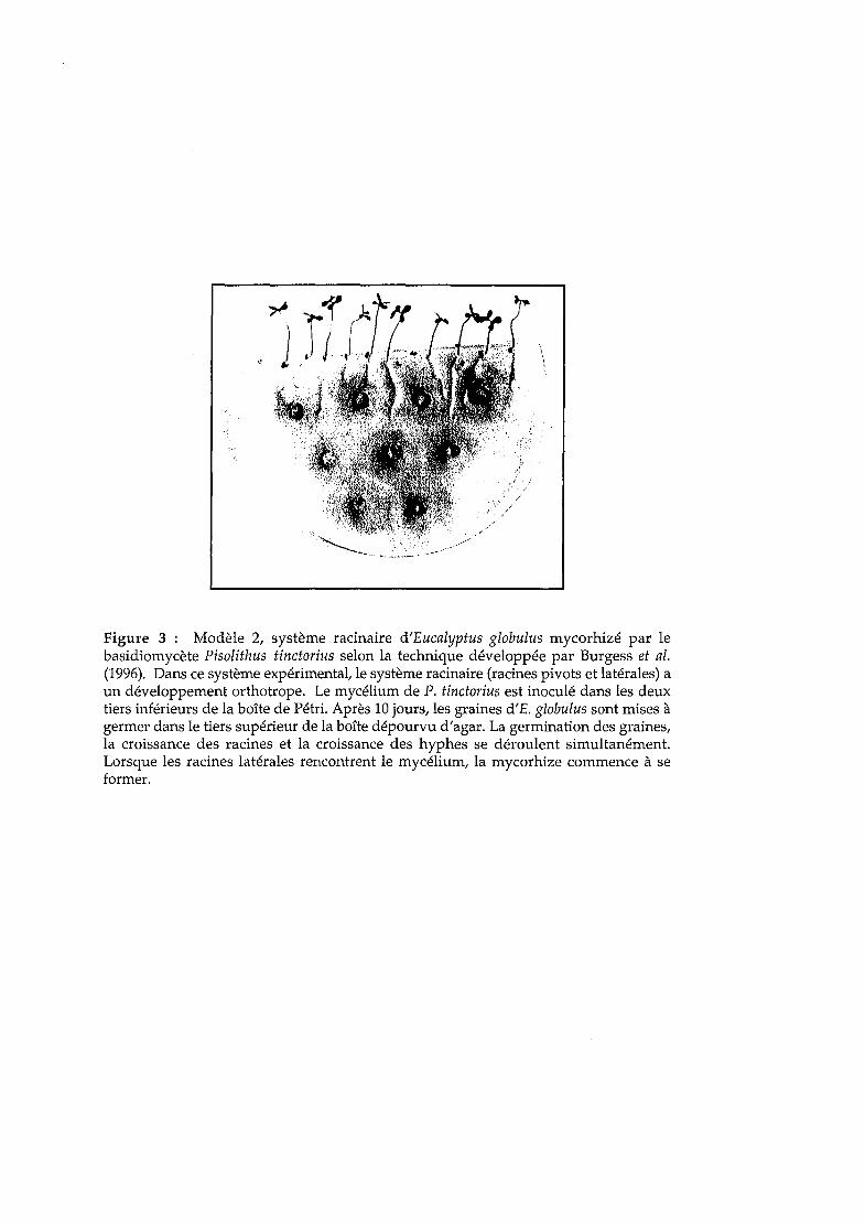
"
Figure 3: Modèle 2, système racinaire d'Eucalyptus globulus mycorhizé par lebasidiomycète Pisolithus tinctorius selon la technique développée par Burgess et al.(1996). Dans ce système expérimental, le système racinaire (racines pivots et latérales) aun développement orthotrope. Le mycélium de P. tinctorius est inoculé dans les deuxtiers inférieurs de la boîte de Pétri. Après 10 jours, les graines d'E. globulus sont mises àgermer dans le tiers supérieur de la boîte dépourvu d'agar. La germination des graines,la croissance des racines et la croissance des hyphes se déroulent simultanément.Lorsque les racines latérales rencontrent le mycélium, la mycorhize commence à seformer.
Dexheimer et al., 1994). Des analyses métaboliques ont démontré la présence d'échanges nutritionnels
entre la plante et le champignon (Rabbani et al., 1992). Enfin, ce modèle a été utilisé pour un grand
nombre d'analyses d'expression de gènes et de protéines (Figure 2; Martin & Tagu 1999). C'est sur ce
système expérimental que la première description du transcriptome de l'ectomycorhize a été obtenue
(Voiblet et al., 2001). Lors de cette première étude, nous avons utilisé des ectomycorhizes âgées de 4
jours. A ce stade de développement intermédiaire, les mycorhizes sont en cours de formation.
L'agrégation du manteau fongique et du réseau de Hartig est bien initiée et un allongement des
cellules rhizodermiques, ainsi que la perte des poils absorbants des racines, sont déjà visibles
(Malajczuk et al., 1990; Dexheimer et al., 1994; Camero Diaz, 1996; Ditengou et al., 2000). Enfin, après
quatre jours de contact, la réorganisation des voies métaboliques est en cours (Rabbani et al., 1992 ;
Boiffin, 1997 ; Boiffin et al., 1998 ; Martin et al., 1998). Ainsi, le stade de développement choisi était
certainement le plus propice à l'identification des gènes impliqués dans la construction des structures
(événements précoces) et dans le fonctionnement métabolique (événements tardifs) propre à la
symbiose.
Le second système expérimental de mycorhization in vitro a été développé par Burgess et al.,
(1996) et permet le développement simultané du système racinaire d'eucalyptus et du mycélium du
mycobionte. Les graines sont mises à germer à l'obscurité dans une boîte de Pétri à distance
d'implants fongiques préalablement inoculés (Figure 3). Ainsi, la germination des graines, le
développement de la racine pivot, et la croissance du mycélium se font simultanément, permettant un
échange progressif des signaux moléculaires nécessaires à la reconnaissance des deux partenaires. Les
racines latérales d'ordre 2 se développent et sont colonisées par le mycélium, alors que la croissance
rapide orthotrope du pivot empêche sa mycorhization (Dexheimer et al., 1994). Ce système de
mycorhization in vitro est vraisemblablement plus proche des ectomycorhizes telles qu'elles se
développent dans la litière. Cependant, ce système présente le désavantage de générer des mycorhizes
dont les stades de développement ne sont pas synchronisés (il n'y a pas synchronisation de
l'apparition des racines latérales). Nous avons décidé d'utiliser ce système expérimental afin de
disposer à la fois des données obtenues sur le modèle des racines pivots mycorhizées et pour la racine
entière portant des racines latérales mycorhizées. De plus, nous avons abordé la régulation temporelle
de l'expression des gènes de l'ectomycorhize d'E. globulus-P. tinctorius, lors des différentes étapes de
développement symbiotique. L'objectif est de déterminer si la mycorhization induit des profils
d'expression variés aux différents stades de développement: phase de colonisation (4 jours post
contact), formation du manteau (7 et 12 jours post-contact), pénétration et croissance intra-racinaire (7,
12 et 15 jours post-contact) et activité métabolique (15,21 et 28 jours post-contact).
Fonctions cellulaires exprimées dans l'ectomycorhize d'Eucalyptus globulus
Pisolithus tinctorius lors de la formation du manteau fongique: analyse des E5Ts.
Les fonctions cellulaires exprimées dans l'ectomycorhize d'E. globulus-P. tinctorius lors de la
différenciation du manteau fongique (racines plagiotropes mycorhizées, 4 jours post contact) ont été
caractérisées en analysant la nature des EST exprimées dans les tissus symbiotiques (Voiblet 1999).
Cette analyse a été effectuée sur un premier lot de 650 ADNc et a été complétée en collaboration avec
92

Nathalie Encelot (Encelot 2000) afin d'atteindre un millier d'EST. Ces EST proviennent de deux types
de criblages d'une banque d'ADNc d'ectomycorhizes Eucalyptus-Pisolithus de 4 jours (Voiblet, 1999).
Cette banque a été établie selon un protocole (CLONTECH) basé sur la reconnaissance de la coiffe
méthylguanoside dans la partie 5' des ARN messagers, par l'hybridation d'un oligonucléotide
spécifique dans cette partie 5' et de l'amplification d'ADNc par RT-PCR. Ce protocole permet un
enrichissement de la banque en ADNc pleine longueur (Diatchenko et al., 1996 ; Sasaki et al., 1998).
L'étape d'amplification par PCR des ADNc n'entraîne pas de biais dans la population d'ADNc
amplifiés par rapport à la population d'ARNm de départ. Sasaki et al. (1998) ont utilisé ce protocole
pour la construction d'une banque de blastocystes de souris et ne soulignent aucun biais dans la
représentation des transcrits. A partir de cette banque d'ADNc, nous avons récupéré aléatoirement
environ 1000 clones bactériens (Voiblet et al., 2001) lors d'un premier criblage aléatoire. Parmi ces
ADNc, environ 700 ont été partiellement séquencés, avec succès, à partir de leur extrémité 5'. Environ
75% de ces clones possèdent un codon ATG (début de traduction) et 71% seraient des clones pleine
longueur, possédant à la fois le codon ATG et une queue polyA+. Un second criblage différentiel par
SSH (Hybridation Soustractive et Suppressive, CLONTECH) a permis de caractériser 182 EST
supplémentaires. Ces EST correspondent à des gènes de mycorhize différentiellement exprimés par
rapport à la racine non mycorhizée et au mycélium libre (Voiblet & Martin, 1999 ; Encelot, 2000 ;
Voiblet et al., 2001).
Les séquences nucléotidiques obtenues ont été éditées (élimination des séquences
correspondant au vecteur, correction des bases ambiguës), puis comparées aux séquences répertoriées
dans les bases de données internationales à l'aide du programme WU-BlastX 2.0 (Altschul et al., 1997).
Les comparaisons de séquences ont été réalisées sur le site Web du Baylor College of Medicine
(Beauty). Ce dernier permet une analyse automatique groupée (en batch) grâce à un script MacPerl,
MacSearchLauncher (ftp:/ / dot.imgen.bcm.tcm.edu/pub / software/ search-launcher).
Les résultats obtenus au cours de ces différents criblages d'ADNc ont été comparés. Plus de 50% des
gènes collectés par la technique de SSH sont des gènes SR (Voiblet, 1999 ; Voiblet & Martin, 1999).
Cette efficacité de la SSH à cloner des gènes différentiellement exprimés ou spécifiques d'un tissu a
déjà été soulignée (Diatchenko et al., 1996 ; Yang et al., 1999). Il faut noter cependant la présence de
18,6% d'ARNr 28S récupérés par SSH. L'analyse de ces séquences d'ADNr indique l'existence d'une
séquence longue très riche en A qui a probablement été reconnue par l'amorce polyT lors de la
synthèse par la transcriptase inverse du premier brin d'ADNc. Environ 50% des ESTs présentent une
similitude avec des séquences présentes dans GenBank. Les séquences annotées sont répertoriées sur
le serveur Web du laboratoire (http://mycor.nancy.inra.fr/pages/DNAdatabases/EctomycorrhizaDB.html).
La répartition quantitative des différentes protéines homologues en catégories correspondant
aux grandes fonctions biochimiques indique que plus de 30% des gènes exprimés dans
l'ectomycorhize codent des protéines impliquées dans la synthèse des ARNm/protéines (protéines
ribosomiques, sous-unités du protéasome, facteurs d'élongation). Près de 13% des gènes identifiés
codent des protéines du métabolisme primaire ou secondaire (ATP synthase, glycéraldéhyde-3-P
déshydrogénase, alanine aminotransférase). Plus de 15% des séquences exprimées codent des
93
protéines pariétales (SRAPs, hydrophobines, extensines). Cette image correspond bien à l'activité d'un
organe en construction à forte activité assimilatrice.
Parmi les 324 protéines pour lesquelles une fonction potentielle a été établie, 89% partagent une
homologie avec des protéines dont la fonction est connue et 11% avec des protéines identifiées, mais
dont la fonction n'est pas élucidée (SRAP17, FUN34). Quarante quatre pour cent des ESTs homologues
présentent une similarité avec des séquences d'origine fongique, essentiellement avec les levures
(Saccharomyces cerevisiae et Schizosaccharomyces pombe), Neurospora crassa ou le Basidiomycète Agaricus
bisporus. Trente sept pour cent des ESTs sont similaires à des séquences d'origine végétale et
notamment d'Arabidopsis thaliana,Oryza sativa, Glycine max et Zea mays. Enfin, 19% des séquences
présentent une homologie avec divers organismes Eucaryotes ou Procaryotes comme Homo sapiens ou
Mus musculus. Nous avons classé ces ESTs (http://mycor.nancy.inra.fr/pages/DNAdatahases
/EctomycorrhizaDB.html) selon la fonction cellulaire potentielle qui leur a été assignée, en 8 catégories
définies dans la base EGAD (Expressed Gene Anatomy Database
http://www.tigr.org/tdb/egad/egad.html).
Nous pouvons remarquer que la majorité des ADNc clonés est homologue de gènes codant des
fonctions de ménage (30,5%). Ceci confirme que l'ectomycorhize est formée de tissus très actifs, lieu de
synthèse protéique importante puisque lors de la différenciation de l'ectomycorhize, les hyphes
fongiques se multiplient abondamment pour former le manteau et le réseau de Hartig (Massicotte et
al., 1990). Le transcriptome de nombreux tissus humains présente un très fort pourcentage (environ
30%) de gènes liés à l'expression des gènes et à la synthèse des protéines (Adams et al., 1995) et la
même observation a pu être faite au regard des analyses génomiques menées chez différents
champignons (Goffeau et al., 1996; Nelson et al., 1997 ; Ospina-Giraldo et al., 2000 ; Qutob et al., 2000)
ou plantes (Hofte et al., 1993; Newman et al., 1994; Gyorgyey et al., 2000 ; White et al., 2000).
Enfin, environ 15% des gènes inventoriés par cette étude correspondent à des protéines
structurales et 13% à des polypeptides intervenant dans les voies de transduction de signaux. Ceci
confirme l'importance de la communication cellulaire et de la restructuration des parois lors du
développement de l'ectomycorhize (Martin et al., 1999 ; Martin et al., 2001)
Grâce aux ESTs que nous avons produites, nous avons pu obtenir, pour la première fois, une image
globale des fonctions exprimées dans une ectomycorhize. Toutefois, la production de près de 1000 ESTs ne
constitue que l'ébauche d'un inventaire des gènes exprimés dans la symbiose. En effet, il est probable que le
nombre de gènes présents chez Eucalyptus globulus soit proche de celui d'Arabidopsis thaliana (soit 25 000
gènes environ; The Arabidopsis Genome Initiative, 2000) et que Pisolithus tinctorius se rapproche des
ascomycètes Neurospora crassa et Aspergillus nidulans pour lesquels environ 12 000 gènes ont été identifiés.
Construction et analyse des filtres à faible densité en ADNe.
Après cette phase d'inventaire, les ADNc séquencés ont été déposés sur des filtres d'ADNc, à faible
densité (96 ADNc par filtre), pour mesurer le niveau d'expression des gènes. Ces étapes ont nécessité
94
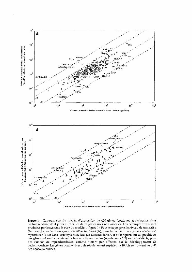
//A
lOS r----------------------------...,....----,.-----,,.".
,..""..""
,.•.,
.,'"..... //
,....•....,....•.. / /// //0 5C6 ///
./". IDA4 789 / ./' .
..,........ ..... lO~g'?9/ 0 0 7E~' / •..•,.•/,••..,••
,,/ ./ S 0 .// 00 lE9 ...-
SC6NIPSN.P,J'J 0// &00 H .Lpt_5
..•..•..° Q .•' 00/ ,-SfJRO O/yaP, .....,../,..{ 4 8<?>000&~//~ eF-la , ..
U6snRNA.9··· 120_,'~Ol/ 0 H llP; 4associaled ).'rtilein a:L IfrD~ Ô () cP./ 'f .
,/.,'.., 6Ô~ ~~ 0 ..•);lyliPI.6
........ ~!WI . 0 ·····IF4A
105 Rahl/Rad5l / ..,.•.,...- ~8#~~~-gà~ F~N34/H1;j;;~e
° ...~/° / cf; 0 89o~p /,...•.,/ 0 0°00/0' ••.•,'..
........ °0 0/00 ....····4 ...•.... / SRAP17 ...7liio
10 • xl0 / 0 ~O . ~.ER6;;··'···/ 0 ..;....
/
mitMDH..,..... ··,··x2.5
..•....•..•..103 L----'x~2-"l.5~__--lI'xlU!0~ ~---------------_--------1
103
Niveaux normalisés des transcrits dans )'eclomycorhize
x2.5
B
108 ,..- --..- ----:_----,
..•,...,..,....,.// /""''Â~: /Ahduce~lrol~~~!k/
MataIlPltil~·~·~;~~ 1/ / ,....,....,...'" ,/...-~./\ !t G3PDH / .---.//
1204 ....,,/ • ",- '" ....... ABA'" ..,..,....•....
......,.. Inducad r aln '"
POI~jJIJ'q'~ltln '" '" '" ,/.,.0"'''••,., '" '" A 1. ,..
./ '" ~ ,/.,.•,/ '" '" '" '" SEND32 ..
,.' '" ~/'" .....ATP ~y.n!h~~e '" 4 ~ ~ '" '" A,...····,··
...... 8·chain '" "'A'" '" '" "')dl'il:Î'...... d ",. '" C0'P9
...... h '" ~ ","'P~!.Ï1'I/Cyt c O~yd'~;e .'//", '" '" ~r
104<10 '" •.,...../.,.""'...---
.....,........,..•...
.~..103 '--_-'-X;:;2.:.;5/ ...:;X:::;10~."_,._·..._·"_· -----------------'
10310
410
5 106 107lOS
Niveaux normalisés des transcrits dans )'eclomycorhize
Figure 4 : Comparaison du niveau d'expression de 400 gènes fongiques et racinaires dansl'ectomycorhize de 4 jours et chez les deux partenaires non associés. Les ectomycorhizes sontproduites par le système in vitro du modèle 1 (figure 1). Pour chaque gène, le niveau de transcrit aété mesuré chez le champignon Pisolithus tinctorius (A), dans la racine d'Eucalyptus globulus nonmycorhizée (B) et dans l'ectomycorhize (axe des abcisses dans A et B) et reporté sur un graphique.Les gènes qui sont localisés entre les deux lignes pleines (régulation x 2,5) sont considérés, pourdes raisons de reproductibilité, comme n'étant pas affectés par le développement del'ectomycorhize. Les gènes dont le niveau de régulation est supérieur à 10 fois se trouvent au delàdes lignes pointillées.

de nombreuses optimisations techniques (Voiblet, 1999) et nous nous sommes attachés par la suite à
développer l'analyse de filtres à haute densité en ADNc (2000 ADNc par filtre).
La première approche a été de déposer manuellement des séries de 96 clones ADNc, amplifiés
par PCR, sur filtres de Nylon. Nous avons ainsi généré plusieurs ensembles de filtres portant au total
plus de 450 ESTs, correspondant aux gènes étudiés et à des gènes marqueurs fongiques et racinaires.
Les gènes correspondant aux ESTs ont été ordonnés par fonctions cellulaires sur les filtres
(métabolisme, expression, stress... ) et nous avons suivi leur expression chez chacun des partenaires en
culture pure et dans l'ectomycorhize au moment de la formation du manteau fongique (4 jours après
contact).
Nous avons ainsi obtenu les profils d'expression de 400 gènes (328 gènes fongiques et 72 gènes
racinaires), générant ainsi la première analyse de transcriptome d'une ectomycorhize (Voiblet et al.,
2001). Dans l'ectomycorhize développée sur la racine plagiotrope d'Eucalyptus, 17 % des gènes sont
régulés (niveau de régulation :2: 2,5) à la fois dans la racine et le champignon (Figures 4A et 4B).
Toutefois, les profils d'expression obtenus n'ont pas mis en évidence des gènes spécifiques uniques de la
symbiose. Le gène le plus fortement stimulé (x 12), eIF4a, code un facteur d'initiation de la traduction
de la famille des "dead-box" hélicases d'origine fongique. D'autres gènes impliqués dans la traduction,
tel que le facteur de transcription tefl, sont stimulés indiquant une régulation de la traduction. Ces
résultats sont en accord avec l'augmentation de la biosynthèse des protéines fongiques observés dans
l'association E. globulus-P. tinctorius (Hilbert et al., 1991) liée à la forte activité de division cellulaire des
hyphes mycéliens lors de la colonisation du système racinaire. Chez le champignon, l'expression de
deux familles de gènes codant des protéines pariétales abondantes (SRAP32 et hydrophobines) sont
également fortement stimulées. La forte synthèse de ces protéines pariétales fongiques avait été
préalablement observée par 2D-PAGE et par l'analyse des niveaux de transcrits par Northern blots
(Tagu et al., 1996; Laurent et al., 1999), validant les données acquises par filtres d'ADNc (Figure 4A).
Chez Eucalyptus globulus (Figure 4B), les gènes fortement stimulés comprennent ceux codant une 7-0
méthyltransférase (synthèse des phénylpropanoïdes) et de nombreux composants des voies de
dégradation des protéines (ubiquitine/protéasome). Ce résultat conforte les observations effectuées
par 2D-PAGE d'une dégradation des protéines racinaires au cours du développement de
l'ectomycorhize (Hilbert et al., 1991). Il est à noter que de nombreux gènes réprimés dans la symbiose
appartiennent à l'hôte végétal: ils sont impliqués notamment dans les réactions de stress et le
métabolisme respiratoire. Enfin, parmi les gènes SR mis en évidence dans cette étude se trouvent de
nombreux gènes sans fonction connue. Ces gènes pourraient coder des fonctions spécifiques à la
morphogénèse mycorhizienne.
A ce stade de la thèse, nous disposions donc d'outils solides pour analyser le transcriptome symbiotique.
Nous avons cherché à compléter l'analyse du développement de la symbiose ectomycorhizienne en suivant les
profils d'expression au cours d'une cinétique de mycorhization. Nous supposions qu'à chaque phase du
développement symbiotique correspondaient des profils d'expression spécifiques (régu Ions).
95
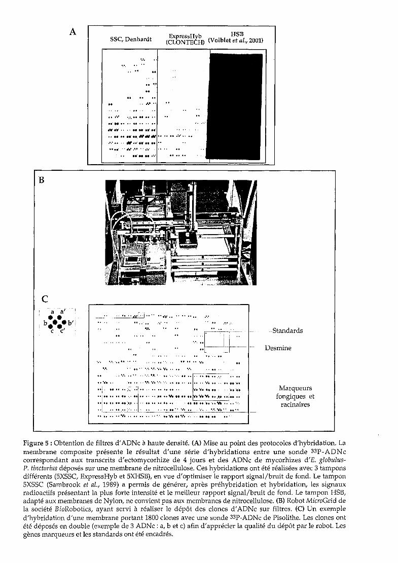
ASSC, Denhardt ExpressHyb HSB
(CLONTECH) (Voiblet et al., 2001)
........ ..
.....••..
tt .. N·t
Il •••• • ••• Il tt Il ••
...... Il •• " fI " Il
N~" .••-... ,'fi""01 •• Il NttItI"" Il Il Il ••••••••
• '.' Il " "fI., " .-•• t.' " "" .
Il .- .
B
c
",," Il ••••••
....",f. Il,,
Il Il tl .,
··::~Ir~·.Standards
Desmine
•••• ••••.•.• Il •• •• Il ••••••••
•••• •• II·· ,,', l,'. •••• •••• •• •• ••••
. , ','. •• ,. '.\ •••• ','. If ',", ..... "•• Il ~-•• Il Il.... 1-' 1.
1 •••••• , ••••• , •••••••••••• ',', Il Il •••• Il •••••• Il •. Il t. '••••~.~. Il ,- Il If Il Il'. • •••••• Il Il, _ ..••.•.•
... Il If Il Il.,' Il Il , •••.•••• v. Il ......•..... v•••.•}; Il Il "
•• Il Il Il Il :',' •••• Il Il Il Il •• ' • Il Il Il '" 'f ',',
............ -f' •••••••• '0' Il •••• Il Il •• '.. ,. '
Marqueursfongiques et
racinaires
Figure 5 : Obtention de filtres d'ADNc à haute densité. (A) Mise au point des protocoles d'hybridation. Lamembrane composite présente le résultat d'une série d'hybridations entre une sonde 33P-ADNccorrespondant aux transcrits d'ectomycorhize de 4 jours et des ADNc de mycorhizes d'E. globulusP. tinctorius déposés sur une membrane de nitrocellulose. Ces hybridations ont été réalisées avec 3 tamponsdifférents (5XSSC, ExpressHyb et 5XHSB), en vue d'optimiser le rapport signal/bruit de fond. Le tampon5XSSC (Sambrook et al., 1989) a permis de générer, après préhybridation et hybridation, les signauxradioactifs présentant la plus forte intensité et le meilleur rapport signal/bruit de fond. Le tampon HSB,adapté aux membranes de Nylon, ne convient pas aux membranes de nitrocellulose. (B) Robot MicroGrid dela société BioRobotics, ayant servi à réaliser le dépôt des clones d'ADNc sur filtres. (C) Un exempled'hybridation d'une membrane portant 1800 clones avec une sonde 33P-ADNc de Pisolithe. Les clones ontété déposés en double (exemple de 3 ADNc : a, b et c) afin d'apprécier la qualité du dépôt par le robot. Lesgènes marqueurs et les standards ont été encadrés.
Caractérisation des profils d'expression génique de Pisolithus tinctorius et
d'Eucalyptus globulus au cours du développement de la symbiose
ectomycorhizienne.
Avant d'aborder l'analyse des différents stades de développement, nous nous sommes attachés à
maîtriser la production et l'analyse de filtres à haute densité en ADNc (2000 ADNc/filtre). Le passage
a une forte densité en acides nucléiques sur le support d'hybridation (environ 25 clones/cm2) a
nécessité une optimisation des techniques utilisées jusqu'alors. A partir de la bibliographie disponible
concernant les études de génomique fonctionnelle menées chez d'autres organismes (Nguyen et al.,
1995; Piétu et al., 1996 ; Richmond et al., 1999; Bowtell, 1999), différentes procédures d'hybridation ont
été testés et la méthode la plus adaptée à notre système a été sélectionnée. Les protocoles
d'hybridation ADNc/ADNc, la nature des tampons d'hybridation, les conditions de lavage des filtres
de nitrocellulose et le radioélément utilisé (Figure SA) ont été choisis afin d'obtenir le meilleur signal
d'hybridation et le rapport signal/bruit de fond le plus élevé (Duplessis & Martin, 2001 ; Chapitre 2,
p.43). Enfin, à partir de gènes marqueurs et de standards internes déposés sur les filtres, nous avons
pu apprécier la reproductibilité entre expérimentations des données et nous assurer ainsi de la fiabilité
des résultats générés.
Neuf cents ADNc ont été amplifiés à partir de colonies bactériennes correspondant à la banque
d'ADNc de l'ectomycorhize E. globulus-P. tinctorius, parmi lesquels 715 ESTs différentes et de
nombreux gènes marqueurs. Les gènes marqueurs correspondent à : un gène humain, la desmine
(Desprez et al., 1998), qui n'existe ni chez la plante, ni chez le champignon; 23 gènes fongiques et 7
gènes racinaires dont la régulation ou la stabilité d'expression est connue et qui ont été placés de 2 à 4
fois en des points différents des filtres; et des contrôles de spécificité d'hybridation (plasmide, site de
multiclonage qui encadre les ADNc, oligonucléotide polyA de 15-18 nt et tampon d'amplification des
réactions de PCR). Nous nous sommes ensuite rendus dans le laboratoire "DNA array" de la société
Eurogentec (Saraing, Belgique) afin de déposer les ADNc amplifiés (25 clones/cm2) à l'aide du robot
MicroGrid (BioRobotics, Figure SB). Les ADNc ont été déposés sur les filtres sans classement préalable
et chaque ADNc a été déposé deux fois, afin de s'assurer de la fiabilité des dépôts par le robot.
Nous avons obtenu les profils d'expression de 715 gènes (608 gènes fongiques et 107 gènes
racinaires) dans la racine d'Eucalyptus non mycorhizée, dans le mycélium végétatif du Pisolithe et
dans les racines mycorhizées 4,7, 12, 15, 21 et 28 jours après contact entre les partenaires (colonisation
de la surface racinaire, formation d'un manteau fongique autour de la racine, pénétration du
mycélium dans le cortex racinaire et formation du réseau de Hartig, organe mixte en fonctionnement).
Cette analyse a généré plus de 20 000 mesures de concentration de transcrits et nécessitait donc
l'introduction d'outils informatiques et mathématiques plus puissants que ceux utilisés préalablement.
En effet, il est difficile d'établir des relations entre les variations d'expression de tous les gènes par une
visualisation en nuage de points (Scatter plot, Figure 6), comme nous l'avions fait précédemment
(Voiblet et al., 2001; Cf. Figure 4). Une telle représentation ne permet de distinguer que les gènes le
plus régulés (~ 2,5 fois).
96

ID'
10',"-0;-'--"''''''''--'=;.------'0.,.'-----'0.,..-----'0.,.'------'".
10·,----------------------,.....__-,--_-,./
,/,,/
ID'
ID'
g ID'
'".5..~ ~. 10'd~~]]:qgo.~
'0'10'
Nonnallzed mRNA Levelln 4-day-old eClomycorrhlza Normalized mRNA Levelln 15-day-old ectomycorrhlza
10'
,,'g'". 10'~~~ ..... .<o.~.~a;s 10''" . "0.~ ~ .';lo.p~
'0'10' 10'
Nonnalized mRNA Levelln '·day-old ectomycorrhlza
'0'
10'
~.5 10'
i~~~~:l' 10's .~
"'::lH~'::o 0
10'Z
'0'lai 10 4 10'
Nonnallzed mRNA Level in 28'11ay-old eclomycorrhiu
10'/'
/,/'
'0' ,/
g,/.
'".../~/,/
~~ --Il'~ §<o.
~·r::l 10'
H~
NOI\-tyrosil\l!prolein!r:ln.ue
1
!10'
10'l.5
Figure 6 : Comparaison du niveau d'expression de 715 gènes fongiques et racinaires dans l'ectomycorhize après 4, 7, 12, 15, 21 et 28 jours dedéveloppement et chez chacun des partenaires non associés. Les mycorhizes sont produites par le système in vitro du modèle 2 (figure 3). Lesniveaux de transcrits normalisés du champignon (ronds) et de la racine (triangles) sont rapportés sur l'axe des ordonnées et les transcrits demycorhize sur l'axe des abcisses. Les traits en pointillés limitent les niveaux de régulation de 2,5 et 10 X (= [ARNm] mycorhize/[ARNm] mycéliumvégétatif ou [ARNm] mycorhize/[ARNm] racine non mycorhizée).

®Synthèse de
mycorhizes in vitro,
~ ..~
auster
Treeview
@
PhosphorIma~er
®
r
gènes marqueurs
!"., •••••• ".' ", y. •••• •••• :.:. ;.;.
••
•
1•Cluster
hydrophobines
®
~p-TranscriV ~
ClusterSRAP17/SRAP32
~ènea
gèneb
~ènec
Figure 7 : De l'ectomycorhize aux profils d'expression... A. Synthèse d'ectomycorhizes d'Eucalyptlls-Pisolithus et récolte à six stadesde leur développement (4, 7, 12, 15, 21 et 28 jours après contact entre les symbiotes). B. Dépôts d'ESIs sur des filtres de nitrocellulose par robotique(Eurogentec, Belgique) panni lesquels 715 ADNe uniques, fongiques et racinaires, et une gamme de gènes marqueurs et de contrôles de spécificitéd'hybridation. Chaque ADNe a été déposé deux fois sur les filtres. C. Hybridation des filtres à haute densité d'ADNe avec les sondes complexesmarquées au [33P)dCfP et [33P)dATP et synthétisées à partir des transcrits extraits des mycorhizes, de mycélium de Pisolithe et de racined'Eucalyptus. D. Qu.antification des niveaux d'expression il l'il'Ïde d'un PhosphorImager et mesure des niveaux de régulation des gènes au cours dudéveloppement de l'ol'ganv.! mixte. Il f..esgèI1P.'t'l O.lIl.t Hé reg,mupés à\ l'aide c!ea logiciels Ouster et Treeview en fonction de la similarité de leur niveaude régulation au. ((Jurs du pt-oœss~lS 1l11j'COI:hi.ziei\i. Les fl'l1es dont, l'e~pressi.ol'i. est sffimul<'~ S\(}nt représentés en rouge; ceux dont l'expression estréprimée sont repré&~nb!:s el'< vert. Le-s gèRes mm re)?iUlés serif el< ooü'S. P. Urte <maJy~ dad.isliqlJ.eJ des gèn('.s codant des protéines structurales duchampignon P. tirrcfcrius. 0Th notera les ~!'I.'ou~s cle gènes presentant d:e.s profils d'expression el d:e varri.ati:ons temporels similaires.

~~~~~~ÀA~
A .-786.-60
. r-l«W, .-2llZ
r-lI>'8, r-13A<1
r-&a.....,.......... f<.--<'C'.-!J('J.1"-2 HOf,--~m
.....<JO<r-7 t:.?r-l3C~
r-1301r-9C8r-11E4r-4A8r-EiPt.<:088('-1Wr-ICf1.-2A4r-llNAJ"-E~,.-2C9r-384r-209r-9A7r-9 E1,-109r-13C8r-13C3r-l3Mr-8 E9r-802r-6A4r-St158r-3B2r-009r-608r-2610,-683r-2 E2r-!Clr-9 E4,.-9 E9r-4A2r-l3M.-1204r-l0A7r-Etptc0127r-EiPt.c«::22r-13C9,-Mar-EiPt;dC32r-EiPtdC48r-EgP~
r-EiTl.ba.-302r-6C7,-3C8r-7 [0r-8C2r-lOO71"-3 E6r-llA9r-EiPt.dD68r-EiPtdC47r-1l82r-2 E8r-l~
r-1003r-l3Alr-2ÇOr-6C5r-14 EZr-fiP~
r-UE61'-1206r-7ll1r-600r-EePtdC82r-Ei ptd0163r-JAlr-6C2r-3O:l"-965r-aOJr-3A6r-8A5r-2010t'-BAlr-9(3r-B08r-EgPW0541"'-966r-EgPtd01391"-6 E-'r-7t8r-I2011'-1010r-14 E4r-7D7r- 0
B
slze=79
size=46
Figure 8 : Recherche des patrons d'expression génique à l'aide des méthodes statistiques deregroupement par profil de régulation. (A) Un exemple de regroupement hiérarchisé réalisé avec lelogiciel Cluster (logiciels Cluster & Treeview, http://rana.lbl.gov)àpartirdesvaleursderégulation* desgènes racinaires. (B) Exemples de profils de régulation générés à l'aide des Self Organizing Maps (logicielGENECLUSTER, http://www.genome.wi.mit.edu/). Les gènes sont placés dans des sous-ensembles dontles moyennes~ et les écart-types r-- sont présentés (ici, 9 sous-ensembles, cO à c8, comprenant de 6 à29 gènes, générés à partir des données d'expression des gènes racinaires). (C) Exemple de profils derégulation obtenus à l'aide des k-means (logiciel EPCLUST sur http:/ / ep.ebi.ac.uk/EP/EPCLUST) àpartir de valeurs de régulation des gènes fongiques. Le logiciel génère un listing (non montré) et unereprésentation graphique des rapports d'expression des gènes pour chaque regroupement effectué. Enexemple, les représentations grap.mques pour deux regroupements de gènes (46 et 79 gènes fongiques)stimulés lors des premiers: sfrades de mycorhÎlzation (le temps de développement est placé en abscisse et leniveau de régulation en mode ïogarithn.1.ique, est donné en ordonnées).
* rapport [transcri!s ectomy,c.odlize]/[transcrits racine non associée]

Différentes approches d'analyses statistiques ont été utilisées ou développées au sein de
plusieurs laboratoires (Eisen et al., 1998; Tamayo et al., 1999). Ces analyses statistiques permettent de
dégager, à partir des données d'expression, des groupes de gènes partageant un mode de régulation
identique ou similaire dans différents traitements ("synexpression"). Parmi ces analyses, on distingue
généralement celles qui se basent sur un tri hiérarchisé (les données sont ordonnées au sein d'un
même groupe) et celles qui procèdent à une classification non hiérarchisée (les données sont triées
sous la forme de sous-ensembles distincts). Nous avons utilisé plusieurs logiciels, permettant de
réaliser ces deux types d'analyses, afin de tirer le maximum d'informations de nos données
d'expression, mais aussi pour corréler ces informations entre elles et rendre nos analyses plus fiables.
En effet, aucun consensus n'existe quant à l'emploi de ces méthodes statistiques. Ces dernières sont
réalisées sur des matrices, de distances euclidiennes ou de corrélations de Pearson, calculées à partir
des rapports d'expression géniques obtenus dans les différents traitements. Ces matrices nécessitent
une transformation préalable des données (normalisation) dans un mode adéquat aux calculs
statistiques (mode logarithmique). Avant de procéder à ces analyses, nous avions réalisé une analyse
de la variance (ANOVA) sur l'ensemble des données. Cette analyse préalable permet de vérifier que
les gènes s'expriment bien d'une manière significativement différente au cours du développement de
l'ectomycorhize.
Les logiciels Cluster et Treeview, développés par M. Eisen (Eisen et al., 1998), ont été utilisés
afin de distinguer les patrons de modulations temporelles de l'expression génique. Ces logiciels
permettent de réaliser des analyses de distance sur matrice euclidienne et ainsi, facilitent le
regroupement cladistique des gènes présentant un comportement d'expression similaire ("guilt-per
association"). La représentation par cladogrammes et matrices colorées des ensembles de gènes et des
niveaux de régulation permet d'acquérir à la fois une vision globale sur l'ensemble des gènes et plus
ciblée sur un gène ou une catégorie cellulaire (Figure 7). Cette visualisation permet à
l'expérimentateur de conserver une partie intuitive dans l'analyse en aval du tri statistique réalisé par
le logiciel (Brown & Botstein, 1999). Si cette approche par tri hiérarchisé permet dans sa représentation
d'avoir une vision simultanée de l'ensemble des données, elle tient compte dans son algorithme de
l'amplitude des régulations de l'expression dans chacun des traitements. Nous avons donc procédé à
des tris non hiérarchisés afin de distinguer des groupes de gènes qui pourraient avoir des profils de
régulation identiques quelque soit leur niveau de régulation.
Nous avons réalisé ces tris à l'aide de deux approches statistiques différentes, les SOMs (pour
Self Organizing Maps) et les k-means (figure 8), qui sont les principaux modes d'analyse qui aient été
appliquées jusqu'à présent lors d'études de transcriptomes (Tamayo et al., 1999). Les algorithmes sur
lesquels reposent ces analyses diffèrent dans le mode de regroupement, mais ils se basent sur une
même représentation vectorielle où 'n' gènes sont représentés dans un espace à 'k' dimensions, 'k' étant
le nombre de traitements de l'expérience. Ainsi, dans le cadre de notre étude, nous avions 715
gènes/vecteurs, dans un espace à 6 dimensions qui correspondaient aux différents temps de la
cinétique de mycorhization. Ces analyses ont été réalisées à l'aide des logiciels Cluster et Genecluster
pour les SOMs et avec EPCLUST, utilisé en ligne sur le site web de l'Institut Européen de
Bioinformatique EBI (http://ep.ebLac.uk/EP/EPCLUST) pour les k-means (figure 8).
97

Tableau 1. Nombre ~t pi}W'Cen.hlge' de gèm,es-SR mis en. évidence lorsdu développement de l'edomycorruze E. globulus-P, tinctorius
Rapport 4db 7d 12d 15d 21d 28dd'expression"
~2 126 (17.5%)< 102 (14.5%) 164 (23%) 162 (23%) 79 (11%) 112 (15.5%)~4 27 (3.5%) 23 (3%) 30 (4%) 19 (2.5%) 12 (1.5%) 10 (1.5%)~ 10 3 (0.5%) 3 (0.5%) 4 (0.5%) 3 (0.5%) 1 «0.5%) 2 «0.5%)
E5Ts d'E. globulus 50 (46.5%)d 55 (51.5%) 54 (50.5%) 59 (55%) 43 (40%) 38 (35.5%)~2
E5Ts de 76 (12.5%)" 47 (8%) 110 (18%) 103 (17%) 36 (6%) 80 (13%)p, tinctorius ~ 2"Rapports des concentrations de transcrits: Mycorhize/Partenaires non associés. Les gènes de ces catégoriesprésentent un rapport supérieur ou égal à la valeur donnée. b stade de développement de l'ectomycorhize (de4 à 28 jours). <Pourcentage calculé sur 715 E5Ts. dPourcentage calculé sur 107 ESTs. " Pourcentage calculé sur608 E5Ts.
-2-----
L,diil*~r-6C3r-l0B4....r-l0A8r-6C!lr-808r-7D7r-1D10r-8A7r-7C8r-9B6r-6 EOr-9C3r-œlr-aA!lr-803r-3œIr-3A6r-701r-6010r-9Il9r-1003r-9A7r-&:7r-1OO7r-EQtt,jpar'r-lD9r-Q)J
r-519r-6A8r-382r-0A4r-aD2,.-9 E4,.-9 E9r-1007r-3C8r-3 ESr-6B3r-9 Elr-WZr-302r-1204r-7 E5r-906r-OAOr-9C8
405 rlbosOMl proutn S5prollne-rlch proutnprol ine-rich protel"ADP-rlbosylatlon f'actor40S rlbosoaal proUln S26urine carbo~tlclase
unknown proutnublqultln conJueatlnt onzyoooC!;jtoplasllI1c aldolaseAIlP-RlBOSYlJlTl~ FACTlRaquaporln "lpCoJbsclslc stress ripent"' protetn60$ ,.IOO$o"al prott'tn LiO60S rlbosoul protetn LiOil!jCoraldohyde-3-phosphalo ~naseJllelallothioneoln-l1k.e protet"rlpening-Induœd protet"Willer-stress II'lliJced protetnglutalll1ne s!;jnt.motasedelta-tonoplast intrlnslc proteln1aquaporlnsenescence-down N'iUlated protetn SEND32cal"oOOl1"~toplas.lc rlbosoNl protet" Sl5aublqultln conJueatlnt onzyoooublqultln-con,jtJgatlne~ E2ribosollllai proteln L39EiHyparhypot~tlcal protelnplrlnublquinol-cyt.octroillll!' c ~tasehl.Jpothetlcal protelnribosollllai proteln S16. IIlIltochonl::rlalphenylalanyl-tRftl ~thetase-llke protetnhypothetlcal protelnalanl~ allllinotransrl!'f"as~
60S rlboso.al protelnepsilon subunlt or IIlIltochondrlal F1-ATPasehistone H4unknown prote!nublqultln-con,jugatlnt en~ E2hypothetlcal proteln40S ribosollllai proteln S5cytochr'olllle c oxidase subunl tZn-blndln~ protelnATP synthase delta chainabsclslc stress rtpenlnt prote1nribosollllai protetn $14r-oroteln BnS1~
0'50 0.500 , 000 '000
Figure 9 : Comparaison des profils d'expression de 48 E5Ts racinaires communes aux deux études du transcriptome del'ectomycorhize E. globulus-P. tinctorius. Mycorhizes du modèle 1 (figure 1) : M4h, Voiblet et al. (2001). Mycorhizes dumodèle 2 (figure 3) : M4v, M7, M12r MlS, M21, M2H, DupRessis & Martin" 2001. Les fonctions correspondant aux E5Tsapparaissent à. droite- du profit, el: les cla.des re:grolillpant les gruupes de gènes sont placés sur la gauche. Le cladogrammeexpdma.nt les relations entre les expérien.œs est: placé au dessus de la matrice. Le clade correspondant à l'expérienceeffectl.lœ sur les racines plagiotmpes mycorhizées (4 jours post-contact) est maLl:qué d'un astérisque *.
Nous avons appliqué ces analyses statistiques aux données obtenues lors de l'expression
génique dans la mycorhize en formation. Elles nous ont permis d'obtenir, pour la première fois, une
vision globale des modifications géniques établies au cours du développement symbiotique. On
observe une chronoséquence dans l'expression des groupes de gènes dans la racine et dans le
champignon. Les ensembles de gènes (clusters) présentant des profils de régulations similaires
(régulons potentiels) au cours du développement de la mycorhize regroupent à la fois des gènes aux
fonctions connues, mais aussi de nombreux gènes sans fonction connue. L'appartenance à un même
cluster, d'un groupe de gènes de fonction connue et d'un gène codant une protéine hypothétique
permet de supposer qu'ils jouent un rôle voisin ou qu'ils sont régulés d'une manière similaire
(régulon).
En fonction du stade de développement symbiotique considéré, 11 à 24% des gènes sont régulés
(niveau de régulation ~ 2). La majorité des gènes racinaires présente une régulation plus marquée au
cours du processus de développement que les gènes fongiques (Tableau 1).
La comparaison des niveaux de régulation de 250 ESTs communes aux deux études du
transcriptome présentées dans ce mémoire (Chapitre 2, Voiblet et al., 2001 ; Duplessis & Martin, 2001)
montre un profil général qualitativement identique avec toutefois des différences quantitatives
marquées. La Figure 9 présente le résultat de cette comparaison pour 48 ESTs de la racine d'E.
globulus. L'analyse cladistique simultanée des deux expériences, avec les logiciels Cluster et Treeview,
place le profil d'expression génique de la radicule plagiotrope mycorhizée (Voiblet et al., 2001) en
dehors de la cinétique de formation de la mycorhize en système vertical (Duplessis & Martin, 2001 ;
Chapitre 2). D'autre part, cette analyse regroupe bien de proche en proche les différents stades étudiés
au cours du développement (de 4 à 28 jours). Le même type d'analyse a été réalisée sur les 202 ESTs
fongiques communes aux deux études du transcriptome d'E. globulus-P. tinctorius et le même type
d'observations a pu être réalisé. Cette analyse comparative ne permet donc pas de regrouper les
données acquises sur la formation des mycorhize en système horizontal avec celles obtenues sur la
cinétique d'expression génique de la mycorhize en cours de développement. Ces observations
confirment ainsi l'importance du choix du modèle de production des mycorhizes in vitro. Parmi les
gènes qui présentent des profils d'expression très différents, dans les deux expériences, se trouvent
quelques gènes racinaires, codant notamment des protéines impliquées dans le métabolisme hydrique
(aquaporines ; Figure 9).
L'analyse ciblée d'un groupe de gènes d'une même classe cellulaire permet de décrire les
principales fonctions régulées au cours de la cinétique de mycorhization. Le profil général des gènes
codant les protéines de structure (parois, membranes, cytosquelette ... ) du champignon Pisolithus
tinctorius qui est retrouvé à la fois par l'analyse cladistique hiérarchisée et par les SOMs, est présenté
sur la Figure 7. On retrouve au sein de ce cluster, les ESTs codant les protéines pariétales SRAP32 et
les hydrophobines. Les gènes codant les hydrophobines sont fortement exprimés après 4 et 7 jours de
contact, puis la concentration de ces transcrits chute plus ou moins rapidement pour revenir au niveau
du mycélium non symbiotique après 21 jours. D'autre part, les gènes codant les protéines pariétales
fongiques SRAP17 et SRAP32 (symbiosis-regulated acidic polypeptides de 17 et 32 kDa) présentent
quant à eux un taux de stimulation plus élevé au cours du développement symbiotique, avant de
98

·2
o2S0
·1
0.500
o
1000 2.0c0 4000
r-r06r-6C3....10f4....1lI2..... 1Ml'r-U'A....aœr-9Il9Ir-4 f'(....9C5....9C1r-2 EtO~
r-906r-7 E2r-1JÇ5r-1301r-9C8r-UE4r-4A8r-I'llf'ttIJ8Ilr-11A3r-ll»ir-2A4r-lH\Jr-I'efI:.lwr-2C9r-364r-2J6....9117r-9 Etr-1D9....13C8r-13C3r-1385r-8 E9r-802r-6A4r-S115Br-382r-~
....608r-2810r-Ql3r-2 E2r-1C7r-9 E4r-9E9r-4il2r-~
r-1204r-10A7r-I'QPtdl127r-I'i!'t«<:22r-1X9r-6A8r-I'iPt«32r-I'll'tdC48r-I'i!'t<Cl6r-I',Tlbar-:J)2r-fJ:.7r-3C8r-7 eir-8C2r-1OO7r-3 E6r-11!l9r-Ei!'t<Di8r-I'll'tdC47r-8B2r-2 E8r-14Mr-1OO3....1:JA1r-~
r-6C!lr-14 E2r-E~r-11E6r-1206r-701Nililr-I'tp1<I:82r-I'ePt<fl163r-7A2....61:2r-3O:l....~r-aoor-3A6r-8A5r-2010r-8A7r-9C3r-rolr-I'i!'tdl54r-!166r-EgPtdJ139r-i; E5r-7C8r-1201r-1010....14 E4r-707r-6ll10
40S rl_l protoln Silproline-rich ,..oteinproll~lch protoln
00""<OS ribosoooi protoln S26.xttnsln IIk.-proll~lchprotoln1Œ-1'1llœ9lotlon Factorcalm.lln
00""00""00""00""rl_l protoln S14<lbscl.lc .u-... rlpenlnt protoln
00""00""IŒ-1'lbosylotlon Factorr-protoln BnS15a""",thttlcal protoln
00""00""00""Il»i
00""IH\JEefI:.lw00""pol~
00"""'1",ltln conJ",atlnt onzyoot>JpothtUcal protoln""",thttlcal protoln
no""""",thttlcal protolnno""o-..tIY:IltransFor...""",thttlcal prot.lnphor>JlallnJl-1RNA S\J'Ithtt...-lIk. protoln"'1",IUrKonJutaUng~rl_1 prot.ln S16, 01_101"'1",lnol~c ro<I.Jct...apoptoUe-nlatod protolnno"""'l",IUrKonJutoUnt onzyoo E2nosoqputatl,," poroxldasoalanine ..lnotransfer~60S rl_1 protolnno""G1o-llk. protolnln-blndlnt protolnopsllon SÛXIllt of oltocllordrlal F1-ATP...no"".1,,",aUon Factor Ef-1 ,-no""""",thtUcal protoln40S RI_l protoln S29""",thtUcal protoin40S Rlbosooal protoln S4E,Tlba~_ e oxl_ SÛXIllt"'1",IUrKonJueatint onzyoo E2hl.ton. H4ATP S\J'Ithaso delta chainprobabl. 60S rlbosooal protoln Li~
rlbosooal protoln L39.....,..., protoln
no""......sœnco llown-Ilotulatod protoln SflC32"'1",ltlrKonJutaUnt onzyoolCRQC1 IIk.40S rl_1 protoln Silno""no""~topl...le rlbosooal proteln 515aeibborollin-Rotulatod Protelnno soqseri.. carboxypepUdas.no soq
-- chaperorIocCIlno""no .oqdelta-tonoplast Intrlnsle prot.lnI_lnel!l=aldel>,jde-J.j:toosphate~~tallothlorein
nosoqhl.ton. H3.3-lIk. protolnnosoqwattr-strtS$ Inwœd prote lnnosoqrlpenlnt-Induced protelnelutaol.. s~thttaso..tallothionoin-llk. protelnnosoqAlP-rlbosylatlon Factor60S rlbosooal proteln LiO.....,..., proteln""",thtUcal protein<lbsclslc .tr... rlpenlnt proteln<lbscl..le-su-.ss rlpenlnt prot.ln60S rlbosooal proteln LiOaquapor ln MI pCno.oqcytOJ)las.lc aldolaseno seq"'1",IUn conJutaUne onzyoo....sœnco-doon roI!Ulated proteln SflC32
Figure 10 : Exemple de dadogramme et de matrice générés avee les logiciels Cluster et Treewiew à partir des taux derégulation mesurés dans l'ectomyeorhize Eucalyptus globulus-PisolitJuls tinetonus en développement. Cet exempleprésente les niveaux de régulation (+ / - logl)' des 107 g~;nes racinairre:s déposés sur filtres à ha.ute densité d'ADNe.

revenir à un niveau d'expression proche de celui du mycélium non symbiotique, dans les dernières
étapes du développement symbiotique (21 et 28 jours). Cette chronoséquence de l'expression génique
est très informative et confirme des analyses antérieures obtenues par Northern blot sur HydPt-2 et
SRAP32-1 (Martin et al., 1999). Elle indique que les gènes codant les protéines les plus abondantes du
manteau fongique et de l'interface plante-champignon présentent deux modes de régulation
temporelle. D'autres gènes codant des protéines de structure, comme la protéine BAP31 riche en
proline ou la ~-tubuline, présentent un patron de stimulation proche de celui des protéines pariétales,
alors que certains gènes gardent un niveau de transcrit plutôt stable tout au long du processus
symbiotique (Fun34). Il est intéressant de noter que l'analyse par regroupement hiérarchisé place les
gènes codant les hydrophobines et les SRAP sur deux clades de la même manière que les SOMs ou les
k-means les regroupent dans deux sous-ensembles distincts (HYDPt et SRAP17 d'une part, et les
SRAP32 d'autre part). L'obtention de tels résultats à travers deux approches différentes renforce l'idée
d'un même mode de régulation pour ces gènes codant des protéines pariétales fongiques. Le fait qu'ils
soient régulés lors de la formation du manteau (hydrophobines) ou dans la mise en place du réseau de
Hartig (SRAP32) indique que ces protéines joueraient des rôles différents dans la construction et
l'organisation de la paroi fongique et de l'interface symbiotique.
Une autre classe de protéines fongiques présente des vagues d'expression au cours de la
mycorhization; il s'agit des métallothionéines et d'autres protéines fixant les métaux (metal-binding
cystein-rich proteins). Ces protéines sont vraisemblablement impliquées dans l'adaptation des hyphes,
progressant dans la racine, aux contraintes imposées par les réactions de défense de la plante
(radicaux libres, ... ) ou dans le transport des métaux entre compartiments fongiques. L'analyse des
multiples ESTs de ces gènes a révélé l'existence de trois groupes de transcrits distincts : deux sont
réprimés durant le développement symbiotique (groupes a et b, p.51) , alors que le troisième groupe
(groupe c, p.51)présente une stimulation de plus en plus marquée au cours de la formation de
l'ectomycorhize. Ces protéines pourraient être impliquées dans des fonctions différentes ou alors, elles
pourraient être exprimées dans différents tissus de l'ectomycorhize.
L'amplitude des taux de régulation, observés chez la plante, est très marquée et on distingue
trois groupes principaux de gènes: des gènes stimulés sur l'ensemble du processus symbiotique, des
gènes marqués par une répression continue et un dernier ensemble moins important de gènes non
affectés par la symbiose (Figure 10). Un tel résultat démontre qu'il existe une réponse forte et
pléiotropique des tissus racinaires au cours de la construction de l'organe mixte. Ce résultat est en
accord avec les profils de biosynthèse protéique décrits par ailleurs pour différentes espèces du genre
Eucalyptus au cours de la formation de la mycorhize (Hilbert et Martin, 1988 ; Hilbert et al., 1991 ;
Burgess et al., 1995). Nos résultats montrent que les régulations concernent des gènes SR (symbiosis
regulated), déjà identifiés par ailleurs dans les racines d'Eucalyptus par des approches ciblées. Par
exemple, la stimulation de l'expression des gènes EgTubAl (Carnero-diaz et al., 1996), EgHypar (Nehls
et al., 1998) et EgIcDH (Boiffin et al., 1998) est soutenue tout au long du processus symbiotique. La
plupart des gènes-SR (Pirin, OMT... ) identifiés dans la racine mycorhizée lors de la formation du
manteau (Voiblet et al., 2001) ont eux aussi une expression stimulée tout au long du développement
symbiotique. La stimulation de l'expression de gènes codant des protéines impliquées dans la
synthèse de protéines (polyA binding protein, Dnaj, ARNt synthétases, protéines ribosomiques)
99

A BManteau Réseau Fonctionnement
de Hartig
--~
----)-----~
I~I
~~
Manteau Réseau Fonctionnementde Hartig
1... ···~H... ....lJ
•••••••• , ,••• ,'
1 1
~........._ t .
.-----r-I---~.. ······ ·· ..··· ·..· 1······..····..·..··· ··········· ..
~' 12
~
~cl: 38
çÂ\,/ \'\
"
21 2847 12 15 21 284
cB: 23
I~:ç:::-7
Figure 11 : Recherche des principaux patrons d'expression des gènes régulés par ledéveloppement symbiotique par regroupements non hiérarchisés. (A) Patrons de régulationidentifiés au sein des 608 gènes fongiques. Les SOMs obtenues avec le logiciel GENECLUSTERsont données en ordonnées (moyenne et écart-types de chaque sous-ensemble). Les résultats sontconcordants avec ceux obtenus à l'aide des k-means et distinguent trois classes principales degènes stimulés aux trois étapes principales de formation de l'ectomycorhize : formation dumanteau, mise en place du réseau de Hartig et fonctionnement de l'organe mature. (B) Patrons derégulation identifiés au sein des 107 gènes racinaires identifiés à partir des SOMs et des k-means.A nouveau, trois principaux profils de régulation sont observés en concordance avec lesdifférentes étapes de formation de l'ectomycorhize.

, Temps1
28 j
CytosqueletteRespirationBiosynthèse
et dégradationdes protéines
1
1
21 j
Fonctionnement
11
15 j
Réseaude Hartig
1
12j
ParoiCytosqueletteSignalisationStress/DétoxificationRespirationMétabolismeBiosynthèse
et dégradationdes protéines
1
SRAP32Protéines G
Ser1thr protein kinasesProtéasome 1
--:::::~...,.,.,~~~~~~~~.".L~~=- ~-tubuline- ~ ... Actine
Protéasome....... ....
1<:8i'!~''''*''i<''';''''''~'':'''';1~'i'''''''''~'''
HYDPtSRAP17
...... "-
..... ....Paroi
1 1
1 1
4j 7j
Formationdu manteau
A
B Métabolisme ~O..... - . - . - . - . - MIPITIP fo • - • - • - ~
ABA/Water Stress-induced proteins
a TubulineAATUbc2
OOC::::::rf'-~~!H~"'N;"r"';""~':""'i"; ",;·.,.':-;:::'.~.:f:.('·"~HF':·i').'-·'ll;'-'~l.'i'~'''Li;~i~,,::::=-IDHCaM
CycleTCARespirationSignalisation
CytosqueletteRespirationMétabolisme NMétabolisme
secondaireDégradation
des protéines
ExtensinesG3PDH IGS
ParoiMétabolisme C & N
1 1 1 1 1 1 "'- Temps1 1 ,4j 7j 12 j 15 j 21 j 28 j
Formation RéseauFonctionnementdu manteau de Hartig
Figure 12. Principales fonctions stimulées aux différents stades de développement del'ectomycorhize (formation du manteau, mise en place du réseau de Hartig, ectomycorhizemature) chez Pisolithus tinctorius (A) et chez Eucalyptus globulus (B). Les fonctions indiquées(engras) correspondent à des groupes de gènes identifiés par analyse statistique (SOMs et k-means).Des exemples de gènes sont donnés pour chaque stade de développement. En (B), les gènescodant les protéines du «métabolisme hydrique» sont réprimés tout au long du développementde l'organe mixte.

pourrait s'expliquer par la forte rhizogénèse induite chez l'Eucalyptus par la présence du mycélium
(Burgess et al., 1995 ; Camera-Diaz et al., 1996). Cette forte synthèse de protéines est accompagnée
d'une stimulation de l'activité respiratoire racinaire (EgIcDH, ubiquinol-cytochrome c reductase, F(I)
F(O)ATP Synthase) confirmant des résultats déjà anciens (Marshall & Perry, 1987). Au contraire, la
diminution de la concentration des transcrits des gènes codant la G3PDH et une aldolase
cytoplasmique suggère une activité glycolytique plus faible. Ce découplage entre glycolyse et
respiration mitochondriale est inattendu et pose le problème de l'origine du carbone utilisé par la
respiration des tissus symbiotiques. Une analyse plus fine des interactions C et N est donc requise.
Elle nécessite le clonage des ADNc codant les enzymes de la voie des pentoses phosphates et de la
dégradation des lipides.
Les gènes réprimés codent des protéines impliquées dans les réactions de stress hydrique
(water- and abscissic-stress induced proteins) et l'absorption d'eau (aquaporines, TIP et MIP)
indiquant que le statut hydrique de la plante serait améliorée au cours du développement de
l'ectomycorhize dans ce système in vitro.
Les résultats des analyses réalisées à l'aide des SOMs et des k-means nous ont permis de
distinguer des ensembles de gènes partageant des profils de régulation au cours du développement.
Ainsi, nous avons pu identifier quatre groupes principaux de régulation similaire pour les gènes
fongiques (Figure llA). On note: (1) un ensemble de gènes stimulés lors de la formation du manteau,
(2) un ensemble de gènes stimulés dans l'ectomycorhize mature, (3) un ensemble de gènes stimulés
lors de la formation du réseau de Hartig (pic de stimulation à 12 jours ou à 15 jours après contact) ou
(4) présentant une stimulation continue durant cette phase médiane du développement de l'organe
symbiotique. Nous avons pu mettre en évidence des patrons similaires pour les gènes racinaires
(Figure l1B). Les informations apportées par ces analyses confirment et complètent les données
obtenues par les regroupements hiérarchisés. Elles permettent surtout d'identifier les fonctions clefs
exprimées chez la plante et chez le champignon, aux différentes étapes de formation de
l'ectomycorhize, révélant ainsi des régulons potentiels (Figure 12).
La mise en place et la maîtrise d'outils performants d'analyse du transcriptome (dépôt d'ADNe assisté
par robotique, hybridation de sondes complexes, logiciels d'acquisition et d'analyse statistique des niveaux
d'expression) ont donc permis d'étudier simultanément l'expression de plus de 700 gènes au cours du
développement de la symbiose Eucalyptus-Pisolithus. Les deux études du transcriptome réalisées sur deux
systèmes expérimentaux in vitro démontrent l'intérêt d'une telle approche pour appréhender pleinement la
biologie du développement de l'organe mycorhizien. Elles ont mis en évidence l'existence de groupes de gènes
présentant le même patron d'expression (régulons). Ces études permettent d'entrevoir, à court terme, l'étude de
l'expression de la totalité des gènes exprimés dans ces symbioses. Au delà de l'analyse globale du transcriptome,
ces études génomiques permettent de cibler des gènes potentiellement importants lors la mise en place de l'organe
symbiotique. Ils peuvent ainsi servir à mettre à jour des fonctions clefs sur lesquelles des études moléculaires
plus poussées pourront être menées afin de démontrer leur activité biologique dans les processus étudiés.
100

A
HydPt-3
HydPt-2
HydPt-l
, , , ,5, , , , ,MFSRV----FEl VS LS LS L L
KFAAVVV.A
, t l ,15, , , , ,AVASLAALAL
. VP. VV. V 1. A. AA. SAET
, , , ,25, , , , ,AGP LSVR - -
. N.. G - -NAQRMA. LP
l , , ,35, , , , 1
--------GTPKAP 1 RRH.
t , , ,45, , , , ,--D-------PSQ------. A. TEKRSHP
, , l ,55, , , , ,HydPt-3 - - - - - -QCNTHydPt-2 - - - - - HydPt-l SS TGGG.
, t l ,65 "t,
GTIQCCQQVQ. P.P.. NT. A
, , , ,75, , , , ,QASYYQSAFQ
T. DL. QFRSTSGSQSGVDE
1 • , ,85, , , , t
EIGLGELLAGSF.. VDA ..LLT. LG.SVP
l , , ,95, ' , , ,VTGQIGTQCSASALV.AN. N
GT. . S.
HydPt-3
HydPt-2HydPt-l
, , l ,105, , , , ,PISVVGASNG. V. L. TG..
.. A. .. S.
B
, , l ,115, , , . ,AQCNAQTVCC
. T. P.SG...
, , , ,125, , , , ,TNTQFNGLI N. SN. ML. AV.EQ. EW. ..
, , , ,135, , , , ,IGCMPINVNAM. L... .. L.
D Contrôle (6 mM glucose et 5 mM ammonium)
• Enrichi en glucose (110 mM glucose et 5 mM ammonium)
E2I Appauvri en glucose (0 mM glucose et 5 mM ammonium)
~ Appauvri en ammonium (5.6 mM glucose et 0 mM ammonium)
200
100
o
-
-- J ---- --- -r- l1, -- ---- --- J
~lj ~lj ~lj ~~. - ~ts
HydPt-l HydPt-2 HydPt-3
Figure 13 : A. Comparaison des séquences polypeptidiques codées par les ADNc d'hydrophobines de
Pisolithus tinctorius (HydPt-l, U29605 i HydPt-2, U29606 i HydPt-3, AF097516). La séquence HydPt-3 correspondau contig SC4 (Voiblet et al., 2001) qui comprend les ESTs SC4, 5C4 et 5D4. Les alignements multiples ont étéréalisés avec le programme SeqApp. Les points indiquent des acides aminés identiques et les tirets indiquentdes interruptions. Les huit cystéines conservées sont en caractères gras. B. Niveaux de régulation destranscrits HydPt-l, HydPt-2 et HydPt-3 dans le mycelium de P. tinctorius ayant été cultivé sur un milieu deculture de Pachlewski standard, ou appauvri en azote et enrichi ou appauvri en glucose. Les niveauxd'expression sont exprimés par rapport à ceux obtenus pour le Pisolithe cultivé en condition standard (valeur100, ligne pointillée). Un test ANOVA à une variable a été appliqué sur les données d'expression pour 3répétitions et aucune différence significative n'a pu être observée (P<0.05).
Caractérisation et étude de l'expression du gène HydPt-3 chez le champignon
ectomycorhizien Pisolithus tinctorius.
De l'analyse du transcriptome ectomycorhizien décrite précédemment, il apparaît que l'expression de
certains membres d'une famille multigénique codant des protéines morphogènes pariétales fongiques,
les hydrophobines, est particulièrement stimulée au cours des premières étapes du développement de
la mycorhize. Lors du séquençage d'ESTs des tissus symbiotiques, nous avons identifié 22 ADNc
d'hydrophobines. Ce sont des protéines de petite taille, pariétales ou excrétées, caractérisées par la
présence de 8 cystéines en des positions conservées dans leurs séquences (Wessels, 1997). Elles jouent
un rôle prépondérant dans les mécanismes d'adhérence des hyphes impliquées dans la morphogénèse
(formation du sporocarpe) ou la pathogénicité (Kershaw & Talbot 1998). Les propriétés d'agrégation et
de fixation à une surface de ces protéines amphiphiles sont probablement fondamentales lors de la
formation de la symbiose mycorhizienne. Tagu et al. (1996) avaient identifié et caractérisé deux ADNc
de Pisolithus tinctorius correspondant à des hydrophobines. Ces deux séquences hydPt-l et hydPt-2 sont
abondamment transcrites dans les hyphes aériens de Pisolithus, et leur accumulation est fortement
stimulée lors des premiers stades de formation de l'ectomycorhize (Tagu et al., 1996; Voiblet et al.,
2001).
Nous avons identifié et caractérisé une autre séquence d'hydrophobine : hydPt-3. Les séquences
des ADNc des trois hydrophobines du Pisolithe ne présentent que 40% de similitude, indiquant qu'il
s'agit d'ARNm provenant de gènes différents (Figure 13A). L'analyse d'empreintes électrophorétiques
selon Southern réalisée sur de l'ADN génomique de Pisolithus indique que hydPt-l, hydPt-2et hydPt-3
sont codés par un petit nombre de gènes. La quantification des transcrits HydPt-3 par northern blot
(Chapitre 3, p. 67, Fig. 3) montre que le gène hydPt-3 est stimulé lors des étapes précoces de
mycorhization de la radicule d'Eucalyptus. Cependant, l'analyse par filtres d'ADNc n'a pas permis de
confirmer cette stimulation (Chapitre 2, p. 51, Fig. 3A). La bonne concordance entre les données
obtenues par northern blot et filtres d'ADNc pour les autres hydrophobines (HydPt-l, HydPt-2), les
SRAPs et d'autres gènes SR (EgTubAl, EgHypar, EgICDH, ...) élimine la possibilité d'un problème lié
aux filtres d'ADNc. Cette différence pourrait être due à une dilution des transcrits fongiques dans le
cas du système de mycorhization de la racine orthotrope, par rapport au système de production de
pivot racinaire plagiotrope. Le premier stade de développement étudié au cours de la cinétique de
formation de la mycorhize étant 4 jours post-contact, le pic de stimulation de HydPt-3 était peut-être
déjà passé.
Si des modulations dans les niveaux de transcrit d'hydrophobines fongiques ont pu être
observées, les facteurs responsables de ces régulations géniques ne sont pas identifiés. Lorsque les
hyphes d'un champignon ectomycorhizien s'approchent de la rhizosphère, il est probable qu'ils
perçoivent une modification de la composition en carbone et en azote de leur environnement. Chez
certains champignons filamenteux, les gènes d'hydrophobines sont régulés par les carences azotées ou
carbonées (Segers et al., 1999). Nous avons émis l'hypothèse que ces gènes SR fongiques étaient
régulés par la disponibilité en carbone et en azote du milieu. Les résultats obtenus indiquent que, dans
nos conditions expérimentales, les gènes hydPt-l, hydPt-2 et hydPt-3 du Pisolithe ne sont pas régulés
par la teneur en ammonium ou en glucose dans le milieu de culture (Figure 13B). Ainsi il est probable
101
que d'autres facteurs - médiateurs symbiotiques spécifiques, exsudats racinaires, pH du milieu,
contacts physiques entre biopolymères pariétaux - interviennent dans la stimulation de l'expression
des gènes d'hydrophobines et d'autres gènes régulés par la symbiose.
Cette étude a donc permis de caractériser le gène hydPt-3 codant une hydrophobine du champignon
ectomycorhizien Pisolithus tinctorius, à partir du contig 8C4 identifiée lors de l'analyse du transcriptome de
l'ectomycorhize Eucalyptus-Pisolithus. Ce gène est différent des gènes hydPt-1 et hydPt-2 déjà caractérisés
chez ce basidiomycète. Le niveau des transcrits est modifié au cours des stades précoces de mycorhization et la
disponibilité en carbone et en azote n'affecte pas l'expression de ce gène.
Caractérisation et étude de l'expression de gènes des voies de transduction chez le
champignon ectomycorhizien Pisolithus tinctorius.
Les protéines des voies de transduction de signaux, conservées, à quelques exceptions près,
chez tous les organismes, sont étudiées pour leur rôle clef dans les processus de prolifération
cellulaire, de différenciation, ou encore de réponse rapide d'acclimatation des cellules face aux
variations de leur environnement (Gadd, 1994). Cette transduction intracellulaire consiste en des
réseaux complexes de médiateurs protéiques (protéines kinases, protéines phosphatases, etc... ). Elles
permettent de faire parvenir une information extérieure depuis les membranes jusqu'au noyau où
l'expression de gènes cibles est modifiée. Ces voies de transduction ont particulièrement bien été
décrites dans le cadre des relations plantes/champignons phytopathogènes, notamment chez certains
champignons filamenteux comme Magnaporthe grisea ou Ustilago maydis. Ainsi, le rôle de plusieurs
protéine kinases (PKA, MAPKinases), de l'adénylate cyclase et de protéines G hétérotrimériques dans
les processus d'infection a pu être démontré chez ces champignons par l'inactivation expérimentale
des gènes qui les codent. Ces inactivations ont permis de montrer le rôle clef des voies de transduction
dans la formation des appressoria qui sont les structures pathogènes développées par le champignon
au contact des feuilles (Regenfelder et al., 1997, Kasahara & Nuss, 1997, Truesdell et al., 2000 ; Coca et
al., 2000).
La ressemblance des processus de colonisation de la surface de l'hôte, de la pénétration de ses
tissus et de la formation des interfaces biotrophes symbiotiques et pathogènes suggère que les
protéines des voies de transduction sont susceptibles de jouer un rôle clef dans le déterminisme du
développement de l'ectomycorhize. Afin de vérifier le rôle de telles protéines, nous avons cloné et
caractérisé plusieurs gènes clefs des voies de transduction chez le champignon ectomycorhizien
Pisolithus tinctorius en vue d'une inactivation ultérieure par transgénèse.
Dans le catalogue d'EST disponibles, nous avons sélectionné six séquences d'origine fongique,
codant des protéines G monomériques (ras) et hétérotrimériques (Gpa, sous-unité Ct. de protéine G),
une protéine G~ à motifs répétés WD (cpc2-1 et cpc2-2), une sérine/thréonine protéine kinase (stk) et
une sous-unité régulatrice d'une protéine phosphatase, la calcineurine (CnB). Les ADNc
102

correspondant à ces ESTs ont été séquencés et la conservation des sites fonctionnels et structuraux,
dans les séquences polypeptidiques déduites, a été vérifiée. La banque d'ADNc ayant été construite à
partir d'un matériel mycorhizien mixte, l'origine fongique de ces gènes, à l'aide d'empreintes
électrophorétiques selon Southern, a aussi été vérifiée. Nous avons ainsi montré que ces gènes étaient
présents dans le génome du champignon en une, ou un très faible nombre, de copies (Chapitre 4).
L'expression basale de ces gènes étant détectable à un niveau suffisant chez le champignon en
culture pure, l'étude de leur expression a pu être envisagée dans différentes conditions
expérimentales. Comme pour les gènes codant les hydrophobines, nous avons suivi l'expression des
gènes ras, Gpa, cpc2, stk et CnB du champignon cultivé sur un milieu de Pachlewski standard, puis
transféré sur le même milieu, enrichi en glucose (110 mM versus 5 mM) ou ne contenant pas d'azote
(0 mM versus 5 mM NH4+). Ces conditions étaient définies afin de mimer le passage du mycélium de
la rhizosphère et de la surface racinaire (relativement pauvres en azote et en carbone) au milieu
intraradiculaire (riche en acides aminés et en glucides). Les modifications de la concentration des
transcrits des gènes étudiés sont de très faible ampleur (Figure 8, p. 76, Verso). Nous avons aussi
montré que l'expression des gènes ras, Gpa et stk du Pisolithe est modifiée lorsque le champignon est
cultivé sur un milieu contenant des exsudats racinaires (Lagrange et al., 2001) d'E. globulus ou une
cytokinine de ces exsudats, la zéatine (Figure 10, p. 77, Verso).
Cette étude a donc permis de caractériser, pour la première fois, la structure et l'expression des gènes
codant une protéine G monomérique Ras, une sous-unité a de protéine G hétérotrimérique, une protéine G{3 à
motifs WD répétés, une sérine/thréonine protéine kinase et une sous-unité régulatrice de la calcineurine du
champignon ectomycorhizien Pisolithus tinctorius. Nous avons montré que ces gènes étaient très proches de
ceux caractérisés chez différents champignons pathogènes et qu'ils existaient en un faible nombre de copie dans
le génome du Pisolithe. Certains de ces gènes sont régulés dans l'ectomycorhize (Gpa, ras). Nous avons cherché
si ces modifications résultaient des changements importants de la concentration en glucose et NH/ dans le
milieu. Ces changements trophiques n'affectent pas l'expression de ces gènes suggérant une régulation par des
signaux symbiotiques ou des modifications post-traductionnelles. Nous avons ainsi montré que les exsudats
racinaires d'E. globulus entraînaient une stimulation de l'expression de certains gènes des voies de
transduction (ras, Gpa, stk). La navette entre l'analyse massive du transcriptome permettant d'identifier des
groupes de gènes régulés et l'étude fine de ces gènes est donc particulièrement fructueuse.
103
Mise en perspectives.
Au cours de ces dernières années, la biologie végétale aura été marquée par le développement
considérable des études de génomique fonctionnelle. Ainsi en Décembre 2000, The Arabidopsis
Genome Initiative publiait la séquence du génome de la plante modèle Arabidopsis thalianna (Nature,
nO 408). Cette séquence annotée sera dans les années à venir une source importante de renseignements
pour l'ensemble de la communauté scientifique. L'accès à la séquence de son génome et aux
nombreuses ESTs disponibles par ailleurs, ont déjà permis d'étudier le transcriptome de cette plante
dans de nombreuses conditions. De telles études ont permis de mettre en évidence de nombreux gènes
régulés par le cycle circadien (Schaffer et al., 2001), par la modification d'un élément du substrat
(induction de gènes par le nitrate, Wang et al., 2000), par le stress hydrique et le froid (Seki et al., 2001)
ou encore au cours du développement des graines (Girke et al., 2000) chez cette plante. S'il est
regrettable qu'A. thaliana n'établisse pas de relations symbiotiques avec des champignons
mycorhiziens, l'étude des interactions de cette plante avec des micro-organismes phytopathogènes est
et sera fortement informative pour la communauté des mycorhizologues (Reymond et al., 2000 ;
Schenk et al., 2000 ; Maleck et al., 2000). La multiplication de tels travaux, à la fois chez les plantes et
chez les champignons pathogènes et mutualistes, devrait permettre à terme d'identifier les
déterminants de ces relations plantes/champignons biotrophes. La mise en place de catalogues
exhaustifs des régulations intervenant dans ces interactions devrait ainsi permettre de comprendre le
fonctionnement et les mécanismes communs mis en jeu lors des interactions entre les plantes et les
micro-organismes.
Ces analyses génomiques montrent aussi qu'il existe encore au sein des génomes une part
très importante de gènes de fonctions inconnues. Dans notre cas, il s'agit de la majorité des ESTs de
l'ectomycorhize Eucalyptus-Pisolithus (parmi lesquelles un grand nombre est régulé). La génomique
couplée aux études du transcriptome et du protéorne devrait permettre de déterminer, par association
avec des gènes de fonctions connues, le rôle de ces nouvelles protéines dans les processus étudiés. Les
études d'expression génique en masse ne nous permettent cependant que de proposer des corrélations
entre des niveaux d'expression génique et un phénotype particulier. Aussi, est-il particulièrement
important de démontrer l'activité biologique des protéines codées par ces gènes régulés. Cette
démonstration repose, par exemple, sur l'inactivation de gènes.
La transformation génétique de Pisolithus tinctorius est une technique indispensable pour une
telle démonstration. Comme l'obtention de protoplastes viables de cette espèce est pour le moment
impossible, nous avons testé la technique de transfert d'ADN recombinant par biolistique sur des
colonies mycéliennes intactes (Duplessis & Podila, résultats non publiés). Cependant, aucun
transformant n'a pu être obtenu. Cet échec démontre la difficulté de la transgénèse du Pisolithe et
d'autres approches alternatives (transformation par Agrobacterium) sont actuellement en cours de
développement (Sorin, 2000).
104

Conclusions ef perspecti~es

Les travaux présentés dans cette thèse concernent l'étude des mécanismes moléculaires contrôlant et
accompagnant l'établissement de la symbiose ectomycorhizienne Eucalyptus globulus-Pisolithus
tinctorius. Ils s'inscrivent dans un programme plus vaste d'étude des interactions
plantes/champignons biotrophes. Ce travail n'est qu'une étape dans les études visant à rechercher et à
caractériser les déterminants de ces interactions et les mécanismes communs mis en jeu. Néanmoins, il
aura permis la mise en place de bases solides pour mener à bien ces études.
Le développement d'outils de génomique fonctionnelle nous a permis d'étudier près d'un
millier de séquences exprimées de l'ectomycorhize Eucalyptus-Pisolithus et de suivre la régulation de
leur expression au cours du développement de l'organe symbiotique.
Parmi les fonctions identifiées dans l'ectomycorhize, les mécanismes impliqués dans la synthèse
des ARNm et des protéines sont fortement exprimées, soulignant ainsi la forte activité mitotique des
tissus fongiques et végétaux dans l'organe mixte en construction. La forte activité respiratoire pourrait
s'expliquer également par cette intense prolifération cellulaire et tissulaire. De nombreux transcrits
codant des protéines de structure et des protéines des voies de signalisation intracellulaires sont aussi
détectés. Les protéines de structure interviennent manifestement dans la constitution du manteau et
du réseau de Hartig. Les composantes des voies de transduction pourraient être impliquées quant à
elle dans le dialogue moléculaire entre la plante et le champignon, ainsi que dans la coordination des
différentes fonctions au sein de l'organe en fonctionnement. Il est important de souligner que la
majorité des séquences ne code aucune fonction connue et correspondent à de nouveaux gènes. C'est
le cas des transcrits les plus abondants dans le Pisolithe (clones ADNc SC6, 7B9, 10A4, ... ). Ces gènes,
qui ne sont pas présents dans les bases de données, pourraient être spécifiques de l'Eucalyptus et du
Pisolithe ou encore des organismes symbiotiques. Ce qui expliquerait qu'ils n'aient pas été identifiés
dans le génome de la levure Saccharomyces cerevisiae, celui d'Arabidopsis thaliana et des organismes dont
les séquences alimentent les bases de données.
Afin d'apprécier l'expression et la régulation des gènes fongiques et racinaires dans
l'ectomycorhize, nous avons développé des techniques permettant de réaliser une étude en masse de
l'expression génique. Nous nous sommes intéressés dans un premier temps, à l'expression d'une
fraction des ESTs disponibles, à partir de filtres à faible densité en ADNc (Vbiblet et al., 2001). Cette
analyse de l'expression de 400 ESTs, nous a permis de montrer que 17% des gènes étaient régulés au
cours de la formation du manteau ectomycorhizien. Nous avons aussi observé que les gènes des deux
partenaires étaient affectés dans leur expression. Toutefois, nous n'avons pas pu détecter de gènes
105

spécifiques de la symbiose. Cela indique que nous avons à faire à une réorganisation des patrons
d'expression chez la plante et son partenaire fongique, plutôt qu'à l'expression de programmes
génétiques spécifiques lors de la mise en place de la mycorhize. Cependant, on ne peut exclure
l'existence de gènes spécifiques codant des facteurs de régulation transcriptionnelle (gènes maîtres)
présents à quelques copies par cellule et de ce fait, indétectables par les techniques de mesure utilisées.
Seule une approche de mutagenèse, suivie de complémentation de gènes, pourraient identifier ces
déterminants symbiotiques.
Au cours de ces travaux, nous avons fait évoluer la technique initialement utilisée (filtres à
faible densité d'ADNc) vers un système permettant de suivre l'expression simultanée de 2000 à 6000
gènes. La maîtrise de ces techniques permet désormais d'envisager l'étude du transcriptome de
l'ectomycorhize à une plus vaste échelle. Notamment, elle nous a permis de suivre à partir de filtres
portant près de 2000 ADNc, les profils d'expression de 715 gènes au cours du développement de la
symbiose, depuis les premiers contacts, jusqu'à la mycorhize en fonctionnement. Cette étude du
développement de l'organe mixte comprend l'étape de colonisation par le mycélium, la formation du
manteau fongique autour de la racine, la mise en place du réseau de Hartig au sein du rhizoderme et
le fonctionnement de la mycorhize mature. Nous avons également développé l'utilisation d'outils
informatiques et statistiques qui permettent de visualiser l'ensemble des régulations affectant
l'expression des gènes au cours du développement symbiotique.
Ainsi, nous avons montré que l'expression génique des tissus racinaires était fortement affectée
par le processus symbiotique, et ce dès les premiers contacts avec le champignon. Les régulations
principales mises en évidence lors de la première étude du transcriptome (Voiblet et al., 2001) ont été
confirmées (Chapitre 2). De plus, nous avons mis en évidence chez le mycélium de Pisolithe, plusieurs
chronoséquences dans l'expression génique concernant des protéines de structure et des protéines de
stress. Ce sont essentiellement des gènes codant les SRAPs (Laurent et al., 1999) et les hydrophobines
(Tagu et al., 1996) qui sont les protéines les plus abondantes du manteau fongique et de l'interface
plante-champignon, mais aussi des protéines qui possèdent des motifs de fixation aux métaux
(métallothionéines). En ce qui concerne les gènes racinaires, nous avons pu confirmer l'existence d'une
régulation forte de l'expression de plusieurs gènes-SR. Nous montrons surtout que cette régulation est
continue durant le développement de l'organe mixte (gènes codant les protéines EgTubA1, EgIcDH,
EgHypar, ... ). Les analyses statistiques nous ont permis de dégager de nombreuses informations
supplémentaires quant aux classes cellulaires principalement exprimées, aux différents stades de
développement, dans les tissus mycorhiziens. Ceci souligne les avantages liés à une telle approche,
qui aurait nécessité avec des techniques plus traditionnelles, une masse de travail beaucoup plus
conséquente (plusieurs centaines de Northern blots). Il est ici important de souligner qu'il est
nécessaire de coupler l'analyse du transcriptome par filtres à haute densité en ADNc, avec les
méthodes statistiques, pour pouvoir dégager l'information parmi les données générées. Nous avons
ainsi pu mettre en évidence des vagues d'expression chez les deux partenaires de la symbiose, mais
aussi montrer qu'il existait parfois des découplages dans certaines voies métaboliques. Par exemple
chez la plante, nous avons pu noter un découplage entre glycolyse (dont les composantes sont
réprimées) et respiration (dont les gènes codant des enzymes essentielles sont stimulés), ou encore un
106

découplage dans l'expression des gènes codant la glutamine synthétase (repression) et l'alanine amino
transférase (stimulation).
Au cours de cette analyse portant sur un nombre plus élevé de gènes, nous n'avons toujours pas
mis en évidence de gènes spécifiques de la symbiose. Cette absence pourrait être liée au stade de
développement de l'ectomycorhize choisi pour construire la banque d'ADNe ayant servi à produire
les ESTs (4 jours post-contact, formation du manteau). Il est vraisemblable qu'en se plaçant à un stade
plus précoce (lors de l'échange de médiateurs chimiques rhizosphériques) nous pourrions identifier
des déterminants de la réponse primaire aux signaux émis et perçus par chacun des symbiotes.
Ces études nous ont permis d'identifier et de caractériser des gènes-SR (HydPt-3), ou des gènes
d'intérêt (voies de transduction) susceptibles de coder des fonctions essentielles à la mycorhization.
Nous avons notamment pu montrer que les gènes de P. tinctorius codant des protéines G (Gpa et ras)
et une sérine/thréonine protéine kinase (stk) étaient régulés par les exsudats racinaires d'E. globulus.
Le gène Gpa est particulièrement intéressant car son rôle-clef dans la virulence de champignons
phytopathogènes a été démontré.
Ces différents résultats confirment et complètent nos connaissances des fonctions régulées par
la symbiose qui accompagnent les modifications morphologiques observées chez les deux symbiotes.
Nous montrons aussi que ces changements dans les profils d'expression sont précoces et ont lieu bien
avant l'existence d'une ectomycorhize fonctionnelle. L'apparition de vagues d'expression de gènes, au
cours du développement de l'organe mixte, implique un rôle majeur des fonctions concernées. Il sera
indispensable de démontrer leur activité biologique dans les processus symbiotiques. Cette
démonstration passe par l'inactivation totale (recombinaison homologue) ou partielle (stratégie
antisens) de l'expression des gènes cibles. Nous nous focaliserons sur les gènes caractérisés par
ailleurs dans cette thèse (hydrophobines, voies de transduction de signaux) qui sont des gènes-SR
candidats particulièrement intéressants pour leur mode de régulation ou leur fonction.
Nous disposons désormais de techniques indispensables à une étude massive de l'expression
génique. Il s'agira désormais de compléter le catalogue des gènes déjà disponibles par plusieurs
milliers d'autres ESTs d'ectomycorhize. Des banques d'ADNe correspondant aux différents stades de
formation de l'organe mixte ont été construites (Tagu & Martin, non publié). Ainsi, les différentes
fonctions mises en évidence au stade intermédiaire de 4 jours seront éventuellement enrichies de
fonctions exprimées à d'autres phases du développement. L'expression génique des symbiotes sous
l'influence des molécules émises lors des stades de préinfection (exsudats racinaires et hypaphorine
fongique) est également en cours d'analyse.
Ce projet s'inscrit dans le cadre plus large de l'analyse comparative avec les autres champignons
symbiotiques (champignons endomycorhiziens et ectomycorhiziens) ou pathogènes interagissant avec
les plantes. Une telle analyse est déjà envisagée au sein du laboratoire à travers différents couples
107

plantes/champignons (Eucalyptus-Pisolithus, Populus-Pisolithus, Populus-Melampsora larici populina, la
rouille foliaire du Peuplier). Ces études devraient permettre de mieux comprendre les bases
moléculaires de ces interactions et d'en découvrir les déterminants. Elles peuvent être décomposées en
trois parties: la création d'un index élargi de gènes (la 000 à 20 000), la réalisation des profils
d'expression et l'étude de ces profils et une analyse des séquences afin d'identifier des groupes de
gènes dont l'expression dépend de régulateurs communs.
Les différentes actions en cours ou à envisager à moyen terme peuvent se résumer comme suit:
Index de gènes
Profils d'expression
(1) la 000 E5Ts de Pisolithe
(2) 1000 à 2000 E5Ts d'Eucalyptus en collaboration avec J. Grima-Pettanati
(Université de Toulouse)
(3) 1000 E5Ts d'ectomycorhizes d'E. globulus-P. tinctorius à partir de banques
d'ectomycorhizes de 4,12 et 21 jours
. (4) 20 000 E5Ts de racines de Populus x interamericana (action transversale
INRA "LIGNOME")
(1) racines d'Eucalyptus et mycélium de Pisolithe sous différentes conditions
de culture afin de déterminer quels sont les facteurs (symbiotiques, trophiques
et morphogènes) qui expliquent la régulation des gènes dans les tissus
symbiotiques
(2) analyse de l'expression de plusieurs dizaines de milliers de gènes au cours
du développement de la mycorhize E. globulus-P. tinctorius
(3) analyse de l'expression de plusieurs dizaines de milliers de gènes au cours
du développement de la mycorhize Peuplier/Pisolithe
(4) Analyse comparative des profils d'expression obtenus sur différentes
mycorhizes: recherche des groupes de gènes différents ou co-régulés
Enfin, lorsque les analyses du transcriptome seront réalisées sur d'autres interactions
(peuplier / endomycorhize ; peuplier / rouille ; peuplier / insectes ; peuplier / nématodes), nous
rechercherons d'éventuels groupes de gènes communs à ces interactions pour analyser leurs
séquences au niveau génomique (gènes candidats, promoteurs)
Analyse de séquences (1) recherche de promoteurs communs aux gènes co-régulés (régulons
potentiels)
(2) recherche de séquences régulatrices communes au sein des promoteurs
(régulons vrais)
108

Références bibliographiques

Les références bibliographiques rapportées dans cette partie correspondent aux références citées dansl'introduction et la discussion générales.
Adams MD, Kerlavage AR, Fleischmann RD, Fuldner RA, BultCJ, Lee NH, Kirkness EF, WeinstockKG, Gocayne JD, White a, Sutton G, Blake JA, Brandon RG, Chiu MW, Clayton RA, Cline RT,Cotton MD, Earlehughes J, Fine LD, Fitzgerald LM, Fitzhugh WM, Fritchman JL, GeoghagenNSM, Glodek A, Gnehm CL, Hanna MC, Hedblom E, Hinkle PS, Kelley JM, Klimek KM,Kelley JC, Liu LI, Marmaros SM, Merrick JM, Morenopalanques RF, Mcdonald LA, NguyenDT, Pellegrino SM, Phillips CA, Ryder SE, Scott JL, Saudek DM, Shirley R, Small KV, SpriggsTA, Utterback TR, Weldman JF, Li Y, Barthlow R, Bednarik DP, Cao L, Cepeda MA, ColemanTA, Collins EJ, Dimke D, Feng P, Ferrie A, Fischer C, Hastings GA, He WW, Hu JS,Huddleston KA, Greene JM, Gruber J, Hudson P, Kim A, Kozak DL, Kunsch C, Ji H, Li H,Meissner PS, Olsen H, Raymond L, Wei YF, Wing J, Xu C, Yu GL, Ruben SM, Dillon PJ,Fannon MR, Rosen CA, HaseltineWA, Fields C, Fraser CM, Venter JC. 1995. Initial assessmentof human gene diversity and expression patterns based upon 83 million nucIeotides of cDNAsequence. Nature 377:Suppl., 3-174.
Altschul SF, Madden TL, Schliffer AA, Zhang J, Zhang Z, Miller W, Lipman DJ. 1997. GappedBLAST and PSI-BLAST: a new generation of protein database search programs. Nucl. Acids Res25:3398-3402.
Béguiristain T, Côté R, Rubini P, Jay-Allemand C, Lapeyrie F. 1995. Hypaphorine accumulation inhyphae of the ectomycorrhizal fungus Pisolithus tinctorius. Phytochem 40:1089-1091.
Béguiristain T, Lapeyrie, F. 1997. Host plant stimulates hypaphorine accumulation in Pisolithustinctorius hyphae during ectomycorrhizal infection while excreted fungal hypaphorine contraIsroot hair development. New Phytol. 136: 525-532.
Boiffin V. 1997. Contribution à l'étude de la régulation du métabolisme carboné de l'ectomycorhized'Eucalyptus globulus - Pisolithus tinctorius. Thèse de Doctorat, Université Paris XI.
Boiffin V, Hodges M, Galvez S, Balestrini R, Bonfante P, Gadal P, Martin F. 1998. Eucalypt NADPdependent isocitrate dehydrogenase. cDNA cloning and expression in ectomycorrhizae. PlantPhysiol117: 939-948.
Botton B, Chalot M. 1999. Nitrogen assimilation: Enzymology in ectomycorrhizas. Pages 325-364 in:Mycorrhiza: Structure, Molecular Biology and Function. A.K. Varma, and B. Hock eds. SpringerVerlag, Berlin, Germany.
Bowtell D D 1. 1999. Options available-from start to finish-for obtaining expression datamicroarray. Nature Genet. Suplement 21: 25-32.
Brown P a, Botstein D. 1999. Exploring the new world of the genome with DNA microarrays. NatureGenet. Supplement 21: 33-37.
Burgess T, Laurent P, Dell B, Malajczuk N, Martin F. 1995. Effect of the fungal isolate infectivity onthe biosynthesis of symbiosis-related polypeptides in differentiating eucalypt ectomycorrhiza.Planta 195: 408-417.
Burgess T, Dell B,Malajczuk N. 1996. In vitro synthesis of Pisolithus - Eucalyptus ectomycorrhiza:synchronization of lateral tip emergence and ectomycorrhizal development. Mycorrhiza 6:189196.
Camero Diaz ME. 1996. Contribution à l'étude des gènes régulés par la symbiose chezl'ectomycorhize d'Eucalyptus globulus - Pisolithus tinctorius. Etude de l'expression des gènes codantpour les a.-tubulines au cours de la morphogenèse de l'ectomycorhize. Thèse de Doctorat,Université de Paris XI.
109
Carnero Diaz E, Martin F, Tagu D. 1996. Eucalypt et.-tubulin: cDNA cloning and increased level oftranscripts in ectomycorrhizal root system. Plant Mol. Biol. 31: 905-910.
Chalot M, Brun A. 1998. Physiology of organie nitrogen acquisition by ectomycorrhizal fungi andectomycorrhizas. FEMS Mierobiol Rev 22: 21-44.
Chambers SM, Cairney JWG. 1999. Pisolithus. In Ectomycorrhizal fungi: key genera in profile (J. W. G.Cairney & s. M. Chambers, ed): 1-31. Springer, Berlin.
Danell E, Camacho FJ. 1997. Successful cultivation of the golden chanterelle. Nature 385:303.
Desprez T, Amselem J, Caboche M, Hofte H. 1998. DifferentiaI gene expression in Arabidopsismonitored using cDNA arrays. Plant J. 14:643-652.
Dexheimer J, Gérard J, Genet P. 1994. Study of transformations of the root system of Eucalyptusglobulus associated with Pisolithus tinctorius. 1. Aptitude to mycorrhization of different kinds ofroots. Phytomorphology 44:235-245.
Diatchenko L, Lau YFC, Campbell AP, Chenchik A, Moqadam F, Huang B, Lukyanov S, LukyanovK, Gurskaya N, Sverdlov ED, Siebert PD. 1996. Suppression subtractive hybridization: a methodfor generating differentially regulated or tissue-specifie cDNA probes and libraries. Proc. Natl.Acad. Sci. USA 93:6025-6030.
Ditengou FA, Béguiristain T, Lapeyrie F. 2000. Root hair elongation is inhibited by hypaphorine, theindole alkaloid from the ectomycorrhizal fungus Pisolithus tinctorius, and restored by IAA. Planta211:722-728.
Ditengou FA, Lapeyrie F. 2000. Hypaphorine from the ectomycorrhizal fungus Pisolithus tinctoriuscounteracts activities of indole-3-acetie acid and ethylene but not synthetic auxins in Eucalyptseedlings. Mol. Plant-Mierobe Interact. 13: 151-158.
Duplessis S., Tagu D. & Martin F. 2001a Living together underground: a molecular glimpse of theectomycorrhizal symbiosis In Molecular biology of fungal development (Osiewacz, ed.). MycologySeries, Dekker & Dekker, New York, sous presse.
Duplessis S, Sorin C, Voiblet C, Palin P, Martin F, Tagu D. 2001b. Cloning and expression analysisof a new hydrophobin cDNA from the ectomycorrhizal basidiomycete Pisolithus. Curr. Genet.,sous presse.
Duplessis S, Martin F. 2001. cDNA array analysis of ectomycorrhiza development. Manuscrit enpréparation pour Plant Physiol.
Eisen MB, Spellman PT, Brown PO, Botstein D 1998 Cluster analysis and display of genome-wideexpression patterns. Proc. Natl. Acad. Sci. USA 95: 14863-14868.
Encelot N. 2000. Caractérisation du transcriptome chez les arbres forestiers: étude de l'expressiongénique de l'ectomycorhize d'Eucalyptus globulus - Pisolithus tinctorius, des racines et des feuillesde Populus x interamericana. Diplôme d'Etudes Approfondies de Biologie Forestière, UniversitéHenri Poincaré - Nancy 1.
Gadd GM. 1994. Signal transduction in fungi. In The Growing Fungus, (Eds, Gow N.A.R., Gadd G.M).Chapman & Hall, London, pp 183-210.
Girke T, Todd J, White J, Benning C, Ohlrogge J. 2000. Mieroarray analysis of developingArabidopsis seeds. Plant Physiol. 124:1570-1581.
Goffeau A, Barrell BG, Bussey H, Davis RW, Dujon B, Feldmann H, Galibert F, Hoheisel JD, JacqC, Johnston M, Louis EJ, Mewes HW, Murakami Y, Philippsen P, Tettelin H, Oliver SG. 1996.Life with 6000 genes. Science 274:546-567.
Grove TS, LeTacon F. 1993. Mycorrhiza in plantation forestry. Adv. Plant Pathol., Acadamie Press,London, San Diego, New York, Sydney, Tokyo, Toronto, Vol. 9, 191-227.
110

Guérin-Laguette A, Plassard C, Mousain D. 2000. Effects of experimental conditions on mycorrhizalrelationships between Pinus sylves tris and Lactarius deliciosus and unprecedented fruit-bodyformation of the Saffron milk cap under controlled soilless conditions. Cano J. Microbiol. 46:790799.
Gyorgyey J, Vaubert D, Jiménez-Zurdo J l, Charon C, Troussard L, Kondorosi A, Kondorosi E. 2000.Analysis of Medicago trunculata nodule expressed sequence tags. Mol. Plant-Microbe Interact.13: 62-71.
Hampp R, Schaeffer C. 1999. Mycorrhiza - Carbohydrates and energy metabolism. Pages 267-296 in:Mycorrhiza: Structure, Molecular Biology and Function. A.K. Varma, and B. Hock eds. SpringerVerlag, Berlin, Germany.
Hilbert JL, Martin F. 1988. Regulation of gene expression in ectomycorrhizas. I. Protein changes andthe presence of ectomycorrhiza-specific polypeptides in the Pisolithus-Eucalyptus symbiosis. NewPhytol. 110:339-346.
Hilbert JL, Costa G, Martin F. 1991. Ectomycorrhizin synthesis and polypeptide changes during theearly stage of eucalypt mycorrhiza development. Plant Physiol. 97:977-984.
Hofte H, Desprez T, Amselem J, Chiapello H, Caboche M, Moisan A, Jourjon MF, Charpenteau JL,Berthomieu P, Guerrier D, Giraudat J, Quigley F, Thomas F, Yu DY, Mache R, Raynal M,Cooke R, Grellet F, Delseny M, Parmentier Y, De Marcillac G, Gigot C, FIeck J, PhiIIips G,Axelos M, Bardet C, Trémousaygue D, Lescure B. 1993. An inventory of 1152 ExpressedSequence Tags obtained by partial sequencing of cDNAs from Arabidopsis thaliana. Plant J. 4 :10511061.
Kasahara S, Nuss DL. 1997. Targeted disruption of a fungal G-protein B subunit gene results inincreased vegetative growth but reduced virulence. Mol. Plant-Microbe Interacti. 10:984-993.
Kershaw KJ, Talbot NJ. 1998. Hydrophobins and repellents: proteins with fundamental raIes infungal morphogenesis. Fungal Genet. Biol. 23:18-33.
Kim SJ, Bernreuther D, Thumm M, Podila GK. 1999. LB-AUT7, a novel symbiosis-regulated genefrom an ectomycorrhizal fungus, Laccaria bicolor, is functionally related to vesicular transport andautophagocytosis. J. Bacteriol. 181:1963-1967.
Lagrange H, Jay-Allemand C, Lapeyrie F. 2001. Rutin, the phenolglycoside from eucalyptus rootexudates, stimulates Pisolithus hyphal growth at picomolar concentrations. New Phytol. 149: 349355.
Laurent P, Voiblet C, Tagu D, de Carvalho D, Nehls U, De BeIIis R, Balestrini R, Bauw G, BonfanteP, Martin F. 1999. A novel class of ectomycorrhiza-regulated cell wall polypeptides in Pisolithustinctorius. Mol. Plant-Microbe Interact. 12: 862-871.
LePage BA, Currah RS, Stockey RA, Rothwell GW. 1997. Fossil ectomycorrhizae from the middleEocene. Am. J. Bot. 84: 410-412.
LoriIIou S. 1995. Contribution à l'étude de la régulation de la glutamate déshydrogénase à NADP chezle basidiomycète ectomycorhizien Laccaria bicolor S238N. Thèse de Doctorat, 'Université HenriPoincaré, Nancy I.
Malajczuk N, Lapeyrie F, Garbaye J. 1990. Infectivity of pine and eucalypt isolates of Pisolithustinctorius on roots of Eucalyptus urophylla in vitro. New Phytol. 114: 627-631.
Maleck K, Levine A, Eulgem T, Morgan A, Schmid J, Lawton KA, Dangl JL, Dietrich RA. 2000. Thetranscriptome of Arabidopsis thaliana during systemic acquired resistance. Nature Genetics. 26: 403-410.
Marshall JO, Perry DA. 1987. Basal and maintenance respiration of mycorrhizal and nonmycorrhizalroot systems of conifers. Cano J. Bot. Res. 17: 872-877.
111

Martin F, Lapeyrie F, Tagu D. 1997. Altered gene expression during ectomycorrhizal development.The Mycota, Vol. V, Part A, Plant Relationships, G Caroll, P Tudzynski (eds), Springer-Verlag,Berlin, Heidelberg, pp. 223-242.
Martin F, Voiblet C. 1998. Large-scale sequencing of anonymous ectomycorrhiza cDNA clones. In"Mycorrhiza Manual", Varma A, ed., Springer Verlag Berlin, Heidelberg, New York, 475-493.
Martin F, Boiffin V, Pfeffer PE. 1998. Carbohydrate and amino acid metabolism in the Eucalyptusglobulus-Pisolithus tinctorius ectomycorrhiza during glucose utilization. Plant Physiol. 118:627-635.
Martin F, Laurent P, de Carvalho D, Voiblet C, Balestrini R, Bonfante P, Tagu D. 1999. Cell wallproteins of the ectomycorrhizal basidiomycete Pisolithus tinctorius: identification, function, andexpression in symbiosis. Fungal Genet. Biol. 27:161-174.
Martin F, Tagu D. 1999. Developmental biology of a plant-fungus symbiosis: the ectomycorrhiza.Mycorrhiza: Structure, Molecular Biology and Function 2nd Edition (A. K. Varma and B. Hock, eds),Springer-Verlag, Berlin Heidelberg, New-York, pp. 51-73.
Martin F, Duplessis S, Ditengou F, Lagrange H, Voiblet C, Lapeyrie F. 2001. Developmental crosstalking in the ectomycorrhizal symbiosis: signaIs and communication genes, New Phytol., souspresse.
Massicotte HB, Peterson RL, Ackerley CA and Melville LH. 1990. Structure and ontogeny of Betulaalleghaniensis - Pisolithus tinctorius ectomycorrhizae. Cano J. Bot. 68:579-593.
Mourer M. 1998. Modification de la biosynthèse des protéines lors du développement del'ectomycorhize Eucalyptus globulus bicostata - Pisolithus tinctorius. Thèse de Doctorat, UniversitéHenri Poincaré - Nancy 1.
Nehls V, Kleber R, Wiese J, Hampp R. 1999. Isolation and characterization of a general amino acidpermease from the ectomycorrhizal fungus Amanita muscaria. New Phytol. 144:343-349.
Nehls V, Béguiristain T, Ditengou F, Lapeyrie F, Martin F. 1998. The expression of a symbiosisregulated gene in eucalypt roots is regulated by auxins and hypaphorine, the tryptophane betaineof the ectomycorrhizal basidiomycete Pisolithus tinctorius. Planta 207: 296-302.
Nelson MA, Kang SC, Braun EL, Crawford ME, Dolan PL, Leonard PM, Mitchell J, Armijo AM,Bean L, Blueyes E, Cushing T, Errett A, Fleharty M, Gorman M, Judson K, Miller R, Ortega J,Pavlova l, Perea J, Todisco S, Trujillo R, Valentine J, Wells A, Werner-Washburne M, Yazzie S,Natvig DO. 1997. Expressed sequences from conidial, mycelial, and sexual stages of Neurosporacrassa. Fungal Genet. Biol. 21:348-363.
Newman T, De Bruijn FJ, Green P, Keegstra K, Kende H, McIntosh L, OhIrogge J, Raikhel N,Somerville S, Thomashow M, Retzel E and Somerville C. 1994. Genes galore: a summary ofmethods for accessing results from large-scale partial sequencing of anonymous ArabidopsiscDNA clones. Plant Physiol. 106:1241-1255.
Nguyen C, Rocha D, Grandjeaud S, Baldit M, Bernard K, Naquet P, Jordan B R. 1995 DifferentiaIexpression in the Murine thymus assayed by quantitative hybridization of arrayed cDNA clone.Genomics 29, 207-216.
Ospina-Giraldo MD, Collopy PD, Romaine CP, Royse DJ. 2000. Classification of sequencesexpressed during the primordial and basidiome stages of the cultivated mushroom Agaricusbisporus. Fungal Genet. and Biol. 29:81-94.
Pietu G, Alibert 0, Guichard V, Lamy B, Bois F, Leroy E, Mariage-Sampson R, Houlgate R,Soularue P, Auffrey C. 1996. Novel gene transcripts preferentially expressed in human musclesrevealed by quantitative hybridization of a high cDNA array. Genome Res. 6: 492-503.
Qutob D, Hraber PT, SabraI BWS, Gijzen M. 2000. Comparative analysis of expressed sequences inPhytophthora sojae. Plant Physiol. 123:243-253.
112

Rabbani S, Chalot M, BoUon B and Martin F. 1992. Utilisation de l'alanine par les plantulesd'Eucalyptus associées au champignon ectomycorhizien Pisolithus tinctorius. Bulletin desAcadémie et Société Lorraines des Sciences 31:131-139.
Regenfelder E, SpelIig T, Hartmann A, Lauenstein S, Bolker M, Kahmann R. 1997. G proteins inUstilago maydis: transmission of multiple signaIs? EMBO J. 16:1934-1942.
Reymond P, Weber H, Damond M, Farmer E E. 2000. DifferentiaI gene expression in response tomechanical wounding and insect feeding in Arabidopsis. Plant Celll2: 707-719.
Richmond C S, Glasner J D, Mau R, Jin H, Blattner FR. 1999. Genome wide expression profiling inEscherichia coli. Nucl. Acids Res. 27:3821-3835.
Rosado SCS, Kropp BR and Piché Y. 1994a. Genetics of ectomycorrhizal symbiosis. 1. Host plantvariability and heritability of ectomycorhizal and root traits. New Phytol. 126:105-110.
Rosado SCS, Kropp BR and Piché Y. 1994b. Genetics of ectomycorrhizal symbiosis. II. Fungalvariability and heritability of ectomycorhizal traits. New Phytol. 126:111-117.
Sambrook J, Fritsch E, Maniatis T 1989. Molecular cloning: a laboratory manual. Ford N, Nolan C,Fergusson M, Eds, Cold Spring Harbor Laboratory Press, Cold Spring Harbor, New York.
Sasaki N, Nagaoka S, Hoh M, Izawa M, Konno H, Carninci P, Yoshiki A, Kusakabe M, Moriuchi T,Muramatsu M, Okazaki Y, Hayashizaki Y. 1998. Characterization of gene expression in mouseblastocyst using single-pass sequencing of 3995 clones. Genomics 49:167-179.
Schaffer R, Landgraf J, Accerbi M, Simon V, Larson M, Wisman E. 2001. Microarray analysis ofdiurnal and circadian-regulated genes in Arabidopsis. Plant Cell13: 113-123.
Schenk P M, Kazan K, Wilson l, Anderson J P, Richmond T, Somerville S C, Manners J M. 2000Coordinated plant defense responses in Arabidopsis revealed by microarray. Proc. Natl. Acad. Sci.USA 97: 11655-11660.
Segers GC, Hamada W, Oliver RP, Spanu PD. 1999. Isolation and characterization of five differenthydrophobin-encoding cDNAs from the fungal tomato pathogen Cladosporium fulvum. Mol. Gen.Genet. 261:644-652.
Seki M, Narusaka M, Abe H, Kasuga M, Yamaguchi-Shinozaki K, Carninci P, Hayashizaki Y,Shinozaki K. 2001. Monitoring the expression pattern of 1300 Arabidopsis genes under droughtand cold stresses by using a full-Iength cDNA microarray. Plant Celll3: 61-72.
Smith SE, Read DJ. 1997. Mycorrhizal Symbiosis. 2nd ed. London: Academic Press.
Sorin C. 2000. Contribution à la caractérisation des rôles de protéines acides pariétales fongiques dansla formation de l'ectomycorhize. Diplôme d'Etudes Approfondies de Biologie, diversité etadaptation des plantes cultivées, option phytopathologie. Université Paris XI.
Tagu D, Python M, Crétin C, Martin F. 1993. Cloning symbiosis-related cDNAs from eucalyptectomycorrhiza by PCR-assisted differential screening. New Phytol. 125:339-343.
Tagu D, Martin F. 1995. Expressed sequence tags of randomly selected cDNA clones from Eucalyptusglobulus-Pisolithus tinctorius ectomycorrhiza. Mol. Plant-Microbe Interact. 8:781-783.
Tagu D, Martin F. 1996. Molecular analysis of cell wall proteins expressed during the early steps ofectomycorrhizal development. New Phytol. 133:73-85.
Tagu D, Nasse B, Martin F. 1996. Cloning and characterization of hydrophobin-encoding cDNAsfrom the ectomycorrhizal basidiomycete Pisolithus tinctorius. Gene 168:93-97.
Tagu D, Faivre-Rampant P, Lapeyrie F, Frey-Klett P, Vion P, Villar M. 2001. Variations in the abilityto form ectomycorrhizas in FI progeny of an interspecific poplar (Populus spp.) cross.Mycorrhiza, sous presse.
113

Tamayo P, Slonim D, Mesirov J, Zhu Q, Kitareewan S, Dmitrovsky E, Lander ES, Golub TR 1999lnterpreting patterns of gene expression with self-organizing maps: methods and application tahematopoietic differentiation. Proc. Natl. Acad. Sei. USA 16:2907-12.
The Arabidopsis Genome Initiative. 2000. Analysis of the genome sequence of the flowering plantArabidopsis thaliana. Nature 408: 796-815.
Truesdell G M, Yang Z, Dickman M B. 2000 A Ga subunit gene from the phytopathogenic fungusColletotrichum trifolii is required for conidial germination. Physiol. Mol. Plant Pathol. 56, 131-140.
Voiblet C, Martin F. 1999. Identifying symbiosis-regulated genes in Eucalyptus globulus-Pisolithustinctorius ectomycorrhiza using suppression subtractive hybridization and cDNA arrays.Molecular Plant-Microbe Interaction, 9th International Congress, Amsterdam July 25-30, PJGMde Wit, T Bisseling and WJ Stiekema Eds, pp. 208-213.
Voiblet C. 1999. Etude des modifications du transcriptome d'Eucalyptus globulus et Pisolithus tinctoriusau cours du développement de la symbiose ectomycorhizienne. Thèse de Doctorat, UniversitéParis XI.
Voiblet C, Duplessis S, Encelot N, Martin F. 2001. Identification of symbiosis-regulated genes inEucalyptus globulus - Pisolithus tinctorius ectomycorrhiza by differential hybridization of arrayedcDNAs. Plant J. 25: 181-191.
Wang R, Guegler K, LaBrie S T, Crawford N M. 2000. Genomic analysis of a nutrient response inArabidopsis reveals diverse expression patterns and novel metabolic and potential regulatorygenes induced by nitrate. Plant Cell12: 1491-1509.
Wessels JGH. 1997. Hydrophobins: proteins that change the nature of the fungal surface. Adv.Microb. Physiol. 38:1-45.
White JA, Todd J, Newman T, Focks N, Girke T, Martinez de Ilàrduya 0, Jaworski JG, Ohlrogge JB,Benning C. 2000. A new set of Arabidopsis expressed sequence tags from developing seeds. Themetabolic pathway from carbohydrates ta seed oil. Plant Physiol. 124:1582-1594.
Yang GP, Ross DT, Kuang WW, Brown PO, Weigel RJ. 1999. Combining SSH and cDNA microarraysfor rapid identification of differentially expressed genes. Nucl. Aeids Res. 27:1517-1523.
114

Caractérisation par ingénierie génomique des profils d'expression génique de Pisolithustinctorius et d'Eucalyptus globulus au cours du développement de la symbioseectomycorhizienne
Résumé
Les ectomycorhizes sont des associations symbiotiques établies entre les racines des arbres etcertains champignons du sol. Ces relations mutualistes, dont les bénéfices sont avant touttrophiques, entraînent des modifications morphologiques et métaboliques importantes chez lesdeux partenaires. La formation de l'ectomycorhize Eucalyptus globulus-Pisolithus tinctorius,s'accompagne également d'un changement du protéome et de l'expression génique des racineset du champignon. Afin d'acquérir une vision globale des gènes exprimés et régulés au cours dela formation de l'ectomycorhize E. globulus-P. tinctorius, nous avons développé une analyse del'expression génique dans l'organe symbiotique via l'analyse du transcriptome. Une premièreétude, nous a permis d'obtenir les profils d'expression de 400 ESTs (Expressed Sequence Tags)dans une ectomycorhize en formation. Nous avons ainsi pu identifier 17% des ESTs qui sontrégulés par la symbiose. Nous avons poursuivi notre analyse sur plus de 700 ESTs au cours dudéveloppement de l'organe symbiotique, depuis les premiers contacts entre les symbiotesjusqu'à la mise en place de l'ectomycorhize fonctionnelle. L'analyse statistique des profilsd'expression nous a permis de mettre en évidence (i) des ensembles de gènes co-régulés(régulons) et (ii) une chronoséquence dans l'expression génique. Parmi les gènes fortementrégulés, nous avons identifié: ceux codant les protéines pariétales fongiques (hydrophobines etSRAP32), plusieurs familles de métallothionéines, des protéines de signalisation, des protéinesde stress hydrique et des enzymes de la respiration mitochondriale. Parmi ces gènes régulés,nous avons caractérisé en détail, un gène codant une nouvelle hydrophobine, HydPt-3, etplusieurs gènes de communication fongiques (ras, ra/, calcineurine)
Mots-clefs : Ectomycorhize - Eucalyptus - Expressed Sequence Tags - Filtres d'ADNe Hydrophobine - Pisolithus - Régulons - Signalisation.
Monitoring Pisolithus tinctorius and Eucalyptus globulus gene expression during theectomycorrhizal symbiosis development using cDNA arrays and c1uster analysis
Summary
Ectomycorrhiza formation and function alter both fungal and plant gene expression. Theidentification of a large number of novel genes expressed exclusively or predominantly in thesymbiosis will contribute greatly to the understanding of the function of the ectomycorrhizalassociation. We have constructed a cDNA library of 4-day-old Eucalyptus globulus-Pisolithustinctorius ectomycorrhiza by random cloning and through suppression subtractivehybridization. We screened 715 arrayed cDNAs to identify symbiosis-regulated genes by usingdifferential hybridization. Gene expression profiles obtained from free-living Pisolithustinctorius, non-inoculated roots and ectomycorrhizas at various developmental stages, from theearliest contacts (4 days) to the functionning symbiotic organ (28 days) were analyzed.Comparisons of free-living partners and symbiotic tissues revealed significant changes in theexpression levels (differential expression ratio> 2.0) for 11 to 23% of the genes analyzed at thedifferent stages of mycorrhiza formation. No ectomycorrhiza-specific gene was detected. Wederived groups of coordinately expressed genes (Le. regulons) using hierarchical c1ustering andSelf Organizing Maps. At least a dozen of distinct temporal patterns of induction/repressionwere observed. The main fungal regulons contained genes coding for ceU-wall and membraneproteins, communication genes, and metallothionein-related poteins. In the host root, a majordown-regulated regulon comprised genes involved in water transport and stress suggestingthat mycorrhiza development improves water uptake. We have furthermore characterizedcDNA clones corresponding to Pisolithus signalling genes (ras, ra! and calcineurine) and to a newhydrophobin gene (HydPt-3).
Key words: cDNA-arrays - Ectomycorrhiza - Expressed Sequence Tags - Hydrophobin Regulon - Signalling - Symbiosis-Regulated Genes.

Monsieur DUPLESSIS Sébastien
DOCTORAT de l'UNIVERSITE HENRI POINCARE, NANCY-I
en BIOLOGIE VEGETALE & FORE5TIERE
Vu, APPROUVÉ ET PERMIS D'IMPRIMER
Nancy, le ,t i ,.,..../~J ':;00.11 r'" e 513
03f001ro03f001roUniversité Henri Poincaré, Nancy-I
24-30 rue Lionnois - B.P. 3069·54013 NANCY CEDEXTél. : 03 83 85 48 00 - Fax: 03 83 85 48 48

!;-
Caractérisation par ingenierie génimllque ùes profils d'e;"pression gemque de PisolithustinôDl'ius et d'Eucalyptus glùbulus au cours du développement de la symbioseedomycorhizienne
Résumé
Les edomycorhizes sont des associations symbiotiques établies entre les racines des arbres etcertains champigpons du 301. Ces relations mutualistes, dont les bén~fices sont avant touttrophi'1ues, entraînent des modifications morphologiques et métaboliques importantes chez lesdeux partenaires. La formation de l'ectomycorhize Eucalyprus globulus-Pisolithus tinctorius~
s'acccmpagne égalemer.t d'un changement du protéome et de l'expression génique des racinesct du champignon. Afin d'acquérir une vision globale des gènes exprimés et régulés au cours dela formation de l'ectomycorhize E. globulus-P. tinctorius, nous avons développé ur,e analyse del'expression sénique dans l'organe symbiotique via l'analyse du transcriptome. Une premi0reétude, nous' a permis d'cHenil' les profils d'expression de 400 ESTs (Expressed Sequer..ce Tags)dans une ectomycorhize en formation. Nous avons ainsi pu identifier 17% des ESTs qui sontrégulés par la symbiose. Nous avons poursuivi notre analyse sur plus de 700 ESTs au cours dudéveloppement de J'organe symbiotique, depuis les premiers contacts entre les symbiotesjusqu'à la mise en place de l'ectomycorhize fonctionnelle. L'analyse statistique des profilsd'expression nous il permis de mettre en évidence (i) des ensembles de gènes co-régulés(résulons) .~t (ii) une chronoséquence dans l'expression génique. Parmi les gènes fortementrég~llés! nous avons identifié: ceux codant les protéines pariétales fongiques (hydrophobines etSRAP32), plusieurs familles de métallothionéines, des protéines de signalisation, des protéinesde stress hydrique et des enzymes de la respiration mitocnondria.le. Parmi ces gènes régulés,nous avons c:.uadérisé en détail, un gène codant une nouvelle hydrophobine, HydPt-3, etplusieurs gène5 de communication fongiques (ras, raI, calcineurine)
Mots-clefs : Ectornycorllize - Eucalyptus - Ex:pressed Sequence Tags - Filtres d'ADNc HydrophoKn.e - Piso!itJws - Régulons - Signalisation.,
1
MonHo:dng Pisolithus tinctùrius and Eucalyptus globllllls gene expression during theectomycorrhizal sympiosis deveJ.opm12nt using cDNA arrays and c1uster analysis 1
Summary
Ectomy~orrhlza for~nation and function alter both funljal :md plant gerte e~pression. TheidEntification of a large r.umber of novel genes expressed exclusively or pred0minantly in thesyrnbiosis will contribute greatly to the understanding of the function of the ectomycorrhizalas',ociatiün. We have constructed a cDNA library of 4-day-old Eucalyphls globulus-Pisolithllstinctorius ectomycorrhiza by ral1dom cloning and th~ough suppression subtractivehybridization. We screened 715 arraYE:d cDNAs to identify symbiosis-regulated genes by usingdifferential hybridiza tion. Gene expression profiles obtained from free-living Pisolithustinctorius, non-inoculated roots and ec:omycorl'hizas at various develop:mental stages, from theearliest contacts (4 days) to the functionning symbiotic organ (28 days) were analyzed.Comparisons of free-living partners and symbidtic tissues revc<lled significant changes in theexpression levels (diffEtrential expression ratio> 2.0) for 11 ta 23% of the genes analyzed at thedifferent stages of mycorrhiza formation. No ectomycorrhiza-specific gene was detec~ed. Wederived groups of coordinately expressed genes (i.e. regè1lons) using hierarchical clustering and.Self Organizing Maps. At least a dozen of distinct temporal patterns of induction/repressionwere observed. The main fungai regulons contained genes coding for cell-wall and membraneproteins, commuri.icê.tion genes, and metallothionein-related poteins. In the 11'ost root, a major
, down-regulated regulon comprised genes involved in vv:ater transport and stress suggestingthat mycorrhiza development improves water upté'.ke.We have furthermol'e characterizedcDNA clones cor'responding to Pisolithus signalling genes (ras, ra! and calcineûrine) and to a newhydrophobin gene (HydPt-3).
Key words: cDNA-arrays . Ectomycorrhiza - Expressed Sequence Tags - Hydrophobin Regulo:.l - Signalling - Symbiosis-Regulated Genes.