Embed Size (px)
Citation preview
Linoleic Acid-Enriched Phospholipids Act through PeroxisomeProliferator-Activated ReceptorsR To Stimulate Hepatic Apolipoprotein A-I
Secretion†
Nihar R. Pandey,‡ Joanna Renwick,§ Ayesha Misquith,‡ Ken Sokoll,‡ and Daniel L. Sparks*,‡,§
Liponex, Inc., 1740 Woodroffe AVenue, Ottawa, Ontario K2G 3R8, Canada, and Lipoprotein and Atherosclerosis ResearchGroup, UniVersity of Ottawa Heart Institute, Ottawa, Ontario K1Y 4W7, Canada
ReceiVed August 21, 2007; ReVised Manuscript ReceiVed NoVember 29, 2007
ABSTRACT: A uniquely formulated soy phospholipid, phosphatidylinositol (PI), is under development asa therapeutic agent for increasing plasma high-density lipoprotein (HDL) levels. Soy PI has been shownto increase plasma HDL and apolipoprotein A-I (apoA-I) levels in phase I human trials. Low micromolarconcentrations of PI increase the secretion of apoA-I in model human hepatoma cell lines, through activationof G-protein and mitogen-activated protein (MAP) kinase pathways. Experiments were undertaken todetermine the importance of the PI head group and acyl chain composition on hepatic apoA-I secretion.Phospholipids with choline and inositol head groups and one or more linoleic acid (LA) acyl chains wereshown to stimulate apoA-I secretion by HepG2 cells and primary human hepatocytes. Phospholipidscontaining two LA groups (dilinoleoylphosphatidylcholine, DLPC) were twice as active as those withonly one LA group and promoted a 4-fold stimulation in apoA-I secretion. Inhibition of cytosolicphospholipase A2 with pyrrolidine 1 (10µM) resulted in complete attenuation of PI- and DLPC-inducedapoA-I secretion. Pretreatment with the peroxisome proliferator-activated receptorR (PPARR) inhibitorMK886 (10 µM) also completely blocked PI- and DLPC-induced apoA-I secretion. Hepatic PPARRexpression was significantly increased by both PI and DLPC. However, in contrast to that seen with thefibrate drugs, PI caused minimal inhibition of catalytic activities of cytochrome P450 and UGT1A1enzymes. These data suggest that LA-enriched phospholipids stimulate hepatic apoA-I secretion througha MAP kinase stimulation of PPARR. LA-enriched phospholipids have a greater apoA-I secretory activitythan the fibrate drugs and a reduced likelihood to interfere with concomitant drug therapies.
Low plasma high-density lipoprotein (HDL)1 levels andelevated low-density lipoprotein (LDL), cholesterol, andtriglycerides (TG) are associated with increased risk ofcardiovascular disease (1). Even with aggressive therapy toreduce plasma levels of LDL-C (i.e., statin therapy), sig-nificant residual cardiovascular risk remains (2, 3). Recent
focus has shifted to targeting HDL elevation as an adjunctivetherapy and have focused on the peroxisome proliferator-activated receptor (PPAR) activation profile of drugs includ-ing fibrates, statins, and niacin drugs (4) by increasing ormodifying levels of HDL components, cholesterol, andapoA-I (5). Niacin (nicotinic acid) and fibrates (PPARRagonists) increase HDL cholesterol levels by 20-30% and10-15%, respectively (reviewed in ref5), as well as reducingplasma triglyceride levels by∼30%. The statin drugseffectively reduce plasma triglyceride (TG) and LDL cho-lesterol levels (2, 6), while niacin and fibrates have beenused to reduce plasma TG levels and raise HDL levels (7,8). We previously have shown that soy PI increases apoA-Iand HDL-cholesterol levels and decreases plasma triglycer-ides in healthy human subjects (9). The therapeutic effectsof PI appeared similar to that of niacin and the fibrate drugs.PI uniquely affects hepatic lipid metabolism in rabbits (10)and in human hepatoma cell systems (11).
PPARs comprise a three-member subgroup (R, γ, andâ/δ)within the nuclear hormone receptor family of ligand-activated transcription factors. Fibrates are considered to bePPAR agonists due to their ligand-specific activation ofPPARR and heterodimerization of the 9-cis-retinoic acidreceptor RXR and are able to uniquely regulate apolipopro-tein C-III and lipoprotein lipase gene expression, key players
† Studies were supported by a grant from the Heart and StrokeFoundation of Canada and from Liponex Inc. J.R. is supported by aPh.D. studentship from the National Sciences and Engineering ResearchCouncil of Canada.
* Corresponding author: University of Ottawa Heart Institute, 40Ruskin Road, Ottawa, Ontario K1Y 4E9, Canada; tel (613) 761-4822;fax (613) 761-5102; e-mail [email protected].
‡ Liponex, Inc.§ University of Ottawa Heart Institute.1 Abbreviations: apoA-I, apolipoprotein A-I; cPLA2, cytosolic Ca2+-
dependent phospholipase A2; CYP, cytochrome P450; DLPC, dilino-leoylphosphatidylcholine; DMSO, dimethyl sulfoxide; EDTA, ethyl-enediaminetetraacetic acid; ELISA, enzyme-linked immunosorbentassay; ERK1/2, extracellular-regulated protein kinases 1 and 2; HDL,high-density lipoprotein; HEPES,N-(2-hydroxyethyl)piperazine-N′-ethanesulfonic acid; LA, linoleic acid; LDL, low-density lipoprotein;MAPK, mitogen-activated protein kinase; PI, phosphatidylinositol;PI3K, phosphatidylinositol 3-kinase; PKB, protein kinase B (Akt); PKC,protein kinase C; PLC, phospholipase C. PLPC, 1-palmitoyl-2-linoleoylphosphatidylcholine; PMSF, phenylmethanesulfonyl fluoride;POPC, 1-palmitoyl-2-oleoylphosphatidylcholine; PPARs, peroxisomeproliferator-activated receptors; PPRE, peroxisome proliferator responseelement; TG, triglyceride; UDPGA, uridine 5′-diphosphoglucuronicacid; UGT, UDP-glucuronosyltransferase.
1579Biochemistry2008,47, 1579-1587
10.1021/bi702148f CCC: $40.75 © 2008 American Chemical SocietyPublished on Web 01/12/2008
in triglyceride metabolism (12-14). PPARR binds to specificresponse elements, peroxisome proliferator response elements(PPREs), in the regulatory regions of target genes (15).PPREs consist of a direct repeat of the degenerated hexamerAGGTCA sequence separated by one nucleotide (DR-1).Fibrates exert their effects on plasma lipids by altering thetranscription of genes involved in lipoprotein metabolism(16). Fibrate action on lipoprotein metabolism is mediatedby PPARR, the principal PPAR form in liver as demonstratedin PPARR-deficient mice (17). Functional PPREs have beenidentified in the promoters of the genes involved in plasmatriglyceride (18), in lipoprotein lipase and apoC-III genes(19), and in HDL metabolism, in both apoA-I (20) and apoA-II genes (21). Fibrate-induced HDL regulation differs invarious animal species and is associated with oppositechanges in apoA-I expression due to differences in cis-element sequence (15). In human plasma, HDL cholesteroland apoA-I levels increase upon fibrate treatment (22), whiledecreases have been observed in rats (23). PPAR regulationis known to differ in small animal models, relative to humans,and as such the human hepatoma cell line HepG2 has beena useful model for studying hepatic lipoprotein metabolism(24, 25) and apoA-I secretion (26). The fibrate drugs appearto interfere in the metabolism of the statin drugs, partly byinhibiting statin hydroxyl acid glucuronidation (27, 28). Thestatin drugs are metabolized by the cytochrome P450enzymes. Several fibrate drugs have been shown to inhibitsome of the CYP P450 enzymes and as such may exacerbatesome of the known toxicities of the statin drugs (28-31). Itis for this reason that statin-fibrate combination therapiesare undertaken cautiously.
Tracking the effect of the PPARR agonists on HDL levelshas been difficult in animal models, as the regulation ofPPARR and its effect on gene transcription are unique inrodent models and generally species-specific (32). For thisreason, the model human hepatocyte cell lines (i.e., HepG2)and primary human cells have been the preferred choice forevaluating the effect of PPARR agonists on HDL syntheticactivity. The human apoA-I gene promoter has been shownto have a PPARR-responsive element (33), and agonists havebeen shown to increase apoA-I gene transcription (16, 20).Unsaturated long-chain fatty acids, notably arachidonic acidand its eicoisanoid metabolites, are thought to be potentnatural ligands for PPARR (34). Intracellular production ofthese ligands is controlled through the action of cytosolicphospholipase A2 (cPLA2), and as such, the enzyme playsan important role in regulating PPAR-mediated gene tran-scription (35-38).
Soy phosphatidylinositol (PI) is being developed as atherapeutic agent for the treatment and prevention of heartdisease associated with dyslipidemia. PI has been shown tostimulate reverse cholesterol transport in animals (10) andto increase HDL and decrease triglyceride levels and inhuman subjects (9). We have shown that a G-protein-dependent activation of mitogen-activated protein kinase(MAPK) by PI is required for apoA-I secretion by HepG2cells (11). Statins and PPAR ligands have also been shownto induce phosphorylation of MAPK family members (39-44). We therefore sought to evaluate whether PI-inducedapoA-I secretion is also mediated by MAPK-dependentactivation of PPARR or PPARγ and whether other phos-pholipids with related acyl chain compositions would act
similarly to impact apoA-I secretion. We show that linoleicacid- (LA-) enriched phospholipids stimulate hepatic apoA-Isecretion through a MAPK stimulation of PPARR. LA-enriched phospholipids have a greater apoA-I secretoryactivity than clofibrate and do not inhibit the cytochromeP450 enzymes.
MATERIALS AND METHODS
Chemicals.All phospholipids, namely, soy PI, POPC,DLPC, PLPC, and LA, were procured from Avanti PolarLipids Inc., Alabaster, AL. MK866 (a noncompetitivePPARR inhibitor), clofibrate (a PPARR agonist), and GW9662(a PPARγ inhibitor) were from Cayman Chemicals, AnnArbor, MI. cPLA2 inhibitor pyrrolidine 1 was from Calbio-chem. Antibodies for PPARR andâ-actin were obtained fromSanta Cruz Biotechnology, Santa Cruz, CA, and phospho-and non-phospho-specific PKB/Akt were from Cell Signal-ing, Beverly, MA. Antibodies for apoA-I were obtained fromBiodesign, Saco, ME. Unless otherwise stated, drugs andinhibitors were of analytical grade and were solubilized indimethyl sulfoxide (DMSO).
Cell Culture. HepG2 cells were cultured in Dulbecco’smodified Eagle medium (DMEM) containing 10% fetalbovine serum (FBS) and 1% penicillin/streptomycin. Almost-confluent cells were subjected to stimulation with drugs for24 h under serum-starved conditions, as indicated. Highglucose experiments were performed with DMEM containing25 mMD-glucose, 10% FBS, and 1% penicillin/streptomycinand with corresponding serum-free medium.
Human Primary Hepatocytes.Collagen-coated, and HIV-I, hepatitis B and C, mycoplasma, bacteria, yeast, and fungitest-negative human primary hepatocytes (HPH) were ob-tained from freshly donated livers supplied by LonzaWalkersville (Walkersville, MD). HPH were incubatedovernight in hepatocyte medium supplemented with trans-ferrin, insulin, and recombinant human epidermal growthfactor (rhEGF). The hepatocytes were incubated for 24 hwith or without drugs, and then conditioned medium andcell protein were collected for analysis.
Preparation of Phospholipid Vesicles.Phospholipid vesiclesin phosphate-buffered saline (PBS; 1 mg/mL) were preparedby sonication as previously described (45). Briefly, phos-pholipids in chloroform were dried down under N2 and 1mL of PBS was added by vortexing. The mix was thensonicated (Branson sonicator set at 100% duty cycle and 10%power) for 1 min. The sonicated preparation was incubatedfor 30 min at 37°C in a water bath, and samples wereresonicated for 5 min at 95% duty cycle and 10% powerand filtered before use. Purity of all phospholipids was>99%(Avanti Polar Lipids) and was verified by HPLC.
ApoA-I ELISA.Protein in conditioned medium from eachstimulation was analyzed by ELISA on a 96 well plateaccording to manufacturer’s instructions, with minor modi-fications. Briefly, the Nunc Immuno-maxisorp 96 well plateswere coated overnight with a mouse anti-human apoA-Imonoclonal antibody. Samples and standards were incubatedin the wells for 2 h, followed by a 1-h incubation with ahorseradish peroxidase-linked goat anti-human apoA-I an-tibody. Both antibodies were purchased from Biodesign.K-blue Max TMB substrate was added to each well and thereaction was stopped with a 1 M HCl solution; and the
1580 Biochemistry, Vol. 47, No. 6, 2008 Pandey et al.
absorbance was recorded at 450 nm. The assay conditionswere optimized to minimize any apoA-I conformationinterference with the apoA-I ELISA.
Western Blot Analysis.After incubation with drugs for theindicated times and doses, cells were washed twice with ice-cold PBS-T on ice. Cells were lysed by adding buffer [NaF1 mmol/L, NaCl 5 mmol/L, EDTA 1 mmol/L, NP40 1mmol/L (Roche Diagnostics, Indianapolis, IN), HEPES 10mmol/L, pepstatin A 1 mg/mL, leupeptin 1 mg/mL, aprotinin1 mg/mL, Na3VO4 1 mmol/L, and PMSF 1 mmol/L] andtotal protein was extracted. An equal amount of cell proteinswere separated by SDS-12% PAGE and were analyzed byWestern blot with specific antibody PPARR (SantaCruzBiotechnology, Santa Cruz, CA), Akt antibody phosphory-lated at residue Ser473, and total Akt antibody. Blots forthe similar experiments were also subjected toâ-actin for aloading control. Band intensity was analyzed with spotdensitometer by AlphaImager software, and obtained PPARRvalues were normalized to the value of correspondingâ-actinvalues.
Cytochrome P450 Study. Inhibition of CYP1A2, CYP2C9,CYP2C19, CYP2D6, CYP3A4, and UGT1A1 enzymes byPI were examined by using model substrates and pooledhuman liver microsomes (CYPs) (BD Gentest catalogue no.452161) and cDNA-derived UGT1A1 (BD Gentest catalogueno. 456411) in microsomes prepared from baculovirus-infected insect cells, as a source of enzyme. The assayconsisted of determination of a 50% inhibitory concentration(IC50) for PI and enzyme/substrate pair as indicated in Table1. Positive controls are compounds that are known to inhibitthe corresponding P450 enzymes. We determined the degreeof enzyme inhibition by various concentrations of PI usinga single concentration of each model substrate (near theapparentKM). Final PI concentrations were 0.0, 0.001, 0.003,0.01, 0.3, 1.0, 3.0, and 10.0µM for CYP inhibition and 0.0,0.01, 0.03, 0.1, 0.3, 1.0, 3.0, 10.0, 30.0, and 100.0µM forUGT inhibition. A 0.25 mL reaction mixture containingvarious dosages of PI, 0.025-0.8 mg/mL microsomalprotein, 1.3 mM NADP+, 3.3 mM glucose 6-phosphate, 0.4unit/mL glucose-6-phosphate dehydrogenase, 3.3 mM MgCl2
and substrates (as indicated in Table 1) were incubated at37 °C for 15 min. For UGT1A1, 50 mM Tris-HCl (pH 7.5),2 mM UDPGA, 10 mM MgCl2, 25µg/mL alamethicin, 150
µM 17â-estradiol, and 0.4 mg/mL enzyme proteins weretaken in a final volume of 0.20 mL and were incubated at37 °C for 20 min. The reaction was stopped and centrifugedto pellet the protein, and the supernatant was injected into a5 µM, C18 HPLC column and separated at 45°C with amobile phase of methanol/water at a flow rate of 1 mL/minfor each samples. The catalytic activities for CYP1A2,CYP2C9, CYP2D6, CYP3A4, and UGT1A1 were calculatedby measurement of metabolite formation in the presence andabsence of the inhibitor, quantified by comparison to standardcurves of known concentrations of analytes using thecoefficient of variation of the metabolite standard peak areas.HPLC absorbance/fluorescence peak areas was convertedinto picomoles of metabolite formed on the basis of the peakarea of the standards. LC/MS peak ratio (metabolite/internalstandard) in the samples was converted into picomoles ofmetabolite formed on the basis of the peak area ratios of thestandards. Catalytic activity for CYP2C19 was determinedradiometrically on the basis of the specific activity of thesubstrate and the detector’s response to radioactivity. Foreach isoform, the IC50 was calculated by linear interpolation.The linear interpolation used the mean percent inhibition foreach concentration of test substance.
Statistical Analysis.Values are shown as mean( SEM,and P < 0.05 was considered significant. Differencesbetween mean values were evaluated by one-way analysisof variance (ANOVA) on ranks by a pairwise multiplecomparison using the Student-Newman-Keuls post-hoc test(SigmaStat; Systat Software, Inc., San Jose, CA) Results inFigure 3 were evaluated by two-way analysis of variance(ANOVA) on ranks by a pairwise multiple comparison usingBonferroni t-test.
RESULTS
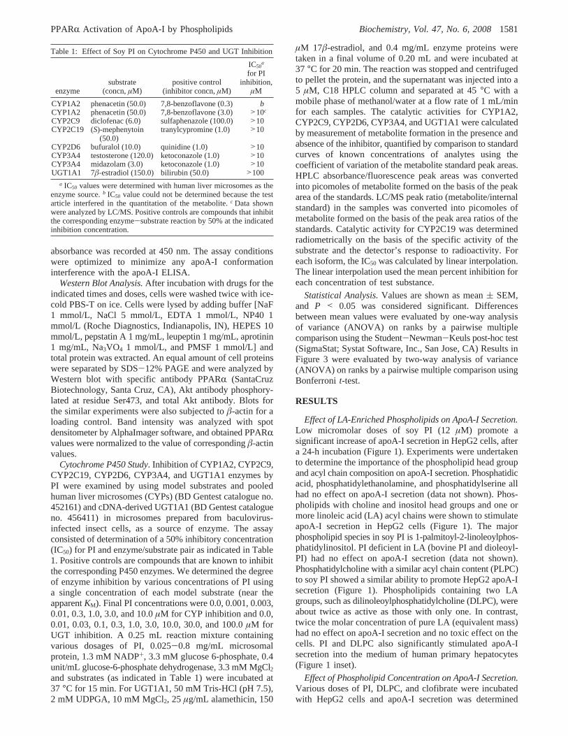
Effect of LA-Enriched Phospholipids on ApoA-I Secretion.Low micromolar doses of soy PI (12µM) promote asignificant increase of apoA-I secretion in HepG2 cells, aftera 24-h incubation (Figure 1). Experiments were undertakento determine the importance of the phospholipid head groupand acyl chain composition on apoA-I secretion. Phosphatidicacid, phosphatidylethanolamine, and phosphatidylserine allhad no effect on apoA-I secretion (data not shown). Phos-pholipids with choline and inositol head groups and one ormore linoleic acid (LA) acyl chains were shown to stimulateapoA-I secretion in HepG2 cells (Figure 1). The majorphospholipid species in soy PI is 1-palmitoyl-2-linoleoylphos-phatidylinositol. PI deficient in LA (bovine PI and dioleoyl-PI) had no effect on apoA-I secretion (data not shown).Phosphatidylcholine with a similar acyl chain content (PLPC)to soy PI showed a similar ability to promote HepG2 apoA-Isecretion (Figure 1). Phospholipids containing two LAgroups, such as dilinoleoylphosphatidylcholine (DLPC), wereabout twice as active as those with only one. In contrast,twice the molar concentration of pure LA (equivalent mass)had no effect on apoA-I secretion and no toxic effect on thecells. PI and DLPC also significantly stimulated apoA-Isecretion into the medium of human primary hepatocytes(Figure 1 inset).
Effect of Phospholipid Concentration on ApoA-I Secretion.Various doses of PI, DLPC, and clofibrate were incubatedwith HepG2 cells and apoA-I secretion was determined
Table 1: Effect of Soy PI on Cytochrome P450 and UGT Inhibition
enzymesubstrate
(concn,µM)positive control
(inhibitor concn,µM)
IC50a
for PIinhibition,
µM
CYP1A2 phenacetin (50.0) 7,8-benzoflavone (0.3) bCYP1A2 phenacetin (50.0) 7,8-benzoflavone (3.0) >10c
CYP2C9 diclofenac (6.0) sulfaphenazole (100.0) >10CYP2C19 (S)-mephenytoin
(50.0)tranylcypromine (1.0) >10
CYP2D6 bufuralol (10.0) quinidine (1.0) >10CYP3A4 testosterone (120.0) ketoconazole (1.0) >10CYP3A4 midazolam (3.0) ketoconazole (1.0) >10UGT1A1 7â-estradiol (150.0) bilirubin (50.0) >100
a IC50 values were determined with human liver microsomes as theenzyme source.b IC50 value could not be determined because the testarticle interfered in the quantitation of the metabolite.c Data shownwere analyzed by LC/MS. Positive controls are compounds that inhibitthe corresponding enzyme-substrate reaction by 50% at the indicatedinhibition concentration.
PPARR Activation of ApoA-I by Phospholipids Biochemistry, Vol. 47, No. 6, 20081581
(Figure 2). PI and DLPC showed a saturable dose-response.PI dose-response began to plateau at a dose of 4µM, whileDLPC response began to plateau at 12µM but was stillincreasing by 35µM. DLPC was almost twice as effectiveas PI and promoted an almost 3-fold stimulation in apoA-Isecretion. Clofibrate was less effective at stimulating apoA-Isecretion, and only about a 1.25-fold stimulation wasobserved at a dose of 10µM. Increasing the dose beyond
10µM blocked the stimulation of apoA-I secretion in HepG2cells.
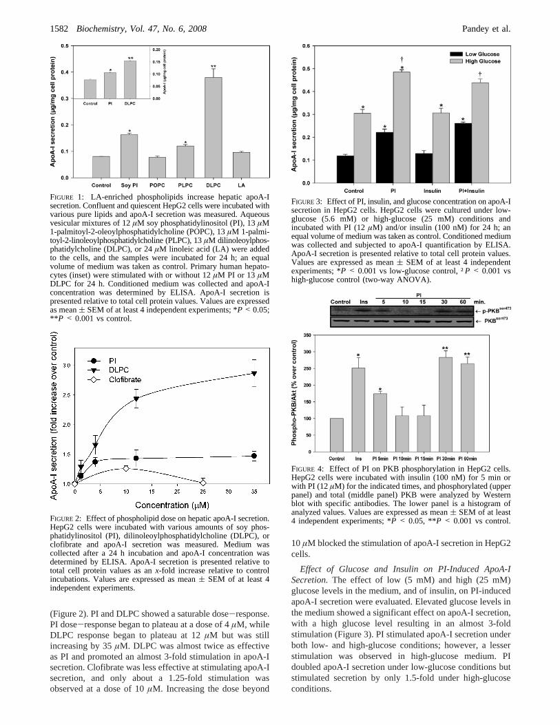
Effect of Glucose and Insulin on PI-Induced ApoA-ISecretion.The effect of low (5 mM) and high (25 mM)glucose levels in the medium, and of insulin, on PI-inducedapoA-I secretion were evaluated. Elevated glucose levels inthe medium showed a significant effect on apoA-I secretion,with a high glucose level resulting in an almost 3-foldstimulation (Figure 3). PI stimulated apoA-I secretion underboth low- and high-glucose conditions; however, a lesserstimulation was observed in high-glucose medium. PIdoubled apoA-I secretion under low-glucose conditions butstimulated secretion by only 1.5-fold under high-glucoseconditions.
FIGURE 1: LA-enriched phospholipids increase hepatic apoA-Isecretion. Confluent and quiescent HepG2 cells were incubated withvarious pure lipids and apoA-I secretion was measured. Aqueousvesicular mixtures of 12µM soy phosphatidylinositol (PI), 13µM1-palmitoyl-2-oleoylphosphatidylcholine (POPC), 13µM 1-palmi-toyl-2-linoleoylphosphatidylcholine (PLPC), 13µM dilinoleoylphos-phatidylcholine (DLPC), or 24µM linoleic acid (LA) were addedto the cells, and the samples were incubated for 24 h; an equalvolume of medium was taken as control. Primary human hepato-cytes (inset) were stimulated with or without 12µM PI or 13 µMDLPC for 24 h. Conditioned medium was collected and apoA-Iconcentration was determined by ELISA. ApoA-I secretion ispresented relative to total cell protein values. Values are expressedas mean( SEM of at least 4 independent experiments; *P < 0.05;** P < 0.001 vs control.
FIGURE 2: Effect of phospholipid dose on hepatic apoA-I secretion.HepG2 cells were incubated with various amounts of soy phos-phatidylinositol (PI), dilinoleoylphosphatidylcholine (DLPC), orclofibrate and apoA-I secretion was measured. Medium wascollected after a 24 h incubation and apoA-I concentration wasdetermined by ELISA. ApoA-I secretion is presented relative tototal cell protein values as anx-fold increase relative to controlincubations. Values are expressed as mean( SEM of at least 4independent experiments.
FIGURE 3: Effect of PI, insulin, and glucose concentration on apoA-Isecretion in HepG2 cells. HepG2 cells were cultured under low-glucose (5.6 mM) or high-glucose (25 mM) conditions andincubated with PI (12µM) and/or insulin (100 nM) for 24 h; anequal volume of medium was taken as control. Conditioned mediumwas collected and subjected to apoA-I quantification by ELISA.ApoA-I secretion is presented relative to total cell protein values.Values are expressed as mean( SEM of at least 4 independentexperiments; *P < 0.001 vs low-glucose control, †P < 0.001 vshigh-glucose control (two-way ANOVA).
FIGURE 4: Effect of PI on PKB phosphorylation in HepG2 cells.HepG2 cells were incubated with insulin (100 nM) for 5 min orwith PI (12µM) for the indicated times, and phosphorylated (upperpanel) and total (middle panel) PKB were analyzed by Westernblot with specific antibodies. The lower panel is a histogram ofanalyzed values. Values are expressed as mean( SEM of at least4 independent experiments; *P < 0.05, **P < 0.001 vs control.
1582 Biochemistry, Vol. 47, No. 6, 2008 Pandey et al.
Effect of PI on PKB Phosphorylation.Insulin is knownto promote the phosphorylation of protein kinase B (PKB/Akt) to induce glucose transport (46). Fibrate drugs arebelieved to inhibit hepatic PKB phosphorylation (47, 48).Experiments were undertaken to determine how PI mayimpact PKB. Figure 4 shows that both insulin (100 nM) andPI (12 µM) induce a significant increase in PKB phospho-rylation by 5 min. With PI, PKB phosphorylation returns tobasal levels at 10 and 15 min and then peaks again at 30and 60 min.
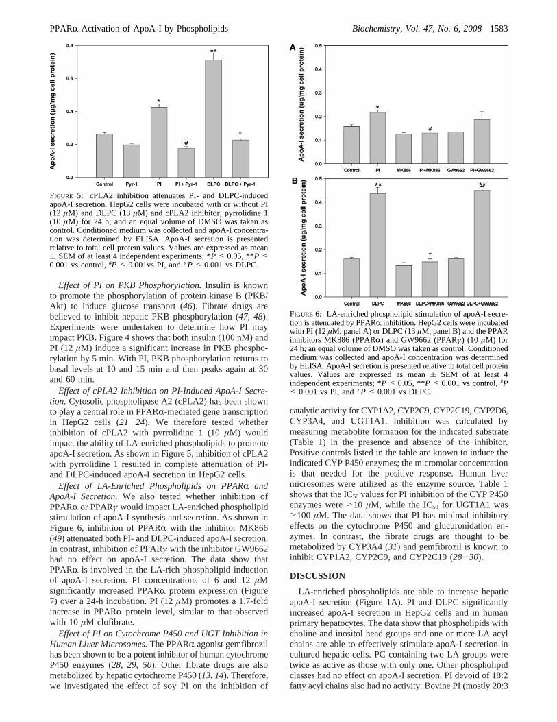
Effect of cPLA2 Inhibition on PI-Induced ApoA-I Secre-tion. Cytosolic phospholipase A2 (cPLA2) has been shownto play a central role in PPARR-mediated gene transcriptionin HepG2 cells (21-24). We therefore tested whetherinhibition of cPLA2 with pyrrolidine 1 (10µM) wouldimpact the ability of LA-enriched phospholipids to promoteapoA-I secretion. As shown in Figure 5, inhibition of cPLA2with pyrrolidine 1 resulted in complete attenuation of PI-and DLPC-induced apoA-I secretion in HepG2 cells.
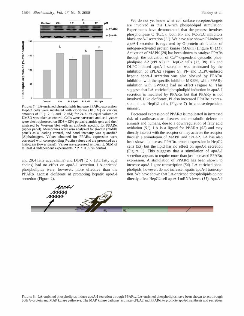
Effect of LA-Enriched Phospholipids on PPARR andApoA-I Secretion.We also tested whether inhibition ofPPARR or PPARγ would impact LA-enriched phospholipidstimulation of apoA-I synthesis and secretion. As shown inFigure 6, inhibition of PPARR with the inhibitor MK866(49) attenuated both PI- and DLPC-induced apoA-I secretion.In contrast, inhibition of PPARγ with the inhibitor GW9662had no effect on apoA-I secretion. The data show thatPPARR is involved in the LA-rich phospholipid inductionof apoA-I secretion. PI concentrations of 6 and 12µMsignificantly increased PPARR protein expression (Figure7) over a 24-h incubation. PI (12µM) promotes a 1.7-foldincrease in PPARR protein level, similar to that observedwith 10 µM clofibrate.
Effect of PI on Cytochrome P450 and UGT Inhibition inHuman LiVer Microsomes.The PPARR agonist gemfibrozilhas been shown to be a potent inhibitor of human cytochromeP450 enzymes (28, 29, 50). Other fibrate drugs are alsometabolized by hepatic cytochrome P450 (13, 14). Therefore,we investigated the effect of soy PI on the inhibition of
catalytic activity for CYP1A2, CYP2C9, CYP2C19, CYP2D6,CYP3A4, and UGT1A1. Inhibition was calculated bymeasuring metabolite formation for the indicated substrate(Table 1) in the presence and absence of the inhibitor.Positive controls listed in the table are known to induce theindicated CYP P450 enzymes; the micromolar concentrationis that needed for the positive response. Human livermicrosomes were utilized as the enzyme source. Table 1shows that the IC50 values for PI inhibition of the CYP P450enzymes were>10 µM, while the IC50 for UGT1A1 was>100 µM. The data shows that PI has minimal inhibitoryeffects on the cytochrome P450 and glucuronidation en-zymes. In contrast, the fibrate drugs are thought to bemetabolized by CYP3A4 (31) and gemfibrozil is known toinhibit CYP1A2, CYP2C9, and CYP2C19 (28-30).
DISCUSSION
LA-enriched phospholipids are able to increase hepaticapoA-I secretion (Figure 1A). PI and DLPC significantlyincreased apoA-I secretion in HepG2 cells and in humanprimary hepatocytes. The data show that phospholipids withcholine and inositol head groups and one or more LA acylchains are able to effectively stimulate apoA-I secretion incultured hepatic cells. PC containing two LA groups weretwice as active as those with only one. Other phospholipidclasses had no effect on apoA-I secretion. PI devoid of 18:2fatty acyl chains also had no activity. Bovine PI (mostly 20:3
FIGURE 5: cPLA2 inhibition attenuates PI- and DLPC-inducedapoA-I secretion. HepG2 cells were incubated with or without PI(12 µM) and DLPC (13µM) and cPLA2 inhibitor, pyrrolidine 1(10 µM) for 24 h; and an equal volume of DMSO was taken ascontrol. Conditioned medium was collected and apoA-I concentra-tion was determined by ELISA. ApoA-I secretion is presentedrelative to total cell protein values. Values are expressed as mean( SEM of at least 4 independent experiments; *P < 0.05, **P <0.001 vs control,#P < 0.001vs PI, and †P < 0.001 vs DLPC.
FIGURE 6: LA-enriched phospholipid stimulation of apoA-I secre-tion is attenuated by PPARR inhibition. HepG2 cells were incubatedwith PI (12µM, panel A) or DLPC (13µM, panel B) and the PPARinhibitors MK886 (PPARR) and GW9662 (PPARγ) (10 µM) for24 h; an equal volume of DMSO was taken as control. Conditionedmedium was collected and apoA-I concentration was determinedby ELISA. ApoA-I secretion is presented relative to total cell proteinvalues. Values are expressed as mean( SEM of at least 4independent experiments; *P < 0.05, **P < 0.001 vs control,#P< 0.001 vs PI, and †P < 0.001 vs DLPC.
PPARR Activation of ApoA-I by Phospholipids Biochemistry, Vol. 47, No. 6, 20081583
and 20:4 fatty acyl chains) and DOPI (2× 18:1 fatty acylchains) had no effect on apoA-I secretion. LA-enrichedphospholipids were, however, more effective than thePPARR agonist clofibrate at promoting hepatic apoA-Isecretion (Figure 2).
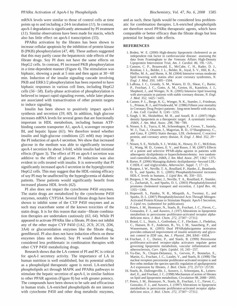
We do not yet know what cell surface receptors/targetsare involved in this LA-rich phospholipid stimulation.Experiments have demonstrated that the process involvesphospholipase C (PLC): both PI- and PC-PLC inhibitorsblock apoA-I secretion (11). We have also shown PI-inducedapoA-I secretion is regulated by G-protein stimulation ofmitogen-activated protein kinase (MAPK) (Figure 8) (11).Activation of MAPK (28) has been shown to catalyze PPARsthrough the activation of Ca2+-dependent cytosolic phos-pholipase A2 (cPLA2) in HepG2 cells (37, 38). PI- andDLPC-induced apoA-I secretion was attenuated by theinhibition of cPLA2 (Figure 5). PI- and DLPC-inducedhepatic apoA-I secretion was also blocked by PPARRinhibition with the specific inhibitor MK886, while PPARγinhibition with GW9662 had no effect (Figure 6). Thissuggests that LA-enriched phospholipid induction in apoA-Isecretion is mediated by PPARR but that PPARγ is notinvolved. Like clofibrate, PI also increased PPARR expres-sion in the HepG2 cells (Figure 7) in a dose-dependentmanner.
Decreased expression of PPARR is implicated in increasedrisk of cardiovascular diseases and metabolic defects inanimals and humans, due to a downregulation of fatty acidoxidation (51). LA is a ligand for PPARR (52) and maydirectly interact with the receptor or may activate the receptorthrough a stimulation of MAPK and cPLA2. LA has alsobeen shown to increase PPARR protein expression in HepG2cells (53) but the lipid has no effect on apoA-I secretion(Figure 1). This suggests that a stimulation of apoA-Isecretion appears to require more than just increased PPARRexpression. A stimulation of PPARR has been shown toincrease apoA-I gene transcription (54). LA-enriched phos-pholipids, however, do not increase hepatic apoA-I transcrip-tion. We have shown that LA-enriched phospholipids do notdirectly affect HepG2 cell apoA-I mRNA levels (11). ApoA-I
FIGURE 7: LA-enriched phospholipids increase PPARR expression.HepG2 cells were incubated with clofibrate (10µM) or variousamounts of PI (1.2, 6, and 12µM) for 24 h; an equal volume ofDMSO was taken as control. Cells were harvested and cell lysateswere electrophoresed on SDS-12% polyacrylamide gels and thenanalyzed by Western blot with an antibody specific for PPARR(upper panel). Membranes were also analyzed forâ-actin (middlepanel) as a loading control, and band intensity was quantified(AlphaImager). Values obtained for PPARR expression werecorrected with correspondingâ-actin values and are presented as ahistogram (lower panel). Values are expressed as mean( SEM ofat least 4 independent experiments; *P < 0.05 vs control.
FIGURE 8: LA-enriched phospholipids induce apoA-I secretion through PPARR. LA-enriched phospholipids have been shown to act throughboth G-protein and MAP kinase pathways. The MAP kinase pathway activates cPLA2 and PPARR to promote apoA-I synthesis and secretion.
1584 Biochemistry, Vol. 47, No. 6, 2008 Pandey et al.
mRNA levels were similar to those of control cells at timepoints up to and including a 24-h incubation (11). In contrast,apoA-I degradation is significantly attenuated by PI treatment(11). Similar observations have been made for niacin, whichalso has little effect on apoA-I transcription (55).
PPARR activation by the fibrates has been shown toincrease cellular apoptosis by the inhibition of protein kinaseB (PKB) phosphorylation (47, 48). These authors suggestedthat this may partly cause the hepatotoxic side effects of thefibrate drugs. Soy PI does not have the same effects onHepG2 cells. In contrast, PI increased PKB phosphorylationin a time-dependent manner (Figure 4). The stimulation wasbiphasic, showing a peak at 5 min and then again at 30-60min. Induction of the insulin signaling cascade involvingPKB and ERK1/2 phosphorylation has been reported to havebiphasic responses in various cell lines, including HepG2cells (56-58). Early-phase activation of phosphorylation isbelieved to impact rapid signaling events, while later phasesare associated with transactivation of other protein targetsto induce signaling.
Insulin has been shown to positively impact apoA-Isynthesis and secretion (59, 60). In addition, high glucoseincreases mRNA levels for several genes that are functionallyimportant in HDL metabolism, including human ATP-binding cassette transporter A1, apoA-I, scavenger receptorBI, and hepatic lipase (61). We therefore tested whetherinsulin and high-glucose conditions (25 mM) may impactthe PI induction of apoA-I secretion. We show that increasedglucose in the medium was able to significantly increaseapoA-I secretion by about 3-fold, while insulin had minimaleffects (Figure 3). The effect of PI on apoA-I secretion wasadditive to the effect of glucose. PI induction was alsoevident in cells treated with insulin. It is noteworthy that PIsignificantly increased apoA-I secretion in the hyperglycemicHepG2 cells. This may suggest that the HDL-raising efficacyof soy PI may be unaffected by the hyperglycemia of diabeticpatients. These patients would significantly benefit fromincreased plasma HDL levels (62).
PI also does not impact the cytochrome P450 enzymes.The statin drugs are metabolized by the cytochrome P450enzymes, notably CYP3A4. Several fibrate drugs have beenshown to inhibit some of the CYP P450 enzymes and assuch may exacerbate some of the known toxicities of thestatin drugs. It is for this reason that statin-fibrate combina-tion therapies are undertaken cautiously (63, 64). While PIappeared to activate PPARR like a fibrate, PI does not inhibitany of the other major CYP P450 (1A2, 2C9, 2C19, 2D6,3A4) or glucuronidation enzymes like the fibrate drug,gemfibrozil. PI also does not have induction effects on theseenzymes (data not shown). Soy PI would therefore beconsidered less problematic in combination therapies withother CYP P450 metabolizing drugs.
Research shows that the LA content of PI and PC is criticalfor apoA-I secretory activity. The importance of LA inhuman nutrition is well established, but its potential utilityas a phospholipid therapeutic agent is novel. LA-enrichedphospholipids act through MAPK and PPARR pathways tostimulate the hepatic secretion of apoA-I, in similar fashionto other PPAR agonists (Figure 8) but with greater potency.The compounds have been shown to be safe and efficaciousin human trials. LA-enriched phospholipids do not interactwith cytochome P450 and other drug metabolic enzymes,
and as such, these lipids would be considered less problem-atic for combination therapies. LA-enriched phospholipidsare therefore novel PPARR therapeutic agents, which havecomparable or better efficacy than the fibrate drugs but lesspotential for hepatic side effects.
REFERENCES
1. Boden, W. E. (2000) High-density lipoprotein cholesterol as anindependent risk factor in cardiovascular disease: assessing thedata from Framingham to the Veterans Affairs High-DensityLipoprotein Intervention Trial,Am. J. Cardiol. 86, 19L-22L.
2. Cannon, C. P., Braunwald, E., McCabe, C. H., Rader, D. J.,Rouleau, J. L., Belder, J. L., Belder, R., Joyal, S. V., Hill, K. A.,Pfeffer, M. A., and Skene, A. M. (2004) Intensive versus moderatelipid lowering with statins after acute coronary syndromes,N.Engl. J. Med. 350, 1495-1504.
3. LaRosa, J. C., Grundy, S. M., Waters, D. D., Shear, C., Barter,P., Fruchart, J. C., Gotto, A. M., Greten, H., Kastelein, J. J.,Shepherd, J., and Wenger, N. K. (2005) Intensive lipid loweringwith atorvastatin in patients with stable coronary disease,N. Engl.J. Med. 352, 1425-1435.
4. Canner, P. L., Berge, K. G., Wenger, N. K., Stamler, J., Friedman,L., Prineas, R. J., and Friedewald, W. (1986) Fifteen year mortalityin Coronary Drug Project patients: long-term benefit with niacin,J. Am. Coll. Cardiol.8, 1245-1255.
5. Singh, I. M., Shishehbor, M. H., and Ansell, B. J. (2007) High-density lipoprotein as a therapeutic target: A systematic review,JAMA, J. Am. Med. Assoc. 298, 786-789.
6. Nissen, S. E., Tuzcu, E. M., Schoenhagen, P., Crowe, T., Sasiela,W. J., Tsai, J., Orazem, J., Magorian, R. D., O’Shaughnessy, C.,and Ganz, P. (2005) Statin therapy, LDL cholesterol, C-reactiveprotein, and coronary artery disease,N. Engl. J. Med. 352, 29-38.
7. Nissen, S. E., Nicholls, S. J., Wolski, K., Howey, D. C., McErlean,E., Wang, M. D., Gomez, E. V., and Russo, J. M. (2007) Effectsof a potent and selective PPAR-alpha agonist in patients withatherogenic dyslipidemia or hypercholesterolemia: two random-ized controlled trials,JAMA, J. Am. Med. Assoc. 297, 1362-1373.
8. Barter, P. (2006) Managing diabetic dyslipidaemiasbeyond LDL-C:HDL-C and triglycerides,Atheroscler. Suppl. 7, 17-21.
9. Burgess, J. W., Neville, T. A., Rouillard, P., Harder, Z., Beanlands,D. S., and Sparks, D. L. (2005) Phosphatidylinositol increasesHDL-C levels in humans,J. Lipid Res. 46, 350-355.
10. Burgess, J. W., Boucher, J., Neville, T. A., Rouillard, P., Stamler,C., Zachariah, S., and Sparks, D. L. (2003) Phosphatidylinositolpromotes cholesterol transport and excretion,J. Lipid Res. 44,1355-1366.
11. Hopewell, S., Pandey, N. R., Misquith, A., Twomey, E., andSparks, D. L. (2007) Phosphatidylinositol Acts Through Mitogen-Activated Protein Kinase to Stimulate Hepatic ApoA-I Secretion,J. Lipid res.(submitted for publication).
12. Peters, J. M., Hennuyer, N., Staels, B., Fruchart, J. C., Fievet, C.,Gonazalez, F. J., and Auwerx, J. (197) Alterations in lipoproteinmetabolism in peroxisome proliferator-activated receptor alpha-deficient mice,J. Biol. Chem. 272, 27307-27312.
13. Brand, C. L., Sturis, J., Gotfredsen, C. F., Fleckner, J., Fledelius,C., Hansen, B. F., Andersen, B., Ye, J. M., Sauerberg, P., andWassermann, K. (2003) Dual PPARalpha/gamma activationprovides enhanced improvement of insulin sensitivity and glyce-mic control in ZDF rats,Am. J. Physiol. 284, E841-E854.
14. Fruchart, J. C., Duriez, P., and Staels, B. (1999) Peroxisomeproliferator-activated receptor-alpha activators regulate genesgoverning lipoprotein metabolism, vascular inflammation andatherosclerosis,Curr. Opin. Lipidol. 10, 245-257.
15. Vu-Dac, N., Chopin-Delannoy, S., Gervois, P., Bonnelye, E.,Martin, G., Fruchart, J. C., Laudet, V., and Staels, B. (1998) Thenuclear receptors peroxisome proliferator-activated receptorR andRev-erbR mediate the species-specific regulation of apolipoproteinA-I expression by fibrates,J. Biol. Chem. 273, 25713-25720.
16. Staels, B., Dallongeville, J., Auwerx, J., Schoonjans, K., Leiters-dorf, E., and Fruchart, J. C. (1998) Mechanism of action of fibrateson lipid and lipoprotein metabolism,Circulation 98, 2088-2093.
17. Peters, J. M., Hennuyer, N., Staels, B., Fruchart, J. C., Fievet, C.,Gonzalez, F. J., and Auwerx, J. (1997) Alterations in lipoproteinmetabolism in peroxisome proliferator-activated receptor alpha-deficient mice,J. Biol. Chem. 272, 27307-27312.
PPARR Activation of ApoA-I by Phospholipids Biochemistry, Vol. 47, No. 6, 20081585
18. Hertz, R., Bishara-Shieban, J., and Bar-Tana, J. (1995) Mode ofaction of peroxisome proliferators as hypolipidemic drugs. Sup-pression of apolipoprotein C-III,J. Biol. Chem. 270, 13470-134775.
19. Schoonjans, K., Peinado-Onsurbe, J., Lefebvre, A. M., Heymen,R. A., Briggs, M., Deeb, S., Staels, B., and Auwerx, J. (1996)PPARalpha and PPARgamma activators direct a distinct tissue-specific transcriptional response via a PPRE in the lipoproteinlipase gene,EMBO J. 15, 5336-5348.
20. Vu-Dac, N., Schoonjans, K., Laine, B., Fruchart, J. C., Auwerx,J., and Staels, B. (1994) Negative regulation of the humanapolipoprotein A-I promoter by fibrates can be attenuated by theinteraction of the peroxisome proliferator-activated receptor withits response element,J. Biol. Chem. 269, 31012-31018.
21. Vu-Dac, N., Schoonjans, K., Kosykh, V., Dellongeville, J.,Fruchart, J. C., Staels, B., and Auwerx, J. (1995) Fibrates increasehuman apolipoprotein A-II expression through activation of theperoxisome proliferator-activated receptor,J. Clin. InVest. 96,741-750.
22. Tikkanen, M. J. (1992) Fibric acid derivatives.Curr. Opin. Lipidol.3, 29 33.
23. Staels, B., Van Tol, A., Andreu, T., and Auwerx, J. (1992) Fibratesinfluence the expression of genes involved in lipoprotein metabo-lism in a tissue-selective manner in the rat.Arterioscler. Thromb.12, 286-94.
24. Zannis, V. I., Breslow, J. L., SanGiacomo, T. R., Aden, D. P.,and Knowles, B. B. (1981) Characterization of the major apoli-poproteins secreted by two human hepatoma cell lines,Biochem-istry 20, 7089-7096.
25. Thrift, R. N., Forte, T. M., Cahoon, B. E., and Shore, V. G. (1986)Characterization of lipoproteins produced by the human liver cellline, Hep G2, under defined conditions,J. Lipid Res. 27, 236-250.
26. Forte, T. M., McCall, M. R., Knowles, B. B., and Shore, V. G.(1989) Isolation and characterization of lipoproteins produced byhuman hepatoma-derived cell lines other than HepG2,J. LipidRes. 30, 817-829.
27. Pruekscaritanont, T., Oui, Y., Mu, L., Subramanian, R., and Lin,R. H. (2002) Effects of fibrates on metabolism of statins in humanhepatocytes,Drug. Metab. Dispos. 30, 1280-1287.
28. Pruekscaritanont, T., Zhao, J. J., Ma, B., Roadcap, B. A., Tang,C., Qiu, Y., Liu, L., Lin, J. H., Pearson, P. G., and Baillie, T. A.(2002) Mechanistic studies on metabolic interactions betweengemfibrozil and statins,J. Pharmacol. Exp. Ther. 301, 1042-1051.
29. Tornio, A., Niemi, M., Neuvonen, P. J., and Backman, J. T. (2007)Stereoselective interaction between the CYP2C8 inhibitors gem-fibrozil and racemic ibuprofen,Eur. J. Clin. Pharmacol. 63, 463-469.
30. Miller, D. B., and Spence, J. D. (1998) Clinical pharmacokineticsof fibric acid derivatives (fibrates),Clin. Pharmacokinet. 34, 155-162.
31. Kajosaari, L. I., Laitila, J., Neuvonen, P. J., and Backman, J. T.(2005) Metabolism of repaglinide by CYP2C8 and CYP3A4 invitro: effect of fibrates and rifampicin,Basic Clin. Pharmacol.Toxicol. 97, 249-56.
32. Kane, C. D., Francone, O. L., and Stevens, K. A. (2006)Differential regulation of cynomolgus, human and rat acyl-CoAoxidase promoters by PPARR, Gene 380, 84-94.
33. Duez, H., Lefebvre, B., Poulain, P., Torra, I. P., Percevault, F.,Luc, G., Peters, J. M., Gonzalez, F. J., Ginste, R., Helleboid, S.,Dzavik, V., Fruchart, J. C., Fievet, C., Lefebvre, P., and Staels,B. (2005) Regulation of human apoA-I by gemfibrozil andfenofibrate through selective peroxisome proliferator-activatedreceptor alpha modulation,Arterioscler. Thromb. Vasc. Biol. 25,585-591.
34. Barbier, O., Fontaine, C., Fruchart, J. C., and Staels, B. (2004)Genomic and non-genomic interactions of PPARR with xenobi-otic-metabolizing enzymes,Trends Endocrinol. Metab. 15, 324-330.
35. Crowl, R. M., Stoller, T. J., Conroy, R. R., and Stoner, C. R. (1991)Induction of phospholipase A2 gene expression in human hepato-ma cells by mediators of the acute phase response,J. Biol. Chem.266, 2647-2651.
36. Dong, L. W., Yang, J., Tong, L. J., Hsu, H. K., and Liu, M. S.(1997) Group II phospholipase A2 gene expression is transcrip-tionally regulated in rat liver during sepsis,Am. J. Physiol. 273,G706-G712.
37. Han, C., Demetris, A. J., Michalopoulos, G., Shelhamer, J. H.,and Wu, T. (2002) 85-kDa cPLA(2) plays a critical role in PPAR-mediated gene transcription in human hepatoma cells,Am. J.Physiol. 282, G586-G597.
38. Agassandian, M., Miakotina, O. L., Andrews, M., Mathur, S. N.,and Mallampalli, R. K. (2007)Pseudomonas aeruginosaandsPLA2 IB stimulate ABCA1-mediated phospholipid efflux viaERK-activation of PPARalpha-RXR,Biochem. J. 403, 409-420.
39. Yano, M., Matsumura, T., Senokuchi, T., Ishii, N., Murata, Y.,Taketa, K., Motoshima, H., Taguchi, T., Sonoda, K., Kukidome,D., Takuwa, Y., kawada, T., Brownlee, M., Nishikawa, T., andAraki, E. (2007) Statins activate peroxisome proliferator-activatedreceptor gamma through extracellular signal-regulated kinase 1/2and p38 mitogen-activated protein kinase-dependent cyclooxy-genase-2 expression in macrophages.Circ. Res. 100, 1442-1451.
40. Rokos, C. L., and Ledwith, B. J. (1997) Peroxisome proliferatorsactivate extracellular signal-regulated kinases in immortalizedmouse liver cells,J. Biol. Chem. 272, 13452-13457.
41. Mounho, B. J., and Thrall, B. D. (1999) The extracellular signal-regulated kinase pathway contributes to mitogenic and antiapo-ptotic effects of peroxisome proliferators in vitro,Toxicol. Appl.Pharmacol. 159, 125-133.
42. Lennon, A. M., Ramauge, M., Dessouroux, A., and Pierre, M.(2002) MAP kinase cascades are activated in astrocytes andpreadipocytes by 15-deoxy-∆12-14-prostaglandin J2 and the thia-zolidinedione ciglitazone through peroxisome proliferator activatorreceptorγ-independent mechanisms involving reactive oxygenatedspecies,J. Biol. Chem. 277, 29681-29685.
43. Teruel, T., Hernandez, R., Benito, M., and Lorenzo, M. (2003)Rosiglitazone and retinoic acid induce uncoupling protein-1 (UCP-1) in a p38 mitogen-activated protein kinase-dependent mannerin fetal primary brown adipocytes,J. Biol. Chem. 278, 263-269.
44. Gardner, O. S., Dewar, B. J., and Graves, L. M. (2005) Activationof mitogen-activated protein kinases by peroxisome proliferator-activated receptor ligands: an example of nongenomic signaling,Mol. Pharmacol. 68, 933-941.
45. Krylova, I. N., Sablin, E. P., Moore, J., Xu, R. X., Waitt, G. M.,MacKay, J. A., Juzumiene, D., Bynum, J. M., Madauss, K.,Montana, V., Lebedeva, L., Suzawa, M., Williams, J. D., Williams,S. P., Guy, R. K., Thornton, J. W., Fletterick, R. J., Willson, T.M., and Ingraham, H. A. (2005) Structural analyses revealphosphatidylinositols as ligands for the NR5 orphan receptors SF-1and LRH-1,Cell 120, 343-355.
46. Saltiel, A. R., and Kahn, C. R. (2001) Insulin signaling and theregulation of glucose and lipid metabolism,Nature 414, 799-806.
47. Kubota, T., Yano, T., Fujisaki, K., Itoh, Y., and Oishi, R. (2005)Fenofibrate induces apoptotic injury in cultured human hepatocytesby inhibiting phosphorylation of Akt,Apoptosis 10, 349-358.
48. Grabacka, M., Plonka, P. M., Urbanska, K., and Reiss, K. (2006)Peroxisome proliferator-activated receptor alpha activation de-creases metastatic potential of melanoma cells in vitro via down-regulation of Akt,Clin. Cancer. Res. 12, 3028-3036.
49. Kehrer, J. P., Biswal, S. S., La, E., Thuillier, P., Datta, K., Fischer,S. M., and Vaden, Heuvel, J. P. (2001) Inhibition of peroxisome-proliferator-activated receptor (PPAR)alpha by MK886,Biochem.J. 356, 899-906.
50. Wen, X., Wang, J. S., Backman, J. T., Kivisto, K. T., andNeuvonen, P. J. (2001) Gemfibrozil is a potent inhibitor of humancytochrome P4502C9,Drug. Metab. Dispos. 29, 1359-1361.
51. Morgan, E. E., Chandler, M. P., Young, M. E., McElfresh, T. A.,Kung, T. A., Rennison, J. H., Tserng, K. Y., Hoit, B. D., andStanley, W. C. (2006) Dissociation between gene and proteinexpression of metabolic enzymes in a rodent model of heart failure,Eur. J. Heart Failure 8, 687-693.
52. Moya-Camarena, S. Y., Vanden Heuvel, J. P., Blanchard, S. G.,Leesnitzer, L. A., and Belury, M. A. (1999) Conjugated linoleicacid is a potent naturally occurring ligand and activator of PPARR,J. Lipid Res. 40, 1426-1433.
53. Akbiyik, F., Ray, D. M., Bozkaya, H., and Demirpence, E. (2004)Ligand- and species-dependent activation of PPARalpha,Cell.Physiol. Biochem. 14, 269-276.
54. Martin, G., Duez, H., Blanquart, C., Berezowski, V., Poulain, P.,Fruchart, J.-C., Najib-Fruchart, J., Glineur, C., and Staels, B.(2001) Statin-induced inhibition of the Rho-signaling pathwayactivates PPARR and induces HDL apoA-I,J. Clin. InVest. 107,1423-1432.
55. Jin, F. Y., Kamanna, V. S., and Kashyap, M. L. (1997) Niacindecreases removal of high-density lipoprotein apolipoprotein A-I
1586 Biochemistry, Vol. 47, No. 6, 2008 Pandey et al.
but not cholesterol ester by Hep G2 cells. Implication for reversecholesterol transport.Arterioscler. Thromb. Vasc. Biol. 17, 2020-2028.
56. Morisco, Condorelli, G., Trimarco, V., Bellis, A., Marrone, C.,Condorelli, G., Sadoshima, J., and Trimarco, B. (2005) Aktmediates the cross-talk between beta-adrenergic and insulinreceptors in neonatal cardiomyocytes,Circ. Res. 96, 180-188.
57. Morino, M., Acconcia, F., and Trentalance, A. (2003) Biphasicestradiol-induced AKT phosphorylation is modulated by PTENvia MAP kinase in HepG2 cells,Mol. Biol. Cell. 14, 2583-2591.
58. Grantcharova, E., Reusch, H.P., Grossmann, S., Eichhorst, J. Krell,H.W., Beyermann, M., Rosenthal, W., and Oksche, A. (2006)N-terminal proteolysis of the endothelin B receptor abolishes itsability to induce EGF receptor transactivation and contractileprotein expression in vascular smooth muscle cells,Arterioscler.Thromb. Vasc. Biol. 26, 1288-1296.
59. Murao, K., Wada, Y., Nakamura, T., Taylor, A. H., Mooradian,A. D., and Wong, N. C. (1998) Effects of glucose and insulin on
rat apolipoprotein A-I gene expression,J. Biol. Chem. 273,18959-18965.
60. Mooradian, A. D., Wong, N. C., and Shah, G. N. (1997)Apolipoprotein A1 expression in young and aged rats is modulatedby dietary carbohydrates,Metabolism 46, 1132-1136.
61. Tu, A. Y., and Albers, J. J. (2001) Glucose regulates thetranscription of human genes relevant to HDL metabolism:responsive elements for peroxisome proliferator-activated receptorare involved in the regulation of phospholipid transfer protein,Diabetes 50, 1851-1856.
62. Quintao, E. C., Medina, W. L., and Passarelli, M. (2000) Reversecholesterol transport in diabetes mellitus,Diabetes Metab. Res.ReV. 16, 237-250.
63. Wierzbicki, A. S., Mikhailidis, D. P., Wray, R., Schacter, M.,Cramb, R., Simpson, W. G., and Byrne, C. B. (2003) Statin-fibrate combination: therapy for hyperlipidemia: a review,Curr.Med. Res. Opin. 19, 155-168.
64. Shek, A., and Ferril, M. J. (2001) Statin-fibrate combinationtherapy,Ann. Pharmacother. 35, 908-917.
BI702148F
PPARR Activation of ApoA-I by Phospholipids Biochemistry, Vol. 47, No. 6, 20081587


![Abb.1: Struktur eines Phospholipids [1]](https://img.pdfslide.tips/doc/110x75/568165f8550346895dd9236e/abb1-struktur-eines-phospholipids-1.jpg)