Embed Size (px)
Citation preview
Cytoplasmic lipid droplets are sites of convergence of proteasomal and autophagic
degradation of apolipoprotein B
Yuki Ohsaki*‡, Jinglei Cheng*, Akikazu Fujita*, Toshinobu Tokumoto†,
and Toyoshi Fujimoto*‡
* Department of Anatomy and Molecular Cell Biology, Nagoya University Graduate School of
Medicine, Nagoya 466-8550, Japan, †Department of Biology and Geosciences, Faculty of
Science, Shizuoka University, Shizuoka 422-8529, Japan
‡Correspondence:
Yuki Ohsaki, Ph.D.
Tel., +81 52 744 2002: Fax, +81 52 744 2011; e-mail, [email protected]
Toyoshi Fujimoto, M.D., Ph.D.
Tel., +81 52 744 2000: Fax, +81 52 744 2011; e-mail, [email protected] Department of Anatomy and Molecular Cell Biology, Graduate School of Medicine,
Nagoya University, 65 Tsurumai, Showa, Nagoya 466-8550, Japan
Running Head: ApoB degradation in CLD
Key Words: apolipoprotein B, lipid droplet, proteasome, autophagy
Abbreviations: ALLN, N-acetyl-L-leucinyl-L-leucinyl-L-norleucinal; ApoB,
apolipoprotein B; ADRP, adipose differentiation-related protein; CLD,
cytoplasmic lipid droplets; 3-MA, 3-methyladenine; VLDL, very
low-density lipoprotein.
2
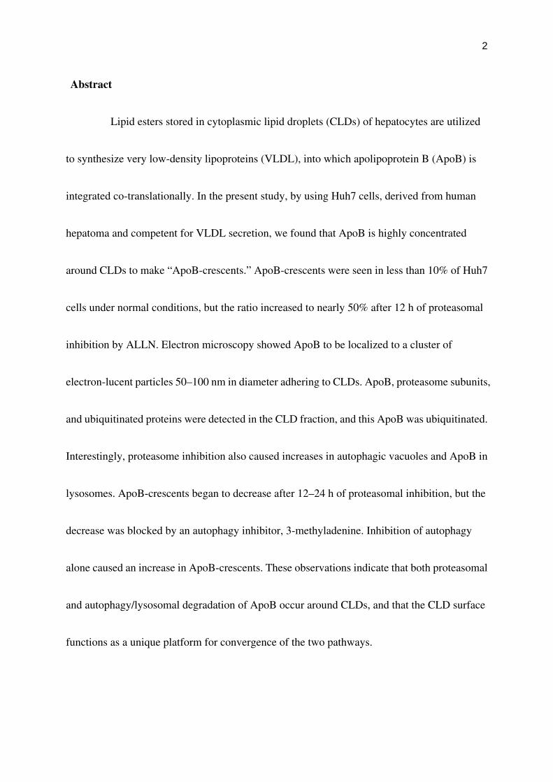
Abstract
Lipid esters stored in cytoplasmic lipid droplets (CLDs) of hepatocytes are utilized
to synthesize very low-density lipoproteins (VLDL), into which apolipoprotein B (ApoB) is
integrated co-translationally. In the present study, by using Huh7 cells, derived from human
hepatoma and competent for VLDL secretion, we found that ApoB is highly concentrated
around CLDs to make “ApoB-crescents.” ApoB-crescents were seen in less than 10% of Huh7
cells under normal conditions, but the ratio increased to nearly 50% after 12 h of proteasomal
inhibition by ALLN. Electron microscopy showed ApoB to be localized to a cluster of
electron-lucent particles 50–100 nm in diameter adhering to CLDs. ApoB, proteasome subunits,
and ubiquitinated proteins were detected in the CLD fraction, and this ApoB was ubiquitinated.
Interestingly, proteasome inhibition also caused increases in autophagic vacuoles and ApoB in
lysosomes. ApoB-crescents began to decrease after 12–24 h of proteasomal inhibition, but the
decrease was blocked by an autophagy inhibitor, 3-methyladenine. Inhibition of autophagy
alone caused an increase in ApoB-crescents. These observations indicate that both proteasomal
and autophagy/lysosomal degradation of ApoB occur around CLDs, and that the CLD surface
functions as a unique platform for convergence of the two pathways.

3
Introduction
Lipid droplets (CLDs) consist of a neutral lipid core with a surrounding
phospholipid monolayer (Murphy and Vance, 1999; Tauchi-Sato et al., 2002). CLDs are
prominent in adipose cells and steroidogenic cells, but they also exist in other cell types. With
the exception of a few cell types, CLDs have been considered as inert excess lipid deposits.
However, recent studies have shown that a variety of proteins are localized in CLDs, suggesting
that they may play more active functional roles than thought previously. In addition to PAT
family proteins, enzymes involved in eicosanoid formation, enzymes for cholesterol synthesis,
signaling proteins, caveolins, and Rab proteins have been reported in CLDs (Ozeki et al., 2005;
and literature cited therein). Proteomic studies identified many more proteins of both known
and unknown functions in the CLD-rich fraction (Brasaemle et al., 2004; Fujimoto et al., 2004;
Liu et al., 2004; Umlauf et al., 2004). The presence of functional proteins implies that CLDs are
not mere lipid storage vessels but may be involved in various cellular activities.
In hepatocytes, the triglycerides in CLDs are thought to be utilized for the synthesis
of very low-density lipoprotein (VLDL) (Gibbons et al., 2000). Enzyme activities that
hydrolyze and re-esterify neutral lipids are found in CLDs and/or ER, but the detailed
mechanism by which the CLD content is mobilized and incorporated into lipoprotein particles
is still unclear (Murphy, 2001). Apolipoprotein B-100 (ApoB), the primary protein component
of VLDL, is assembled with lipids in the ER lumen, and further lipidation and maturation of the

4
lipoprotein particles occur in the ER or the pre-Golgi compartment (Olofsson et al., 1999; Pan
et al., 2002). ApoB is a huge amphipathic protein of 4,536 amino acids, and when the assembly
of VLDL does not proceed effectively, excess ApoB molecules are destined for degradation by
both proteasomal and non-proteasomal pathways (Fisher et al., 1997; Cavallo et al., 1999). In
contrast to most other proteins subjected to ER-associated degradation, with a notable
exception of major histocompatibility complex class I heavy chains in the presence of US2 or
US11 (Wiertz et al., 1996a, b), ApoB is ubiquitinated and transferred to proteasomes directly
from the ER membrane without full translocation into the lumen (Mitchell et al., 1998;
Pariyarath et al., 2001).
In studying the functional role of CLDs in VLDL assembly, we found serendipitously
that ApoB accumulated heavily around CLDs. Subsequent experiments revealed that the ApoB
accumulation around CLDs increased markedly when either proteasomal function or autophagy
were inhibited. Ubiquitinated ApoB, proteasomal subunits, and autophagic vacuoles were all
found in or around the ApoB-positive CLDs. The observation indicates that CLDs provide a
site to hold amphipathic ApoB without gross aggregation, where proteasomal and autophagic
pathways converge for degradation.

5
Materials and Methods
Cells and transfection
The human hepatocellular carcinoma cell lines, Huh7 and HepG2, were obtained
from the Japanese Collection of Research Bioresources Cell Bank. They were cultured in
DMEM supplemented with 10% FCS, 50 U/ml penicillin, and 0.05 mg/ml streptomycin at
37°C in a humidified atmosphere containing 5% CO2. Frozen human hepatocytes were
purchased from Tissue Transformation Technologies, and cultured in the incubation medium
obtained from BIOPREDIC International. When appropriate, cells were incubated in medium
containing 10 µM N-acetyl-L-leucinyl-L-leucinyl-L-norleucinal (ALLN; Sigma) or MG132
(Sigma) to inhibit proteasome functions, and/or 10 mM 3-methyladenine (3-MA; Sigma) to
inhibit autophagy. Lipoprotein-deficient serum (LPDS) was prepared from FCS as described
(Goldstein et al., 1983).
In some experiments, cells were transfected with cDNA by using Lipofectamine
2000 (Invitrogen) according to the manufacturer's instruction. Expression vectors for
dynamin-2 and myc/His6-tagged ubiquitin were kindly donated by Drs. Kazuhisa Nakayama
(Kyoto University) and Ron R. Kopito (Stanford University), respectively.
6
Antibodies and reagents
Mouse monoclonal anti-human ApoB-100 (clone 6H12 from INTRACEL; clone 5F8
from MONOSAN), goat polyclonal anti-human ApoB-100 (Rockland), mouse anti-adipose
differentiation-related protein (ADRP) (Progen), mouse anti-ubiquitin (clone FK1; Affinity
Bioreagents), mouse anti-EEA1 (Transduction Lab.), and mouse anti-Lamp1 antibody (clone
H4A3; Developmental Studies Hybridoma Bank at the University of Iowa) were obtained from
the respective suppliers. Mouse anti-lysobisphosphatidic acid, rabbit anti-GM130, rabbit
anti-ADRP, rabbit anti-LC3, and rabbit anti-polyubiquitin antibodies were kindly provided by
Drs. Toshihide Kobayashi (RIKEN), Nobuhiro Nakamura (Kanazawa University), Tom
Keenan (Virginia Polytechnic Institute), Yasuo Uchiyama (Osaka University), and Sadaki
Yokota (Yamanashi University), respectively. Mouse GC3α antibody recognizing α2, α6, and
α7 proteasomal subunits and guinea pig anti-α6 antibodies were obtained as described
previously (Tokumoto et al., 1995; Wakata et al., 2004). Rabbit anti-TIP47 antibody was raised
against a human TIP47 peptide (residues 305–318) as reported previously (Miura et al., 2002).
Secondary antibodies conjugated to fluorochromes (Molecular Probes; Jackson
ImmunoResearch Lab.), and streptavidin-colloidal gold (BioCel) were purchased from the
respective suppliers. Mevastatin, mevalonolactone, brefeldin A, and rhodamine-transferrin
7
were obtained from Sigma.
Subcellular fractionation and proteasome activity assay
Cells were disrupted by nitrogen cavitation and subjected to sucrose density- gradient
ultracentrifugation as described (Fujimoto et al., 2001). Absence of contamination by other
organelles was confirmed by Western blotting of marker molecules as described previously
(Tauchi-Sato et al., 2002; Ozeki et al., 2005). The proteasomal activity was assayed using 100
µM N-succinyl-Leu-Leu-Val-Tyr-7-amino 4-methylcoumarin (BACHEM) as the substrate
according to the published protocol (Gaczynska et al., 1994).
Immunoprecipitation and Western blotting
The lipid droplet fraction obtained from the top of the sucrose density gradient was
dissolved in lysis buffer (50 mM Tris, 150 mM NaCl, 1 mM EDTA, 1% NP-40, 0.5% sodium
deoxycholate, 0.1% SDS, and protease inhibitor cocktail (Sigma), pH 8.0). Lysates were
precleared using protein A-agarose (Santa Cruz Biotech), and then incubated with 2 µg of goat
anti-ApoB antibody for 8 h, followed by incubation with rabbit anti-goat IgG antibody for 2 h
and then with protein A-agarose for 2 h. The agarose beads were washed in lysis buffer and
8
bound proteins were eluted by heating at 60°C for 10 min in SDS sample buffer. All the
immunoprecipitation procedure was carried out at 4°C.
In some experiments, subcellular fractions were precipitated with 10%
trichloroacetic acid, dissolved in sample buffer, and subjected to Western blotting. After
incubation with HRP-conjugated second antibodies (Pierce), the blots were developed using
Super Signal West Dura Substrate (Pierce).
Immunofluorescence microscopy and data analysis
Cells were fixed with 3% formaldehyde in 0.1 M phosphate buffer for 15 min for
double labeling of ApoB and organelle markers, or with a mixture of 3% formaldehyde and
0.025% glutaraldehyde in 0.1 M phosphate buffer for 15 min for double labeling of ApoB and
either ADRP or TIP47. To visualize acidic organelles, cells were incubated with 500 nM
Lysotracker-Red (Molecular Probes) for 2 h before fixation. For immunolabeling, the fixed
cells were permeabilized with 0.01% digitonin in PBS for 30 min, and treated with 3% bovine
serum albumin in PBS for 10 min. The cells were then incubated for 1 h with goat or mouse
anti-ApoB antibody in combination with another antibody in 1% BSA in PBS, and then with
secondary antibodies for 1 h. CLDs and nuclei were stained with BODIPY493/503 (Molecular

9
Probes) and DAPI, respectively. Images were captured using an LSM 5 PASCAL/Axioscope 2
laser scanning microscope or Apotome/Axiovert 200M microscope (Carl Zeiss), using an
Apochromat 63x lens with a 1.40 numerical aperture. Specimens were moved in a
pre-determined manner along the x-y axis using the mechanical stage of the microscope, and
pictures were taken arbitrarily along the path. Color, brightness, and contrast of the images
were adjusted using Adobe Photoshop 7.0. Cell numbers and colocalization of ApoB and other
markers were quantified using ImageJ (http://rsb.info.nih.gov/ij/).
Conventional electron microscopy and immunoelectron microscopy
For conventional electron microscopy, cells cultured on coverslips were fixed with
2.5% glutaraldehyde in 0.1 M sodium cacodylate buffer (pH 7.4), post-fixed with 1% osmium
tetroxide in the same buffer, stained en block with uranyl acetate, and embedded in Epon for
thin sectioning. For immunoelectron microscopy, cells were fixed with 3% formaldehyde and
0.025% glutaraldehyde in 0.1 M PIPES buffer (pH 7.4) for 60 min, infiltrated with a mixture of
sucrose and polyvinylpyrolidone, and frozen in liquid nitrogen. Ultrathin cryosections were
treated sequentially with goat anti-ApoB-100 antibody, biotinylated horse anti-goat IgG
antibody, and streptavidin-colloidal gold (10 nm). Sections were embedded in 2%
10
methylcellulose containing 0.3–0.5% uranyl acetate (Liou et al., 1996), and observed using a
JEOL 1200CX electron microscope operated at 100 kV.

11
Results
ApoB around CLDs is increased by proteasome inhibitors
Huh7 cells are derived from human hepatoma and retain the ability to secrete VLDL
(Higashi et al., 2002, 2003; Lalanne et al., 2005). At least 25% of ApoB in the conditioned
medium was recovered as lipoproteins of < 1.063 g/cm3 (data not shown). When Huh7 cells
cultured in standard medium were labeled for ApoB, intense labeling was observed in crescent
or circular shapes in occasional cells. Double labeling with BODIPY493/503 showed that the
crescent-shaped or circular labeling of ApoB was localized around CLDs (Figure 1A). Based
on the characteristic shape, we called the ApoB labeling around CLDs the “ApoB-crescent.”
ApoB-crescents were labeled in the same manner by three different antibodies to ApoB (mouse
monoclonal clone 6H12 and goat polyclonal, Figure 1A; mouse monoclonal clone 5F8, not
shown). ApoB-crescents were also identified in another hepatoma-derived cell line, HepG2,
and in primary human hepatocyte, although the frequency was lower than in Huh7 cells
(Supplementary Figure 1).
The frequency of ApoB-crescents increased markedly when the cells were treated by
proteasome inhibitors, ALLN or MG132. ALLN at 10 µM increased the percentage of Huh7
cells showing ApoB-crescents from less than 10% at 0 h to nearly 50% at 12 h (Figures 1B, C).
12
The ALLN treatment also increased the ratio of CLDs bearing ApoB-crescents in a similar
manner (Figure 1D). The increases were significant as early as 1 hr after addition of ALLN.
These observations suggest that ApoB-crescents are related to the ubiquitin-proteasome
degradation of ApoB. MG132 at 50 µM or ALLN at concentrations >20 µM caused a similar
increase, but affected the overall cell shape. Therefore, we mainly used 10 µM ALLN for the
subsequent experiments. Notably, the frequency of ApoB-crescents decreased to the basal level
after 24 h of ALLN treatment (Figure 1C), and ApoB labeling in other locations increased as
described below.
The increase of ApoB-crescents was also observed when cells were cultured in DME with 10%
LPDS instead of 10% FCS (Supplementary Figure 2). The frequency of ApoB-crescent further
increased by adding mevastatin and mevalonolactone to the LPDS medium to inhibit de novo
synthesis of cholesterol without affecting isoprenylation of Ras family proteins.
Mevastatin/mevalonolactone alone also induced a slight but significant increase of
ApoB-crescents. These results showed that ApoB-crescents were formed when lipid supply
was not adequate, and that they did not form only as a result of non-specific stress response to
the protease inhibition. Brefeldin A, which blocks vesicular transport from ER to Golgi,
induced a sharp increase of ApoB-crescents as expected.

13
ApoB-crescents were complementary to ADRP and TIP47 around CLDs
Among the PAT proteins that coat CLDs in mammalian cells, ADRP and TIP47 are
expressed in non-adipocytes (Miura et al., 2002; Ohsaki et al., 2005; Wolins et al., 2005). On
immunofluorescence microscopy of Huh7 cells in the standard culture condition, ADRP was
observed in most CLDs, while TIP47 was seen in about 10% of CLDs (Ohsaki et al.,
unpublished data). Both ADRP and TIP47 were seen to encircle the whole surface of CLDs that
lacked ApoB labeling, but where ApoB-crescents were observed, the two proteins were
excluded from the ApoB-positive area (Figure 2). Vertical reconstruction of confocal images
showed that CLD globules contained clear complementary ADRP/TIP47-positive and
ApoB-positive hemispheres (Figure 2). By triple labeling of ApoB, ADRP, and TIP47, ADRP
and TIP47 were found to coexist in those CLDs (data not shown). Another CLD-specific
protein, Rab18 (Ozeki et al., 2005), was never found in CLDs with ApoB-crescents, but
occurred in those without ApoB-crescents (data not shown).
CLD fraction contained ubiquitinated ApoB and proteasomal subunits
On sucrose density-gradient centrifugation of disrupted Huh7 cells, CLDs were
recovered in the top floating fraction as shown by ADRP labeling (Figure 3A). The density of

14
ApoB-containing lipoproteins secreted from Huh7 cells was reported as 1.04–1.06 g/cm3
(Higashi et al., 2002), and that of premature lipoproteins in the secretory pathway must be no
less than that, whereas the top three layers used for the CLD isolation were 0.99, 1.03, and 1.04
g/cm3 in density. Thus lipoproteins in the secretory pathway were not likely to be included in
the top fraction. Markers of other organelles and soluble proteins were detected only in the
bottom fractions (Lamp1 as shown in Figure 3A; Ozeki et al., 2005), which confirmed the
purity of the CLD fraction. ApoB was recovered in both the CLD fraction and in the bottom
fractions. By measuring the reaction intensity in the Western blotting, the proportion of ApoB
in the CLD fraction increased significantly from less than 1% in the control to about 15% in the
ALLN-treated cell (n = 3; Supplementary Figure 3).
Immunofluorescence analyses suggested that CLDs are a site of
ubiquitin-proteasome-dependent ApoB degradation. To test this assumption, we examined
whether proteasome components and ubiquitinated proteins were associated with CLDs. By
Western blotting, proteasome subunits were detected in the CLD fraction (Figure 3B); by
densitometry, the amount of GC3α-reactive bands in the CLD fraction (fraction #1) was
estimated to be about 0.23% of the fraction #8, containing the cytosol. Moreover, by
immunofluorescence microscopy, α6 was found around CLDs adjacent to apoB-crescents
15
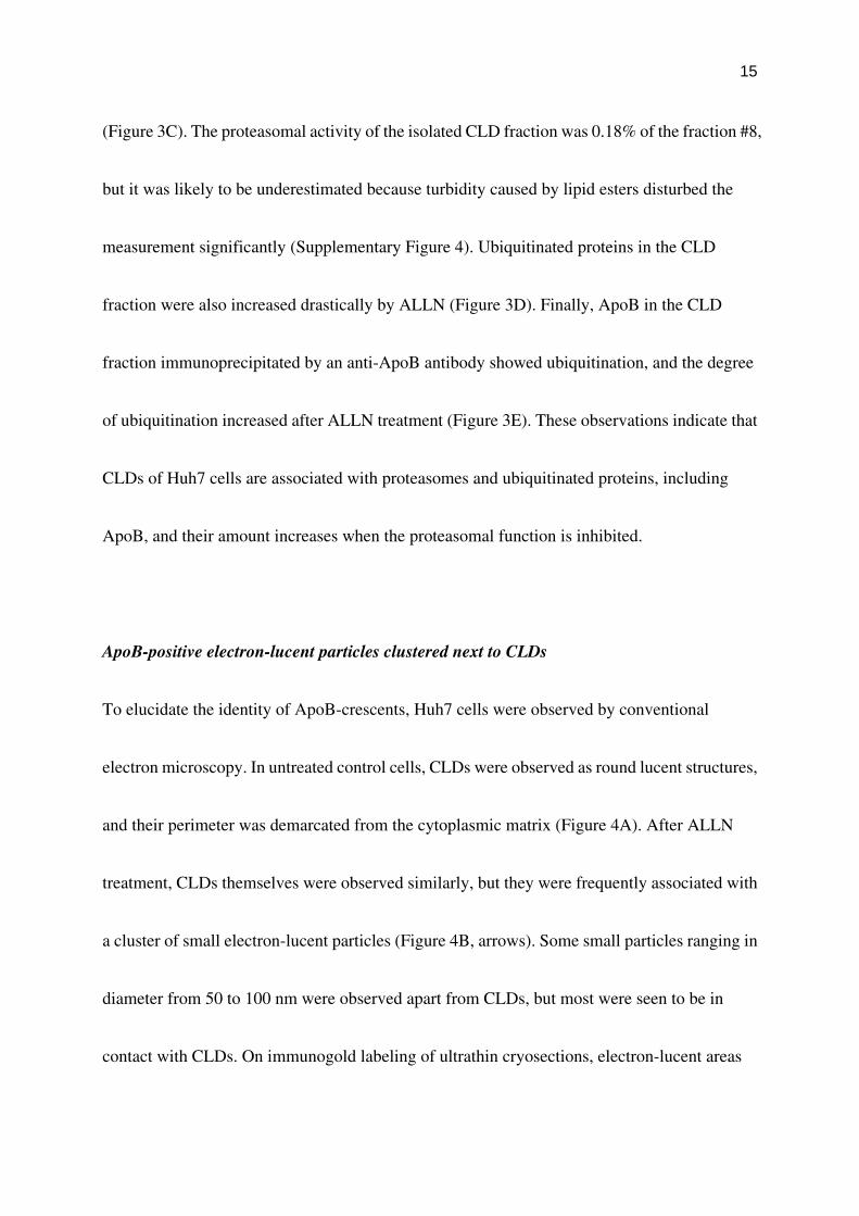
(Figure 3C). The proteasomal activity of the isolated CLD fraction was 0.18% of the fraction #8,
but it was likely to be underestimated because turbidity caused by lipid esters disturbed the
measurement significantly (Supplementary Figure 4). Ubiquitinated proteins in the CLD
fraction were also increased drastically by ALLN (Figure 3D). Finally, ApoB in the CLD
fraction immunoprecipitated by an anti-ApoB antibody showed ubiquitination, and the degree
of ubiquitination increased after ALLN treatment (Figure 3E). These observations indicate that
CLDs of Huh7 cells are associated with proteasomes and ubiquitinated proteins, including
ApoB, and their amount increases when the proteasomal function is inhibited.
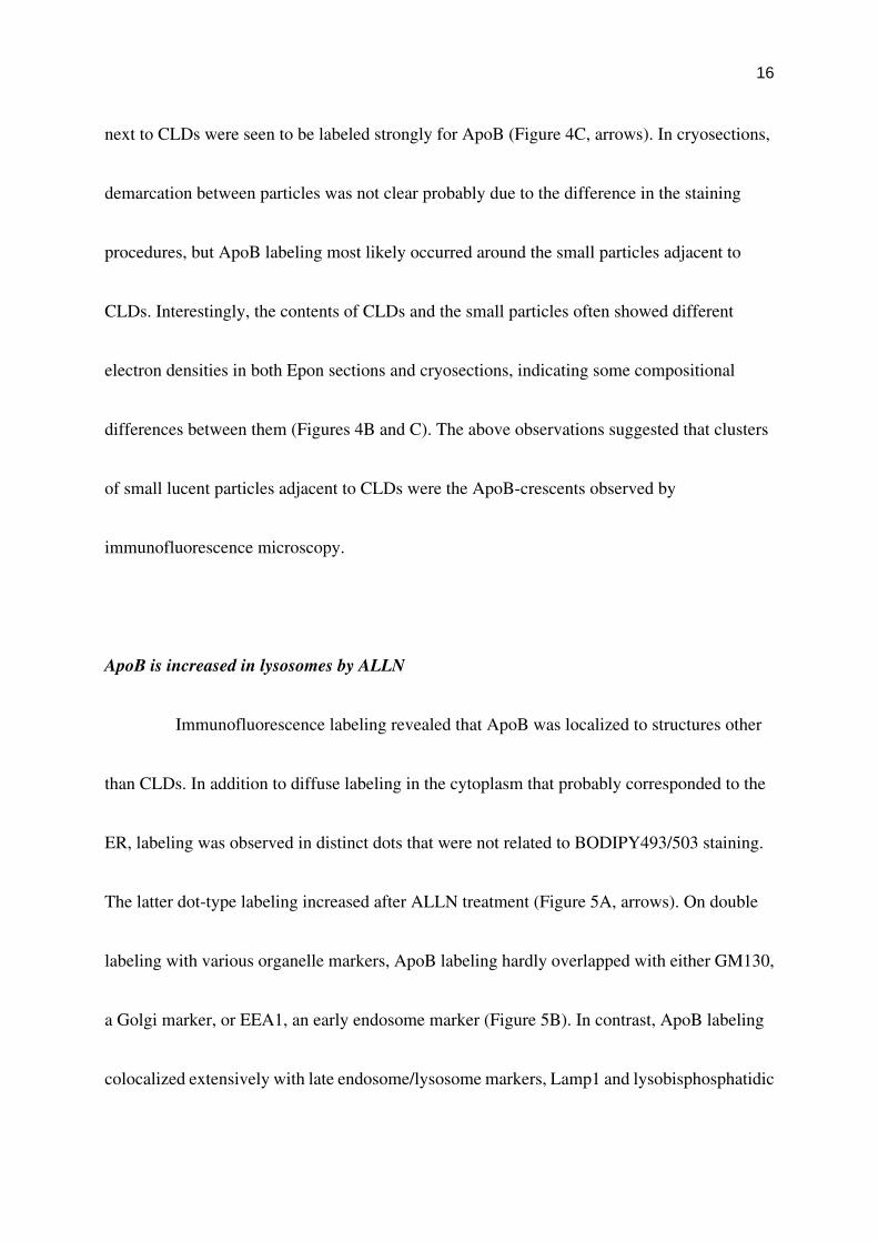
ApoB-positive electron-lucent particles clustered next to CLDs
To elucidate the identity of ApoB-crescents, Huh7 cells were observed by conventional
electron microscopy. In untreated control cells, CLDs were observed as round lucent structures,
and their perimeter was demarcated from the cytoplasmic matrix (Figure 4A). After ALLN
treatment, CLDs themselves were observed similarly, but they were frequently associated with
a cluster of small electron-lucent particles (Figure 4B, arrows). Some small particles ranging in
diameter from 50 to 100 nm were observed apart from CLDs, but most were seen to be in
contact with CLDs. On immunogold labeling of ultrathin cryosections, electron-lucent areas
16
next to CLDs were seen to be labeled strongly for ApoB (Figure 4C, arrows). In cryosections,
demarcation between particles was not clear probably due to the difference in the staining
procedures, but ApoB labeling most likely occurred around the small particles adjacent to
CLDs. Interestingly, the contents of CLDs and the small particles often showed different
electron densities in both Epon sections and cryosections, indicating some compositional
differences between them (Figures 4B and C). The above observations suggested that clusters
of small lucent particles adjacent to CLDs were the ApoB-crescents observed by
immunofluorescence microscopy.
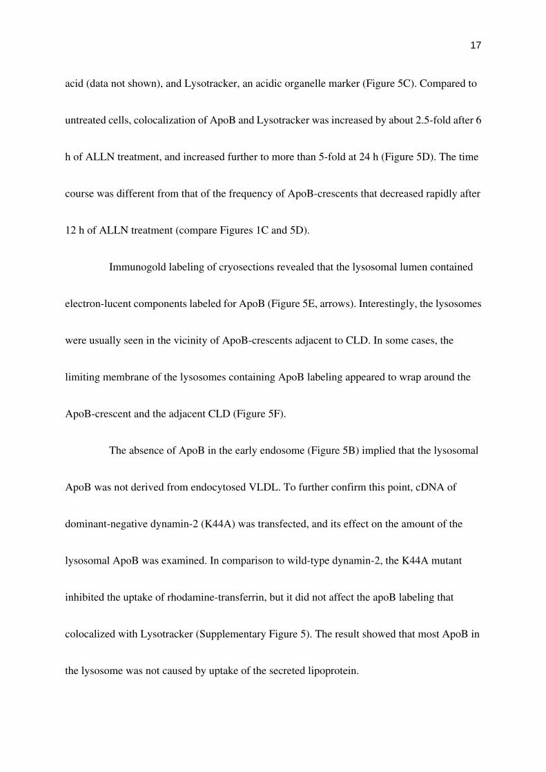
ApoB is increased in lysosomes by ALLN
Immunofluorescence labeling revealed that ApoB was localized to structures other
than CLDs. In addition to diffuse labeling in the cytoplasm that probably corresponded to the
ER, labeling was observed in distinct dots that were not related to BODIPY493/503 staining.
The latter dot-type labeling increased after ALLN treatment (Figure 5A, arrows). On double
labeling with various organelle markers, ApoB labeling hardly overlapped with either GM130,
a Golgi marker, or EEA1, an early endosome marker (Figure 5B). In contrast, ApoB labeling
colocalized extensively with late endosome/lysosome markers, Lamp1 and lysobisphosphatidic
17
acid (data not shown), and Lysotracker, an acidic organelle marker (Figure 5C). Compared to
untreated cells, colocalization of ApoB and Lysotracker was increased by about 2.5-fold after 6
h of ALLN treatment, and increased further to more than 5-fold at 24 h (Figure 5D). The time
course was different from that of the frequency of ApoB-crescents that decreased rapidly after
12 h of ALLN treatment (compare Figures 1C and 5D).
Immunogold labeling of cryosections revealed that the lysosomal lumen contained
electron-lucent components labeled for ApoB (Figure 5E, arrows). Interestingly, the lysosomes
were usually seen in the vicinity of ApoB-crescents adjacent to CLD. In some cases, the
limiting membrane of the lysosomes containing ApoB labeling appeared to wrap around the
ApoB-crescent and the adjacent CLD (Figure 5F).
The absence of ApoB in the early endosome (Figure 5B) implied that the lysosomal
ApoB was not derived from endocytosed VLDL. To further confirm this point, cDNA of
dominant-negative dynamin-2 (K44A) was transfected, and its effect on the amount of the
lysosomal ApoB was examined. In comparison to wild-type dynamin-2, the K44A mutant
inhibited the uptake of rhodamine-transferrin, but it did not affect the apoB labeling that
colocalized with Lysotracker (Supplementary Figure 5). The result showed that most ApoB in
the lysosome was not caused by uptake of the secreted lipoprotein.
18
ApoB-crescents are processed by autophagy
The above result as well as immunoelectron microscopy of ALLN-treated cells
suggested that the ApoB-positive particles in the lysosome were largely caused by autophagy.
In fact, a previous study demonstrated that inhibition of proteasomes in Huh7 cells induced
autophagic vacuoles (Harada et al., 2003). The increase in autophagic vacuoles was confirmed
by labeling with LC3 (Kabeya et al., 2000); by ALLN treatment, the increase of LC3-positive
particles was already evident at 1 hr, and became drastic at 12 h (Figure 6A; Supplementary
Figure 6). By triple labeling for ApoB, LC3, and neutral lipids, the LC3-positive
autophagosomes were often seen adjacent to ApoB-crescents (Figure 6B, arrows). By treating
the cells with leupeptin for 30 min to inhibit the lysosomal digestion, ApoB-crescents in the
Lamp1-positive organelles were detected (Figure 6C, arrow). These observations suggest that
inhibition of proteasomal function induced autophagic vacuoles, which may then engulf
ApoB-crescents and adjacent CLDs. The decrease in ApoB-crescents after 12 h of ALLN
treatment indicated that autophagy was activated to a sufficient degree by that time.
The above assumption was tested by inhibiting autophagy by 3-MA. The
ALLN-induced increase in LC3 was suppressed when the medium contained 10 mM 3-MA
(Figure 6A), which confirmed the effectiveness of the reagent. In cells treated with 3-MA at 12
19
h after the beginning of ALLN treatment, the decrease in ApoB-crescents between 12–24 h was
suppressed (Figure 6D). The suppressive effect was more obvious when cells were treated with
3-MA and ALLN from the beginning (Figure 6D). This observation verified that autophagy is
important in the processing of ApoB-crescents. Furthermore, it suggested that even when
proteasomal function is normal, autophagy may function in ApoB degradation. This
assumption proved correct because 3-MA alone caused an increase in ApoB-crescent-positive
cells and CLDs (Figures 6E and F). The ratio of ApoB-crescent-positive cells increased to more
than 30% at 12 h after addition of 3-MA. The results of the present study indicate that
proteasomal and autophagocytic systems collaborate to process ApoB, and that CLDs provide a
site at which the two degradation systems converge.
20
Discussion
CLDs as sites of proteasomal processing of ApoB
ApoB is the primary structural protein of VLDL. It is synthesized on
membrane-bound ribosomes, and translocated to the ER lumen cotranslationally. Nascent
lipoprotein particles are first assembled in the rough ER, and after maturation in the smooth
ER and possibly in the Golgi, they are secreted by exocytosis (for review, see Davis (1999)).
Thus, along with other secretory proteins, ApoB has been shown to exist in the ER and Golgi
(Sakata et al., 2001). In addition, labeling for ApoB was also observed in lysosomes (Du et al.,
1998), which may be derived, at least partially, from secreted and then endocytosed VLDL
(Williams et al., 1990; Twisk et al., 2000). However, to our knowledge, there have been no
previous reports of localization of ApoB on the CLD surface. The cluster of ApoB-positive
particles adjacent to CLDs, which we designated the “ApoB-crescent,” was observed in both
Huh7 and HepG2 cells, and increased markedly when proteasome function was inhibited. In
HepG2 cells, inhibition of proteasomes was reported to cause particulate accumulation of
ApoB in association with the ER, but the ApoB-crescent was not described (Fisher et al.,
1997; Pariyarath et al., 2001). ApoB-crescent may not have been identified previously because
of its relatively low frequency in HepG2 cells. Furthermore, without CLD staining, it may be

21
difficult to discriminate between ApoB-crescents and dense particulate labeling.
Degradation of ApoB has been reported to occur by both proteasomal and
non-proteasomal pathways (Cavallo et al., 1999; Fisher et al., 2001). When conditions are not
appropriate for VLDL assembly, ubiquitination and proteasomal degradation of ApoB
increase. In contrast to most other proteins that probably undergo retrotranslocation from the
ER lumen for ER-associated degradation, ApoB is thought to remain halfway through the
translocon before being extracted to the cytoplasm for degradation (Fisher and Ginsberg,
2002), and thus proteasomes where ApoB is degraded are likely to be in the proximity of the
ER. In the present study, we showed that proteasome subunits are located near CLDs. CLDs
are generally distributed in the proximity of the ER (Murphy and Vance, 1999), and lipids
stored in CLDs are thought to be mobilized for pre-VLDL assembly in synchronization with
ApoB translation (Gibbons et al., 2000). This functional relationship should require that CLDs
and the translocons for ApoB synthesis exist in close proximity. Thus CLDs are likely to be in
the vicinity of both the ApoB translation site and proteasomes, which location appears
suitable to harbor ubiquitinated ApoB for proteasomal degradation.
In addition to proximity, the CLD surface may offer another important advantage
for the processing of ApoB. That is, even if the proteasomes are highly efficient in degrading

22
multi-ubiquitinated proteins, there is a danger that proteins to be degraded may form
aggregates in the cytoplasm through hydrophobic interactions (Kopito, 2000). Such
aggregates were shown to impair proteasomal processing (Bence et al., 2001), and may
become toxic to the cells. In this respect, CLDs may provide a surface to hold large
amphipathic proteins, such as ApoB, in an appropriate conformation and prevent them
forming aggregates. This assumption is supported by the structural resemblance of CLDs and
lipoprotein particles in that both consist of a neutral lipid core and a phospholipid monolayer.
The importance of the CLD surface for ApoB degradation might be tested by manipulating
the expression of CLD-related proteins.
The relationship between CLDs and electron-lucent particles in the ApoB-crescent
Immunoelectron microscopy indicated that ApoB was localized around small
electron-lucent particles that clustered around CLDs. Their appearance suggested that the
particles are lipidic in nature, but their electron density often looked different from that of
CLDs, suggesting that the contents of the particles and CLDs are not the same. The
electron-lucent particles in the ApoB-crescent were 50–100 nm in diameter, roughly matching
that of VLDL and/or premature VLDL (Murphy, 2001; Swift et al., 2001). The origin of the

23
particles is not known, but one possibility is that binding of ApoB induced segmentation of
CLDs solely depending on the molecular nature of ApoB, and gave rise to particles of that
size (Segrest et al., 2001). Alternatively, lipid esters made in the ER membrane may bud to the
cytoplasmic side; in this case, the particles are thought to be an aberrant form of CLDs coated
with ApoB instead of PAT proteins. Finally, as a third possibility, the particles could be VLDL
and/or pre-VLDL particles themselves. It is difficult to explain how they could extravasate
from the luminal side, but lipidic particles bearing apolipoproteins can exist in the cytoplasm
(Ito et al., 2002).
In CLDs associated with ApoB-crescents, ADRP and TIP47 were seen only in the
area complementary to the crescent. This distribution was reminiscent of the displacement of
ADRP from CLDs when Rab18 was overexpressed (Ozeki et al., 2005), which then induced
close apposition of CLDs and the ER-derived membrane. The ApoB-crescent is different from
the above case in that Rab18 was not upregulated (data not shown), and that both ADRP and
TIP47 were expelled from the area apposing the ApoB-crescent. The function of ADRP (and
possibly TIP47) may be to shield the CLD surface from other organelles (Ozeki et al., 2005).
The absence of PAT proteins may be necessary for the adherence of lipidic particles to CLDs
and the formation of the ApoB-crescent.
24
Convergence of ubiquitin-proteasome pathway and autophagy-lysosome pathway in CLDs
ApoB level in the lysosome was increased by inhibition of the proteasome, and it
persisted even after the frequency of ApoB-crescents began to decrease after 12 h. Two
different mechanisms have been proposed to explain the existence of ApoB in the lysosome.
One was the reuptake of secreted VLDL through LDL receptors (Williams et al., 1990; Twisk
et al., 2000). The reuptake may also occur in Huh7 cells, but it is likely to be of minor
importance because the lysosomal ApoB labeling persisted even in cell expressing the
dominant-negative dynamin-2. The other was the proteasomal processing of ApoB resulting in
a single-pass transmembrane protein (Du et al., 1998). However, labeling for ApoB was
observed throughout the lysosomal lumen in our immunoelectron microscopic study, which
was unlikely to be caused by a transmembrane protein in the limiting membrane.
The ALLN-induced increase in ApoB in the lysosome was probably caused by
autophagy. First, LC3, a specific marker of the autophagosomal membrane (Kabeya et al.,
2000), was increased by ALLN, and was found near the ApoB-crescent. Second, electron
microscopy showed autophagic structures engulfing the ApoB-crescent, and ApoB in the
lysosomal lumen was found around small electron-lucent particles resembling those in the
ApoB-crescent. Third, the decrease in ApoB-crescents between 12–24 h of ALLN treatment
25
was blocked by 3-MA, an inhibitor of autophagy. These results suggest that the
ApoB-crescents were subjected to autophagy when the proteasome function was inhibited.
Previously, ALLN treatment was shown to induce ubiquitin-rich intermediate filament
inclusion bodies in Huh7 cells, which were removed by autophagic vacuoles (Harada et al.,
2003). In view of the close relationship of CLDs with intermediate filaments (Franke et al.,
1987; Almahbobi, 1995), it is likely that the inclusion bodies contain CLDs and the adhering
ApoB-crescents.
The participation of autophagy in processing ApoB is not apparently limited to
conditions where the proteasomal function is compromised. Inhibition of autophagy caused a
similar increase in ApoB-crescent frequency as inhibition of the proteasome. This observation
suggests that autophagy plays a major role in degradation of ApoB in Huh7 cells. Previously,
autophagy was shown to process aggresomes in Schwann cells (Fortun et al., 2003). More
recently, using Atg7-deficient mice, ubiquitinated proteins were shown to aggregate even in
the presence of functional proteasomes, and the aggregates were shown to be eliminated by
the autophagic process (Komatsu et al., 2005). Notably, the accumulation of ubiquitinated
proteins was observed around CLDs. The authors proposed that ubiquitination may serve as a
signal for the autophagic process as well as to the proteasome pathway. The results of the

26
present study fit well with their proposal and show that such a mechanism works
physiologically to process ubiquitinated proteins in vivo. On the other hand, the relative
importance of the proteasomal and autophagic pathways for the ApoB degradation may not be
the same in different cell lines. For example, in HepG2 cells, a significant increase of
ApoB-crescents were observed by ALLN, but not by 3-MA (data not shown). In the present
study, Huh7 cells were used because the cell line retains the ability to secrete VLDL (Higashi
et al., 2002, 2003; Lalanne et al., 2005), whereas HepG2 cells require exogenous fatty acids for
the secretion due to the deficiency of triacylglycerol hydrolase (Lehner et al., 1999). But it does
not mean that other physiological mechanisms to regulate ApoB are preserved in Huh7 cells
as well. The detailed degradation mechanism of ApoB in vivo needs to be studied under
physiological settings.
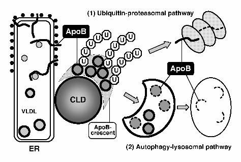
A diagram depicting the major features of the ApoB degradation in Huh7 cells is shown in
Figure 7. CLDs are likely to provide a surface to integrate the two degradation
systems—proteasomal and lysosomal—for ApoB. The unique structure of CLDs must be
important for their role as discussed above. In this way, CLDs in hepatocytes may act not only
as a reservoir of lipids for VLDL formation, but also as a platform on which to process the
unused ApoB effectively, and to prevent the unusually large amphipathic molecules from
27
forming toxic aggregates. An intriguing possibility is that the role of CLDs as a site of
convergence of proteasomal and autophagic processing may not be limited to ApoB but may
be extended to other amphipathic/hydrophobic proteins. In this context, it is noteworthy that
oligomers of α-synuclein have a propensity to bind CLDs (Cole et al., 2002). α-Synuclein is a
component of intraneuronal inclusion bodies that are found in a number of neurodegenerative
diseases, including Parkinson’s disease (Spillantini et al., 1998). In the neuronal cell, CLDs
are scarce and labeling for ADRP is not observed (Heid et al., 1998). If CLDs are important
for the safe processing of aggregation-prone proteins, the paucity of CLDs may explain why
toxic aggregates are formed preferentially in neuronal cells. Further studies to determine
whether manipulation of CLD proteins and lipids would modify aggregate formation are
warranted.
28
Acknowledgments
We thank Drs. Kazuhisa Nakayama, Ron R. Kopito, Toshihide Kobayashi, Nobuhiro
Nakamura, Tom Keenan, Yasuo Uchiyama, and Sadaki Yokota for providing vectors and
antibodies. We are also grateful to Dr. Shigeo Murata for invaluable advise on the proteasomal
activity assay, and to Ms. Kumi Tauchi-Sato and Mr. Tetsuo Okumura for technical assistance.
This work was supported by Grants-in-Aid for Scientific Research and the 21st Century COE
Program “Integrated Molecular Medicine for Neuronal and Neoplastic Disorders” of the
Ministry of Education, Culture, Sports, Science, and Technology of the Japanese Government.
29
References
Almahbobi, G. (1995). Adhesion of intermediate filaments and lipid droplets in adrenal cells
studied by field emission scanning electron microscopy. Cell Tissue Res 281, 387-390.
Bence, N.F., Sampat, R.M., and Kopito, R.R. (2001). Impairment of the Ubiquitin-Proteasome
System by Protein Aggregation. Science 292, 1552-1555.
Brasaemle, D.L., Dolios, G., Shapiro, L., and Wang, R. (2004). Proteomic analysis of proteins
associated with lipid droplets of basal and lipolytically stimulated 3T3-L1 adipocytes. J Biol
Chem 279, 46835-46842.
Cavallo, D., Rudy, D., Mohammadi, A., Macri, J., and Adeli, K. (1999). Studies on degradative
mechanisms mediating post-translational fragmentation of apolipoprotein B and the generation
of the 70-kDa fragment. J Biol Chem 274, 23135-23143.
Cole, N.B., Murphy, D.D., Grider, T., Rueter, S., Brasaemle, D., and Nussbaum, R.L. (2002).
Lipid droplet binding and oligomerization properties of the Parkinson's disease protein
alpha-synuclein. J Biol Chem 277, 6344-6352.
Davis, R.A. (1999). Cell and molecular biology of the assembly and secretion of apolipoprotein
B-containing lipoproteins by the liver. Biochim Biophys Acta 1440, 1-31.
Du, X., Stoops, J.D., Mertz, J.R., Stanley, C.M., and Dixon, J.L. (1998). Identification of two
regions in apolipoprotein B100 that are exposed on the cytosolic side of the endoplasmic
reticulum membrane. J Cell Biol 141, 585-599.
Fisher, E.A., and Ginsberg, H.N. (2002). Complexity in the secretory pathway: the assembly
and secretion of apolipoprotein B-containing lipoproteins. J Biol Chem 277, 17377-17380.
Fisher, E.A., Pan, M., Chen, X., Wu, X., Wang, H., Jamil, H., Sparks, J.D., and Williams, K.J.
(2001). The triple threat to nascent apolipoprotein B. Evidence for multiple, distinct
degradative pathways. J Biol Chem 276, 27855-27863.
Fisher, E.A., Zhou, M., Mitchell, D.M., Wu, X., Omura, S., Wang, H., Goldberg, A.L., and
Ginsberg, H.N. (1997). The degradation of apolipoprotein B100 is mediated by the
ubiquitin-proteasome pathway and involves heat shock protein 70. J Biol Chem 272,
20427-20434.
Fortun, J., Dunn, W.A., Jr., Joy, S., Li, J., and Notterpek, L. (2003). Emerging role for
autophagy in the removal of aggresomes in Schwann cells. J Neurosci 23, 10672-10680.
Franke, W.W., Hergt, M., and Grund, C. (1987). Rearrangement of the vimentin cytoskeleton
during adipose conversion: formation of an intermediate filament cage around lipid globules.
Cell 49, 131-141.
Fujimoto, T., Kogo, H., Ishiguro, K., Tauchi, K., and Nomura, R. (2001). Caveolin-2 is targeted
30
to lipid droplets, a new "membrane domain" in the cell. J Cell Biol 152, 1079-1085.
Fujimoto, Y., Itabe, H., Sakai, J., Makita, M., Noda, J., Mori, M., Higashi, Y., Kojima, S., and
Takano, T. (2004). Identification of major proteins in the lipid droplet-enriched fraction isolated
from the human hepatocyte cell line HuH7. Biochim Biophys Acta 1644, 47-59.
Gaczynska, M., Rock, K..L, Spies, T., and Goldberg, A.L. (1994) Peptidase activities of
proteasomes are differentially regulated by the major histocompatibility complex-encoded
genes for LMP2 and LMP7. Proc. Natl. Acad. Sci. USA 91, 9213-9217. Gibbons, G.F., Islam, K., and Pease, R.J. (2000). Mobilisation of triacylglycerol stores.
Biochim Biophys Acta 1483, 37-57.
Goldstein, J. L., Basu, S. K. and Brown, M. S. (1983) Receptor-mediated endocytosis of
low-density lipoprotein in cultured cells. Methods Enzymol 98, 241–260.
Harada, M., Kumemura, H., Omary, M.B., Kawaguchi, T., Maeyama, N., Hanada, S., Taniguchi,
E., Koga, H., Suganuma, T., Ueno, T., and Sata, M. (2003). Proteasome inhibition induces
inclusion bodies associated with intermediate filaments and fragmentation of the Golgi
apparatus. Exp Cell Res 288, 60-69.
Heid, H.W., Moll, R., Schwetlick, I., Rackwitz, H.R., and Keenan, T.W. (1998). Adipophilin is
a specific marker of lipid accumulation in diverse cell types and diseases. Cell Tissue Res 294,
309-321.
Higashi, Y., Itabe, H., Fukase, H., Mori, M., Fujimoto, Y., Sato, R., Imanaka, T., and Takano, T.
(2002). Distribution of microsomal triglyceride transfer protein within sub-endoplasmic
reticulum regions in human hepatoma cells. Biochim Biophys Acta 1581, 127-136.
Higashi, Y., Itabe, H., Fukase, H., Mori, M., Fujimoto, Y., and Takano, T. (2003)
Transmembrane lipid transfer is crucial for providing neutral lipids during very low density
lipoprotein assembly in endoplasmic reticulum. J Biol Chem 278, 21450-21458.
Ito, J., Nagayasu, Y., Kato, K., Sato, R., and Yokoyama, S. (2002). Apolipoprotein A-I induces
translocation of cholesterol, phospholipid, and caveolin-1 to cytosol in rat astrocytes. J Biol
Chem 277, 7929-7935.
Kabeya, Y., Mizushima, N., Ueno, T., Yamamoto, A., Kirisako, T., Noda, T., Kominami, E.,
Ohsumi, Y., and Yoshimori, T. (2000). LC3, a mammalian homologue of yeast Apg8p, is
localized in autophagosome membranes after processing. EMBO J 19, 5720-5728.
Komatsu, M., Waguri, S., Ueno, T., Iwata, J., Murata, S., Tanida, I., Ezaki, J., Mizushima, N.,
Ohsumi, Y., Uchiyama, Y., Kominami, E., Tanaka, K., and Chiba, T. (2005). Impairment of
starvation-induced and constitutive autophagy in Atg7-deficient mice. J Cell Biol 169, 425-434.
Kopito, R.R. (2000). Aggresomes, inclusion bodies and protein aggregation. Trends Cell Biol
10, 524-530.
31
Lalanne, F., Lambert, G., Amar, M. J., Chetiveaux, M., Zair, Y., Jarnoux, A. L., Ouguerram, K.,
Friburg, J., Seidah, N. G., Brewer, H. B. Jr, Krempf, M., and Costet, P. (2005) Wild-type
PCSK9 inhibits LDL clearance but does not affect apoB-containing lipoprotein production in
mouse and cultured cells. J Lipid Res 46, 1312-1319.
Lehner, R, Cui, Z., and Vance, D.E. (1999). Subcellullar localization, developmental expression
and characterization of a liver triacylglycerol hydrolase. Biochem J 338, 761-768.
Liou, W., Geuze, H.J., and Slot, J.W. (1996). Improving structural integrity of cryosections for
immunogold labeling. Histochem Cell Biol 106, 41-58.
Liu, P., Ying, Y., Zhao, Y., Mundy, D.I., Zhu, M., and Anderson, R.G. (2004). Chinese hamster
ovary K2 cell lipid droplets appear to be metabolic organelles involved in membrane traffic. J
Biol Chem 279, 3787-3792.
Mitchell, D.M., Zhou, M., Pariyarath, R., Wang, H., Aitchison, J.D., Ginsberg, H.N., and Fisher,
E.A. (1998). Apoprotein B100 has a prolonged interaction with the translocon during which its
lipidation and translocation change from dependence on the microsomal triglyceride transfer
protein to independence. Proc Natl Acad Sci U S A 95, 14733-14738.
Miura, S., Gan, J.W., Brzostowski, J., Parisi, M.J., Schultz, C.J., Londos, C., Oliver, B., and
Kimmel, A.R. (2002). Functional conservation for lipid storage droplet association among
Perilipin, ADRP, and TIP47 (PAT)-related proteins in mammals, Drosophila, and
Dictyostelium. J Biol Chem 277, 32253-32257.
Murphy, D.J. (2001). The biogenesis and functions of lipid bodies in animals, plants and
microorganisms. Prog Lipid Res 40, 325-438.
Murphy, D.J., and Vance, J. (1999). Mechanisms of lipid-body formation. Trends Biochem Sci
24, 109-115.
Ohsaki, Y., Maeda, T., and Fujimoto, T. (2005) Fixation and permeabilization protocol is
critical for immunolabeling of lipid droplet protein. Histochem Cell Biol 124, 445-452.
Olofsson, S.O., Asp, L., and Boren, J. (1999). The assembly and secretion of apolipoprotein
B-containing lipoproteins. Curr Opin Lipidol 10, 341-346.
Ozeki, S., Cheng, J.-L., Tauchi-Sato, K., Hatano, N., Taniguchi, H., and Fujimoto, T. (2005).
Rab18 localizes to lipid droplets and induces their close apposition to the endoplasmic
reticulum-derived membrane. J Cell Sci 118, 2601-2611.
Pan, M., Liang Js, J.S., Fisher, E.A., and Ginsberg, H.N. (2002). The late addition of core lipids
to nascent apolipoprotein B100, resulting in the assembly and secretion of triglyceride-rich
lipoproteins, is independent of both microsomal triglyceride transfer protein activity and new
triglyceride synthesis. J Biol Chem 277, 4413-4421.
Pariyarath, R., Wang, H., Aitchison, J.D., Ginsberg, H.N., Welch, W.J., Johnson, A.E., and
32
Fisher, E.A. (2001). Co-translational interactions of apoprotein B with the ribosome and
translocon during lipoprotein assembly or targeting to the proteasome. J Biol Chem 276,
541-550.
Sakata, N., Phillips, T.E., and Dixon, J.L. (2001). Distribution, transport, and degradation of
apolipoprotein B-100 in HepG2 cells. J Lipid Res 42, 1947-1958.
Segrest, J.P., Jones, M.K., De Loof, H., and Dashti, N. (2001). Structure of apolipoprotein
B-100 in low density lipoproteins. J Lipid Res 42, 1346-1367.
Spillantini, M.G., Crowther, R.A., Jakes, R., Hasegawa, M., and Goedert, M. (1998). alpha
-Synuclein in filamentous inclusions of Lewy bodies from Parkinson's disease and dementia
with Lewy bodies. PNAS 95, 6469-6473.
Swift, L.L., Valyi-Nagy, K., Rowland, C., and Harris, C. (2001). Assembly of very low density
lipoproteins in mouse liver: evidence of heterogeneity of particle density in the Golgi apparatus.
J Lipid Res 42, 218-224.
Tauchi-Sato, K., Ozeki, S., Houjou, T., Taguchi, R., and Fujimoto, T. (2002). The surface of
lipid droplets is a phospholipid monolayer with a unique Fatty Acid composition. J Biol Chem
277, 44507-44512.
Tokumoto, T., Yamashita, M., Yoshikuni, M., Kajiura, H., and Nagahama, Y. (1995).
Purification of latent proteasome (20S proteasome) and demonstration of active proteasome in
goldfish (Carassius auratus) oocyte cytosol. Biomed Res 16, 173-186.
Twisk, J., Gillian-Daniel, D.L., Tebon, A., Wang, L., Barrett, P.H., and Attie, A.D. (2000). The
role of the LDL receptor in apolipoprotein B secretion. J Clin Invest 105, 521-532.
Umlauf, E., Csaszar, E., Moertelmaier, M., Schuetz, G.J., Parton, R.G., and Prohaska, R. (2004).
Association of stomatin with lipid bodies. J Biol Chem 279, 23699-23709.
Wakata, Y., Tokumoto, M., Horiguchi, R., Ishikawa, K., Nagahama, Y., and Tokumoto, T.
(2004) Identification of alpha-type subunits of the Xenopus 20S proteasome and analysis of
their changes during the meiotic cell cycle. BMC Biochem 5, 18.
Wiertz, E.J., Jones, T.R., Sun, L., Bogyo, M., Geuze, H.J., and Ploegh, H.L. (1996a). The
human cytomegalovirus US11 gene product dislocates MHC class I heavy chains from the
endoplasmic reticulum to the cytosol. Cell 84, 769-779.
Wiertz, E.J., Tortorella, D., Bogyo, M., Yu, J., Mothes, W., Jones, T.R., Rapoport, T.A., and
Ploegh, H.L. (1996b). Sec61-mediated transfer of a membrane protein from the endoplasmic
reticulum to the proteasome for destruction. Nature 384, 432-438.
Williams, K.J., Brocia, R.W., and Fisher, E.A. (1990). The unstirred water layer as a site of
control of apolipoprotein B secretion. J Biol Chem 265, 16741-16744.
Wolins, N.E., Quaynor, B.K., Skinner, J.R., Schoenfish, M.J., Tzekov, A., and Bickel, P.E.
33
(2005). S3-12, adipophilin, and TIP47 package lipid in adipocytes. J Biol Chem 280,
19146-19155.
34
Figure Legends
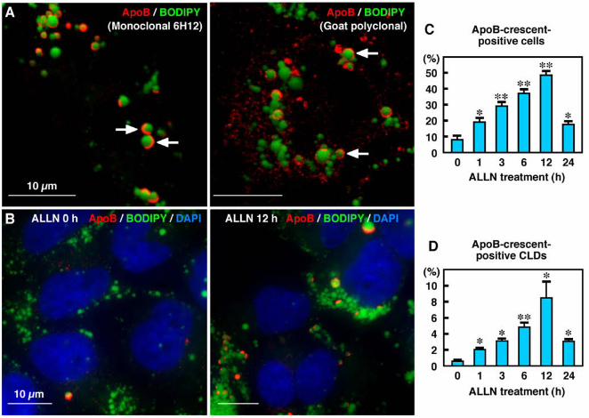
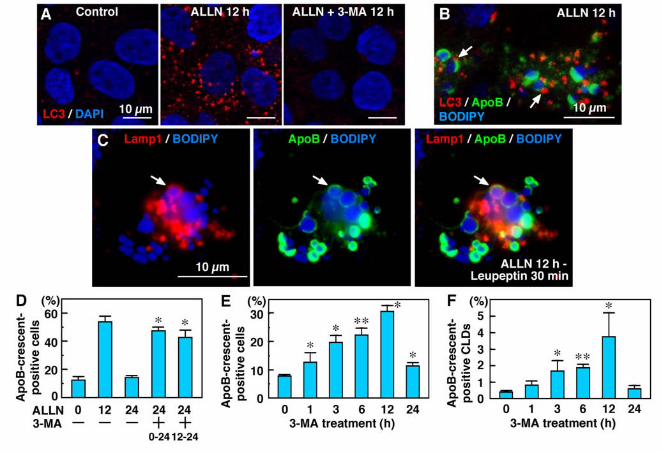
Figure 1. ApoB was localized in crescent-shaped areas adjacent to lipid droplets. (A) Huh7
cells cultured in normal medium. Some cells showed labeling for ApoB (red) in a crescent
shape adjacent to lipid droplets stained by BODIPY493/503 (green). Two different anti-ApoB
antibodies, mouse monoclonal (clone 6H12; left) and goat polyclonal (right), gave essentially
the same results. Bars, 10 µm. (B) The crescent-shaped ApoB labeling increased markedly
when Huh7 cells were treated with 10 µM ALLN for 12 h. Cell nuclei were labeled with DAPI.
Bars, 10 µm. (C, D) Percentage of cells (C) and CLDs (D) showing ApoB-crescents. The
frequency of crescent-positive cells and CLDs increased gradually up to 12 h by ALLN
treatment, and then decreased at 24 h. Results of three independent experiments were averaged;
statistical difference from the control (0 h) was examined by Student’s t-test (* p < 0.01, ** p <
0.001).
Figure 2. The crescent-shaped ApoB was complementary to ADRP and TIP47 in the CLD
surface. Confocal images of ADRP (red) and ApoB (green) [upper panel], and TIP47 (red) and
ApoB (green) [lower panel] in Huh7 cells treated with 10 μM ALLN for 12 h. Images
reconstructed in the x-z axis along the red line are shown above the upper panel. In most CLDs,
35
the ApoB labeling occupied 1/3 to 2/3 of the CLD surface (arrows), whereas both ADRP and
TIP47 labeling were seen in the area complementary to the ApoB-positive area (arrowheads).
Bars, 10 µm.
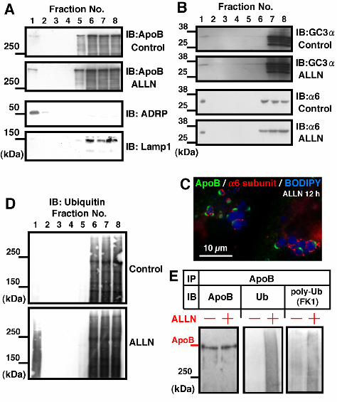
Figure 3. The CLD fraction was enriched with ApoB, proteasome subunits, and ubiquitinated
proteins. (A, B, D) Huh7 cells were cultured with or without 10 µM ALLN for 12 h, and
fractions obtained by sucrose density-gradient centrifugation were subjected to western blotting.
(A) ApoB (goat anti-ApoB antibody). Fraction #1 obtained from the top of the gradient
contained ApoB and its amount was greater in the ALLN-treated sample than in the control (see
Supplementary Figure 3 for quantitation). The concentration of ADRP (3rd panel) and the lack
of Lamp1 (4th panel) verified that the fraction was highly enriched with CLD. The result shown
is representative of three independent experiments. (B) Western blotting by GC3α and
anti-α6 antibody recognizing proteasomal subunits. The CLD fraction showed positive bands
with both antibodies. (C) The proteasome subunit α6 was localized adjacent to CLDs bearing
ApoB-crescents. α6 was also seen around CLDs that do not bear ApoB-crescents, and even
without ALLN treatment (data not shown). (D) Ubiquitin. The ubiquitinated proteins
recovered in the CLD fraction were increased significantly by ALLN treatment. (E) The CLD

36
fractions taken from control and ALLN-treated cells were adjusted to the same volume, lysed,
and immunoprecipitated with goat anti-ApoB antibody. The precipitated proteins were
subjected to Western blotting with anti-ApoB (left), anti-ubiquitin (middle), or
anti-polyubiquitin antibodies (right). The ubiquitination of ApoB was increased significantly in
the ALLN-treated sample. Cells transfected with ubiquitin cDNA were used for this experiment,
but essentially the same result was obtained by using cells without the overexpression.
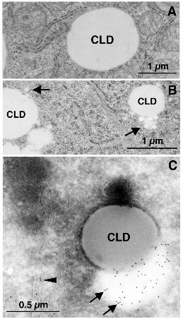
Figure 4. ApoB-positive low-density particles appeared adjacent to CLD in ALLN-treated
Huh7 cells. (A and B) Huh7 cells were cultured with or without 10 µM ALLN for 12 h, and
observed by conventional electron microscopy. In control cells (A), the rim of CLDs was
demarcated by the cytoplasm. In ALLN-treated cells (B), CLDs were often associated with a
cluster of small round electron-lucent particles (arrows). The small particles ranged from 50 to
100 nm in diameter. Bars, 1 μm. (C) Immunogold labeling of ApoB in Huh7 cells treated with
10 µM ALLN for 12 h. ApoB was localized to the electron-lucent particles adjacent to CLDs
(arrows). The electron density of the ApoB-positive particles was lower than that of CLDs,
implying a difference in their composition. ApoB labeling was also seen in the ER lumen
(arrowheads). Bar, 0.5 μm.
37
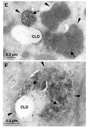
Figure 5. ApoB in lysosomal structures was increased by ALLN treatment. Bars, 10 μm. (A,
B) Huh7 cells treated with 10 µM ALLN for 12 h. Many ApoB-positive structures were not
colocalized with BODIPY493/503 (arrows in A), GM130 (B-left), or EEA1 (B-right). (C)
Colocalization of ApoB (green) and Lysotracker (red) increased drastically after ALLN
treatment. (D) Huh7 cells treated with 10 µM ALLN for the indicated times were incubated
with 500 nM Lysotracker-Red for 2 h before fixation and immunolabeling. Colocalization of
ApoB and Lysotracker-Red was quantified by measuring the number of double-positive pixels
in confluent cells. Results of three independent experiments were averaged; statistical
difference from the control (0 h) was examined by Student’s t-test (* p < 0.05, ** p < 0.001).
(E, F) Immunogold labeling of ALLN-treated Huh7 cells. Bars, 0.2 μm. (E) Lysosomes
(arrowheads) contained ApoB-positive electron-lucent particles (arrows), and adhered to the
ApoB-crescent area adjacent to CLDs. (F) In some cases, the lysosomes containing ApoB
labeling (arrows) wrapped around the ApoB-crescent and the adjacent CLD. The limiting
membrane of the lysosome is marked by arrowheads.
Figure 6. Autophagy is involved in processing the ApoB-crescent. (A) LC3 was observed
only infrequently in Huh7 cells cultured under normal conditions. ALLN (10 μM, 12 h)
38
increased LC3 markedly, but co-treatment with 3-MA (10 mM) suppressed the increase. Cell
nuclei were labeled by DAPI. Bars, 10 μm. (B) Triple labeling for LC3 (red), ApoB (green),
and BODIPY493/503 (blue) in ALLN-treated cells. LC3 was seen near the ApoB-crescent and
its adjacent CLD (arrows). Bar, 10 μm. (C) Triple labeling for Lamp1 (red), ApoB (green), and
BODIPY493/503 (blue). Cells were cultured with 10 µM ALLN for 12 h, and added with 100
µM leupeptin for 30 min. The ApoB-crescent and the adjacent CLD was found in the
Lamp1-positive late endosome/lysosome (arrow). (D) In comparison to cells cultured with
ALLN alone for 24 h, addition of 3-MA at 0 h or 12 h increased the ratio of
ApoB-crescent-positive cells at 24 h. The difference was statistically significant by Student’s
t-test (n = 3; *p < 0.001). (E, F) Percentage of cells (E) and CLDs (F) showing
ApoB-crescents in cells treated with 3-MA alone. The frequency of crescent-positive cells and
CLDs reached a maximum at 12 h. Results of three independent experiments were averaged;
statistical difference from the control (0 h) was examined by Student’s t-test (E: * p < 0.01, ** p
< 0.001; F: p < 0.05, ** p < 0.001).
Figure 7. The role of CLDs in ApoB degradation. CLDs exist in the vicinity of the ER, and may
serve to hold ApoB-positive lipidic particles as the ApoB-crescent. Both the
39
ubiquitin-proteasome pathway and the autophagy-lysosomal pathway are involved in the
degradation of ApoB in normal cells. The ApoB-crescent is thought to be the site where the two
degradation systems converge.