Embed Size (px)
Citation preview

LIU Chuan Yong
刘传勇
Institute of Physiology
Medical School of SDU
Tel 88381175 (lab)
88382098 (office)
Email: [email protected]
Website: www.physiology.sdu.edu.cn
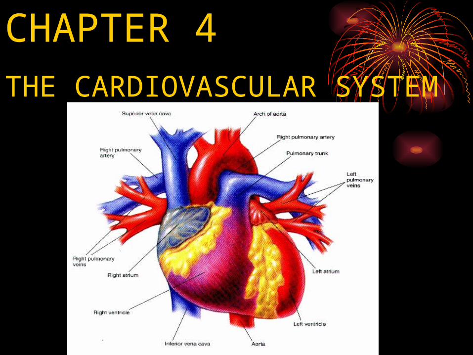
CHAPTER 4
THE CARDIOVASCULAR SYSTEM
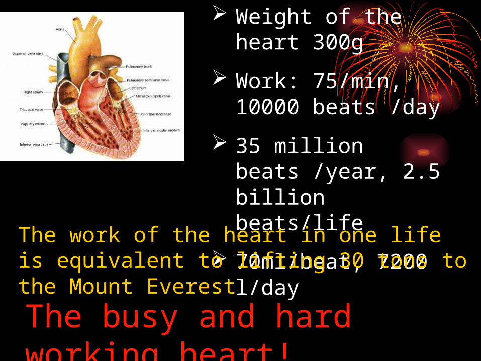
Weight of the heart 300g
Work: 75/min, 10000 beats /day
35 million beats /year, 2.5 billion beats/life
70ml/beat, 7200 l/day
The work of the heart in one life is equivalent to lifting 30 tons to the Mount Everest
The busy and hard working heart!

MAIN FUNCTIONS OF THE MAIN FUNCTIONS OF THE CIRCULATORY SYSTEMCIRCULATORY SYSTEM
Transport and distribute essential substances to the tissues.
Remove metabolic byproducts. Adjustment of oxygen and nutrient supply in
different physiologic states. Regulation of body temperature. Humoral communication.
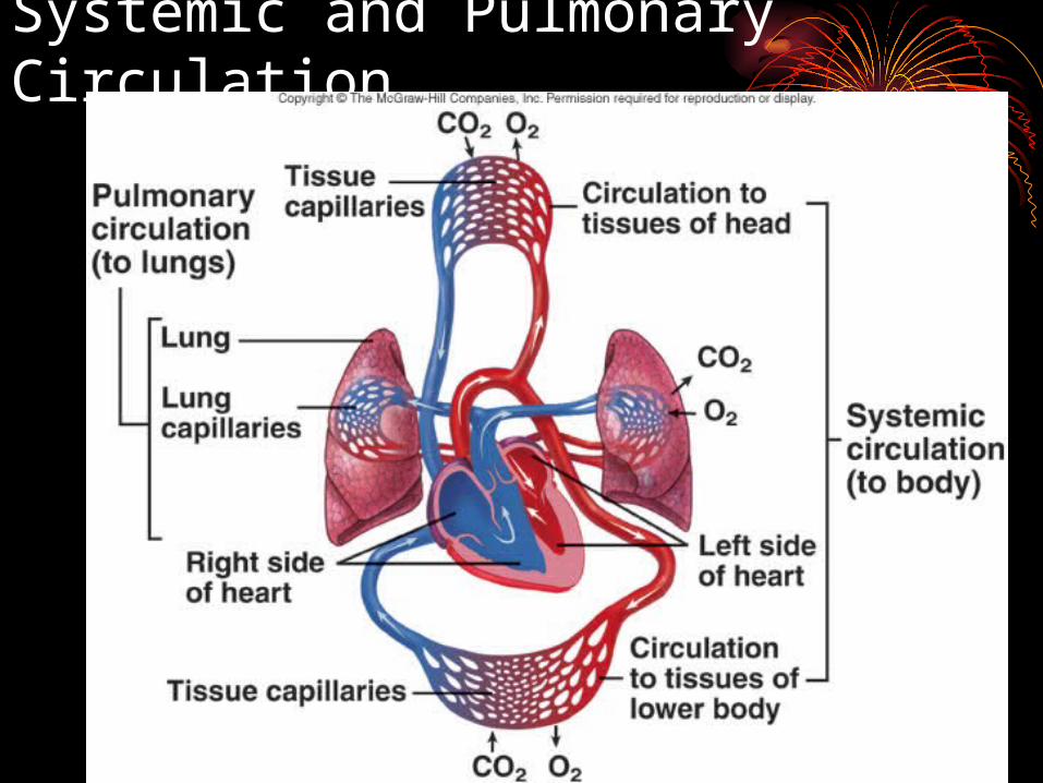
Systemic and Pulmonary Circulation
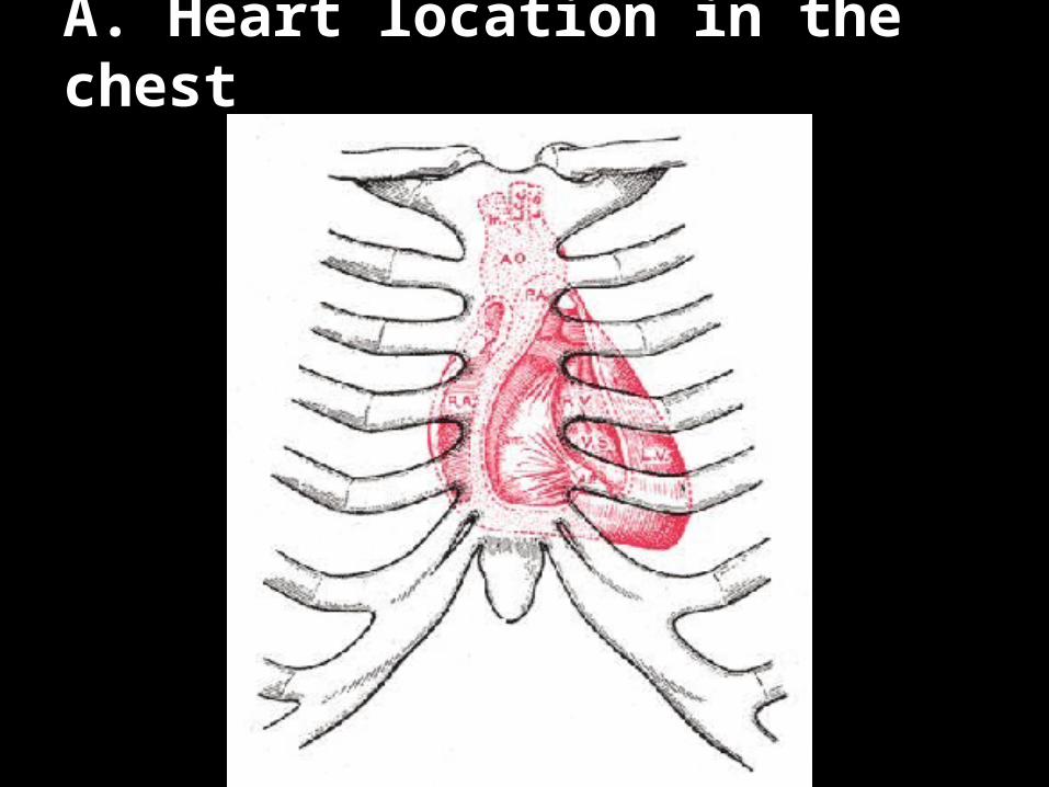
A. Heart location in the chest
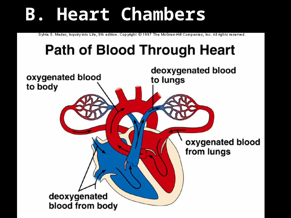
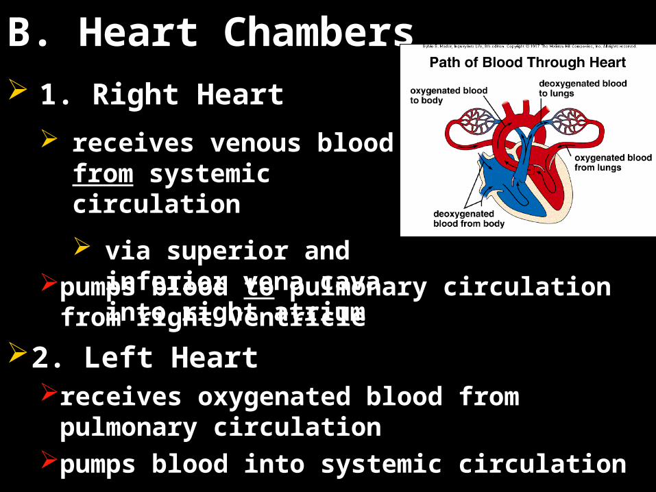
B. Heart Chambers
B. Heart Chambers
pumps blood to pulmonary circulation from right ventricle
2. Left Heartreceives oxygenated blood from pulmonary
circulationpumps blood into systemic circulation
1. Right Heart
receives venous blood from systemic circulation
via superior and inferior vena cava into right atrium
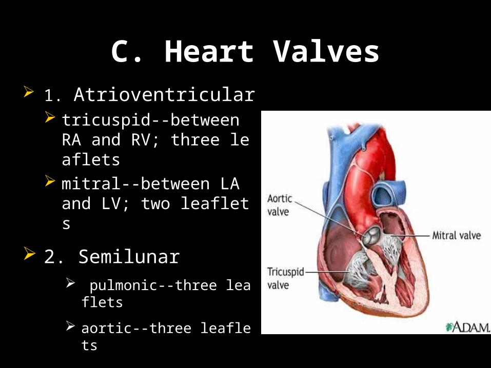
C. Heart Valves 1. Atrioventricular
tricuspid--between RA and RV; three leaflets
mitral--between LA and LV; two leaflets
2. Semilunar pulmonic--three leaflets
aortic--three leaflets
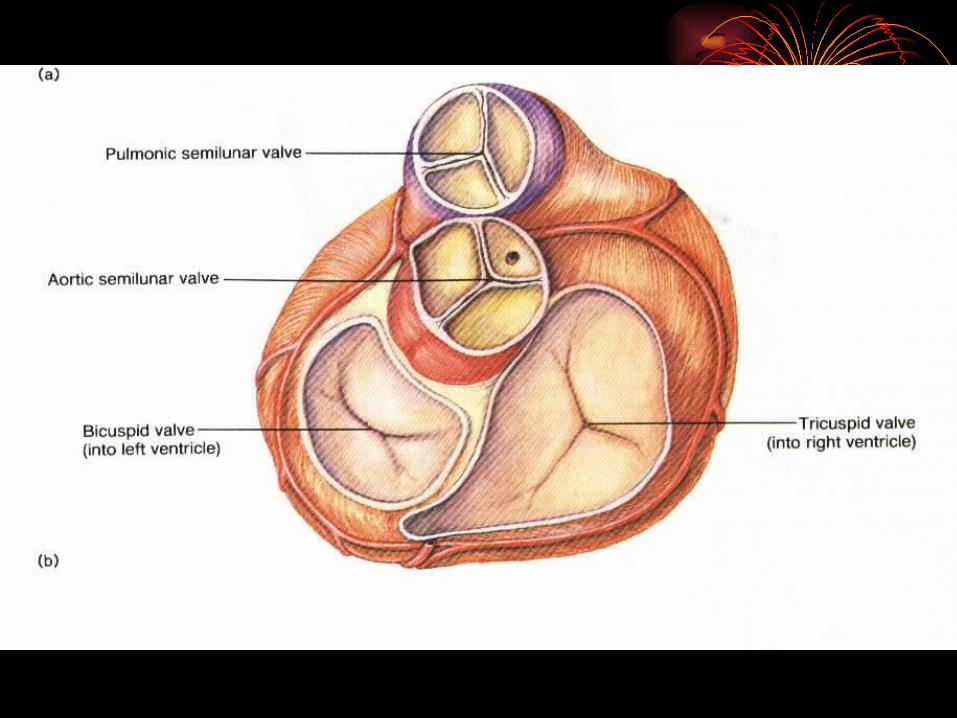

Prevent backward regurgitation
Provide low resistance to forward flow
Heart ValvesHeart Valves
Section 1 The Heart as a Pump
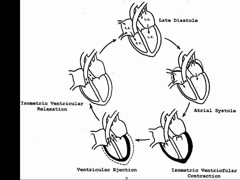
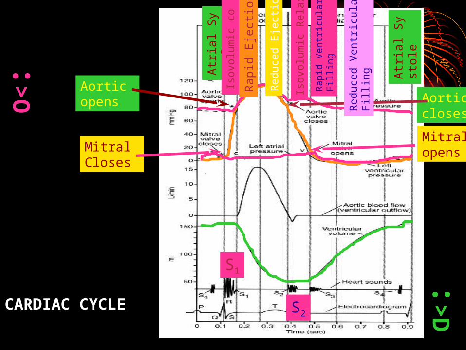
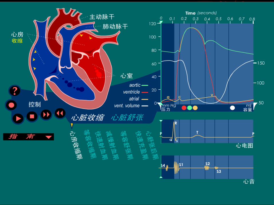
I Cardiac Cycle The period from the end of one heart
contraction to the end of the next
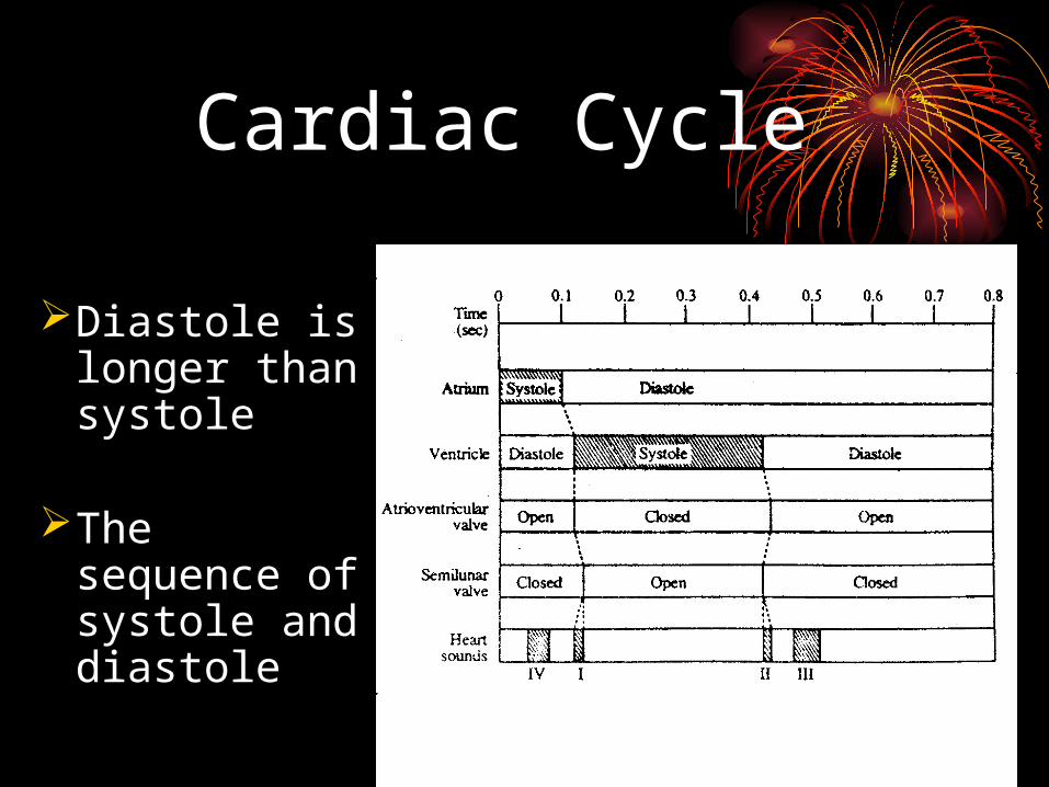
Cardiac Cycle
Diastole is longer than systole
The sequence of systole and diastole
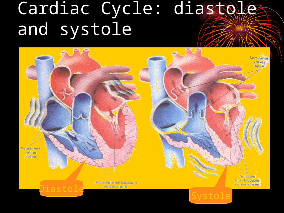
Cardiac Cycle: diastole and systole
DiastoleSystole
2 The Phases of the Cardiac Cycle
(1)Period of isometric (isovolumetric
or isovolumic) contraction
Events: ventricular contraction
ventricular pressure rise
atrioventricular valve close
the ventricular pressure increase sharply
Period: 0.05 sec
Importance: enable the ventricular pressure to rise from 0 to the level of aortic pressure (after-load)
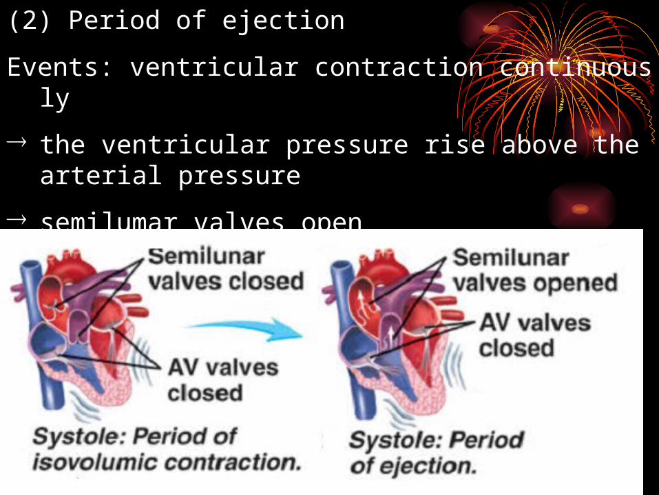
(2) Period of ejection
Events: ventricular contraction continuously
the ventricular pressure rise above the arterial pressure
semilumar valves open
blood pours out of the ventricles
Rapid ejection period (0.10s, 60% of the stroke volume)
Reduced ejection period (0.15s, 40% of the stroke volume)
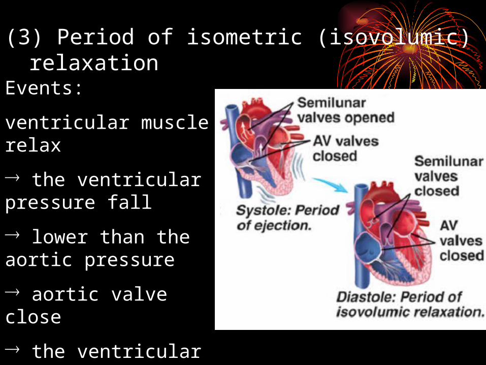
(3) Period of isometric (isovolumic) relaxation
Events:
ventricular muscle relax
the ventricular pressure fall
lower than the aortic pressure
aortic valve close
the ventricular pressure fall sharply
Period: 0.06-0.08 s
Importance: Enable the ventricular pressure fall to the level near the atrial pressure
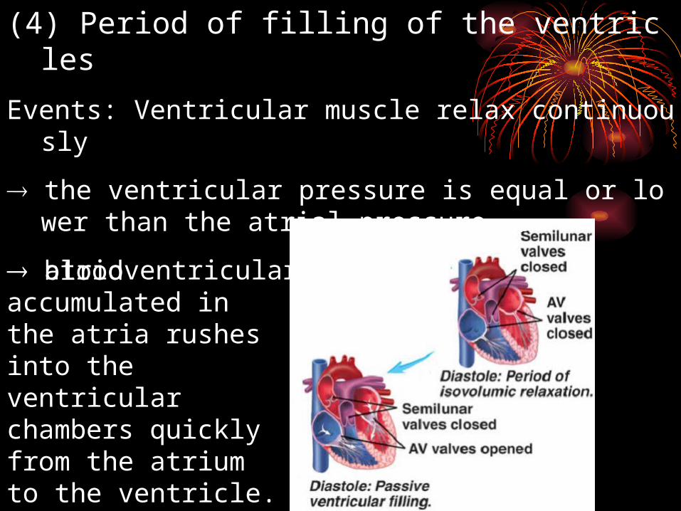
(4) Period of filling of the ventricles
Events: Ventricular muscle relax continuously
the ventricular pressure is equal or lower than the atrial pressure
atrioventricular valve open
blood accumulated in the atria rushes into the ventricular chambers quickly from the atrium to the ventricle.
Period of rapid filling. (0.11s, amount of filling, 2/3)
Period of reduced filling (0.22s, little blood fills into the ventricle)
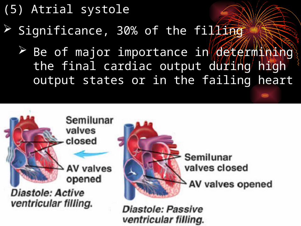
(5) Atrial systole
Significance, 30% of the filling
Be of major importance in determining the final cardiac output during high output states or in the failing heart
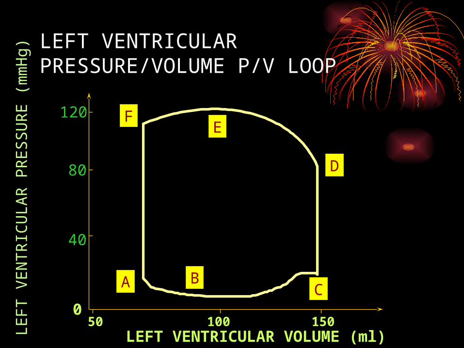
LEFT VENTRICULAR PRESSURE/VOLUME P/V LOOP
LE
FT
VE
NT
RIC
UL
AR
PR
ES
SU
RE
(m
mH
g)
LEFT VENTRICULAR VOLUME (ml)
A BC
D
EF
100 150500
120
40
80
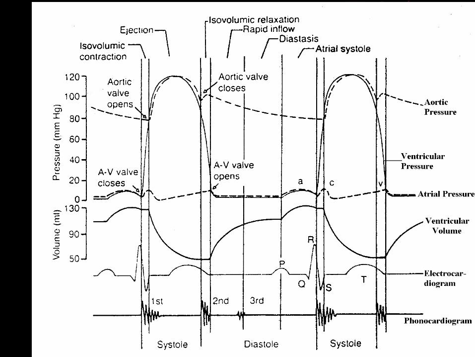
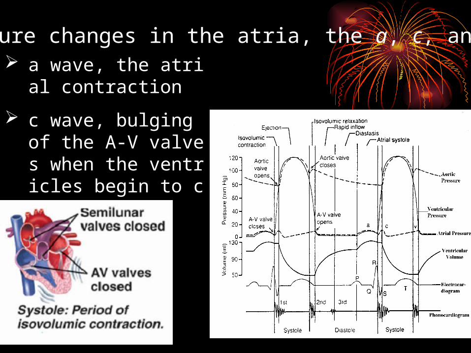
2 ) Pressure changes in the atria, the a, c, and v waves. a wave, the atrial contrac
tion
c wave, bulging of the A-V valves when the ventricles begin to contract
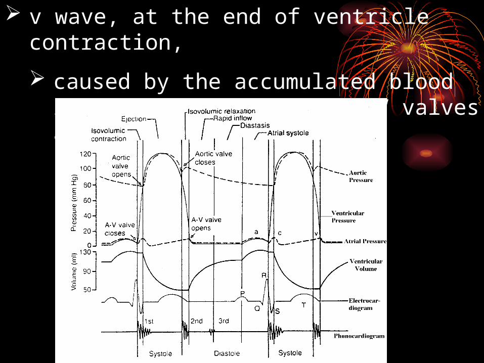
v wave, at the end of ventricle contraction,
caused by the accumulated blood in the atria while the A-V valves are closed
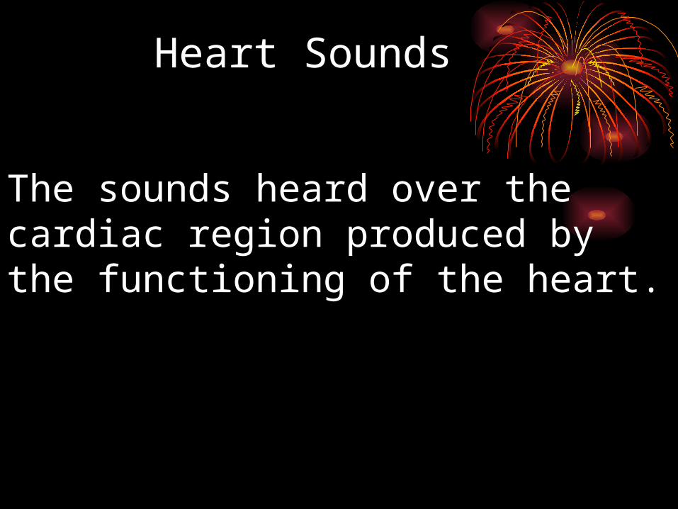
The sounds heard over the cardiac region produced by the functioning of the heart.
Heart SoundsHeart Sounds
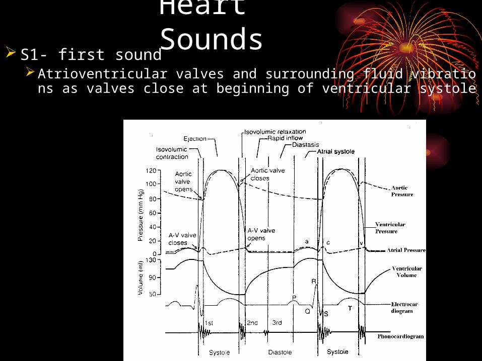
Heart Sounds S1- first sound
Atrioventricular valves and surrounding fluid vibrations as valves close at beginning of ventricular systole
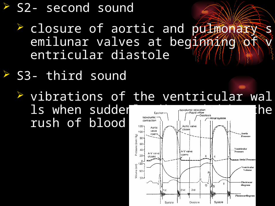
S2- second sound
closure of aortic and pulmonary semilunar valves at beginning of ventricular diastole
S3- third sound
vibrations of the ventricular walls when suddenly distended by the rush of blood from the atria
CARDIAC CYCLE
Atr
ial S
ysto
le
Mitral Closes
Isov
olum
ic c
ontr
act.
Aortic opens
S1
Rap
id E
ject
ion
Red
uced
Eje
ctio
n
Isov
olum
ic R
elax
.
Aorticcloses
Rap
id V
entr
icul
arF
illi
ng
Mitralopens
S2
Red
uced
Ven
tric
ular
F
illi
ng Atr
ial S
ysto
le
:>O
:>D
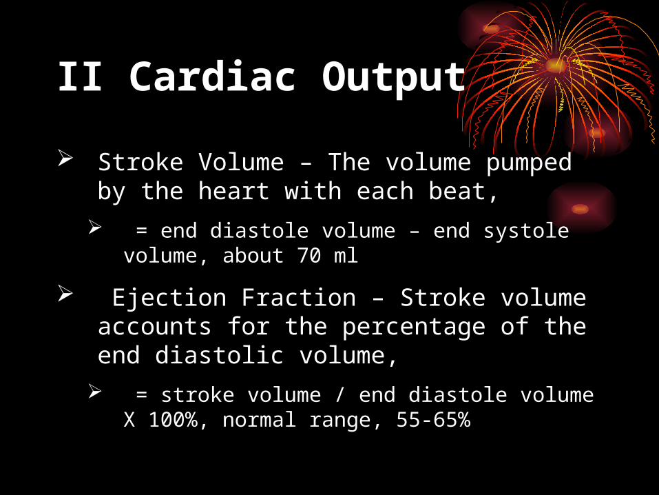
II Cardiac Output
Stroke Volume – The volume pumped by the heart with each beat,
= end diastole volume – end systole volume, about 70 ml
Ejection Fraction – Stroke volume accounts for the percentage of the end diastolic volume,
= stroke volume / end diastole volume X 100%, normal range, 55-65%
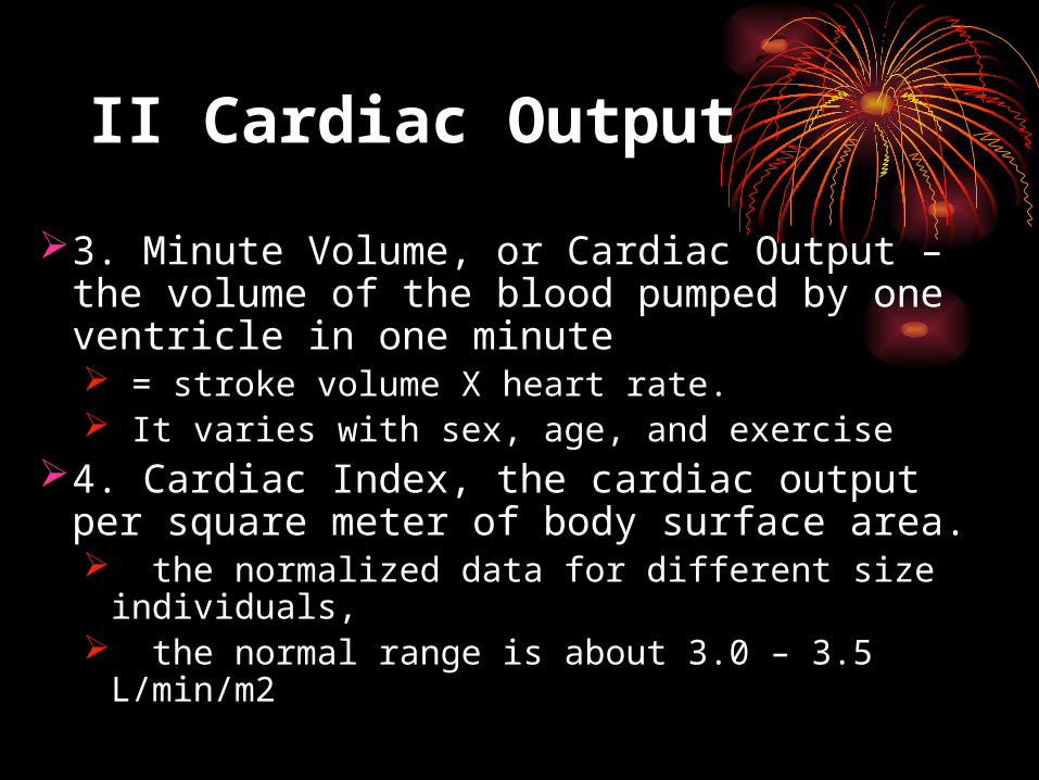
II Cardiac Output
3. Minute Volume, or Cardiac Output – the volume of the blood pumped by one ventricle in one minute = stroke volume X heart rate. It varies with sex, age, and exercise
4. Cardiac Index, the cardiac output per square meter of body surface area. the normalized data for different size individuals, the normal range is about 3.0 – 3.5 L/min/m2
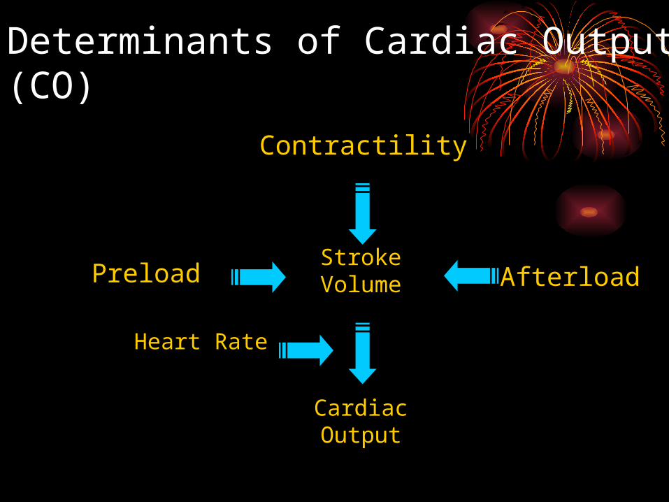
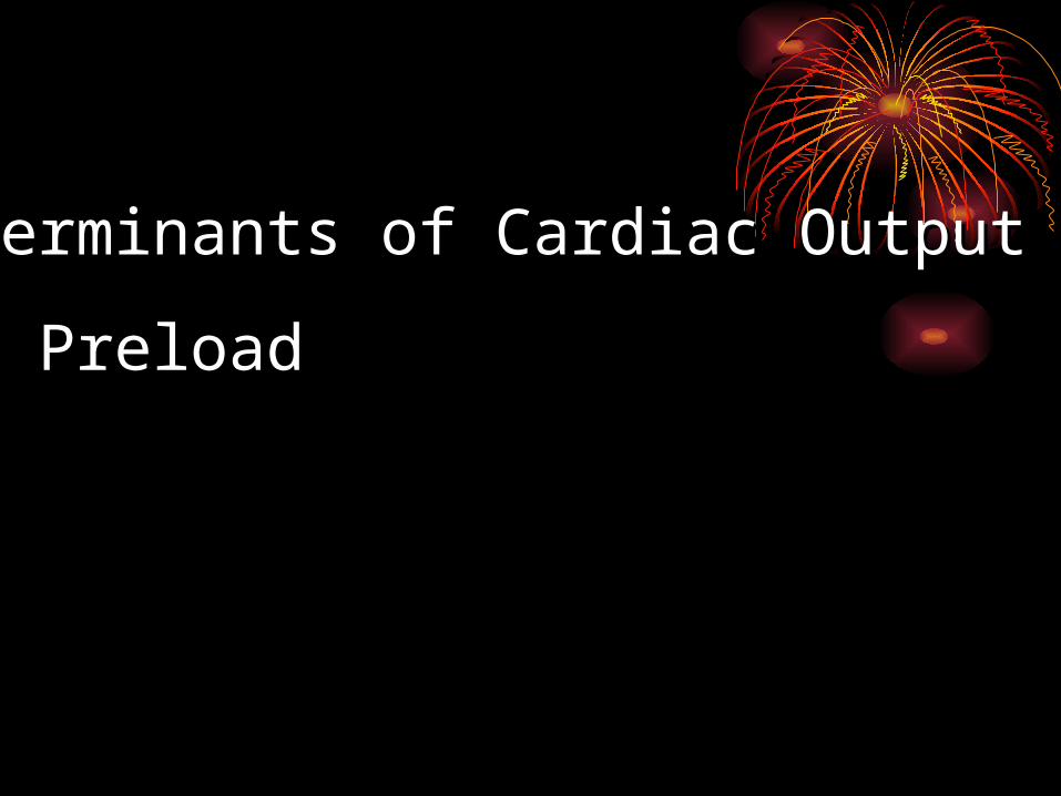
Determinants of Cardiac Output (CO)
Preload
Heart Rate
Afterload
Contractility
Cardiac Output
Stroke Volume
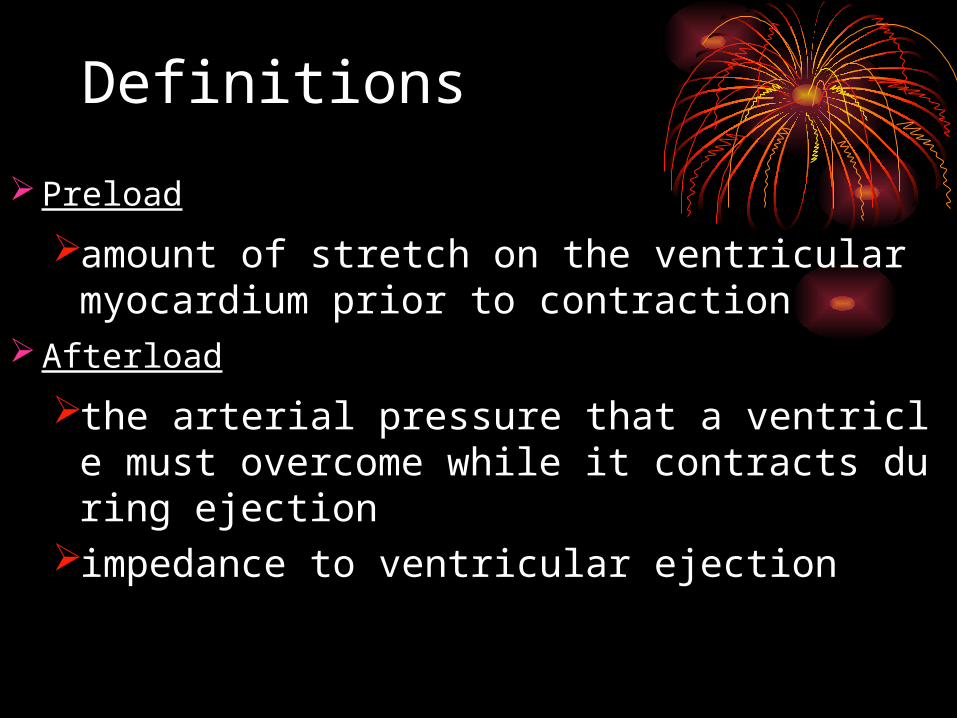
Definitions
Preload
amount of stretch on the ventricular myocardium prior to contraction
Afterload
the arterial pressure that a ventricle must overcome while it contracts during ejection
impedance to ventricular ejection
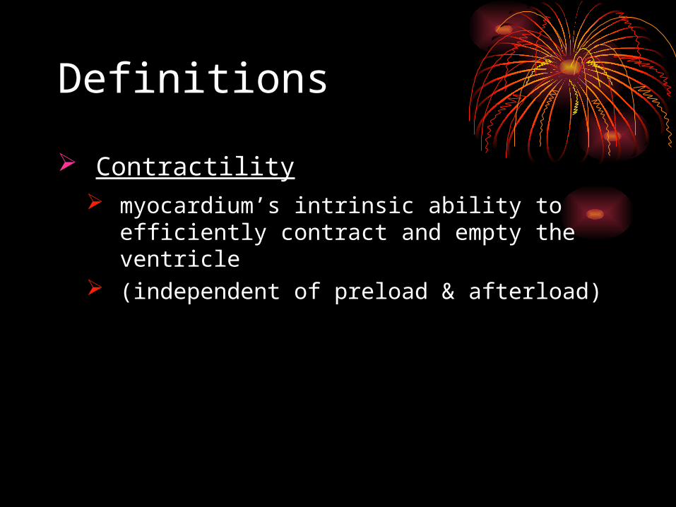
Definitions
Contractility myocardium’s intrinsic ability to
efficiently contract and empty the ventricle
(independent of preload & afterload)
Determinants of Cardiac OutputDeterminants of Cardiac Output
1. Preload1. Preload
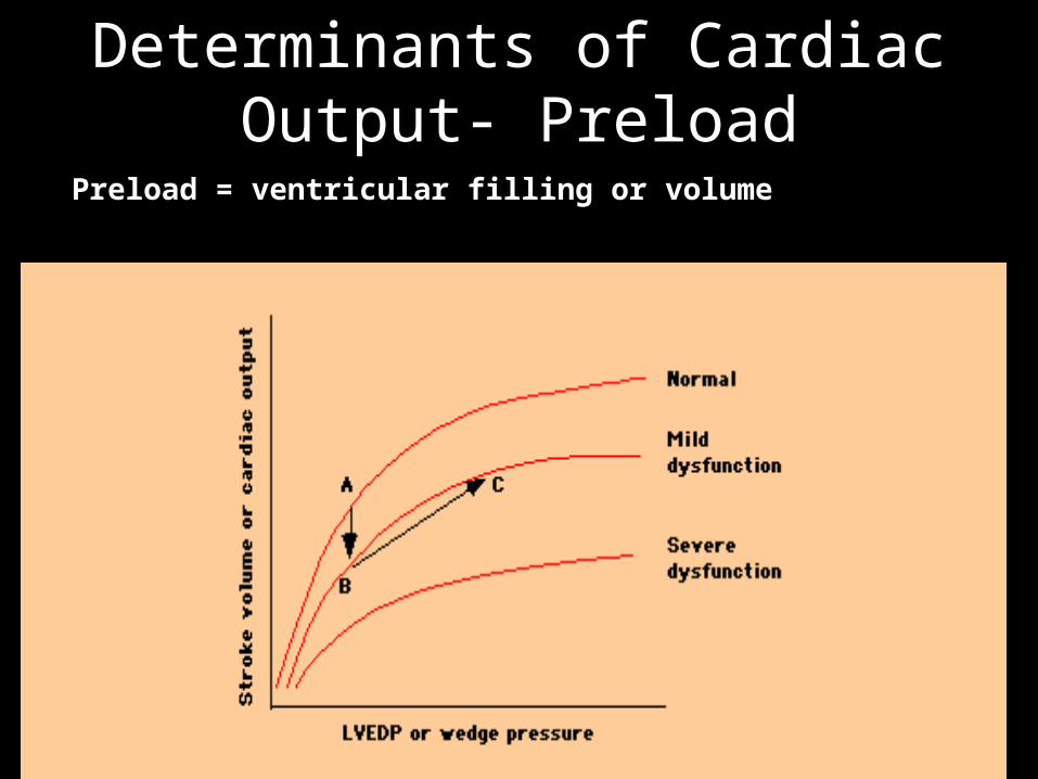
Preload = ventricular filling or volume
Determinants of Cardiac Determinants of Cardiac Output- PreloadOutput- Preload
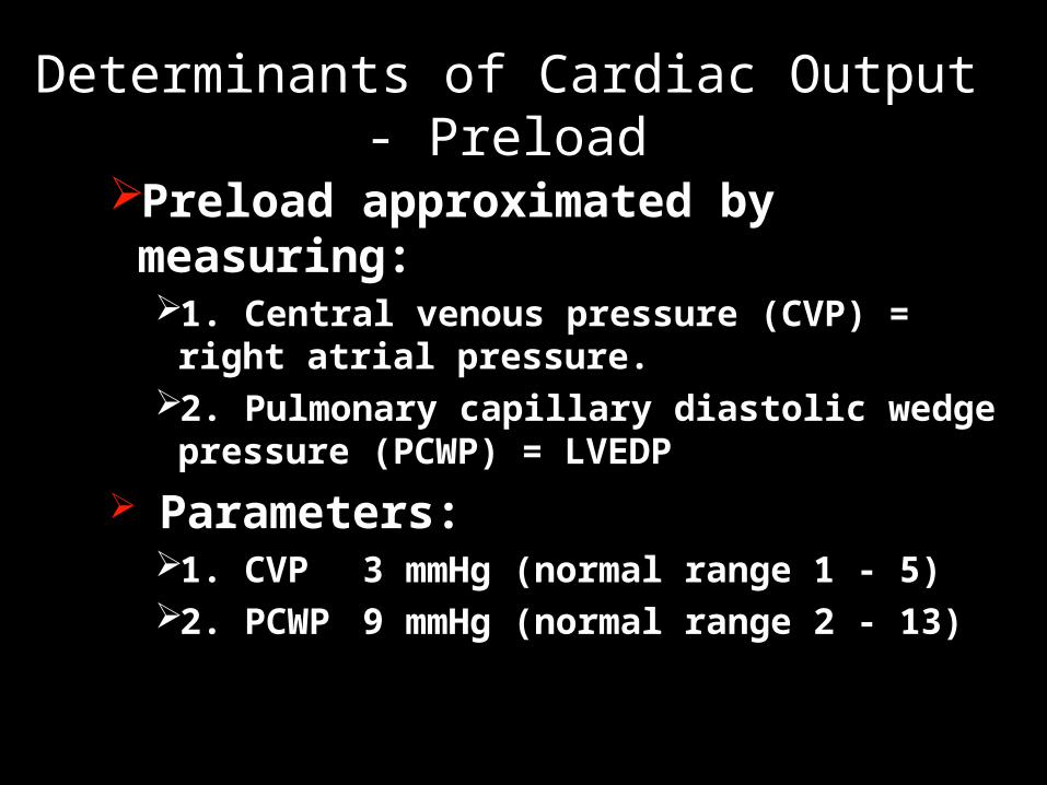
Preload approximated by measuring:1. Central venous pressure (CVP) = right
atrial pressure.2. Pulmonary capillary diastolic wedge
pressure (PCWP) = LVEDP
Parameters:1. CVP 3 mmHg (normal range 1 - 5)2. PCWP 9 mmHg (normal range 2 - 13)
Determinants of Cardiac Output - PreloadDeterminants of Cardiac Output - Preload
Frank-Starling Mechanism of the Heart
The intrinsic ability of the heart to adapt to changing volumes of inflowing blood
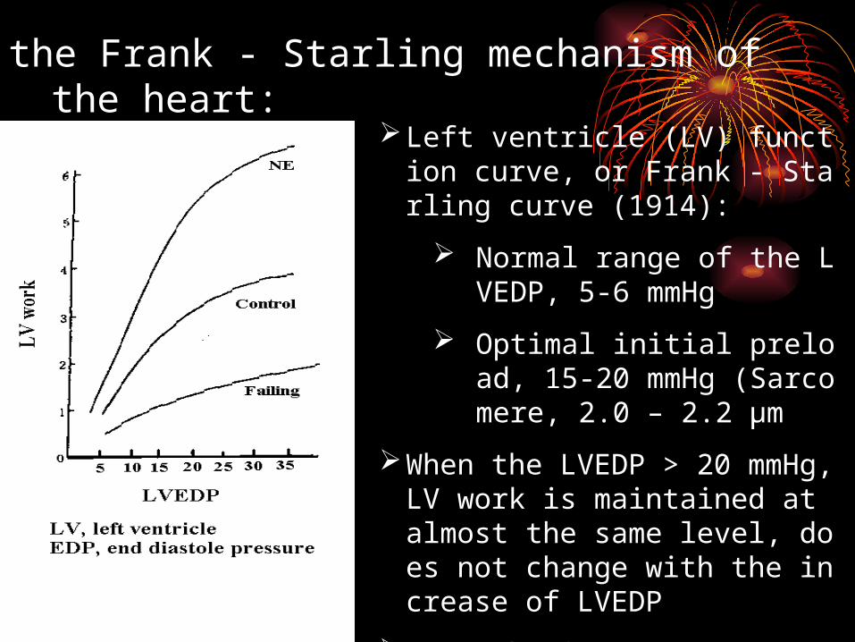
the Frank - Starling mechanism of the heart:
Left ventricle (LV) function curve, or Frank - Starling curve (1914):
Normal range of the LVEDP, 5-6 mmHg
Optimal initial preload, 15-20 mmHg (Sarcomere, 2.0 – 2.2 µm
When the LVEDP > 20 mmHg, LV work is maintained at almost the same level, does not change with the increase of LVEDP
Mechanism
Concept of heterometric regulation
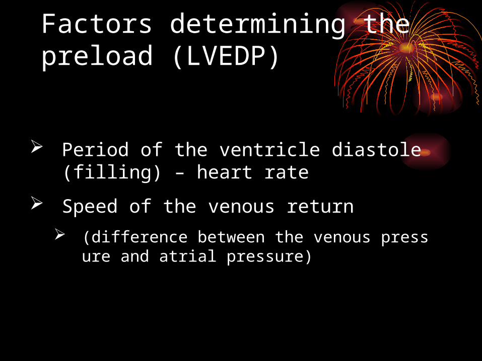
Factors determining the preload (LVEDP)
Period of the ventricle diastole (filling) – heart rate
Speed of the venous return
(difference between the venous pressure and atrial pressure)
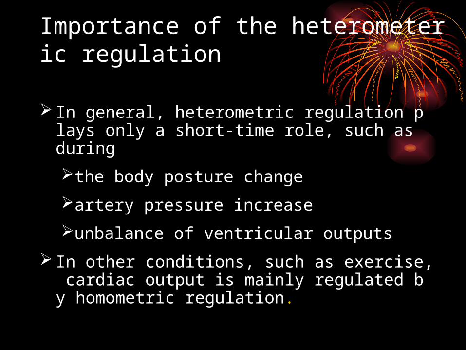
Importance of the heterometeric regulation
In general, heterometric regulation plays only a short-time role, such as during
the body posture change
artery pressure increase
unbalance of ventricular outputs
In other conditions, such as exercise, cardiac output is mainly regulated by homometric regulation.
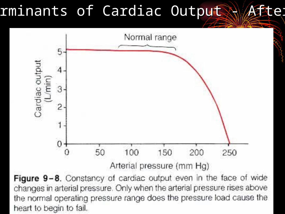
Determinants of Cardiac Output - AfterloadDeterminants of Cardiac Output - Afterload
Short time change of the arterial pressure
Transit arterial pressure rise
isovolumetric contraction phase become longer
period of ejection shorter
stroke volume less
more blood left in the ventricle left
LVEDP increase
through heterometeric regulation
stroke volume return to normal in next beat.
Long time high arterial pressure
through neural and humoral regulation
the stroke volume is maintained at normal level
pathogenesis of the cardiovascular system
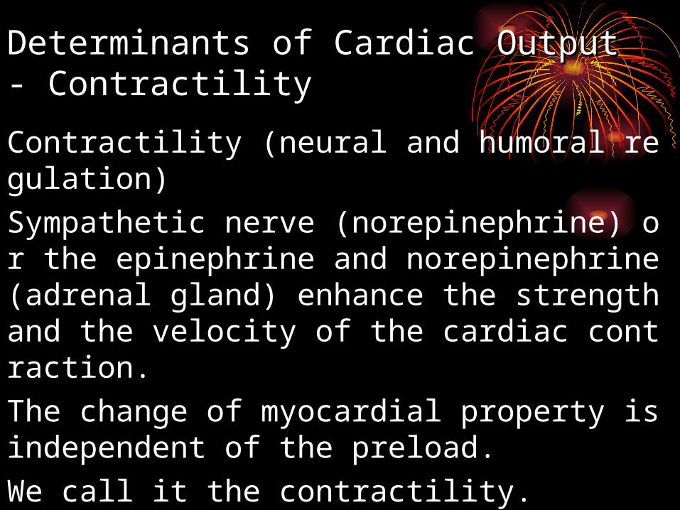
Contractility (neural and humoral regulation)
Sympathetic nerve (norepinephrine) or the epinephrine and norepinephrine (adrenal gland) enhance the strength and the velocity of the cardiac contraction.
The change of myocardial property is independent of the preload.
We call it the contractility.
Importance: exert a long – time influence on the cardiac output.
Determinants of Cardiac Output Determinants of Cardiac Output - Contractility- Contractility
Definitions
Contractilitymyocardium’s intrinsic ability to
efficiently contract and empty the ventricle
(independent of preload & afterload)
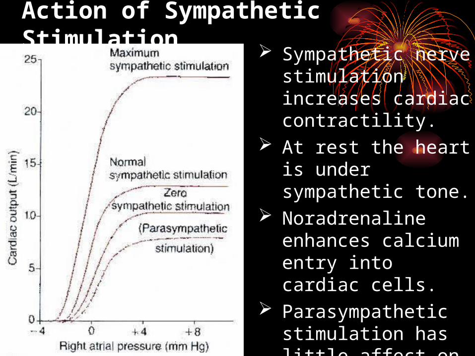
Action of Sympathetic Stimulation Sympathetic nerve
stimulation increases cardiac contractility.
At rest the heart is under sympathetic tone.
Noradrenaline enhances calcium entry into cardiac cells.
Parasympathetic stimulation has little affect on contractility due to the innervation pattern of the heart.
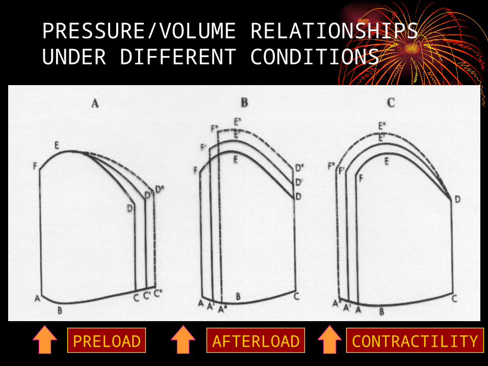
PRESSURE/VOLUME RELATIONSHIPS UNDER DIFFERENT CONDITIONS
PRELOAD AFTERLOAD CONTRACTILITY
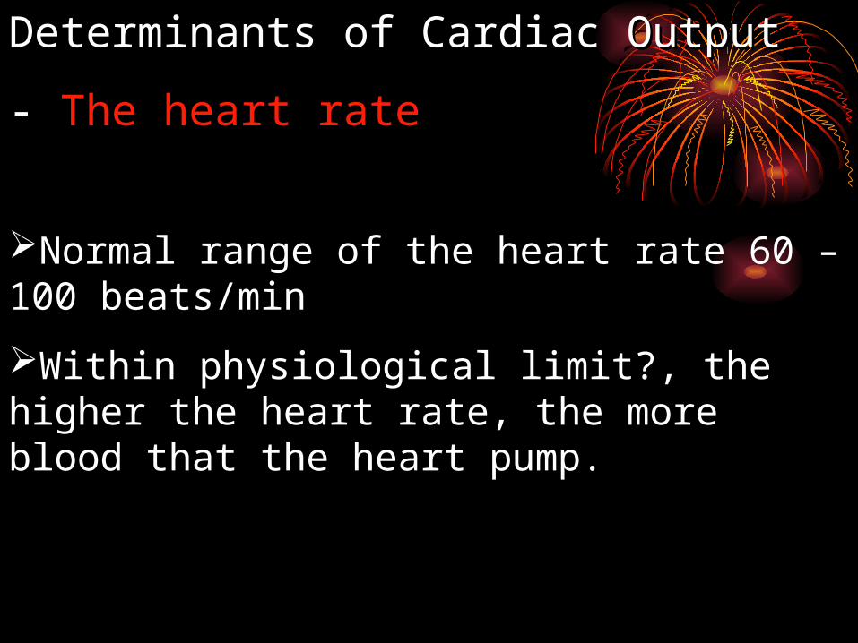
Normal range of the heart rate 60 – 100 beats/min
Within physiological limit?, the higher the heart rate, the more blood that the heart pump.
Determinants of Cardiac Output Determinants of Cardiac Output
- - The heart rate

1, at rest (without any regulation)
2, during exercise (with humoral and neural regulation)
IV Cardiac Output ReserveThe maximal cardiac output subtracts the normal value.
It reflects the ability of the heart to adapt the change of environment (internal or external)
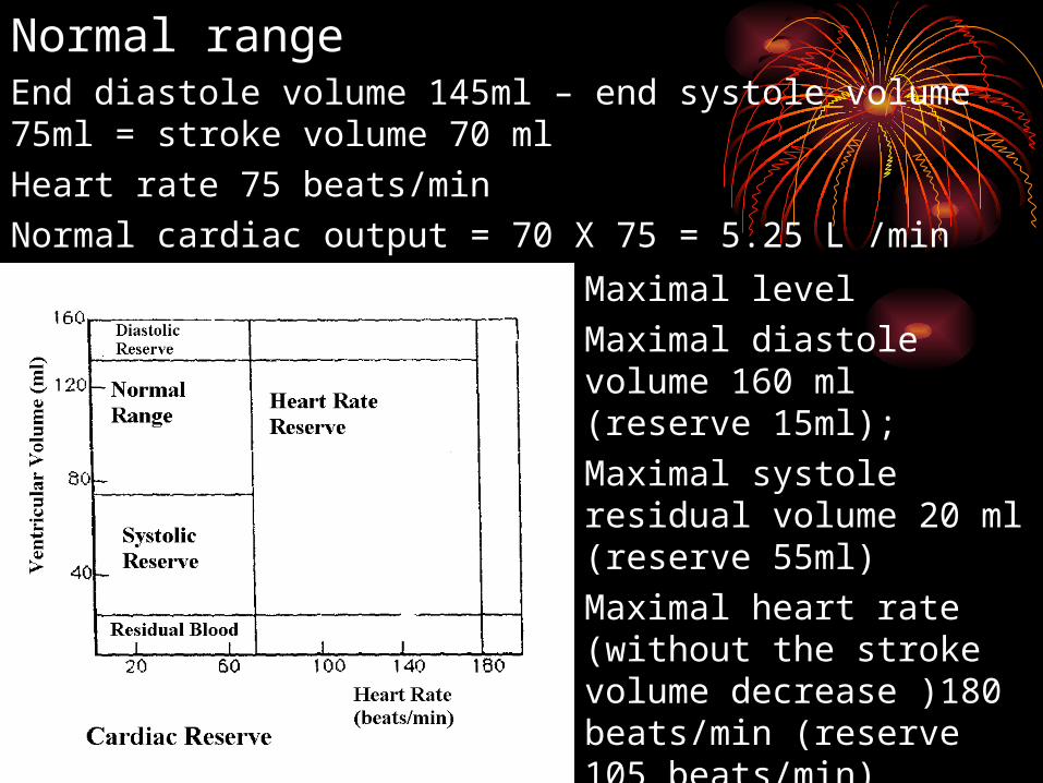
Maximal level
Maximal diastole volume 160 ml (reserve 15ml);
Maximal systole residual volume 20 ml (reserve 55ml)
Maximal heart rate (without the stroke volume decrease )180 beats/min (reserve 105 beats/min)
Maximal cardiac output (160 – 20) X 180 = 25.2 L/min
Normal rangeEnd diastole volume 145ml – end systole volume 75ml = stroke volume 70 ml
Heart rate 75 beats/min
Normal cardiac output = 70 X 75 = 5.25 L /min