Embed Size (px)
Citation preview
Lola regulates glutamate receptor expression at theDrosophila neuromuscular junction
Ai Fukui1, Mikiko Inaki1, Gaku Tonoe1, Hiroki Hamatani2, Mizuho Homma3, Takako Morimoto3,Hiroyuki Aburatani4 and Akinao Nose1,2,*1Department of Physics, Graduate School of Science, University of Tokyo, Hongo, Bunkyo-ku, Tokyo 113-0033, Japan2Department of Complexity Science and Engineering, Graduate School of Frontier Sciences, University of Tokyo, Kashiwanoha 5-1-5, Kashiwa,Chiba 277-8561, Japan3School of Life Science, Tokyo University of Pharmacy and Life Sciences, Hachioji, Tokyo 192-0392, Japan4Research Center of Advanced Science and Technology, University of Tokyo, Meguro-ku, Tokyo 153-8904, Japan
*Author for correspondence ([email protected])
Biology Open 1, 362–375doi: 10.1242/bio.2012448
SummaryCommunication between pre- and post-synaptic cells is a key
process in the development and modulation of synapses.
Reciprocal induction between pre- and postsynaptic cells
involves regulation of gene transcription, yet the underlying
genetic program remains largely unknown. To investigate
how innervation-dependent gene expression in postsynaptic
cells supports synaptic differentiation, we performed
comparative microarray analysis of Drosophila muscles
before and after innervation, and of prospero mutants,
which show a delay in motor axon outgrowth. We identified
84 candidate genes that are potentially up- or downregulated
in response to innervation. By systematic functional analysis,
we found that one of the downregulated genes, longitudinals
lacking (lola), which encodes a BTB-Zn-finger transcription
factor, is required for proper expression of glutamate
receptors. When the function of lola was knocked down in
muscles by RNAi, the abundance of glutamate receptors
(GluRs), GluRIIA, GluRIIB and GluRIII, as well as that of
p-21 activated kinase (PAK), was greatly reduced at the
neuromuscular junctions (NMJs). Recordings of the synaptic
response revealed a decrease in postsynaptic quantal size,
consistent with the reduction in GluR levels. Lola appears to
regulate the expression of GluRs and PAK at the level of
transcription, because the amount of mRNAs encoding these
molecules was also reduced in the mutants. The
transcriptional level of lola, in turn, is downregulated by
increased neural activity. We propose that Lola coordinates
expression of multiple postsynaptic components by
transcriptional regulation.
� 2012. Published by The Company of Biologists Ltd. This is
an Open Access article distributed under the terms of the
Creative Commons Attribution Non-Commercial Share Alike
License (http://creativecommons.org/licenses/by-nc-sa/3.0).
Key words: Synapse formation, Transcriptional regulation,
Neuromuscular junction, Drosophila, longitudinals lacking (lola),
Glutamate receptor
IntroductionDuring initial synapse formation, reciprocal interaction between
innervating neurons and their targets are essential for assembly of
synaptic components, cytoskeletal organization and activation of
gene expression (Goda and Davis, 2003; Li and Sheng, 2003;
McAllister, 2007). Similarly, mutual trans-synaptic signaling is
important for activity-dependent refinement of differentiated
synapses (Kandel, 2001; Flavell and Greenberg, 2008). While
short-term changes in synaptic properties can be induced by
modulation of pre-existing proteins and/or mRNAs, such as
trafficking, modification and local translation, long-term changes
require transcriptional control of gene expression. During the
formation of vertebrate neuromuscular junctions (NMJs), signals
from presynaptic motor neurons are necessary for the regulation
of gene expression of postsynaptic transmitter receptors, which
are acetylcholine receptors (AChRs) (Sanes and Lichtman, 2001;
Schaeffer et al., 2001; Kummer et al., 2006). Many studies have
focused on the role of immediate-early genes (IEGs) and CREB-
mediated transcriptional regulation in long-term synaptic
plasticity and memory formation (Flavell and Greenberg, 2008;
Cohen and Greenberg, 2008). Nonetheless, a large gap still exists
in our knowledge about how multiple molecular pathways
integrate and orchestrate the development and plasticity of
synapses. In particular, despite extensive work on activity-
induced genes, very few studies have established functional links
between these activity-induced genes and the downstream target
genes that ultimately regulate synaptic properties.
In this study, we used the Drosophila NMJ as a model to study
gene expression changes in postsynaptic muscle cells in response
to presynaptic innervation. The Drosophila NMJ is a
glutamatergic synapse expressing ionotropic glutamate
receptors (GluRs) and contains a number of synaptic
components commonly found in mammalian synapses, such as
the postsynaptic density protein Discs-Large/PSD-95 (Keshishian
et al., 1996; Griffith and Budnik, 2006). Previous studies showed
that immediate–early transcription factors such as CREB and AP-
1 regulate the strength and/or morphology of this synapse (Davis
et al., 1996; Sanyal et al., 2002) (reviewed in Sanyal and
Ramaswami, 2006). Signaling pathways mediated by secreted
factors, such as Wnts and Bmps, are known to regulate
anterograde and/or retrograde interaction between the motor
neurons and muscles that are important for synaptic development
362 Research Article
Bio
logy
Open
by guest on September 17, 2018http://bio.biologists.org/Downloaded from
(McCabe et al., 2003; Ataman et al., 2008; Korkut et al., 2009)
(reviewed in Griffith and Budnik, 2006). However, the finaltargets of these signaling cascades—the molecules that directlyregulate the changes in synaptic structure and function—remain
largely unknown.
In this study, we performed genome-wide microarray analysesof specific muscle cells and identified 84 candidate genes whose
expression changed in response to innervation. By systematicfunctional analyses of the candidate genes, we found thatlongitudinals lacking (lola), a gene downregulated by
innervation, plays a prominent role in the transcriptional controlof a number of postsynaptic components. lola encodes a BTB-Zn-finger transcription factor with a number of different isoforms(Goeke et al., 2003; Horiuchi et al., 2003). This transcription
factor, Lola, has been implicated in a wide range of developmentaland cellular processes including axon guidance, neuralspecification and tumorigenesis (Madden et al., 1999; Crowner
et al., 2002; Goeke et al., 2003; Ferres-Marco et al., 2006; Spletteret al., 2007). Previous studies suggest that Lola may execute itsfunction by directly binding to DNA and regulating the expression
of the target genes. Here we show that postsynaptic Lolatranscriptionally regulates the expression level of the glutamatereceptors GluRIIA, GluRIIB and GluRIII, as well as p-21 activated
kinase (PAK). We also show that the transcriptional level of lola isdownregulated by increased neural activity. We propose thatpostsynaptic Lola functions as a transcription factor that controlssynapse formation and/or maturation by regulating the expression
of multiple synaptic components.
Materials and MethodsFly stocksFor microarray analysis, we used an allele of prospero (prosM4) (Broadie and Bate,1993b) and y, w. Forced expression and RNA interference (RNAi) analyses wereperformed using the GAL4-UAS system (Brand and Perrimon, 1993). Elav-gal4(Luo et al., 1994), 24B-Gal4 (Brand and Perrimon, 1993) or G14-Gal4 (Shishidoet al., 1998), or 5053A-Gal4 (Ritzenthaler et al., 2000) were used to induceexpression in all neurons, all muscles, or in M12, respectively. UAS-RNAi lineswere obtained from the Vienna Drosophila RNAi Center (VDRC) and Fly Stocksof the National Institute of Genetics (NIG) (Dietzl et al., 2007). Lines and allelesused for the systematic functional analyses are listed in supplementary materialTable S2. Animals were raised at 29 C for RNAi analyses and at 25 C for otheranalyses. Alleles of lola, lolaORC46 (Crowner et al., 2002) and lolaORE119 (Goeke etal., 2003), were used.
Microarray AnalysisThe collection of embryonic somatic muscles was performed as previouslydescribed (Inaki et al., 2007) with the following modifications. For collection ofmuscles at 18 hr after egg laying (AEL), the preparation was treated with 1 mg / mlcollagenase (Sigma, St. Louis, Missouri) for ,30 sec to weaken intersegmentalmuscle-muscle attachments. For chip analysis, we prepared three samples for eachdevelopmental stage or genotype: one sample was prepared from 200 collectedcells and two from 50 collected cells. We performed two or three rounds of cRNAamplification before biotin labeling for samples prepared from 200 cells or thoseprepared from 50 cells, respectively. Synthesis of biotin-labeled cRNA wasperformed with the IVT Labeling Kit (Affymetrix, Santa Clara, CA).Hybridization of the fragmented and labeled cRNA to Affymetrix DrosophilaGenome 2.0 Genechip arrays were performed according to the GenechipExpression Analysis technical manual (Affymetrix). Gene expression dataanalyses were performed using Affymetrix GeneChip Operating Software 1.4(Affymetrix). By comparing gene expression signals between the samples preparedfrom the same number of cells, we obtained the Change Call (I, increase; D,decrease; NC, no change). After three pairs of comparisons, we selected the genesthat displayed Change Call ‘I’ in all three pairs. We carried out Gene Ontologyanalyses by batch query using NetAffx (Affymetrix).
Real-time reverse-transcription PCRReal-time reverse-transcription PCR (RT-PCR) assays were performed with ABIPrism 7000 SDS or Applied Biosystems StepOne Real Time PCR System (AppliedBiosystems, Foster City, CA) with SYBR Green fluorescence according to the
manufacturer protocol. We used as templates the second-round amplified cDNAsof wild type muscles and pros mutant muscles, prepared as described formicroarray analyses. The gene expression values were normalized using theMyosin heavy chain (Mhc) gene as a reference. To examine gene expression inlarval muscles, total RNA was extracted from the body wall of wall-climbingthird-instar larvae using Sepasol RNA I Super (Nacalai, Kyoto, Japan) according tothe manufacturer instructions. Approximately 2 mg of total RNA was reverse-transcribed into cDNA using oligo-dT primer and SuperScript III ReverseTranscriptase (Invitrogen, Carlsbad, CA). The obtained values were normalizedusing the Gapdh1 gene as a reference.
ImmunohistochemistryImmunohistochemical staining of dissected larvae and embryos was performed asdescribed previously (Inaki et al., 2007). The following primary antibodies wereused at the indicated concentrations: goat anti-horse radish peroxidase (HRP;Jackson, West Grove, PA, 1:4000); rabbit anti-Lola polyclonal antibodies (1:150)(Giniger et al., 1994); mouse monoclonal anti-GluRIIA (8B4D2) and mousemonoclonal anti-Dlg (Developmental Studies Hybridoma Bank, University ofIowa, Iowa City, IA, 1:10 and 1:50, respectively); rabbit anti-GluRIIB and rabbitanti-GluRIII (1:2500, and 1:5000, respectively) (Marrus et al., 2004); mouse Nc82(1:100) (Wagh et al., 2006); rabbit anti-Pak antibody (1:500) (Sone et al., 2000).
Quantification of NMJ morphology and immunofluorescence signalsWe quantified the morphology of the NMJ with Imaris software (Bitplane, Zurich,Switzerland). The number of boutons and branches and terminal length werescored based on the morphology visualized with anti-HRP staining as previouslydescribed (Coyle et al., 2004). The terminal length was normalized to muscle area.The bouton and branch number were also normalized to muscle area in theanalyses of pst RNAi mutants because there was a slight change in muscle area inthe mutants (surface area, 84.362.8 [n510] in pst-RNAi-8588R-4 [p,0.05] and66.462.7 [n517] in pst-RNAi-107243 [p,0.05] compared to 75.063.0 [n532] incontrol, 6 103 mm2). To assess the content of particular molecules at synapses,confocal images were projected to create a two-dimensional image and analyzedwith IPLab software (Scanalytics, Fairfax, VA, USA). To control for relativeintensity, experimental and control samples were stained in the same dish andimaged under identical conditions. We defined the synaptic area as delimited byHRP immunoreactivity on the muscle surface and the synaptic intensity of GluR,PAK or BRP as the average immunoreactive signals within the synaptic area. Thetotal area of GluRIIA-, PAK- and BRP-positive clusters were defined by the pixelswith an intensity above a threshold set arbitrarily, and were normalized by the totalarea of muscles. The GluRIIA expression level of the embryonic NMJ was definedas the total immunoreactive signals within the area of GluRIIA-positive clusters.For analysis of Dlg levels, we measured net intensity of immunoreactive signalsnormalized by the stained area. The extra-junctional GluR expression level wasdefined as the average immunoreactive signal within arbitrarily chosen regionscovering ,1% of muscle surface area. All statistical analyses were done byStudent’s t-test. All quantitative data in the larvae were obtained from muscle 6/7NMJs in abdominal segment 2.
Immunoblot analysisTwo hundred first-instar larvae were ground in RIPA buffer containing proteaseinhibitors (Complete mini; Roche Diagnostics, Mannheim, Germany) as describedby Thomas et al. (Thomas et al., 1997) and loaded onto 4%–12% SDS-PAGE gels(TEFCO, Tokyo, Japan). Immunoblotting was performed using mouse monoclonalanti-Tubulin (12G10; Developmental Studies Hybridoma Bank, 1:2000), anti-GluRIIA (MAb 8B4D2 described above, 1:50), goat anti-mouse IgG HRPconjugate (Jackson, 1:1000) and donkey anti-goat IgG IRDye 680(LI-CORBiosciences, Lincoln, NE, USA, 1:2500) and imaged and quantified by Odysseyinfrared imaging system (LI-OCR). To control for loading, the level of GluRIIAwas normalized to the level of Tubulin.
ElectrophysiologyIntracellular recordings were made from muscle 6 in abdominal segments 3 and 4,and excitatory junctional potentials (EJPs) and miniature EJPs were recorded asdescribed previously (Morimoto et al., 2010). HL3-containing medium at varyingconcentrations of extracellular Ca2+ were used (0.375 mM for mEJP recordingsand 1.5 mM for EJP recordings). The peak amplitudes of ten EJPs were measuredand averaged in each recording. All statistical analyses were done by Student’st-test.
Manipulation of neural activity with dTRPA1Embryos of elav-Gal4/UAS-dTRPA1 (Pulver et al., 2009) were raised at 22 C,collected at 18 hr AEL and transferred to a PCR tube containing 25 ml ofHalocarbon 700 oil (Sigma). Repetitive, spaced stimuli, consisting of 2-minintervals at a higher temperature (27 C) spaced by 5 min of rest (22 C) wereapplied in a PCR machine for 12 hrs. The larvae were then transferred to an agar
Gene regulation of synaptic components 363
Bio
logy
Open
by guest on September 17, 2018http://bio.biologists.org/Downloaded from
plate and reared at 22 C until third instar. Control embryos (elav-Gal4/+)
underwent the same temperature cycling.
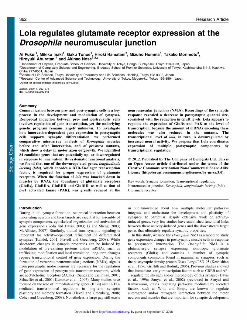
ResultsGene expression changes in innervated muscle cells
In the Drosophila NMJ, synaptogenesis starts at ,13 hr AEL,
when motor neurons contact the target muscles for the first time
(Broadie and Bate, 1993a). During the process of intimate
interaction between the presynaptic motor neurons and the
postsynaptic muscles, the synapses gradually mature and become
fully functional by 17 hr AEL (Broadie and Bate, 1993a;
Kohsaka et al., 2007). Using Affymetrix DNA microarrays, we
performed two series of comparative gene expression analysis of
muscles (Fig. 1A). First, we compared gene expression profiling
of muscles before (12.75 hr AEL) and after (18 hr AEL)
innervation (‘‘before versus after’’ comparison). Second, we
compared gene expression in muscles of wild-type versus
prospero (pros) mutants; ,80% of the muscles in pros mutants
lack innervation but otherwise differentiate normally (‘‘with
versus without’’ comparison, at 18 hr AEL) (Broadie and Bate,
1993b). Genes that are differentially expressed in the first
comparison would include genes that are up- or downregulated
by innervation. However, these would also include genes that are
involved in other aspects of muscle differentiation and whose
expression is regulated at this stage, such as genes involved in the
maturation of the muscle contraction apparatus. On the other
hand, the second comparison would identify genes that are
specifically regulated by innervation because the differentiation
of muscle itself is normal in pros mutants (Broadie and Bate,
1993b). By conducting comparative analyses based on these two
categories, we aimed to identify genes that function in synapse
formation and whose expression is regulated by innervation.
Using the single-cell collection technique that we developed
previously (Inaki et al., 2007), we performed microarray analyses
of four neighboring ventral muscles, M6, M7, M12 and M13,
using Affymetrix Drosophila Genome chips (see Materials and
Methods for detail). Because these muscles show similar
morphology, run in parallel and attach to adjacent muscle
insertion sites, they likely share common genetic programs for
synapse formation. We therefore used a combination of the four
muscles as a source for the microarray analyses. We prepared
three independent samples each for the two comparative
expression analyses described above (‘‘before versus after’’ and
‘‘with versus without’’ innervation) and focused on genes that
displayed differential expression reproducibly. We identified 72
genes that showed increased signals in muscles ‘‘after’’ and
‘‘with’’ innervation compared respectively to muscles ‘‘before’’
and ‘‘without’’ innervation. We defined these as ‘‘genes
upregulated by innervation’’ (Fig. 1B; Table 1). We also
identified 12 genes that showed decreased signals in the same
comparisons and defined them as ‘‘genes downregulated by
innervation’’ (Fig. 1C; Table 1). To validate the microarray
results, we analyzed the expression of randomly-chosen subsets
of the identified genes by quantitative RT-PCR (qPCR).
Differential expression (‘‘with vs. without’’) was confirmed for
22 of the 26 genes assayed, indicating 85% concordance
(Table 2).
pastrel is involved in synaptic growth
The 72 upregulated and 12 downregulated candidate genes
described above were categorized into multiple functional classes
of proteins (supplementary material Table S1). Among the
upregulated genes was cactus (cact), one of the few genes
known to function in muscles to regulate synaptic development
and/or function (Cantera et al., 1999; Heckscher et al., 2007). To
study whether other candidate genes also have roles in synaptic
development, we performed loss-of-function (LOF) analyses
on 37 of the candidate genes whose predicted molecular
structure and/or function are related to synaptogenesis
(supplementary material Table S2), using existing LOF mutant
alleles and RNA interference (RNAi)-mediated gene knock-
down. We visualized the NMJ by staining the presynaptic
membrane (anti-HRP) and postsynaptic GluRIIA, and looked for
changes in the terminal morphology and/or receptor clustering in
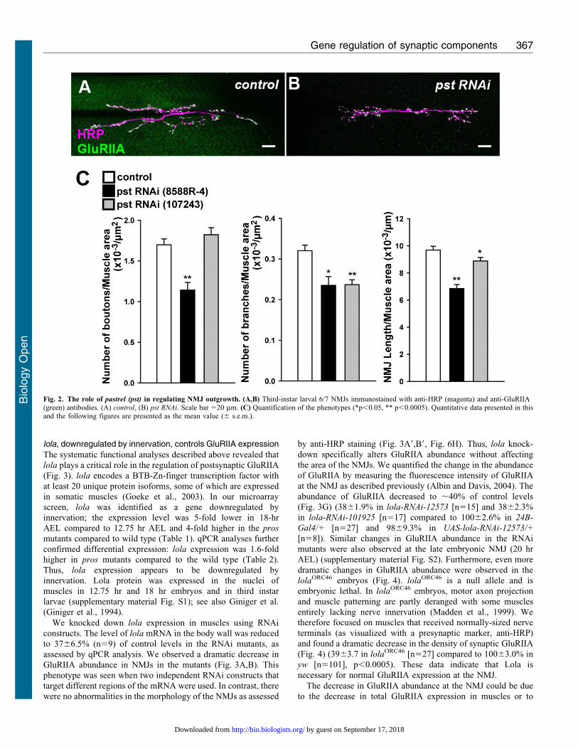
the mutants. This screening revealed a function for pastrel (pst)
in proper synaptic growth and lola in proper GluRIIA expression.
No dramatic phenotypes were observed in the LOF mutants of
the remaining genes examined.
RNAi knock-down of pastrel (pst) with two independent
constructs that target different portions of the mRNA resulted in
decreased bouton number, branch number and NMJ length
(Fig. 2) (number of boutons per muscle area, 1.160.094 in pst-
RNAi-8588R-4 [p,0.0005] versus 1.760.072 in control, 61023/mm2; number of branches per muscle area, 0.2460.021 in
pst-RNAi-8588R-4 [p,0.05] and 0.2460.012 in pst-RNAi-
107243 [p,0.0005] versus 0.3260.014 in control, 6 1023/
mm2; NMJ length per muscle area, 6.960.3 in pst-RNAi-8588R-4
[p,0.0005] and 8.960.3 in pst-RNAi-107243 [p,0.05] versus
9.760.3 in control, 6 1023 mm21; n510, 17, 32).
pst has been implicated in the processes of learning and
memory (Dubnau et al., 2003). The expression of pst appears to
be upregulated with innervation. Therefore, pst may be part of the
genetic program that is induced by innervation to promote
synaptic growth.
Fig. 1. Microarray analyses of genes whose expression is regulated by
innervation during synaptogenesis. (A) Schematic drawings of theexperimental design. (B,C) Venn diagrams showing the number of probe setsthat displayed similar or differential regulation in two series of gene expressioncomparative analyses. For the probe sets that displayed similar regulation in thetwo comparisons, the number of the corresponding genes is also shownin parenthesis.
Gene regulation of synaptic components 364
Bio
logy
Open
by guest on September 17, 2018http://bio.biologists.org/Downloaded from
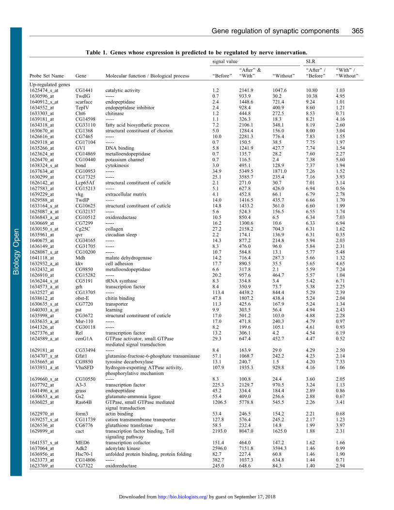
Table 1. Genes whose expression is predicted to be regulated by nerve innervation.
Molecular function / Biological process
signal value SLR
Probe Set Name Gene ‘‘Before’’‘‘After’’ &‘‘With’’ ‘‘Without’’
‘‘After’’ /‘‘Before’’
‘‘With’’ /‘‘Without’’
Up-regulated genes1625474_s_at CG1441 catalytic activity 1.2 2141.9 1047.6 10.80 1.031630596_at TwdlG ----- 0.7 933.9 30.2 10.38 4.951640912_s_at scarface endopeptidase 2.4 1448.6 721.4 9.24 1.011634552_at TepIV endopeptidase inhibitor 2.4 928.4 400.9 8.60 1.211633303_at Cht6 chitinase 1.2 444.8 272.5 8.53 0.711639181_at CG14598 ----- 1.1 326.3 18.3 8.21 4.161634318_at CG33110 fatty acid biosynthetic process 7.2 2106.1 348.1 8.19 2.601630670_at CG1368 structural constituent of chorion 5.0 1284.4 156.0 8.00 3.041626616_at CG7465 ----- 10.0 2281.3 776.4 7.83 1.551629318_at CG17104 ----- 0.7 150.5 38.5 7.75 1.971635266_at GV1 DNA binding 5.8 1241.9 427.7 7.74 1.541623624_at CG14869 metalloendopeptidase 0.7 135.7 28.2 7.60 2.271626470_at CG10440 potassium channel 0.7 116.5 2.4 7.38 5.601638324_s_at bond cytokinesis 3.0 495.1 128.9 7.37 1.941637634_at CG10953 ----- 34.9 5349.5 1871.0 7.26 1.521630299_at CG17325 ----- 25.1 3585.7 235.4 7.16 3.931626142_at Lcp65Af structural constituent of cuticle 2.1 271.0 30.7 7.01 3.141627583_at CG15213 ----- 5.1 627.8 426.0 6.94 0.561639229_at vkg extracellular matrix 4.1 452.8 66.1 6.79 2.781629588_at TwdlP ----- 14.0 1416.5 435.7 6.66 1.701633164_s_at CG10625 structural constituent of cuticle 14.8 1433.2 361.0 6.60 1.991625087_a_at CG32137 ----- 5.6 524.3 156.5 6.55 1.741636843_a_at CG10512 oxidoreductase 10.5 850.4 6.5 6.34 7.031630669_at CG7299 ----- 16.2 1300.6 10.6 6.33 6.941630150_s_at Cg25C collagen 27.2 2158.2 704.3 6.31 1.621635961_at qvr circadian sleep 2.2 174.1 136.9 6.31 0.351640675_at CG34165 ----- 14.3 877.2 214.8 5.94 2.031636149_at CG31705 ----- 8.3 476.0 96.0 5.84 2.311628087_s_at CG10200 ----- 10.7 584.8 13.1 5.77 5.481641118_at Mdh malate dehydrogenase 14.2 716.4 287.3 5.66 1.321632932_a_at kkv cell adhesion 17.7 890.5 35.5 5.65 4.651632432_at CG9850 metalloendopeptidase 6.6 317.8 2.1 5.59 7.241626910_at CG15282 ----- 20.2 957.6 464.7 5.57 1.041636244_s_at CG5191 tRNA synthase 8.3 354.8 3.4 5.42 6.711634573_a_at grh transcription factor 8.4 350.9 73.7 5.38 2.251632527_at CG13705 ----- 113.4 4438.2 844.4 5.29 2.391638612_at obst-E chitin binding 47.8 1807.2 438.4 5.24 2.041630635_s_at CG7720 transporter 11.3 425.6 167.9 5.24 1.341640303_a_at pst learning 9.9 303.5 56.4 4.94 2.431635998_at CG3672 structural constituent of cuticle 17.0 501.2 103.0 4.88 2.281635635_a_at Msr-110 ----- 17.0 471.8 240.3 4.79 0.971641326_at CG30118 ----- 8.2 199.6 105.1 4.61 0.931627376_at Rel transcription factor 13.2 306.1 4.2 4.54 6.191624589_a_at cenG1A GTPase activator, small GTPase
mediated signal transduction29.3 647.4 452.7 4.47 0.52
1629181_at CG33494 ----- 8.4 163.9 29.0 4.29 2.501634707_s_at Gfat1 glutamine-fructose-6-phosphate transaminase 57.1 1068.7 242.2 4.23 2.141635665_at CG9850 tyrosine decarboxylase 13.1 240.7 1.5 4.20 7.331633931_a_at VhaSFD hydrogen-exporting ATPase activity,
phosphorylative mechanism107.9 1935.3 929.8 4.16 1.06
1639660_s_at CG10550 ----- 8.3 100.8 24.4 3.60 2.051637792_at A3-3 transcription factor 225.3 2129.7 970.5 3.24 1.131641496_a_at grass endopeptidase 45.2 334.4 184.4 2.89 0.861630653_a_at Gs2 glutamate-ammonia ligase 55.4 409.0 256.6 2.88 0.671636025_at Ras64B GTPase, small GTPase mediated
signal transduction1206.5 5778.8 545.5 2.26 3.41
1622970_at form3 actin binding 53.4 246.5 154.2 2.21 0.681639257_s_at CG11739 cation transmembrane transporter 127.8 576.4 245.2 2.17 1.231626536_at CG6776 glutathione transferase 58.5 232.4 14.8 1.99 3.971629899_at cact transcription factor binding, Toll
signaling pathway2193.0 8047.0 1625.0 1.88 2.31
1641537_s_at MED6 transcription cofactor 151.4 464.0 147.2 1.62 1.661637064_at Adk2 adenylate kinase 2596.0 7151.8 3594.3 1.46 0.991636956_at Hsc70-1 unfolded protein binding, protein folding 82.7 227.4 60.8 1.46 1.901623373_at CG14806 ----- 382.7 1037.3 634.8 1.44 0.711623769_at CG7322 oxidoreductase 245.0 648.6 84.3 1.40 2.94
Gene regulation of synaptic components 365
Bio
logy
Open
by guest on September 17, 2018http://bio.biologists.org/Downloaded from
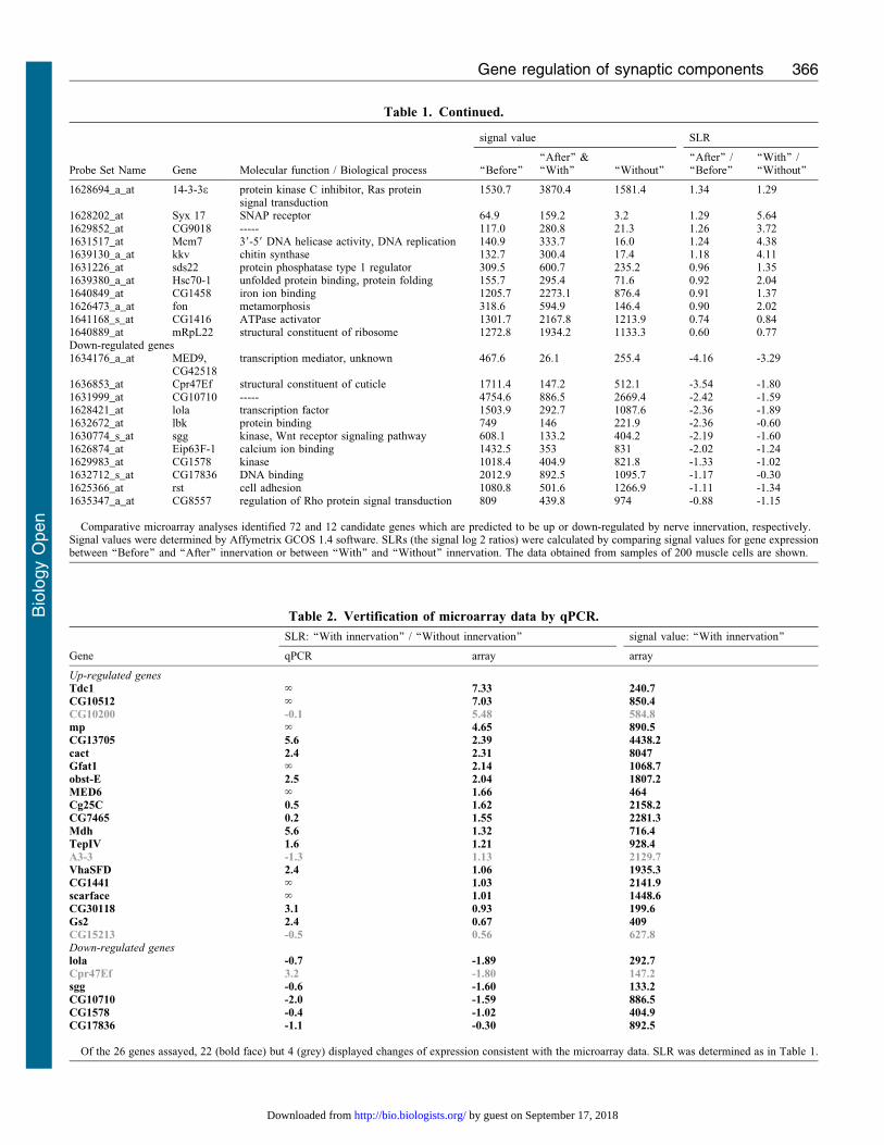
Table 1. Continued.
Molecular function / Biological process
signal value SLR
Probe Set Name Gene ‘‘Before’’‘‘After’’ &‘‘With’’ ‘‘Without’’
‘‘After’’ /‘‘Before’’
‘‘With’’ /‘‘Without’’
1628694_a_at 14-3-3e protein kinase C inhibitor, Ras proteinsignal transduction
1530.7 3870.4 1581.4 1.34 1.29
1628202_at Syx 17 SNAP receptor 64.9 159.2 3.2 1.29 5.641629852_at CG9018 ----- 117.0 280.8 21.3 1.26 3.721631517_at Mcm7 39-59 DNA helicase activity, DNA replication 140.9 333.7 16.0 1.24 4.381639130_a_at kkv chitin synthase 132.7 300.4 17.4 1.18 4.111631226_at sds22 protein phosphatase type 1 regulator 309.5 600.7 235.2 0.96 1.351639380_a_at Hsc70-1 unfolded protein binding, protein folding 155.7 295.4 71.6 0.92 2.041640849_at CG1458 iron ion binding 1205.7 2273.1 876.4 0.91 1.371626473_a_at fon metamorphosis 318.6 594.9 146.4 0.90 2.021641168_s_at CG1416 ATPase activator 1301.7 2167.8 1213.9 0.74 0.841640889_at mRpL22 structural constituent of ribosome 1272.8 1934.2 1133.3 0.60 0.77Down-regulated genes1634176_a_at MED9,
CG42518transcription mediator, unknown 467.6 26.1 255.4 -4.16 -3.29
1636853_at Cpr47Ef structural constituent of cuticle 1711.4 147.2 512.1 -3.54 -1.801631999_at CG10710 ----- 4754.6 886.5 2669.4 -2.42 -1.591628421_at lola transcription factor 1503.9 292.7 1087.6 -2.36 -1.891632672_at lbk protein binding 749 146 221.9 -2.36 -0.601630774_s_at sgg kinase, Wnt receptor signaling pathway 608.1 133.2 404.2 -2.19 -1.601626874_at Eip63F-1 calcium ion binding 1432.5 353 831 -2.02 -1.241629983_at CG1578 kinase 1018.4 404.9 821.8 -1.33 -1.021632712_s_at CG17836 DNA binding 2012.9 892.5 1095.7 -1.17 -0.301625366_at rst cell adhesion 1080.8 501.6 1266.9 -1.11 -1.341635347_a_at CG8557 regulation of Rho protein signal transduction 809 439.8 974 -0.88 -1.15
Comparative microarray analyses identified 72 and 12 candidate genes which are predicted to be up or down-regulated by nerve innervation, respectively.Signal values were determined by Affymetrix GCOS 1.4 software. SLRs (the signal log 2 ratios) were calculated by comparing signal values for gene expressionbetween ‘‘Before’’ and ‘‘After’’ innervation or between ‘‘With’’ and ‘‘Without’’ innervation. The data obtained from samples of 200 muscle cells are shown.
Table 2. Vertification of microarray data by qPCR.
SLR: ‘‘With innervation’’ / ‘‘Without innervation’’ signal value: ‘‘With innervation’’
Gene qPCR array array
Up-regulated genesTdc1 ‘ 7.33 240.7CG10512 ‘ 7.03 850.4CG10200 -0.1 5.48 584.8mp ‘ 4.65 890.5CG13705 5.6 2.39 4438.2cact 2.4 2.31 8047Gfat1 ‘ 2.14 1068.7obst-E 2.5 2.04 1807.2MED6 ‘ 1.66 464Cg25C 0.5 1.62 2158.2CG7465 0.2 1.55 2281.3Mdh 5.6 1.32 716.4TepIV 1.6 1.21 928.4A3-3 -1.3 1.13 2129.7VhaSFD 2.4 1.06 1935.3CG1441 ‘ 1.03 2141.9scarface ‘ 1.01 1448.6CG30118 3.1 0.93 199.6Gs2 2.4 0.67 409CG15213 -0.5 0.56 627.8Down-regulated geneslola -0.7 -1.89 292.7Cpr47Ef 3.2 -1.80 147.2sgg -0.6 -1.60 133.2CG10710 -2.0 -1.59 886.5CG1578 -0.4 -1.02 404.9CG17836 -1.1 -0.30 892.5
Of the 26 genes assayed, 22 (bold face) but 4 (grey) displayed changes of expression consistent with the microarray data. SLR was determined as in Table 1.
Gene regulation of synaptic components 366
Bio
logy
Open
by guest on September 17, 2018http://bio.biologists.org/Downloaded from
lola, downregulated by innervation, controls GluRIIA expression
The systematic functional analyses described above revealed that
lola plays a critical role in the regulation of postsynaptic GluRIIA
(Fig. 3). lola encodes a BTB-Zn-finger transcription factor with
at least 20 unique protein isoforms, some of which are expressed
in somatic muscles (Goeke et al., 2003). In our microarray
screen, lola was identified as a gene downregulated by
innervation; the expression level was 5-fold lower in 18-hr
AEL compared to 12.75 hr AEL and 4-fold higher in the pros
mutants compared to wild type (Table 1). qPCR analyses further
confirmed differential expression: lola expression was 1.6-fold
higher in pros mutants compared to the wild type (Table 2).
Thus, lola expression appears to be downregulated by
innervation. Lola protein was expressed in the nuclei of
muscles in 12.75 hr and 18 hr embryos and in third instar
larvae (supplementary material Fig. S1); see also Giniger et al.
(Giniger et al., 1994).
We knocked down lola expression in muscles using RNAi
constructs. The level of lola mRNA in the body wall was reduced
to 3766.5% (n59) of control levels in the RNAi mutants, as
assessed by qPCR analysis. We observed a dramatic decrease in
GluRIIA abundance in NMJs in the mutants (Fig. 3A,B). This
phenotype was seen when two independent RNAi constructs that
target different regions of the mRNA were used. In contrast, there
were no abnormalities in the morphology of the NMJs as assessed
by anti-HRP staining (Fig. 3A9,B9, Fig. 6H). Thus, lola knock-
down specifically alters GluRIIA abundance without affecting
the area of the NMJs. We quantified the change in the abundance
of GluRIIA by measuring the fluorescence intensity of GluRIIA
at the NMJ as described previously (Albin and Davis, 2004). The
abundance of GluRIIA decreased to ,40% of control levels
(Fig. 3G) (3861.9% in lola-RNAi-12573 [n515] and 3862.3%
in lola-RNAi-101925 [n517] compared to 10062.6% in 24B-
Gal4/+ [n527] and 9869.3% in UAS-lola-RNAi-12573/+
[n58]). Similar changes in GluRIIA abundance in the RNAi
mutants were also observed at the late embryonic NMJ (20 hr
AEL) (supplementary material Fig. S2). Furthermore, even more
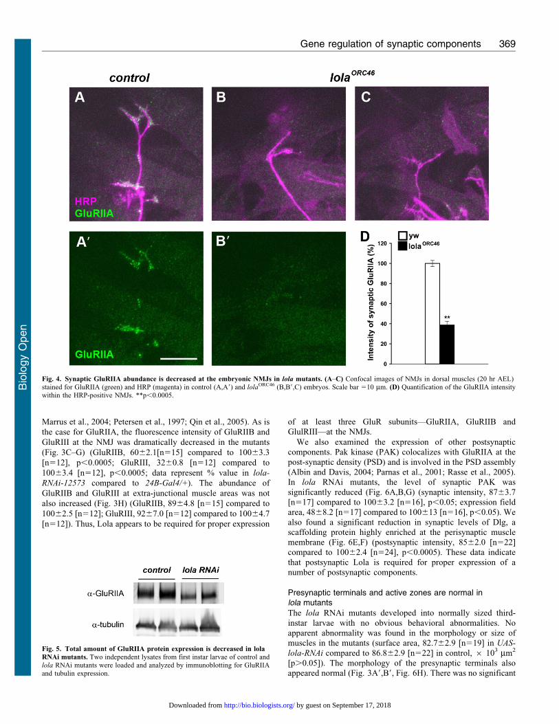
dramatic changes in GluRIIA abundance were observed in the
lolaORC46 embryos (Fig. 4). lolaORC46 is a null allele and is
embryonic lethal. In lolaORC46 embryos, motor axon projection
and muscle patterning are partly deranged with some muscles
entirely lacking nerve innervation (Madden et al., 1999). We
therefore focused on muscles that received normally-sized nerve
terminals (as visualized with a presynaptic marker, anti-HRP)
and found a dramatic decrease in the density of synaptic GluRIIA
(Fig. 4) (3963.7 in lolaORC46 [n527] compared to 10063.0% in
yw [n5101], p,0.0005). These data indicate that Lola is
necessary for normal GluRIIA expression at the NMJ.
The decrease in GluRIIA abundance at the NMJ could be due
to the decrease in total GluRIIA expression in muscles or to
Fig. 2. The role of pastrel (pst) in regulating NMJ outgrowth. (A,B) Third-instar larval 6/7 NMJs immunostained with anti-HRP (magenta) and anti-GluRIIA(green) antibodies. (A) control, (B) pst RNAi. Scale bar 520 mm. (C) Quantification of the phenotypes (*p,0.05, ** p,0.0005). Quantitative data presented in thisand the following figures are presented as the mean value (6 s.e.m.).
Gene regulation of synaptic components 367
Bio
logy
Open
by guest on September 17, 2018http://bio.biologists.org/Downloaded from
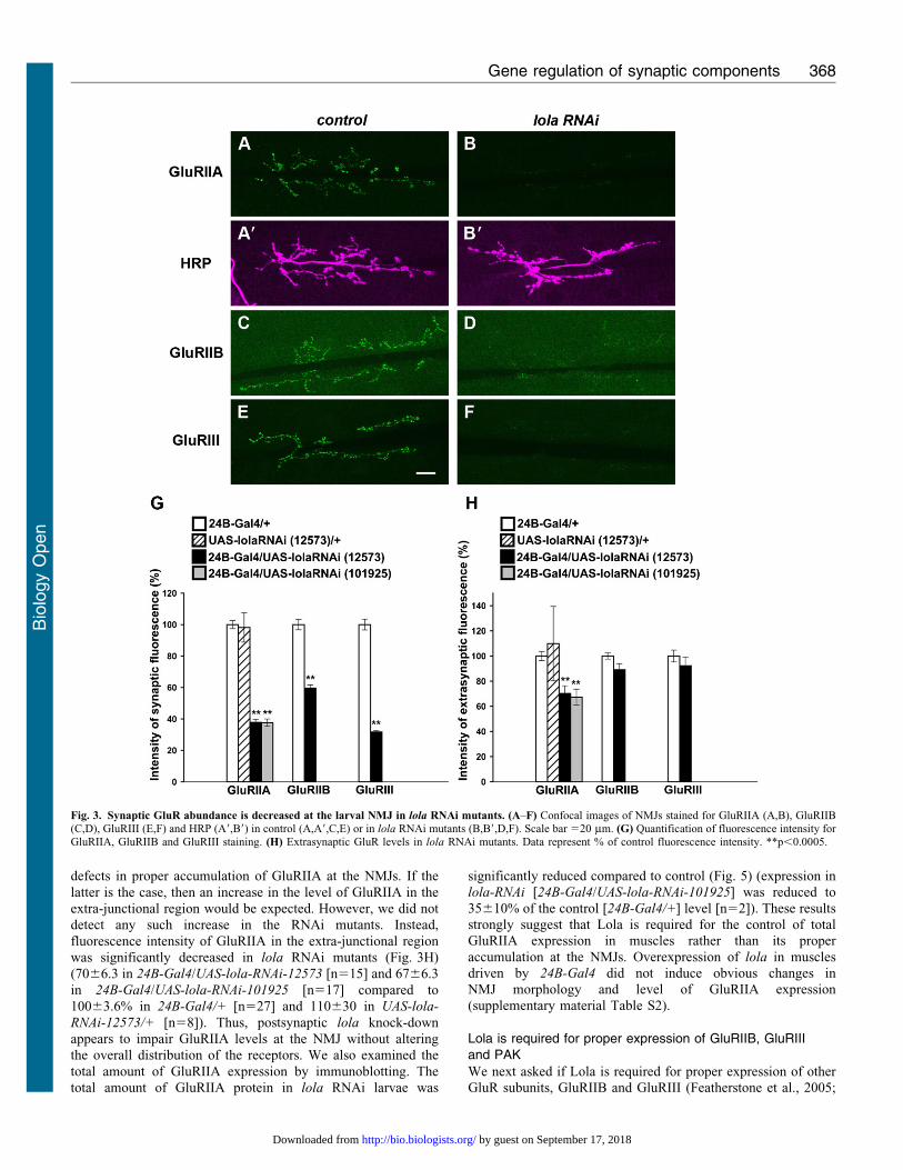
defects in proper accumulation of GluRIIA at the NMJs. If the
latter is the case, then an increase in the level of GluRIIA in the
extra-junctional region would be expected. However, we did not
detect any such increase in the RNAi mutants. Instead,
fluorescence intensity of GluRIIA in the extra-junctional region
was significantly decreased in lola RNAi mutants (Fig. 3H)
(7066.3 in 24B-Gal4/UAS-lola-RNAi-12573 [n515] and 6766.3
in 24B-Gal4/UAS-lola-RNAi-101925 [n517] compared to
10063.6% in 24B-Gal4/+ [n527] and 110630 in UAS-lola-
RNAi-12573/+ [n58]). Thus, postsynaptic lola knock-down
appears to impair GluRIIA levels at the NMJ without altering
the overall distribution of the receptors. We also examined the
total amount of GluRIIA expression by immunoblotting. The
total amount of GluRIIA protein in lola RNAi larvae was
significantly reduced compared to control (Fig. 5) (expression inlola-RNAi [24B-Gal4/UAS-lola-RNAi-101925] was reduced to
35610% of the control [24B-Gal4/+] level [n52]). These resultsstrongly suggest that Lola is required for the control of totalGluRIIA expression in muscles rather than its proper
accumulation at the NMJs. Overexpression of lola in musclesdriven by 24B-Gal4 did not induce obvious changes inNMJ morphology and level of GluRIIA expression
(supplementary material Table S2).
Lola is required for proper expression of GluRIIB, GluRIIIand PAK
We next asked if Lola is required for proper expression of otherGluR subunits, GluRIIB and GluRIII (Featherstone et al., 2005;
Fig. 3. Synaptic GluR abundance is decreased at the larval NMJ in lola RNAi mutants. (A–F) Confocal images of NMJs stained for GluRIIA (A,B), GluRIIB(C,D), GluRIII (E,F) and HRP (A9,B9) in control (A,A9,C,E) or in lola RNAi mutants (B,B9,D,F). Scale bar 520 mm. (G) Quantification of fluorescence intensity forGluRIIA, GluRIIB and GluRIII staining. (H) Extrasynaptic GluR levels in lola RNAi mutants. Data represent % of control fluorescence intensity. **p,0.0005.
Gene regulation of synaptic components 368
Bio
logy
Open
by guest on September 17, 2018http://bio.biologists.org/Downloaded from
Marrus et al., 2004; Petersen et al., 1997; Qin et al., 2005). As is
the case for GluRIIA, the fluorescence intensity of GluRIIB and
GluRIII at the NMJ was dramatically decreased in the mutants
(Fig. 3C–G) (GluRIIB, 6062.1[n515] compared to 10063.3
[n512], p,0.0005; GluRIII, 3260.8 [n512] compared to
10063.4 [n512], p,0.0005; data represent % value in lola-
RNAi-12573 compared to 24B-Gal4/+). The abundance of
GluRIIB and GluRIII at extra-junctional muscle areas was not
also increased (Fig. 3H) (GluRIIB, 8964.8 [n515] compared to
10062.5 [n512]; GluRIII, 9267.0 [n512] compared to 10064.7
[n512]). Thus, Lola appears to be required for proper expression
of at least three GluR subunits—GluRIIA, GluRIIB andGlulRIII—at the NMJs.
We also examined the expression of other postsynapticcomponents. Pak kinase (PAK) colocalizes with GluRIIA at the
post-synaptic density (PSD) and is involved in the PSD assembly(Albin and Davis, 2004; Parnas et al., 2001; Rasse et al., 2005).In lola RNAi mutants, the level of synaptic PAK was
significantly reduced (Fig. 6A,B,G) (synaptic intensity, 8763.7[n517] compared to 10063.2 [n516], p,0.05; expression fieldarea, 4868.2 [n517] compared to 100613 [n516], p,0.05). We
also found a significant reduction in synaptic levels of Dlg, ascaffolding protein highly enriched at the perisynaptic musclemembrane (Fig. 6E,F) (postsynaptic intensity, 8562.0 [n522]compared to 10062.4 [n524], p,0.0005). These data indicate
that postsynaptic Lola is required for proper expression of anumber of postsynaptic components.
Presynaptic terminals and active zones are normal inlola mutants
The lola RNAi mutants developed into normally sized third-
instar larvae with no obvious behavioral abnormalities. Noapparent abnormality was found in the morphology or size ofmuscles in the mutants (surface area, 82.762.9 [n519] in UAS-
lola-RNAi compared to 86.862.9 [n522] in control, 6 103 mm2
[p.0.05]). The morphology of the presynaptic terminals alsoappeared normal (Fig. 3A9,B9, Fig. 6H). There was no significant
Fig. 4. Synaptic GluRIIA abundance is decreased at the embryonic NMJs in lola mutants. (A–C) Confocal images of NMJs in dorsal muscles (20 hr AEL)stained for GluRIIA (green) and HRP (magenta) in control (A,A9) and lolaORC46 (B,B9,C) embryos. Scale bar 510 mm. (D) Quantification of the GluRIIA intensitywithin the HRP-positive NMJs. **p,0.0005.
Fig. 5. Total amount of GluRIIA protein expression is decreased in lola
RNAi mutants. Two independent lysates from first instar larvae of control andlola RNAi mutants were loaded and analyzed by immunoblotting for GluRIIAand tubulin expression.
Gene regulation of synaptic components 369
Bio
logy
Open
by guest on September 17, 2018http://bio.biologists.org/Downloaded from
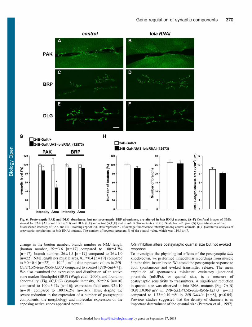
change in the bouton number, branch number or NMJ length
(bouton number, 9263.6 [n517] compared to 10064.2%
[n517]; branch number, 2661.5 [n519] compared to 2661.0
[n522]; NMJ length per muscle area, 8.160.4 [n519] compared
to 9.060.4 [n522], 6 1023 mm21; data represent values in 24B-
Gal4/UAS-lola-RNAi-12573 compared to control [24B-Gal4/+]).
We also examined the expression and distribution of an active
zone marker Bruchpilot (BRP) (Wagh et al., 2006), and found no
abnormality (Fig. 6C,D,G) (synaptic intensity, 9262.6 [n510]
compared to 10063.4% [n516]; expression field area, 92610
[n510] compared to 10066.2% [n516]). Thus, despite the
severe reduction in the expression of a number of postsynaptic
components, the morphology and molecular expression of the
apposing active zones appeared normal.
lola inhibition alters postsynaptic quantal size but not evokedresponse
To investigate the physiological effects of the postsynaptic lola
knock-down, we performed intracellular recordings from muscle
6 in the third-instar larvae. We tested the postsynaptic response to
both spontaneous and evoked transmitter release. The mean
amplitude of spontaneous miniature excitatory junctional
potentials (mEJPs), or quantal size, is a measure of
postsynaptic sensitivity to transmitters. A significant reduction
in quantal size was observed in lola RNAi mutants (Fig. 7A,B)
(0.9160.068 mV in 24B-GAL4/UAS-lola-RNAi-12573 [n511]
compared to 1.3360.10 mV in 24B-Gal4/+ [n58], p,0.05).
Previous studies suggested that the density of channels is an
important determinant of the quantal size (Petersen et al., 1997).
Fig. 6. Postsynaptic PAK and DLG abundance, but not presynaptic BRP abundance, are altered in lola RNAi mutants. (A–F) Confocal images of NMJsstained for PAK (A,B) and BRP (C,D) and DLG (E,F) in control (A,C,E) and in lola RNAi mutants (B,D,F). Scale bar 520 mm. (G) Quantification of thefluorescence intensity of PAK and BRP staining (*p,0.05). Data represent % of average fluorescence intensity among control animals. (H) Quantitative analysis ofpresynaptic morphology in lola RNAi mutants. The number of boutons represent % of the control value, which was 110.664.7.
Gene regulation of synaptic components 370
Bio
logy
Open
by guest on September 17, 2018http://bio.biologists.org/Downloaded from
Therefore, our data are consistent with the decreased GluR
expression observed in the imaging analysis. However, there was
no significant change in the amplitude of excitatory junctional
potential (EJP) in lola RNAi mutants, despite the decrease in the
quantal size (Fig. 7C,D) (36.062.07 mV in lola-RNAi [n511]
compared to 35.864.58 mV in control [n58]). Similar
compensation for the decrease in quantal size has been reported
for several mutants, including GluRIIA (Davis et al., 1998;
Paradis et al., 2001; Petersen et al., 1997). These previous studies
suggested that retrograde signals from the postsynaptic cells
increased the presynaptic transmitter release to compensate for
postsynaptic defects. The mechanism for the compensation in
lola mutants remains to be determined.
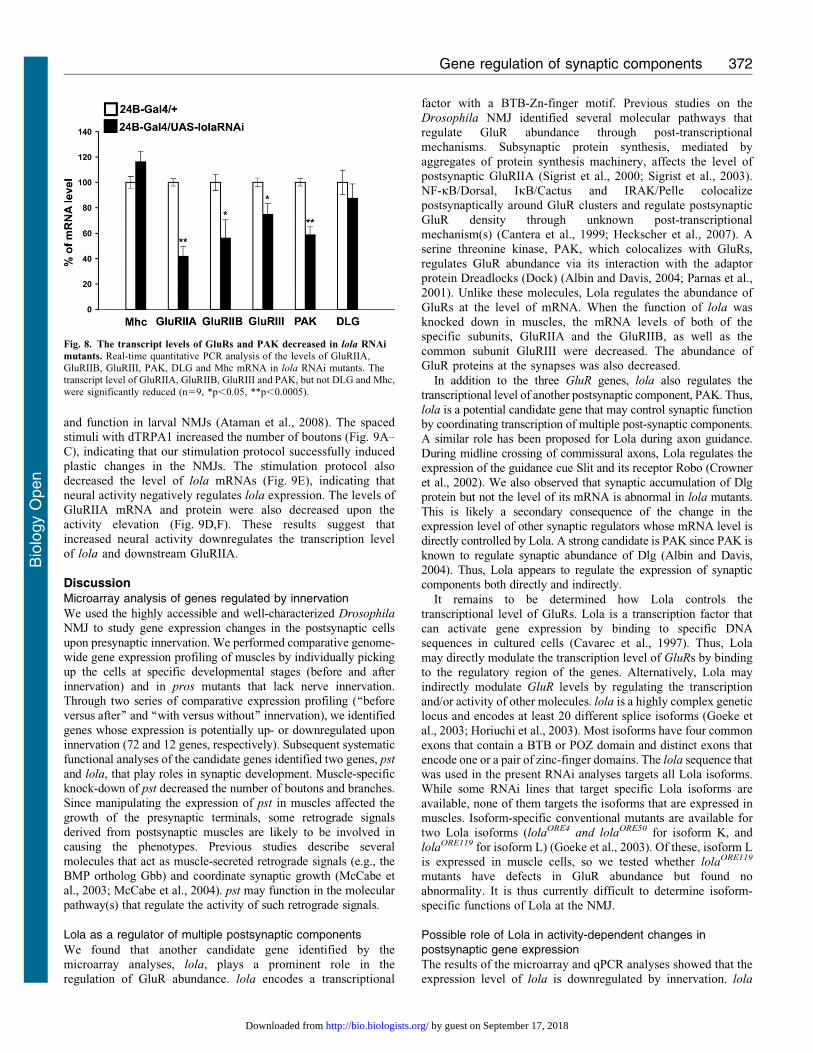
Lola regulates transcription level of GluRs and PAK
The imaging and physiological analyses described above show
that postsynaptic Lola is necessary for normal expression of
GluRs and other synaptic components. Since lola encodes a
BTB-Zn-finger transcription factor (Goeke et al., 2003; Cavarec
et al., 1997), Lola might regulate the transcriptional level of
GluRs. To assess this possibility, we examined the level of GluR
mRNAs in the mutants. We extracted total RNA from the larval
body wall and performed real-time quantitative PCR. We found
that the levels of GluRIIA and GluRIIB mRNAs in lola RNAi
mutants are dramatically reduced (Fig. 8) (GluRIIA, 4267.9
[n59] compared to 10062.9 [n59], p,0.0005; GluRIIB,
56614.4 [n59] compared to 10066.4 [n59], p,0.05; data
represent % values in lola-RNAi-12573 compared to control
[24B-Gal4/+]). We also found a smaller but significant decrease
in the level of GluRIII (7568.7 [n59] compared to 10063.5
[n59], p,0.05). Furthermore, we found a significant reduction
of PAK mRNAs (5966.4 [n59] compared to 10062.9 [n59],
p,0.0005). On the other hand, the mRNA transcript level of Dlg
and Myosin heavy chain (Mhc), a protein involved in muscle
contraction, was not affected (Dlg, 87611.3 [n59] compared to
10069.6 [n59]; Mhc,11668.4 [n59] compared to 10064.5
[n59]). These data indicate that Lola positively regulates the
transcription level of glutamate receptors and PAK but not Dlg.
Neural activity induces changes in lola expression
Having shown that Lola is involved in the transcriptional
regulation of a number of synaptic components, we next asked
how the expression of lola itself is regulated. Our microarray
analyses showed that the level of lola transcript is changed by
innervation. Therefore, one possibility is that neural activity
modulates lola expression. To address this, we used the
Drosophila warmth-activated cation channel TRPA1 (dTRPA1)
(Pulver et al., 2009) to activate all neurons during late embryonic
and early 1st instar larval stages and studied the effect on the
expression of lola in the third instar larvae. We applied repetitive
2-min intervals at a higher temperature (27 C) spaced by 5 min of
rest (22 C), for 12 hrs from AEL18. Such spaced induction of
increased activity with Channelrhodopsin or high K+ has
previously been shown to induce changes in synaptic structure
Fig. 7. Quantal size of NMJs is decreased in
lola RNAi mutants. (A) Representative tracesof spontaneous neurotransmitter release in
0.375 mM calcium in control (upper trace) andin lola RNAi mutants (lower trace).(B) Quantification of quantal size. *p,0.05.(C) Representative traces of evokedtransmitter release in 1.5 mM calcium incontrol (left) and lola RNAi mutants (right).
(D) Quantification of the amplitude of EJPs.
Gene regulation of synaptic components 371
Bio
logy
Open
by guest on September 17, 2018http://bio.biologists.org/Downloaded from
and function in larval NMJs (Ataman et al., 2008). The spacedstimuli with dTRPA1 increased the number of boutons (Fig. 9A–C), indicating that our stimulation protocol successfully induced
plastic changes in the NMJs. The stimulation protocol alsodecreased the level of lola mRNAs (Fig. 9E), indicating thatneural activity negatively regulates lola expression. The levels of
GluRIIA mRNA and protein were also decreased upon theactivity elevation (Fig. 9D,F). These results suggest thatincreased neural activity downregulates the transcription level
of lola and downstream GluRIIA.
DiscussionMicroarray analysis of genes regulated by innervation
We used the highly accessible and well-characterized Drosophila
NMJ to study gene expression changes in the postsynaptic cells
upon presynaptic innervation. We performed comparative genome-wide gene expression profiling of muscles by individually pickingup the cells at specific developmental stages (before and after
innervation) and in pros mutants that lack nerve innervation.Through two series of comparative expression profiling (‘‘beforeversus after’’ and ‘‘with versus without’’ innervation), we identified
genes whose expression is potentially up- or downregulated uponinnervation (72 and 12 genes, respectively). Subsequent systematicfunctional analyses of the candidate genes identified two genes, pst
and lola, that play roles in synaptic development. Muscle-specificknock-down of pst decreased the number of boutons and branches.Since manipulating the expression of pst in muscles affected the
growth of the presynaptic terminals, some retrograde signalsderived from postsynaptic muscles are likely to be involved incausing the phenotypes. Previous studies describe several
molecules that act as muscle-secreted retrograde signals (e.g., theBMP ortholog Gbb) and coordinate synaptic growth (McCabe etal., 2003; McCabe et al., 2004). pst may function in the molecularpathway(s) that regulate the activity of such retrograde signals.
Lola as a regulator of multiple postsynaptic components
We found that another candidate gene identified by the
microarray analyses, lola, plays a prominent role in theregulation of GluR abundance. lola encodes a transcriptional
factor with a BTB-Zn-finger motif. Previous studies on the
Drosophila NMJ identified several molecular pathways thatregulate GluR abundance through post-transcriptionalmechanisms. Subsynaptic protein synthesis, mediated by
aggregates of protein synthesis machinery, affects the level ofpostsynaptic GluRIIA (Sigrist et al., 2000; Sigrist et al., 2003).NF-kB/Dorsal, IkB/Cactus and IRAK/Pelle colocalizepostsynaptically around GluR clusters and regulate postsynaptic
GluR density through unknown post-transcriptionalmechanism(s) (Cantera et al., 1999; Heckscher et al., 2007). Aserine threonine kinase, PAK, which colocalizes with GluRs,
regulates GluR abundance via its interaction with the adaptorprotein Dreadlocks (Dock) (Albin and Davis, 2004; Parnas et al.,2001). Unlike these molecules, Lola regulates the abundance of
GluRs at the level of mRNA. When the function of lola wasknocked down in muscles, the mRNA levels of both of thespecific subunits, GluRIIA and the GluRIIB, as well as the
common subunit GluRIII were decreased. The abundance ofGluR proteins at the synapses was also decreased.
In addition to the three GluR genes, lola also regulates thetranscriptional level of another postsynaptic component, PAK. Thus,
lola is a potential candidate gene that may control synaptic functionby coordinating transcription of multiple post-synaptic components.A similar role has been proposed for Lola during axon guidance.
During midline crossing of commissural axons, Lola regulates theexpression of the guidance cue Slit and its receptor Robo (Crowneret al., 2002). We also observed that synaptic accumulation of Dlgprotein but not the level of its mRNA is abnormal in lola mutants.
This is likely a secondary consequence of the change in theexpression level of other synaptic regulators whose mRNA level isdirectly controlled by Lola. A strong candidate is PAK since PAK is
known to regulate synaptic abundance of Dlg (Albin and Davis,2004). Thus, Lola appears to regulate the expression of synapticcomponents both directly and indirectly.
It remains to be determined how Lola controls the
transcriptional level of GluRs. Lola is a transcription factor thatcan activate gene expression by binding to specific DNAsequences in cultured cells (Cavarec et al., 1997). Thus, Lola
may directly modulate the transcription level of GluRs by bindingto the regulatory region of the genes. Alternatively, Lola mayindirectly modulate GluR levels by regulating the transcription
and/or activity of other molecules. lola is a highly complex geneticlocus and encodes at least 20 different splice isoforms (Goeke etal., 2003; Horiuchi et al., 2003). Most isoforms have four common
exons that contain a BTB or POZ domain and distinct exons thatencode one or a pair of zinc-finger domains. The lola sequence thatwas used in the present RNAi analyses targets all Lola isoforms.While some RNAi lines that target specific Lola isoforms are
available, none of them targets the isoforms that are expressed inmuscles. Isoform-specific conventional mutants are available fortwo Lola isoforms (lolaORE4 and lolaORE50 for isoform K, and
lolaORE119 for isoform L) (Goeke et al., 2003). Of these, isoform Lis expressed in muscle cells, so we tested whether lolaORE119
mutants have defects in GluR abundance but found no
abnormality. It is thus currently difficult to determine isoform-specific functions of Lola at the NMJ.
Possible role of Lola in activity-dependent changes inpostsynaptic gene expression
The results of the microarray and qPCR analyses showed that theexpression level of lola is downregulated by innervation. lola
Fig. 8. The transcript levels of GluRs and PAK decreased in lola RNAi
mutants. Real-time quantitative PCR analysis of the levels of GluRIIA,GluRIIB, GluRIII, PAK, DLG and Mhc mRNA in lola RNAi mutants. Thetranscript level of GluRIIA, GluRIIB, GluRIII and PAK, but not DLG and Mhc,
were significantly reduced (n59, *p,0.05, **p,0.0005).
Gene regulation of synaptic components 372
Bio
logy
Open
by guest on September 17, 2018http://bio.biologists.org/Downloaded from
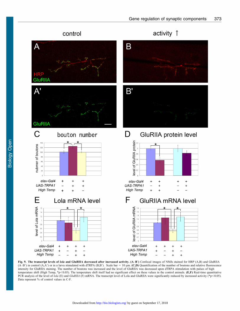
Fig. 9. The transcript levels of lola and GluRIIA decreased after increased activity. (A–B9) Confocal images of NMJs stained for HRP (A,B) and GluRIIA(A–B9) in control (A,A9) or in a larva stimulated with dTRPA (B,B9). Scale bar 5 10 mm. (C,D) Quantification of the number of boutons and relative fluorescenceintensity for GluRIIA staining. The number of boutons was increased and the level of GluRIIA was decreased upon dTRPA stimulation with pulses of hightemperature shift (High Temp, *p,0.05). The temperature shift itself had no significant effect on these values in the control animals. (E,F) Real-time quantitativePCR analysis of the level of lola (E) and GluRIIA (F) mRNA. The transcript level of Lola and GluRIIA were significantly reduced by increased activity (*p,0.05).
Data represent % of control values in C-F.
Gene regulation of synaptic components 373
Bio
logy
Open
by guest on September 17, 2018http://bio.biologists.org/Downloaded from
expression was downregulated after innervation and upregulated
in pros mutants, which lack innervation. Similarly, increasing
neural activity decreased the transcriptional level of lola. Thus,
lola mRNA expression appears to be negatively regulated by
innervation and neural activity. Since the expression of GluRs
and other synaptic components are in turn regulated by lola, lola
may link changes in neural activity to changes in postsynaptic
gene expression. Consistent with this view, increased neural
activity decreased the expression of the GluRIIA transcript and
protein, concomitant with the decrease in lola expression. It is
however currently unknown whether lola is solely responsible for
the changes in GluRIIA expression in the activity misregulated
NMJs. We also could not detect obvious changes in the
expression of Lola protein in the aneural muscles (in pros
mutants) or in activity-misregulated muscles (in dTRPA1
expressing larvae). Role of lola in coordinating activity-
dependent changes in postsynaptic components remains to be
explored in future studies.
How lola expression is regulated by neural innervation and/or
activity also remains to be determined. Signaling mediated by
PKA and CaMKII has been previously implicated in activity-
dependent regulation of GluRIIA expression (Davis et al., 1998;
Haghighi et al., 2003; Kazama et al., 2003; Morimoto-Tanifuji et
al., 2004). Whether lola is regulated by these or other activity-
dependent signaling mechanisms will be an interesting question
to address in the future.
The negative regulation of lola by innervation and increased
activity is somewhat counterintuitive in light of the fact that lola
positively regulates the level of GluR and other postsynaptic
components. The synaptic expression level of GluRs is known to
increase after innervation (Broadie and Bate, 1993c; Chen and
Featherstone, 2005). Some protocols that increase larval activity
are known to induce an increase in GluRIIA expression
(Schuster, 2006). Increasing motoneuron activity also increases
their terminal size (Ataman et al., 2008; Budnik et al., 1990). One
possibility is that Lola is part of the homeostatic mechanism(s)
that maintain a proper level of synaptic strength. It has been
shown that a decrease in postsynaptic function is compensated by
an increase in presynaptic synaptic release at the Drosophila
NMJs (Davis, 2006; Dickman and Davis, 2009; Frank et al.,
2009). Similarly, Lola may control the appropriate level of
postsynaptic components according to the level of presynaptic
activity. For example, when presynaptic activity is too strong,
expression of lola may be downregulated to reduce the level of
GluRs at the postsynaptic cells. Conversely, when presynaptic
innervation drops to an insufficient level in pros mutants, lola
may be upregulated to compensate for the decreased transmission
by increasing the level of GluRs.
Another possibility is involvement in the pruning of ectopic
synapses. During the formation of neuromuscular connectivity in
the embryos, reduction in electric activity in motor neurons
results in an increase in ectopic synapses on non-target muscles
(Jarecki and Keshishian, 1995; Carrillo et al., 2010). While
involvement of presynaptic mechanisms has been shown by a
recent study (Carrillo et al., 2010), postsynaptic Lola may also
contribute to the regulation of ectopic synapses. For example,
lola expression may be downregulated after innervation by proper
motor neurons, to prevent the formation of ectopic synapses.
Possible roles of lola in these processes will be determined in
future studies.
Several studies suggest that Lola can regulate the chromatin
structure and function in an epigenetic manner (Ferres-Marco etal., 2006; Zhang et al., 2003). Thus, Lola may be involved in the
epigenetic control of synaptic plasticity, as shown in othersystems (Guan et al., 2004; Levenson and Sweatt, 2005).
Identification of Lola as a regulator of multiple postsynaptic
components will provide a unique entry point to theunderstanding of transcriptional regulation of synapse
formation and plasticity at the Drosophila NMJ.
AcknowledgementsWe are grateful to E. Giniger, T. Aigaki, A. DiAntonio, M. Saitoe, A.Hofbauer, C. Hama, Bloomington and Kyoto Stock Center, ViennaDrosophila RNAi Center (VDRC), Fly Stocks of the NationalInstitute of Genetics (NIG), The Exelixis Collection at the HarvardMedical School, and the Developmental Studies Hybridoma Bankfor fly stocks and reagents. We acknowledge H. Miyakawa andmembers of the Nose lab for helpful discussion, H. Kohsaka for helpin staining of the embryos, Y. Fujioka, H. Kato and M. Sotodate fortechnical assistance. This study was supported by a Grant-in-Aid forScientific Research B and for Scientific Research on Priority Areas-Molecular Brain Science-from the MEXT to A.N.
Competing InterestsWe declare that there are no competing interests.
ReferencesAlbin, S. D. and Davis, G. W. (2004). Coordinating structural and functional synapse
development: postsynaptic p21-activated kinase independently specifies glutamatereceptor abundance and postsynaptic morphology. J. Neurosci. 24, 6871-6879.
Ataman, B., Ashley, J., Gorczyca, M., Ramachandran, P., Fouquet, W., Sigrist, S.
and Budnik, V. (2008). Rapid activity-dependent modifications in synaptic structureand function require bidirectional Wnt signaling. Neuron 57, 705-718.
Brand, A. H. and Perrimon, N. (1993). Targeted gene expression as a means of alteringcell fates and generating dominant phenotypes. Development 118, 401-415.
Broadie, K. S. and Bate, M. (1993a). Development of the embryonic neuromuscularsynapse of Drosophila melanogaster. J. Neurosci. 13, 144-166.
Broadie, K. and Bate, M. (1993b). Muscle development is independent of innervationduring Drosophila embryogenesis. Development 119, 533-543.
Broadie, K. and Bate, M. (1993c). Innervation directs receptor synthesis andlocalization in Drosophila embryo synaptogenesis. Nature 361, 350-353.
Budnik, V., Zhong, Y. and Wu, C. F. (1990). Morphological plasticity of motor axonsin Drosophila mutants with altered excitability. J. Neurosci. 10, 3754-3768.
Cantera, R., Kozlova, T., Barillas-Mury, C. and Kafatos, F. C. (1999). Musclestructure and innervation are affected by loss of Dorsal in the fruit fly, Drosophilamelanogaster. Mol. Cell. Neurosci. 13, 131-141.
Carrillo, R. A., Olsen, D. P., Yoon, K. S. and Keshishian, H. (2010). Presynapticactivity and CaMKII modulate retrograde semaphorin signaling and synapticrefinement. Neuron 68, 32-44.
Cavarec, L., Jensen, S., Casella, J. F., Cristescu, S. A., Heidmann, T. and Chen, K.
(1997). Molecular cloning and characterization of a transcription factor for the copiaretrotransposon with homology to the BTB-containing lola neurogenic factor. Mol.
Cell. Biol. 17, 482-494.
Chen, K. and Featherstone, D. E. (2005). Discs-large (DLG) is clustered bypresynaptic innervation and regulates postsynaptic glutamate receptor subunitcomposition in Drosophila. BMC Biol. 3, 1.
Cohen, S. and Greenberg, M. E. (2008). Communication between the synapse and thenucleus in neuronal development, plasticity, and disease. Annu. Rev. Cell Dev. Biol.
24, 183-209.
Coyle, I. P., Koh, Y. H., Lee, W. C., Slind, J., Fergestad, T., Littleton, J. T. and
Ganetzky, B. (2004). Nervous wreck, an SH3 adaptor protein that interacts with Wsp,regulates synaptic growth in Drosophila. Neuron 41, 521-534.
Crowner, D., Madden, K., Goeke, S. and Giniger, E. (2002). Lola regulates midlinecrossing of CNS axons in Drosophila. Development 129, 1317-1325.
Davis, G. W. (2006). Homeostatic control of neural activity: from phenomenology tomolecular design. Annu. Rev. Neurosci. 29, 307-323.
Davis, G. W., Schuster, C. M. and Goodman, C. S. (1996). Genetic dissection ofstructural and functional components of synaptic plasticity. III. CREB is necessary forpresynaptic functional plasticity. Neuron 17, 669-679.
Davis, G. W., DiAntonio, A., Petersen, S. A. and Goodman, C. S. (1998). PostsynapticPKA controls quantal size and reveals a retrograde signal that regulates presynaptictransmitter release in Drosophila. Neuron 20, 305-315.
Dickman, D. K. and Davis, G. W. (2009). The schizophrenia susceptibility genedysbindin controls synaptic homeostasis. Science 326, 1127-1130.
Gene regulation of synaptic components 374
Bio
logy
Open
by guest on September 17, 2018http://bio.biologists.org/Downloaded from

Dietzl, G., Chen, D., Schnorrer, F., Su, K. C., Barinova, Y., Fellner, M., Gasser, B.,Kinsey, K., Oppel, S., Scheiblauer, S. et al. (2007). A genome-wide transgenicRNAi library for conditional gene inactivation in Drosophila. Nature 448, 151-156.
Dubnau, J., Chiang, A. S., Grady, L., Barditch, J., Gossweiler, S., McNeil, J., Smith,
P., Buldoc, F., Scott, R., Certa, U. et al. (2003). The staufen/pumilio pathway isinvolved in Drosophila long-term memory. Curr. Biol. 13, 286-296.
Featherstone, D. E., Rushton, E., Rohrbough, J., Liebl, F., Karr, J., Sheng, Q.,
Rodesch, C. K. and Broadie, K. (2005). An essential Drosophila glutamate receptorsubunit that functions in both central neuropil and neuromuscular junction. J.
Neurosci. 25, 3199-3208.Ferres-Marco, D., Gutierrez-Garcia, I., Vallejo, D. M., Bolivar, J., Gutierrez-Avino,
F. J. and Dominguez, M. (2006). Epigenetic silencers and Notch collaborate topromote malignant tumours by Rb silencing. Nature 439, 430-436.
Flavell, S. W. and Greenberg, M. E. (2008). Signaling mechanisms linking neuronalactivity to gene expression and plasticity of the nervous system. Annu. Rev. Neurosci.
31, 563-590.Frank, C. A., Pielage, J. and Davis, G. W. (2009). A presynaptic homeostatic signaling
system composed of the Eph receptor, ephexin, Cdc42, and CaV2.1 calcium channels.Neuron 61, 556-569.
Giniger, E., Tietje, K., Jan, L. Y. and Jan, Y. N. (1994). lola encodes a putativetranscription factor required for axon growth and guidance in Drosophila.Development 120, 1385-1398.
Goda, Y. and Davis, G. W. (2003). Mechanisms of synapse assembly and disassembly.Neuron 40, 243-264.
Goeke, S., Greene, E. A., Grant, P. K., Gates, M. A., Crowner, D., Aigaki, T. andGiniger, E. (2003). Alternative splicing of lola generates 19 transcription factorscontrolling axon guidance in Drosophila. Nat. Neurosci. 6, 917-924.
Griffith, L. and Budnik, V. (2006). Plasticity and second messengers during synapsedevelopment. Int. Rev. Neurobiol. 75, 237-265.
Guan, J. S., Haggarty, S. J., Giacometti, E., Dannenberg, J. H., Joseph, N., Gao, J.,Nieland, T. J., Levenson, J. M., O’Riordan, K. J., Brown, K. D. et al. (2004).Regulation of histone acetylation during memory formation in the hippocampus. J.
Biol. Chem. 279, 40545-40559.Haghighi, A. P., McCabe, B. D., Fetter, R. D., Palmer, J. E., Hom, S. and Goodman,
C. S. (2003). Retrograde control of synaptic transmission by postsynaptic CaMKII atthe Drosophila neuromuscular junction. Neuron 39, 255-267.
Heckscher, E. S., Fetter, R. D., Marek, K. W., Albin, S. D. and Davis, G. W. (2007).NF-kappaB, IkappaB, and IRAK control glutamate receptor density at the DrosophilaNMJ. Neuron 55, 859-873.
Horiuchi, T., Giniger, E. and Aigaki, T. (2003). Alternative trans-splicing of constantand variable exons of a Drosophila axon guidance gene, lola. Genes Dev. 17, 2496-2501.
Inaki, M., Yoshikawa, S., Thomas, J. B., Aburatani, H. and Nose, A. (2007). Wnt4 isa local repulsive cue that determines synaptic target specificity. Curr. Biol. 17, 1574-1579.
Jarecki, J. and Keshishian, H. (1995). Role of neural activity during synaptogenesis inDrosophila. J. Neurosci. 15, 8177-8190.
Kandel, E. R. (2001). The molecular biology of memory storage: a dialogue betweengenes and synapses. Science 294, 1030-1038.
Kazama, H., Morimoto-Tanifuji, T. and Nose, A. (2003). Postsynaptic activation ofcalcium/calmodulin-dependent protein kinase II promotes coordinated pre- andpostsynaptic maturation of Drosophila neuromuscular junctions. Neuroscience 117,615-625.
Keshishian, H., Broadie, K., Chiba, A. and Bate, M. (1996). The drosophilaneuromuscular junction: a model system for studying synaptic development andfunction. Annu. Rev. Neurosci. 19, 545-575.
Kohsaka, H., Takasu, E. and Nose, A. (2007). In vivo induction of postsynapticmolecular assembly by the cell adhesion molecule Fasciclin2. J. Cell Biol. 179, 1289-1300.
Korkut, C., Ataman, B., Ramachandran, P., Ashley, J., Barria, R., Gherbesi, N. and
Budnik, V. (2009). Trans-Synaptic Transmission of Vesicular Wnt Signals throughEvi/Wntless. Cell 139, 393-404.
Kummer, T. T., Misgeld, T. and Sanes, J. R. (2006). Assembly of the postsynapticmembrane at the neuromuscular junction: paradigm lost. Curr. Opin. Neurobiol. 16,74-82.
Levenson, J. M. and Sweatt, J. D. (2005). Epigenetic mechanisms in memoryformation. Nat. Rev. Neurosci. 6, 108-118.
Li, Z. and Sheng, M. (2003). Some assembly required: the development of neuronalsynapses. Nat. Rev. Mol. Cell Biol. 4, 833-841.
Luo, L., Liao, Y. J., Jan, L. Y. and Jan, Y. N. (1994). Distinct morphogeneticfunctions of similar small GTPases: Drosophila Drac1 is involved in axonaloutgrowth and myoblast fusion. Genes Dev. 8, 1787-1802.
Madden, K., Crowner, D. and Giniger, E. (1999). LOLA has the properties of a masterregulator of axon-target interaction for SNb motor axons of Drosophila. Dev. Biol.
213, 301-313.Marrus, S. B., Portman, S. L., Allen, M. J., Moffat, K. G. and DiAntonio, A. (2004).
Differential localization of glutamate receptor subunits at the Drosophila neuromus-cular junction. J. Neurosci. 24, 1406-1415.
McAllister, A. K. (2007). Dynamic aspects of CNS synapse formation. Annu. Rev.
Neurosci. 30, 425-450.
McCabe, B. D., Marques, G., Haghighi, A. P., Fetter, R. D., Crotty, M. L., Haerry,
T. E., Goodman, C. S. and O’Connor, M. B. (2003). The BMP homolog Gbb
provides a retrograde signal that regulates synaptic growth at the Drosophila
neuromuscular junction. Neuron 39, 241-254.
McCabe, B. D., Hom, S., Aberle, H., Fetter, R. D., Marques, G., Haerry, T. E., Wan,
H., O’Connor, M. B., Goodman, C. S., Haghighi, A. P. et al. (2004). Highwire
regulates presynaptic BMP signaling essential for synaptic growth. Neuron 41, 891-905.
Morimoto-Tanifuji, T., Kazama, H. and Nose, A. (2004). Developmental stage-
dependent modulation of synapses by postsynaptic expression of activated calcium/
calmodulin-dependent protein kinase II. Neuroscience 128, 797-806.
Morimoto, T., Nobechi, M., Komatsu, A., Miyakawa, H. and Nose, A. (2010).
Subunit-specific and homeostatic regulation of glutamate receptor localization by
CaMKII in Drosophila neuromuscular junctions. Neuroscience 65, 1284-1292.
Paradis, S., Sweeney, S. T. and Davis, G. W. (2001). Homeostatic control of
presynaptic release is triggered by postsynaptic membrane depolarization. Neuron 30,
737-749.
Parnas, D., Haghighi, A. P., Fetter, R. D., Kim, S. W. and Goodman, C. S. (2001).
Regulation of postsynaptic structure and protein localization by the Rho-type guanine
nucleotide exchange factor dPix. Neuron 32, 415-424.
Petersen, S. A., Fetter, R. D., Noordermeer, J. N., Goodman, C. S. and DiAntonio,
A. (1997). Genetic analysis of glutamate receptors in Drosophila reveals a retrograde
signal regulating presynaptic transmitter release. Neuron 19, 1237-1248.
Pulver, S. R., Pashkovski, S. L., Hornstein, N. J., Garrity, P. A. and Griffith, L. C.
(2009). Temporal dynamics of neuronal activation by Channelrhodopsin-2 and
TRPA1 determine behavioral output in Drosophila larvae. J. Neurophysiol. 101,
3075-3088.
Qin, G., Schwarz, T., Kittel, R. J., Schmid, A., Rasse, T. M., Kappei, D.,
Ponimaskin, E., Heckmann, M. and Sigrist, S. J. (2005). Four different subunits are
essential for expressing the synaptic glutamate receptor at neuromuscular junctions ofDrosophila. J. Neurosci. 25, 3209-3218.
Rasse, T. M., Fouquet, W., Schmid, A., Kittel, R. J., Mertel, S., Sigrist, C. B.,
Schmidt, M., Guzman, A., Merino, C., Qin, G. et al. (2005). Glutamate receptordynamics organizing synapse formation in vivo. Nat. Neurosci. 8, 898-905.
Ritzenthaler, S., Suzuki, E. and Chiba, A. (2000). Postsynaptic filopodia in muscle
cells interact with innervating motoneuron axons. Nat. Neurosci. 3, 1012-1017.
Sanes, J. R. and Lichtman, J. W. (2001). Induction, assembly, maturation and
maintenance of a postsynaptic apparatus. Nat. Rev. Neurosci. 2, 791-805.
Sanyal, S. and Ramaswami, M. (2006). Activity-dependent regulation of transcriptionduring development of synapses. Int. Rev. Neurobiol. 75, 287-305.
Sanyal, S., Sandstrom, D. J., Hoeffer, C. A. and Ramaswami, M. (2002). AP-1
functions upstream of CREB to control synaptic plasticity in Drosophila. Nature 416,870-874.
Schaeffer, L., de Kerchove d’Exaerde, A. and Changeux, J. P. (2001). Targeting
transcription to the neuromuscular synapse. Neuron 31, 15-22.
Schuster, C. M. (2006). Experience-dependent potentiation of larval neuromuscular
synapses. Int. Rev. Neurobiol. 75, 307-322.
Shishido, E., Takeichi, M., and Nose, A. (1998). Drosophila synapse formation:regulation by transmembrane protein with Leu-rich repeats, CAPRICIOUS. Science
280, 2118-2121.
Sigrist, S. J., Thiel, P. R., Reiff, D. F., Lachance, P. E., Lasko, P. and Schuster, C. M.
(2000). Postsynaptic translation affects the efficacy and morphology of neuromus-
cular junctions. Nature 405, 1062-1065.
Sigrist, S. J., Reiff, D. F., Thiel, P. R., Steinert, J. R. and Schuster, C. M. (2003).
Experience-dependent strengthening of Drosophila neuromuscular junctions. J.
Neurosci. 23, 6546-6556.
Sone, M., Suzuki, E., Hoshino, M., Hou, D., Kuromi, H., Fukata, M., Kuroda, S.,
Kaibuchi, K., Nabeshima, Y. and Hama, C. (2000). Synaptic development iscontrolled in the periactive zones of Drosophila synapses. Development 127, 4157-4168.
Spletter, M. L., Liu, J., Liu, J., Su, H., Giniger, E., Komiyama, T., Quake, S. and
Luo, L. (2007). Lola regulates Drosophila olfactory projection neuron identity andtargeting specificity. Neural. Dev. 2, 14.
Thomas, U., Kim, E., Kuhlendahl, S., Koh, Y. H., Gundelfinger, E. D., Sheng, M.,
Garner, C. C. and Budnik, V. (1997). Synaptic clustering of the cell adhesionmolecule fasciclin II by discs-large and its role in the regulation of presynaptic
structure. Neuron. 4, 787-799.
Wagh, D. A., Rasse, T. M., Asan, E., Hofbauer, A., Schwenkert, I., Durrbeck, H.,
Buchner, S., Dabauvalle, M. C., Schmidt, M., Qin, G. et al. (2006). Bruchpilot, a
protein with homology to ELKS/CAST, is required for structural integrity and
function of synaptic active zones in Drosophila. Neuron 49, 833-844.
Zhang, W., Wang, Y., Long, J., Girton, J., Johansen, J. and Johansen, K. M. (2003).A developmentally regulated splice variant from the complex lola locus encoding
multiple different zinc finger domain proteins interacts with the chromosomal kinase
JIL-1. J. Biol. Chem. 278, 11696-11704.
Gene regulation of synaptic components 375
Bio
logy
Open
by guest on September 17, 2018http://bio.biologists.org/Downloaded from