Embed Size (px)
Citation preview
Lymphatic System
The “Other” Circulatory System
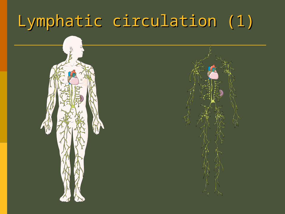
Lymphatic circulation (1)Lymphatic circulation (1)
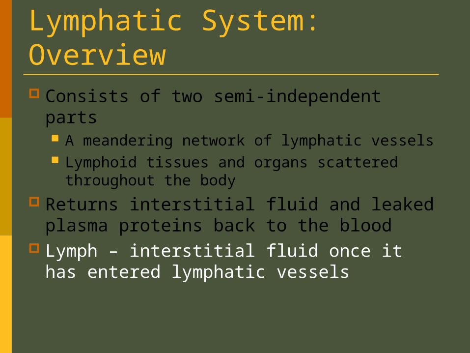
Lymphatic System: Overview Consists of two semi-independent parts
A meandering network of lymphatic vessels Lymphoid tissues and organs scattered
throughout the body Returns interstitial fluid and leaked plasma
proteins back to the blood Lymph – interstitial fluid once it has
entered lymphatic vessels
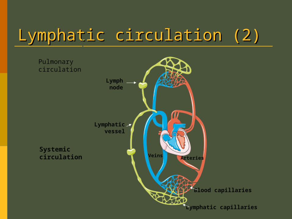
Lymphatic circulation (2)Lymphatic circulation (2)Pulmonarycirculation
Systemiccirculation ArteriesVeins
Blood capillaries
Lymphatic capillaries
Lymphnode
Lymphaticvessel
Lymphatic System: Overview
Lymphatic System: Overview
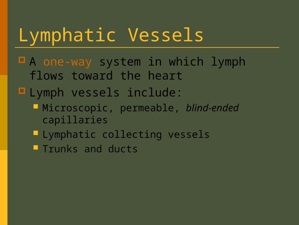
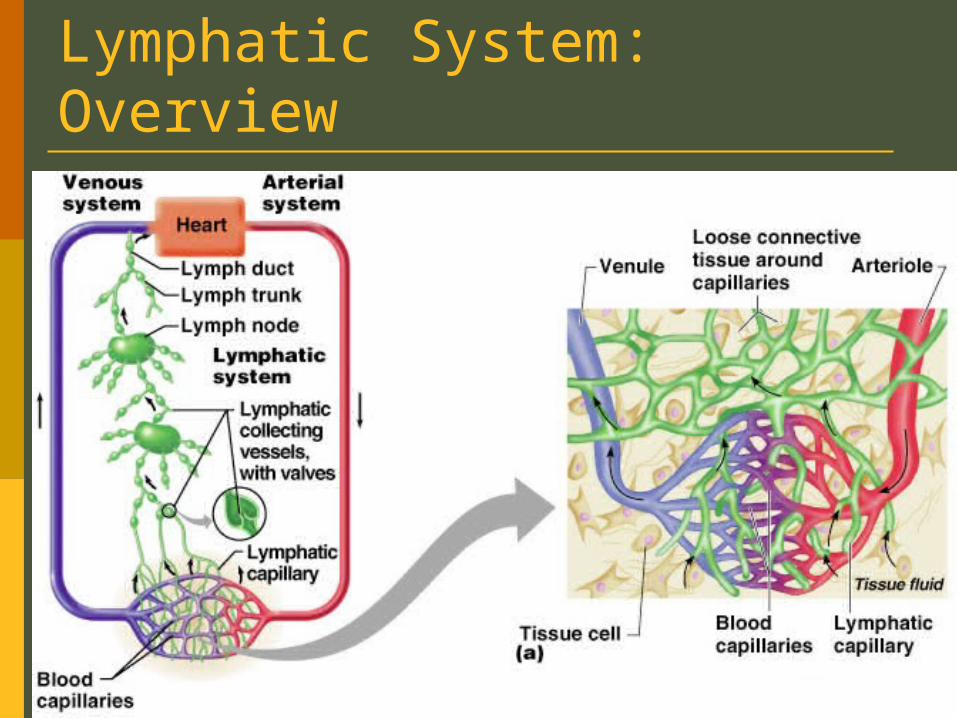
Lymphatic Vessels A one-way system in which lymph flows
toward the heart Lymph vessels include:
Microscopic, permeable, blind-ended capillaries Lymphatic collecting vessels Trunks and ducts
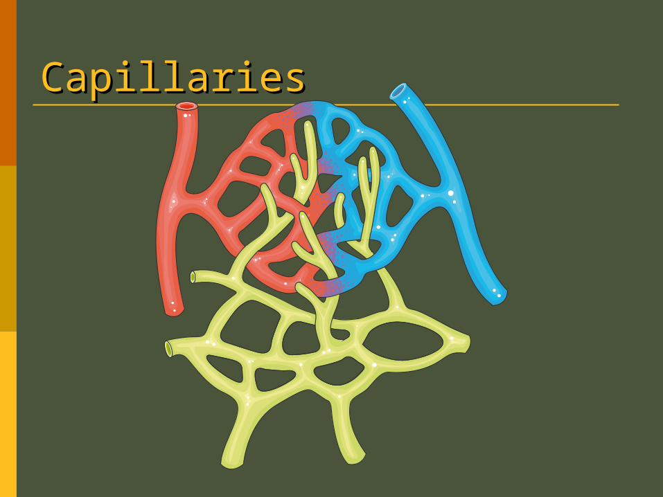
CapillariesCapillaries
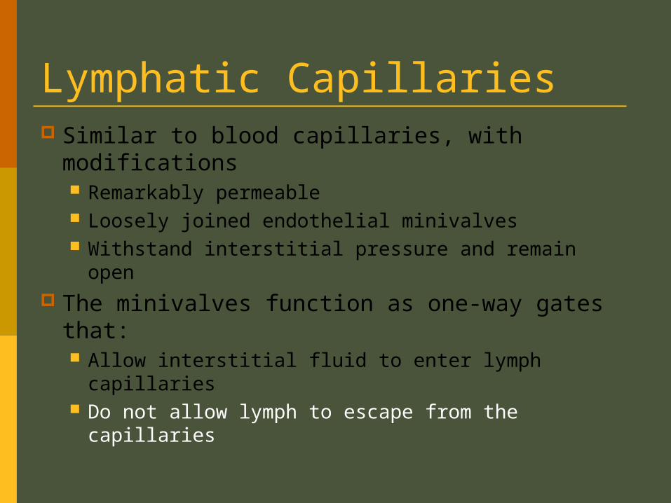
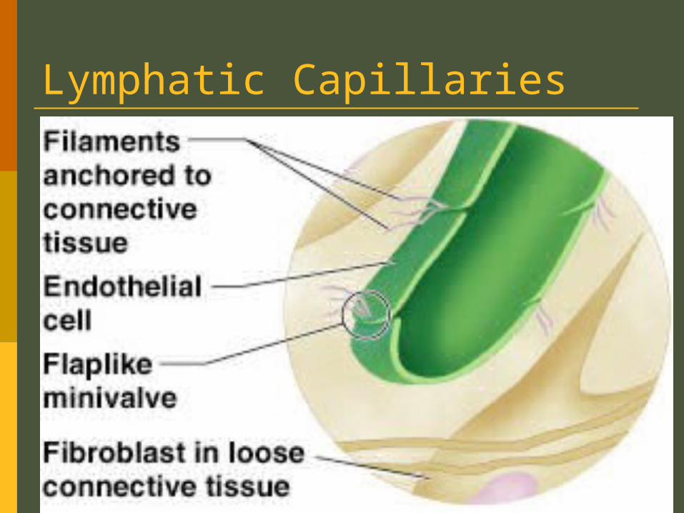
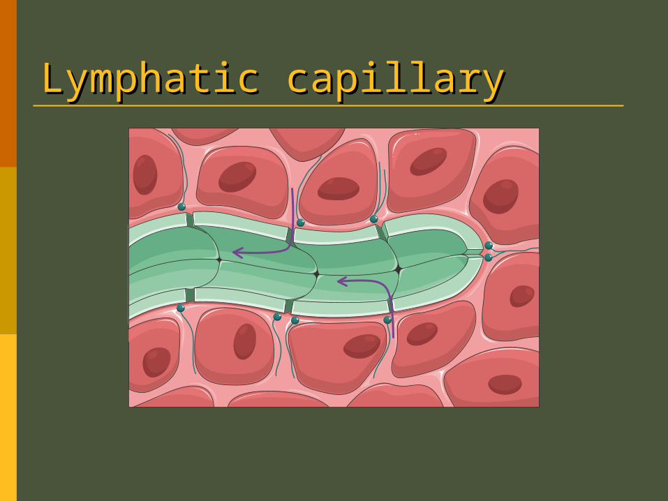
Lymphatic Capillaries Similar to blood capillaries, with
modifications Remarkably permeable Loosely joined endothelial minivalves Withstand interstitial pressure and remain open
The minivalves function as one-way gates that: Allow interstitial fluid to enter lymph capillaries Do not allow lymph to escape from the
capillaries
Lymphatic Capillaries
Lymphatic capillaryLymphatic capillary
Lymphatic Capillaries During inflammation, lymph capillaries can
absorb: Cell debris Pathogens Cancer cells
Cells in the lymph nodes: Cleanse and “examine” this debris
Lacteals – specialized lymph capillaries present in intestinal mucosa Absorb digested fat, mix it with lymph and
deliver it to the blood
Lymphatic Collecting Vessels
Have the same three tissue layers as veins
Have thinner walls, with more internal valves
come together or open into each other more frequently
Collecting vessels in the skin travel with superficial veins
Deep vessels travel with arteries Nutrients are supplied from branching
vasa vasorum (Small blood vessels that supply the walls of large vessels with oxygen and nutrients)
Lymphatic Trunks Lymphatic trunks are formed by the union
of the largest collecting ducts Major trunks include:
Paired lumbar, bronchomediastinal, subclavian, and jugular trunks
A single intestinal trunk
Lymphatic Trunks Lymph is delivered into one of two large
trunks Right lymphatic duct – drains the right upper
arm and the right side of the head and thorax Thoracic duct – arises from the cisterna chyli
and drains the rest of the body
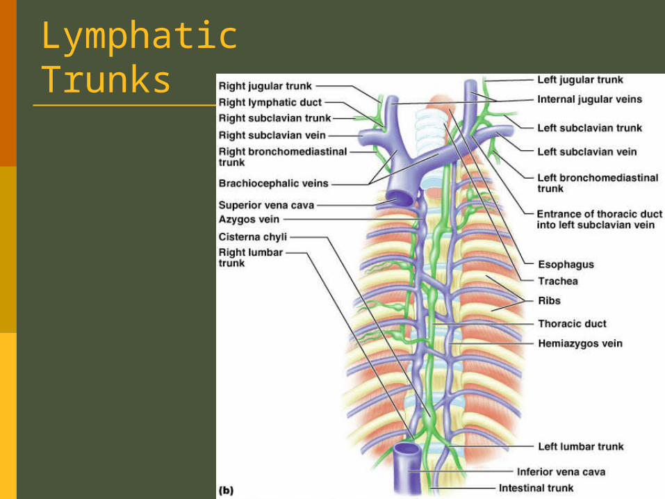
Lymphatic Trunks
Figure 20.2b
Lymph Transport The lymphatic system lacks an organ that
acts as a pump Vessels are low-pressure conduits Uses the same methods as veins to propel
lymph Pulsations of nearby arteries Contractions of smooth muscle in the walls of
the lymphatics
Lymphoid Cells Lymphocytes are the main cells involved
in the immune response The two main varieties are:
T cells B cells
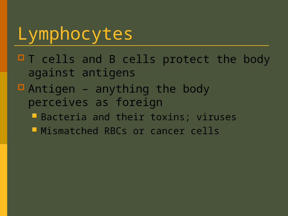
Lymphocytes T cells and B cells protect the body against
antigens Antigen – anything the body perceives as
foreign Bacteria and their toxins; viruses Mismatched RBCs or cancer cells
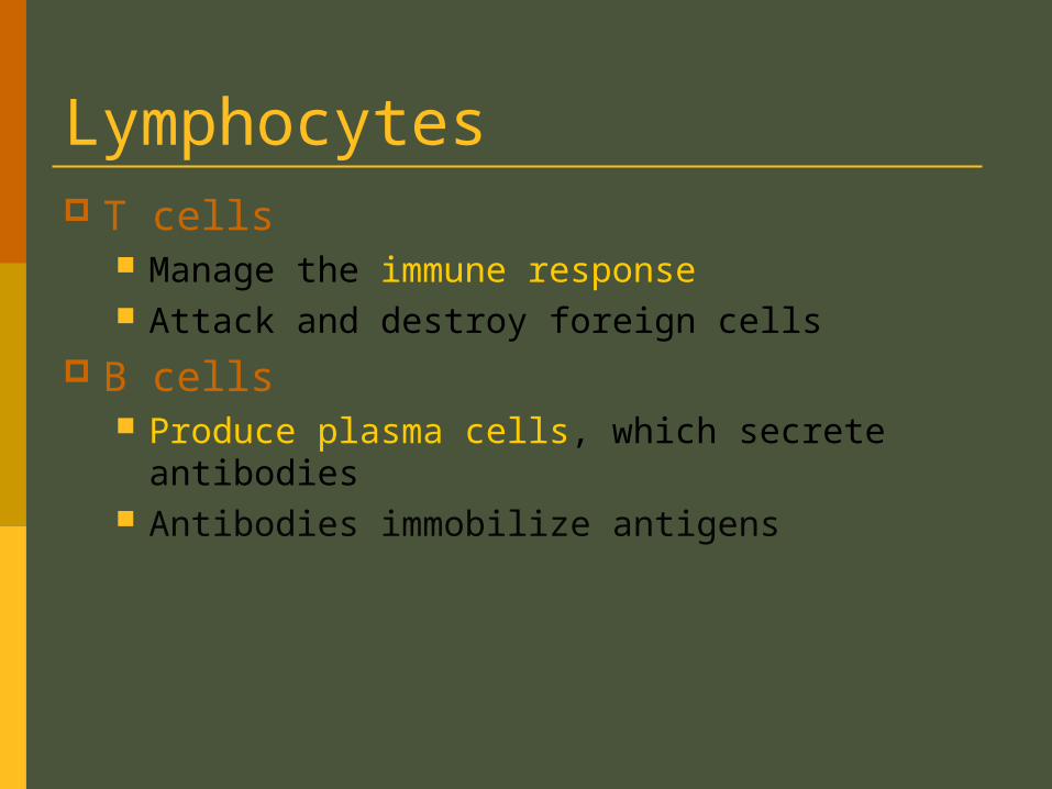
Lymphocytes T cells
Manage the immune response Attack and destroy foreign cells
B cells Produce plasma cells, which secrete antibodies Antibodies immobilize antigens
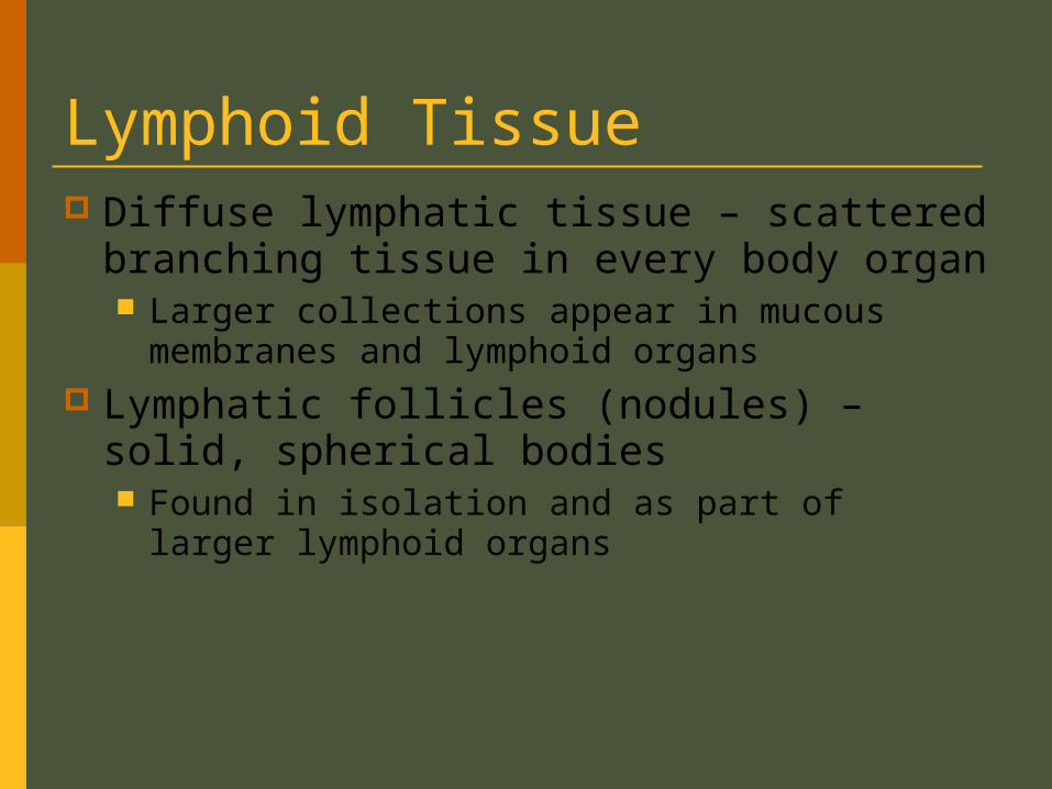
Lymphoid Tissue Diffuse lymphatic tissue – scattered
branching tissue in every body organ Larger collections appear in mucous
membranes and lymphoid organs Lymphatic follicles (nodules) – solid,
spherical bodies Found in isolation and as part of larger
lymphoid organs
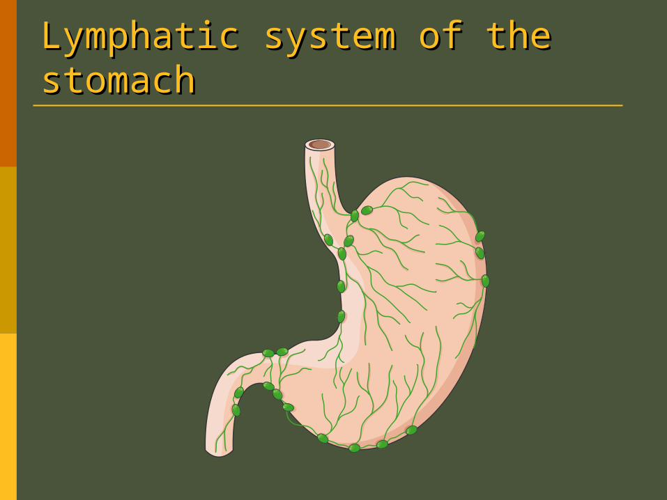
Lymphatic system of the Lymphatic system of the stomachstomach
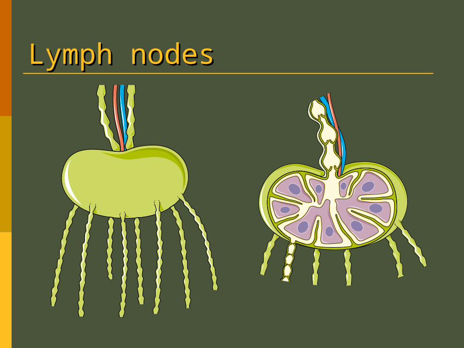
Lymph nodesLymph nodes
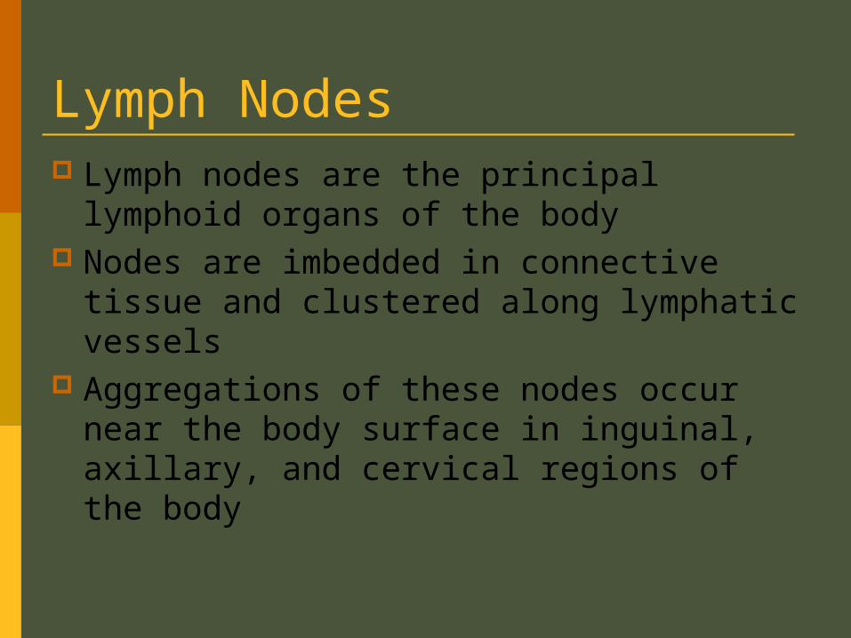
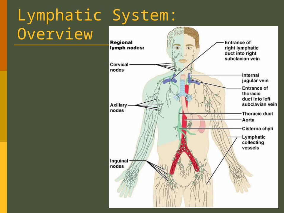
Lymph Nodes Lymph nodes are the principal lymphoid
organs of the body Nodes are imbedded in connective tissue
and clustered along lymphatic vessels Aggregations of these nodes occur near
the body surface in inguinal, axillary, and cervical regions of the body
Lymph Nodes Their two basic functions are:
Filtration – macrophages destroy microorganisms and debris
Immune system activation – monitor for antigens and mount an attack against them
Structure of a Lymph Node Nodes are bean shaped and surrounded by
a fibrous capsule
Nodes have two distinct regions: a cortex and a medulla
Structure of a Lymph Node The cortex contains follicles with germinal
centers, heavy with dividing B cells
The deep cortex houses T cells in transit T cells circulate continuously among the
blood, lymph nodes, and lymphatic stream
Structure of a Lymph Node Medullary cords extend from the cortex
and contain B cells, T cells, and plasma cells
Throughout the node are lymph sinuses crisscrossed by branching fibers Macrophages reside on these fibers and
phagocytize foreign matter
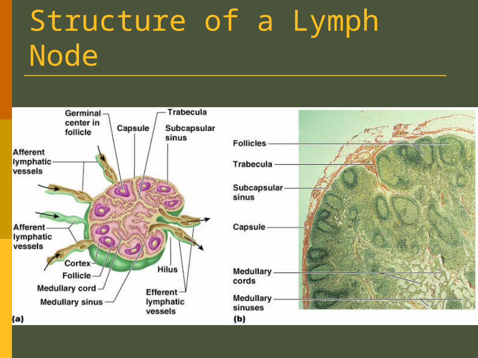
Structure of a Lymph Node
Circulation in the Lymph Nodes Lymph enters via a number of afferent
lymphatic vessels It then enters a large sinus and travels into
a number of smaller sinuses It meanders through these sinuses and
exits the node via efferent vessels Because there are fewer efferent vessels,
lymph stagnates somewhat in the node This allows lymphocytes and macrophages
time to carry out their protective functions
Other Lymphoid Organs The spleen, thymus gland, and tonsils Peyer’s patches and bits of lymphatic
tissue scattered in connective tissue All are composed of reticular connective
tissue and all help protect the body Only lymph nodes filter lymph
Spleen Largest lymphoid organ, located on the
left side of the abdominal cavity beneath the diaphragm
It extends to curl around the anterior part of the stomach
Functions Site of lymphocyte multiplication Immune surveillance and response Cleanses the blood
Additional Spleen Functions Stores breakdown products of RBCs for
later reuse Spleen macrophages salvage and store iron for
later use by bone marrow Site of fetal erythrocyte (RBC) production
(normally ceases after birth) Stores blood platelets
Structure of the Spleen Two distinct areas of the spleen are:
White pulp – area containing mostly lymphocytes suspended on reticular fibers and involved in immune functions
Red pulp – remaining splenic tissue concerned with disposing of worn-out RBCs and bloodborne pathogens
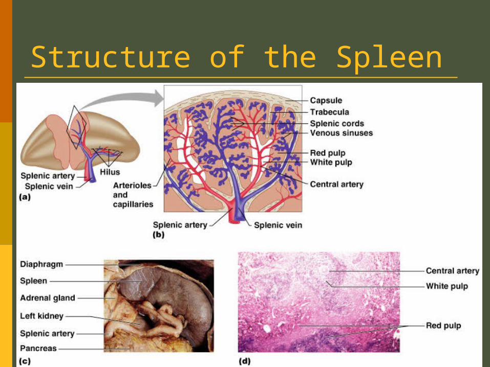
Structure of the Spleen
Figure 20.6a-d
Thymus A bi-lobed organ that secrets hormones
that cause T lymphocytes to become immunocompetent
The size of the thymus varies with age In infants, it is found in the inferior neck and
extends into the mediastinum where it partially overlies the heart
It increases in size and is most active during childhood
It stops growing during adolescence and then gradually atrophies
Tonsils Simplest lymphoid organs; form a ring of
lymphatic tissue around the pharynx Location of the tonsils
Palatine tonsils – either side of the posterior end of the oral cavity
Lingual tonsils – lie at the base of the tongue Pharyngeal tonsil – posterior wall of the
nasopharynx Tubal tonsils – surround the openings of the
auditory tubes into the pharynx
Tonsils Lymphoid tissue of tonsils contains follicles
with germinal centers Deep grooves, called crypts, trap and
destroy bacteria and particulate matter
Aggregates of Lymphoid Follicles Peyer’s patches – isolated clusters of
lymphoid tissue, similar to tonsils Found in the wall of the distal portion of the
small intestine Similar structures are found in the appendix
Peyer’s patches and the appendix: Destroy bacteria, preventing them from
breaching the intestinal wall Generate “memory” lymphocytes for long-term
immunity
Resources Anatomy Drill and Practice Practice Flashcards