Embed Size (px)
Citation preview
Magnesium calcium phosphate/b-tricalciumphosphate incorporation into gelatin scaffold:an in vitro comparative studyAhmed Hussain1*, Kazuhisa Bessho1, Katsu Takahashi1 and Yasuhiko Tabata21Department of Oral and Maxillofacial Surgery, Graduate School of Medicine, Kyoto University, Japan2Department of Biomaterials, Field of Tissue Engineering, Institute for Frontier Medical Sciences, Kyoto University, Japan
Abstract
Gelatin scaffolds incorporating or not 50wt% of magnesium calcium phosphate (MCP) or b-tricalciumphosphate (bTCP) were prepared and the in vitro osteogenic differentiation of rat bone marrowmesenchymal stem cells (MSCs) in the scaffolds was investigated. The pore sizes of the scaffolds werein the range 123.8�47.2–153�60.72mm in diameter, while the porosity was 33.3�2–44.9�3.4%.The compressionmodulus of the sponges was about 2.04–2.24mPa. There was no significant differenceamong groups regarding the physical and mechanical properties. When seeded into the sponges by anagitation method, the MSCs were distributed throughout the scaffold. Higher MSC proliferation wasobserved for scaffolds incorporatingminerals. Following the incubation ofMSCs in scaffolds incorporat-ing MCP, the alkaline phosphatase activity was significantly higher at weeks 2, 3 and 4 in comparisonwith other scaffolds; however, the osteocalcin levels ofMSCs did not show significant differences amonggroups. These findings indicate that MSCs seeded in scaffolds incorporating MCP showed significantlysuperior biological results in terms of proliferation and osteogenic differentiation in comparison withother scaffold types. Copyright © 2012 John Wiley & Sons, Ltd.
Received 11 October 2011; Revised 20 June 2012; Accepted 19 July 2012
Keywords gelatin scaffolds; MSCs; MCP; bTCP; in vitro; osteogenic differentiation
1. Introduction
One of the tissue engineering-based approaches to regen-erate bone lost to birth defects, trauma or disease is to useporous biodegradable scaffolds, which degrade at a ratesimilar to the rate of tissue regeneration, are osteoconduc-tive and have an open interconnected pore structure withpores large enough to allow bone ingrowth (> 100mm)and sufficient mechanical strength to support healingtissues (Logeart-Avramoglou et al., 2005; Stock andVacanti, 2001; Liu and Ma, 2004).
Gelatin is one of the materials extensively used in bio-medical fields. It is a biodegradable polymer that showseasy chemical modification and has been applied for
pharmaceutical, medical and food usage. One the otherhand, b-tricalcium phosphate (bTCP) is a biodegradableand osteoconductive material that has been extensivelyinvestigated as a scaffold for bone regeneration. Weprepared gelatin sponge with or without tricalciumphosphate as a scaffold for bone regeneration and foundthat the ALP activity and osteocalcin content of MSCs, asosteogenic differentiation markers, were highest forgelatin sponge incorporating 50wt% bTCP (Takahashiet al., 2005). For this reason, we chose gelatin spongeswith 50wt% bTCP as the positive control in this study.
Magnesium has recently received considerable attention.It is the fourth most abundant cation in the human bodyand is found naturally in bone (Straiger et al., 2006). Ithas been reported that magnesium is highly involved inbone formation and promotes cell attachment or spreadingon other surfaces (Revel et al., 2004). From the chemicalview point, the presence of magnesium ions can decreasethe crystallinity of calcium phosphate and increase thewater solubility of phosphates (Marchi et al., 2007).
*Correspondence to: A. Hussain, Department of Oral and Max-illofacial Surgery, Graduate School of Medicine, Kyoto University,54 Kawahara-cho, Shogoin, Sakyo-ku, Kyoto 606–8507, Japan.E-mail: [email protected]
Copyright © 2012 John Wiley & Sons, Ltd.
JOURNAL OF TISSUE ENGINEERING AND REGENERATIVE MEDICINE RESEARCH ARTICLEJ Tissue Eng Regen Med (2012)Published online in Wiley Online Library (wileyonlinelibrary.com) DOI: 10.1002/term.1596
Different cells have been used to test the biological func-tions of scaffolds. Among them are mesenchymal stem cells(MSCs), which are clinically popular in regenerative medi-cine because they can be readily isolated from the bonemarrow (Lennon et al., 2000). It is well recognized thatMSCs have an inherent potential to differentiate into celllineages of various types (Grigoriadis et al., 1988). Duringthe osteogenic differentiation of MSCs, it is known thatthey differentiate into osteoprogenitors, with a limitedself-renewal capacity, then to pre-osteoblasts, with limitedproliferation, and finally mature into osteoblasts, whichsecrete osteoid (Aubin and Triffitt, 2002).
The objective of this study was to evaluate and comparethe characteristics of gelatin sponges incorporating, or not,50wt% bTCP or MCP as a scaffold for MSCs from the view-point of their physical, mechanical and biological properties.
2. Materials and methods
2.1. Materials
A gelatin samplewith an isoelectric point (IEP) of 9.0 (BloomNo. 285g) was kindly supplied by Nitta Gelatin Co. (Osaka,Japan). Calcium dihydrogen phosphate and magnesiumoxide (90% and 99%, respectively) were obtained fromNacalai Tesque Ltd (Osaka, Japan) and used withoutfurther purification. bTCP granules (average diameter:2.89 mm, surface area 3.22m2/g, density 2.640, Ca:Pratio 1.50, purity 99.6%) were obtained from OlympusBiomaterials Co. Ltd (Tokyo, Japan). Culture media wereobtained from Invitrogen (Carlsbad, USA) .Other chemi-cals were obtained from Wako Pure Chemical Industries(Osaka, Japan).
2.2. Preparation of gelatin sponges incorporatingMCP or bTCP
Gelatin sponges containing 50wt% MCP or bTCP wereprepared, as were gelatin sponges with 0wt%, by thedehydrothermal crosslinking of gelatin. Briefly, calciumdihydrogen phosphate and magnesium oxide at a molarratio of 2:1 (Jia et al., 2010), or bTCP, were mixed with3wt% gelatin aqueous solution at 0 and 50wt%. Themixedsolutionwas agitated at 5000 rpm for 3min using a homog-enizer (ED-12, Nihonseiki Co., Tokyo, Japan). The resultingfoamy solution was cast into a polypropylene dish of138�138 cm and 5mm depth and then immediatelyfrozen at �80 �C. Then, the sponges were freeze-driedand dehydrothermally crosslinked at 140 �C for 96h.
2.3. Physical and chemical characterization ofgelatin sponges incorporating MCP or bTCP
The inner structure of the sponges was viewed using a scan-ning electron microscope (SEM; S2380N, Hitachi, Japan)after sputter-coating with gold/palladium. The percentage
porosity and mean diameter of the pores were determinedusing ImageJ v 1.43 (Wayne Rasband, National Institutesof Health, USA).
The pore size was determined by using the scale bar ofthe image as a reference. Percentage porosity was deter-mined by converting the image into binary vision mode(i.e. black and white), where the black represented thepore area while the white represented the gelatin spongearea; both areas were measured by ImageJ. By dividingthe black area to the white area and multiplying the resultby 100, we obtained the percentage porosity. More thanone picture for different areas was used.
The compression moduli of the freeze-dried gelatinsponges, 5� 5� 5mm in size, were measured using amechanical apparatus (AG-5000B, Shimadzu, Kyoto, Japan)at a rate of 1mm/min. The samples were dried at roomtemperature during the modulus of compression testing;no water or PBS was added to the samples under testing.A stress–strain curve was obtained and the compressionmoduli of the samples were calculated from the initial slopeof the load–deformation curve. Four samples were used foreach concentration to calculate the average value and thestandard deviation (SD) of the mean.
For determination of the calcium:phosphorus (Ca:P)ratio, each scaffold was immersed in 1ml 1 N HCL aqueoussolution for> 12h. Complete dissolution of the mineralsand degradation of gelatin were observed.
Calcium was determined quantitively using the CalciumE-Test Kit (Wako Pure Chemical Industries, Osaka, Japan).The molybdenum blue method was used to determine thephosphorus content of the scaffolds; the absorption wasmeasured at 820nm wavelength, using a Beckman CoulterDU 800 UV-visible spectrophotometer.
2.4. MSCs preparation and culture
MSCs were isolated from the bone shaft of femurs of3week-old male Fisher 344 rats, according to the tech-nique reported by Lennon et al. (1995). Briefly, bothends of rat femurs were cut away from the epiphysisand the bone marrow was flushed out by a syringe(21-gauge needle) with 1ml a-MEM supplemented with15 vol% fetal calf serum (FCS) and 50 IU/ml penicillinand streptomycin. The cell suspension (5ml) was placedinto a T-75 culture flask (Sumilon, Sumitomo BakeliteCo. Ltd, Tokyo, Japan). The medium was changed every3–4 days of culture. When the cells became subconflu-ent, they were detached by 0.25wt% trypsin, 0.02wt%ethylenediaminetetra-acetic acid (EDTA) and subcultured.Bone marrow cells of the third passage at subconfluencewere used for all the experiments.
2.5. MSCs seeding into gelatin spongesincorporating MCP or bTCP and culture
Gelatin sponges with or without MCP or bTCP were cutinto cylinders of 8mm diameter and 1.25� 0.25mm
A. Hussain et al.
Copyright © 2012 John Wiley & Sons, Ltd. J Tissue Eng Regen Med (2012)DOI: 10.1002/term
length, using a biopsy punch (Kai Industries Co. Ltd, Seki,Japan). MSCs were seeded into the sponges by theagitated seeding method because it was demonstrated thatthis method was effective in seeding cells homogeneouslythroughout three-dimensional (3D) porous scaffolds (Kimet al., 1999; Takahashi and Tabata, 2003). Briefly, 50ml cellsuspension (5�105 cells) was dropped onto each sponge,which had been placed into each well of 48-well multi-tissue culture plate (Iwaki Glass Co. Ltd, Chiba, Japan)and agitated on an orbital shaker (Bellco Glass Inc.) at180 rpm for 1h. Then, a further 1ml culture medium wasadded and the shaking was continued for a further 5 h.The cell-seeded sponges were placed into six-well tissueculture plates (3815–012, Iwaki Glass Co. Ltd). Thesponges were incubated in Dulbecco’s modified Eagle’smedium (DMEM) supplemented with 15 vol% FCS, 10nMdexamethasone, 50mg/ml ascorbic acid and 10mM b-glycerophosphate (osteogenic differentiation medium) at37 �C in a 5% CO2/95% air atmosphere. The medium waschanged twice a week. The number of sponges used foreach experimental group was two or three.
2.6. SEM observation of MSCs cultured ingelatin sponges incorporating MCP or bTCP
Gelatin sponges cultured with cells for 6 h were fixed with2.5wt% glutaraldehyde solution in 1� PBS, pH 7.4. Afterrinsing in PBS and subsequent dehydration with gradedaqueous ethanol solutions, the dehydrated samples wereimmersed in t-butanol and dried on a critical point dryer(ES-2030, Hitachi). After sputter-coating with gold/palla-dium, the samples were viewed by SEM.
2.7. Evaluation of cell behaviour after incubationin gelatin sponges incorporating MCP or bTCP
The number of MSCs attached to the gelatin sponges withor without mineral incorporation was determined by thefluorometric quantification of cellular DNA according tothe method reported by Rao and Otto (1992). Briefly,the cell-seeded sponges were lysed in 500 ml 30mM
sodium citrate-buffered saline solution (SSC), pH 7.4,containing 0.2mg/ml sodium dodecylsulphate (SDS),using a tissue lyser (Retsch Qiagen 85210) for 10min at20Hz, and then the samples were incubated at 37 �C for1 h. The cell lysates were centrifuged at 14 000 rpm and4 �C for 5min to separate the cell lysate from the spongeremnants. The cell lysate (100ml) was mixed with 100mldye solution (30mM SSC, 1mg/ml Hoechst 33258 dye)and the fluorescence intensity of the mixed solution wasmeasured by a fluorescence spectrometer (F-2000, Hitachi)at excitation and emission wavelengths of 355 and 460nm,respectively. The calibration curve between DNA and cellnumber was prepared by using cells of known numbers.As a measure of MSCs osteogenic differentiation, thealkaline phosphatase (ALP) and osteocalcin activity were
evaluated. ALP was determined by using the conventionalp-nitrophenylphosphate method (Kobayashi et al., 1996).The osteocalcin content was determined by enzyme-linkedimmunosorbent assay (ELISA). Briefly, MSCs culturedin sponges for different time periods were mixed with1ml 40 vol% formic acid for> 12 h to decalcify, using amixer (CM-1000, Eyela Co. Ltd, Miyagi, Japan). Afterthe decalcified samples had been centrifuged, the super-natant of cell extraction was subjected to gel filtrationusing a SephadexTM G-25 column (PD-10, AmershamPharmacia Biotech AB, Sweden). The resulting solutionwas freeze-dried, redissolved in double-distilled waterand subjected to a Rat Osteocalcin ELISA (BiomedicalTechnologies Inc., MA. USA).
2.8. Statistical analysis
All the data were analysed by one-way analysis of variance(ANOVA) with Tukey’s test to compare significance, andstatistical significance was accepted at p< 0.05. The exper-imental results were expressed as mean� SD.
3. Results
3.1. Characterization of gelatin spongesincorporating MCP or bTCP
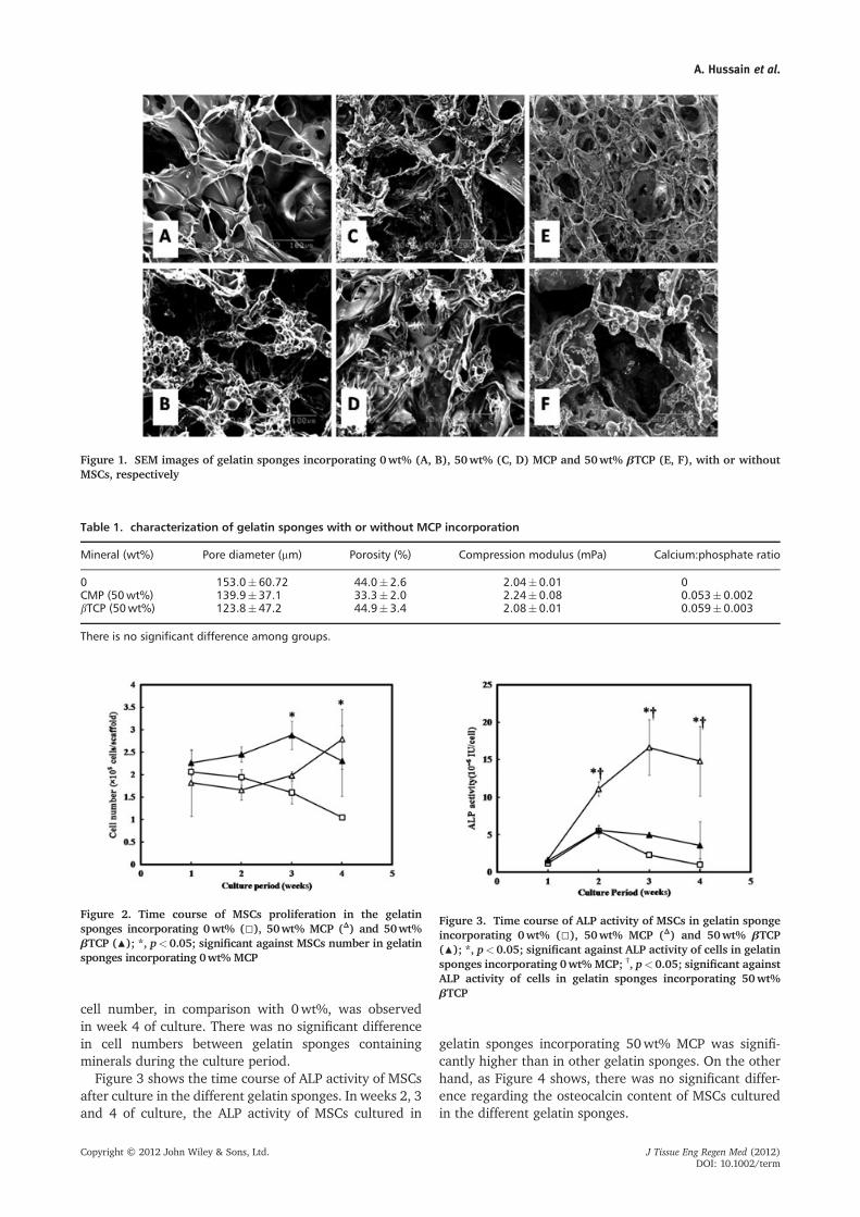
Figure 1 shows SEM images of gelatin sponges, with orwithout MCP or bTCP, both empty and cell-seeded.Irrespective of the mineral content, a similar infrastruc-ture was observed, in which every sponge had aninterconnected porous structure with the MCP or bTCPappearing to be incorporated into the matrix of the spongewith no precipitation or two-layer formation. The averagepore size range was 123.8� 47.2–153� 60.72mm, whilethe percentage porosity was 33.3� 2–44.9� 3.4%. Nosignificant difference was observed among groups re-garding pore size, percentage porosity or compressionmodulus, the latter in the range 2.04–2.24mPa. Simi-larly, no significant difference between groups wasobserved regarding the Ca:P ratio (Table 1). In the cell-seeded sponges it appeared that the cells becameattached and distributed with no morphological changesin all types of sponge (Figure 1).
3.2. MSCs proliferation and osteogenicdifferentiation in gelatin sponges incorporatingMCP or bTCP
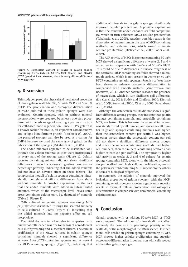
Figure 2 shows the number of MSCs proliferated in thegelatin sponges with or without MCP or bTCP. In week 1of culture, no significant difference was observed in cellnumbers among groups. However, in week 3 of culturegelatin sponges containing bTCP showed a significantdifference in comparison with the 0wt% sponges. Ingelatin sponges containing MCP a significant increase in
MCP/bTCP gelatin scaffolds: comparative study
Copyright © 2012 John Wiley & Sons, Ltd. J Tissue Eng Regen Med (2012)DOI: 10.1002/term
cell number, in comparison with 0wt%, was observedin week 4 of culture. There was no significant differencein cell numbers between gelatin sponges containingminerals during the culture period.
Figure 3 shows the time course of ALP activity of MSCsafter culture in the different gelatin sponges. In weeks 2, 3and 4 of culture, the ALP activity of MSCs cultured in
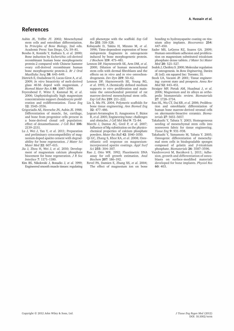
gelatin sponges incorporating 50wt% MCP was signifi-cantly higher than in other gelatin sponges. On the otherhand, as Figure 4 shows, there was no significant differ-ence regarding the osteocalcin content of MSCs culturedin the different gelatin sponges.
Figure 1. SEM images of gelatin sponges incorporating 0wt% (A, B), 50wt% (C, D) MCP and 50wt% bTCP (E, F), with or withoutMSCs, respectively
Table 1. characterization of gelatin sponges with or without MCP incorporation
Mineral (wt%) Pore diameter (mm) Porosity (%) Compression modulus (mPa) Calcium:phosphate ratio
0 153.0�60.72 44.0�2.6 2.04�0.01 0CMP (50wt%) 139.9�37.1 33.3�2.0 2.24�0.08 0.053�0.002bTCP (50wt%) 123.8�47.2 44.9�3.4 2.08�0.01 0.059�0.003
There is no significant difference among groups.
Figure 2. Time course of MSCs proliferation in the gelatinsponges incorporating 0wt% (□), 50wt% MCP (Δ) and 50wt%bTCP (▲); *, p<0.05; significant against MSCs number in gelatinsponges incorporating 0wt% MCP
Figure 3. Time course of ALP activity of MSCs in gelatin spongeincorporating 0wt% (□), 50wt% MCP (Δ) and 50wt% bTCP(▲); *, p<0.05; significant against ALP activity of cells in gelatinsponges incorporating 0wt% MCP; †, p<0.05; significant againstALP activity of cells in gelatin sponges incorporating 50wt%bTCP
A. Hussain et al.
Copyright © 2012 John Wiley & Sons, Ltd. J Tissue Eng Regen Med (2012)DOI: 10.1002/term
4. Discussion
This study compared the physical andmechanical propertiesof three gelatin scaffolds, 0%, 50wt% MCP and 50wt %bTCP. The proliferation and osteogenic differentiationof MSCs cultured in these gelatin sponges were alsoevaluated. Gelatin sponges, with or without mineralincorporation, were prepared by an easy one-step proce-dure, with the advantage of creating a porous structurefor cell-based bone regeneration. Since I.E.P.9 gelatin isa known carrier for BMP-2, an important osteoinductiveand ectopic bone-forming protein (Bessho et al., 2000),the prepared sponges can also be used as a carrier forBMP-2 because we used the same type of gelatin in thefabrication of the sponges (Takahashi et al., 2005).
The added minerals appeared to be distributed wellthrough the gelatin sponges as they were incorporatedin every part of the sponge walls (Figure 1). Gelatinsponges containing minerals did not show significantdifferences from other sponges regarding pore size orpercentage porosity, indicating that the added mineralsdid not have an adverse effect on these factors. Thecompression moduli of gelatin sponges containing miner-als did not show significant differences from thosewithout minerals. A possible explanation is the factthat the added minerals were added in sub-saturatedamounts, which at the microscopic level leaves someareas containing gelatin only, i.e. denuded of minerals(Table 1, Figure 1).
Cells cultured in gelatin sponges containing MCPor bTCP were distributed through the scaffold similarlyto cells cultured in 0wt% (Figure 1), indicating thatthe added minerals had no negative effect on cellmorphology.
The initial decrease in cell number in comparison withnumber of cells loaded was due to the loss of non-adherentcells during washing and subsequent culture. The cellularproliferation of the MSCs cultured in gelatin spongescontaining minerals showed a significant differenceat week 3 for bTCP-containing sponges and at week 4for MCP-containing sponges (Figure 2), indicating that
addition of minerals to the gelatin sponges significantlyimproved cellular proliferation. A possible explanationis that the minerals added enhance scaffold compatibil-ity, which in turn enhances MSCs cellular proliferation(Takahashi et al., 2005). Another possible reason is thedissolution of magnesium, in the case of MCP-containingscaffolds, and calcium ions, which would stimulatecellular proliferation (Dietrich et al., 2009; Sader et al.,2009).
The ALP activity of MSCs in sponges containing 50wt%MCP showed a significant difference at weeks 2, 3 and 4of culture in comparison with 0wt% and 50wt% BTCP.This could be due to differences in surface roughness ofthe scaffolds; MCP-containing scaffolds showed a micro-rough surface, which is not present in 0wt% or 50wt%BTCP-containing gelatin sponges. Rough surfaces havebeen shown to enhance osteogenic differentiation incomparison with smooth surfaces (Vandrovcová andBacáková, 2011). Another possible reason is the presenceof magnesium, which in turn facilitates cell differentia-tion (Lu et al., 2011; Sodek and Cheifetz, 2000; Dietrichet al., 2009; Sun et al., 2006; Qi et al., 2008; Feyerabendet al., 2006).
Although the osteocalcin results did not show a signif-icant difference among groups, they indicate that gelatinsponges containing minerals, and especially containingMCP, are better. This is because the osteocalcin contentwas standardized by cell number, and since the cell num-ber in gelatin sponges containing minerals was higher,then the osteocalcin content per scaffold was higher.In other words, since the osteocalcin content per celldid not show a significant difference among groups,and since the mineral-containing scaffolds had highercell numbers, then the mineral-containing scaffolds hadhigher osteocalcin per scaffold. The significantly higherALP activity at weeks 2, 3 and 4 of culture for gelatinsponge containing MCP, along with the higher osteocal-cin per scaffold and high cellular proliferation, makesthe gelatin scaffold containingMCP superior to the othersin terms of biological properties.
In summary, the addition of minerals improved thebiological properties of gelatin sponges, with the MCP-containing gelatin sponges showing significantly superiorresults in terms of cellular proliferation and osteogenicdifferentiation in comparison with zero mineral-containinggelatin sponges.
5. Conclusion
Gelatin sponges with or without 50wt% MCP or bTCPwere prepared. The addition of minerals did not affectadversely the pore size or percentage porosity of thescaffolds, or the morphology of the MSCs seeded. Further-more, cells seeded in gelatin sponges containing 50wt%MCP showed higher cellular proliferation and superiorosteogenic differentiation in comparison with cells seededin the other gelatin sponges.
Figure 4. Osteocalcin content of MSCs in gelatin spongecontaining 0wt% (white), 50wt% MCP (black) and 50wt%bTCP (grey) at 2 and 4weeks; there is no significant differenceamong groups
MCP/bTCP gelatin scaffolds: comparative study
Copyright © 2012 John Wiley & Sons, Ltd. J Tissue Eng Regen Med (2012)DOI: 10.1002/term
References
Aubin JE, Triffitt JT. 2002; Mesenchymalstem cells and osteoblast differentiation.In Principles of Bone Biology, 2nd edn.Academic Press: San Diego, CA; 59–81.
Bessho K, Konishi Y, Kaihara S, et al. 2000;Bone induction by Escherichia coli-derivedrecombinant human bone morphogeneticprotein-2 compared with Chinese hamsterovary cell-derived recombinant humanbone morphogenetic protein-2. Br J OralMaxillofac Surg 38: 645–649.
Dietrich E, Oudadesse H, Lucas-Girot A, et al.2009; In vitro bioactivity of melt-derivedglass 46 S6 doped with magnesium. JBiomed Mater Res A 88: 1087–1096.
Feyerabend F, Witte F, Kammal M, et al.2006; Unphysiologically high magnesiumconcentrations support chondrocyte prolif-eration and redifferentiation. Tissue Eng12: 3545–3556.
Grigoriadis AE, Heersche JN, Aubin JE. 1988;Differentiation of muscle, fat, cartilage,and bone from progenitor cells present ina bone-derived clonal cell population:effect of dexamethasone. J Cell Biol 106:2139–2151.
Lu J, Wei J, Yan Y, et al. 2011; Preparationand preliminary cytocompatibility of mag-nesium doped apatite cement with degrad-ability for bone regeneration. J Mater SciMater Med 22: 607–615.
Jia J, Zhou H, Wei J, et al. 2010; Develop-ment of magnesium calcium phosphatebiocement for bone regeneration. J R SocInterface 7: 1171–1180.
Kim BS, Nikolovski J, Bonadio J, et al. 1999;Engineered smoothmuscle tissues: regulating
cell phenotype with the scaffold. Exp CellRes 251: 318–328.
Kobayashi D, Takita H, Mizuno M, et al.1996; Time-dependent expression of bonesialoprotein fragments in osteogenesisinduced by bone morphogenetic protein.J Biochem 119: 475–481.
Lennon DP, Haynesworth SE, Arm DM, et al.2000; Dilution of human mesenchymalstem cells with dermal fibroblasts and theeffects on in vitro and in vivo osteochon-drogenesis. Dev Dyn 219: 50–62.
Lennon DP, Haynesworth SE, Young RG,et al. 1995; A chemically defined mediumsupports in vitro proliferation and main-tains the osteochondral potential of ratmarrow-derived mesenchymal stem cells.Exp Cell Res 219: 211–222.
Liu X, Ma PX. 2004; Polymeric scaffolds forbone tissue engineering. Ann Biomed Eng32: 477–486.
Logeart-Avramoglou D, Anagnostou F, BiziosR, et al. 2005; Engineering bone: challengesand obstacles. J Cell Mol Med 9: 72–84.
Marchi J, Dantas AC, Greil P, et al. 2007;Influence of Mg-substitution on the physico-chemical properties of calcium phosphatepowders. Mater Res Bull 42: 1040–1050.
Qi GC, Zhang S, Khor KA, et al. 2008; Oste-oblastic cell response on magnesium-incorporated apatite coatings. Appl SurfSci 255: 304–307.
Rao J, Otto WR. 1992; Fluorimetric DNAassay for cell growth estimation. AnalBiochem 207: 186–192.
Revel PA, Damien E, Zhang XS, et al. 2004;The effect of magnesium ion on bone
bonding to hydroxyapatite coating on tita-nium alloy implants. Bioceramics 254:447–450.
Sader MS, LeGeros RZ, Soares GA. 2009;Human osteoblasts adhesion and prolifera-tion on magnesium substituted tricalciumphosphate dense tablets. J Mater Sci MaterMed 20: 521–527.
Sodek J, Cheifetz S. 2000;Molecular regulationof osteogenesis. In Bone Engineering, DaviesJE (ed). em squared Inc: Toronto; 31.
Stock UA, Vacanti JP. 2001; Tissue engineer-ing: current state and prospects. Annu RevMed 52: 443–451.
Straiger MP, Pietak AM, Huadmai J, et al.2006; Magnesium and its alloys as ortho-pedic biomaterials: review. Biomaterials27: 1728–1734.
Sun HL, Wu CT, Dai KR, et al. 2006; Prolifera-tion and osteoblastic differentiation ofhuman bone marrow-derived stromal cellson akermanite-bioactive ceramics. Bioma-terials 27: 5651–5657.
Takahashi Y, Tabata Y. 2003; Homogeneousseeding of mesenchymal stem cells intononwoven fabric for tissue engineering.Tissue Eng 9: 931–938.
Takahashi Y, Yamamoto M, Tabata Y. 2005;Osteogenic differentiation of mesenchy-mal stem cells in biodegradable spongescomposed of gelatin and b-tricalciumphosphate. Biomaterials 26: 3587–3596.
Vandrovcová M, Bacáková L. 2011; Adhe-sion, growth and differentiation of osteo-blasts on surface-modified materialsdeveloped for bone implants. Physiol Res60: 403.
A. Hussain et al.
Copyright © 2012 John Wiley & Sons, Ltd. J Tissue Eng Regen Med (2012)DOI: 10.1002/term