Embed Size (px)
Citation preview

UNIVERSIDADE FEDERAL DE GOIÁS PROGRAMA DE PÓS-GRADUAÇÃO EM MEDICINA TROPICAL
E SAÚDE PÚBLICA
MARIELLE GARCIA SILVA
ANÁLISE PROTEÔMICA DE MEMBRANAS DE Paracoccidioides sp. DURANTE PRIVAÇÃO DE ZINCO
Orientadora: Dra. Célia Maria de Almeida Soares
Goiânia 2014
2

iii
UNIVERSIDADE FEDERAL DE GOIÁS PROGRAMA DE PÓS-GRADUAÇÃO EM MEDICINA TROPICAL
E SAÚDE PÚBLICA
MARIELLE GARCIA SILVA
ANÁLISE PROTEÔMICA DE MEMBRANAS DE Paracoccidioides sp. DURANTE PRIVAÇÃO DE ZINCO
Dissertação apresentada ao Programa de Pós-Graduação em Medicina Tropical e Saúde Pública da Universidade Federal de Goiás como requisito para obtenção do Título de Mestre em Medicina Tropical e Saúde Pública.
Orientador: Prof. Dra. Célia Maria de Almeida Soares
Goiânia 2014

iv

v

vi
Programa de Pós-Graduação em Medicina Tropical e Saúde Pública
da Universidade Federal de Goiás
BANCA EXAMINADORA DA DISSERTAÇÃO DE MESTRADO
Aluno (a): Marielle Garcia Silva
Orientador (a): Profª. Dra. Célia Maria de Almeida Soares
DATA: 04/08/2014
MEMBROS Dr. Célia Maria de Almeida Soares
Instituto de Ciências Biológicas, Universidade Federal de Goiás
Dra. Luciana Casaletti
Instituto de Ciências Biológicas, Universidade Federal de Goiás Dra. Patrícia de Sousa Lima
Instituto de Ciências Biológicas, Universidade Federal de Goiás

vii
Dedico este trabalho ás pessoas mais importantes da minha vida...
... meus pais José e Lucélia, que sempre fizeram de tudo para me oferecer boa educação e sempre me
apoiaram nas decisões tomadas. ... à minha irmã Mirelle, que sempre me ajudou nas
tarefas e decisões. Obrigada pelo apoio! Se não fossem vocês, hoje não estaria
onde estou! Obrigada por tudo! Amo vocês!

viii
AGRADECIMENTOS
A Deus, pela oportunidade da vida e por me abençoar a cada dia.
À minha orientadora Célia Maria de Almeida Soares, por me receber no seu grupo de
trabalho. Agradeço pela orientação e pela confiança em mim depositada. Pelo esforço
em oferecer aos seus alunos condições para realização de trabalho de qualidade. Admiro
sua competência e profissionalismo na coordenação do laboratório.
Ao meu cunhado Alexandre, que nunca negou ajuda durante a realização dos
experimentos. Obrigada pelo apoio, pelas sugestões e pela amizade.
Aos professores do Laboratório de Biologia Molecular, da Universidade Federal de
Goiás, Maristela Pereira, Alexandre Bailão, Clayton Borges Juliana Parente e Sílvia
Salém-Izzac, pelo auxílio oferecido.
Aos membros da banca de qualificação, Patrícia Lima, Lilian Baeza e Clayton Borges
pelas sugestões e contribuições.
À Juliana Santana, pelo companheirismo durante a realização dos experimentos e das
discussões. Obrigada pela amizade profissional e pessoal.
À Luciana, pela amizade, pelos conselhos e momentos de diversão. Obrigada pela ajuda
durante a realização dos experimentos e por toda contribuição durante esses anos. Muito
obrigada Lu.
À Patrícia Lima, Elisa Flávia, Lucas Nojosa e Mariana Tomazett, pelos momentos de
descontração, que ajudam a aliviar o cansaço durante o trabalho, e pela disponibilidade
em sempre me ajudar. Obrigada a todos vocês!
À Edilânia, Amanda, Danielle, Alessandro, Karla, Gabriel, Leandro Prado, André,
Vanessa, Luiz Paulo, Igor, Fabiana, Leandro, Paula Francienete, Laura, Diandra,
Laurine, Wesley, Lívia, Joyce, Kleber. Agradeço pelo convívio e contribuições.
À todos os membros da minha família pelo apoio e palavras de incentivo. Obrigada
pelas orações!

ix
SUMÁRIO
Pág.
LISTA DE ABREVIATURAS
xi
RESUMO
xii
ABSTRACT
xiii
1. INTRODUÇÃO .............................................................................................. 14
1.1. Paracoccidioides spp. e Paracoccidioidomicose – Aspectos gerais .......... 14
1.2. Membranas celulares ................................................................................. 19
1.3. Importância dos íons metálicos em processos celulares ............................ 26
2. JUSTIFICATIVA ........................................................................................... 33
3. OBJETIVOS ................................................................................................... 34
3.1 Geral ........................................................................................................... 34
3.2 Específicos ................................................................................................. 34
4. MANUSCRITO .............................................................................................. 35
5. DISCUSSÃO ................................................................................................... 99
6. CONCLUSÃO................................................................................................. 102
7. PERSPECTIVAS ........................................................................................... 103
8. REFERÊNCIAS ............................................................................................. 104

x
LISTA DE FIGURAS E TABELAS Dissertação Pág. Figura 1: Morfologia de Paracoccidioides spp.
13
Figura 2: Distribuição geográfica da paracoccidioidomicose.
15
Figura 3: Aspectos clínicos da PCM.
16
Figura 4: Estrutura da membrana celular.
17
Figura 5: Associação das proteínas à membrana celular.
19
Figura 6: Proteínas de membrana ligadas a lipídeos.
21
Figura 7: Estrutura geral de uma âncora GPI ligando uma proteína à membrana plasmática.
23
Figura 8: Modelo do transporte de zinco.
27
Figura 9: Alinhamento de sequências de aminoácidos de S. cerevisiae, Paracoccidioides spp. e espécies de Cryptococcus.
28
Manuscrito
Figura 1: Microscopia Eletrônica de Transmissão (MET) do extrato de membrana de Paracoccidioides sp.
45
Figura 2:
Porcentagem de proteínas de membrana identificadas. 46
Figura 3: Proteínas de membrana reguladas.
46
Figure 4: Classificação funcional das proteínas de membrana.
47
Tabela 1: Proteínas de membrana identificadas em Paracoccidioides sp. na presença ou na ausência de zinco.
48
Tabela 2: Proteínas de membrana de Paracoccidioides sp. reprimidas em privação de zinco e suas funções biológicas.
56
Tabela 3: Proteínas de membrana de Paracoccidioides sp. induzidas em privação de zinco e suas funções biológicas.
63
Figura suplementar – 1 Tipo de detecção dos peptídeos para amostras do controle e de privação de zinco.
65
Figura suplementar – 2 Faixa de detecção dinâmica dos experimentos.
66
Figura suplementar – 3 Localização subcelular das proteínas identificadas
67
Tabela suplementar – 1 Proteínas reguladas em células de levedura de Paracoccidioides sp. sob privação de zinco e as suas funções biológicas previstas.
68

xi
LISTA DE ABREVIATURAS PCM: Paracoccidioidomicose MET: Microscopia eletrônica de transmissão S1: Espécie 1 PS2: Espécie filogenética 2 PS3: Espécie filogenética 3 PS4: Espécie filogenética 4 DTT: Ditiotreitol TPEN: N,N,N_,N_- tetrakis (2-pyridyl-methyl) ethylenediamine - Quelante
específico de zinco PIM: Proteínas integrais de membrana GPI: Glicosilfosfatidilinositol MPTs: Modificações pós-traducionais PATs: Palmitoil-transferases FTase: Farnesiltransferase Zrt1: Transportador de zinco de alta afinidade Zrt2: Transportador de zinco de baixa afinidade Irt1: Transportador de ferro CDF: Facilitador de difusão de cátions GM-CSF: Fator estimulante de colônias de granulócitos e macrófagos PA: Ácido fosfatídico LPA: Ácido lisofosfatídico 1-AGPAT: 1-acil-sn-glicerol-3-phosphate aciltransferase beta PS: Fosfatidilserina PI: Fosfatidilinositol PE: Fosfatidiletanolamina PC: Fosfatidilcolina Sac1: Fosfatase fosfoinositidica Dpm1: Dolicol fosfato manose sintase Mkc1: Proteína quinase ativada por mitógeno MMcM: Meio mínimo Mc Veigh Morton AF: Ammonium formate PLGS: ProteinLynx Global Server UPLC-MSE: Cromatografia de ultraperformace acoplada à espectrometria de massas

xii
RESUMO
Paracoccidioides spp. são fungos patogênicos, causadores da paracoccidioidomicose,
uma importante micose sistêmica nos países latino-americanos. O sucesso da infecção
depende da capacidade do patógeno obter micronutrientes essenciais (metais) a partir do
hospedeiro. Este processo de captação de nutrientes ocorre através de transportadores
associados às membranas. As membranas são constituídas por uma bicamada lipídica
com proteínas associadas e estão envolvidas em diferentes processos durante o
estabelecimento da infecção, como transporte de nutrientes e regulação homeostática da
célula. Como o zinco é um metal que desempenha um papel importante na regulação da
interação patógeno-hospedeiro e distúrbios na homeostase deste micronutriente estão
implicados na patogênese de doenças infecciosas, o objetivo deste estudo foi identificar
as proteínas de membranas expressas em condições de privação de zinco. A técnica
NanoUPLC-MSE foi empregada para identificar proteínas de membranas de células de
levedura (Pb01) cultivadas em meio quimicamente definido na presença e ausência de
zinco. Microscopia eletrônica de transmissão foi realizada para confirmar o
enriquecimento da amostra com membranas. Posteriormente, as amostras foram
digeridas e analisadas por NanoUPLC-MSE. Análises in silico foram realizadas para
determinação da localização de 460 proteínas obtidas do extrato proteico de Pb01
cultivado em meio com zinco (controle) e TPEN (tratado). Do total de proteínas
identificadas, 141 proteínas foram classificadas como pertencentes a membranas e
destas, 120 proteínas foram reprimidas e 21 foram induzidas durante privação de zinco.
Dentre as 141 proteínas de membranas, 81 apresentaram domínio transmembrana e 9
foram classificadas como sendo de membrana, em decorrência de modificação pós-
traducional. Um total de 15 proteínas de membrana apresentou peptídeo sinal. Análises
da função das proteínas de membranas permitiu a descrição de que o metabolismo de
fosfolipídeos e a integridade celular foram afetados pela privação de zinco.
Palavras chave: Proteoma, proteínas de membranas, privação de zinco

xiii
ABSTRACT
Paracoccidioides spp. are pathogenic fungi that causes paracoccidioidomycosis, an
important systemic mycosis in Latin American countries. The success of infection
depends on the pathogen ability to obtain essential micronutrients (metals) from the
host. This process of nutrient uptake occurs through membrane associated transporters.
The membranes are constituted of a lipid bilayer with associated proteins and are
involved in different processes during the establishment of infection, such as transport
of nutrients and homeostatic regulation. As zinc is a metal that plays an important role
in the regulation of host-pathogen interaction and changes in this micronutrient
homeostasis are implicated in the pathogenesis of infectious diseases, the aim of this
study was to identify the membrane proteins expressed under conditions of zinc
deprivation. NanoUPLC-MSE technique was employed in order to identify membrane
proteins of yeast cells (Pb01) grown in chemically defined media in the presence and
absence of zinc. Transmission electron microscopy was performed to confirm the
sample enrichment with membranes. Subsequently, the samples were digested and
analyzed by NanoUPLC-MSE. In silico analysis was performed to determine the
location of 460 proteins identified in extracts of Pb01 grown in + Zn (control) and
TPEN (treated) medium. Among the identified proteins, 141 were classified as
belonging to membranes and of those, 120 proteins were repressed and 21 were induced
during zinc deprivation. Among the 141 membrane proteins, 81 showed transmembrane
domains and 9 were classified as membranes proteins with post-transcriptional
modification. A total of 15 membrane proteins showed signal peptide. Analysis of the
function of membrane proteins, allowed the description that phospholipid metabolism
and cell integrity maintenance pathways were affected by zinc deprivation.
Keywords: Proteome, membranes proteins, zinc deprivation

14
1. INTRODUÇÃO
1.1 Paracoccidioides spp. e Paracoccidioidomicose – Aspectos gerais
Paracoccidioides spp. são fungos patogênicos, causadores da
paracoccidioidomicose (PCM), uma importante micose sistêmica nos países Latino-
Americanos. O fungo pertence ao filo Ascomicota, ordem Onygenales e família
Ajellomycetaceae (Onygenaceae). Análises filogenéticas de isolados de
Paracoccidioides spp. resultaram na diferenciação do gênero em duas espécies:
Paracoccidioides brasiliensis e Paracoccidioides lutzii. De acordo com dados do
sequenciamento nucleotídico de 8 loci de 65 isolados, o complexo P. brasiliensis é
constituído por três linhagens filogenéticas: S1 (com 38 isolados), PS2 (com seis
isolados, sendo 5 de origem brasileira e 1 de origem venezuelana) e PS3 (com 21
isolados colombianos). Recentemente foi identificada uma nova linhagem filogenética,
PS4, que inclui 5 isolados clínicos da Venezuela (SALGADO-SALAZAR et al., 2010;
BOCCA et al., 2013). O complexo P. lutzii representa os isolados semelhantes ao Pb01
(isolado 1 de Paracoccidioides lutzii), que apresentaram diferenças morfológicas e
genômicas em relação aos outros isolados classificados como P. brasiliensis (MATUTE
et al., 2006; CARRERO et al., 2008; TEIXEIRA et al., 2009; TEIXEIRA et al., 2014).
Os membros do complexo P. brasiliensis estão distribuídos entre Brasil, Argentina,
Uruguai, Paraguai, Peru e Colômbia. Já os membros do complexo P. lutzii foram
encontrados na região Centro-Oeste do Brasil, Rondônia e Equador (MATUTE et al.,
2006; TEIXEIRA et al., 2009; TEIXEIRA et al., 2014).
Alguns organismos são capazes de ocupar mais de um nicho ecológico durante
seu ciclo de vida, como é o caso dos fungos patogênicos termodimórficos. Durante a
fase de micélio (saprófita) o fungo está sob a influência de diferentes condições, como
mudança frequente de temperatura e umidade e em competição com outros micro-
organismos. Já durante a fase de levedura, estes patógenos se adaptam a um habitat
diferente, tais como os órgãos internos do corpo (influência da temperatura, hormônios
e do sistema imunológico) (BAGAGLI et al., 2008). Paracoccidioides spp. são
encontrados na forma miceliana no meio ambiente (solo) ou em condições in vitro nas
temperaturas de 22ºC-28ºC, enquanto nos tecidos do hospedeiro ou quando cultivado
entre 35ºC-37ºC, é encontrado como levedura (BAGAGLI et al., 2006). Em relação à

15
morfologia, micélio e levedura de ambas as espécies do gênero Paracoccidioides são
similares. Ambas as formas são multinucleadas, enquanto os conídios produzidos pelo
micélio possuem só um núcleo (CANO et al., 1998; MCEWEN et al., 1987b).
Microscopicamente, na fase de levedura as colônias de Paracoccidioides spp. são
caracterizadas por múltiplos brotamentos originados de uma célula-mãe, a qual é central
e circundada por células periféricas menores, apresentando uma estrutura semelhante a
uma roda de leme de navio (Figura 1A). A forma miceliana é caracterizada por hifas
septadas com a presença de conídios terminais ou intercalares (Figura 1C)
(RESTREPO-MORENO, 2003; QUEIROZ-TELLES, 1994). Macroscopicamente, as
colônias de levedura são rugosas e amareladas (Figura 1B); já as colônias de micélio são
pequenas, irregulares e com aspecto algodonoso (Figura 1D) (BRUMMER et al., 1993).
Figura 1: Morfologia de Paracoccidioides spp. : Características microscópicas: A- Levedura,
C- Micélio. Aspecto de colônias crescidas à 37ºC (B – Levedura) e 25ºC (D – Micélio).
Aumento: A e B – 40 vezes. Fonte: Laboratório de Biologia Molecular, Universidade Federal de
Goiás; Sturme et al. (2011); Lacaz et al., (1999).
Paracoccidioides spp. já foram isolados de tatus (Dasypus novemcinctus e
Cabassous centralis) e detectados em animais domésticos, como cachorro (BAGAGLI
et al., 1998; RICCI et al., 2004; CORREDOR et al., 1999). Por poderem ser acometidos

16
pela doença, estes organismos devem ser considerados hospedeiros acidentais e não
reservas naturais do fungo (CONTI DIAZ, 2007). No solo, em amostras recolhidas ao
redor e dentro da toca dos tatus, o fungo foi detectado através de métodos moleculares
(THEODORO et al., 2005). Os tatus podem infectar-se várias vezes, pois ficam em
contato constante com o solo e possuem uma resposta imunológica celular fraca
(RICHINI-PEREIRA et al., 2009). Infecção por P. brasiliensis também já foi reportada
em porcos domésticos, mas o fungo não foi isolado dos animais (BELITARDO et al.,
2014). A identificação de Paracoccidioides spp. em 20% dos tatus examinados da
região de Tucuruí (Pará) sugere que o animal possa exercer algum papel no ciclo natural
do fungo (NAIFF et al., 1986).
A paracoccidioidomicose afeta principalmente trabalhadores rurais do sexo
masculino que estão em contato direto com o solo. A doença é restrita às Américas
Central e do Sul, com maior prevalência no Brasil, Colômbia, Venezuela e Argentina
(Figura 2). No Brasil, entre 1980 e 1995, a PCM foi considerada a oitava causa de morte
por doença infecciosa e entre as micoses sistêmicas apresenta taxa de mortalidade média
anual de 1,45/milhão de habitantes. As regiões Sudeste (São Paulo, Minas Gerais e Rio
de Janeiro) e Sul (Paraná e Rio Grande do Sul) apresentaram maior índice de
mortalidade. Acredita-se que a incidência anual em áreas endêmicas varie de 3 a 4
novos casos/milhão (PRADO et al., 2009; COUTINHO et al., 2002; BRUMMER et al.,
1993; SHIKANAI-YASUDA et al., 2006).
A principal via de infecção por Paracoccidioides spp. é a inalação (ALMEIDA,
2005). Os conídios e/ou propágulos produzidos pela forma miceliana, após serem
inalados e atingirem os alvéolos pulmonares, transitam para levedura, a forma
parasitária (MCEWEN et al., 1987a). Após a transição, o fungo pode disseminar-se
pelas vias linfáticas e/ou hematogênicas, para outros locais do organismo hospedeiro,
podendo ser fatal para o paciente (CAMARGO e FRANCO, 2000; VALERA et al.,
2008). A PCM é predominante no sexo masculino, com razão de 10 a 12 homens para 1
mulher, em adultos (SHIKANAI-YASUDA et al., 2006). Esta diferença é explicada
pela presença de hormônios esteróides, que inibem a transição de micélio ou conídio
para levedura (LOOSE et al., 1983). Análise da transição dimórfica de isolados de
Paracoccidioides spp. na presença de 17β-estradiol demonstraram que o número de
células que transitaram para a forma parasitária foi menor que na ausência do hormônio
(RESTREPO et al., 1984). Em estudo realizado com camundongos foi demonstrado o
bloqueio da transição dimórfica em fêmeas (ARISTIZABAL et al., 1998). Além disso,

17
análises transcricionais indicaram que as ações inibitórias de 17β-estradiol são
decorrentes da ação de genes sinalizadores que regulam o dimorfismo (SHANKAR et
al., 2011).
Figura 2: Distribuição geográfica da paracoccidioidomicose. Fonte: Shikanai-
Yasuda et al. 2006.
A classificação clínica da PCM, definida no Colóquio Internacional de
Paracoccidioidomicose em 1986, inclui: PCM infecção e PCM doença (forma
aguda/subaguda e forma crônica). Na PCM infecção o paciente não apresenta sinais ou
sintomas da doença mas possui reatividade positiva à paracoccidioidina (antígeno)
(FRANCO et al., 1987). A forma aguda (tipo juvenil) possui evolução mais rápida e
severa, representa apenas 3 a 5% de todos os casos e é caracterizada pelo envolvimento
do sistema fagocítico mononuclear (linfonodos, fígado, baço e medula óssea) (Figura
3A). Predomina em crianças e adolescentes e sua distribuição é semelhante em crianças
do gênero feminino e masculino. O quadro clínico é caracterizado por hipertrofia do
sistema retículo-endotelial, disfunção da medula óssea, linfadenomegalia, manifestações
digestivas, hepatoesplenomegalia, envolvimento ósteo-articular e lesões cutâneas
(LONDERO e MELO, 1983; SHIKANAI-YASUDA et al., 2006). Dependendo do grau
de disseminação, a forma aguda pode ser subdividida em grave ou moderada. A forma

18
grave acomete principalmente órgãos linfáticos, fígado, baço e medula óssea. Já a forma
moderada acomete somente um sistema ou uma cadeia linfática (FRANCO et al., 1987).
A PCM crônica apresenta evolução mais lenta, podendo levar anos até ser
diagnosticada. Ocorre em mais de 90% dos casos e acomete adultos entre 30 e 60 anos,
predominantemente do sexo masculino. O quadro clínico é caracterizado por
manifestações pulmonares, presentes em 90% dos pacientes. Quando a infecção
acomete somente um órgão é chamada de apresentação unifocal e quando está presente
em mais de um órgão (pulmão, mucosa e pele) é chamada de multifocal. Os pulmões
são os órgãos mais afetados, seguidos pela pele e membranas mucosas, oral e nasal
principalmente (Figura 3B) (FRANCO et al., 1987; SHIKANAI-YASUDA et al., 2006).
Os cinco principais aspectos patológicos descritos por Tuder e colaboradores (1985) em
pacientes com PCM crônica foram: reação pneumônica, formação granulomatosa
precoce, granulomas maduros, padrão misto (granuloma precoce e maduro na mesma
área pulmonar) e fibrose pulmonar.
A cura espontânea não ocorre na PCM, por isso é necessária a realização do
tratamento que inclui o uso de drogas antifúngicas, suporte nutricional, tratamento de
eventual sequelas e prevenção de doenças oportunistas (SILVA e SARAIVA, 2008). As
drogas antifúngicas utilizadas para o tratamento da PCM são: anfotericina B,
sulfamídicos (sulfadiazina, associação sulfametoxazol/trimetoprim, sulfadoxina) e
azólicos (cetoconazol, fluconazol, itraconazol). A avaliação da resposta do hospedeiro
ao tratamento é realizada através de provas sorológicas específicas, que também são
importantes para auxiliar no diagnóstico. De forma geral o tratamento é de longa
Figura 3: Aspectos clínicos da PCM. A – Forma aguda apresentando linfonodos
aumentados, linfoadenomegalia. B - Acometimento peri-oral e mentoniano na forma
crônica. Fonte: Shikanai-Yasusa et al., 2006.

19
duração, mas não existe um consenso quanto à melhor droga a ser utilizada e quanto a
duração do tratamento (BOCCA et al., 2013; SHIKANAI-YASUDA et al., 2006).
1.2 Membranas celulares
A membrana plasmática desempenha importante papel nas células, funcionando
como barreira física, regulando a troca de informações, íons e metabólitos entre a célula
e o meio ambiente (EPHRITIKHINE et al., 2004). Além disso, as membranas são
responsáveis pela compartimentalização intracelular de organelas, como núcleo,
mitocôndria, retículo endoplasmático, aparelho de Golgi, lisossomos e vesículas
secretoras (TAN et al., 2008).
As membranas são formadas por proteínas e lipídeos, que são responsáveis por
quase toda a massa das membranas biológicas, e carboidratos, presentes como parte de
glicoproteínas e glicolipídeos (Figura 4) (NELSON e COX, 2011).
Figura 4: Estrutura da membrana celular. Formada por proteínas e lipídeos e carboidratos.
As proteínas integrais flutuam entre os lipídeos, mantidas por interações hidrofóbicas com as
cadeias laterais de seus aminoácidos apolares. As porções de carboidratos ligadas a algumas
proteínas e lipídeos da membrana plasmática são expostas na superfície extracelular da
membrana. Fonte: Nelson e Cox, 2001.
Os principais lipídeos constituintes da membrana plasmática são os fosfolipídeos,
que formam uma bicamada lipídica, na qual regiões apolares das moléculas lipídicas são

20
orientadas para o centro da bicamada e seus grupos polares são orientados para fora. As
membranas são impermeáveis à maioria de solutos polares ou carregados e permeáveis
a compostos apolares (ALBERTS et al., 2004; NELSON e COX, 2011).
As glicoproteínas são formadas por um ou mais oligossacarídeos de complexidades
variadas covalentemente ligados a uma proteína. São encontradas na superfície externa
da membrana plasmática, na matriz extracelular e no sangue. Desempenham papel
fundamental em processos biológicos, incluindo adesão celular, dobramento de
proteínas e transdução de sinal (NELSON e COX, 2011; VARKI, 1993). Já os
glicolipídeos são esfingolipídeos de membrana, que são compostos por uma molécula
de aminoálcool, uma molécula de ácido graxo de cadeia longa e um grupo polar que
está unido por uma ligação glicosídica ou fosfodiéster. Estes esfingolipídeos são
divididos em três subclasses de acordo com o grupo da cabeça: esfingomielinas,
glicolipídeos neutros e gangliosídeos. As esfingomielinas contêm fosfocolina ou
fosfoetanolamina como grupo polar da cabeça. Já os glicolipídeos neutros e
gangliosídeos contêm oligossacarídeos simples e oligossacarídeos complexos ligados à
ceramida, respectivamente (NELSON e COX, 2011).
A estrutura da membrana biológica, como modelo de mosaico fluido, foi
inicialmente proposta por Singer e Nicolson (1972). De acordo com o modelo de
mosaico fluido, a membrana biológica é formada por uma bicamada lipídica com
proteínas embebidas. No entanto, no interior da bicamada existem regiões específicas
em função, estrutura e composição denominadas plataformas lipídicas as quais
englobam o colesterol e os esfingolipídeos. Estas plataformas muitas vezes são
resistentes ao tratamento com detergentes e são insolúveis na presença de Triton
(MALINSKY et al., 2013). Em células de Saccharomyces cerevisiae e animais, a
associação de proteínas com estas plataformas lipídicas surgiu como importante
regulador da transdução de sinal, da reorganização do citoesqueleto e da infecção por
patógenos (MONGRAND et al., 2004).
A estrutura e função da bicamada lipídica são influenciadas pela constituição
proteica. Diante da grande variedade de funções desempenhadas pelas proteínas de
membrana (transporte, adesão celular, receptores de sinais, captação de nutrientes)
existem muitas proteínas diferentes que permitem o bom funcionamento da célula e sua
interação com o ambiente. Em eucariotos, estima-se que aproximadamente 30% genoma
codifica proteínas de membrana (TAN et al., 2008).

21
De acordo com o tipo de associação, as proteínas de membrana podem ser
classificadas em três categorias: as proteínas integrais de membrana (PIM), as
periféricas e as proteínas ancoradas a membrana por glicosilfosfatidilinositol (GPI)
(YADETA et al., 2013). As PIMs são anfipáticas (possuem regiões hidrofóbicas e
regiões hidrofílicas), compostas de um ou mais domínios transmembrana e estão
firmemente associadas à membrana pela interação hidrofóbica entre seus domínios
hidrofóbicos e os lipídeos de membrana (Figura 5) (NELSON e COX, 2011; ALBERTS
et al., 2004). Para ocorrer a inserção da proteína integral na membrana plasmática é
necessária a presença de um peptídeo sinal na porção N-terminal da proteína para
secreção e direcionamento à membrana através do retículo endoplasmático e aparato de
Golgi (YADETA et al., 2013).
Figura 5: Associação das proteínas à membrana celular. Acredita-se que a maioria das
proteínas transmembrana atravessam a bicamada como (1) uma α hélice única, (2) como
múltiplas α hélices ou (3) como uma folha β pregueada (um barril β). Algumas destas proteínas
de “passagem única” ou de “múltiplas passagens” através da membrana têm uma cadeia de
ácidos graxos covalentemente ligada, inserida na monocamada lipídica citosólica (1). Fonte:
Alberts et al. 2004.
As ligações peptídicas na bicamada lipídica tendem a formar pontes de hidrogênio
entre elas, pois são polares e há ausência de água. Assim, com o objetivo de formar
pontes de hidrogênio, as PIMs podem formar dois tipos de estruturas, as α hélices e
conformação β (ALBERTS et al., 2004). Na conformação β as cadeias polipeptídicas
atravessam a membrana várias vezes e são organizadas em cilindros fechados. A

22
conformação β é encontrada na membrana externa de bactérias gram-negativas,
mitocôndrias e cloroplastos, onde permite o efluxo/influxo passivo de pequenas
moléculas (TORRES et al., 2003; SPEERS e WU, 2007). Proteínas contendo α hélice
são encontradas em todos os tipos de membrana, com exceção da membrana externa de
bactérias gram-negativas e são constituídas por um ou mais domínios hidrofóbicos
(OTT e LINGAPPA, 2002; TORRES et al., 2003).
As PIMs são classificadas em três tipos de acordo com as propriedades do domínio
transmembrana. As proteínas do tipo I apresentam apenas um domínio transmembrana
cuja porção N-terminal é orientada para o espaço extracelular. As proteínas do tipo II
apresentam um domínio transmembrana e a porção N-terminal da proteína é voltada
para o meio intracelular, enquanto as proteínas do tipo III constituem as proteínas
multipasso, formadas por múltiplos domínios transmembrana cujas porções N- e C-
terminais são orientadas para o espaço luminal ou extracelular (OTT e LINGAPPA,
2002; ALBERTS et al., 1994).
As PIMs possuem várias funções e são as únicas proteínas que podem atuar nos
dois lados da bicamada ou transportar moléculas através dela. Estão envolvidas na
comunicação intra- e extracelular, no reconhecimento da resposta imune, no controle da
adesão celular, no controle dos processos metabólicos, no tráfego de vesículas, no
transporte de íons, na propagação da cascata de sinalização e são alvos para muitos
fármacos no tratamento de infeções (GILMORE e WASHBURN, 2010; TAN et al.,
2008; OTT e LINGAPPA, 2002).
Outra categoria das proteínas de membrana são as proteínas periféricas (Figura 6).
Estas proteínas não atravessam a membrana, mas são associadas à ela através de
interações com os lipídios ou interações não-covalentes proteína-proteína
(MARMAGNE et al., 2007; TAN et al., 2008). As proteínas periféricas estão
localizadas na parte citosólica da membrana e estão envolvidas nos processos de
sinalização da membrana plasmática com o interior da célula (YADETA et al., 2013).
As interações com os lipídios ocorrem através de modificações pós-traducionais
(MPTs), que podem ocorrer no citoplasma ou na face citoplasmática da membrana e no
lúmen da via secretora. As modificações que ocorrem no citoplasma são a miristoilação,
palmitoilação e a prenilação. Outra modificação pós-traducional que ocorre no lúmen da
via secretora é a fixação de âncoras glicosilfosfatidilinositol (GPI) (NADOLSKI e
LINDER, 2007).

23
Na miristoilação a enzima N-miristoil transferase catalisa a transferência do
miristato (ácido graxo saturado de 14 carbonos) de miristoil-CoA para substrato
peptídico ou proteico adequado. O miristato é especificamente ligado ao resíduo de
glicina N-terminal via ligação amida. Proteínas destinadas à miristoilação apresentam
uma sequência N-terminal formada pelos aminoácidos metionina e glicina. Muitas
proteínas miristoiladas são encontradas na membrana (membrana plasmática ou
membranas intracelulares) e vários estudos comprovaram que a miristoilação é
necessária para a ligação de proteínas à membrana (RESH, 1999). Já a palmitoilação, é
catalisada pela classe das palmitoil-transferases (PATs) ligadas à membrana. Ocorre
através da adição reversível do palmitato, um ácido graxo saturado de 16 carbonos, por
meio de uma ligação tioéster ao resíduo de cisteína da proteína. Como a palmitoilação é
reversível, uma outra classe de enzima atua sobre a regulação do estado de
palmitoilação, as aciltioesterases, responsáveis pela remoção do palmitato das proteínas.
A palmitoilação também ocorre nas PIMs e nas proteínas que se associam à membrana
por interações. Assim, por ser uma ligação reversível e por atuar em proteínas que já
estão presentes na membrana, a palmitoilação pode ser caracterizada como um sinal
secundário para a associação das proteínas à membrana (GREAVES e
CHAMBERLAIN, 2007; NADOLSKI e LINDER, 2007).
Figura 6: Proteínas de membrana ligadas a lipídeos. Lipídeos ligados covalentemente
ancoram proteínas de membrana à bicamada lipídica. Um grupo palmitoil é mostrado ligado via
ligação tioéster a um resíduo de Cys. Um grupo N-miristoil geralmente é ligado a uma Gly
aminoterminal; os grupos farnesil e geranilgeranil ligados a resíduos de Cys carboxiterminais

24
são isoprenoides de 15 a 20 carbonos, respectivamente. Todos estes conjuntos de lipídeos-
proteínas são encontrados apenas na face interna da membrana. Âncoras de
glicosilfosfatidilinositol (GPI) são derivadas do fosfatidilinositol, no qual o inositol conduz um
oligossacarídeo pequeno covalentemente ligado por meio da fosfoetanolamina, ao resíduo
carboxiterminal de uma proteína. Proteínas ligadas ao GPI estão sempre na face extracelular da
membrana plasmática. Fonte: Nelson e Cox, 2011.
Outra modificação lipídica pós-traducional é a prenilação, envolvida em processos
de direcionamento de proteínas à membrana. Na prenilação ocorre a adição de um
isoprenóide, farnesil de 15 carbonos, ou geranil de 20 carbonos, ao resíduo de cisteína
próximo a porção C-terminal da proteína através de uma ligação tioéster. Três enzimas
catalisam a adição do isoprenóide à proteína, são elas: farnesiltransferase (FTase),
geranil-transferase do tipo I e geranil-transferase do tipo II. A FTase e a geranil-
transferase do tipo I transferem os grupos farnesil e geranil, respectivamente, à proteínas
com motivo CaaX, sendo que (C) é uma cisteína, (aa) aminoácido alifático e (X) um
aminoácido variado que determina qual enzima atuará sobre a proteína. Já a geranil-
transferase do tipo II transfere o grupo geranil a proteínas com motivo CxC (cisteína,
aminoácido variado, cisteína) ou CC (cisteína/cisteína) (MOORES et al., 1991; ZHANG
e CASEY, 1996).
A modificação pós-traducional que ocorre no lúmen da via secretora é a fixação de
âncoras glicosilfosfatidilinositol (GPI). Caracterizando a terceira categoria de proteínas
de membrana estas âncoras associam proteínas à superfície externa da membrana
plasmática (ENGLUND, 1993; NADOLSKI e LINDER, 2007). A associação ocorre
através da ligação da âncora GPI em um motivo existente na região C-terminal da
proteína (FERGUSON et al., 1988). A âncora GPI é composta por um grupo inositol e
glicosamina, três grupos manose e um grupo fosfoetanolamina, que é o ponto onde
ocorre a ligação da âncora com a região C-terminal da proteína (PITTET e
CONZELMANN, 2007) (Figura 7). As proteínas que se associarão à membrana pela
âncora GPI contêm um peptídeo sinal na porção N-terminal, que direciona a proteína ao
retículo endoplasmático e um segundo sinal para a ligação à âncora GPI na porção C-
terminal da proteína (CARAS et al., 1987).
Estudo realizado com o objetivo de avaliar o perfil transcricional de
Paracoccidioides sp. (Pb01) durante as etapas iniciais da transição de micélio para
levedura, por meio de análises das sequências expressas (do inglês Expressed Sequence

25
Tags, EST), revelaram aumento da expressão de genes envolvidos no remodelamento e
biossíntese da parede celular e membrana plasmática. Trinta e quatro genes,
relacionados à parede celular e membrana, apresentaram expressão aumentada em
resposta à mudança na temperatura. Genes que codificam a enzima ácido graxo
desaturase (desA) e as enzimas da via de biossíntese do ergosterol foram induzidos. A
presença destas duas classes de enzimas evidencia que durante a transição de micélio
para levedura o remodelamento da membrana é ativado (BASTOS et al., 2007). Além
disso, proteínas da família beta-1,3-glucanosiltransferase, que são associadas à
membrana plasmática através da âncora de GPI, foram caracterizadas em
Paracoccidioides. Estudo em S. cerevisiae demonstrou que a diminuição da atividade
da proteína da família beta-1,3-glucanosiltransferase promove alterações na parede
celular. Estas alterações leva à diminuição no crescimento, alteração na morfologia
celular, sensibilidade ao fluorocromo que se liga a quitina e aumento da permeabilidade
da parede celular (LIMA et al., 2012; POPOLO e VAI, 1999; CASTRO et al., 2009;
RAM et al., 1998).
Figura 7: Estrutura geral de uma âncora GPI ligando uma proteína à membrana
plasmática: A proteína é ancorada à fosfoetanolamina através de sua extremidade C-terminal.
Man, manose; GlcN, glicosamina. Adaptação:
http://www.ncbi.nlm.nih.gov/bookshelf/br.fcgi?book=glyco2&part=ch21

26
1.3 Importância dos íons metálicos em processos celulares
Os íons metálicos são elementos vitais que participam de numerosos processos
metabólicos em todos os tipos celulares, sendo ligados diretamente ao metabolismo do
DNA e ao processamento pós-transcricional da maioria das proteínas. Assim, alterações
nas concentrações celulares dos íons metálicos podem causar danos aos elementos
metabólicos vitais, que podem conduzir à morte celular (NELSON, 1999).
Ferro, cobre e zinco são os três metais cuja oferta suficiente é essencial para
proliferação do organismo. Sob a forma heme e ferro-enxofre, o ferro é um cofator
essencial de várias enzimas, transportadores de oxigênio e de sistemas de transferência
de elétrons envolvidos em funções celulares vitais que vão desde a respiração até a
replicação do DNA (SCHAIBLE e KAUFMANN, 2004). É requerido para biossíntese
de aminoácidos, proteínas, esteróis e ácidos graxos e para o metabolismo de
xenobióticos (SHAKOURY-ELIZEH et al., 2010). O cobre é um íon metálico redutor
essencial para os organismos aeróbios, que também atua como cofator catalítico e
estrutural para as enzimas envolvidas na geração de energia, aquisição de ferro,
transporte de oxigênio e o metabolismo celular (KIM et al., 2008a).
Assim como o ferro e o cobre, o zinco é um metal de extrema importância para o
desenvolvimento do organismo. O zinco forma o centro catalítico em numerosas
enzimas, desempenha papéis importantes na funcionalidade e na definição da forma de
uma grande variedade de proteínas (apesar de não ser um metal redutor ativo), previne a
formação de radicais livres, participa dos processos de divisão e diferenciação celulares
e garante o funcionamento correto de vários tecidos, órgãos e sistemas
(MOCCHEGIANI e MUZZIOLI, 2000; VAN HO et al., 2002).
Apesar da grande importância para o desenvolvimento do organismo, ferro,
cobre e zinco podem ser tóxicos se os seus níveis e distribuição não forem regulados
(PENA et al., 1999; RUTHERFORD e BIRD, 2004; EIDE, 2003; FINNEY e
O'HALLORAN, 2003). O excesso de zinco pode provocar a ligação do metal a sítios
impróprios em proteínas ou cofatores, tornando-se tóxico para célula (EIDE, 2003). Por
outro lado, se os níveis destes metais estiverem abaixo do necessário, podem ocorrer
consequências graves como anemia e alterações no sistema imune, em humanos
(MACDIARMID et al., 2000). Assim, os organismos expressam uma variedade de
mecanismos de captação para absorvê-los. Dentre os mecanismos que já foram

27
identificados e estudados destaca-se o controle da transcrição de genes envolvidos na
aquisição, distribuição e armazenamento de metais (RUTHERFORD e BIRD, 2004).
A capacidade de invasores microbianos obterem micronutrientes (metais) a
partir do hospedeiro é um componente importante de virulência. Para impedir o
crescimento microbiano o hospedeiro limita as concentrações destes metais, processo
conhecido como imunidade nutritiva (WEINBERG, 2009; APPELBERG, 2006). Estudo
realizado com Staphylococcus aureus identificou uma proteína do hospedeiro,
calprotectina, que inibiu o crescimento de S. aureus dentro do abscesso através da
quelação de nutrientes Mn+2 e Zn+2 resultando na reprogramação do transcritoma da
bactéria. Este resultado define que a quelação de metal é uma estratégia imune para
controlar a infecção, uma vez que em abscessos sem calprotectina ocorreu proliferação
microbiana (CORBIN et al., 2008).
A homeostase de zinco é mantida em S. cerevisiae através da atividade regulada
de proteínas transportadoras presentes na membrana plasmática e na membrana de
compartimentos intracelulares. Esta regulação é mediada em ambos os níveis,
transcricional e pós-traducional e ocorre em resposta a alterações dos níveis de zinco
intracelulares (EIDE, 2003). Em S. cerevisiae a captação de zinco é mediada por dois
sistemas (Figura 8). O sistema de captação de alta afinidade, o qual é ativo em
condições limitadas de zinco (ZHAO e EIDE, 1996b), e o sistema de captação de baixa
afinidade, que é ativo na presença de concentrações suficientes de zinco (ZHAO e
EIDE, 1996a). A expressão e a atividade do transportador de zinco de alta afinidade,
Zrt1, e do transportador de zinco de baixa afinidade, Zrt2, são regulados por dois fatores
de transcrição, Zap1 e o Mtf1, e em nível pós-traducional (ZHAO e EIDE, 1997;
GITAN et al., 1998).
Estes transportadores de zinco de alta e baixa afinidade são codificados pelos
genes ZRT1 e ZRT2 e pertencem à família ZIP. O nome desta família refere-se aos
primeiros membros que foram caracterizados funcionalmente, os transportadores de
zinco Zrt1 e Zrt2 (zinc-regulated transporter) em S. cerevisiae e o transportador de ferro
Irt1 (iron-regulated transporter) em Arabidopsis thaliana (EIDE, 2004). Os
transportadores desta família, que estão associados com o transporte de zinco,
transportam o metal do meio extracelular para o citoplasma ou intracelular através das
membranas celulares (GAITHER e EIDE, 2001); a maioria destas proteínas
transportadoras tem oito domínios transmembrana-DTM (EIDE, 2004). Outro gene que
pertence à família ZIP foi identificado por Macdiarmid e colaboradores (2000), o ZRT3,

28
que está localizado na membrana de vacúolos que acumulam zinco, os chamados
“zincosomos”. Este gene codifica uma proteína que transporta zinco do interior do
vacúolo para o citoplasma (SIMM et al., 2007).
Outra família de transportadores de zinco é a CDF (Cation Diffusion
facilitator/Facilitador de difusão de cátions) que é dividida em diferentes sub-grupos de
acordo com a similaridade de sequências das proteínas. Os transportadores pertencentes
a esta família transportam o zinco do meio intracelular para o meio extracelular (direção
oposta à das proteínas da família ZIP) ou do meio intracelular para dentro de organelas
(vacúolo) (GAITHER e EIDE, 2001).
O controle da expressão gênica em resposta à deficiência de zinco é feito por
dois fatores de transcrição, o fator de transcrição responsivo ao zinco Zap1 e o Mtf1. O
ZAP1 codifica um ativador transcricional que, em S. cerevisiae, induz a expressão de
ZRT1 e ZRT2 (RUTHERFORD e BIRD, 2004). O Mtf1, em humanos, desempenha um
papel central na proteção das células contra a toxicidade de zinco (RUTHERFORD e
BIRD, 2004). Em Candida albicans, Kim e colaboradores (2008b) identificaram um
fator de transcrição responsivo ao zinco, Csr1 (Candida Supressor of ROK1), que é
codificado por um gene homólogo ao gene ZAP1 em S. cerevisiae. Em Aspergillus
fumigatus, Moreno e colaboradores em 2007 evidenciaram o papel do gene ZAFA
(zinc-responsive transcriptional activator), homólogo à ZAP1 de S. cerevisiae. Este
gene codifica um fator de transcrição para proteínas responsivas ao zinco na regulação
da homeostase de zinco e na virulência (MORENO et al., 2007).
A regulação do transportador Zrt1 em nível pós-traducional ocorre quando a
célula, sob baixas concentrações de zinco, é exposta a níveis elevados de zinco
extracelular. Esta exposição provoca a internalização da proteína Zrt1 através de
endocitose e sua subsequente degradação no vacúolo. Assim, a regulação pós-
traducional limita a absorção de zinco impedindo a acumulação potencialmente
prejudicial do metal (GITAN et al., 1998).
Estudos prévios têm demonstrado que o vacúolo é um importante local de
estoque de zinco e que a concentração de zinco nesta organela é controlado pelo fator de
transcrição Zap1. O transporte de zinco do meio intracelular para dentro do vacúolo
ocorre através de transportadores, Zrc1 e Cot1, localizados na membrana vacuolar, que
pertencem à família CDF (WHITE e GADD, 1987; MACDIARMID et al., 2000; SIMM
et al., 2007). O armazenamento de zinco no vacúolo provavelmente é um importante

29
fator durante o processo de infecção, já que uma resposta do hospedeiro é diminuir a
disponibilidade de zinco para o patógeno (LULLOFF et al., 2004).
Figura 8 – Modelo do transporte de zinco (Zn). Zrt1p (alta afinidade) e Zrt2p (baixa
afinidade) transportam Zn+2 para dentro da célula. Os genes que codificam Zrt1p e Zrt2p são
regulados transcricionalmente pela atividade de Zap1p, que ativa a transcrição sob condições de
limitação de zinco. Em nível pós-traducional, Zrt1p sofre endocitose e é degradado dentro dos
vacúolos. Modificado: Van Ho et al. (2002).
Análise do genoma de Paracoccidioides spp. revelou ortólogos aos
transportadores de zinco que foram previamente descritos em fungos e que são
localizados na membrana plasmática, vacuolar e do retículo endoplasmático. Genes que
codificam para transportadores da família ZIP, com homologia para Zrt1 (Figura 9) ou
Zrt2 de S. cerevisiae, estão no banco de dados do genoma de Paracoccidioides spp..
Um total de cinco genes, que codificam transportadores de zinco, foram identificados de
acordo com análise genômica. O isolado Pb01 apresenta 5 transportadores de zinco,
codificado pelos genes ZRT1, ZRT2, ZRT3, ZRC1 e COT1, sendo os três últimos de
membrana vacuolar. Já os isolados Pb03 e Pb18 possuem quatro genes (COT1, ZRT1,
ZRT2 E ZRT3). Um ortólogo para o fator de transcrição Zap1p de S. cerevisiae também
está presente nos três isolados de Paracoccidioides spp. (SILVA et al., 2011).

30
Figura 9 - Alinhamento de sequências de aminoácidos de S. cerevisiae, Paracoccidioides
spp. e espécies de Cryptococcus. Os domínios transmembrana previstos estão apresentados em
caixas cinza. As caixas pretas no interior do segmento transmembrana contém resíduos
conservados de histidina-serina e glicina. As histidinas encontradas na região amino-terminal de
Zrt1p de espécies de Cryptococcus e no laço entre domínios transmembrana III e IV em
Paracoccidioides spp. e S. cerevisiae são encaixotadas. Os asteríscos indicam identidade de
aminoácidos e pontos representam substituições conservadas. Adaptado: Silva et al., 2011.

31
Bailão e colaboradores (2006) desenvolveram um estudo com o objetivo de
identificar genes envolvidos na adaptação e sobrevivência de Paracoccidioides spp. no
hospedeiro durante a infecção. Transcritos homólogos à Zrt1 foram induzidos durante o
processo infectivo e em condições que mimetizam a via hematogênica de disseminação
fúngica. Estes transcritos foram observados em estudos realizados por Bailão e
colaboradores (2007) ao analisarem células do fungo incubadas com plasma humano
durante 10 e 60 minutos.
Análise proteômica de leveduras de Paracoccidioides spp. submetidas à
privação de zinco permitiu a identificação de 100 proteínas que foram diferencialmente
reguladas, sendo que, 46 foram induzidas e 54 foram reprimidas. Proteínas relacionadas
à virulência, resgate celular e resposta ao estresse foram induzidas. Outro processo
induzido pela privação de zinco foi a gliconeogênese, enquanto o ciclo do metilcitrato
foi reprimido (PARENTE et al., 2013).
Estudos tem mostrado que o zinco é um micronutriente essencial para
proliferação de fungos patogênicos. Foi demostrado que a privação de zinco é um
mecanismo de defesa do hospedeiro utilizado pelos macrófagos durante a infecção por
Histoplasma capsulatum. Winters e colaboradores (2010) observaram que durante a
infecção há redução dos níveis de Zn+2 e Fe+2/+3 em macrófagos (Mφ) ativados com
GM-CSF (fator estimulante de colônias de granulócitos e macrófagos) em comparação
com Mφ não ativados. A quelação de Zn+2 no meio e no interior de Mφ não ativados
reduziu o crescimento do H. capsulatum. Diante destes resultados os autores concluíram
que o zinco é requerido para o crescimento de H. capsulatum no meio e pode influenciar
o crescimento intracelular. Posteriormente foi demonstrado que GM-CSF regula a
expressão de exportadores de Zn (Slc30a4 e Slc30a7), que transportam o zinco dos
fagossomos para dentro do aparato de Golgi. O aparato de Golgi atua como um local de
armazenamento de Zn livre ou como um “depósito de Zn” restringindo o acesso ao Zn
por patógenos intracelulares, reservando-o para a função fagocitária. Além disso, foi
demonstrado que macrófagos ativados com GM-CSF sequestram o zinco livre
induzindo a sua ligação a metalotioneínas. Consequentemente, a replicação e o processo
de infecção fúngica por H. capsulatum foi interrompido pela privação de Zn. Estas
estratégias de sequestro de zinco livre em macrófagos ativados aumentou o estresse
oxidativo, que também inibiu o crescimento de H. capsulatum (VIGNESH et al., 2013;
PALMITER e HUANG, 2004; MCCORMICK et al., 2010).

32
Diante da importância do metal, do fato de transcritos serem expressos durante o
processo infectivo e em condições que mimetizam a via hematogênica de disseminação
fúngica, juntamente com a diferença de expressão das proteínas durante a privação de
zinco tem-se como perspectiva ampliar os estudos da homeostase deste metal em
Paracoccidioides spp. principalmente sob ênfase das proteínas das membranas
celulares. Para tal estudo a técnica de cromatografia líquida de alta eficiência
(NanoUPLC) acoplada ao espectrômetro de massas foi utilizada. Esta técnica permite a
análise de amostras complexas de forma eficiente e com alta sensibilidade (LIMA et al.,
2014; BAILAO et al., 2014).

33
2. JUSTIFICATIVA
Proteínas de membranas celulares são importantes no crescimento e
desenvolvimento celulares e durante o processo de interação patógeno-hospedeiro,
participam de inúmeros processos como transporte de nutrientes e íons, adesão celular,
resposta ao estresse, produção de energia, captação de estímulos ambientais e captação
de nutrientes. Dentre estes nutrientes destacam-se os íons metálicos que são importantes
para o metabolismo e desenvolvimento dos organismos. Assim, quando estes
micronutrientes estão em concentrações limitadas no meio, os organismos empregam
mecanismos para captá-lo, o que é considerado um fator de virulência.
Estudos prévios realizados no Laboratório de Biologia Molecular, revelaram que
Paracoccidioides spp. possui transportadores de zinco de alta e baixa afinidades, os
quais são expressos em condições que mimetizam o processo infeccioso. Além disso,
foi observado que a deficiência de zinco induz proteínas citoplasmáticas relacionadas
com a resposta ao estresse oxidativo. Diante destes resultados, o estudo das proteínas
de membranas do fungo expressas em condições de privação de zinco, um ambiente que
mimetiza o encontrado no hospedeiro, se torna importante para compreensão dos
mecanismos utilizados pelo patógeno para a obtenção deste nutriente essencial.

34
3. OBJETIVOS
3.1 Geral
O objetivo geral é identificar as proteínas de membranas de Paracoccidioides sp.
diferencialmente expressas sob condição de privação de zinco.
3.2 Específicos
àObter fração enriquecida de membranas e realizar de microscopia eletrônica de
transmissão (MET);
àObter o extrato proteico da fração enriquecida de membranas de Paracoccidioides sp.
na presença (controle) e ausência (tratado) de zinco;
àDigerir, identificar e classificar funcionalmente as proteínas da fração enriquecida de
membranas diferencialmente expressas em condições de privação de zinco;
àRealizar análises in silico das proteínas identificadas à fim de identificar a localização
subcelular, a presença de domínio transmembrana, a associação com membrana, a
presença de peptídeo sinal e classificação funcional;
àSugerir mecanismos utilizados por Paracoccidioides sp. na homeostase de zinco
relacionados à fração de membranas do fungo.

35
4. MANUSCRITO Proteomic analysis of Paracoccidioides sp. membranes during zinc deprivation SILVA, M.G.1; de CURCIO, J.S.1; BAILÃO, M.G.S.1; CASALETTI L.1; BÁO, S. N.2; BAILÃO, A.M.1; BORGES C.L.1, SOARES, C.M.A1 1Laboratório de Biologia Molecular, Instituto de Ciências Biológicas, Universidade Federal de Goiás, Goiânia, Goiás, Brazil. 2Laboratório de Microscopia, Universidade de Brasília, Brasília, Brazil. e-mail: [email protected]
Abstract
Paracoccidioides spp. are a pathogenic fungi that causes paracoccidioidomycosis, an
important systemic mycosis in Latin American countries. The plasma membrane
constituted of a lipid bilayer with associated proteins is involved in different processes
during the establishment of infection, such as transport of nutrients and homeostatic
regulation. As zinc is a metal that plays an important role in the regulation of host-
pathogen interaction and changes in this micronutrient homeostasis are implicated in the
pathogenesis of infectious diseases, the aim of this study was to identify the membrane
proteins of Pb01 expressed under condition zinc deprivation. NanoUPLC-MSE
technique was employed in order to identify membrane proteins of yeast cells grown in
chemically defined media in the presence (0.03 mM de ZnSO4.7H2O) and absence
(0.05 mM de TPEN) of zinc. After obtaining protein extracts, transmission electron
microscopy was performed to confirm the enrichment with membranes of
Paracoccidoides sp. Subsequently, the samples were digested and analyzed by
NanoUPLC-MSE. In silico analysis allowed the determination of the location of 460
proteins identified in the protein extracts of Pb01 grown in + Zn (control) and TPEN
(treated) medium. Of the total identified proteins, 141 were classified as belonging to
membranes plasmatic, mitochondrial, peroxisome, the endoplasmatic reticulum and the
golgi complex. Of these 120 proteins were repressed and 21 were induced during zinc
deprivation. Among the 141 membrane proteins, 81 proteins showed transmembrane
domain and 9 were classified as membrane by post-transcriptional modification. A total
of 15 membrane proteins showed signal peptide. With these results we observed that
phospholipid metabolism and cell integrity were affected by zinc deprivation.

36
Introduction
Zinc is a metal of importance to the development of organisms. It serves as a
structural or catalytic cofactor for many proteins and participates in the processes of cell
division and differentiation (MOCCHEGIANI and MUZZIOLI, 2000). If levels and
distribution of this metal are not controlled, the excess can be toxic to the cell (FINNEY
and O'HALLORAN, 2003). The zinc homeostasis is maintained at both the
transcriptional and post-translational levels and occurs in response to changes in the
zinc intracellular levels (EIDE, 2003).
The uptake of zinc in Saccharomyces cerevisiae is mediated by two systems.
The high-affinity uptake system, that is active in zinc limited conditions (ZHAO and
EIDE, 1996b), and the low-affinity uptake system that is active in the presence of
sufficient concentrations of zinc (ZHAO and EIDE, 1996a). The expression and activity
of the high-affinity zinc transporter (Zrt1) and low-affinity zinc transporter (Zrt2) are
regulated by the transcription factor Zap1 and at the posttranslational level (ZHAO and
EIDE, 1997; GITAN et al., 1998). In conditions of zinc limitation Zap1 induces the
expression of Zrt1 and Zrt2 (RUTHERFORD and BIRD, 2004). The regulation at the
posttranslational level occurs when the cell under low zinc concentration is exposed to
high extracellular levels of zinc. This exposure causes the internalization of Zrt1 protein
via endocytosis and subsequent degradation in the vacuole (GITAN et al., 1998).
The plasma membrane plays an important role in cells, acting as a physical
barrier, regulating the exchange of information, ions and metabolites between the cell
and the environment (EPHRITIKHINE et al., 2004). Moreover, the membranes are
responsible for the intracellular compartmentalization of organelles (TAN et al., 2008).
The structure and function of the lipid bilayer are influenced by the protein
composition. The membrane proteins perform a variety of functions such as transport,
cell adhesion, signal receptors and nutrient uptake. Zinc transporters (Zrt1 and Zrt2) are
located in the plasma membrane depending on the concentration of zinc in the medium
(TAN et al., 2008; GITAN et al., 1998).
Paracoccidioides is pathogenic thermodimorphic fungus that causes
paracoccidioidomycosis, an important systemic mycosis in Latin American countries. In
host tissue or in vitro at 36ºC this organism grows as yeast and in the environment or
when grown at temperatures below 28 ° C grows in the mycelial form (BAGAGLI et
al., 2006). The main route of infection by Paracoccidioides spp. is inhalation. Conidia

37
and/or propagules produced by the mycelial form, after being inhaled, reach the
pulmonary alveoli and transit to pathogenic yeast form (MCEWEN et al., 1987).
Due to relevance of micronutrients to fungal homeostasis and pathogenesis, our
group has conducted studies with several metals in Paracoccidioides spp. In silico
analysis demonstrated that Paracoccidioides spp. has genes that encode zinc
transporters of the ZIP family with homology to S. cerevisiae Zrt1 and Zrt2 (SILVA et
al., 2011). It was demonstrated that these Paracoccidioides sp. homologous genes are
induced under zinc deprivation (PARENTE et al., 2013). In addition, it was observed
that zinc deficiency induces cytoplasmic proteins related to oxidative stress response,
such as thioredoxin, glutathione reductase and thioredoxin reductase (PARENTE et al.,
2013). In the present work we performed proteomic analysis of Paracoccidioides sp.
membranes during zinc deprivation by using NanoUPLC-MSE strategy. We identified
460 proteins differentially regulated, of which 156 were up regulated and 304 were
down regulated under zinc deficiency. A total of 141 membrane proteins were identified
in our analysis. We observed that phospholipid metabolism and cellular integrity were
affected by zinc deprivation.
Materials and methods
Fungal isolate and culture conditions
The experiments were performed with Pb01 (ATCC MYA-826) of
Paracoccidioides lutzii. The yeast phase was grown in vitro for five days in Fava
Netto´s solid medium (FAVA-NETTO, 1955) at 36ºC. Fava Netto´s solid medium
components are the following: 1% (w/v) peptone, 0.5% (w/v) yeast extract, 0.3% (w/v)
proteose peptone, 0.5% (w/v) beef extract, 0.5% (w/v) NaCl, 4% (w/v) glucose, 1.2%
(w/v) agar, pH 7.2.
Obtainment of the cells in the yeast phase
After five days of growth in the medium above, the yeast cells were inoculated
in Fava Netto’ liquid medium for 72 h at 36ºC, 150 rpm. Posteriorly, the yeast cells
were centrifuged at 1,200 g for 10 min at 4ºC and washed in sterile phosphate buffered
saline 1 x (PBS 1X - 0.14 mM NaCl, 2.7 mM KCL, 1.8 mM KH2HPO4, 10 mM

38
Na2HPO4, pH 7.2). The supernatant was discarded and the cells were then resuspended
in PBS 1X. The cells were inoculated in McVeigh/Morton’ liquid medium, (MMcM -
4% (w/v) glucose, 0.15% (w/v) KH2PO4, 0.05% (w/v) MgSO4.7H2O, 0.015% (w/v)
CaCl2.2H2O, 0.2% (w/v) (NH4)2SO4, 0.2% (w/v) L-Asparagine, 0.02% (w/v) L-Cystine,
1% (v/v) vitamin supplement, 0.1% (v/v) trace elements supplement, pH 7.0)
(RESTREPO and JIMENEZ, 1980) for 24 h at 36°C under agitation of 150 rpm.
Posteriorly, the cells were centrifuged at 1,200 g for 10 min at 4ºC and washed in PBS
1X. The supernatant was discarded and the cells were then resuspended in PBS 1X. The
cells were counted in a Neubauer’ chamber, and 2 x 10⁶ cells/mL were inoculated in
MMcM medium with ZnSO4 and in MMcM medium supplemented with the zinc
chelator, as described below (PARENTE et al., 2013).
Cultivation of the fungus in conditions of zinc deprivation
The yeast cells (2 x 10⁶ cells/mL) were grown in MMcM medium in the
presence (control - 30 µM ZnSO4.7H2O) and absence of zinc. The depleted medium
was prepared without addition of ZnSO4 and supplemented with the zinc chelator
N,N,N,N-tetrakis (2-pyridyl-methyl) ethylenediamine (TPEN; Sigma Aldrich, Co., St.
Louis, MO) at a concentration of 0.05 mM. The cultures were incubated with gentle
shaking at 36ºC for 24 h. The time of cell exposure to zinc starvation was determined by
cell viability analysis (PARENTE et al., 2013).
Extraction of membrane proteins
The protocol described by Vidakovics and colleagues (2007) was used with
some modifications. The yeast cells cultured in MMcM medium (with ZnSO4 and zinc
chelator) were centrifuged at 1,200 x g for 10 min at 4ºC, frozen in liquid nitrogen, and
disrupted by maceration using a mortar and pestle until a fine powder was obtained
(DA FONSECA et al., 2001). After this step, the sample was transferred to a conical
tube and resuspended in 50 mM Tris-HCl, pH 7.5. Glass beads were added to the
conical tube, and after agitation for 20 min at 4ºC, the sample was subjected to
centrifugation at 8,000 g for 10 min at 4ºC. The supernatant was diluted in 10 mL of 0.1
M sodium carbonate (Na2CO3) pH 11, and slowly stirred on ice, for 1 h. The carbonate-
treated sample was submitted to ultracentrifugation in a Beckman Coulter Optima L-

39
90K centrifuge at 115,000 g for 1 h, at 4ºC. The supernatant was discarded and the
pellet containing the membrane fraction was resuspended in 8 mL of 50 mM Tris/HCL
pH 7.5 using a glass homogenizer (grinder) in ice to solubilize the membranes fraction.
The sample was submitted the other step of ultracentrifugation at 115,000 g for 1 h at
4ºC. Part of the formed pellet was removed for performing transmission electron
microscopy (TEM) and the remainder was resuspended in 50 mM ammonium
bicarbonate pH 8.0 and stored at – 80ºC.
Transmission Electron Microscopy (TEM)
The transmission electron microscopy was performed to confirm the quality of
the obtained sample. For the realization of the TEM of the membrane fraction the
procedures employed were based on the methodology described by Barbosa and
colleagues (2006) with some modifications. The sample of the membrane fraction was
fixed in 2% (w/v) paraformaldehyde, 2% (v/v) glutaraldehyde, in 0.1 M sodium
cacodylate buffer, pH 7.2, for 2 h at room temperature. After washing in 0.1 M sodium
cacodylate buffer pH 7.2, the sample was post fixed in 1.6% (w/v) (1:1) solution of
osmium tetroxide and potassium ferrocyanide for 1 h. Another washing in 0.1 M
sodium cacodylate buffer pH 7.2 and in distilled water, was performed and the sample
was maintained for 12 h in the solution of 0.5 % (v/v) aqueous uranyl acetate at 4ºC.
The sample was washed in distilled water, and dehydrated in an ascending series of
acetone (v/v) (30, 50, 70, 90, and 100 %). The inclusion of the material occurred in a
mixture of (3:1) acetone/ Spurr resin (Eletron Microscopy Sciences, Co) for 6 h, (2:1)
acetone/Spurr resin overnight, (1:1) acetone/Spurr resin for 6 h, (2:1) Spurr
resin/acetone overnight and finally in pure resin for 6 h. The sample was imbebed in
Spurr resin for 3 days in a incubator at 60°C. The ultrathin sections were contrasted
with 3% aqueous uranyl acetate and 10% lead citrate and the sample of the membranes
fraction was analyzed in the Transmission Electron Microscope (TEM) type JEM 1011
(Electron Microscopy Sciences, Co.).

40
Quantification of protein extract and preparation of protein samples for
NanoUPLC-MSE
After obtaining the samples, the quantification of the protein extract was
performed using the Bradford reagent (Sigma-Aldrich) (BRADFORD, 1976). The
samples were analyzed using nanoscale liquid chromatography coupled with tandem
mass spectrometry. Aliquot of the sample (100 µg) was prepared for NanoUPLC-MSE
as previously described (MURAD et al., 2011b; LIMA et al., 2014; BAILAO et al.,
2014). Briefly, 50 mM ammonium bicarbonate pH 8.5 was added and was followed by
addition of 75 µL of RapiGEST (0.2% v/v) (Waters Corp, Milford, MA). After, the
solution was vortexed and incubated in dry bath for 15 min at 80ºC. After incubation,
the samples were centrifuged and the proteins reduced by adding of 2.5 µL of a 100 mM
DTT solution followed by incubation for 30 min at 60ºC. The alkylation of proteins was
performed by addition 2.5 µL of 300 mM iodoacetamide and incubation in a dark room
for 30 min. A 40 µL aliquot of trypsin (Promega, Madison, WI, USA) prepared with 50
mM ammonium bicarbonate, to 50 ng/µL, was added. The sample was vortexed slightly
and digested at 37ºC for 16 h. Following the digestion, the hydrolysis of RapiGEST was
performed by addition of 10 µL of 5% (v/v) trifluoroacetic acid, followed by incubation
at 37ºC for 90 min. The sample was centrifuged at 20,000 x g at 6ºC for 30 min, and the
supernatant was transferred to microfuge tubes and dried in a speed vacuum. Peptide
was solubilized in 30 µL of ultrapure water and submitted to purification and
concentration using a pipette tip with a bed of chromatographic media (ZipTips® C18
Pipette Tips, Milipore, Bedford, MA, USA) and dried in a speed vacuum. The peptide
was ressuspended with a solution of one pmol/µl MassPREP Digestion Standard [rabbit
phosphorilase B (PHB)] (Waters Corp) to prepare the final concentration of 200 fmol/µl
of the PHB. The buffer solution of 20 mM ammonium formate (AF) was used to
increase the pH. After solubilization, the peptide was transferred to a Waters Total
Recovery vial (Waters Corp).
NanoUPLC-MSE analysis and data processing
The digested peptides were separated by NanoUPLC-MSE through
nanoACQUITYTM system (Waters-Micromass, Manchester, UK) coupled to a Synapt
HDMSTM mass spectrometer (Waters-Micromass, Manchester, UK). The

41
nanoACQUITYTM system is equipped with a Trap Symmetry C18, 5µm pre-column,
180 µm x 20mm (Part No 186006527, Waters), XBridge C18, 5µm separation column,
300µm x 50mm (Part No 186003682, Waters), and analytical column 1.7 µm, 100 µm x
100 mm (Part No 186005779, Waters). The columns were utilized in the separation of
the peptides. Acetonitrile, formic acid and water were used to promote a gradient of
acetonitrile with different concentrations in order to elute the peptides retained on the
column according to the hydrophobicity. According to acetonitrile gradient (10, 14, 16,
20, 65 %), the sample was fractionated five times in a reverse phase column. The lock
mass calibrant standard glufibrinopeptide B (GFP-B) was used as reference spray of the
NanoLockSpray source. All analyses were performed with nanoelectrospray ionization
in the positive ion mode nanoESI(+) with a NanoLockSpray source (MURAD et al.,
2011b).
The MS data obtained from NanoUPLC-MSE were processed and searched using
the ProteinLynx (PLGs) version 2.4 (Waters Corporation, Manchester, UK) as
previously described by Murad and colleagues (2011a). Protein identifications and
quantitative data packaging were performed using dedicated algorithms and a search
against the Paracoccidioides database
(http://www.broadinstitute.org/annotation/genome/paracoccidioides_brasiliensis/MultiHome
.html). The ion detection, clustering, and log-scale parametric normalizations were
performed in PLGS with an ExpressionE license installed (Waters, Manchester, UK).
Protein and peptides tables generated by PLGS were merged and the dynamic range of
the experiments, peptides detection type, and mass accuracy were determined for each
condition (MURAD and RECH, 2012). Software programs FBAT (LAIRD et al., 2000),
MassPivot (kindly provided by Dr. Andre M. Murad), Spotfire® (© TIBCO Software
Inc.) and Microsoft Office Excel (Microsoft©) were used. To identify differentially
expressed proteins, the quantification data were normalized to the protein that showed
the lowest coefficient of variation between the six replicates.
In silico Analysis
The data obtained by NanoUPLC-MSE were subjected to in silico analysis in
databases to identify the subcellular localization, the presence of the transmembrane
domain, the association with membrane, the presence of signal peptide and functional
classification.

42
In determination of the subcellular localization of proteins, the programs Wolf
PSORT (http://www.genscript.com/psort/wolf_psort.html) and Pedant with the GO
database (http://pedant.gsf.de/). The search for transmembrane domain was performed
with the program TMHMM version 2.0 (http://www.cbs.dtu.dk/services/TMHMM/). The
search for signal peptide was done with the program SignalP version 4.1
(http://www.cbs.dtu.dk/services/SignalP/). The program used to predict GPI anchor was
the big-PI Fungal Predictor (http://mendel.imp.ac.at/gpi/fungi_server.html). In the search
for myristoylated proteins it were used the programs Myristoylator
(http://web.expasy.org/myristoylator/) and TermiNator (http://www.isv.cnrs-
gif.fr/terminator3/index.html). For the prediction of palmitoylated proteins, the
TermiNator program was used and to predict prenylated proteins, the PrePS-Prenylation
Prediction Suite (http://mendel.imp.ac.at/sat/PrePS/index.html) software was used. For
functional classification the database Funcat2
(http://pedant.gsf.de/pedant3htmlview/pedant3view?Method=analysis&Db=p3_r48325_Par_
brasi_Pb01) was employed.
RESULTS
Transmission Electron Microscopy (TEM)
The transmission electron microscopy (TEM) was performed to confirm that the
obtained sample was enriched with cell membranes of Paracoccidioides sp.. The
sample was subjected to the fixation step, washing, resin inclusion, ultrathin sections
and photographed in a transmission electron microscope. The result obtained by
microscopy confirm that the sample was enriched with cell membranes (Figure 1).
Proteomics analysis
The resulting NanoUPLC-MSE protein and peptide data generated by PLGS
process are shown in supplementary figures 1 and 2. The experiments resulted in 2,938
and 2,237 identified peptides which 55 and 59% were obtained from peptide match type
data in the first pass to control and zinc deprivation conditions, respectively, and 16%
were obtained in the second pass in both conditions (Supplementary figure 1). A total of
14% of the peptides were identified by a missed trypsin cleavage in both conditions,

43
whereas an in-source fragmentation rate of 4 and 1% was obtained to control and zinc
deprivation conditions, respectively (Supplementary figure 1). The results obtained
from dynamic range detection are presented in supplementary figure 2. This graphic
represents the concentration of protein identified in its detection range. The proteins
identified showed a range of similar concentration.
A total of six hundred and eleven proteins were identified, from which four
hundred and sixty (Table S1) were regulated in Pb01 yeast cells under zinc deprivation.
A 1.5 fold change was used as a threshold to determine the up and down regulated
proteins.
Subcellular localization of identified proteins by NanoUPLC-MSE in the
membranes fraction of Paracoccidioides sp.
The prediction of the subcellular localization of identified proteins was
determined by in silico analysis. These analysis was performed in the database in order
to identify proteins that showed some association with membranes. The Wolf PSORT
software (predicts only proteins of plasma membrane) and the TMHMM, TermiNator,
Myristoylator, PrePS-Prenilation Prediction Suite and Gene Ontology (GO) programs
were used for identify proteins belonging the cell membranes of Paracoccidioides sp..
According to the analysis in silico the main subcellular localization of the identified
proteins in the membrane fraction was in the cell membranes (30,65%) followed by
localization in mitochondrial region (25,22%) and cytoplasmatic region (23,7%)
(Supplementary figure 3).
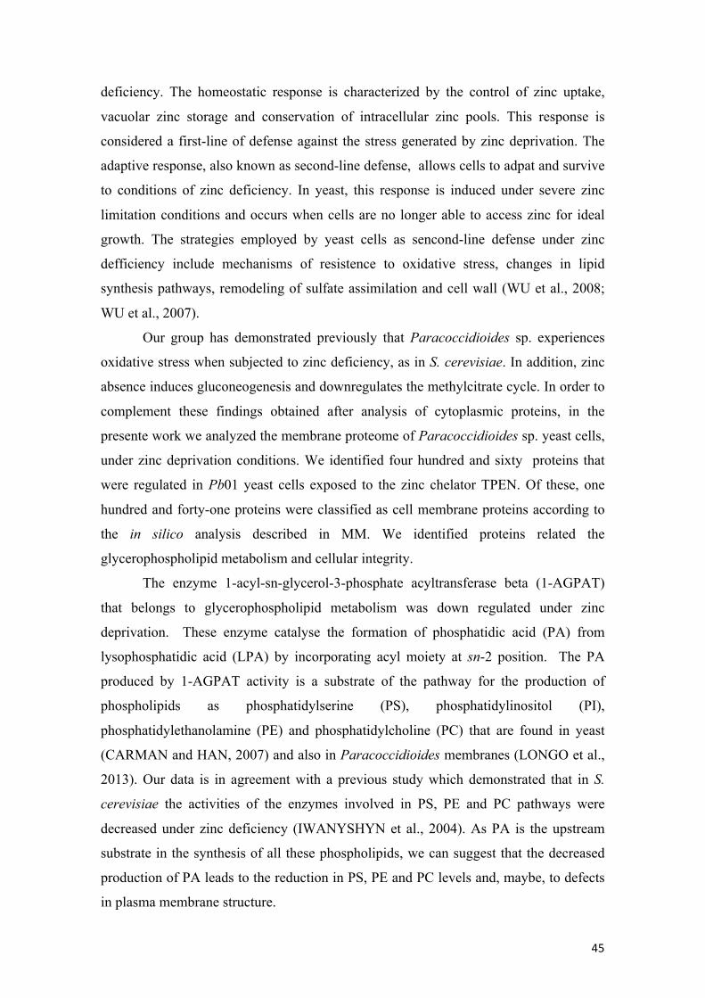
Membrane proteins of Paracoccidioides sp. identified by in silico analysis
From the total of the regulated proteins, one hundred and forty-one (31%) (Table 1)
were classified as cell membrane proteins. Eighty-one proteins presented
transmembrane domain and nine proteins were classified belonging to the cell
membrane according to the association with membrane by myristoylation (one protein),
palmitoylation and prenylation (eight proteins). The others fifty-one proteins not present
transmembrane domain or post translational modification but showed that the
subcellular localization according to Wolf PSORT and cellular component in gene
ontology (GO) were in regions of cell membranes (Figure 2). Between the proteins that

44
presented transmembrane domain, seventy proteins were down regulated and eleven
proteins were up regulated. According to the subcellular localization, forty-one proteins
were down regulated and ten proteins were up regulated. All myristoylated and
prenylated proteins were down regulated (Figure 3).
Membrane proteins of Paracoccidioides sp. down regulated in zinc deprivation
One hundred and twenty proteins (Table 2) of cell membranes were down
regulated in zinc deprivation. Eighty-six of these proteins were only identified in the
control condition. Besides the in silico analysis performed to identify proteins that
showed some association with the membrane, the search for proteins secreted by
classical and no classical pathways was also performed. Among the one hundred and
twenty proteins of cell membranes, thirteen were predicted to be secreted by classical
pathways, due to the the presence of signal peptide and transmembrane domain.
The functional classification of down regulated membrane proteins, revealed
that 26,67% and 18,33% of proteins were involved in transport events and metabolism,
respectively (Figure 4).
Membrane proteins of Paracoccidioides sp. up regulated in zinc deprivation
A total of twenty-one proteins (Table 3) of cell membranes, were up regulated in
zinc privation. Nineteen of these proteins were only identified in the zinc deprivation
condition. Between the twenty-one proteins of cell membranes, it were identified two
proteins that were predicted to be secereted by classical pathways and also presented
transmembrane domain. A total of 47,62% of these proteins of cell membranes up
regulated were involved in transporte events and 14,29 % were inolved in protein
synthesis (Figure 4).
DISCUSSION
Zinc as other metals, is an essential nutrient and serves as a structural or
catalytic cofactor for many proteins. In conditions of zinc deprivation the cellular
response is regulated mainly at the transcriptional level (ZHAO and EIDE, 1997). In S.
cerevisiae, studies have revealed both homeostatic and adaptive responses to zinc
45
deficiency. The homeostatic response is characterized by the control of zinc uptake,
vacuolar zinc storage and conservation of intracellular zinc pools. This response is
considered a first-line of defense against the stress generated by zinc deprivation. The
adaptive response, also known as second-line defense, allows cells to adpat and survive
to conditions of zinc deficiency. In yeast, this response is induced under severe zinc
limitation conditions and occurs when cells are no longer able to access zinc for ideal
growth. The strategies employed by yeast cells as sencond-line defense under zinc
defficiency include mechanisms of resistence to oxidative stress, changes in lipid
synthesis pathways, remodeling of sulfate assimilation and cell wall (WU et al., 2008;
WU et al., 2007).
Our group has demonstrated previously that Paracoccidioides sp. experiences
oxidative stress when subjected to zinc deficiency, as in S. cerevisiae. In addition, zinc
absence induces gluconeogenesis and downregulates the methylcitrate cycle. In order to
complement these findings obtained after analysis of cytoplasmic proteins, in the
presente work we analyzed the membrane proteome of Paracoccidioides sp. yeast cells,
under zinc deprivation conditions. We identified four hundred and sixty proteins that
were regulated in Pb01 yeast cells exposed to the zinc chelator TPEN. Of these, one
hundred and forty-one proteins were classified as cell membrane proteins according to
the in silico analysis described in MM. We identified proteins related the
glycerophospholipid metabolism and cellular integrity.
The enzyme 1-acyl-sn-glycerol-3-phosphate acyltransferase beta (1-AGPAT)
that belongs to glycerophospholipid metabolism was down regulated under zinc
deprivation. These enzyme catalyse the formation of phosphatidic acid (PA) from
lysophosphatidic acid (LPA) by incorporating acyl moiety at sn-2 position. The PA
produced by 1-AGPAT activity is a substrate of the pathway for the production of
phospholipids as phosphatidylserine (PS), phosphatidylinositol (PI),
phosphatidylethanolamine (PE) and phosphatidylcholine (PC) that are found in yeast
(CARMAN and HAN, 2007) and also in Paracoccidioides membranes (LONGO et al.,
2013). Our data is in agreement with a previous study which demonstrated that in S.
cerevisiae the activities of the enzymes involved in PS, PE and PC pathways were
decreased under zinc deficiency (IWANYSHYN et al., 2004). As PA is the upstream
substrate in the synthesis of all these phospholipids, we can suggest that the decreased
production of PA leads to the reduction in PS, PE and PC levels and, maybe, to defects
in plasma membrane structure.

46
The phosphoinositide phosphatase (Sac1) is a transmembrane protein found in
the endoplasmatic reticulum (ER) and Golgi complex (KONRAD et al., 2002;
WHITTERS et al., 1993). This enzyme has a specific role in secretion and acts as an
antagonist of the phosphatidylinositol 4-kinase Pikp in Golgi trafficking (SCHORR et
al., 2001). The rapid change of the localization of Sac1 between the ER and the Golgi
occurs in response to growth conditions. The localization of Sac1 at the ER occurs only
during times of exponential growth and is mediated by interaction with dolichol-
phosphate mannose synthase (Dpm1) through direct contact between the
transmembrane domains (FAULHAMMER et al., 2005). The Sac1 and Dpm1 proteins
are required for dolichol oligosaccharide biosynthesis and in this study these proteins
were down regulated suggesting that carbohydrate metabolism was affected by zinc
deprivation, as described by Wu and colleague (2008).
Studies have shown que Sac1 is related to the integrity of the cell wall due to
defects in chitin synthase trafficking and chitin biosynthesis observed in sac1 mutant.
This phenotype was more severe for the sac1 and slt2 double mutant. Slt2 is a non-
membrane MAP kinase involved in cell wall rearrangment in response to cell wall stress
(HEINISCH et al., 1999). Therefore, these proteins have a synergistic role in regulating
cell wall construction (TAHIROVIC et al., 2003). A Slt2 homologous was found in our
membrane proteome, the mitogen-activated protein kinase Mkc1. Interestingly, Mkc1
was up regulated under zinc deficiency while Sac1 was down regulated. As Sac1 is
involved in cell wall integrity, we suggest that zinc absence triggers cell wall defects, as
described in S. cerevisiae (WU et al., 2008). To counteract the cell wall stress, Mkc1
was induced. In agreement with the effect of zinc starvation on cell wall, we found that
the chitin synthase B, a membrane protein, was also downregulated in the absence of
zinc.
To our knowledge, this is the first report that describes the response of
Paracoccidioides spp. to micronutrient starvation at the membranes proteome level.The
new findings showed here, associated with the responses to zinc deficiency at
citoplasmic level, will add important information to the growing knowledge of how
Paracoccidioides spp. behaves in conditions of metal starvation. As nutritional
immunity is a strategy of host to avoid infection, the mechanisms employed by the
fungus to survive and adapt to this condition are paramount for pathogenicity.

47
CONCLUSION
Nutritional immunity is one of the strategies used by the host to prevent the
growth of pathogenic microorganisms. Thus, Paracoccidioides sp, facing a condition of
stress (nutrient deprivation) uses mechanisms to adapt and survive. The development of
these mechanisms are essential for its pathogenicity. With the results obtained in the
present study, we can conclude that zinc deprivation Paracoccidioides sp. promotes
changes in the cell integrity and metabolism of phospholipids and these changes can
affect the cellular development.
Figures and Table
Figure 1: Transmission Electron Microscopy of membrane extract of Paracoccidioides
spp.. The large figure show the presence of membrane fragments of the fungus, as indicated in
the white arrow. In detail, a fragment showing the lipid bilayer of the membrane (300 thousand
fold). Magnitude bar (0.5 µm = 50 thousand fold).
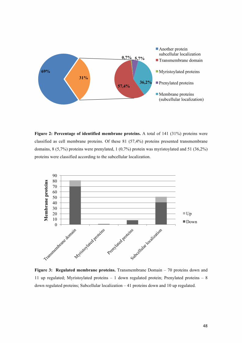
48
Figure 2: Percentage of identified membrane proteins. A total of 141 (31%) proteins were
classified as cell membrane proteins. Of these 81 (57,4%) proteins presented transmembrane
domains, 8 (5,7%) proteins were prenylated, 1 (0,7%) protein was myristoylated and 51 (36,2%)
proteins were classified according to the subcellular localization.
Figure 3: Regulated membrane proteins. Transmembrane Domain – 70 proteins down and
11 up regulated; Myristoylated proteins – 1 down regulated protein; Prenylated proteins – 8
down regulated proteins; Subcellular localization – 41 proteins down and 10 up regulated.
Another protein subcellular localizationTransmembrane domain
Myristoylated proteins
Prenylated proteins
Membrane proteíns (subcellular localization)
0102030405060708090
Mem
bran
e pr
otei
ns
Up
Down
69% 31%
36,2%
5,7% 0,7%
57,4%

49
Figure 4: Functional classification of the membrane proteins. The database FunCat2 was
used to perform this classification. Most down regulated proteins are involved in transport and
metabolism and the up regulated proteins are involved in transport and protein synthesis.
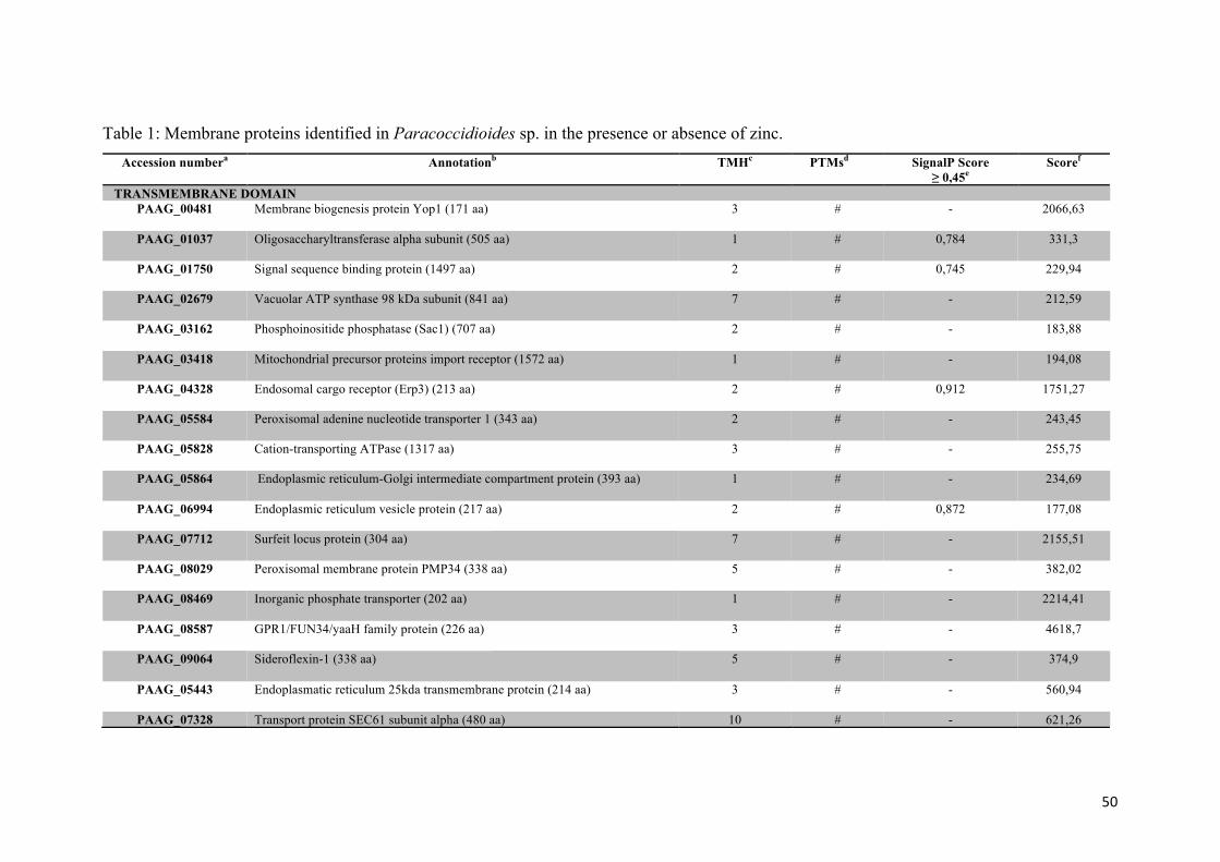
50
Table 1: Membrane proteins identified in Paracoccidioides sp. in the presence or absence of zinc. Accession numbera Annotationb TMHc PTMsd SignalP Score
≥ 0,45e Scoref
TRANSMEMBRANE DOMAIN
PAAG_00481
Membrane biogenesis protein Yop1 (171 aa) 3 # - 2066,63
PAAG_01037
Oligosaccharyltransferase alpha subunit (505 aa) 1 # 0,784 331,3
PAAG_01750
Signal sequence binding protein (1497 aa) 2 # 0,745 229,94
PAAG_02679
Vacuolar ATP synthase 98 kDa subunit (841 aa) 7 # - 212,59
PAAG_03162
Phosphoinositide phosphatase (Sac1) (707 aa) 2 # - 183,88
PAAG_03418
Mitochondrial precursor proteins import receptor (1572 aa) 1 # - 194,08
PAAG_04328
Endosomal cargo receptor (Erp3) (213 aa) 2 # 0,912 1751,27
PAAG_05584
Peroxisomal adenine nucleotide transporter 1 (343 aa) 2 # - 243,45
PAAG_05828
Cation-transporting ATPase (1317 aa) 3 # - 255,75
PAAG_05864
Endoplasmic reticulum-Golgi intermediate compartment protein (393 aa) 1 # - 234,69
PAAG_06994
Endoplasmic reticulum vesicle protein (217 aa) 2 # 0,872 177,08
PAAG_07712
Surfeit locus protein (304 aa) 7 # - 2155,51
PAAG_08029
Peroxisomal membrane protein PMP34 (338 aa) 5 # - 382,02
PAAG_08469
Inorganic phosphate transporter (202 aa) 1 # - 2214,41
PAAG_08587
GPR1/FUN34/yaaH family protein (226 aa) 3 # - 4618,7
PAAG_09064
Sideroflexin-1 (338 aa) 5 # - 374,9
PAAG_05443 Endoplasmatic reticulum 25kda transmembrane protein (214 aa)
3 # - 560,94
PAAG_07328 Transport protein SEC61 subunit alpha (480 aa) 10 # - 621,26

51
PAAG_08620
ADP,ATP carrier protein (310 aa) 3 # - 10453,18
PAAG_08082
Plasma membrane ATPase (930 aa) 9 # - 7230,75
PAAG_01041
Prohibitin-2 (311 aa) 1 # - 4227,94
PAAG_00231
Cytochrome P450 61 (354 aa) 1 # - 303,05
PAAG_00827
Cytochrome P450 51 (603 aa) 4 # 0,749 308,44
PAAG_01390
Endoplasmic reticulum protein (570 aa) 1 # 0,658 223,43
PAAG_03024
3-ketodihydrosphingosine reductase tsc10 (386 aa) 1 # - 253,4
PAAG_03722
Acyl-CoA desaturase (467 aa) 4 # - 738,82
PAAG_03974
NADPH cytochrome P450 oxidoreductase (379 aa) 1 # - 310,79
PAAG_04719
Oligosaccharyl transferase subunit (739 aa) 13 # - 379,35
PAAG_05077
Class I alpha-mannosidase 1ª (869 aa) 1 # - 360,41
PAAG_05644
Estradiol 17-beta-dehydrogenase 12-A (340 aa) 1 # - 208,66
PAAG_05910
Dolichyl-phosphate-mannose-protein mannosyltransferase (917 aa) 8 # - 657,3
PAAG_08431
Dolichyl-phosphate-mannose-protein mannosyltransferase (746 aa) 9 # - 289,29
PAAG_09072
Mitochondrial 51ucleasse (336 aa) 1 # - 3854,97
PAAG_06668
Mitochondrial metallochaperone Sco1 (317 aa) 1 # - 253,54
PAAG_04025
Osmosensor protein (278 aa) 2 # - 510,94
PAAG_06815
Tricalbin-3 (1516 aa) 2 # - 165,67
PAAG_02462
Alpha-1,2 mannosyltransferase KTR1 (505 aa) 1 # - 300,78
PAAG_05468 Translocation protein sec63 (693 aa) 3 # - 324,18

52
PAAG_06464
Disulfide-isomerase (728 aa) 1 # 0,851 252,29
PAAG_07238
Alpha-1,2 mannosyltransferase KTR1 (397 aa) 1 # 0,732 671,57
PAAG_04110
Dolichyl-di-phosphooligosaccharide-protein glycotransferase (471 aa) 1 # 0.949 977,57
PAAG_00622
Mitochondria fission 1 protein (154 aa) 1 # - 1001,94
PAAG_02917
GTP-binding protein Sey1 (873 aa) 2 # - 176,93
PAAG_03391
Chitin synthase B (928 aa) 6 # - 282,35
PAAG_03489
Cell lysis protein cwl1 (358 aa) 4 # - 1188,55
PAAG_07503
1-acyl-sn-glycerol-3-phosphate acyltransferase beta (304 aa) 2 # - 425,34
PAAG_01262
Hsp70-like protein (629 aa) 1 # 0,864 3655,29
PAAG_08209
Peroxin-11 (239 aa) 1 # - 3889,83
PAAG_00125 Conserved hypothetical protein (480 aa)
3 # - 206,51
PAAG_00184
Conserved hypothetical protein (299 aa) 4 # - 304,39
PAAG_00187
Conserved hypothetical protein (241 aa) 3 # - 808,12
PAAG_01890
Conserved hypothetical protein (190 aa) 2 # - 251,17
PAAG_04529
Conserved hypothetical protein (117 aa) 1 # - 1843,87
PAAG_04540
Conserved hypothetical protein (180 aa) 1 # - 247,42
PAAG_04911
Conserved hypothetical protein (300 aa) 4 # - 422,48
PAAG_05272
Conserved hypothetical protein (435 aa) 1 # - 446,27
PAAG_05875
Hypothetical protein (115 aa) 1 # - 434,68
PAAG_06568 Conserved hypothetical protein (415 aa) 1 # - 345,74

53
PAAG_08987
Conserved hypothetical protein (317 aa) 1 # - 170,75
PAAG_05911
Conserved hypothetical protein (147 aa) 1 # - 8129,16
PAAG_07960
Conserved hypothetical protein (152 aa) 4 # - 1003,46
PAAG_08051
Hypothetical protein (279 aa) 2 # 0,594 217,91
PAAG_02265
Mitochondrial F1F0 ATP synthase subunit (102 aa) 1 # - 8032,77
PAAG_03160
Formation of crista junctions protein 1(686 aa)
1 # - 346,12
PAAG_05091
NADH-ubiquinone oxidoreductase 213 kDa subunit (200 aa) 3 # - 357,62
PAAG_08517
Propionate-CoA ligase (711 aa) 1 # - 271,08
PAAG_06654
Signal sequence receptor alpha chain (275 aa) 1 # 0,769 390,4
PAAG_07846
CBS and PB1 domain-containing protein (672 aa) 1 # - 496,37
PAAG_04157 DUF1620 domain-containing protein (954 aa)
1 # 0,863 382,34
PAAG_12011
Mitochondria cytochrome c oxidase subunit 2 (254 aa) 2 # - 2138,11
PAAG_06701
PCI domain-containing protein (750 aa) 1 # 0,790 352,24
PAAG_08352
NADH-ubiquinone oxidoreductase (120 aa)
1 # - 375,07
PAAG_01425
Eukaryotic translation initiation factor 3 (478 aa) 2 # - 281,8
PAAG_05425
Golgi membrane protein (Coy1) (766 aa) 1 # - 118,61
PAAG_07589
SNARE domain-containing protein (271 aa) 1 # - 124,41
PAAG_00404
Mitochondrial sodium/hydrogen exchanger (710 aa) 9 # - 123,07
PAAG_00739
Peptidyl-prolyl cis-trans isomerase B (211 aa) 1 # 0,641 212,21
PAAG_06946 Mitochondrial import inner membrane translocase subunit tim14 1 # - 330,93

54
PAAG_03316
Conserved hypothetical protein (422 aa) 1 # - 225,85
PAAG_07599
Conserved hypothetical protein (312 aa) 1 # - 158,75
PAAG_01417
ER membrane DUF1077 domain-containing protein (105 aa)
2 # - 311,13
MYRISTOYLATION PAAG_03988
ADP-ribosylation factor (187 aa) * C 14 - 243,52
PRENYLATION
PAAG_01602
Ras-like GTP-binding protein (312 aa) * C 20 - 1037,04
PAAG_07175
GTP-binding protein YPT52 (294 aa) * C 20 - 1651,53
PAAG_08093
GTP-binding protein ypt3 (221 aa) * C 20 - 692,85
PAAG_01500
GTP-binding protein SAS1 (207 aa) * C 20 - 1546,88
PAAG_02458
GTP-binding protein ypt7 (206 aa) * C 20 - 1281,11
PAAG_07634
Small GTPase RhoA (192 aa) * C 15/C 20 - 6087,45
PAAG_02230
Cell division control protein (193 aa) * C 15/C 20 - 638,27
PAAG_00797
Mitochondrial protein import protein MAS5 (411 aa) * C 15 - 982
SUBCELLULAR LOCALIZATION PAAG_02796
Mitochondrial 2-oxodicarboxylate carrier 1 (315 aa) * # - 217,42
PAAG_02824
Mitochondrial inner membrane translocase subunit TIM44 (534 aa) * # - 198,45
PAAG_03644
Mitochondrial import receptor subunit tom-40 (356 aa) * # - 651,53
PAAG_05350
Mitochondrial phosphate carrier protein (422 aa) * # - 3006,85
PAAG_08010
Mitochondrial carrier protein (311 aa * # - 1443,87
PAAG_08086
Mitochondrial DNA replication protein YHM2 (317 aa) * # - 514,67

55
PAAG_07564
Outer mitochondrial membrane protein porin (285 aa) * # - 9018,79
PAAG_00639
Phosphate carrier protein (385 aa) * # - 1566,86
PAAG_05107
AAA ATPase (921 aa) * # - 351,17
PAAG_07669
Mannan polymerase II complex ANP1 subunit (461 aa) * # - 580,61
PAAG_00230
Cytochrome P450 61 (167 aa) * # - 652,31
PAAG_01833
2-succinylbenzoate-CoA ligase (583 aa) * # - 456,85
PAAG_02604
L-tyrosine decarboxylase (578 aa) * # - 169,59
PAAG_04238
Succinate dehydrogenase flavoprotein subunit (655 aa) * # - 179,7
PAAG_04564
Betaine aldehyde dehydrogenase (635 aa) * # - 193,12
PAAG_01575
Sterol 24-C-methyltransferase (378 aa) * # - 1375,67
PAAG_01874
Dolichol-phosphate mannosyltransferase (280 aa) * # - 1456,63
PAAG_00850
Glucosamine-fructose-6-phosphate aminotransferase (489 aa) * # - 3602,18
PAAG_06289
Tryptophan synthase (725 aa) * # - 390,84
PAAG_06961
Aldehyde dehydrogenase (496 aa) * # - 744,17
PAAG_07640
Casein kinase I (284 aa) * # - 196,41
PAAG_07235
SNARE domain-containing protein (252 aa) * # - 430,59
PAAG_04458
Prohibitin-1 (281 aa) * # - 2586,79
PAAG_03292
Cytochrome c peroxidase (375 aa) * # - 6884,11
PAAG_05415
TIM22 inner membrane protein import complex anchor subunit Tim18 (199 aa)
* # - 201,95
PAAG_00151
NADH-cytochrome b5 reductase (310 aa)* * # - 465,02

56
PAAG_00948
NADH dehydrogenase iron-sulfur protein (229 aa) * # - 195,5
PAAG_01044
NADH-ubiquinone oxidoreductase 24 kDa subunit (260 aa) * # - 368,56
PAAG_02656
NADH-ubiquinone oxidoreductase 51 kDa subunit (516 aa) * # - 617,71
PAAG_05735
NADH-ubiquinone oxidoreductase 49 kDa subunit (480 aa) * # - 2857,94
PAAG_05876
Cytochrome c oxidase subunit VIa (143 aa)
* # - 2405,87
PAAG_06796
Cytochrome c oxidase polypeptide IV (198 aa) * # - 1101,38
PAAG_08088
Cytochrome b-c1 complex subunit 2 (464 aa) * # - 2793,7
PAAG_08057
Cytochrome c oxidase polypeptide V (200 aa) * # - 969,86
PAAG_04570
ATP synthase D chain, mitochondrial (175 aa) * # - 6113,62
PAAG_05031
NADH-ubiquinone oxidoreductase 40 kDa subunit (294 aa) * # - 1979,87
PAAG_07672
Cytochrome b-c1 complex subunit 7 (123 aa) * # - 13389,36
PAAG_02653
Acetyl-coenzyme A synthetase (670 aa) * # - 1504,51
PAAG_06541
Erythrocyte band 7 integral membrane protein (361 aa) * # - 867,73
PAAG_01439
Predicted protein (375 aa) * # - 205,68
PAAG_03337
Predicted protein (327 aa) * # - 202,04
PAAG_03780
ATP-dependent RNA helicase DBP5 (505 aa) * # - 131,49
PAAG_01345
Importin-8 (1032 aa) * # - 133,85
PAAG_03042
Vacuolar ATP synthase subunit c (394 aa) * # - 195,78
PAAG_05036
Transport protein SEC24 (939 aa) * # - 141,36
PAAG_05589
Transport protein SEC23 (773 aa) * # - 211,4

57
PAAG_07874
Hepatocellular carcinoma down-regulated mitochondrial carrier protein (302 aa)*
* # - 152,37
PAAG_08834
Tricarboxylate transport protein (297 aa) * # - 135,81
PAAG_06344
Rab GDP-dissociation inhibitor (469 aa) * # - 532,2
PAAG_08164
Homogentisate 1,2-dioxygenase (455 aa) * # - 187,81
PAAG_06136
Conserved hypothetical protein (414 aa) * # - 159,87
aAccession number– Accession number of the protein in the database of Paracoccidioides spp. (http://www.broadinstitute.org/annotation/genome/paracoccidioides_brasiliensis/MultiHome.html) bProtein annotation from Paracoccidioides genome database or by homology in NCBI database (http://www.ncbi.nlm.nih.gov/). cRepresents the amount of transmembrane domains identified in proteins using the program TMHMM version 2.0 (http://www.cbs.dtu.dk/services/TMHMM/). dPresence of posttranslational modification. For myristoylated protein (C14) programs Terminator (http://www.isv.cnrs-gif.fr/terminator3/test.php) and Myristoylator (http://web.expasy.org/myristoylator/) were used. Terminator program was used to identify palmitoylated proteins. Research by prenylated proteins was done by the program PresPs-Prenylation Prediction Suite (http://mendel.imp.ac.at/sat/PrePS/index.html) and to predict GPI anchor the program big-Pi fungi predictor (http://mendel.imp.ac.at/gpi/fungi_server.html) was used. C15 and C20 indicates the presence of a recognition site for the addition of a farnesyl or geranyl group (FTase and geranyltransferase type I, Rab geranylgeranyl transferase). ePresence of signal peptide was analyzed by program SignalP version 4.1 (http://www.cbs.dtu.dk/services/SignalP/). The values indicate the score of the proteins with the signal peptide, this value must be equal to or greater than 0.45. fIndicates the score of the protein *: Proteins that no presented transmembrane domain #: Proteins that no presented posttranslational modification -: Proteins that no presented signal peptide C: Proteins only identified in the control condition T: Proteins only identified in the zinc deprivation condition The functional classification was performed used the database of FunCat2 (http://pedant.gsf.de/pedant3htmlview/pedant3view?Method=analysis&Db=p3_r48325_Par_brasi_Pb01). The database Gene Ontology (GO) and WolfPSORT was employed to evaluate the possible localization of proteins in cell membranes of Paracoccidioides sp.. Some proteins did not show any form of classical association with the lipid bilayer, but showed that the localization subcellular were in regions of cell membranes, with score ≥ 50.

58
Table 2: Membrane proteins of Paracoccidioides sp. down regulated in zinc deprivation and their biological functions provided – FunCat2 Access Number a Annotationb TMHc PTMsd SignalP Score
≥ 0,45e Scoref
Fold change (log2)g
CELLULAR TRANSPORT, TRANSPORT FACILITIES AND TRANSPORT ROUTESPAAG_00481
Membrane biogenesis protein Yop1 (171 aa) 3 # - 2066,63
C
PAAG_01037
Oligosaccharyltransferase alpha subunit (505 aa) 1 # 0,784 331,3
C
PAAG_01750
Signal sequence binding protein (1497 aa) 2 # 0,745 229,94
C
PAAG_02679
Vacuolar ATP synthase 98 kDa subunit (841 aa) 7 # - 212,59
C
PAAG_02796
Mitochondrial 2-oxodicarboxylate carrier 1 (315 aa) * # - 217,42
C
PAAG_02824
Mitochondrial inner membrane translocase subunit TIM44 (534 aa) * # - 198,45
C
PAAG_03162
Phosphoinositide phosphatase (Sac1) (707 aa) 2 # - 183,88
C
PAAG_03418
Mitochondrial precursor proteins import receptor (1572 aa) 1 # - 194,08
C
PAAG_03644
Mitochondrial import receptor subunit tom-40 (356 aa) * # - 651,53
C
PAAG_03988
ADP-ribosylation factor (187 aa) * C 14 - 243,52
C
PAAG_04328
Endosomal cargo receptor (Erp3) (213 aa) 2 # 0,912 1751,27
C
PAAG_05350
Mitochondrial phosphate carrier protein (422 aa) * # - 3006,85
C
PAAG_05584
Peroxisomal adenine nucleotide transporter 1 (343 aa) 2 # -- 243,45
C
PAAG_05828
Cation-transporting ATPase (1317 aa) 3 # - 255,75
C
PAAG_05864
Endoplasmic reticulum-Golgi intermediate compartment protein (393 aa) 1 # - 234,69
C
PAAG_06994
Endoplasmic reticulum vesicle protein (217 aa) 2 # 0,872 177,08
C
PAAG_07712
Surfeit locus protein (304 aa) 7 # - 2155,51
C
PAAG_08010 Mitochondrial carrier protein (311 aa) * # - 1443,87 C

59
PAAG_08029
Peroxisomal membrane protein PMP34 (338 aa) 5 # - 382,02
C
PAAG_08086
Mitochondrial DNA replication protein YHM2 (317 aa) * # - 514,67
C
PAAG_08469
Inorganic phosphate transporter (202 aa) 1 # - 2214,41
C
PAAG_08587
GPR1/FUN34/yaaH family protein (226 aa) 3 # - 4618,7
C
PAAG_09064
Sideroflexin-1 (338 aa) 5 # - 374,9
C
PAAG_05443 Endoplasmatic reticulum 25kda transmembrane protein (214 aa)
3 # - 560,94 C
PAAG_07328
Transport protein SEC61 subunit alpha (480 aa) 10 # - 621,26
-3,910
PAAG_07564
Outer mitochondrial membrane protein porin (285 aa) * # - 9018,79
-3,910
PAAG_01602
Ras-like GTP-binding protein (312 aa) * C 20 - 1037,04
-3,506
PAAG_00639
Phosphate carrier protein (385 aa) * # - 1566,86
-2,871
PAAG_08620
ADP,ATP carrier protein (310 aa) 3 # - 10453,18
-2,207
PAAG_05107
AAA ATPase (921 aa) * # - 351,17
-1,500
PAAG_08082
Plasma membrane ATPase (930 aa) 9 # - 7230,75
-2,828
PAAG_07175
GTP-binding protein YPT52 (294 aa) * C 20 - 1651,53
-2,077
CELL CYCLE AND DNA PROCESSING PAAG_07669
Mannan polymerase II complex ANP1 subunit (461 aa) * # - 580,61
C
PAAG_01041
Prohibitin-2 (311 aa) 1 # - 4227,94
-1,428
METABOLISMPAAG_00230
Cytochrome P450 61 (167 aa) * # - 652,31
C
PAAG_00231
Cytochrome P450 61 (354 aa) 1 # - 303,05
C
PAAG_00827 Cytochrome P450 51 (603 aa) 4 # 0,749 308,44 C

60
PAAG_01390
Endoplasmic reticulum protein (570 aa) 1 # 0,658 223,43
C
PAAG_01833
2-succinylbenzoate-CoA ligase (583 aa) * # - 456,85
C
PAAG_07503
1-acyl-sn-glycerol-3-phosphate acyltransferase beta (304 aa) 2 # - 425,34
C
PAAG_02604
L-tyrosine decarboxylase (578 aa) * # - 169,59
C
PAAG_03024
3-ketodihydrosphingosine reductase tsc10 (386 aa) 1 # - 253,4
C
PAAG_03722
Acyl-CoA desaturase (467 aa) 4 # - 738,82
C
PAAG_03974
NADPH cytochrome P450 oxidoreductase (379 aa) 1 # - 310,79
C
PAAG_04238
Succinate dehydrogenase flavoprotein subunit (655 aa) * # - 179,7
C
PAAG_04564
Betaine aldehyde dehydrogenase (635 aa) * # - 193,12
C
PAAG_04719
Oligosaccharyl transferase subunit (739 aa) 13 # - 379,35
C
PAAG_05077
Class I alpha-mannosidase 1A (869 aa) 1 # - 360,41
C
PAAG_05644
Estradiol 17-beta-dehydrogenase 12-A (340 aa) 1 # - 208,66
C
PAAG_05910
Dolichyl-phosphate-mannose-protein mannosyltransferase (917 aa) 8 # - 657,3
C
PAAG_08431
Dolichyl-phosphate-mannose-protein mannosyltransferase (746 aa) 9 # - 289,29
C
PAAG_01575
Sterol 24-C-methyltransferase (378 aa) * # - 1375,67
-3,044
PAAG_01874
Dolichol-phosphate mannosyltransferase (280 aa) * # - 1456,63
-2,366
PAAG_00850
Glucosamine-fructose-6-phosphate aminotransferase (489 aa) * # - 3602,18
-1,991
PAAG_09072
Mitochondrial nuclease (336 aa) 1 # - 3854,97
-1,616
PAAG_06289
Tryptophan synthase (725 aa) * # - 390,84
-1,443
PAAG_06961 Aldehyde dehydrogenase (496 aa) * # - 744,17 -0,909

61
PROTEIN SYNTHESIS
PAAG_06668
Mitochondrial metallochaperone Sco1 (317 aa) 1 # - 253,54
C
CELLULAR COMMUNICATION/SIGNAL TRANSDUCTION MECHANISM PAAG_04025
Osmosensor protein (278 aa) 2 # - 510,94
C
PAAG_06815
Tricalbin-3 (1516 aa) 2 # - 165,67
C
PAAG_07640
Casein kinase I (284 aa) * # - 196,41
C
PAAG_08093
GTP-binding protein ypt3 (221 aa) * C 20 - 692,85
C
PAAG_01500
GTP-binding protein SAS1 (207 aa) * C 20 - 1546,88
-2,813
PAAG_02458
GTP-binding protein ypt7 (206 aa) * C 20 - 1281,11
-2,481
PAAG_07634
Small GTPase RhoA (192 aa) * C 15/C 20 - 6087,45
-1,053
PROTEIN FATE (FOLDING, MODIFICATION, DESTINATION)PAAG_02462
Alpha-1,2 mannosyltransferase KTR1 (505 aa) 1 # - 300,78
C
PAAG_05468
Translocation protein sec63 (693 aa) 3 # - 324,18
C
PAAG_06464
Disulfide-isomerase (728 aa) 1 # 0,851 252,29
C
PAAG_07235
SNARE domain-containing protein (252 aa) * # - 430,59
C
PAAG_07238
Alpha-1,2 mannosyltransferase KTR1 (397 aa) 1 # 0,732 671,57
C
PAAG_04458
Prohibitin-1 (281 aa) * # - 2586,79
-3,145
PAAG_04110
Dolichyl-di-phosphooligosaccharide-protein glycotransferase (471 aa) 1 # 0.949 977,57
-0,837
BIOGENESIS OF CELLULAR COMPONENTS PAAG_00622
Mitochondria fission 1 protein (154 aa) 1 # - 1001,94
C
PAAG_02917
GTP-binding protein Sey1 (873 aa) 2 # - 176,93
C
PAAG_03391 Chitin synthase B (928 aa) 6 # - 282,35 C

62
PAAG_03489
Cell lysis protein cwl1 (358 aa) 4 # - 1188,55
C
PAAG_08209
Peroxin-11 (239 aa) 1 # - 3889,83
C
CELL RESCUE, DEFENSE AND VIRULENCE PAAG_02230
Cell division control protein (193 aa) * C 15/C 20 - 638,27
C
PAAG_01262
Hsp70-like protein (629 aa) 1 # 0,864 3655,29
-2,640
PAAG_00797
Mitochondrial protein import protein MAS5 (411 aa) * C 15 - 982
-2,582
PAAG_03292
Cytochrome c peroxidase (375 aa) * # - 6884,11
-0,808
HYPOTHETICAL PROTEINSPAAG_00125 Conserved hypothetical protein (480 aa)
3 # - 206,51 C
PAAG_00184
Conserved hypothetical protein (299 aa) 4 # - 304,39
C
PAAG_00187
Conserved hypothetical protein (241 aa) 3 # - 808,12
C
PAAG_01890
Conserved hypothetical protein (190 aa) 2 # - 251,17
C
PAAG_04529
Conserved hypothetical protein (117 aa) 1 # - 1843,87
C
PAAG_04540
Conserved hypothetical protein (180 aa) 1 # - 247,42
C
PAAG_04911
Conserved hypothetical protein (300 aa) 4 # - 422,48
C
PAAG_05272
Conserved hypothetical protein (435 aa) 1 # - 446,27
C
PAAG_05415
TIM22 inner membrane protein import complex anchor subunit Tim18 (199 aa)
* # - 201,95
C
PAAG_05875
Hypothetical protein (115 aa) 1 # - 434,68
C
PAAG_06568
Conserved hypothetical protein (415 aa)
1 # - 345,74
C
PAAG_08051
Hypothetical protein (279 aa) 2 # 0,594 217,91
C
PAAG_08987 Conserved hypothetical protein (317 aa) 1 # - 170,75 C

63
PAAG_05911
Conserved hypothetical protein (147 aa) 1 # - 8129,16
-1,876
PAAG_07960
Conserved hypothetical protein (152 aa) 4 # - 1003,46
-1,573
ENERGY
PAAG_00151
NADH-cytochrome b5 reductase (310 aa) * # - 465,02
C
PAAG_00948
NADH dehydrogenase iron-sulfur protein (229 aa) * # - 195,5
C
PAAG_01044
NADH-ubiquinone oxidoreductase 24 kDa subunit (260 aa) * # - 368,56
C
PAAG_02265
Mitochondrial F1F0 ATP synthase subunit (102 aa) 1 # - 8032,77
C
PAAG_02656
NADH-ubiquinone oxidoreductase 51 kDa subunit (516 aa) * # - 617,71
C
PAAG_03160
Formation of crista junctions protein 1 (686 aa)
1 # - 346,12
C
PAAG_05091
NADH-ubiquinone oxidoreductase 213 kDa subunit (200 aa) 3 # - 357,62
C
PAAG_05735
NADH-ubiquinone oxidoreductase 49 kDa subunit (480 aa) * # - 2857,94
C
PAAG_05876
Cytochrome c oxidase subunit VIa (143 aa) * # - 2405,87
C
PAAG_08517
Propionate-CoA ligase (711 aa) 1 # - 271,08
C
PAAG_06796
Cytochrome c oxidase polypeptide IV (198 aa) * # - 1101,38
-3,160
PAAG_08088
Cytochrome b-c1 complex subunit 2 (464 aa) * # - 2793,7
-2,481
PAAG_08057
Cytochrome c oxidase polypeptide V (200 aa) * # - 969,86
-2,150
PAAG_04570
ATP synthase D chain, mitochondrial (175 aa) * # - 6113,62
-1,890
PAAG_05031
NADH-ubiquinone oxidoreductase 40 kDa subunit (294 aa) * # - 1979,87
-1,169
PAAG_07672
Cytochrome b-c1 complex subunit 7 (123 aa) * # - 13389,36
-1,082
PAAG_02653
Acetyl-coenzyme A synthetase (670 aa) * # - 1504,51
-1,702
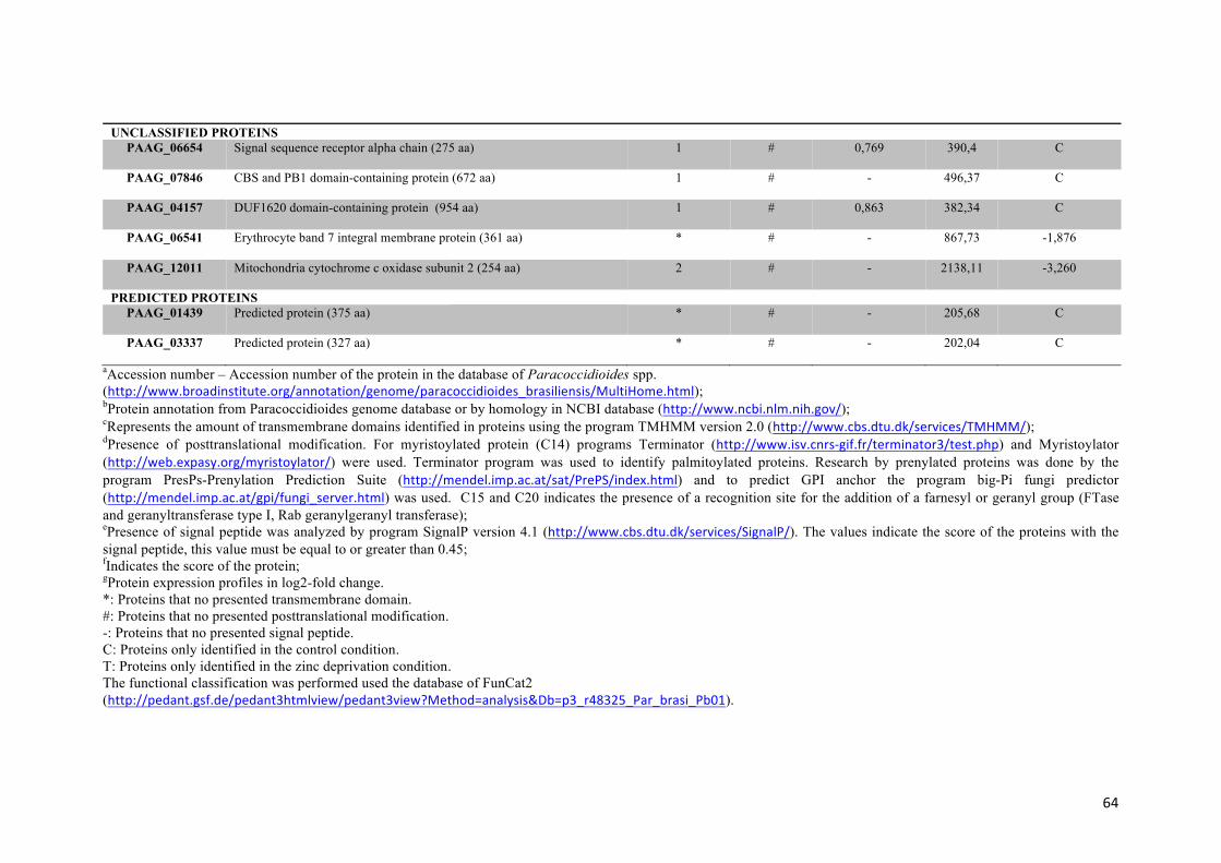
64
UNCLASSIFIED PROTEINSPAAG_06654
Signal sequence receptor alpha chain (275 aa) 1 # 0,769 390,4
C
PAAG_07846
CBS and PB1 domain-containing protein (672 aa) 1 # - 496,37
C
PAAG_04157 DUF1620 domain-containing protein (954 aa)
1 # 0,863 382,34
C
PAAG_06541
Erythrocyte band 7 integral membrane protein (361 aa) * # - 867,73
-1,876
PAAG_12011
Mitochondria cytochrome c oxidase subunit 2 (254 aa) 2 # - 2138,11
-3,260
PREDICTED PROTEINSPAAG_01439
Predicted protein (375 aa) * # - 205,68
C
PAAG_03337
Predicted protein (327 aa) * # - 202,04
C
aAccession number – Accession number of the protein in the database of Paracoccidioides spp. (http://www.broadinstitute.org/annotation/genome/paracoccidioides_brasiliensis/MultiHome.html); bProtein annotation from Paracoccidioides genome database or by homology in NCBI database (http://www.ncbi.nlm.nih.gov/); cRepresents the amount of transmembrane domains identified in proteins using the program TMHMM version 2.0 (http://www.cbs.dtu.dk/services/TMHMM/); dPresence of posttranslational modification. For myristoylated protein (C14) programs Terminator (http://www.isv.cnrs-gif.fr/terminator3/test.php) and Myristoylator (http://web.expasy.org/myristoylator/) were used. Terminator program was used to identify palmitoylated proteins. Research by prenylated proteins was done by the program PresPs-Prenylation Prediction Suite (http://mendel.imp.ac.at/sat/PrePS/index.html) and to predict GPI anchor the program big-Pi fungi predictor (http://mendel.imp.ac.at/gpi/fungi_server.html) was used. C15 and C20 indicates the presence of a recognition site for the addition of a farnesyl or geranyl group (FTase and geranyltransferase type I, Rab geranylgeranyl transferase); ePresence of signal peptide was analyzed by program SignalP version 4.1 (http://www.cbs.dtu.dk/services/SignalP/). The values indicate the score of the proteins with the signal peptide, this value must be equal to or greater than 0.45; fIndicates the score of the protein; gProtein expression profiles in log2-fold change. *: Proteins that no presented transmembrane domain. #: Proteins that no presented posttranslational modification. -: Proteins that no presented signal peptide. C: Proteins only identified in the control condition. T: Proteins only identified in the zinc deprivation condition. The functional classification was performed used the database of FunCat2 (http://pedant.gsf.de/pedant3htmlview/pedant3view?Method=analysis&Db=p3_r48325_Par_brasi_Pb01).

65
The database Gene Ontology (GO) and WolfPSORT was employed to evaluate the possible localization of proteins in cell membranes of Paracoccidioides sp.. Some proteins did not show any form of classical association with the lipid bilayer, but showed that the localization subcellular were in regions of cell membranes, with score ≥ 50. Table 3: Membrane proteins of Paracoccidioides sp. up regulated in zinc deprivation and their provided biological functions – FunCat2
Access Numbera Annotationb TMHsc PTMsd SignalP Score ≥ 0,45e
Scoref
Fold change (log2)g
PROTEIN SYNTHESISPAAG_03780
ATP-dependent RNA helicase DBP5 (505 aa) * # - 131,49
T
PAAG_06701
PCI domain-containing protein (750 aa) 1 # 0,790 352,24
T
PAAG_01425
Eukaryotic translation initiation factor 3 (478 aa) 2 # - 281,8
1,197
CELLULAR TRANSPORT, TRANSPORT FACILITIES AND TRANSPORT ROUTESPAAG_01345
Importin-8 (1032 aa) * # - 133,85
T
PAAG_03042
Vacuolar ATP synthase subunit c (394 aa) * # - 195,78
T
PAAG_05036
Transport protein SEC24 (939 aa) * # - 141,36
T
PAAG_05425
Golgi membrane protein (Coy1) (766 aa) 1 # - 118,61
T
PAAG_05589
Transport protein SEC23 (773 aa) * # - 211,4
T
PAAG_07589
SNARE domain-containing protein (271 aa) 1 # - 124,41
T
PAAG_07874
Hepatocellular carcinoma down-regulated mitochondrial carrier protein (302 aa)
* # - 152,37
T
PAAG_08834
Tricarboxylate transport protein (297 aa) * # - 135,81
T
PAAG_00404
Mitochondrial sodium/hydrogen exchanger (710 aa) 9 # - 123,07
T
PAAG_06344
Rab GDP-dissociation inhibitor (469 aa) * # - 532,2
1,847
METABOLISM PAAG_08164
Homogentisate 1,2-dioxygenase (455 aa) * # - 187,81
T
ENERGY PAAG_08352 NADH-ubiquinone oxidoreductase (120 aa) 1 # - 375,07 T
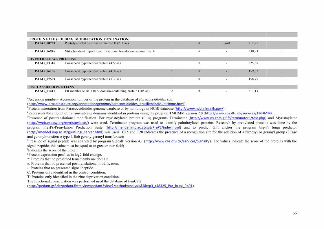
66
PROTEIN FATE (FOLDING, MODIFICATION, DESTINATION)
PAAG_00739
Peptidyl-prolyl cis-trans isomerase B (211 aa) 1 # 0,641 212,21
T
PAAG_06946
Mitochondrial import inner membrane translocase subunit tim14 1 # - 330,93
T
HYPOTHETICAL PROTEINS PAAG_03316
Conserved hypothetical protein (422 aa) 1 # - 225,85
T
PAAG_06136
Conserved hypothetical protein (414 aa) * # - 159,87
T
PAAG_07599
Conserved hypothetical protein (312 aa) 1 # - 158,75
T
UNCLASSIFIED PROTEINS PAAG_01417
ER membrane DUF1077 domain-containing protein (105 aa)
2 # - 311,13
T
aAccession number– Accession number of the protein in the database of Paracoccidioides spp. (http://www.broadinstitute.org/annotation/genome/paracoccidioides_brasiliensis/MultiHome.html); bProtein annotation from Paracoccidioides genome database or by homology in NCBI database (http://www.ncbi.nlm.nih.gov/); cRepresents the amount of transmembrane domains identified in proteins using the program TMHMM version 2.0 (http://www.cbs.dtu.dk/services/TMHMM/); dPresence of posttranslational modification. For myristoylated protein (C14) programs Terminator (http://www.isv.cnrs-gif.fr/terminator3/test.php) and Myristoylator (http://web.expasy.org/myristoylator/) were used. Terminator program was used to identify palmitoylated proteins. Research by prenylated proteins was done by the program PresPs-Prenylation Prediction Suite (http://mendel.imp.ac.at/sat/PrePS/index.html) and to predict GPI anchor the program big-Pi fungi predictor (http://mendel.imp.ac.at/gpi/fungi_server.html) was used. C15 and C20 indicates the presence of a recognition site for the addition of a farnesyl or geranyl group (FTase and geranyltransferase type I, Rab geranylgeranyl transferase); ePresence of signal peptide was analyzed by program SignalP version 4.1 (http://www.cbs.dtu.dk/services/SignalP/). The values indicate the score of the proteins with the signal peptide, this value must be equal to or greater than 0.45; fIndicates the score of the protein; gProtein expression profiles in log2-fold change. *: Proteins that no presented transmembrane domain. #: Proteins that no presented posttranslational modification. -: Proteins that no presented signal peptide. C: Proteins only identified in the control condition. T: Proteins only identified in the zinc deprivation condition. The functional classification was performed used the database of FunCat2 (http://pedant.gsf.de/pedant3htmlview/pedant3view?Method=analysis&Db=p3_r48325_Par_brasi_Pb01).

67
The database Gene Ontology (GO) and WolfPSORT was employed to evaluate the possible localization of proteins in cell membranes of Paracoccidioides sp.. Some proteins did not show any form of classical association with the lipid bilayer, but showed that the localization subcellular were in regions of cell membranes, with score ≥ 50.
Supplementary figure 1: Peptide detection type to control and zinc deprivation samples. The graphics A and B represent peptide detection type to
control and zinc deprivation samples, respectively.PepFrag1 and PepFrag2 correspond to the peptide matched when compared to database by PLGS, VarMod
that corresponds to variable modifications, InSource that corresponds to fragmentation that occurred on ionization source, MissedCleavage that indicates the
missed cleaveage by trypsin and Neutral loss H2O and NH3 that correspond to water and ammonia precursor losses.

68
Supplementary figure 2: Detection dynamic range of the experiments. For each conditions the dynamic range of proteomic experiments was performed.
Graphic A represent the control and graphic B zinc deprivation. The square gray represent the regular proteins, black circle represent the reverse proteins and
triangle black, indicated by the black arrow, represent the standard proteins (rabbit phosphorylase B).

69
Supplementary figure 3: Subcellular localization of identified proteins. The programs used
for in silico analysis of the subcellular localization of proteins was Wolf PSORT and Gene
Ontology (GO). The presence of transmembrane domain and post translational modification
were also considered during the classification of subcellular localization of identified proteins.
The main subcellular localizations of the identified proteins in the membrane fraction were in
the cell membranes (30,65%) followed by localization in mitochondrial region (25,22%) and
cytoplasmatic region (23,7%).
0
5
10
15
20
25
30
35
Perc
enta
gem
of p
rote
íns
%

70
Supplementary table-1: Proteins regulated in Paracoccidioides sp. yeast cells under zinc deprivation and their provided biological functions – FunCat2
Access Number a Annotationb TMHc PTMsd SignalP Score ≥ 0,45e
Scoref
Fold change (log2)g
CELLULAR TRANSPORT, TRANSPORT FACILITIES AND TRANSPORT ROUTESPAAG_00481
Membrane biogenesis protein Yop1 (171 aa) 3 # - 2066,63
C
PAAG_01037
Oligosaccharyltransferase alpha subunit (505 aa) 1 # 0,784 331,3
C
PAAG_01750
Signal sequence binding protein (1497 aa) 2 # 0,745 229,94
C
PAAG_02679
Vacuolar ATP synthase 98 kDa subunit (841 aa) 7 # - 212,59
C
PAAG_02796
Mitochondrial 2-oxodicarboxylate carrier 1 (315 aa) * # - 217,42
C
PAAG_02824
Mitochondrial inner membrane translocase subunit TIM44 (534 aa) * # - 198,45
C
PAAG_03162
Phosphoinositide phosphatase (Sac1) (707 aa) 2 # - 183,88
C
PAAG_03418
Mitochondrial precursor proteins import receptor (1572 aa) 1 # - 194,08
C
PAAG_03644
Mitochondrial import receptor subunit tom-40 (356 aa) * # - 651,53
C
PAAG_03988
ADP-ribosylation factor (187 aa) * C 14 - 243,52
C
PAAG_04328
Endosomal cargo receptor (Erp3) (213 aa) 2 # 0,912 1751,27
C
PAAG_05350
Mitochondrial phosphate carrier protein (422 aa) * # - 3006,85
C
PAAG_05584
Peroxisomal adenine nucleotide transporter 1 (343 aa) 2 # - 243,45
C
PAAG_05828
Cation-transporting ATPase (1317 aa) 3 # - 255,75
C
PAAG_05848
Reduced viability upon starvation protein (418 aa) * # - 485,7
C
PAAG_05864
Endoplasmic reticulum-Golgi intermediate compartment protein (393 aa) 1 # - 234,69
C
PAAG_06994 Endoplasmic reticulum vesicle protein (217 aa) 2 # 0,872 177,08 C

71
PAAG_07712
Surfeit locus protein (304 aa) 7 # - 2155,51
C
PAAG_08010
Mitochondrial carrier protein (311 aa) * # - 1443,87
C
PAAG_08029
Peroxisomal membrane protein PMP34 (338 aa) 5 # - 382,02
C
PAAG_08086
Mitochondrial DNA replication protein YHM2 (317 aa * # - 514,67
C
PAAG_08469
Inorganic phosphate transporter (202 aa) 1 # - 2214,41
C
PAAG_08587
GPR1/FUN34/yaaH family protein (226 aa) 3 # - 4618,7
C
PAAG_09064
Sideroflexin-1 (338 aa) 5 # - 374,9
C
PAAG_05443 Endoplasmatic reticulum 25kda transmembrane protein (214 aa)
3 # - 560,94 C
PAAG_07328
Transport protein SEC61 subunit alpha (480 aa) 10 # - 621,26
-3,910
PAAG_07564
Outer mitochondrial membrane protein porin (285 aa) * # - 9018,79
-3,910
PAAG_01602
Ras-like GTP-binding protein (312 aa) * C 20 - 1037,04
-3,506
PAAG_00639
Phosphate carrier protein (385 aa) * # - 1566,86
-2,871
PAAG_08620
ADP,ATP carrier protein (310 aa) 3 # - 10453,18
-2,207
PAAG_06233
Vesicular-fusion protein SEC17 (293 aa) * # - 3732,83
-1,789
PAAG_05107
AAA ATPase (921 aa) * # - 351,17
-1,500
PAAG_00782
Small COPII coat GTPase sar1 (190 aa) * # - 1439,66
-1,241
PAAG_05518
Cell division cycle protein (821 aa) * # - 445,04
-0,736
PAAG_08082
Plasma membrane ATPase (930 aa) 9 # - 7230,75
-2,828
PAAG_07175
GTP-binding protein YPT52 (294 aa) * C 20 - 1651,53
-2,077
PAAG_01345 Importin-8 (1032 aa) * # - 133,85 T

72
PAAG_01947
ATPase GET3 (342 aa) * # - 218,13
T
PAAG_02625
Vesicle transport v-SNARE protein vti1 (209 aa) * # - 256
T
PAAG_03042
Vacuolar ATP synthase subunit c (394 aa * # - 195,78
T
PAAG_03820
Sorting nexin-3 (143 aa) * # - 168,23
T
PAAG_04914
Zinc finger protein gcs1 (407 aa) * # - 191,11
T
PAAG_05036
Transport protein SEC24 (939 aa) * # - 141,36
T
PAAG_05425
Golgi membrane protein (Coy1) (766 aa) 1 # - 118,61
T
PAAG_05589
Transport protein SEC23 (773 aa) * # - 211,4
T
PAAG_05716
Carbonic anhydrase (283 aa) * # - 198,09
T
PAAG_05827
Glycolipid transferprotein HET-C2 (209 aa) * # - 198,19
T
PAAG_06249
Transport protein SEC31 (1274 aa) * # - 144,74
T
PAAG_06255
Mitochondrial co-chaperone GrpE (254 aa) * # - 2023,92
T
PAAG_07589
SNARE domain-containing protein (271 aa) 1 # - 124,41
T
PAAG_07845
Karyopherin (1035 aa) * # 0,661 139,85
T
PAAG_07874
Hepatocellular carcinoma down-regulated mitochondrial carrier protein (302 aa)*
* # - 152,37
T
PAAG_08359
Coatomer subunit alpha (1211 aa) * # - 140,04
T
PAAG_08834
Tricarboxylate transport protein (297 aa) * # - 135,81
T
PAAG_00404
Mitochondrial sodium/hydrogen exchanger (710 aa) 9 # - 123,07
T
PAAG_06344
Rab GDP-dissociation inhibitor (469 aa) * # - 532,2
1,847
CELL CYCLE AND DNA PROCESSING
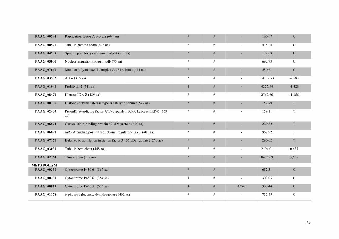
73
PAAG_00294
Replication factor-A protein (604 aa) * # - 190,97
C
PAAG_00570
Tubulin gamma chain (448 aa) * # - 435,26
C
PAAG_04999
Spindle pole body component alp14 (911 aa) * # - 172,63
C
PAAG_05000
Nuclear migration protein nudF (75 aa) * # - 692,73
C
PAAG_07669
Mannan polymerase II complex ANP1 subunit (461 aa) * # - 580,61
C
PAAG_03532
Actin (376 aa) * # - 14339,53
-2,683
PAAG_01041
Prohibitin-2 (311 aa) 1 # - 4227,94
-1,428
PAAG_08471
Histone H2A.Z (139 aa) * # - 2767,66
-1,356
PAAG_00106
Histone acetyltransferase type B catalytic subunit (547 aa) * # - 152,79
T
PAAG_02403
Pre-mRNA-splicing factor ATP-dependent RNA helicase PRP43 (769 aa)
* # - 159,11
T
PAAG_06574
Curved DNA-binding protein 42 kDa protein (420 aa) * # - 229,32
T
PAAG_06891
mRNA binding post-transcriptional regulator (Csx1) (401 aa) * # - 962,92
T
PAAG_07170
Eukaryotic translation initiation factor 3 135 kDa subunit (1270 aa)
* # - 290,02
T
PAAG_03031
Tubulin beta chain (448 aa) * # - 2194,01
0,635
PAAG_02364
Thioredoxin (117 aa) * # - 8475,69
3,636
METABOLISMPAAG_00230
Cytochrome P450 61 (167 aa) * # - 652,31
C
PAAG_00231
Cytochrome P450 61 (354 aa) 1 # - 303,05
C
PAAG_00827
Cytochrome P450 51 (603 aa) 4 # 0,749 308,44
C
PAAG_01178 6-phosphogluconate dehydrogenase (492 aa) * # - 752,45
C

74
PAAG_01222
Acyl-CoA dehydrogenase family protein (506 aa) * # - 367,58
C
PAAG_01390
Endoplasmic reticulum protein (570 aa) 1 # 0,658 223,43
C
PAAG_07503
1-acyl-sn-glycerol-3-phosphate acyltransferase beta (304 aa) 2 # - 425,34
C
PAAG_01542
Glutamyl-tRNA(Gln) amidotransferase subunit A (512 aa) * # - 210,86
C
PAAG_01833
2-succinylbenzoate-CoA ligase (583 aa) * # - 456,85
C
PAAG_01882
Acetolactate synthase catalytic subunit (702 aa) * # - 376,47
C
PAAG_01931
Phosphoacetylglucosamine mutase (549 aa) * # - 1039,05
C
PAAG_01974
Mitochondrial methylglutaconyl-CoA hydratase (306 aa) * # - 201,77
C
PAAG_02604
L-tyrosine decarboxylase (578 aa)* * # - 169,59
C
PAAG_02644
Kynurenine-oxoglutarate transaminase (452 aa) * # - 263,08
C
PAAG_02693
Saccharopine dehydrogenase (451 aa) * # - 215,45
C
PAAG_03024
3-ketodihydrosphingosine reductase tsc10 (386 aa) 1 # - 253,4
C
PAAG_03659
Protein kinase gsk3 (395 aa) * # - 248,12
C
PAAG_03722
Acyl-CoA desaturase (467 aa) 4 # - 738,82
C
PAAG_03974
NADPH cytochrome P450 oxidoreductase (379 aa) 1 # - 310,79
C
PAAG_04099
Methylcrotonoyl-CoA carboxylase subunit alpha (727 aa) * # - 205,93
C
PAAG_04238
Succinate dehydrogenase flavoprotein subunit (655 aa) * # - 179,7
C
PAAG_04564
Betaine aldehyde dehydrogenase (635 aa) * # - 193,12
C
PAAG_04719
Oligosaccharyl transferase subunit (739 aa) 13 # - 379,35
C
PAAG_05077
Class I alpha-mannosidase 1A (869 aa) 1 # - 360,41
C

75
PAAG_05644
Estradiol 17-beta-dehydrogenase 12-A (340 aa) 1 # - 208,66
C
PAAG_05910
Dolichyl-phosphate-mannose-protein mannosyltransferase (917 aa) 8 # - 657,3
C
PAAG_05972
N-acetyltransferase ECO1 (426 aa) * # - 236,06
C
PAAG_06217
Acetylornithine aminotransferase (485 aa) * # - 203,45
C
PAAG_06825
Fatty-acid amide hydrolase (607 aa) * # - 212,72
C
PAAG_06996
G-protein comlpex beta subunit CpcB (317 aa) * # - 587,65
C
PAAG_07631
Short chain dehydrogenase/reductase family protein (254 aa) * # - 222,77
C
PAAG_07682
Ribonucleoside-diphosphate reductase large chain (879 aa) * # - 272,71
C
PAAG_07785
116 kDa U5 small nuclear ribonucleoprotein component (990 aa) * # - 185,43
C
PAAG_08052
Long-chain-fatty-acid-CoA ligase (708 aa) * # - 251,97
C
PAAG_08100
O-acetylhomoserine (thiol)-lyase (440 aa) * # - 373,09
C
PAAG_08349
C-1-tetrahydrofolate synthase (940 aa) * # - 237,4
C
PAAG_08431
Dolichyl-phosphate-mannose-protein mannosyltransferase (746 aa) 9 # - 289,29
C
PAAG_08718
Succinate-semialdehyde dehydrogenase (498 aa) * # - 235,42
C
PAAG_08880
Cell division control protein (386 aa) * # - 207,96
C
PAAG_01575
Sterol 24-C-methyltransferase (378 aa) * # - 1375,67
-3,044
PAAG_07729
Isocitrate dehydrogenase subunit 2 (342 aa) * # - 2867,17
-2,510
PAAG_03978
3-hydroxyisobutyrate dehydrogenase (345 aa) * # - 394,12
-2,453
PAAG_01874
Dolichol-phosphate mannosyltransferase (280 aa) * # - 1456,63
-2,366
PAAG_02603 Aspartate aminotransferase (430 aa) * # - 1320,11
-2,279

76
PAAG_05419
rRNA 2'-O-methyltransferase fibrillarin (319 aa) * # - 662,01
-2,164
PAAG_00850
Glucosamine-fructose-6-phosphate aminotransferase (489 aa) * # - 3602,18
-1,991
PAAG_08174
Mannose-1-phosphate guanyltransferase subunit beta-A (506 aa) * # - 1194,16
-1,789
PAAG_00856
Isocitrate dehydrogenase subunit 1 (389 aa) * # - 2920,71
-1,731
PAAG_09072
Mitochondrial nuclease (336 aa) 1 # - 3854,97
-1,616
PAAG_06289
Tryptophan synthase (725 aa) * # - 390,84
-1,443
PAAG_02718
Mannose-1-phosphate guanyltransferase (416 aa) * # - 845,97
-1,428
PAAG_01002
NAD-specific glutamate dehydrogenase (1104 aa) * # - 691,16
-1,284
PAAG_00435
(R)-benzylsuccinyl-CoA dehydrogenase (445 aa) * # - 1227,68
-1,068
PAAG_06961
Aldehyde dehydrogenase (496 aa) * # - 744,17
-0,909
PAAG_01525
Fatty acid synthase subunit alpha reductase (1884 aa) * # - 389,8
-0,793
PAAG_00317
Septin 4 (346 aa) * # - 444,9
T
PAAG_00417
Succinyl-CoA ligase subunit alpha (332 aa) * # - 681,58
T
PAAG_01324
Folic acid synthesis protein (601 aa) * # - 193,22
T
PAAG_01787
Protein phosphatase PP2A regulatory subunit A (617 aa) * # - 185,69
T
PAAG_02011
Phosphoglucomutase (763 aa) * # - 140,67
T
PAAG_02016
Hydroxymethylglutaryl-CoA synthase (653 aa) * # - 368,86
T
PAAG_02336
Nudix hydrolase (323 aa)
* # - 114,18
T
PAAG_02554
3-hydroxyisobutyryl-CoA hydrolase (512 aa) * # - 243,08
T
PAAG_02859
Adenosylhomocysteinase (448 aa) * # - 491,12
T

77
PAAG_03643
6-phosphofructo-2-kinase (535 aa) * # - 156,97
T
PAAG_03689
3-ketoacyl-CoA thiolase B (420 aa) * # - 140,26
T
PAAG_04102
Isovaleryl-CoA dehydrogenase (431 aa) * # - 988,61
T
PAAG_04528
Acetylglutamate kinase (905 aa) * # - 184,26
T
PAAG_04856
Acetyl-CoA hydrolase (526 aa) * # - 152,67
T
PAAG_04966
Hydrolase (298 aa) * # - 155,53
T
PAAG_05005
Anthranilate synthase component 1 (556 aa) * # - 147,53
T
PAAG_05151
ATP-citrate-lyase (487 aa) * # - 655,78
T
PAAG_05198
Chorismate mutase (330 aa) * # - 286,91
T
PAAG_05253
Delta-1-pyrroline-5-carboxylate dehydrogenase (574 aa) * # - 208,72
T
PAAG_05328
3-isopropylmalate dehydrogenase A (363 aa) * # - 198,35
T
PAAG_05355
Acetamidase (543 aa) * # - 174,47
T
PAAG_05392
Betaine aldehyde dehydrogenase (479 aa) * # - 1998,5
T
PAAG_05406
Histidine biosynthesis trifunctional protein (871 aa) * # - 217,33
T
PAAG_06431
Ornithine aminotransferase (461 aa) * # - 1076,09
T
PAAG_06458
Dihydroorotate dehydrogenase (529 aa) * # - 172,97
T
PAAG_06596
Alcohol dehydrogenase (320 aa) * # - 173,77
T
PAAG_06953
Short chain dehydrogenase/reductase family (261 aa) * # - 204,27
T
PAAG_07605
Acetolactate synthase small subunit (338 aa) * # - 241,99
T
PAAG_07626
Cobalamin-independent synthase (772 aa) * # - 546,81
T

78
PAAG_07746
3-ketoacyl-CoA thiolase (422 aa) * # - 464,73
T
PAAG_07804
D-lactate dehydrogenase (525 aa) * # - 265,01
T
PAAG_08164
Homogentisate 1,2-dioxygenase (455 aa) * # - 187,81
T
PAAG_08512
Serine hydroxymethyltransferase (536 aa) * # - 672,51
T
PAAG_08731
Acetyl-CoA carboxylase (2273 aa) * # - 127,68
T
PAAG_08910
D-3-phosphoglycerate dehydrogenase (609 aa) * # - 218,07
T
PAAG_06506
Aspartate aminotransferase (464 aa) * # - 166,79
0,707
PAAG_08075
Citrate synthase (475 aa) * # - 1939,32
0,750
PAAG_07786
Acetyl-CoA acetyltransferase (400 aa) * # - 565,13
1,111
PAAG_01336
Long-chain-fatty-acid-CoA ligase (669 aa) * # - 166,58
1,284
PAAG_00468
4-aminobutyrate aminotransferase (286 aa) * # - 399,07
1,601
PAAG_07276
Glycogen synthase (711 aa) * # - 292,89
1,601
PAAG_01321
Oxidoreductase 2-nitropropane dioxygenase family (356 aa) * # - 4962,24
1,919
PROTEIN SYNTHESIS PAAG_00648
60S ribosomal protein L33-B (110 aa) * # - 1964,82
C
PAAG_01001 40S ribosomal protein S29 (261 aa) * # - 1992,17 C
PAAG_01050
Cytosolic large ribosomal subunit protein L30 (110 aa) * # - 3129,24
C
PAAG_01330
Eukaryotic translation initiation factor 3 RNA-binding subunit (289 aa) * # - 249,8
C
PAAG_01834
60S ribosomal protein L16 (203 aa) * # - 1527,43
C
PAAG_02889
Ribosomal protein L35 (126 aa) * # - 4498,1
C
PAAG_03019
60S ribosomal protein L6-B (205 aa) * # - 945,94
C

79
PAAG_03322
40S ribosomal protein S20 (117 aa) * # - 6248,36
C
PAAG_04253
Mitochondrial 54S ribosomal protein IMG1 (219 aa) * # - 350,09
C
PAAG_05233
60S ribosomal protein L26 (135 aa) * # - 1520,52
C
PAAG_05805
40S ribosomal protein S21 (89 aa) * # - 1297,9
C
PAAG_06668
Mitochondrial metallochaperone Sco1 (317 aa) 1 # - 253,54
C
PAAG_07381
Eukaryotic peptide chain release factor subunit 1 (475 aa) * # - 283,46
C
PAAG_07385
60S ribosomal protein L23a (154 aa) * # - 5895,08
C
PAAG_07707
60S ribosomal protein L10a (230 aa) * # - 1338,26
C
PAAG_05337 40S ribosomal protein S22 (131 aa)
* # - 12504,44 -4,588
PAAG_00724 Ribosomal protein 11 large subunit (176 aa) * # - 9552,25
-4,400
PAAG_01785 40S ribosomal protein S3 (271 aa) * # - 21541,52
-4,371
PAAG_01433
40S ribosomal protein S14 (152 aa) * # - 17244
-3,679
PAAG_04998
60S ribosomal protein L8-B (262 aa) * # - 2775,79
-3,549
PAAG_05379
60S ribosomal protein L17 (185 aa) * # - 1113,95
-3,520
PAAG_00765
60S ribosomal protein L36 (104 aa) * # - 2664,35
-3,477
PAAG_06569
60S ribosomal protein L43 (93 aa) * # - 5061,11
-3,477
PAAG_05017
40S ribosomal protein S10-A (166 aa) * # - 679,84
-3,333
PAAG_00952
60S ribosomal protein L20 (175 aa) * # - 5734,02
-3,304
PAAG_08888
60S ribosomal protein L4-A (373 aa) * # - 8767,51
-3,246
PAAG_03513
40S ribosomal protein S18 (156 aa) * # - 14815,58
-3,174
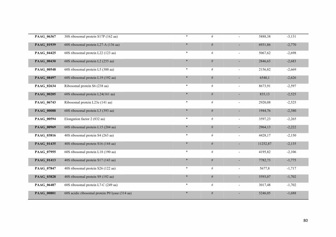
80
PAAG_06367
30S ribosomal protein S17P (162 aa) * # - 5888,38
-3,131
PAAG_01939
60S ribosomal protein L27-A (136 aa) * # - 6931,86
-2,770
PAAG_04425
60S ribosomal protein L22 (123 aa) * # - 5067,62
-2,698
PAAG_00430
60S ribosomal protein L2 (255 aa) * # - 2846,63
-2,683
PAAG_00548 60S ribosomal protein L5 (300 aa) * # - 2156,82
-2,669
PAAG_08497
60S ribosomal protein L19 (192 aa) * # - 6540,1
-2,626
PAAG_02634
Ribosomal protein S6 (238 aa) * # - 8673,91
-2,597
PAAG_00205
60S ribosomal protein L24(161 aa) * # - 835,13
-2,525
PAAG_06743
Ribosomal protein L23e (141 aa) * # - 2920,08
-2,525
PAAG_00088
60S ribosomal protein L3 (393 aa) * # - 1944,76
-2,380
PAAG_00594
Elongation factor 2 (832 aa) * # - 3597,23
-2,265
PAAG_00969
60S ribosomal protein L15 (204 aa) * # - 2964,13
-2,222
PAAG_03816
40S ribosomal protein S4 (263 aa) * # - 4428,17
-2,150
PAAG_01435
40S ribosomal protein S16 (144 aa) * # - 11252,87
-2,135
PAAG_07955
60S ribosomal protein L18 (190 aa) * # - 4195,82
-2,106
PAAG_01413
40S ribosomal protein S17 (143 aa) * # - 7782,73
-1,775
PAAG_07847
40S ribosomal protein S26 (122 aa) * # - 5677,8
-1,717
PAAG_03828
40S ribosomal protein S9 (192 aa) * # - 5593,07
-1,702
PAAG_06487
60S ribosomal protein L7-C (249 aa) * # - 3017,48
-1,702
PAAG_00801
60S acidic ribosomal protein P0 lyase (314 aa) * # - 5246,05
-1,688

81
PAAG_04965
60S ribosomal protein L31 (124 aa) * # - 4718
-1,688
PAAG_00347
60S ribosomal protein L9-B (193 aa) * # - 857,94
-1,587
PAAG_09043
40S ribosomal protein S2 (261 aa) * # - 6516,56
-1,298
PAAG_06320
60S ribosomal protein L13 (218 aa) * # - 7295,81
-1,342
PAAG_00264
40S ribosomal protein S8-B (203 aa) * # - 4093,71
-1,183
PAAG_06882
40S ribosomal protein S24 (135 aa) * # - 2114,3
-0,838
PAAG_02111
40S ribosomal protein S0 (296 aa) * # - 6272,65
-0,692
PAAG_03827
60S ribosomal protein L21-A (161 aa) * # - 5832
-0,692
PAAG_00150
Arginyl-tRNA synthetase (585 aa) * # - 290,14
T
PAAG_00240
Eukaryotic translation initiation factor 5A (139 aa) * # - 193,53
T
PAAG_00244
Polyadenylate-binding protein (823 aa) * # - 359,49
T
PAAG_00747
Eukaryotic translation initiation factor 2 subunit gamma (521 aa) * # - 297,63
T
PAAG_00815
Eukaryotic translation initiation factor 3 subunit A (1072 aa) * # - 123,62
T
PAAG_01141
Tyrosyl-tRNA synthetase (411 aa) * # - 375,36
T
PAAG_01786
Phenylalanyl-tRNA synthetase beta chain (601 aa) * # - 238,36
T
PAAG_02553
54S ribosomal protein YmL6 (347 aa) * # - 331,5
T
PAAG_03780
ATP-dependent RNA helicase DBP5 (505 aa) * # - 131,49
T
PAAG_04916
Elongation factor G (904 aa) * # - 118,6
T
PAAG_05015
Leucyl-tRNA synthetase (1119 aa) * # - 177,07
T
PAAG_05806
Phenylalanyl-tRNA synthetase alpha chain (521 aa) * # - 169,92
T

82
PAAG_06701
PCI domain-containing protein (750 aa) 1 # 0,790 352,24
T
PAAG_09083
TCTP family protein (183 aa) * # - 8742,68
T
PAAG_00238
Ubiquitin (219 aa) * # - 274,05
0,678
PAAG_08540
40S ribosomal protein S25 (158 aa) * # - 1542,68
0,721
PAAG_08955
40S ribosomal protein S3aE (256 aa) * # - 8490,22
1,125
PAAG_01425
Eukaryotic translation initiation factor 3 (478 aa) 2 # - 281,8
1,197
PAAG_04690
40S ribosomal protein S15 (154 aa) * # - 14548,56
1,399
CELLULAR COMMUNICATION/SIGNAL TRANSDUCTION MECHANISM PAAG_04025
Osmosensor protein (278 aa) 2 # - 510,94
C
PAAG_04784
COP9 signalosome complex subunit 1 (490 aa) * # - 362,35
C
PAAG_06815
Tricalbin-3 (1516 aa) 2 # - 165,67
C
PAAG_07640
Casein kinase I (284 aa) * # - 196,41
C
PAAG_08093
GTP-binding protein ypt3 (221 aa) * C 20 - 692,85
C
PAAG_08247
Calmodulin (150 aa) * # - 943,74
C
PAAG_01500
GTP-binding protein SAS1 (207 aa) * C 20 - 1546,88
-2,813
PAAG_02458
GTP-binding protein ypt7 (206 aa) * C 20 - 1281,11
-2,481
PAAG_07734
Adenylyl cyclase-associated protein (531 aa) * # - 244,83
-2,265
PAAG_07634
Small GTPase RhoA (192 aa) * C 15/C 20 - 6087,45
-1,053
PAAG_02817
Stomatin family protein (457 aa) * # - 362,08
-0,866
PAAG_05737
Calcium/calmodulin-dependent protein kinase (352 aa) * # - 125
T

83
PAAG_05737
Calcium/calmodulin-dependent protein kinase (386 aa) * # - 125
T
PAAG_05737 Calcium/calmodulin-dependent protein kinase (412 aa)
* # - 125 T
PAAG_05737 Calcium/calmodulin-dependent protein kinase (446 aa)
* # - 125 T
PAAG_06948
Mitogen-activated protein kinase MKC1 (432 aa) * # - 827,05
T
PAAG_02820
GTP-dependent nucleic acid-binding protein engD (413 aa) * # - 284,62
1,674
PAAG_00535
Mitogen-activated protein kinase HOG1 (361 aa) * # - 176,87
1,688
PROTEIN FATE (FOLDING, MODIFICATION, DESTINATION)PAAG_00188
Mitochondrial intermembrane space protein Mia40(401 aa) * # - 426,79
C
PAAG_00986
Disulfide isomerase Pdi1 (539 aa) * # 0,772 1771,33
C
PAAG_01706
26S proteasome regulatory subunit RPN10 (301 aa) * # - 265,68
C
PAAG_02085
Signal sequence processing protein SEC11 (198 aa) * # - 363,12
C
PAAG_02462
Alpha-1,2 mannosyltransferase KTR1 (505 aa) 1 # - 300,78
C
PAAG_02588
Peptidyl-prolyl cis-trans isomerase cyp8 (442 aa) * # - 213,93
C
PAAG_04882
Tetratricopeptide repeat domain-containing protein (415 aa) * # - 368,2
C
PAAG_04917
Vacuolar protein sorting-associated protein vps17 (582 aa) * # - 301,54
C
PAAG_05468
Translocation protein sec63 (693 aa) 3 # - 324,18
C
PAAG_05509
Proteasome non-ATPase regulatory subunit 11 (425 aa) * # - 174,96
C
PAAG_05962
Proteasome non-ATPase regulatory subunit 3 (642 aa) * # - 227,53
C
PAAG_06464
Disulfide-isomerase (728 aa) 1 # 0,851 252,29
C
PAAG_07235
SNARE domain-containing protein (252 aa) * # - 430,59
C
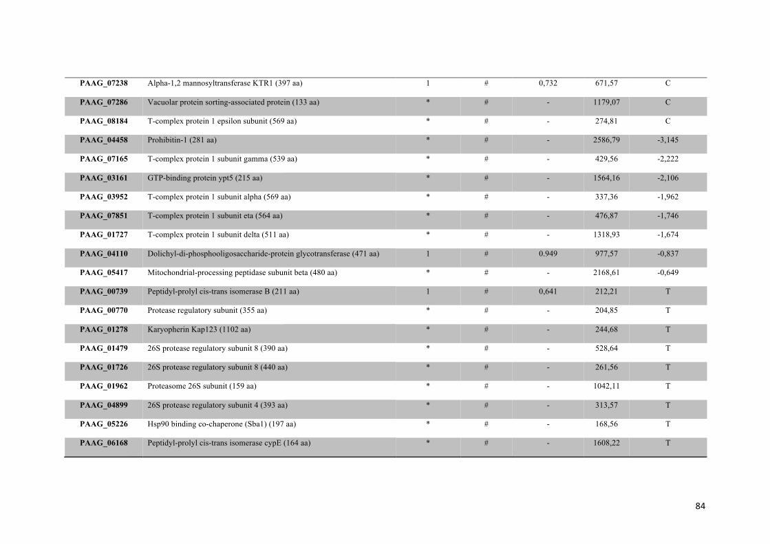
84
PAAG_07238
Alpha-1,2 mannosyltransferase KTR1 (397 aa) 1 # 0,732 671,57
C
PAAG_07286
Vacuolar protein sorting-associated protein (133 aa) * # - 1179,07
C
PAAG_08184
T-complex protein 1 epsilon subunit (569 aa) * # - 274,81
C
PAAG_04458
Prohibitin-1 (281 aa) * # - 2586,79
-3,145
PAAG_07165
T-complex protein 1 subunit gamma (539 aa) * # - 429,56
-2,222
PAAG_03161
GTP-binding protein ypt5 (215 aa) * # - 1564,16
-2,106
PAAG_03952
T-complex protein 1 subunit alpha (569 aa) * # - 337,36
-1,962
PAAG_07851
T-complex protein 1 subunit eta (564 aa) * # - 476,87
-1,746
PAAG_01727
T-complex protein 1 subunit delta (511 aa) * # - 1318,93
-1,674
PAAG_04110
Dolichyl-di-phosphooligosaccharide-protein glycotransferase (471 aa) 1 # 0.949 977,57
-0,837
PAAG_05417
Mitochondrial-processing peptidase subunit beta (480 aa) * # - 2168,61
-0,649
PAAG_00739
Peptidyl-prolyl cis-trans isomerase B (211 aa) 1 # 0,641 212,21
T
PAAG_00770
Protease regulatory subunit (355 aa) * # - 204,85
T
PAAG_01278
Karyopherin Kap123 (1102 aa) * # - 244,68
T
PAAG_01479
26S protease regulatory subunit 8 (390 aa) * # - 528,64
T
PAAG_01726
26S protease regulatory subunit 8 (440 aa) * # - 261,56
T
PAAG_01962
Proteasome 26S subunit (159 aa) * # - 1042,11
T
PAAG_04899
26S protease regulatory subunit 4 (393 aa) * # - 313,57
T
PAAG_05226
Hsp90 binding co-chaperone (Sba1) (197 aa) * # - 168,56
T
PAAG_06168
Peptidyl-prolyl cis-trans isomerase cypE (164 aa) * # - 1608,22
T

85
PAAG_06741
Proteasome 26S subunit (191 aa) * # - 194,8
T
PAAG_06946
Mitochondrial import inner membrane translocase subunit tim14 1 # - 330,93
T
PAAG_07394
T-complex protein 1 subunit zeta (541 aa) * # - 346,29
T
PAAG_00730
Clathrin heavy chain 1 (1650 aa) * # - 216,55
1,731
BIOGENESIS OF CELLULAR COMPONENTS PAAG_00622
Mitochondria fission 1 protein (154 aa) 1 # - 1001,94
C
PAAG_02917
GTP-binding protein Sey1 (873 aa) 2 # - 176,93
C
PAAG_03391
Chitin synthase B (928 aa) 6 # - 282,35
C
PAAG_03489
Cell lysis protein cwl1 (358 aa) 4 # - 1188,55
C
PAAG_05312
Coatomer subunit delta (566 aa) * # - 394,31
C
PAAG_08104
Alpha-tubulin (73 aa) * # - 449
C
PAAG_08209
Peroxin-11 (239 aa) 1 # - 3889,83
C
PAAG_06192
ARP2/3 complex 20 kDa subunit (170 aa) * # - 310,7
T
TRANSCRIPTION PAAG_02173
ATP-dependent RNA helicase mss116 (680 aa) * # - 163,62
C
PAAG_05498
Nucleolar protein NOP58 (890 aa) * # - 167,15
C
PAAG_07099
Histone H3.3 (137 aa) * # - 749,86
C
PAAG_00126
HistonE H4 2 (104 aa) * # - 9528,42
-2,727
PAAG_04651
GTP-binding nuclear protein GSP1/Ran (216 aa) * # - 4111,59
-2,597
PAAG_08833
ATP-dependent RNA helicase DHH1 (516 aa) * # - 496,42
-1,630
PAAG_07283
ATP-dependent RNA helicase FAL1 (409 aa) * # - 449,27
-0,923

86
PAAG_00734
ATP-dependent RNA helicase DED1 (684 aa) * # - 1519,36
-0,620
PAAG_04511
ATP-dependent RNA helicase SUB2 (468 aa) * # - 1837,61
-2,496
PAAG_02018
Transcription initiation factor IIB (353 aa) * # - 134,94
T
PAAG_03277
Pre-mRNA-processing factor 39 (542 aa) * # - 156,26
T
PAAG_04662
Cleavage and polyadenylation specificity factor subunit 5 (279 aa) * # - 473,08
T
PAAG_04726
Pirin (316 aa) * # - 125,14
T
PAAG_05087
RNP domain-containing protein (320 aa) * # - 178,49
T
PAAG_01062
Small nuclear ribonucleoprotein Sm D1 (122 aa) * # - 320,61
1,962
CELL RESCUE, DEFENSE AND VIRULENCE PAAG_02230
Cell division control protein (193 aa) * C 15/C 20 - 638,27
C
PAAG_05142
10 kDa heat shock protein, mitochondrial (104 aa) * # - 2943,71
C
PAAG_01262
Hsp70-like protein (629 aa) 1 # 0,864 3655,29
-2,640
PAAG_00797
Mitochondrial protein import protein MAS5 (411 aa) * C 15 - 982
-2,582
PAAG_07775
Heat shock protein SSB1 (558 aa) * # - 1741,24
-1,226
PAAG_08003
Hsp70-like protein (655 aa) * # - 3719,92
-1,010
PAAG_03292
Cytochrome c peroxidase (375 aa) * # - 6884,11
-0,808
PAAG_05679
Heat shock protein (696 aa) * # - 4757,84
-0,765
PAAG_05249
Aldehyde dehydrogenase (497 aa) * # - 1041,64
-0,721
PAAG_08664 Phenylacetate 2-hydroxylase (373 aa) * # - 427,39
-2,380
PAAG_00660 Replication factor C subunit 2 (405 aa) * # - 198,98 TPAAG_06811 Heat shock protein STI1 (579 aa) * # - 386,58
T

87
PAAG_07444
Chaperone protein dnaK (579 aa) * # - 140,36
T
PAAG_02686
Hsp90 co-chaperone AHA1 (330 aa) * # - 349,98
1,298
HYPOTHETICAL PROTEINSPAAG_00125 Conserved hypothetical protein (480 aa)
3 # - 206,51 C
PAAG_00171
Conserved hypothetical protein (204 aa) * # - 349,48
C
PAAG_00184
Conserved hypothetical protein (299 aa) 4 # - 304,39
C
PAAG_00187
Conserved hypothetical protein (241 aa) 3 # - 808,12
C
PAAG_00335
Conserved hypothetical protein (370 aa) * # - 223,46
C
PAAG_01045
Conserved hypothetical protein (151 aa) * # - 228,82
C
PAAG_01806
Conserved hypothetical protein (105 aa) * # - 991,89
C
PAAG_01870
Conserved hypothetical protein (296 aa) * # - 372,04
C
PAAG_01890
Conserved hypothetical protein (190 aa) 2 # - 251,17
C
PAAG_02068
Conserved hypothetical protein (450 aa) * # - 776,87
C
PAAG_02192
Conserved hypothetical protein (397 aa) * # - 270,83
C
PAAG_03505
Conserved hypothetical protein (203 aa) * # 0,840 350,01
C
PAAG_04529
Conserved hypothetical protein (117 aa) 1 # - 1843,87
C
PAAG_04540
Conserved hypothetical protein (180 aa) 1 # - 247,42
C
PAAG_04911
Conserved hypothetical protein (300 aa) 4 # - 422,48
C
PAAG_04991 Conserved hypothetical protein (618 aa)
* # - 160,24 C
PAAG_05224
Hypothetical protein (282 aa) * # - 382,88
C
PAAG_05272 Conserved hypothetical protein (435 aa) 1 # - 446,27 C
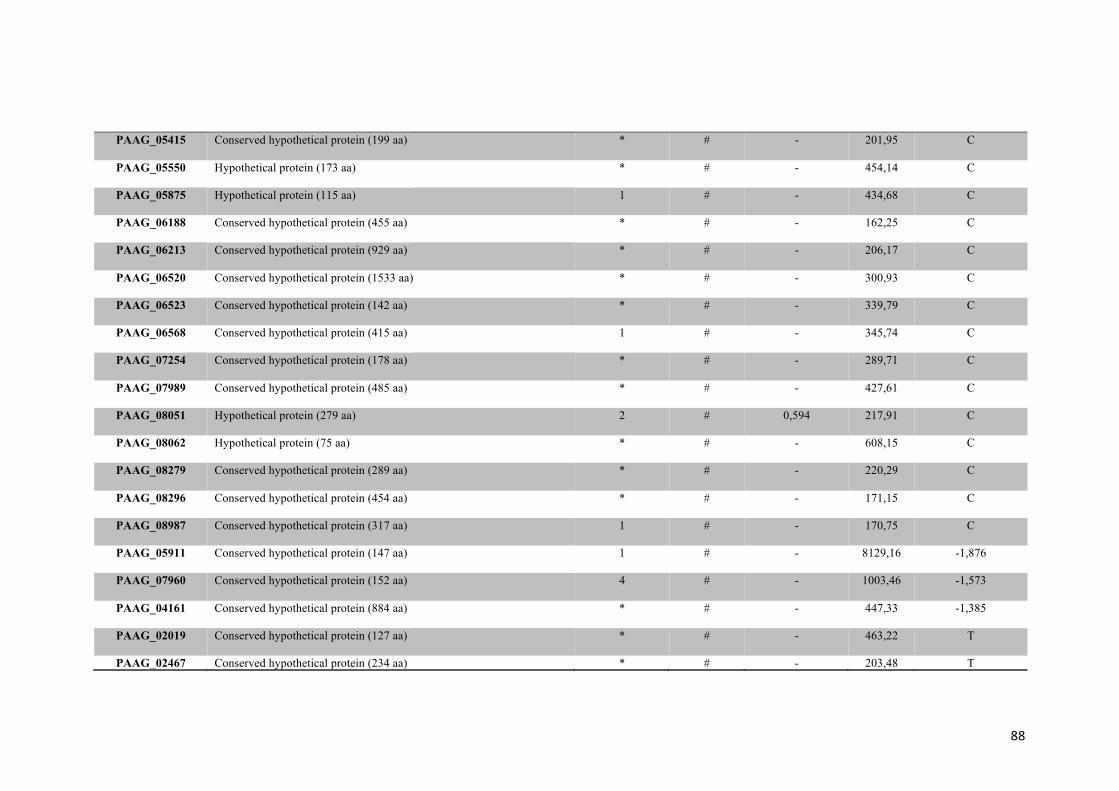
88
PAAG_05415
Conserved hypothetical protein (199 aa) * # - 201,95
C
PAAG_05550
Hypothetical protein (173 aa) * # - 454,14
C
PAAG_05875
Hypothetical protein (115 aa) 1 # - 434,68
C
PAAG_06188
Conserved hypothetical protein (455 aa) * # - 162,25
C
PAAG_06213
Conserved hypothetical protein (929 aa) * # - 206,17
C
PAAG_06520
Conserved hypothetical protein (1533 aa)
* # - 300,93
C
PAAG_06523
Conserved hypothetical protein (142 aa) * # - 339,79
C
PAAG_06568
Conserved hypothetical protein (415 aa)
1 # - 345,74
C
PAAG_07254
Conserved hypothetical protein (178 aa) * # - 289,71
C
PAAG_07989
Conserved hypothetical protein (485 aa) * # - 427,61
C
PAAG_08051
Hypothetical protein (279 aa) 2 # 0,594 217,91
C
PAAG_08062
Hypothetical protein (75 aa) * # - 608,15
C
PAAG_08279
Conserved hypothetical protein (289 aa) * # - 220,29
C
PAAG_08296
Conserved hypothetical protein (454 aa) * # - 171,15
C
PAAG_08987
Conserved hypothetical protein (317 aa) 1 # - 170,75
C
PAAG_05911
Conserved hypothetical protein (147 aa) 1 # - 8129,16
-1,876
PAAG_07960
Conserved hypothetical protein (152 aa) 4 # - 1003,46
-1,573
PAAG_04161
Conserved hypothetical protein (884 aa) * # - 447,33
-1,385
PAAG_02019
Conserved hypothetical protein (127 aa) * # - 463,22
T
PAAG_02467 Conserved hypothetical protein (234 aa) * # - 203,48 T
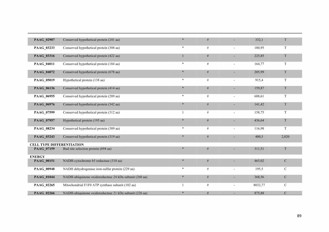
89
PAAG_02907 Conserved hypothetical protein (241 aa) * # - 332,1
T
PAAG_03233
Conserved hypothetical protein (308 aa) * # - 180,95
T
PAAG_03316
Conserved hypothetical protein (422 aa) 1 # - 225,85
T
PAAG_04011
Conserved hypothetical protein (184 aa) * # - 164,77
T
PAAG_04072
Conserved hypothetical protein (678 aa) * # - 205,99
T
PAAG_05019
Hypothetical protein (138 aa) * # - 915,4
T
PAAG_06136
Conserved hypothetical protein (414 aa) * # - 159,87
T
PAAG_06955
Conserved hypothetical protein (289 aa) * # - 688,61
T
PAAG_06976
Conserved hypothetical protein (342 aa) * # - 141,42
T
PAAG_07599
Conserved hypothetical protein (312 aa) 1 # - 158,75
T
PAAG_07957
Hypothetical protein (195 aa) * # - 436,64
T
PAAG_08234
Conserved hypothetical protein (389 aa)
* # - 116,98
T
PAAG_03243
Conserved hypothetical protein (319 aa) * # - 400,5
2,020
CELL TYPE DIFFERENTIATION PAAG_07159
Bud site selection protein (694 aa) * # - 311,51
T
ENERGYPAAG_00151
NADH-cytochrome b5 reductase (310 aa) * # - 465,02
C
PAAG_00948
NADH dehydrogenase iron-sulfur protein (229 aa) * # - 195,5
C
PAAG_01044
NADH-ubiquinone oxidoreductase 24 kDa subunit (260 aa) * # - 368,56
C
PAAG_02265
Mitochondrial F1F0 ATP synthase subunit (102 aa) 1 # - 8032,77
C
PAAG_02266 NADH-ubiquinone oxidoreductase 21 kDa subunit (226 aa) * # - 875,88 C

90
PAAG_02297
Cytochrome b-c1 complex subunit Rieske (251 aa) * # - 923,89
C
PAAG_02656
NADH-ubiquinone oxidoreductase 51 kDa subunit (516 aa) * # - 617,71
C
PAAG_03051
NADH-ubiquinone oxidoreductase 20.8 kDa subunit (161 aa) * # - 320,19
C
PAAG_03138
Alanine-glyoxylate aminotransferase (494 aa) * # - 280,59
C
PAAG_03160
Formation of crista junctions protein 1(686 aa)
1 # - 346,12
C
PAAG_04539
NADH-ubiquinone oxidoreductase 105 kDa subunit (95 aa) * # - 362,72
C
PAAG_04698
NADH-ubiquinone oxidoreductase 178 kDa subunit(177 aa)
* # - 784,8
C
PAAG_05091
NADH-ubiquinone oxidoreductase 213 kDa subunit (200 aa) 3 # - 357,62
C
PAAG_05454
Acyl-CoA dehydrogenase (440 aa) * # - 737,07
C
PAAG_05735
NADH-ubiquinone oxidoreductase 49 kDa subunit (480 aa) * # - 2857,94
C
PAAG_05876
Cytochrome c oxidase subunit VIa (143 aa)
* # - 2405,87
C
PAAG_06103
Succinate dehydrogenase iron-sulfur subunit (192 aa) * # - 511,18
C
PAAG_07791
NADH-ubiquinone oxidoreductase (212 aa) * # - 1529,82
C
PAAG_07868
NADH-quinone oxidoreductase subunit I 2 (230 aa) * # - 374,77
C
PAAG_08517
Propionate-CoA ligase (711 aa) 1 # - 271,08
C
PAAG_08916
LYR family protein (133 aa) * # - 1206,08
C
PAAG_06796
Cytochrome c oxidase polypeptide IV(198 aa) * # - 1101,38
-3,160
PAAG_08088
Cytochrome b-c1 complex subunit 2 (464 aa * # - 2793,7
-2,481
PAAG_08966
NADH-ubiquinone oxidoreductase 78 kDa subunit (729 aa) * # - 985,25
-2,366
PAAG_08057
Cytochrome c oxidase polypeptide V (200 aa) * # - 969,86
-2,150

91
PAAG_04838
ATP synthase subunit 4 (245 aa) * # - 8091,03
-2,034
PAAG_04570
ATP synthase D chain, mitochondrial (175 aa) * # - 6113,62
-1,890
PAAG_00771
Enolase (405 aa) * # - 2583,52
-1,775
PAAG_06380
Pyruvate kinase (535 aa) * # - 829,73
-1,385
PAAG_04166
Transaldolase (325 aa) * # - 1355,79
-1,342
PAAG_05031
NADH-ubiquinone oxidoreductase 40 kDa subunit (294 aa) * # - 1979,87
-1,169
PAAG_07672
Cytochrome b-c1 complex subunit 7 (123 aa) * # - 13389,36
-1,082
PAAG_04820
ATPase alpha subunit (466 aa) * # - 8922,32
-0,923
PAAG_01534
Pyruvate dehydrogenase E1 component subunit beta (378 aa) * # - 1193,17
-0,750
PAAG_08468
Glyceraldehyde-3-phosphate dehydrogenase (339 aa) * # - 20985,51
-3,708
PAAG_02653
Acetyl-coenzyme A synthetase (670 aa) * # - 1504,51
-1,702
PAAG_01725
Succinate dehydrogenase flavoprotein subunit (597 aa) * # - 393,21
-1,558
PAAG_00050
Pyruvate dehydrogenase protein X component (490 aa) * # - 283,14
T
PAAG_00173
Electron transfer flavoprotein subunit alpha (350 aa) * # - 684,28
T
PAAG_00403
Alcohol dehydrogenase (355 aa) * # - 685,88
T
PAAG_00726
Pyruvate carboxylase (1197 aa) * # - 147,03
T
PAAG_01650
Monothiol glutaredoxin-5 (277 aa) * # - 244,35
T
PAAG_02769
Pyruvate dehydrogenase protein X component (496 aa) * # - 388,71
T
PAAG_04374
Nonspecific lipid-transfer protein (399 aa) * # - 754,71
T
PAAG_04931
Electron transfer flavoprotein subunit beta (200 aa) * # - 1245,14
T

92
PAAG_07410
2,3-bisphosphoglycerate-independent phosphoglycerate mutase (522 aa) * # - 231,99
T
PAAG_08352
NADH-ubiquinone oxidoreductase (120 aa)
1 # - 375,07
T
PAAG_08551
ATP synthase subunit g (200 aa) * # - 3438,37
0,793
PAAG_04444
Transketolase (686 aa)* * # - 300,17
1,226
UNCLASSIFIED PROTEINSPAAG_02600
Peptidyl-tRNA hydrolase (254 aa) * # 0,665 7026,78
C
PAAG_03531
Phosphoribosylamine-glycine ligase (154 aa)
* # - 238,23
C
PAAG_06654
Signal sequence receptor alpha chain (275 aa) 1 # 0,769 390,4
C
PAAG_07680
Phosphoribulokinase/uridine kinase family protein (238 aa) * # - 246,36
C
PAAG_07846
CBS and PB1 domain-containing protein (672 aa) 1 # - 496,37
C
PAAG_04157 DUF1620 domain-containing protein (954 aa)
1 # 0,863 382,34
C
PAAG_06541
Erythrocyte band 7 integral membrane protein (361 aa) * # - 867,73
-1,876
PAAG_12011
Mitochondria cytochrome c oxidase subunit 2 (254 aa) 2 # - 2138,11
-3,260
PAAG_01280
Ser/Thr protein phosphatase family protein (680 aa) * # - 364,72
T
PAAG_01417
ER membrane DUF1077 domain-containing protein (105 aa) 2 # - 311,13
T
PAAG_01900
Serine/threonine-protein kinase psk1 (289 aa) * # - 128,89
T
PAAG_03152
CobW domain-containing protein (434 aa) * # - 146,14
T
PAAG_03741
HD domain-containing protein (143 aa) * # - 1373,61
T
PAAG_06995
TPR repeat protein (115 aa) * # - 161,42
T
PAAG_01347
Actin cytoskeleton protein (VIP1) (267 aa) * # - 317,17
-1,183
PREDICTED PROTEINS

93
PAAG_01245
Predicted protein (115 aa) * # - 289,73
C
PAAG_01439
Predicted protein (375 aa) * # - 205,68
C
PAAG_02238
Predicted protein (134 aa) * # - 251,65
C
PAAG_03337
Predicted protein (327 aa) * # - 202,04
C
PAAG_04219
Predicted protein (132 aa) * # - 311,02
C
PAAG_04363
Predicted protein (371 aa) * # - 230,13
C
PAAG_06540
Predicted protein (127 aa) * # - 218,9
C
PAAG_01478
Predicted protein (113 aa) * # - 151,67
T
PAAG_01702
Predicted protein (723 aa) * # - 119,2
T
PAAG_04993
Predicted protein (230 aa) * # - 213,59
TaAccession number– Accession number of the protein in the database of Paracoccidioides spp. (http://www.broadinstitute.org/annotation/genome/paracoccidioides_brasiliensis/MultiHome.html); bProtein annotation from Paracoccidioides genome database or by homology in NCBI database (http://www.ncbi.nlm.nih.gov/); cRepresents the amount of transmembrane domains identified in proteins using the program TMHMM version 2.0 (http://www.cbs.dtu.dk/services/TMHMM/); dPresence of posttranslational modification. For myristoylated protein (C14) programs Terminator (http://www.isv.cnrs-gif.fr/terminator3/test.php) and Myristoylator (http://web.expasy.org/myristoylator/) were used. Terminator program was used to identify palmitoylated proteins. Research by prenylated proteins was done by the program PresPs-Prenylation Prediction Suite (http://mendel.imp.ac.at/sat/PrePS/index.html) and to predict GPI anchor the program big-Pi fungi predictor (http://mendel.imp.ac.at/gpi/fungi_server.html) was used. C15 and C20 indicates the presence of a recognition site for the addition of a farnesyl or geranyl group (FTase and geranyltransferase type I, Rab geranylgeranyl transferase); ePresence of signal peptide was analyzed by program SignalP version 4.1 (http://www.cbs.dtu.dk/services/SignalP/). The values indicate the score of the proteins with the signal peptide, this value must be equal to or greater than 0.45; fIndicates the score of the protein; gProtein expression profiles in log2-fold change; *: Proteins that no presented transmembrane domain. #: Proteins that no presented posttranslational modification. -: Proteins that no presented signal peptide. C: Proteins only identified in the control condition. T: Proteins only identified in the zinc deprivation condition. The functional classification was performed used the database of FunCat2 (http://pedant.gsf.de/pedant3htmlview/pedant3view?Method=analysis&Db=p3_r48325_Par_brasi_Pb01).

94
The database Gene Ontology (GO) and WolfPSORT was employed to evaluate the possible localization of proteins in cell membranes of Paracoccidioides sp.. Some proteins did not show any form of classical association with the lipid bilayer, but showed that the localization subcellular were in regions of cell membranes, with score ≥ 50.

95
REFERENCES
BAGAGLI, E.; BOSCO, S. M.; THEODORO, R. C.; FRANCO, M. Phylogenetic and evolutionary aspects of Paracoccidioides brasiliensis reveal a long coexistence with animal hosts that explain several biological features of the pathogen. Infect Genet Evol 6(5): 344-351, 2006.
BAILAO, E. F.; PARENTE, J. A.; PIGOSSO, L. L.; DE CASTRO, K. P.; FONSECA, F. L.; SILVA-BAILAO, M. G.; BAO, S. N.; BAILAO, A. M.; RODRIGUES, M. L.; HERNANDEZ, O.; MCEWEN, J. G.; SOARES, C. M. Hemoglobin uptake by Paracoccidioides spp. is receptor-mediated. PLoS Negl Trop Dis 8(5): e2856, 2014.
BARBOSA, M. S.; BAO, S. N.; ANDREOTTI, P. F.; DE FARIA, F. P.; FELIPE, M. S.; DOS SANTOS FEITOSA, L.; MENDES-GIANNINI, M. J.; SOARES, C. M. Glyceraldehyde-3-phosphate dehydrogenase of Paracoccidioides brasiliensis is a cell surface protein involved in fungal adhesion to extracellular matrix proteins and interaction with cells. Infect Immun 74(1): 382-389, 2006.
BRADFORD, M. M. A rapid and sensitive method for the quantitation of microgram quantities of protein utilizing the principle of protein-dye binding. Anal Biochem 72: 248-254, 1976.
CARMAN, G. M.; HAN, G. S. Regulation of phospholipid synthesis in Saccharomyces cerevisiae by zinc depletion. Biochim Biophys Acta 1771(3): 322-330, 2007.
DA FONSECA, C. A.; JESUINO, R. S.; FELIPE, M. S.; CUNHA, D. A.; BRITO, W. A.; SOARES, C. M. Two-dimensional electrophoresis and characterization of antigens from Paracoccidioides brasiliensis. Microbes Infect 3(7): 535-542, 2001.
EIDE, D. J. Multiple regulatory mechanisms maintain zinc homeostasis in Saccharomyces cerevisiae. J Nutr 133(5 Suppl 1): 1532S-1535S, 2003.
EPHRITIKHINE, G.; FERRO, M.; ROLLAND, N. Plant membrane proteomics. Plant Physiol Biochem 42(12): 943-962, 2004.
FAULHAMMER, F.; KONRAD, G.; BRANKATSCHK, B.; TAHIROVIC, S.; KNODLER, A.; MAYINGER, P. Cell growth-dependent coordination of lipid signaling and glycosylation is mediated by interactions between Sac1p and Dpm1p. J Cell Biol 168(2): 185-191, 2005.
FAVA-NETTO, C. Estudos quantitativos sobre a fixação de complemento na blastomicose sul americana, com antígeno polissacarídico. Arq Cir Clin Exp 18: 197- 254, 1955.
FINNEY, L. A.; O'HALLORAN, T. V. Transition metal speciation in the cell: insights from the chemistry of metal ion receptors. Science 300(5621): 931-936, 2003.

96
GITAN, R. S.; LUO, H.; RODGERS, J.; BRODERIUS, M.; EIDE, D. Zinc-induced inactivation of the yeast ZRT1 zinc transporter occurs through endocytosis and vacuolar degradation. J Biol Chem 273(44): 28617-28624, 1998.
HEINISCH, J. J.; LORBERG, A.; SCHMITZ, H. P.; JACOBY, J. J. The protein kinase C-mediated MAP kinase pathway involved in the maintenance of cellular integrity in Saccharomyces cerevisiae. Mol Microbiol 32(4): 671-680, 1999.
IWANYSHYN, W. M.; HAN, G. S.; CARMAN, G. M. Regulation of phospholipid synthesis in Saccharomyces cerevisiae by zinc. J Biol Chem 279(21): 21976-21983, 2004.
KONRAD, G.; SCHLECKER, T.; FAULHAMMER, F.; MAYINGER, P. Retention of the yeast Sac1p phosphatase in the endoplasmic reticulum causes distinct changes in cellular phosphoinositide levels and stimulates microsomal ATP transport. J Biol Chem 277(12): 10547-10554, 2002.
LAIRD, N. M.; HORVATH, S.; XU, X. Implementing a unified approach to family-based tests of association. Genet Epidemiol 19 Suppl 1: S36-42, 2000.
LIMA, P. S.; CASALETTI, L.; BAILAO, A. M.; DE VASCONCELOS, A. T.; FERNANDES GDA, R.; SOARES, C. M. Transcriptional and proteomic responses to carbon starvation in Paracoccidioides. PLoS Negl Trop Dis 8(5): e2855, 2014.
LONGO, L. V.; NAKAYASU, E. S.; GAZOS-LOPES, F.; VALLEJO, M. C.; MATSUO, A. L.; ALMEIDA, I. C.; PUCCIA, R. Characterization of cell wall lipids from the pathogenic phase of Paracoccidioides brasiliensis cultivated in the presence or absence of human plasma. PLoS One 8(5): e63372, 2013.
MCEWEN, J. G.; BEDOYA, V.; PATINO, M. M.; SALAZAR, M. E.; RESTREPO, A. Experimental murine paracoccidiodomycosis induced by the inhalation of conidia. J Med Vet Mycol 25(3): 165-175, 1987.
MOCCHEGIANI, E.; MUZZIOLI, M. Therapeutic application of zinc in human immunodeficiency virus against opportunistic infections. J Nutr 130(5S Suppl): 1424S-1431S, 2000.
MURAD, A. M.; SOUZA, G.; GARCIA, J.; RECH, E. Characterisation and quantitation expression analysis of recombinant proteins in plant complex mixtures using nanoUPLC mass spectrometry. Protocol Exchange: 1-17, 2011a.
MURAD, A. M.; SOUZA, G. H.; GARCIA, J. S.; RECH, E. L. Detection and expression analysis of recombinant proteins in plant-derived complex mixtures using nanoUPLC-MS(E). J Sep Sci 34(19): 2618-2630, 2011b.
MURAD, A. M.; RECH, E. L. NanoUPLC-MSE proteomic data assessment of soybean seeds using the Uniprot database. BMC Biotechnol 12: 82, 2012.

97
PARENTE, A. F.; DE REZENDE, T. C.; DE CASTRO, K. P.; BAILAO, A. M.; PARENTE, J. A.; BORGES, C. L.; SILVA, L. P.; SOARES, C. M. A proteomic view of the response of Paracoccidioides yeast cells to zinc deprivation. Fungal Biol 117(6): 399-410, 2013.
RESTREPO, A.; JIMENEZ, B. E. Growth of Paracoccidioides brasiliensis yeast phase in a chemically defined culture medium. J Clin Microbiol 12(2): 279-281, 1980.
RUTHERFORD, J. C.; BIRD, A. J. Metal-responsive transcription factors that regulate iron, zinc, and copper homeostasis in eukaryotic cells. Eukaryot Cell 3(1): 1-13, 2004.
SCHORR, M.; THEN, A.; TAHIROVIC, S.; HUG, N.; MAYINGER, P. The phosphoinositide phosphatase Sac1p controls trafficking of the yeast Chs3p chitin synthase. Curr Biol 11(18): 1421-1426, 2001.
SILVA, M. G.; SCHRANK, A.; BAILAO, E. F.; BAILAO, A. M.; BORGES, C. L.; STAATS, C. C.; PARENTE, J. A.; PEREIRA, M.; SALEM-IZACC, S. M.; MENDES-GIANNINI, M. J.; OLIVEIRA, R. M.; SILVA, L. K.; NOSANCHUK, J. D.; VAINSTEIN, M. H.; DE ALMEIDA SOARES, C. M. The homeostasis of iron, copper, and zinc in Paracoccidioides brasiliensis, Cryptococcus neoformans var. grubii, and Cryptococcus gattii: a comparative analysis. Front Microbiol 2: 49, 2011.
TAHIROVIC, S.; SCHORR, M.; THEN, A.; BERGER, J.; SCHWARZ, H.; MAYINGER, P. Role for lipid signaling and the cell integrity MAP kinase cascade in yeast septum biogenesis. Curr Genet 43(2): 71-78, 2003.
TAN, S.; TAN, H. T.; CHUNG, M. C. Membrane proteins and membrane proteomics. Proteomics 8(19): 3924-3932, 2008.
VIDAKOVICS, M. L.; PABA, J.; LAMBERTI, Y.; RICART, C. A.; DE SOUSA, M. V.; RODRIGUEZ, M. E. Profiling the Bordetella pertussis proteome during iron starvation. J Proteome Res 6(7): 2518-2528, 2007.
WHITTERS, E. A.; CLEVES, A. E.; MCGEE, T. P.; SKINNER, H. B.; BANKAITIS, V. A. SAC1p is an integral membrane protein that influences the cellular requirement for phospholipid transfer protein function and inositol in yeast. J Cell Biol 122(1): 79-94, 1993.
WU, C. Y.; BIRD, A. J.; WINGE, D. R.; EIDE, D. J. Regulation of the yeast TSA1 peroxiredoxin by ZAP1 is an adaptive response to the oxidative stress of zinc deficiency. J Biol Chem 282(4): 2184-2195, 2007.
WU, C. Y.; BIRD, A. J.; CHUNG, L. M.; NEWTON, M. A.; WINGE, D. R.; EIDE, D. J. Differential control of Zap1-regulated genes in response to zinc deficiency in Saccharomyces cerevisiae. BMC Genomics 9: 370, 2008.

98
ZHAO, H.; EIDE, D. The ZRT2 gene encodes the low affinity zinc transporter in Saccharomyces cerevisiae. J Biol Chem 271(38): 23203-23210, 1996a.
ZHAO, H.; EIDE, D. The yeast ZRT1 gene encodes the zinc transporter protein of a high-affinity uptake system induced by zinc limitation. Proc Natl Acad Sci U S A 93(6): 2454-2458, 1996b.
ZHAO, H.; EIDE, D. J. Zap1p, a metalloregulatory protein involved in zinc-responsive transcriptional regulation in Saccharomyces cerevisiae. Mol Cell Biol 17(9): 5044-5052, 1997.

99
5. DISCUSSÃO
As proteínas de membrana são de importância para o desenvolvimento celular
pois executam uma variedade de processos como adesão, sinalização, transporte celular
e captação de nutrientes (GILMORE e WASHBURN, 2010). Diante disso, estudos
foram desenvolvidos para caracterização destas proteínas em diferentes organismos e
condições. Em S. cerevisiae, análise proteômica da membrana plasmática durante o
estresse salino revelou mudanças significantes na abundância de várias proteínas de
membrana plasmática, as quais podem afetar a morfologia da membrana plasmática e a
homeostase iônica (SZOPINSKA et al., 2011). Em plantas, foi realizada análise
proteômica com o objetivo de avaliar a regulação e a função de proteínas de membrana
na absorção de ferro e estresse oxidativo. Os resultados obtidos indicam que as
proteínas envolvidas no transporte e sinalização foram as mais abundantes (HOPFF et
al., 2013).
Zinco, como outros metais, é um nutriente essencial e desempenha papeis como
um co-factor estrutural ou catalítico para muitas proteínas. Em condições de privação de
zinco a resposta celular é regulada, principalmente ao nível da transcrição (ZHAO e
EIDE, 1997). Em S. cerevisiae, estudos têm revelado tanto respostas homeostáticas
como respostas adaptativas à deficiência de zinco. A resposta homeostática é
caracterizada pelo controle da absorção de zinco, armazenamento vacuolar de zinco e
conservação de grandes quantidades de zinco intracelulares. Esta resposta é considerada
uma primeira linha de defesa contra o estresse gerado pela privação de zinco. A resposta
adaptativa, também conhecida como segunda linha de defesa, permite adaptação celular
em condições de deficiência de zinco. Em levedura, esta resposta é induzida em
condições de limitação de zinco graves e ocorre quando as células já não são capazes de
captar o zinco para o crescimento ideal. As estratégias utilizadas pelas células de
levedura como segunda linha de defesa sobre deficiência de zinco incluem mecanismos
de resistência ao estresse oxidativo, alterações nas vias de síntese de lipídios,
remodelação de assimilação de sulfato e parede celular (WU et al., 2008; WU et al.,
2007).
Nosso grupo demonstrou anteriormente que quando células de Paracoccidioides
sp. são incubadas em condições de privação de zinco proteínas relacionadas ao estresse
oxidativo são induzidas, assim como em S. cerevisiae. Além disso, a ausência de zinco
100
induz gliconeogênese e reprime o ciclo do metil-citrato (PARENTE et al., 2013). Para
complementar estes resultados obtidos após a análise de proteínas citoplasmáticas, no
presente trabalho analisamos o proteoma de membrana de células de levedura de
Paracoccidioides sp. em condições de privação de zinco. Foram identificados 460
proteínas que foram regulados em células de levedura de Pb01 expostas ao quelante de
zinco TPEN. Destes, 141 foram classificados como proteínas da membrana celular de
acordo com a análise in silico realizada. Identificamos proteínas relacionadas
metabolismo glicerofosfolipídios e integridade celular.
A enzima 1-AGPAT (1-acil-sn-glicerol-3-phosphate aciltransferase beta), que
apresenta dois domínios transmembrana e que pertence ao metabolismo de
glicerofosfolipídeos, foi reprimida em condições de privação de zinco. Esta enzima
catalisa a formação do ácido fosfatídico (PA) a partir de ácido lisofosfatídico (LPA),
incorporando um grupo acil na posição sn-2. O PA produzido pela atividade da 1-
AGPAT é substrato para via de fosfolípidos para produção de fosfatidilserina (PS),
fosfatidilinositol (PI), fosfatidiletanolamina (PE) e fosfatidilcolina (PC), que são
encontradas em leveduras (CARMAN e HAN, 2007) e também nas membranas
Paracoccidioides spp. (LONGO et al., 2013). Nossos dados estão de acordo com um
estudo anterior que demonstrou que em S. cerevisiae as atividades das enzimas
envolvidas na via de biossintese de PS, PE e PC foram reduzidas sob a deficiência de
zinco (IWANYSHYN et al., 2004). Como PA é o substrato a principal na síntese de
todos estes fosfolípidos, podemos sugerir que a diminuição da produção de PA leva à
redução dos níveis de PS, PE e PC e, talvez, a defeitos na estrutura da membrana
plasmática.
A fosfatase fosfoinositidica (Sac1) é uma proteína que apresenta domínio
transmembrana, encontrada no retículo endoplasmático (RE) e complexo de Golgi
(KONRAD et al., 2002; WHITTERS et al., 1993). Esta enzima tem papel específico na
secreção e age como antagonista da fosfatidilinositol 4-quinase PiKp no tráfego de
Golgi (SCHORR et al., 2001). A rápida mudança de localização da Sac1 entre o RE e o
Golgi ocorre em resposta a condições de crescimento. A localização da Sac1 no RE
ocorre apenas durante os períodos de crescimento exponencial e é mediada pela
interação com dolicol fosfato manose sintase (Dpm1) através do contato direto entre os
domínios transmembrana (FAULHAMMER et al., 2005). As proteínas Sac1 e Dpm1
são necessárias para a biossíntese de oligossacarídeos e neste estudo estas proteínas

101
foram reguladas negativamente sugerindo que o metabolismo de carboidratos foi
afetado pela privação de zinco como descrito por Wu e colaboradores (2008).
Estudos tem mostrado que Sac1 está relacionada a integridade da parede celular
devido a defeitos no tráfego da quitina sintase e biossíntese de quitina observados no
mutante para Sac1. Esse fenótipo foi mais severo para o duplo mutante sac1 e slt2. Slt2
é uma proteína que não está ligada à membrana, envolvida na reorganização da parede
celular em resposta ao estresse causado na parede (HEINISCH et al., 1999). Por
conseguinte, estas proteínas têm um papel sinergístico na regulação da construção da
parede celular (TAHIROVIC et al., 2003). Um homólogo à Slt2 foi encontrado no
nosso proteoma de membrana, proteína quinase ativada por mitógeno, Mkc1.
Curiosamente, Mkc1 foi induzida pela deficiência de zinco, enquanto Sac1 foi
reprimida. Como Sac1 está envolvida na integridade da parede celular, sugere-se que a
ausência de zinco provoque defeitos da parede celular, como descrito em S. cerevisiae
(WU et al., 2008). Para amenizar o estresse na parede celular, Mkc1 foi induzida. De
acordo com o efeito da privação de zinco na parede da célula, verificou-se que a quitina-
sintase B, uma proteína de membrana, foi também reprimida na ausência de zinco.
Para o nosso conhecimento, este é o primeiro trabalho que descreve a resposta
de Paracoccidioides spp. a privação de micronutrientes a nível proteômico de
membranas. As novas descobertas mostradas aqui, associadas com as respostas à
deficiência de zinco a nível citoplasmático, irá acrescentar informações importantes
para o crescente conhecimento de como Paracoccidioides spp. comporta-se em
condições de privação de metais. Como a imunidade nutricional é uma estratégia de
para evitar a infecção, os mecanismos utilizados pelo fungo para sobreviver e se adaptar
a essa condição são fundamentais para patogenicidade.

102
6. CONCLUSÃO
A imunidade nutricional é uma das estratégias utilizada pelo hospedeiro para
impedir o desenvolvimento de micro-organismos patogênicos. Assim, o micro-
organismo, diante de uma condição de estresse (privação de nutrientes no meio) utiliza
mecanismos para se adaptar e sobreviver. O desenvolvimento destes mecanismos são
fundamentais para sua patogenicidade. Com os resultados obtidos no presente trabalho,
podemos concluir que durante a privação de zinco o Paracoccidioides sp. apresentou
alterações na sua estrutura como na integridade celular e no metabolismo de
fosfolipídeos.

103
7. PERSPECTIVAS
- Imunofluorêscencia com calcofluor white;
- Western blot com anticorpos específicos para marcadores de membranas;
- Análise da expressão de genes por qRT-PCRs.

104
8. REFERÊNCIAS
ALBERTS, B.; JOHNSON, A.; LEWIS, J.; RAFF, M.; ROBERTS, K.; WALTER, P. Molecular Biology of the Cell. In. New York: Garland Publishing, 1994. 485-488.
ALBERTS, B.; JOHNSON, A.; LEWIS, J.; RAFF, M.; ROBERTS, K.; WALTER, P. Biologia molecular da célula. In. Porto Alegre, 2004.
ALMEIDA, S. M. Central nervous system paracoccidioidomycosis: an overview. Braz J Infect Dis 9(2): 126-133, 2005.
APPELBERG, R. Macrophage nutriprive antimicrobial mechanisms. J Leukoc Biol 79(6): 1117-1128, 2006.
ARISTIZABAL, B. H.; CLEMONS, K. V.; STEVENS, D. A.; RESTREPO, A. Morphological transition of Paracoccidioides brasiliensis conidia to yeast cells: in vivo inhibition in females. Infect Immun 66(11): 5587-5591, 1998.
BAGAGLI, E.; SANO, A.; COELHO, K. I.; ALQUATI, S.; MIYAJI, M.; DE CAMARGO, Z. P.; GOMES, G. M.; FRANCO, M.; MONTENEGRO, M. R. Isolation of Paracoccidioides brasiliensis from armadillos (Dasypus noveminctus) captured in an endemic area of paracoccidioidomycosis. Am J Trop Med Hyg 58(4): 505-512, 1998.
BAGAGLI, E.; BOSCO, S. M.; THEODORO, R. C.; FRANCO, M. Phylogenetic and evolutionary aspects of Paracoccidioides brasiliensis reveal a long coexistence with animal hosts that explain several biological features of the pathogen. Infect Genet Evol 6(5): 344-351, 2006.
BAGAGLI, E.; THEODORO, R. C.; BOSCO, S. M.; MCEWEN, J. G. Paracoccidioides brasiliensis: phylogenetic and ecological aspects. Mycopathologia 165(4-5): 197-207, 2008.
BAILAO, A. M.; SCHRANK, A.; BORGES, C. L.; DUTRA, V.; WALQUIRIA INES MOLINARI-MADLUM, E. E.; SOARES FELIPE, M. S.; SOARES MENDES-GIANNINI, M. J.; MARTINS, W. S.; PEREIRA, M.; MARIA DE ALMEIDA SOARES, C. Differential gene expression by Paracoccidioides brasiliensis in host interaction conditions: representational difference analysis identifies candidate genes associated with fungal pathogenesis. Microbes Infect 8(12-13): 2686-2697, 2006.
BAILAO, A. M.; SHRANK, A.; BORGES, C. L.; PARENTE, J. A.; DUTRA, V.; FELIPE, M. S.; FIUZA, R. B.; PEREIRA, M.; DE ALMEIDA SOARES, C. M. The transcriptional profile of Paracoccidioides brasiliensis yeast cells is influenced by human plasma. FEMS Immunol Med Microbiol 51(1): 43-57, 2007.
BAILAO, E. F.; PARENTE, J. A.; PIGOSSO, L. L.; DE CASTRO, K. P.; FONSECA, F. L.; SILVA-BAILAO, M. G.; BAO, S. N.; BAILAO, A. M.; RODRIGUES, M. L.;

105
HERNANDEZ, O.; MCEWEN, J. G.; SOARES, C. M. Hemoglobin uptake by Paracoccidioides spp. is receptor-mediated. PLoS Negl Trop Dis 8(5): e2856, 2014.
BASTOS, K. P.; BAILAO, A. M.; BORGES, C. L.; FARIA, F. P.; FELIPE, M. S.; SILVA, M. G.; MARTINS, W. S.; FIUZA, R. B.; PEREIRA, M.; SOARES, C. M. The transcriptome analysis of early morphogenesis in Paracoccidioides brasiliensis mycelium reveals novel and induced genes potentially associated to the dimorphic process. BMC Microbiol 7: 29, 2007.
BELITARDO, D. R.; CALEFI, A. S.; BORGES, I. K.; DE OLIVEIRA, G. G.; SBEGHEN, M. R.; ITANO, E. N.; DE CAMARGO, Z. P.; ONO, M. A. Detection of antibodies against Paracoccidioides brasiliensis in free-range domestic pigs (Sus scrofa). Mycopathologia 177(1-2): 91-95, 2014.
BOCCA, A. L.; AMARAL, A. C.; TEIXEIRA, M. M.; SATO, P.; SHIKANAI-YASUDA, M. A.; SOARES FELIPE, M. S. Paracoccidioidomycosis: eco-epidemiology, taxonomy and clinical and therapeutic issues. Future Microbiol 8(9): 1177-1191, 2013.
BRUMMER, E.; CASTANEDA, E.; RESTREPO, A. Paracoccidioidomycosis: an update. Clin Microbiol Rev 6(2): 89-117, 1993.
CAMARGO, Z. P.; FRANCO, M. F. Current knowledge on pathogenesis and immunodiagnosis of paracoccidioidomycosis. Rev Iberoam Micol 17(2): 41-48, 2000.
CANO, M. I.; CISALPINO, P. S.; GALINDO, I.; RAMIREZ, J. L.; MORTARA, R. A.; DA SILVEIRA, J. F. Electrophoretic karyotypes and genome sizing of the pathogenic fungus Paracoccidioides brasiliensis. J Clin Microbiol 36(3): 742-747, 1998.
CARAS, I. W.; WEDDELL, G. N.; DAVITZ, M. A.; NUSSENZWEIG, V.; MARTIN, D. W., JR. Signal for attachment of a phospholipid membrane anchor in decay accelerating factor. Science 238(4831): 1280-1283, 1987.
CARMAN, G. M.; HAN, G. S. Regulation of phospholipid synthesis in Saccharomyces cerevisiae by zinc depletion. Biochim Biophys Acta 1771(3): 322-330, 2007.
CARRERO, L. L.; NINO-VEGA, G.; TEIXEIRA, M. M.; CARVALHO, M. J.; SOARES, C. M.; PEREIRA, M.; JESUINO, R. S.; MCEWEN, J. G.; MENDOZA, L.; TAYLOR, J. W.; FELIPE, M. S.; SAN-BLAS, G. New Paracoccidioides brasiliensis isolate reveals unexpected genomic variability in this human pathogen. Fungal Genet Biol 45(5): 605-612, 2008.
CASTRO, N. S.; DE CASTRO, K. P.; ORLANDI, I.; FEITOSA LDOS, S.; ROSA E SILVA, L. K.; VAINSTEIN, M. H.; BAO, S. N.; VAI, M.; SOARES, C. M. Characterization and functional analysis of the beta-1,3-glucanosyltransferase 3 of the human pathogenic fungus Paracoccidioides brasiliensis. FEMS Yeast Res 9(1): 103-114, 2009.

106
CONTI DIAZ, I. A. On the unknown ecological niche of Paracoccidioides brasiliensis: our hypothesis of 1989: present status and perspectives. Rev Inst Med Trop Sao Paulo 49(2): 131-134, 2007.
CORBIN, B. D.; SEELEY, E. H.; RAAB, A.; FELDMANN, J.; MILLER, M. R.; TORRES, V. J.; ANDERSON, K. L.; DATTILO, B. M.; DUNMAN, P. M.; GERADS, R.; CAPRIOLI, R. M.; NACKEN, W.; CHAZIN, W. J.; SKAAR, E. P. Metal chelation and inhibition of bacterial growth in tissue abscesses. Science 319(5865): 962-965, 2008.
CORREDOR, G. G.; CASTANO, J. H.; PERALTA, L. A.; DIEZ, S.; ARANGO, M.; MCEWEN, J.; RESTREPO, A. Isolation of Paracoccidioides brasiliensis from the nine-banded armadillo Dasypus novemcinctus, in an endemic area for paracoccidioidomycosis in Colombia. Rev Iberoam Micol 16(4): 216-220, 1999.
COUTINHO, Z. F.; SILVA, D.; LAZERA, M.; PETRI, V.; OLIVEIRA, R. M.; SABROZA, P. C.; WANKE, B. Paracoccidioidomycosis mortality in Brazil (1980-1995). Cad Saude Publica 18(5): 1441-1454, 2002.
EIDE, D. J. Multiple regulatory mechanisms maintain zinc homeostasis in Saccharomyces cerevisiae. J Nutr 133(5 Suppl 1): 1532S-1535S, 2003.
EIDE, D. J. The SLC39 family of metal ion transporters. Pflugers Arch 447(5): 796-800, 2004.
ENGLUND, P. T. The structure and biosynthesis of glycosyl phosphatidylinositol protein anchors. Annu Rev Biochem 62: 121-138, 1993.
EPHRITIKHINE, G.; FERRO, M.; ROLLAND, N. Plant membrane proteomics. Plant Physiol Biochem 42(12): 943-962, 2004.
FAULHAMMER, F.; KONRAD, G.; BRANKATSCHK, B.; TAHIROVIC, S.; KNODLER, A.; MAYINGER, P. Cell growth-dependent coordination of lipid signaling and glycosylation is mediated by interactions between Sac1p and Dpm1p. J Cell Biol 168(2): 185-191, 2005.
FERGUSON, M. A.; HOMANS, S. W.; DWEK, R. A.; RADEMACHER, T. W. Glycosyl-phosphatidylinositol moiety that anchors Trypanosoma brucei variant surface glycoprotein to the membrane. Science 239(4841 Pt 1): 753-759, 1988.
FINNEY, L. A.; O'HALLORAN, T. V. Transition metal speciation in the cell: insights from the chemistry of metal ion receptors. Science 300(5621): 931-936, 2003.
FRANCO, M.; MONTENEGRO, M. R.; MENDES, R. P.; MARQUES, S. A.; DILLON, N. L.; MOTA, N. G. Paracoccidioidomycosis: a recently proposed classification of its clinical forms. Rev Soc Bras Med Trop 20(2): 129-132, 1987.

107
GAITHER, L. A.; EIDE, D. J. Eukaryotic zinc transporters and their regulation. Biometals 14(3-4): 251-270, 2001.
GILMORE, J. M.; WASHBURN, M. P. Advances in shotgun proteomics and the analysis of membrane proteomes. J Proteomics 73(11): 2078-2091, 2010.
GITAN, R. S.; LUO, H.; RODGERS, J.; BRODERIUS, M.; EIDE, D. Zinc-induced inactivation of the yeast ZRT1 zinc transporter occurs through endocytosis and vacuolar degradation. J Biol Chem 273(44): 28617-28624, 1998.
GREAVES, J.; CHAMBERLAIN, L. H. Palmitoylation-dependent protein sorting. J Cell Biol 176(3): 249-254, 2007.
HEINISCH, J. J.; LORBERG, A.; SCHMITZ, H. P.; JACOBY, J. J. The protein kinase C-mediated MAP kinase pathway involved in the maintenance of cellular integrity in Saccharomyces cerevisiae. Mol Microbiol 32(4): 671-680, 1999.
HOPFF, D.; WIENKOOP, S.; LUTHJE, S. The plasma membrane proteome of maize roots grown under low and high iron conditions. J Proteomics 91: 605-618, 2013.
IWANYSHYN, W. M.; HAN, G. S.; CARMAN, G. M. Regulation of phospholipid synthesis in Saccharomyces cerevisiae by zinc. J Biol Chem 279(21): 21976-21983, 2004.
KIM, B. E.; NEVITT, T.; THIELE, D. J. Mechanisms for copper acquisition, distribution and regulation. Nat Chem Biol 4(3): 176-185, 2008a.
KIM, M. J.; KIL, M.; JUNG, J. H.; KIM, J. Roles of Zinc-responsive transcription factor Csr1 in filamentous growth of the pathogenic Yeast Candida albicans. J Microbiol Biotechnol 18(2): 242-247, 2008b.
KONRAD, G.; SCHLECKER, T.; FAULHAMMER, F.; MAYINGER, P. Retention of the yeast Sac1p phosphatase in the endoplasmic reticulum causes distinct changes in cellular phosphoinositide levels and stimulates microsomal ATP transport. J Biol Chem 277(12): 10547-10554, 2002.
LACAZ , C. S.; VIDAL, M. S.; HEINS-VACCARI, E. M.; DE MELO, N. T.; DEL NEGRO, G. M.; ARRIAGADA, G. L.; FREITAS RDOS, S. Paracoccidioides brasiliensis. A mycologic and immunochemical study of two strains. Rev Inst Med Trop Sao Paulo 41(2): 79-86, 1999.
LIMA, P. S.; BAILAO, E. F.; SILVA, M. G.; CASTRO NDA, S.; BAO, S. N.; ORLANDI, I.; VAI, M.; DE ALMEIDA SOARES, C. M. Characterization of the Paracoccidioides beta-1,3-glucanosyltransferase family. FEMS Yeast Res 12(6): 685-702, 2012.

108
LIMA, P. S.; CASALETTI, L.; BAILAO, A. M.; DE VASCONCELOS, A. T.; FERNANDES GDA, R.; SOARES, C. M. Transcriptional and proteomic responses to carbon starvation in Paracoccidioides. PLoS Negl Trop Dis 8(5): e2855, 2014.
LONDERO, A. T.; MELO, I. S. Paracoccidioidomycosis in childhood. A critical review. Mycopathologia 82(1): 49-55, 1983.
LONGO, L. V.; NAKAYASU, E. S.; GAZOS-LOPES, F.; VALLEJO, M. C.; MATSUO, A. L.; ALMEIDA, I. C.; PUCCIA, R. Characterization of cell wall lipids from the pathogenic phase of Paracoccidioides brasiliensis cultivated in the presence or absence of human plasma. PLoS One 8(5): e63372, 2013.
LOOSE, D. S.; STOVER, E. P.; RESTREPO, A.; STEVENS, D. A.; FELDMAN, D. Estradiol binds to a receptor-like cytosol binding protein and initiates a biological response in Paracoccidioides brasiliensis. Proc Natl Acad Sci U S A 80(24): 7659-7663, 1983.
LULLOFF, S. J.; HAHN, B. L.; SOHNLE, P. G. Fungal susceptibility to zinc deprivation. J Lab Clin Med 144(4): 208-214, 2004.
MACDIARMID, C. W.; GAITHER, L. A.; EIDE, D. Zinc transporters that regulate vacuolar zinc storage in Saccharomyces cerevisiae. EMBO J 19(12): 2845-2855, 2000.
MALINSKY, J.; OPEKAROVA, M.; GROSSMANN, G.; TANNER, W. Membrane microdomains, rafts, and detergent-resistant membranes in plants and fungi. Annu Rev Plant Biol 64: 501-529, 2013.
MARMAGNE, A.; FERRO, M.; MEINNEL, T.; BRULEY, C.; KUHN, L.; GARIN, J.; BARBIER-BRYGOO, H.; EPHRITIKHINE, G. A high content in lipid-modified peripheral proteins and integral receptor kinases features in the Arabidopsis plasma membrane proteome. Mol Cell Proteomics 6(11): 1980-1996, 2007.
MATUTE, D. R.; MCEWEN, J. G.; PUCCIA, R.; MONTES, B. A.; SAN-BLAS, G.; BAGAGLI, E.; RAUSCHER, J. T.; RESTREPO, A.; MORAIS, F.; NINO-VEGA, G.; TAYLOR, J. W. Cryptic speciation and recombination in the fungus Paracoccidioides brasiliensis as revealed by gene genealogies. Mol Biol Evol 23(1): 65-73, 2006.
MCCORMICK, N.; VELASQUEZ, V.; FINNEY, L.; VOGT, S.; KELLEHER, S. L. X-ray fluorescence microscopy reveals accumulation and secretion of discrete intracellular zinc pools in the lactating mouse mammary gland. PLoS One 5(6): e11078, 2010.
MCEWEN, J. G.; BEDOYA, V.; PATINO, M. M.; SALAZAR, M. E.; RESTREPO, A. Experimental murine paracoccidiodomycosis induced by the inhalation of conidia. J Med Vet Mycol 25(3): 165-175, 1987a.

109
MCEWEN, J. G.; RESTREPO, B. I.; SALAZAR, M. E.; RESTREPO, A. Nuclear staining of Paracoccidioides brasiliensis conidia. J Med Vet Mycol 25(5): 343-345, 1987b.
MOCCHEGIANI, E.; MUZZIOLI, M. Therapeutic application of zinc in human immunodeficiency virus against opportunistic infections. J Nutr 130(5S Suppl): 1424S-1431S, 2000.
MONGRAND, S.; MOREL, J.; LAROCHE, J.; CLAVEROL, S.; CARDE, J. P.; HARTMANN, M. A.; BONNEU, M.; SIMON-PLAS, F.; LESSIRE, R.; BESSOULE, J. J. Lipid rafts in higher plant cells: purification and characterization of Triton X-100-insoluble microdomains from tobacco plasma membrane. J Biol Chem 279(35): 36277-36286, 2004.
MOORES, S. L.; SCHABER, M. D.; MOSSER, S. D.; RANDS, E.; O'HARA, M. B.; GARSKY, V. M.; MARSHALL, M. S.; POMPLIANO, D. L.; GIBBS, J. B. Sequence dependence of protein isoprenylation. J Biol Chem 266(22): 14603-14610, 1991.
MORENO, M. A.; IBRAHIM-GRANET, O.; VICENTEFRANQUEIRA, R.; AMICH, J.; AVE, P.; LEAL, F.; LATGE, J. P.; CALERA, J. A. The regulation of zinc homeostasis by the ZafA transcriptional activator is essential for Aspergillus fumigatus virulence. Mol Microbiol 64(5): 1182-1197, 2007.
NADOLSKI, M. J.; LINDER, M. E. Protein lipidation. FEBS J 274(20): 5202-5210, 2007.
NAIFF, R. D.; FERREIRA, L. C.; BARRETT, T. V.; NAIFF, M. F.; ARIAS, J. R. Enzootic paracoccidioidomycosis in armadillos (Dasypus novemcinctus) in the State of Para. Rev Inst Med Trop Sao Paulo 28(1): 19-27, 1986.
NELSON, D. L.; COX, M. M. Princípio de bioquímica de Lehninger. In. Artmed, 2011.
NELSON, N. Metal ion transporters and homeostasis. EMBO J 18(16): 4361-4371, 1999.
OTT, C. M.; LINGAPPA, V. R. Integral membrane protein biosynthesis: why topology is hard to predict. J Cell Sci 115(Pt 10): 2003-2009, 2002.
PALMITER, R. D.; HUANG, L. Efflux and compartmentalization of zinc by members of the SLC30 family of solute carriers. Pflugers Arch 447(5): 744-751, 2004.
PARENTE, A. F.; DE REZENDE, T. C.; DE CASTRO, K. P.; BAILAO, A. M.; PARENTE, J. A.; BORGES, C. L.; SILVA, L. P.; SOARES, C. M. A proteomic view of the response of Paracoccidioides yeast cells to zinc deprivation. Fungal Biol 117(6): 399-410, 2013.

110
PENA, M. M.; LEE, J.; THIELE, D. J. A delicate balance: homeostatic control of copper uptake and distribution. J Nutr 129(7): 1251-1260, 1999.
PITTET, M.; CONZELMANN, A. Biosynthesis and function of GPI proteins in the yeast Saccharomyces cerevisiae. Biochim Biophys Acta 1771(3): 405-420, 2007.
POPOLO, L.; VAI, M. The Gas1 glycoprotein, a putative wall polymer cross-linker. Biochim Biophys Acta 1426(2): 385-400, 1999.
PRADO, M.; SILVA, M. B.; LAURENTI, R.; TRAVASSOS, L. R.; TABORDA, C. P. Mortality due to systemic mycoses as a primary cause of death or in association with AIDS in Brazil: a review from 1996 to 2006. Mem Inst Oswaldo Cruz 104(3): 513-521, 2009.
QUEIROZ-TELLES, F. Paracoccidoiodes brasiliensis ultrastructural findings. In: ______. Paracoccidioidomycosis. M. Franco, C. S. Lacaz , A. Restrepo-Moreno and G. Del Negro. London: CRC Press, 1994. 27-44.
RAM, A. F.; KAPTEYN, J. C.; MONTIJN, R. C.; CARO, L. H.; DOUWES, J. E.; BAGINSKY, W.; MAZUR, P.; VAN DEN ENDE, H.; KLIS, F. M. Loss of the plasma membrane-bound protein Gas1p in Saccharomyces cerevisiae results in the release of beta1,3-glucan into the medium and induces a compensation mechanism to ensure cell wall integrity. J Bacteriol 180(6): 1418-1424, 1998.
RESH, M. D. Fatty acylation of proteins: new insights into membrane targeting of myristoylated and palmitoylated proteins. Biochim Biophys Acta 1451(1): 1-16, 1999.
RESTREPO-MORENO, A. Paracoccidioidomycosis. In: ______. Clinical Mycology. W. E. Dismukes, P. G. Pappas and J. Sobel. New York: Oxford University Press, 2003. 328-345.
RESTREPO, A.; SALAZAR, M. E.; CANO, L. E.; STOVER, E. P.; FELDMAN, D.; STEVENS, D. A. Estrogens inhibit mycelium-to-yeast transformation in the fungus Paracoccidioides brasiliensis: implications for resistance of females to paracoccidioidomycosis. Infect Immun 46(2): 346-353, 1984.
RICCI, G.; MOTA, F. T.; WAKAMATSU, A.; SERAFIM, R. C.; BORRA, R. C.; FRANCO, M. Canine paracoccidioidomycosis. Med Mycol 42(4): 379-383, 2004.
RICHINI-PEREIRA, V. B.; BOSCO, S. M.; THEODORO, R. C.; BARROZO, L.; PEDRINI, S. C.; ROSA, P. S.; BAGAGLI, E. Importance of xenarthrans in the eco-epidemiology of Paracoccidioides brasiliensis. BMC Res Notes 2: 228, 2009.
RUTHERFORD, J. C.; BIRD, A. J. Metal-responsive transcription factors that regulate iron, zinc, and copper homeostasis in eukaryotic cells. Eukaryot Cell 3(1): 1-13, 2004.

111
SALGADO-SALAZAR, C.; JONES, L. R.; RESTREPO, A.; MCEWEN, J. G. The human fungal pathogen Paracoccidioides brasiliensis (Onygenales: Ajellomycetaceae) is a complex of two species: phylogenetic evidence from five mitochondrial markers. Cladistics 26(6): 613-624, 2010.
SCHAIBLE, U. E.; KAUFMANN, S. H. Iron and microbial infection. Nat Rev Microbiol 2(12): 946-953, 2004.
SCHORR, M.; THEN, A.; TAHIROVIC, S.; HUG, N.; MAYINGER, P. The phosphoinositide phosphatase Sac1p controls trafficking of the yeast Chs3p chitin synthase. Curr Biol 11(18): 1421-1426, 2001.
SHAKOURY-ELIZEH, M.; PROTCHENKO, O.; BERGER, A.; COX, J.; GABLE, K.; DUNN, T. M.; PRINZ, W. A.; BARD, M.; PHILPOTT, C. C. Metabolic response to iron deficiency in Saccharomyces cerevisiae. J Biol Chem 285(19): 14823-14833, 2010.
SHANKAR, J.; WU, T. D.; CLEMONS, K. V.; MONTEIRO, J. P.; MIRELS, L. F.; STEVENS, D. A. Influence of 17beta-estradiol on gene expression of Paracoccidioides during mycelia-to-yeast transition. PLoS One 6(12): e28402, 2011.
SHIKANAI-YASUDA, M. A.; TELLES FILHO FDE, Q.; MENDES, R. P.; COLOMBO, A. L.; MORETTI, M. L. Guidelines in paracoccidioidomycosis. Rev Soc Bras Med Trop 39(3): 297-310, 2006.
SILVA, M. G.; SCHRANK, A.; BAILAO, E. F.; BAILAO, A. M.; BORGES, C. L.; STAATS, C. C.; PARENTE, J. A.; PEREIRA, M.; SALEM-IZACC, S. M.; MENDES-GIANNINI, M. J.; OLIVEIRA, R. M.; SILVA, L. K.; NOSANCHUK, J. D.; VAINSTEIN, M. H.; DE ALMEIDA SOARES, C. M. The homeostasis of iron, copper, and zinc in Paracoccidioides brasiliensis, Cryptococcus neoformans var. grubii, and Cryptococcus gattii: a comparative analysis. Front Microbiol 2: 49, 2011.
SILVA, M. R.; SARAIVA, L. E. S. Paracoccidioidomycosis. Dermatol Clin 26(2): 257-269, vii, 2008.
SIMM, C.; LAHNER, B.; SALT, D.; LEFURGEY, A.; INGRAM, P.; YANDELL, B.; EIDE, D. J. Saccharomyces cerevisiae vacuole in zinc storage and intracellular zinc distribution. Eukaryot Cell 6(7): 1166-1177, 2007.
SINGER, S. J.; NICOLSON, G. L. The fluid mosaic model of the structure of cell membranes. Science 175(4023): 720-731, 1972.
SPEERS, A. E.; WU, C. C. Proteomics of integral membrane proteins--theory and application. Chem Rev 107(8): 3687-3714, 2007.

112
STURME, M. H. J.; PUCCIA, R.; GOLDMAN, G. H.; RODRIGUES, F. Molecular biology of the dimorphic fungi Paracoccidioides spp. Fungal biology reviews 25(2): 89-97, 2011.
SZOPINSKA, A.; DEGAND, H.; HOCHSTENBACH, J. F.; NADER, J.; MORSOMME, P. Rapid response of the yeast plasma membrane proteome to salt stress. Mol Cell Proteomics 10(11): M111 009589, 2011.
TAHIROVIC, S.; SCHORR, M.; THEN, A.; BERGER, J.; SCHWARZ, H.; MAYINGER, P. Role for lipid signaling and the cell integrity MAP kinase cascade in yeast septum biogenesis. Curr Genet 43(2): 71-78, 2003.
TAN, S.; TAN, H. T.; CHUNG, M. C. Membrane proteins and membrane proteomics. Proteomics 8(19): 3924-3932, 2008.
TEIXEIRA, M. M.; THEODORO, R. C.; DE CARVALHO, M. J.; FERNANDES, L.; PAES, H. C.; HAHN, R. C.; MENDOZA, L.; BAGAGLI, E.; SAN-BLAS, G.; FELIPE, M. S. Phylogenetic analysis reveals a high level of speciation in the Paracoccidioides genus. Mol Phylogenet Evol 52(2): 273-283, 2009.
TEIXEIRA, M. M.; THEODORO, R. C.; OLIVEIRA, F. F.; MACHADO, G. C.; HAHN, R. C.; BAGAGLI, E.; SAN-BLAS, G.; SOARES FELIPE, M. S. Paracoccidioides lutzii sp. nov.: biological and clinical implications. Med Mycol 52(1): 19-28, 2014.
THEODORO, R. C.; CANDEIAS, J. M.; ARAUJO, J. P., JR.; BOSCO SDE, M.; MACORIS, S. A.; PADULA, L. O.; FRANCO, M.; BAGAGLI, E. Molecular detection of Paracoccidioides brasiliensis in soil. Med Mycol 43(8): 725-729, 2005.
TORRES, J.; STEVENS, T. J.; SAMSO, M. Membrane proteins: the 'Wild West' of structural biology. Trends Biochem Sci 28(3): 137-144, 2003.
TUDER, R. M.; EL IBRAHIM, R.; GODOY, C. E.; DE BRITO, T. Pathology of the human pulmonary paracoccidioidomycosis. Mycopathologia 92(3): 179-188, 1985.
VALERA, E. T.; MORI, B. M.; ENGEL, E. E.; COSTA, I. S.; BRANDAO, D. F.; NOGUEIRA-BARBOSA, M. H.; QUEIROZ, R. G.; SILVEIRA VDA, S.; SCRIDELI, C. A.; TONE, L. G. Fungal infection by Paracoccidioides brasiliensis mimicking bone tumor. Pediatr Blood Cancer 50(6): 1284-1286, 2008.
VAN HO, A.; WARD, D. M.; KAPLAN, J. Transition metal transport in yeast. Annu Rev Microbiol 56: 237-261, 2002.
VARKI, A. Biological roles of oligosaccharides: all of the theories are correct. Glycobiology 3(2): 97-130, 1993.

113
VIGNESH, K. S.; FIGUEROA, J. A. L.; POROLLO, A.; CARUSO, J. A.; DEEPE, G. S., JR. Granulocyte macrophage-colony stimulating factor induced Zn sequestration enhances macrophage superoxide and limits intracellular pathogen survival. Immunity 39(4): 697-710, 2013.
WEINBERG, E. D. Iron availability and infection. Biochim Biophys Acta 1790(7): 600-605, 2009.
WHITE, C.; GADD, G. M. The uptake and cellular distribution of zinc in Saccharomyces cerevisiae. J. Gen. Microbiol: 727-737, 1987.
WHITTERS, E. A.; CLEVES, A. E.; MCGEE, T. P.; SKINNER, H. B.; BANKAITIS, V. A. SAC1p is an integral membrane protein that influences the cellular requirement for phospholipid transfer protein function and inositol in yeast. J Cell Biol 122(1): 79-94, 1993.
WINTERS, M. S.; CHAN, Q.; CARUSO, J. A.; DEEPE, G. S., JR. Metallomic analysis of macrophages infected with Histoplasma capsulatum reveals a fundamental role for zinc in host defenses. J Infect Dis 202(7): 1136-1145, 2010.
WU, C. Y.; BIRD, A. J.; WINGE, D. R.; EIDE, D. J. Regulation of the yeast TSA1 peroxiredoxin by ZAP1 is an adaptive response to the oxidative stress of zinc deficiency. J Biol Chem 282(4): 2184-2195, 2007.
WU, C. Y.; BIRD, A. J.; CHUNG, L. M.; NEWTON, M. A.; WINGE, D. R.; EIDE, D. J. Differential control of Zap1-regulated genes in response to zinc deficiency in Saccharomyces cerevisiae. BMC Genomics 9: 370, 2008.
YADETA, K. A.; ELMORE, J. M.; COAKER, G. Advancements in the analysis of the Arabidopsis plasma membrane proteome. Front Plant Sci 4: 86, 2013.
ZHANG, F. L.; CASEY, P. J. Protein prenylation: molecular mechanisms and functional consequences. Annu Rev Biochem 65: 241-269, 1996.
ZHAO, H.; EIDE, D. The ZRT2 gene encodes the low affinity zinc transporter in Saccharomyces cerevisiae. J Biol Chem 271(38): 23203-23210, 1996a.
ZHAO, H.; EIDE, D. The yeast ZRT1 gene encodes the zinc transporter protein of a high-affinity uptake system induced by zinc limitation. Proc Natl Acad Sci U S A 93(6): 2454-2458, 1996b.
ZHAO, H.; EIDE, D. J. Zap1p, a metalloregulatory protein involved in zinc-responsive transcriptional regulation in Saccharomyces cerevisiae. Mol Cell Biol 17(9): 5044-5052, 1997.