Embed Size (px)
Citation preview
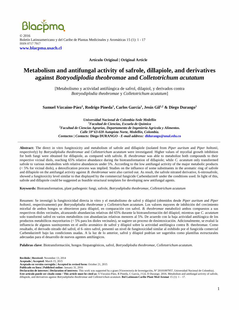
© 2016 Boletín Latinoamericano y del Caribe de Plantas Medicinales y Aromáticas 15 (1): 1 - 17
ISSN 0717 7917
www.blacpma.usach.cl
Artículo Original | Original Article
1
Metabolism and antifungal activity of safrole, dillapiole, and derivatives
against Botryodiplodia theobromae and Colletotrichum acutatum
[Metabolismo y actividad antifúngica de safrol, dilapiol, y derivados contra
Botryodiplodia theobromae y Colletotrichum acutatum]
Samuel Vizcaíno-Páez1, Rodrigo Pineda1, Carlos García1, Jesús Gil1,2 & Diego Durango1
Universidad Nacional de Colombia-Sede Medellín 1Facultad de Ciencias, Escuela de Química
2Facultad de Ciencias Agrarias, Departamento de Ingeniería Agrícola y Alimentos.
Calle 59ª 63-020 Autopista Norte, Medellín, Colombia.
Contactos | Contacts: Diego DURANGO - E-mail address: [email protected]
Abstract: The direct in vitro fungitoxicity and metabolism of safrole and dillapiole (isolated from Piper auritum and Piper holtonii,
respectively) by Botryodiplodia theobromae and Colletotrichum acutatum were investigated. Higher values of mycelial growth inhibition for both fungi were obtained for dillapiole, as compared with safrole. B. theobromae was able to metabolize both compounds to their
respective vicinal diols, reaching 65% relative abundance during the biotransformation of dillapiole; while C. acutatum only transformed
safrole to various metabolites with relative abundances under 5%. According to the low antifungal activity of the major metabolic products
(< 5% for vicinal diols), a detoxification process was implied. Studies on the influence of some substituents in the aromatic ring of safrole and dillapiole on the antifungal activity against B. theobromae were also carried out. As result, the safrole nitrated derivative, 6-nitrosafrole,
showed a fungitoxicity level similar to that displayed by the commercial fungicide Carbendazim® under the conditions used. In light of this,
safrole and dillapiole could be suggested as feasible structural templates for developing new antifungal agents.
Keywords: Biotransformation, plant pathogenic fungi, safrole, Botryodiplodia theobromae, Colletotrichum acutatum
Resumen: Se investigó la fungitoxicidad directa in vitro y el metabolismo de safrol y dilapiol (obtenidos desde Piper auritum and Piper holtonii, respectivamente) por Botryodiplodia theobromae y Colletotrichum acutatum. Los valores mayores de inhibición del crecimiento
micelial de ambos hongos se obtuvieron para dilapiol, en comparación con safrol. B. theobromae metabolizó ambos compuestos a sus
respectivos dioles vecinales, alcanzando abundancias relativas del 65% durante la biotransformación del dilapiol; mientras que C. acutatum solo transformó safrol en varios metabolitos con abundancias relativas menores al 5%. De acuerdo con la baja actividad antifúngica de los
productos metabólicos mayoritarios (< 5% para los dioles vecinales), se sugiere un proceso de desintoxicación. Adicionalmente, se evaluó la
influencia de algunos sustituyentes en el anillo aromático de safrol y dilapiol sobre la actividad antifúngica contra B. theobromae. Como
resultado, el derivado nitrado del safrol, el 6–nitro safrol, presentó un nivel de fungitoxicidad similar al exhibido por el fungicida comercial Carbendazim® bajo las condiciones usadas. A la luz de lo anterior, safrol y dilapiol podrían ser sugeridos como plantillas estructurales
adecuadas para el desarrollo de nuevos agentes antifúngicos.
Palabras clave: Biotransformación, hongos fitopatogénicos, safrol, Botryodiplodia theobromae, Colletotrichum acutatum.
Recibido | Received: November 13, 2014
Aceptado | Accepted: March 15, 2015
Aceptado en versión corregida | Accepted in revised form: October 21, 2015
Publicado en línea | Published online: January 31, 2016
Declaración de intereses | Declaration of interests: This work was supported by a grant (Vicerrectoría de Investigación, Nº 20101007957, Universidad Nacional de Colombia).
Este artículo puede ser citado como / This article must be cited as: S Vizcaíno-Páez, R Pineda, C García, J Gil, D Durango. 2016. Metabolism and antifungal activity of safrole,
dillapiole, and derivatives against Botryodiplodia theobromae and Colletotrichum acutatum. Bol Latinoam Caribe Plant Med Aromat 15 (1): 1 – 17.
Vizcaíno-Páez et al. Metabolism and Antifungal Activity of Safrole and Dillapiole
Boletin Latinoamericano y del Caribe de Plantas Medicinales y Aromáticas/2
INTRODUCTION
Anthracnose and stem-end rot (caused by fungi
Colletotrichum acutatum and Botryodiplodia
theobromae, respectively) are two severe diseases
that contribute considerably to the pre- and
postharvest losses of economically important fruits
and vegetables around the world (Damm et al., 2012;
Twumasi et al., 2014). Conventionally, both diseases
have been successfully controlled through the
application of non-selective fungicides (Gaviria-
Hernández et al., 2013; Syed et al., 2014).
Nonetheless, phytopathogenic microorganisms have
been progressively developing resistance against
commonly used fungicides, which has reduced the
effectiveness of chemical treatments during recent
years (Gutiérrez et al., 2003; Hahn, 2014).
Furthermore, the use of non-selective fungicides to
control deterioration of fruits and vegetables has been
restricted as a result of negative public perception of
their possible undesirable side effects on human
beings. Therefore, researchers are actively working
on alternative methods to combat fungal diseases. In
this way, the spraying of essential oils, plant extracts,
and their main constituents may be an attractive
method for controlling pre- and postharvest fruit
diseases. Particularly, the discovery of novel
antifungal agents representing new chemical classes
with different toxicities and modes of action is highly
desirable (Lopéz-García et al., 2012). Hence,
evaluating antifungal activity of natural products
offers an interesting approach to identify potential
new fungicides and discover valuable structural
templates that may be transformed on antifungal
agents. In addition, the study of the microbial
metabolism of potential antifungal agents using
phytopathogenic fungi could provide information on
the detoxification mechanism used by these
microorganisms and give an indication of the
structural modifications that may be necessary if
certain substrates are to be further developed as
selective fungal control agents (Daoubi et al., 2005a;
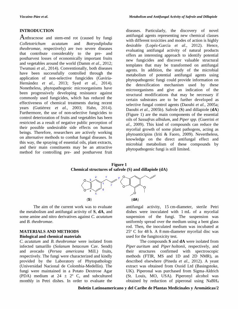
Daoubi et al., 2005b). Safrole (S) and dillapiole (dA)
(Figure 1) are the main components of the essential
oils of Sassafras albidum, and Piper spp. (Guerrini et
al., 2009). This kind of compounds can reduce the
mycelial growth of some plant pathogens, acting as
phytoanticipins (Iriti & Faoro, 2009). Nevertheless,
knowledge on the direct antifungal effect and
microbial metabolism of these compounds by
phytopathogenic fungi is still limited.
Figure 1
Chemical structures of safrole (S) and dillapiole (dA)
The aim of the current work was to evaluate
the metabolism and antifungal activity of S, dA, and
some amine and nitro derivatives against C. acutatum
and B. theobromae.
MATERIALS AND METHODS
Biological and chemical materials
C. acutatum and B. theobromae were isolated from
infected tamarillo (Solanum betaceum Cav. Sendt)
and avocado (Persea americana Mill.) fruits,
respectively. The fungi were characterized and kindly
provided by the Laboratory of Phytopathology
(Universidad Nacional de Colombia-Medellín). The
fungi were maintained in a Potato Dextrose Agar
(PDA) medium at 24 ± 2º C, and subcultured
monthly in Petri dishes. In order to evaluate the
antifungal activity, 15 cm-diameter, sterile Petri
dishes were inoculated with 1 mL of a mycelial
suspension of the fungi. The suspension was
uniformly spread over the medium using a bent glass
rod. Then, the inoculated medium was incubated at
25º C for 48 h. A 8 mm-diameter mycelial disc was
used for the fungitoxicity test.
The compounds S and dA were isolated from
Piper auritum and Piper holtonii, respectively, and
their structures confirmed with spectroscopic
methods (FTIR, MS and 1D and 2D NMR), as
described elsewhere (Pineda et al., 2012). A yeast
extract was obtained from Oxoid Ltd (Basingstoke,
UK). Piperonal was purchased from Sigma-Aldrich
(St. Louis, MO, USA). Piperonyl alcohol was
obtained by reduction of piperonal using NaBH4
Vizcaíno-Páez et al. Metabolism and Antifungal Activity of Safrole and Dillapiole
Boletin Latinoamericano y del Caribe de Plantas Medicinales y Aromáticas/3
(87% yield) (Smith, 2013). Nitro- and amine
derivatives were prepared through nitration
(HNO3/H2SO4) and further reduction (Sn/HCl) of
nitro-derivatives (Olah et al., 1989; Singh et al.,
2006). The compounds 1-allyl - 5 , 6 - dimethoxy -
2 - nitro - 3, 4 -methylenedioxybenzene (2-nitro
dillapiole, 43% yield) and 1-allyl-5,6-dimethoxy - 2-
amino -3,4 - methylenedioxy benzene (2-amino
dillapiole, 80% yield) were obtained from the
dillapiole, whereas, 1-allyl-6-nitro-3,4-methylene-
dioxy benzene (6-nitro safrole, 92% yield) and 1-
allyl-6-amino-3,4-methylenedioxy benzene (6-amino
safrole, 83% yield) were synthesized from safrole.
Fungitoxicity bioassays of dA and S
In order to investigate the toxicity of dA and S
against C. acutatum and B. theobromae, the poisoned
food technique was used (Grover & Moore, 1962;
Velasco et al., 2010). Different concentrations of dA
(50, 75, 100, 300 and 600 μg mL-1) and S (250, 500,
750, 1000, 1250, 1500 and 1750 μg mL-1) dissolved
in ethanol (0.2%, v/v) were diluted in Petri dishes
with PDA. All of the concentrations were tested in
triplicate, and the results are shown as mean values of
three replications of colony diameters [± standard
deviation (SD)]. Petri dishes without compounds
were used as negative control, which only contained
ethanol (0.2%, v/v) in the PDA medium. The
common fungicide Carbendazim (methyl
benzimidazol-2-yl carbamate) was used as positive
control at 50 μg mL-1. The Petri dishes were
incubated at room temperature and the diameter of
the mycelial growth was measured every 12 (for
B. theobromae) and 24 hours (for C. acutatum). The
incubation was stopped when the mycelial mass of
control Petri dishes almost filled them (ca. 10 and 4
days for C. acutatum and B. theobromae
respectively). The fungitoxicity of S and dA in terms
of inhibition percentage of the radial growth was
calculated by using the formula:
The statistical analysis of least significant
difference between means was carried out employing
the ANOVA and LSD test in Statgraphics
(Statgraphics Centurion XV, Version 15.2.06).
Metabolism of dA and S
Preculture of C. acutatum and B. theobromae Mycelia of C. acutatum or B. theobromae from a 2-
day-old culture were employed, following a
methodology described in a previous work
(Numpaque et al., 2011).
Preparative-Scale Metabolism
The mycelia of C. acutatum and B. theobromae were
transferred into six 1.0 L Erlenmeyer flasks
containing 0.5 L of a sterilized broth and dA or S
dissolved in 96% ethanol (final concentration of
0.2%, v/v). When C. acutatum was used as a
biocatalyst, dA and S were incorporated into the
culture medium at 50 and 400 μg mL-1, respectively.
In the case of B. theobromae, 100 and 800 μg mL-1
concentrations of dA and S, respectively, were
employed. The cultivation was developed at room
temperature with stirring at 120 rpm for 360 h. After
the incubation period, the culture medium and
mycelia were separated by filtration. Controls
(without substrate) were performed in order to verify
the presence of similar compounds in the fungi
culture (Correa et al., 2009).
Isolation and identification of metabolic products Crude extract from culture medium was obtained as
described by Velasco et al. (2012). Then, the extract
was chromatographed on a silica gel column. Elution
was performed with an n-hexane-EtOAc gradient
system. Extracts from the metabolism of dA and S by
B. theobromae were separately fractionated to afford
ten fractions, grouped according to TLC profiles. The
seventh (n-hexane:EtOAc, 4:6) fraction from S and
the eighth (n-hexane:EtOAc, 3:7) fraction from dA
were re-chromatographed over silica gel column
using n-hexane-EtOAc as eluent to yield two
metabolic compounds (1 and 2). The identification of
these metabolites was based on the interpretation of
their nuclear magnetic resonance (NMR), infrared,
and mass spectra, and by contrast with the NIST 2002
Mass Spectral Library. In addition, the GC-MS
analysis detected the metabolite (3) from the
biotransformation of S.
Furthermore, six metabolites were detected in
the transformation of S by C. acutatum (including 1
and 3), which were identified according to the mass
spectral data and comparison with authentic samples.
Unfortunately, the very low abundance of some of
Vizcaíno-Páez et al. Metabolism and Antifungal Activity of Safrole and Dillapiole
Boletin Latinoamericano y del Caribe de Plantas Medicinales y Aromáticas/4
the compounds in the biotransformation excluded
isolation, enrichment and further direct spectroscopic
characterization through NMR. Metabolic products
were not detected by GC-MS in the metabolism of
dA using C. acutatum.
Time-course experiments
Precultured B. theobromae and C. acutatum were
separately transferred into fifteen 250 mL Erlenmeyer
flasks containing 125 mL of the Czapek-Dox medium
and the compounds dA and S. The flasks were stirred
under the same conditions as the preculture. The
concentrations of dA and S were the same as
described for the preparative-scale metabolism. The
plant pathogenic fungi were cultivated at 120 rpm for
216 and 360 h for B. theobromae and C. acutatum,
respectively. The culture medium was taken from
each flask daily for C. acutatum and every 12 h
(during the first 5 days) and 48 h (during the last 4
days) for B. theobromae. Then, the crude extract was
obtained as described by Velasco et al. (2012), re-
dissolved in 5 mL of chloroform and analyzed by
TLC and GC-MS. The ratios among the substrate and
products were determined based on the GC peak area;
the results were expressed as relative abundances.
Additionally, control cultivations without dA or S
were performed.
Antifungal activity of some metabolic products
and derivatives of dA and S
Major metabolites resulting from the metabolism of S
and dA by C. acutatum and B. theobromae were
tested for antifungal activity against C. acutatum at a
200 μg mL-1 concentration in triplicate. Finally, in
order to evaluate the effects of electron-releasing and
electron-withdrawing groups on the antifungal
activity, both nitro and amino derivatives of S and dA
were also analyzed. These derivatives were prepared
by conventional nitration and amination procedures
(Smith, 2013). Structural elucidation was performed
by NMR.
Analytical methods
Thin layer chromatography (TLC) was carried out on
Merck Kiesegel 60 F254 (0.25 mm thick). Mixtures of
n-hexane:EtOAc were used as the mobile phase.
Column chromatography (CC) was performed using
silica gel 60 (0.040-0.063 mm; Merck). GC–MS
analysis was performed using a Hewlett-Packard
6890 (Agilent Technologies) gas chromatograph
coupled with a HP 5973 MSD (Mass selective
detector-Quadrupole type). A Zebron ZB 35 column
(30 m x 0.25 mm i.d.; coating thickness 0.25 μm) was
employed. The chromatographic conditions were:
column temperature, 50-250° C at 10° C min-1 and
maintained for five minutes; injector temperature,
150° C; detector temperature, 280° C; and carrier gas,
helium at 1.0 mL min-1. The relative composition of
each constituent was established from the average
peak area obtained in the GC. The nuclear magnetic
resonance spectra were measured with a Bruker
AMX 300 NMR spectrometer. The proton chemical
shifts (δ) and coupling constants (J) are given in ppm
and hertz, respectively. The attribution of 13C NMR
signals for some compounds has been done using
JMOD experiments. FTIR spectra were carried out
using CHCl3 on a Perkin-Elmer RXI. Optical
rotations were measured in a CHCl3 solution at 25° C
with a JASCO P-2000 digital polarimeter.
RESULTS AND DISCUSSION
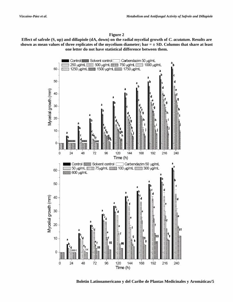
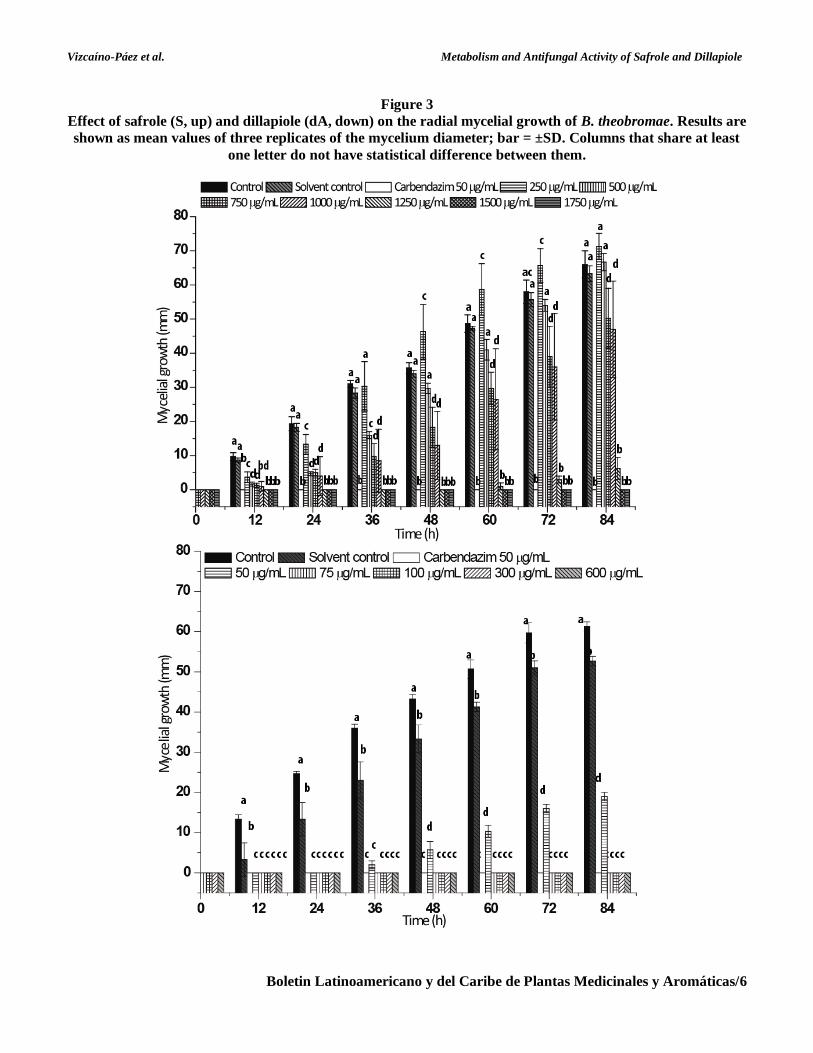
Antifungal activity of safrole S and dillapiole dA The inhibitory effects of dA and S against
C. acutatum and B. theobromae are shown in Figures
2 and 3. For both fungi, the mycelial growth was only
completely inhibited during the evaluation time by S
at 1500 μg mL-1 and above. After the first 24 h, S
exhibited a total inhibition of radial growth in
C. acutatum at all evaluated concentrations; at
1250 μg mL-1 and above, however, this effect
remained for 72 h. Then, the inhibitory effects
strongly decreased at all the evaluated concentrations.
Particularly at 250 μg mL-1, S presented radial growth
inhibitions for C. acutatum of from 100 to 25%, after
240 h. Only at 1250 μg mL-1 and above did S exhibit
inhibitions higher than 70% after 240 h.
For B. theobromae, S reduced the radial
growth of B. theobromae completely during the first
48 h at 1250 μg mL-1; then, the inhibitory effect
decreased by almost 80% after 96 h. The antifungal
activity of S found in the present study is in
agreement with previous reports. Simic et al. (2004)
reported that S is the major constituent of the
essential oil (85%) of Sassafras albidum (Lauraceae),
which has shown an antifungal activity similar to that
of the commercial drug bifonazol. In addition, some
authors (Kubo et al., 1993; Fujita & Kubo, 2004)
have reported that S possesses moderate activity
against S. cerevisiae and Candida utilis, with MICs
(Minimum inhibitory concentration) of 200 μg mL-1.
They suggested that the allyl moiety in S is a
minimum structural requirement necessary to display
its antifungal activity.
Vizcaíno-Páez et al. Metabolism and Antifungal Activity of Safrole and Dillapiole
Boletin Latinoamericano y del Caribe de Plantas Medicinales y Aromáticas/5
Figure 2
Effect of safrole (S, up) and dillapiole (dA, down) on the radial mycelial growth of C. acutatum. Results are
shown as mean values of three replicates of the mycelium diameter; bar = ± SD. Columns that share at least
one letter do not have statistical difference between them.
Vizcaíno-Páez et al. Metabolism and Antifungal Activity of Safrole and Dillapiole
Boletin Latinoamericano y del Caribe de Plantas Medicinales y Aromáticas/6
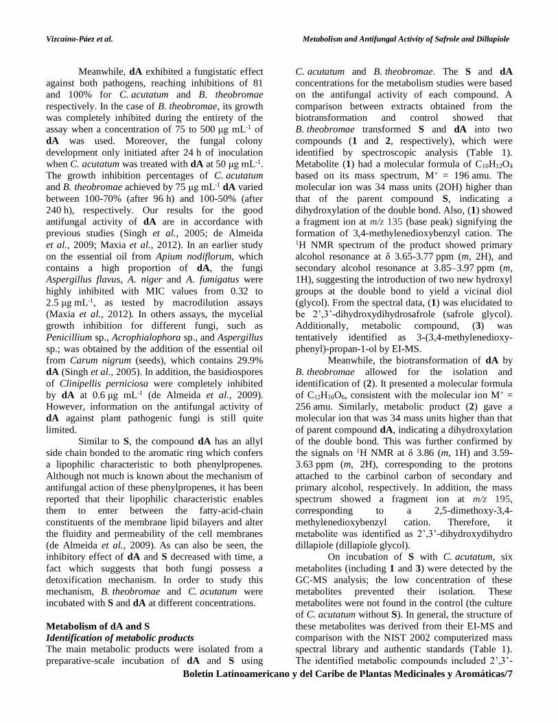
Figure 3
Effect of safrole (S, up) and dillapiole (dA, down) on the radial mycelial growth of B. theobromae. Results are
shown as mean values of three replicates of the mycelium diameter; bar = ±SD. Columns that share at least
one letter do not have statistical difference between them.
Vizcaíno-Páez et al. Metabolism and Antifungal Activity of Safrole and Dillapiole
Boletin Latinoamericano y del Caribe de Plantas Medicinales y Aromáticas/7
Meanwhile, dA exhibited a fungistatic effect
against both pathogens, reaching inhibitions of 81
and 100% for C. acutatum and B. theobromae
respectively. In the case of B. theobromae, its growth
was completely inhibited during the entirety of the
assay when a concentration of 75 to 500 μg mL-1 of
dA was used. Moreover, the fungal colony
development only initiated after 24 h of inoculation
when C. acutatum was treated with dA at 50 μg mL-1.
The growth inhibition percentages of C. acutatum
and B. theobromae achieved by 75 μg mL-1 dA varied
between 100-70% (after 96 h) and 100-50% (after
240 h), respectively. Our results for the good
antifungal activity of dA are in accordance with
previous studies (Singh et al., 2005; de Almeida
et al., 2009; Maxia et al., 2012). In an earlier study
on the essential oil from Apium nodiflorum, which
contains a high proportion of dA, the fungi
Aspergillus flavus, A. niger and A. fumigatus were
highly inhibited with MIC values from 0.32 to
2.5 μg mL-1, as tested by macrodilution assays
(Maxia et al., 2012). In others assays, the mycelial
growth inhibition for different fungi, such as
Penicillium sp., Acrophialophora sp., and Aspergillus
sp.; was obtained by the addition of the essential oil
from Carum nigrum (seeds), which contains 29.9%
dA (Singh et al., 2005). In addition, the basidiospores
of Clinipellis perniciosa were completely inhibited
by dA at 0.6 μg mL-1 (de Almeida et al., 2009).
However, information on the antifungal activity of
dA against plant pathogenic fungi is still quite
limited.
Similar to S, the compound dA has an allyl
side chain bonded to the aromatic ring which confers
a lipophilic characteristic to both phenylpropenes.
Although not much is known about the mechanism of
antifungal action of these phenylpropenes, it has been
reported that their lipophilic characteristic enables
them to enter between the fatty-acid-chain
constituents of the membrane lipid bilayers and alter
the fluidity and permeability of the cell membranes
(de Almeida et al., 2009). As can also be seen, the
inhibitory effect of dA and S decreased with time, a
fact which suggests that both fungi possess a
detoxification mechanism. In order to study this
mechanism, B. theobromae and C. acutatum were
incubated with S and dA at different concentrations.
Metabolism of dA and S
Identification of metabolic products The main metabolic products were isolated from a
preparative-scale incubation of dA and S using
C. acutatum and B. theobromae. The S and dA
concentrations for the metabolism studies were based
on the antifungal activity of each compound. A
comparison between extracts obtained from the
biotransformation and control showed that
B. theobromae transformed S and dA into two
compounds (1 and 2, respectively), which were
identified by spectroscopic analysis (Table 1).
Metabolite (1) had a molecular formula of C10H12O4
based on its mass spectrum, M+ = 196 amu. The
molecular ion was 34 mass units (2OH) higher than
that of the parent compound S, indicating a
dihydroxylation of the double bond. Also, (1) showed
a fragment ion at m/z 135 (base peak) signifying the
formation of 3,4-methylenedioxybenzyl cation. The 1H NMR spectrum of the product showed primary
alcohol resonance at δ 3.65-3.77 ppm (m, 2H), and
secondary alcohol resonance at 3.85–3.97 ppm (m,
1H), suggesting the introduction of two new hydroxyl
groups at the double bond to yield a vicinal diol
(glycol). From the spectral data, (1) was elucidated to
be 2’,3’-dihydroxydihydrosafrole (safrole glycol).
Additionally, metabolic compound, (3) was
tentatively identified as 3-(3,4-methylenedioxy-
phenyl)-propan-1-ol by EI-MS.
Meanwhile, the biotransformation of dA by
B. theobromae allowed for the isolation and
identification of (2). It presented a molecular formula
of C12H16O6, consistent with the molecular ion M+ =
256 amu. Similarly, metabolic product (2) gave a
molecular ion that was 34 mass units higher than that
of parent compound dA, indicating a dihydroxylation
of the double bond. This was further confirmed by
the signals on 1H NMR at δ 3.86 (m, 1H) and 3.59-
3.63 ppm (m, 2H), corresponding to the protons
attached to the carbinol carbon of secondary and
primary alcohol, respectively. In addition, the mass
spectrum showed a fragment ion at m/z 195,
corresponding to a 2,5-dimethoxy-3,4-
methylenedioxybenzyl cation. Therefore, it
metabolite was identified as 2’,3’-dihydroxydihydro
dillapiole (dillapiole glycol).
On incubation of S with C. acutatum, six
metabolites (including 1 and 3) were detected by the
GC-MS analysis; the low concentration of these
metabolites prevented their isolation. These
metabolites were not found in the control (the culture
of C. acutatum without S). In general, the structure of
these metabolites was derived from their EI-MS and
comparison with the NIST 2002 computerized mass
spectral library and authentic standards (Table 1).
The identified metabolic compounds included 2’,3’-

Vizcaíno-Páez et al. Metabolism and Antifungal Activity of Safrole and Dillapiole
Boletin Latinoamericano y del Caribe de Plantas Medicinales y Aromáticas/8
dihydroxydihydrosafrole (1), 3-(3,4-methylenedioxy-
phenyl)-propan-1-ol (3), 3,4-methylenedioxy cinna-
mylalcohol (4), 1-(3,4-methylenedioxyphenyl)-
propan-1-one (5), piperonyl alcohol (6), and
piperonal (7).
Table 1
Metabolic compounds identified from the biotransformation of S and dA by C. acutatum and B. theobromae
Metabolite Rt
(min)
Isolated
from Spectroscopic data
(1) 20.2
Both fungi/S
1H-NMR (CDCl3, 300 MHz): δ 6.77-6.75 (d, 1H, H6), 6.73 (s, 1H,
H2), 6.68-6.66 (d, 1H, H5), 5.94 (s, 2H, –OCH2O–), 3.85-3.97 (m, 1H,
H2’), 3.65-3.77 (m, 2H, H3’), 3.49-3.59 (m, 2H, H1’). MS-EI m/z,
(fragment) [% rel. int.]: 196 (M+) [18], 177 [6], 166 [12], 165 (M-
H2CO-H) [6], 136 [31], 135 (M-C2H5O2; C8H7O2+) [100], 122 [35],
107 [10], 79 [13], 77 [20], 51 [10], 43 [7]. (CHCl3): +8.59°.
(3) 18.6
MS-EI m/z (fragment) [% rel. int.]: 180 (M+) [40], 162 (M–H2O)
[7], 161 (M-H2O-H) [7], 149 (M-H2CO-H; α-cleavage of primary
alcohol group) [7], 136 [71], 135 (base peak, formation of 3,4-
methylenedioxybenzyl cation: C8H7O2+) [100], 106 [20], 105 [7], 91
(C7H7+) [10], 78 [18], 77 (C6H5
+) [35], 65 (C5H5+) [7], 51 [29], 45 [27].
Molecular formula: C10H12O3. Tentatively identified as 3-(3,4-
methylenedioxyphenyl)-propan-1-ol.
(2) 25.5
B.
theobromae/
dA
1H-NMR (CDCl3, 300 MHz): δ 6.33 (s, 1H, H6), 5.97 (s, 2H, –
OCH2O–), 3.92 (s, 3H, –OMe), 3.86 (s, 3H, –OMe), 3.82-3.89 (m, 1H,
H2’), 3.60-3.64 (dd, 1H, J=11.4, 3.5, H3’a), 3.47-3.51 (dd, 1H, J=11.4,
6.0, H3’b), 2.76-2.81 (dd, 1H, J=13.6, 5.7, H1’a), 2.70-2.75 (dd, 1H,
J=13.6, 7.3, H-1’b). MS-EI m/z, (fragment) [% rel. int.]: 256 (M+)
[39], 225 (M–H2CO–H) [67], 195 (C10H11O4+) [100], 180 [17], 165 [7],
135 [7]. IR ῡ (cm-1): 3289 (OH), 3206 (OH), 3015, 1611, 1503, 1435.
(CHCl3): -7.13°.
(4) 17.9
C.
acutatum/S
MS-EI m/z, (fragment) [% rel. int.]: 178 (M+) [21], 161 (M–OH) [6],
149 (M–CO–H; C9H9O2+) [38], 135 (base peak, formation of 3,4-
methylendioxybenzyl cation, M–CO–CH3; C8H7O2+) [100], 123 [50],
121 (C7H5O2+) [28], 103 [25], 93 [75], 91 (C7H7
+) [25], 77 (C6H5+)
[16], 65 (C5H5+) [27]. Metabolite (4) was elucidated to be 3,4-
methylenedioxy cinnamyl alcohol.
(5) 19.8
MS-EI m/z, (fragment) [% rel. int.]: 178 (M+) [21], 149 (base peak,
formation of 3,4-methylenedioxybenzoyl cation -acylium ion-, due to
α-cleavage of the benzyl bond: C8H5O3+) [100], 135 [50], 121
(formation of 3,4-methylenedioxyphenyl cation, C7H5O2+) [30], 91
(C7H7+) [9], 77 (C6H5
+) [7], 65 (C5H5+) [31], 63 [25], 62 [11], 29 [16],
27 [13]. Metabolite (5) was tentatively identified as 1-(3,4-
methylenedioxyphenyl)-propan-1-one
(6) 15.3
MS-EI m/z, (fragment) [% rel. int.]: 152 (M+) [100], 151 (M–H)
[37], 135 (base peak, formation of 3,4-methylendioxybenzyl cation:
C8H7O2+) [53], 123 [30], 122 [27], 93 [59], 91 (C7H7
+) [30], 77 (C6H5+)
[18], 65 (C5H5+) [32]. Molecular formula: C8H8O3. Metabolite (6) was
elucidated as piperonyl alcohol.
(7) 14.2
MS-EI m/z, (fragment) [% rel. int.]: 150 (M+) [86], 149 (M–H, base
peak, formation of 3,4-methylenedioxybenzoyl cation -acylium ion-,
due to α-cleavage of the benzyl bond) [100], 121 (M+–CO–H,
formation of 3,4-methylenedioxyphenyl cation, C7H5O2+) [34], 91
(C7H7+) [13], 65 (C5H5
+) [21], 63 [34], 61 [10]. Metabolite (7) was
identified as piperonal.
Vizcaíno-Páez et al. Metabolism and Antifungal Activity of Safrole and Dillapiole
Boletin Latinoamericano y del Caribe de Plantas Medicinales y Aromáticas/9
To the best of our knowledge, the metabolism
of S and dA by plant fungal pathogens was evaluated
for the first time in this study. On the other hand,
C. acutatum was not able to transform dA.
Thus, the high antifungal activity of dA and
the low level of microbial transformation, makes dA
a promising candidate to control C. acutatum. It is
important to note that the biotransformation effected
by C. acutatum and B. theobromae on S and dA did
not affect the dioxole moiety. Earlier works have
reported that microbial conversion of S by an
Arthrobacter strain and its t-anethole blocked
mutants yield a dihydroxybenzene derivative, i.e.
hydroxichavicol (Shimoni et al., 2003). Similarly, in
mammalians, the P450 type enzyme converts S to
hydroxychavicol in the liver (Scheline, 1991). Unlike
the Arthrobacter strain and its t-anethole blocked
mutants, the transformation of S by C. acutatum and
B. theobromae involves the oxidation of the allyl side
chain.
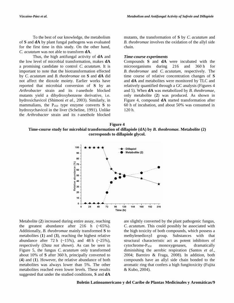
Time-course experiments Compounds S and dA were incubated with the
microorganisms during 216 and 360 h for
B. theobromae and C. acutatum, respectively. The
time course of relative concentration changes of S
and dA and metabolites were monitored by TLC and
relatively quantified through a GC analysis (Figures 4
and 5). When dA was metabolized by B. theobromae,
only metabolite (2) was produced. As shown in
Figure 4, compound dA started transformation after
60 h of incubation, and about 50% was consumed in
120 h.
Figure 4
Time-course study for microbial transformation of dillapiole (dA) by B. theobromae. Metabolite (2)
corresponds to dillapiole glycol.
Metabolite (2) increased during entire assay, reaching
the greatest abundance after 216 h (~65%).
Additionally, B. theobromae mainly transformed S to
metabolites (1) and (3), reaching the highest relative
abundance after 72 h (~15%), and 48 h (~25%),
respectively (Data not shown). As can be seen in
Figure 5, the fungus C. acutatum only transformed
about 10% of S after 360 h, principally converted to
(4) and (1). However, the relative abundance of both
metabolites was always lower than 5%. The other
metabolites reached even lower levels. These results
suggested that under the studied conditions, S and dA
are slightly converted by the plant pathogenic fungus,
C. acutatum. This could possibly be associated with
the high toxicity of both compounds, which possess a
methylenedioxyl group. Substances with that
structural characteristic act as potent inhibitors of
cytochrome-P450 monoxygenases, dramatically
diminishing the aerobic respiration (Santos et al.,
2004; Barreiro & Fraga, 2008). In addition, both
compounds have an allyl side chain bonded to the
aromatic ring that confers a high fungitoxicity (Fujita
& Kubo, 2004).

Vizcaíno-Páez et al. Metabolism and Antifungal Activity of Safrole and Dillapiole
Boletin Latinoamericano y del Caribe de Plantas Medicinales y Aromáticas/10
Based on the structure of the identified
metabolites, a metabolic pathway for the
biotransformation of S and dA by C. acutatum and
B. theobromae was proposed (Figure 6). The
transformation of S and dA may resemble that of
eugenol due to the similarity in its 2-propenyl side
chain. Many bacteria and fungi, such as
Corynebacterium, Pseudomonas, Streptomyces,
Byssochlamys, Penicillium and Rhodococcus can
transform eugenol (Plaggenborg et al., 2006; Jin et
al., 2007). Studies have revealed that microorganisms
follow different metabolic pathways for eugenol
degradation. Recently, six possible biotransformation
routes were presented (Mishra et al., 2013). Thus, the
formation of eugenol epoxide, followed by hydrolysis
to yield a diol (eugenol-diol), have been proposed as
the initial reactions of degradation in Pseudomonas
sp. (Tadasa & Kayahara, 1983). Epoxides and
dihydrodioles were also found in urine and liver of
rats pretreated with of eugenol (and analogs such as
safrole, estragole, and eugenol methyl ether)
(Delaforge et al., 1980). However, eugenol epoxide
and eugenol-diol have not been identified from
microbial metabolism (Xu et al., 2007). Additionally,
it has been suggested that the microbial metabolic
pathway of eugenol involves the intermediates
coniferyl alcohol (4-hydroxy-3-methoxy cinnamyl
alcohol), coniferyl aldehyde and ferulic acid (Mishra
et al., 2013). Some of the genes that are essential to
the degradation of eugenol by Pseudomonas sp. strain
HR199 have already been identified (Overhage et al.,
1999; Priefert et al., 1999).
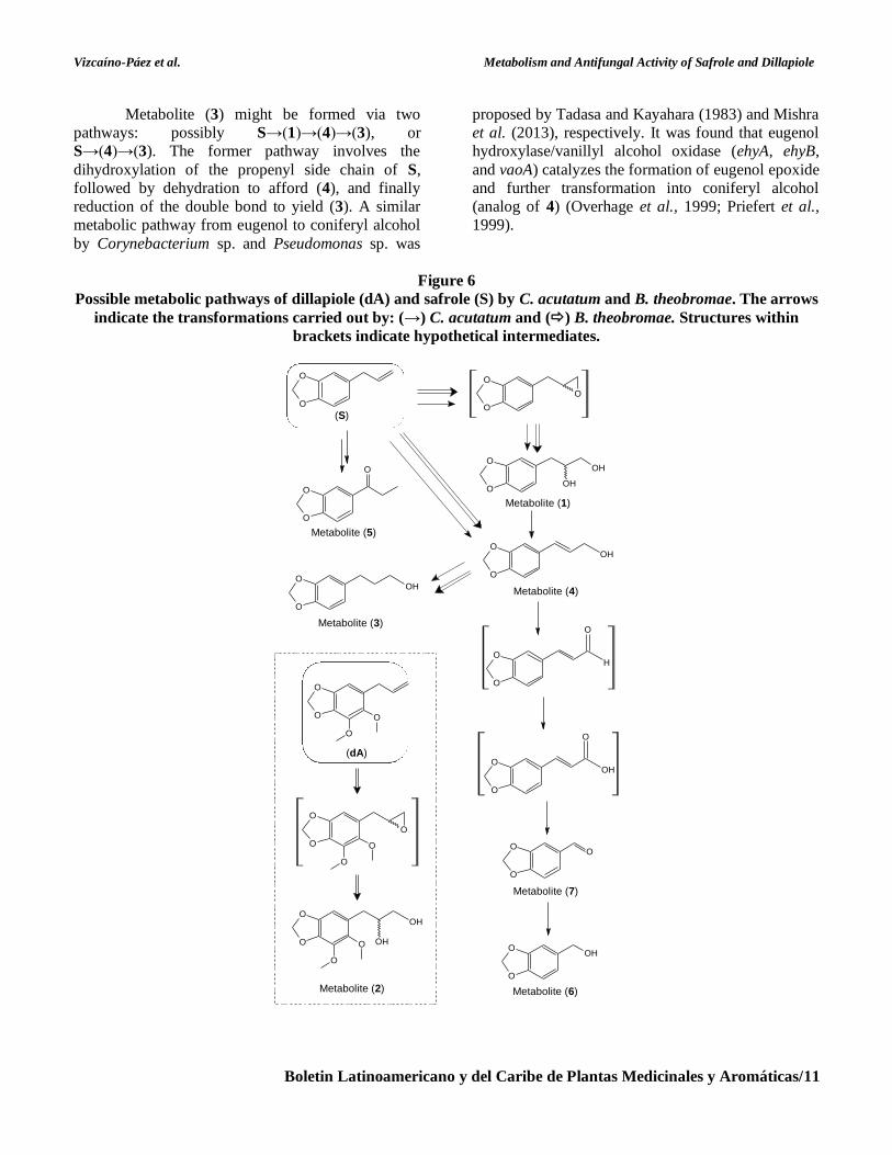
According to our results, B. theobromae was
able to dihydroxylate the double bond of S and dA,
generating the major products: the glycols (1) and (2)
respectively. Metabolite (1) was also detected in the
biotransformation of S by C. acutatum (only 3% after
360 h). The identification of (1) and (2) implies that
both are intermediates in the metabolic pathway from
S and dA. These major metabolites are presumed to
be formed via epoxidation at the double bond and
subsequent hydrolysis of the epoxides. A similar
epoxide intermediate was suggested in the
degradation pathway of eugenol by a
Corynebacterium sp. strain, but its existence has not
been confirmed (Tadasa, 1977; Xu et al., 2007).
Safrole and dillapiole epoxide could be rapidly
detoxicated by the formation of diols, possibly by an
enzyme, since epoxide was not detected at any
appreciable concentration (Guenthner & Luo, 2001).
On the other hand, the opening of the epoxides could
also be performed during the acidic extraction
(Mohan et al., 2000). The epoxidation of C-C double
bonds, and further hydrolysis to yield vicinal diols,
have been widely reported for the Colletotrichum
genus (García-Pajón et al., 2003). To the best of our
knowledge, this is the first report on the degradation
of 2-propenylbenzenes through an epoxide-diol route
in both fungi.
Figure 5
Time-course study for microbial transformation of safrole (S) by C. acutatum. Metabolites (1), (3), (4), (5), (6)
and (7) correspond to safrole glycol; 3-(3,4-methylenedioxyphenyl)-propan-1-ol; 3,4-methylenedioxy
cinnamyl alcohol; 1-(3,4-methylenedioxyphenyl)-propan-1-one; piperonyl alcohol; and piperonal,
respectively.
Vizcaíno-Páez et al. Metabolism and Antifungal Activity of Safrole and Dillapiole
Boletin Latinoamericano y del Caribe de Plantas Medicinales y Aromáticas/11
Metabolite (3) might be formed via two
pathways: possibly S→(1)→(4)→(3), or
S→(4)→(3). The former pathway involves the
dihydroxylation of the propenyl side chain of S,
followed by dehydration to afford (4), and finally
reduction of the double bond to yield (3). A similar
metabolic pathway from eugenol to coniferyl alcohol
by Corynebacterium sp. and Pseudomonas sp. was
proposed by Tadasa and Kayahara (1983) and Mishra
et al. (2013), respectively. It was found that eugenol
hydroxylase/vanillyl alcohol oxidase (ehyA, ehyB,
and vaoA) catalyzes the formation of eugenol epoxide
and further transformation into coniferyl alcohol
(analog of 4) (Overhage et al., 1999; Priefert et al.,
1999).
Figure 6
Possible metabolic pathways of dillapiole (dA) and safrole (S) by C. acutatum and B. theobromae. The arrows
indicate the transformations carried out by: (→) C. acutatum and () B. theobromae. Structures within
brackets indicate hypothetical intermediates.
(S)
O
O
O
O
O
Metabolite (5)
O
O
OH
OH
Metabolite (1)
O
O
O
O
O
OH
Metabolite (4)
O
O
H
O
O
O
OH
O
O
O
O
Metabolite (7)
O
O
OH
Metabolite (6)
O
O
O
O
(dA)
O
O
O
O
O
Metabolite (2)
O
O OH
OH
O
O
O
O
OH
Metabolite (3)
Vizcaíno-Páez et al. Metabolism and Antifungal Activity of Safrole and Dillapiole
Boletin Latinoamericano y del Caribe de Plantas Medicinales y Aromáticas/12
Metabolite (4) may also be formed directly
from S by a safrole hydroxylase. This analogous
route, with a reaction mechanism including a quinone
propenide intermediate, has been suggested for the
catabolism of eugenol in Pseudomonas sp. strain
HR199 (Overhage et al., 1999). However, the exact
side-chain oxidation mechanism of eugenol to
coniferyl alcohol is still unknown. Finally, the
reduction of (4) leads to (3). The ability of C.
acutatum to reduce allylic alcohols was previously
reported by Correa et al. (2009) and Velasco et al.
(2012).
Furthermore, C. acutatum was able to
transform S to (5); therefore, two pathways could be
considered on the formation of (5). The substrate S
could be reduced at the double bond, hydroxylated in
the benzylic carbon and subsequently oxidized to
produce the metabolite (5). Nevertheless, the
reduction of the double bond may proceed before or
after the hydroxylation. Thus, two different
intermediates could be suggested: 1’-hydroxysafrole
or 3,4-methylenedioxyphenylpropane. This oxidation
of the propenyl side chain is unusual and has not been
reported before in the microbial metabolism of 2-
propenylbenzene compounds.
Metabolic product (7) is hypothesized as
possible being derived from (1) via dehydration of
the secondary alcohol function to afford (4), which is
then oxidized to 3,4-methylenedioxycinnamyl
aldehyde and further oxidized to the corresponding
carboxylic acid (3,4-methylenedioxy cinnamic acid).
A similar route has been proposed to the metabolism
of eugenol by bacteria and fungi (Xu et al., 2007;
Mishra et al., 2013; Han et al., 2013). In this type of
pathway, coniferyl alcohol is oxidized to coniferyl
aldehyde by coniferyl alcohol dehydrogenase (CaIA)
and further oxidized to ferulic acid by coniferyl
aldehyde dehydrogenase (CaIB).
Then, the elimination of an acetate moiety
from the unsaturated side-chain of 3,4-
methylenedioxy cinnamic acid directly yields (7).
The intermediate carboxylic acid, however, was not
detected in the medium. A similar deacetylation has
been proposed in the conversion of ferulic acid to
vanillin (employing 4-hydroxycinnamate CoA ligase
and 4-hydroxycinnamate CoA hydratase/ligase) for
different bacteria and some fungi, such as Aspergillus
sp., Fomes fomentarius, Fusarium solani, Polyporus
versicolor, and Rhodotorula rubra (Huang et al.,
1993; Gasson et al., 1998; Overhage et al., 2003;).
Moreover, a coenzyme-A-dependent mechanism has
been proposed for the conversion of substituted
cinnamic acids in Pseudomonas putida, and ferulic
acid in Rhodotorula rubra (Huang et al., 1993).
However, the precise mechanism of such
deacetylation has not been fully established. In a
previous work, Santos et al. (2003) reported that
metabolite (7) was formed on the biotransformation
of isosafrole by Cladosporium sphaerospermum.
Subsequently, the reduction of the carbonyl group
from (7) affords (6). These results indicate that
C. acutatum and B. theobromae were able to modify
the allyl chain side of S and dA. Additionally, our
results support the fact that Colletotrichum sp.
species have considerable oxido-reductase activity
(García-Pajón et al., 2003).
Antifungal activity of some metabolic products
and derivatives of dA and S
Antifungal activity of metabolic products To determine if S and dA were metabolized by
C. acutatum and B. theobromae through
detoxification pathways, the inhibitory effects of
metabolites (1) and (2) against B. theobromae, and
(6) and (7) against C. acutatum were evaluated.
Metabolites (1) and (2) were analyzed at 50 μg mL-1
whereas (6) and (7) were evaluated at 200 μg mL-1
concentration. All assays were carried out in
triplicate. For C. acutatum, the mycelial growth
inhibitions achieved by (7) were between 80.0 (after
24 h) and 30.8% (after 240 h). Meanwhile, metabolite
(6) showed only about 36% growth inhibition after
24 h of incubation. Then, the antifungal activity
steadily decreased, reaching only 25% inhibition after
240 h. Overall, the radial growth inhibition displayed
by metabolites (6) and (7) decreased progressively in
accordance with the incubation time. At a similar
concentration, S only exhibited a mycelial growth
inhibition of C. acutatum between 50 (at 24 h) and
19% (after 240 h). Therefore, the results of the
antifungal bioassays showed that metabolic product
(6) was less potent at inhibiting the fungal growth of
C. acutatum than its precursor S. Since metabolite (6)
was less fungistatic than S, it may suggest that the
metabolism of S by C. acutatum was a detoxification
process. Interestingly, metabolite (7) displayed a
higher fungistatic effect against C. acutatum than S.
So, it is possible to think that the low conversion of S
to (7) by C. acutatum seeks to ensure no-inhibitory
levels of (7) in the medium. In fact, the amount of (7)
produced during the biotransformation of S by this
plant pathogenic fungus (the minor metabolite) was
almost negligible in comparison to that used in the
bioassays.
Vizcaíno-Páez et al. Metabolism and Antifungal Activity of Safrole and Dillapiole
Boletin Latinoamericano y del Caribe de Plantas Medicinales y Aromáticas/13
Otherwise, the inhibitory effect of
metabolites (1) and (2) against B. theobromae was
weak. Mycelial growth inhibitions exhibited by both
glycols were lesser than 5%. The higher fungistatic
effect of S and dA against B. theobromae, as
compared to metabolic products (1) and (2), suggests
that its metabolism is a detoxification process. As
mentioned above, the allyl side chain bonded to the
aromatic ring confers a lipophilic characteristic and
the ability to disrupt fungal membranes to S and dA.
However, the dihydroxylation of the allyl chain to
afford (1) and (2) reduced the lipophilic characteristic
and abolished the antifungal activity, as compared to
the parent compounds. Previously, it was reported
that the relatively high antifungal activity of
compounds that contain the methylendioxyl group is
due to its capacity to act as a cytochrome P450
inhibitor (Santos et al., 2004). However, our results
indicated that the allyl substituent is even more
important in displaying such an inhibitory effect.
Thus, our results are in agreement with a previous
report that 1-allyl moiety plays a positive role in the
antifungal behavior (Carrasco et al., 2012). Indeed,
these authors found that the replacement of the allyl
radical resulted in the disappearance of the antifungal
activity. Glycols (1) and (2) were not further
metabolized under the conditions studied by the
fungus B. theobromae.
Antifungal activity of derivatives from S and dA It is noteworthy that, although the structural
difference between dA and S is only seen in the
presence of two methoxyl groups on the aromatic
ring, dA was significantly more active than S against
both fungi. These results indicate that electronic
and/or steric factors in phenylpropenes might be
important for antifungal activity. In order to
investigate the relationship between the ring
substitution and the antifungal activity of these
compounds, the nitro and amino derivatives were also
tested against the fungus B. theobromae. The
compounds nitro safrole (1-allyl-6-nitro-3,4-
methylenedioxybenzene) and (amino safrole) 1-allyl-
6-amino-3,4-methylenedioxybenzene were prepared
from S; whereas, nitro dillapiole (1-allyl-5,6-
dimethoxy - 2 - nitro - 3, 4 - methylenedioxy
benzene) and amino dillapiole (1-allyl-5,6-
dimethoxy- 2 -amino - 3,4-methylenedioxy benzene)
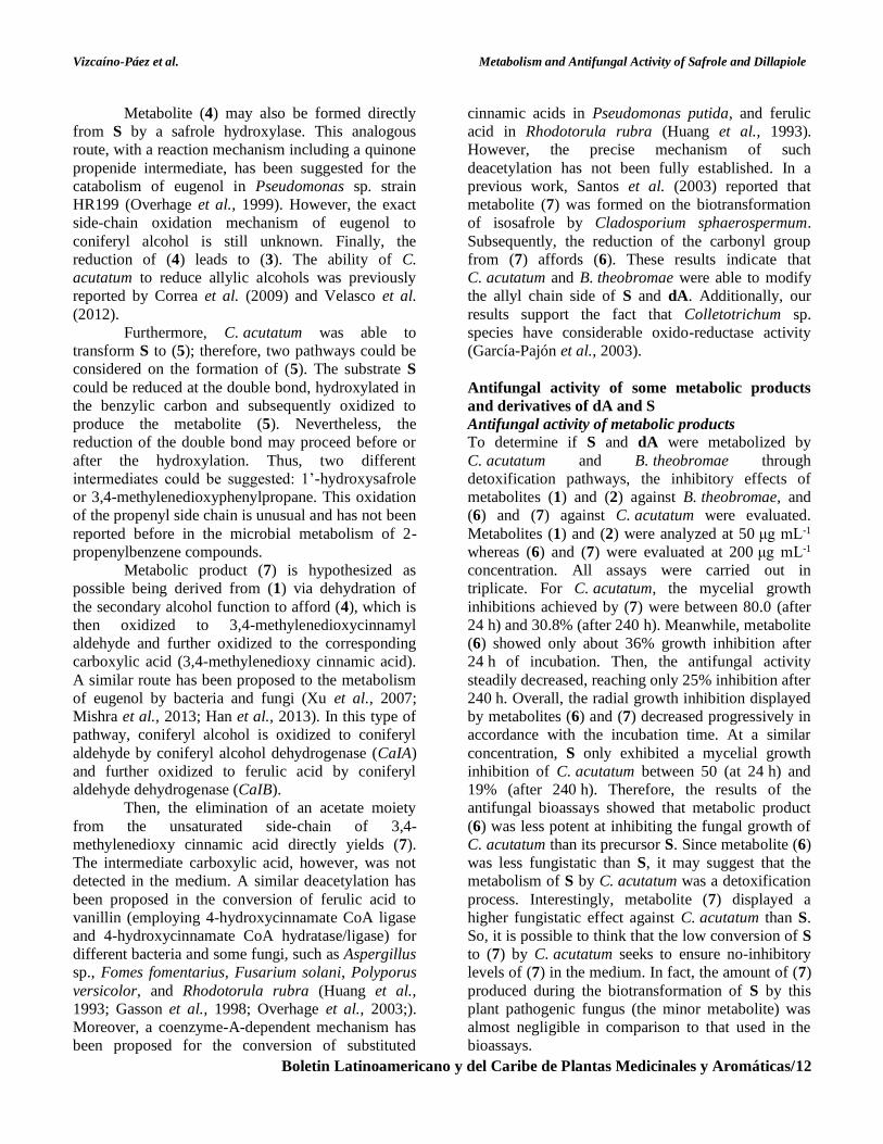
(Figure 7) were synthesized from dA.
Figure 7
Chemical structure amino and nitro derivatives of S and dA.
Confirmation of the structures of the
derivatives was carried out using spectroscopic data
(Table 2).
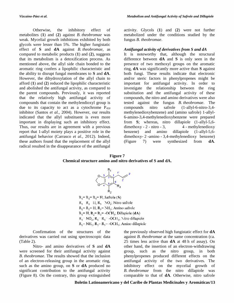
Nitro- and amino derivatives of S and dA
were screened for their antifungal activity against
B. theobromae. The results showed that the inclusion
of an electron-releasing group in the aromatic ring,
such as the amino group, on S or dA produced no
significant contribution to the antifungal activity
(Figure 8). On the contrary, this group extinguished
the previously observed high fungistatic effect for dA
against B. theobromae at the same concentration (ca.
25 times less active than dA at 48 h of assay). On
other hand, the insertion of an electron-withdrawing
group, such as the nitro group, in both
phenylpropenes produced different effects on the
antifungal activity of the two derivatives. The
inhibitory effect on the mycelial growth of
B. theobromae from the nitro dillapiole was
comparable to that of dA. Otherwise, nitro safrole
Vizcaíno-Páez et al. Metabolism and Antifungal Activity of Safrole and Dillapiole
Boletin Latinoamericano y del Caribe de Plantas Medicinales y Aromáticas/14
was found to be the most active compound against
B. theobromae, being almost 49 times higher than S
at the same concentration and after 48 h of the assay.
Table 2
Spectroscopic data of safrole and dillapiole derivatives
Compound Spectroscopic data
Nitro safrole
1H-NMR (CDCl3, 300 MHz): δ 7.51 (s, 1H, H3), 6.78 (s, 1H, H6), 6.12 (s,
2H, –OCH2O–), 6.06-5.92 (ddt, 1H, J = 16.8, 10.2, 6.6, H2’), 5.09-4.99 (dd,
2H, J = 13.6, 1.8, H3’a,b), 3.69-3.66 (d, 2H, J = 6.6, H1’a,b). 13C-NMR
(CDCl3, 75 MHz): 151.7 (C1), 146.5 (C2), 110.4 (C3), 142.8 (C4), 132.2
(C5), 105.7 (C6), 37.62 (C1’), 135.2 (C2’), 117.0 (C3’), 102.8 (–OCH2O–).
Amino safrole
1H-NMR (CDCl3, 300 MHz): δ 6.57 (s, 1H, H3), 6.29 (s, 1H, H6), 5.96-
5.85 (ddt, 1H, J = 16.8, 10.2, 6.3, H2’), 5.85 (s, 2H, –OCH2O–), 5.13-5.09
(dd, 2H, J = 6.0, 1.8, H3’a), 5.06-5.05 (d, J = 1.5, H3’b), 3.23-3.21 (d, 2H, J
= 6.0, H1’a,b). 13C-(JMOD) NMR (CDCl3, 75 MHz): 135.6 (CH, C2’),
115.5 (CH2, C3’), 109.6 (CH, C5), 100.2 (–OCH2O–), 97.9 (CH, C2), 35.8
(CH2, C1’).
Nitro dillapiole
1H-NMR (CDCl3, 300 MHz): δ 6.03 (s, 2H, –OCH2O–), 5.91-5.77 (ddt,
1H, J = 15.7, 9.6, 6.6, H2’), 5.05-5.01 (dd, 2H, J = 12.4, 1.1, H3’a,b), 3.98 (–
OMe), 3.93 (–OMe), 3.28-3.26 (d, 2H, J = 6.6, H1’a,b). 13C-(JMOD) NMR
(CDCl3, 75 MHz): 135.2 (CH, C2’), 116.7 (CH2, C3’), 102.8 (–OCH2O–),
61.5 (-OMe), 60.8 (-OMe), 30.2 (CH2, C1’).
Amino dillapiole
1H-NMR (CDCl3, 300 MHz): δ 6.00 (s, 2H, –OCH2O–), 5.99-5.86 (m,
1H, H2’), 5.08-4.96 (m, 2H, H3’a,b), 3.81 (–OMe), 3.70 (–OMe), 3.21-3.18
(m, 2H, H1’a,b). The 13C-NMR analysis was not possible due to insufficient
quantity.
Figure 8
Effect of safrole (S), dillapiole (dA), and their nitro and amino derivatives on the radial mycelial growth of
B. theobromae. Results are shown as mean values of three replicates of the mycelium diameter; bar = ± SD.
Vizcaíno-Páez et al. Metabolism and Antifungal Activity of Safrole and Dillapiole
Boletin Latinoamericano y del Caribe de Plantas Medicinales y Aromáticas/15
The data showed that the antifungal activity
of nitro safrole was comparable to that exerted by the
commercial synthetic fungicide Carbendazim at the
same concentration. In contrast to a previous report
(Carrasco et al., 2012), our results indicated that the
introduction of a NO2 group led to an increase in the
antifungal activity of S. Thus, it is possible to believe
that the presence of the two methoxyl groups in dA
or the nitro group in C6 in nitro safrole may lead to a
correct balance of hydrophilicity-lipophilicity, which
is an essential factor for the antifungal properties seen
in these compounds.
CONCLUSIONS
The present paper reports on the ability of
B. theobromae to convert S and dA to the
corresponding vicinal diols (glycols), which could be
used in a wide variety of applications. Time-course
studies indicated that S is converted to safrole glycol
at a rapid rate. Also, it was demonstrated that
C. acutatum transformed S into various metabolites at
a low proportion, including safrole glycol, piperonal,
piperonyl alcohol, among others. However,
C. acutatum was ineffective in transforming dA.
Additionally, the fungitoxicity of S and dA, their
metabolic products, and nitro- and amino-derivatives
were evaluated against both fungi. S and dA may
effectively inhibit the mycelial growth of the
evaluated pathogenic fungi. However, dA was the
most active. Also, the products from the metabolism
of S and dA displayed lower antifungal activities than
the parent compounds. So, it was concluded that the
metabolism of S and dA is a detoxification process. It
can also be concluded that allyl moiety plays a
positive role in the inhibitory effect of S and dA
against C. acutatum and B. theobromae. Finally, the
most active derivative against the fungus
B. theobromae was nitro safrole, 49 times more
active than S at the same concentration and assay
time. Nitro safrole displayed an activity against
B. theobromae that was comparable to that of the
commercial fungicide Carbendazim®. Therefore, it is
possible to believe that S and dA offer interesting
structural templates that may be transformed for
developing new antifungal agents.
ACKNOWLEDGEMENTS This work was supported by a grant (Vicerrectoría de
Investigación, Nº 20101007957, Universidad
Nacional de Colombia). We thank to Dr. Afanador-
Kafuri for providing cultures of the plant pathogen
fungi.
REFERENCES
Barreiro EJ, Fraga MCA. 2008. Química Médica:
As Bases Moleculares da Ação dos
Fármacos. 2ª Ed. Artmed, Porto Alegre,
Brasil.
Correa Y, Durango D, García C. 2009.
Transformación microbiana del
arilpropanoide cinamaldehído con el hongo
fitopatógeno Colletotrichum acutatum. Vitae
16: 83 - 91.
Carrasco H, Raimondi M, Svetaz L, Liberto MD,
Rodriguez MV, Espinoza L, Madrid A,
Zacchino S. 2012. Antifungal activity of
eugenol analogues. Influence of different
substituents and studies on mechanism of
action. Molecules 17: 1002 - 1024.
Damm U, Cannon PF, Woudenberg JHC, Crous PW.
2012. The Colletotrichum acutatum species
complex. Stud Mycol 73: 37 - 113.
Daoubi M, Deligeorgopoulou A, Macías-Sánchez AJ,
Hernández-Galán R, Hitchcock PB, Hanson
JR, Collado IG. 2005a. Antifungal activity
and biotransformation of diisophorone by
Botrytis cinerea. J Agric Food Chem 53:
6035 - 6039.
Daoubi M, Hernandez-Galan R, Benharref A,
Collado IG. 2005b. Screening study of lead
compounds for natural product-based
fungicides: antifungal activity and
biotransformation of 6α,7α-dihydroxy-β-
himachalene by Botrytis cinerea. J Agric
Food Chem 53: 6673 - 6677.
de Almeida RRP, Souto RNP, Bastos CN, da Silva
MHL, Maia JGS. 2009. Chemical variation in
Piper aduncum and biological properties of
its dillapiole-rich essential oil. Chem
Biodivers 6: 1427 - 1434.
Delaforge M, Janiaud P, Levi P, Morizot JP. 1980.
Biotransformation of allylbenzene analogues
in vivo and in vitro through the epoxide-diol
pathway. Xenobiotica 10: 737 - 744.
Fujita KI, Kubo I. 2004. Potentiation of fungicidal
activities of trans-anethole against
Saccharomyces cerevisiae under hypoxic
conditions. J Biosci Bioeng 98: 490 - 492.
García-Pajón CM, Henández-Galán R, Collado IG.
2003. Biotransformations by Colletotrichum
species. Tetrahedron: Asymmetry 14: 1229
- 1239.
Gasson MJ, Kitamura Y, McLauchlan WR, Narbad
A, Parr AJ, Parsons EL, Payne J, Rhodes MJ,
Walton NJ. 1998. Metabolism of ferulic acid
Vizcaíno-Páez et al. Metabolism and Antifungal Activity of Safrole and Dillapiole
Boletin Latinoamericano y del Caribe de Plantas Medicinales y Aromáticas/16
to vanillin. A bacterial gene of the enoyl-
SCoA hydratase/isomerase superfamily
encodes an enzyme for the hydration and
cleavage of a hydroxycinnamic acid SCoA
thioester. J Biol Chem 237: 4163 - 4170.
Gaviria-Hernández V, Patiño-Hoyos LF, Saldarriaga-
Cardona A. 2013. Evaluación in vitro de
fungicidas comerciales para el control de
Colletotrichum spp., en mora de castilla.
Corpoica Cienc Tecnol Agropec 14: 67 -
75.
Grover RK, Moore JD. 1962. Toxicometric studies of
fungicides against the browning organisms
Sclerotinia fructicola and S. lava.
Phytopathology 52: 876 - 880.
Guenthner TM, Luo G. 2001. Investigation of the
role of the 2',3'-epoxidation pathway in the
bioactivation and genotoxicity of dietary
allylbenzene analogs. Toxicology 160: 47 -
58.
Guerrini A, Sacchetti G, Rossi D, Paganetto G,
Muzzoli M, Andreotti E, Tognolini M,
Maldonado ME, Bruni R. 2009. Bioactivities
of Piper aduncum L. and Piper obliquum
Ruiz & Pavon (Piperaceae) essential oils
from eastern Ecuador. Environ Toxicol
Pharm 27: 39 - 48.
Gutiérrez O, Gutiérrez JG, Ángel DN, Ortiz DT,
Mejía EZ, Sánchez FD, Huerta HV. 2003.
Resistencia a benomil y tiabendazol en
aislamientos de Colletotrichum
gloeosporioides (Penz.) Penz. y Sacc.
obtenidos de mango (Mangifera indica L.) en
cinco regiones de méxico. Rev Mex
Fitopatol 21: 260 - 266.
Hahn M. 2014. The rising threat of fungicide
resistance in plant pathogenic fungi: Botrytis
as a case study. J Chem Biol 7: 133 - 141.
Han D, Ryu JY, Lee H, Hur HG. 2013. Bacterial
biotransformation of phenylpropanoid
compounds fro producing flavor and fragance
compounds. J Korean Soc Appl Biol Chem
56: 125 - 133.
Huang Z, Dostal L, Rosazza JP. 1993. Mechanisms
of ferulic acid conversions to vanillic acid
and guaiacol by Rhodotorula rubra. J Biol
Chem 268: 23954 - 23958.
Iriti M, Faoro F. 2009. Chemical diversity and
defence metabolism: How plants cope with
pathogens and ozone pollution. Int J Mol Sci
10: 3371 - 3399.
Jin J, Mazon H, van den Heuvel RHH, Janssen DB.
2007. Discovery of a eugenol oxidase from
Rhodococcus sp. strain RHA1. FEBS J 274:
2311 - 2321.
Kubo I, Muroi H, Himejima M. 1993. Combination
effects of antifungal nagilactones against
Candida albicans and two other fungi with
phenylpropanoids. J Nat Prod 56: 220 - 226.
López-García B, Hernández M, Segundo BS. 2012.
Bromelain, a cysteine protease from
pineapple (Ananas comosus) stem, is an
inhibitor of fungal plant pathogens. Lett
Appl Microbiol 55: 62 - 67.
Maxia A, Falconieri D, Piras A, Porcedda S,
Marongiu B, Frau M, Gonçalves M, Cabral
C, Cavaleiro C, Salgueiro L. 2012. Chemical
composition and antifungal activity of
essential oils and supercritical CO2 extracts
of Apium nodiflorum (L.) Lag.
Mycopathologia 174: 61 - 67.
Mishra S, Sachan A, Sachan SG. 2013. Production of
natural value-added compounds: an insight
into the eugenol biotransformation pathway.
J Ind Microbiol Biotechnol 40: 545 - 550.
Mohan RS, Gavardinas K, Kyere S, Whalen DL.
2000. Spontaneous hydrolysis reactions of
cis- and trans-β-methyl-4-methoxystyrene
oxides (anethole oxides): Buildup of trans-
anethole oxide as an intermediate in the
spontaneous reaction of cis-anethole oxide. J
Org Chem 65: 1407 - 1413.
Numpaque MA, Oviedo LA, Gil JH, García CM,
Durango DL. 2011. Thymol and carvacrol:
biotransformation and antifungal activity
against the plant pathogenic fungi
Colletotrichum acutatum and Botryodiplodia
theobromae. Trop Plant Pathol 36: 3 - 13.
Olah GA, Malhotra R, Narang SC. 1989. Nitration.
Methods and Mechanisms. VCH
Publishers, New York, USA.
Overhage J, Priefert H, Steinbüchel A. 1999.
Biochemical and genetic analyses of ferulic
acid catabolism in Pseudomonas sp. strain
HR199. Appl Environ Microb 65: 4837 -
4847.
Overhage J, Steinbüchel A, Priefert H. 2003. Highly
efficient biotransformation of eugenol to
ferulic acid and further conversion to vanillin
in recombinant strains of Escherichia coli.
Appl Environ Microbiol 69: 6569 - 6576.
Pineda R, Vizcaíno S, García CM, Gil JH, Durango
DL. 2012. Chemical composition and
Vizcaíno-Páez et al. Metabolism and Antifungal Activity of Safrole and Dillapiole
Boletin Latinoamericano y del Caribe de Plantas Medicinales y Aromáticas/17
antifungal activity of Piper auritum Kunth
and Piper holtonii C. DC. against
phytopathogenic fungi. Chil J Agric Res 72:
507 - 515.
Plaggenborg R, Overhage O, Loos A, Archer JA,
Lessard P, Sinskey AJ, Steinbüchel A,
Priefert H. 2006. Potential of Rhodococcus
strains fro biotechnological vanillin
production from ferulic acid and eugenol.
Appl Microbiol Biotechnol 56: 457 - 461.
Priefert H, Overhage J, Steinbüchel A. 1999.
Identification and molecular characterization
of the eugenol hydroxylase genes
(ehyA/ehyB) of Pseudomonas sp. strain
HR199. Arch Microbiol 172: 354 - 363.
Santos AS, Pereira Jr N, da Silva IM, Sarquis MIM,
Antunes OAC. 2004. Peroxidase catalyzed
microbiological oxidation of isosafrol into
piperonal. Process Biochem 39: 2269 - 2275.
Santos AS, Pereira NP, da Silva IM, Sarquis MIM,
Antunes OA. 2003. Microbiologic oxidation
of isosafrole into piperonal. Appl Biochem
Biotechnol 105-108: 649 - 657.
Scheline RR. 1991. CRC Handbook of Mammalian
Metabolism of Plant Compounds. 1ª Ed.
CRC Press LLC, Boca Raton, FL, USA.
Shimoni E, Baasov T, Ravid U, Shoham Y. 2003.
Biotransformations of propenylbenzenes by
an Arthrobacter sp. and its t-anethole blocked
mutants. J Biotech 105: 61 - 70.
Singh G, Marimuthu P, de Heluani CS, Catalan CAN.
2005. Antioxidant and biocidal activities of
Carum nigrum (Seed) essential oil, oleoresin,
and their selected components. J Agric Food
Chem 54: 174 - 181.
Singh V, Kanojiyab S, Batra S. 2006. Studies on the
reduction of the nitro group in 3-aryl-2-
methylene-4-nitro-alkanoates afforded by the
Baylis–Hillman adducts: synthesis of 4-aryl-
3-methylene-2-pyrrolidinones and 3-(1-
alkoxycarbonyl - vinyl) - 1H - indole-2-car-
boxylates. Tetrahedron 62: 10100 - 10110.
Simic A, Sokovic MD, Ristic M, Grujic-Jovanovic S,
Vukojevic J, Marin PD. 2004. The chemical
composition of some Lauraceae essential oils
and their antifungal activities. Phytother Res
18: 713 - 717.
Smith MB. 2013. March's Advanced Organic
Chemistry: Reactions, Mechanisms, and
Structure. 7 ed. John Wiley & Sons.
Hoboken, New Jersey, USA.
Syed RN, Mansha N, Khaskheli MA, Khanzada MA,
Lodhi AM. 2014. Chemical control of stem
end rot of mango caused by Lasiodiplodia
theobromae. Pak J Phytopathol 26: 201 -
206.
Tadasa K. 1977. Degradation of eugenol by a
microorganism. Agric Biol Chem 41: 925 -
929.
Tadasa K, Kayahara H. 1983. Initial steps of eugenol
degradation pathway of a microorganism.
Agric Biol Chem 47: 2639 - 2640.
Twumasi P, Ohene-Mensah G, Moses E. 2014. The
rot fungus Botryodiplodia theobromae strains
cross infect cocoa, mango, banana and yam
with significant tissue damage and economic
losses. Afr J Agric Res 9: 613 - 619.
Velasco R, Gil JH, García CM, Durango DL. 2010.
Production of 2-phenylethanol in the
biotransformation of cinnamyl alcohol by the
plant pathogenic fungus Colletotrichum
acutatum. Vitae 17: 272 - 280.
Velasco R, Gil JH, García CM, Durango DL. 2012.
Structural modification of trans-cinnamic
acid using Colletotrichum acutatum. Rev Fac
Ing Univ Antioquia 63: 20 - 29.
Xu P, Hua D, Ma C. 2007. Microbial transformation
of propenylbenzenes for natural flavour
production. Trends Biotechnol 25: 571 -
576.


![Pharmacologically active boranes*old.iupac.org/publications/pac/2006/pdf/7807x1425.pdf · 2017. 7. 24. · teresting pharmacological activity such as antifungal [21], peptidomimetic](https://img.pdfslide.tips/doc/110x75/60d89ee659f0b108862ebb5e/pharmacologically-active-boranesoldiupacorgpublicationspac2006pdf-2017.jpg)