Embed Size (px)
Citation preview

MIC & Etest
Dr. M. Talebi Ph.D of Bacteriology Tehran University of Medical Sciences
MIC The minimum inhibitory concentration (MIC)
is defined as the lowest concentration of the
antimicrobial agent required to prevent
growth of a microorganism under defined
experimental conditions, giving a quantitative
measure of bacterial susceptibility
MIC The lowest concentration of an antimicrobial agent that prevents visible growth of a microorganism
in an agar or broth dilution susceptibility test.
THE CLINICAL IMPORTANCE OF THE
MINIMUM
INHIBITORY CONCENTRATION
• Disc testing can give unreliable or misleading results
• Clinical microbiology laboratories should be
putting more emphasis on antimicrobial resistance testing (ART)
• A method is chosen to either predict or test for
small changes in the bacterial phenotype that may lead to clinical resistance
THE CLINICAL IMPORTANCE OF THE
MINIMUM INHIBITORY CONCENTRATION
• Organisms are grouped into various categories:
▫ Susceptible (S)
▫ Intermediate (I)
▫ Resistant (R)
• But, for serious infections, and those in sites where antibiotics
penetrate poorly, and among immune-suppressed patients,
categorical results have limited predictive value
THE CLINICAL IMPORTANCE OF THE MINIMUM
INHIBITORY CONCENTRATION
• Assessment of the MIC values of several agents will provide a selective choice as to which antibiotic possesses the widest margin between the MIC and the susceptible breakpoint. For the patient, this could be used both as a safety and efficacy margin
• This is particularly relevant for infections in sites into which antibiotics penetrate poorly. For example, in osteomyelitis or prosthetic joint infections, the MIC values are critical along with knowledge of penetration of different agents to select optimal therapy
Clinical priorities for MIC testing include
• Sterile site infections e.g. endocarditis
• Serious nosocomial infections
• Chronic infections e.g. cystic fibrosis
• ICU and other high risk patients
So, for:
• Resistance surveillances
• Drug evaluations
• Clinical trials
only quantitative MIC data can be used.
Methods for determination of MIC
• 1. Broth dilution method
• 2. Agar dilution method
• 3. E- test
Broth Dilution Method
Macro broth dilution
Micro broth dilution
Procedure
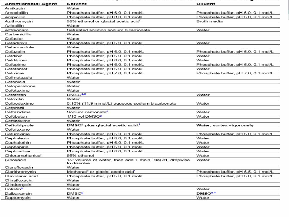
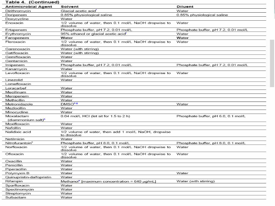
• Antimicrobial Agents
• Obtain antimicrobial standards or reference powders directly from the drug manufacturer
• Acceptable powders ▫ Drug’s generic name
▫ lot number
▫ potency [μg] or [IU] per mg
▫ expiration date
• Store the powders at ≤-20 °C

Weighing Antimicrobial Powders
• All antimicrobial agents are assayed for standard units of activity
• The assay units may differ widely from the actual weight of the powder and often may differ between drug production lots.
• Thus, a laboratory must standardize its antimicrobial solutions based on assays of the lots of antimicrobial powders that are used to make stock solutions.
The value for potency supplied by the manufacturer
should include consideration of :
• Measures of purity (usually by HPLC assay)
• Water content
• The salt/counter-ion fraction
• The potency may be expressed as a percentage, or in units of μg/mg (w/w).

Weighing Antimicrobial Powders
•Preparing Solutions

Direct Colony Suspension Method
Direct colony suspension is the recommended method for testing the fastidious organisms
• Haemophilus spp
• N. gonorrhoeae
• N. meningitidis
• Streptococci
• for testing staphylococci for potential methicillin or oxacillin resistance.

Growth Method
• The growth method can be used alternatively and is sometimes preferable when colony growth is difficult to suspend directly and a smooth suspension cannot be made.
• It can also be used for non fastidious organisms (except staphylococci) when fresh (24 hour) colonies, as required for the direct colony suspension method, are not available.
• Select at least three to five well-isolated colonies of the same morphologic type from an agar plate culture.
Procedure
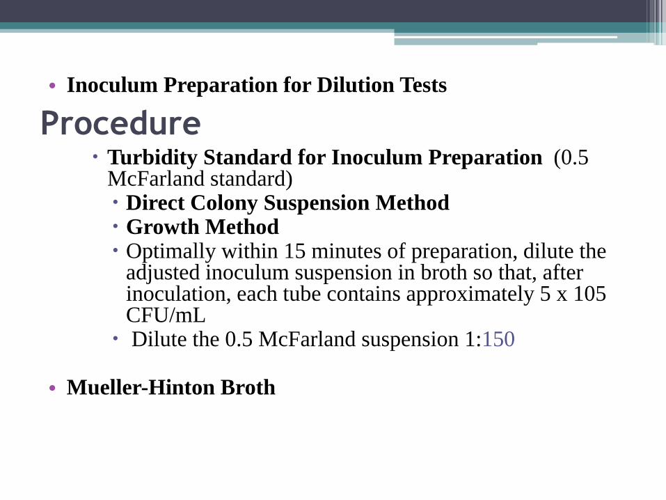
• Inoculum Preparation for Dilution Tests
Turbidity Standard for Inoculum Preparation (0.5
McFarland standard) Direct Colony Suspension Method Growth Method Optimally within 15 minutes of preparation, dilute the
adjusted inoculum suspension in broth so that, after inoculation, each tube contains approximately 5 x 10⁵ CFU/mL Dilute the 0.5 McFarland suspension 1:150
• Mueller-Hinton Broth

Mueller-Hinton Broth
• Mueller-Hinton broth is recommended as the medium of choice for susceptibility testing of commonly isolated, rapidly growing aerobic, or facultative organisms.
• Mueller-Hinton broth demonstrates good batch-to-batch reproducibility for susceptibility testing
• Is low in sulfonamide, trimethoprim, and tetracycline inhibitors; and yields satisfactory growth of most pathogens.
• In addition, a large body of data and experience has been gathered about tests performed with this medium.
Number of Concentrations Tested
• The concentrations to be tested for a particular antimicrobial agent should encompass the interpretive breakpoints shown in CLSI Tables
• But the actual number of concentrations tested is the decision of the laboratory
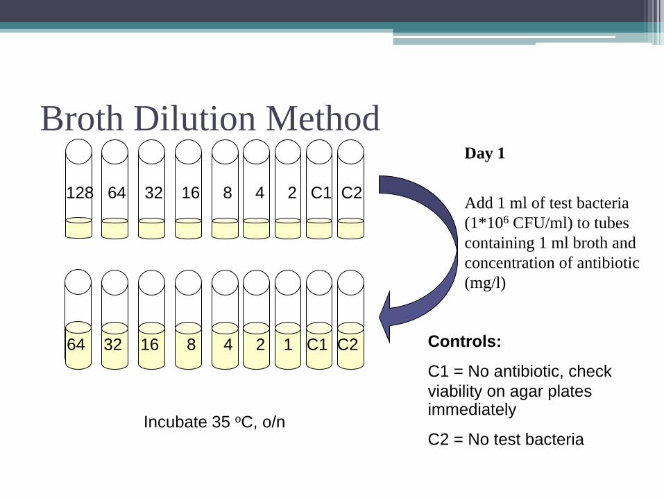
Broth Dilution Method Day 1
Add 1 ml of test bacteria
(1*106 CFU/ml) to tubes
containing 1 ml broth and
concentration of antibiotic
(mg/l)
Controls:
C1 = No antibiotic, check
viability on agar plates immediately
C2 = No test bacteria
Incubate 35 oC, o/n
128 64 32 16 8 4 2 C1 C2
64 32 16 8 4 2 1 C1 C2
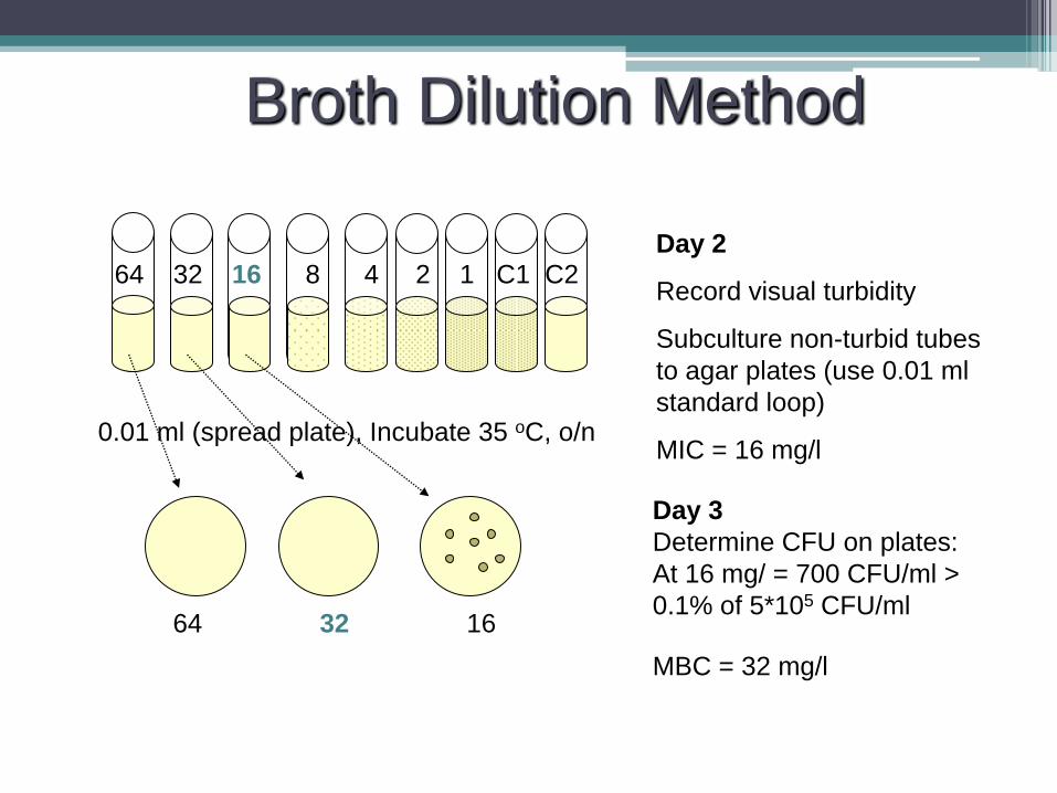
Broth Dilution Method
Day 2
Record visual turbidity
Subculture non-turbid tubes
to agar plates (use 0.01 ml
standard loop)
MIC = 16 mg/l
64 32 16 8 4 2 1 C1 C2
0.01 ml (spread plate), Incubate 35 oC, o/n
64 32 16
Day 3
Determine CFU on plates:
At 16 mg/ = 700 CFU/ml >
0.1% of 5*105 CFU/ml
MBC = 32 mg/l
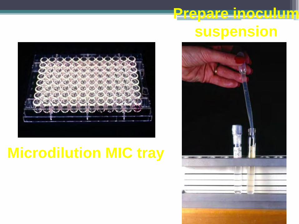
Microdilution MIC tray
Prepare inoculum
suspension
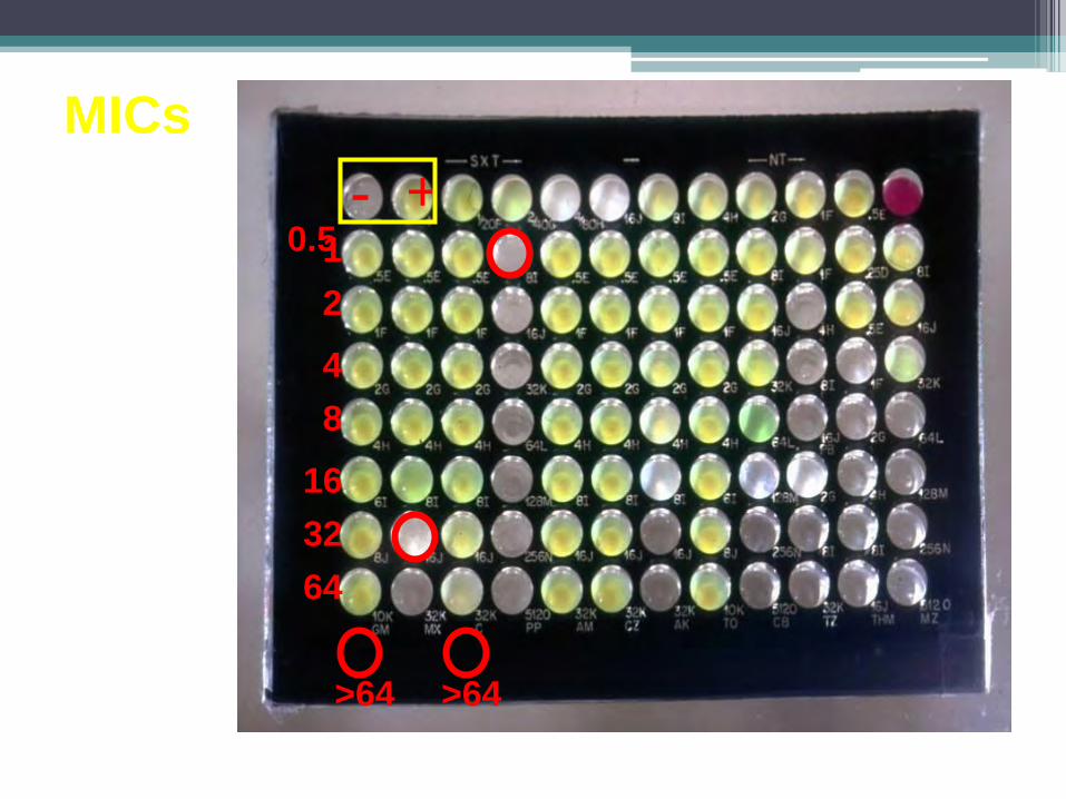
- +
64
32
16
8
4
2
1
>64
0.5
MICs
>64

Agar Dilution Method
• Procedure
▫ Making dilutions of antimicrobial agent in melted media and pouring plates
One concentration of antibiotic/ plate
Possible for several different strains/plate
64 ug/ml 32 ug/ml 16 ug/ml

Agar Dilution Method
Procedure
Inoculation of bacterial inoculum (McFarland
No. 0.5) Delivers 0.001 ml of bacterial inoculum
Incubation
Spot of growth
MIC
32 ug/ml
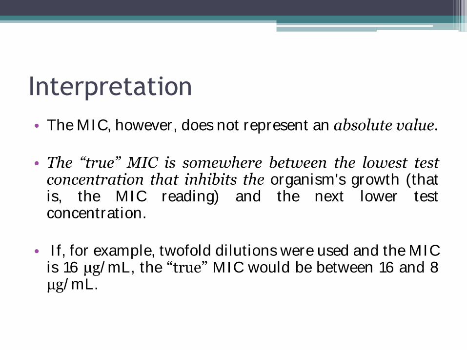
Interpretation
• The MIC, however, does not represent an absolute value. • The “true” MIC is somewhere between the lowest test
concentration that inhibits the organism's growth (that is, the MIC reading) and the next lower test concentration.
• If, for example, twofold dilutions were used and the MIC
is 16 μg/mL, the “true” MIC would be between 16 and 8 μg/mL.

Fastidious Organisms
Haemophilus influenzae and
H. parainfluenzae • MIC testing using Haemophilus Test Medium
(HTM)
• Only for broth dilution as described below
• The agar dilution method using HTM has not been studied
HTM
• Mueller-Hinton broth
• β-nicotinamide adenine dinucleotide (NAD)
• Bovine hematin
• Yeast extract
• Thymidine phosphorylase

Neisseria gonorrhoeae
MIC testing of N. gonorrhoeae has been
developed only for
• agar dilution
• using GC agar base
• growth supplement
Broth microdilution and agar dilution susceptibility
testing of N. meningitidis have been validated
Streptococcus pneumoniae and Other
Streptococcus spp
• MIC testing of Streptococcus spp.
• Using CAMHB with 2.5 to 5% lysed horse blood has been developed
• Only for broth dilution
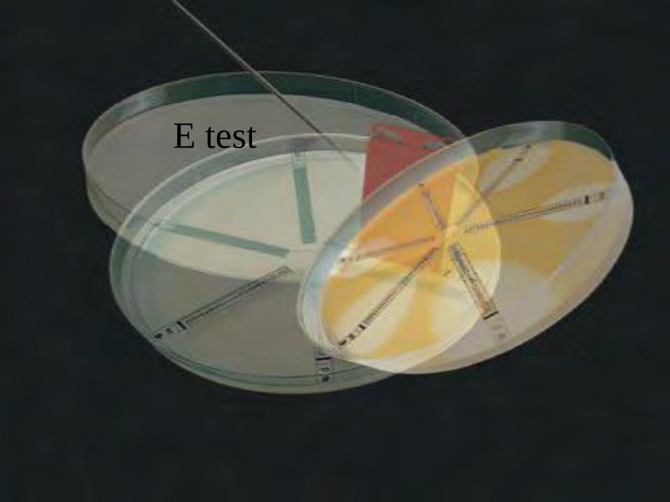
E test
E test
Etest is a quantitative technique for determining
the Minimum Inhibitory Concentration (MIC)
of antimicrobial agents against microorganisms
and for detection of resistance mechanisms
ART method Resistance Testing
Needs to be flexible and should fulfill some of the
following characteristics:
▫ Allowing the use of heavier inoculum to detect low
level resistance
▫ Adaptable to conditions optimal for resistance
extended incubation (glycopeptide resistance)
• Applicable to fastidious, uncommon and slow
growing organisms
• Provide quantitative MIC values over a wide
concentration range (>10 dilutions)
• Decreases in susceptibility to be reliably detected
E test
• Concentration range across 15 dilutions
• Precise, continuous and stable gradient
• Visual recognition of resistant phenotypes on agar

E test
This test may have
• It has a continuous concentration gradient and is able to
show subtle changes in susceptibility
• The wide concentration gradients cover the MIC ranges
of a wide variety of pathogens
• Allow both low level and high level resistance to be
detected.
• The Etest is reportedly easy to use in most laboratory
settings and requires no complicated procedures
Robust to fastidious
• Easily adaptable to different test conditions
▫ robust aerobes to anaerobes ▫ Pneumococci ▫ Meningococci ▫ H. pylori ▫ Bartonella ▫ Bordetella ▫ Franscisella ▫ Brucella ▫ Actinomycetes such as nocardia and rhodococcus ▫ Fungi including yeasts, moulds ▫ Mycobacteria
E test
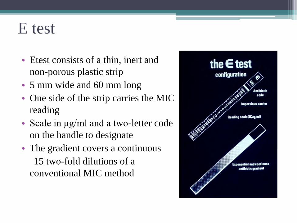
• Etest consists of a thin, inert and
non-porous plastic strip
• 5 mm wide and 60 mm long
• One side of the strip carries the MIC
reading
• Scale in μg/ml and a two-letter code
on the handle to designate
• The gradient covers a continuous
15 two-fold dilutions of a
conventional MIC method
STORAGE
• All packages, must be stored in a freezer at -20°C until the expiry date.
• Etest strips left over from an opened package must be stored at -20°C in an airtight storage container or tube • Strips in storage containers can be used until the expiry date if correctly stored and handled. • Store only one antibiotic type per storage tube. • Prevent moisture from penetrating into or forming within the package or storage tubes. Etest strips must be kept
dry.

Procedure
• Medium
• Ensure that the agar plate has a depth of 4.0 ± 0.5 mm and pH 7.2 - 7.4
• The medium and required supplements will depend on the bacterial species being tested
• Inoculum preparation
• Inoculation
The surface is completely dry before applying the Etest strips.

Swab plate
Remove sample
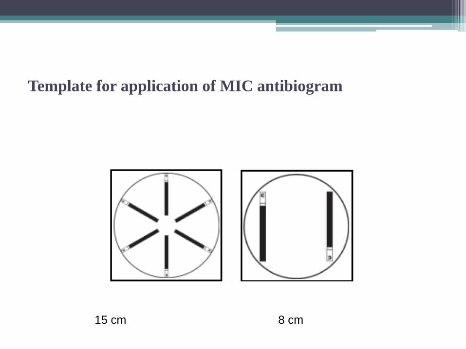
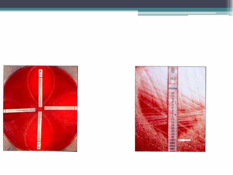
Template for application of MIC antibiogram
15 cm 8 cm
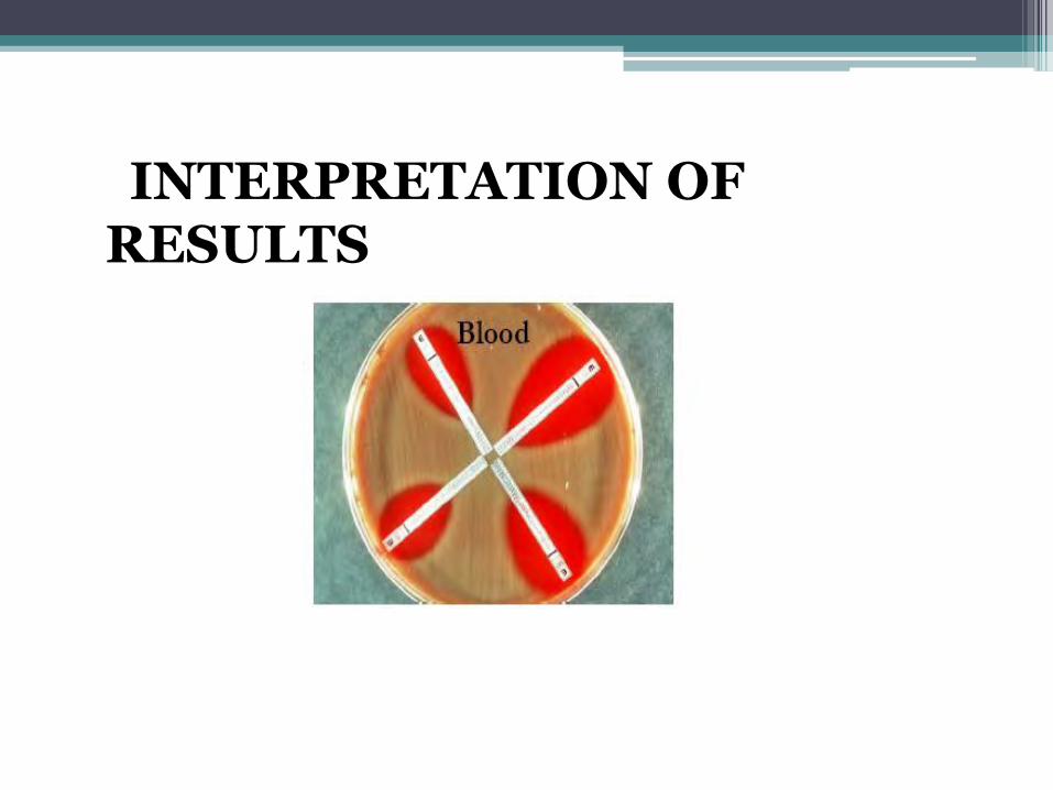
INTERPRETATION OF RESULTS
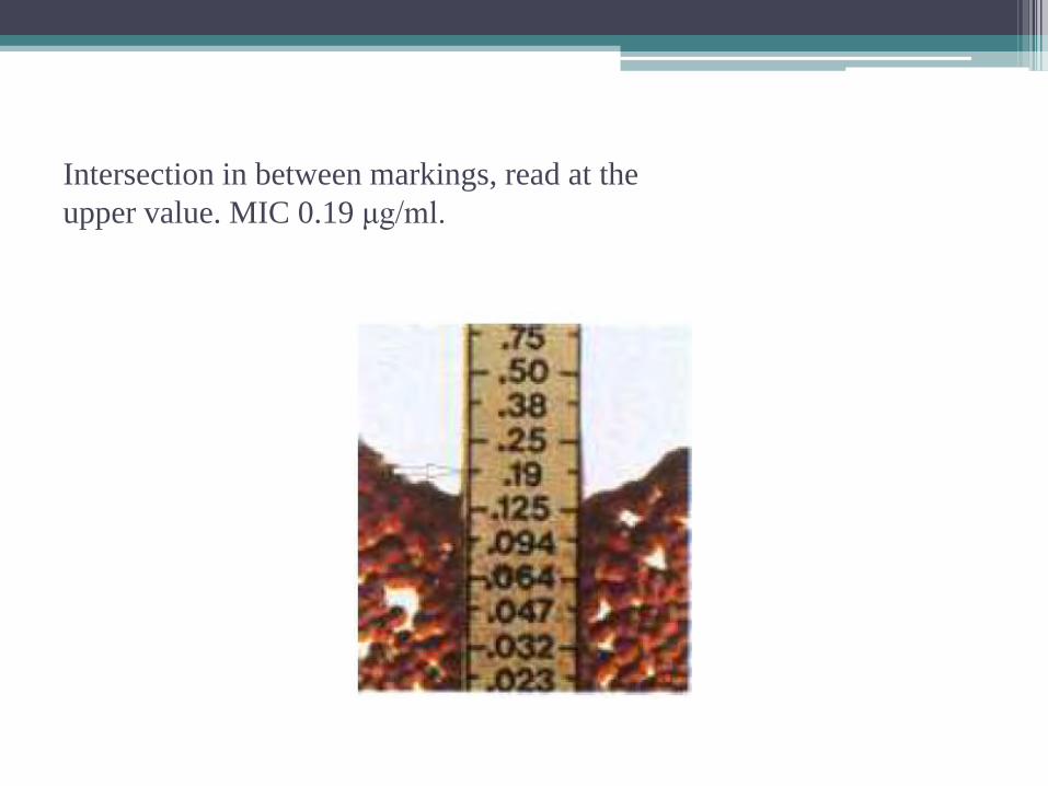
Intersection in between markings, read at the
upper value. MIC 0.19 μg/ml.

Bactericidal agents like aminoglycosides
give sharp ellipses. MIC 0.064 μg/ml
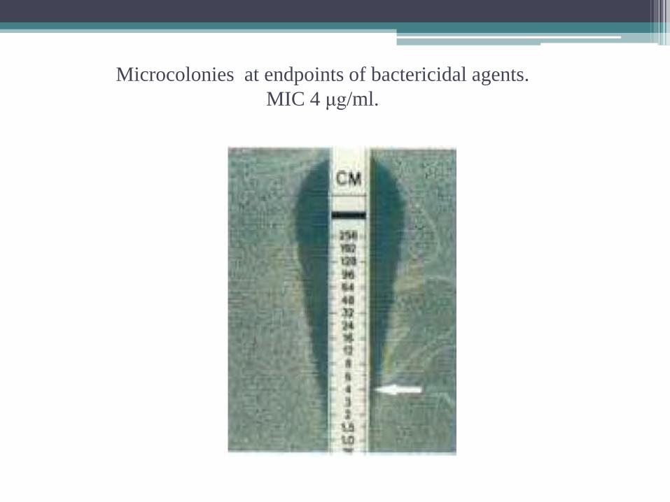
Microcolonies at endpoints of bactericidal agents.
MIC 4 μg/ml.
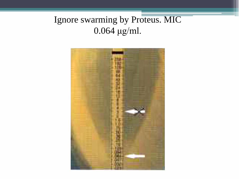
Ignore swarming by Proteus. MIC
0.064 μg/ml.
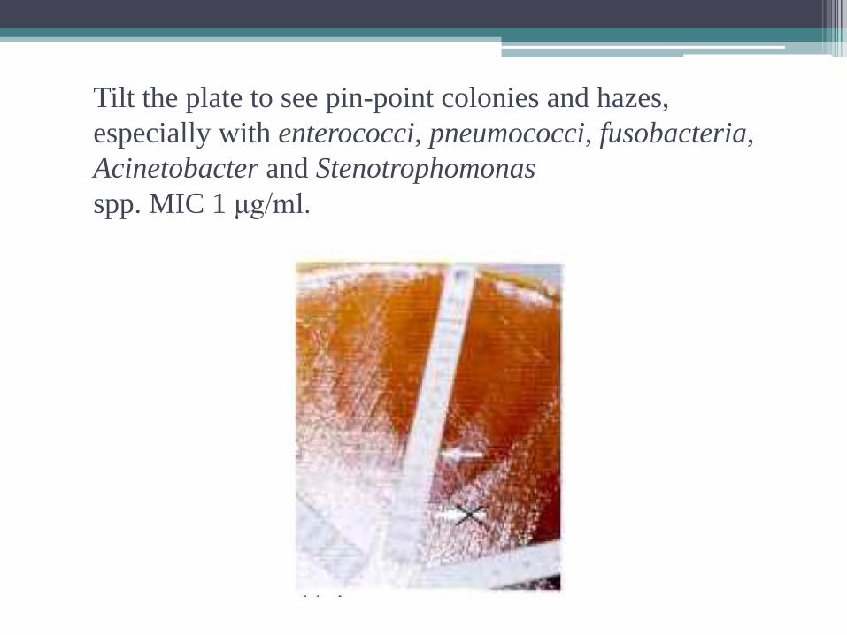
Tilt the plate to see pin-point colonies and hazes,
especially with enterococci, pneumococci, fusobacteria,
Acinetobacter and Stenotrophomonas
spp. MIC 1 μg/ml.
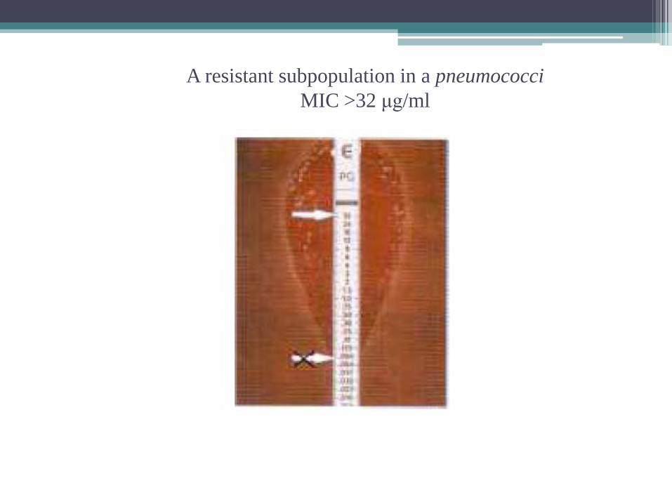
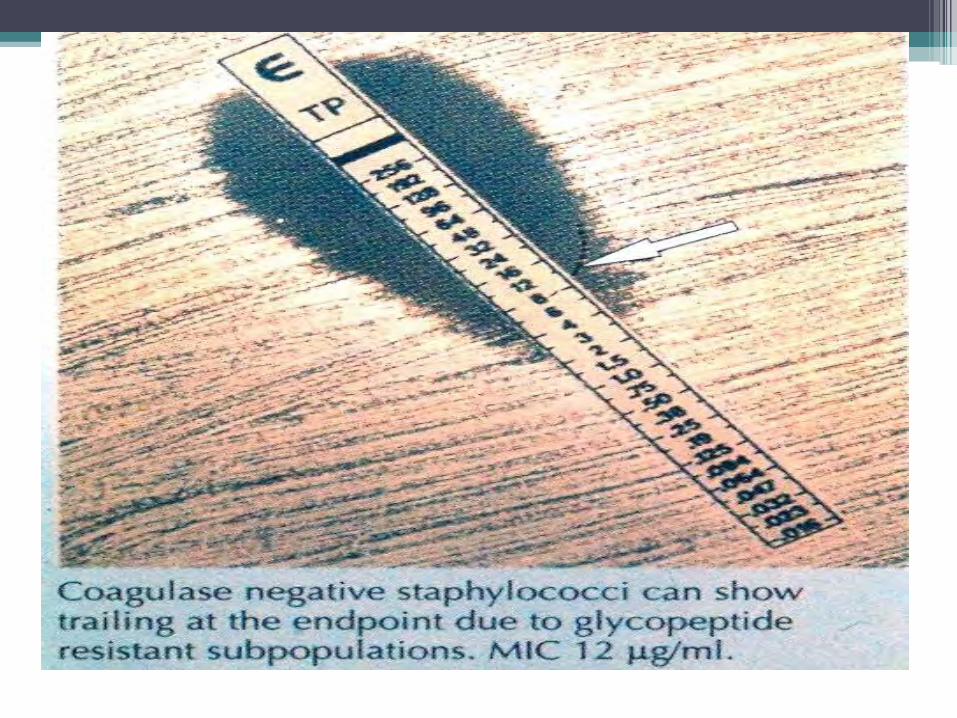
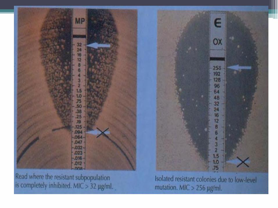
A resistant subpopulation in a pneumococci
MIC >32 μg/ml
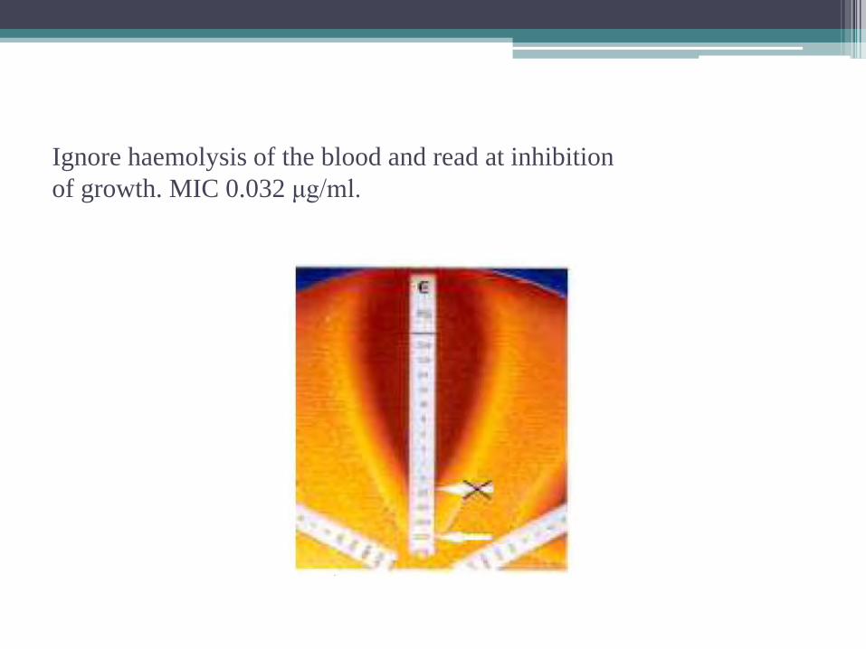
Ignore haemolysis of the blood and read at inhibition
of growth. MIC 0.032 μg/ml.
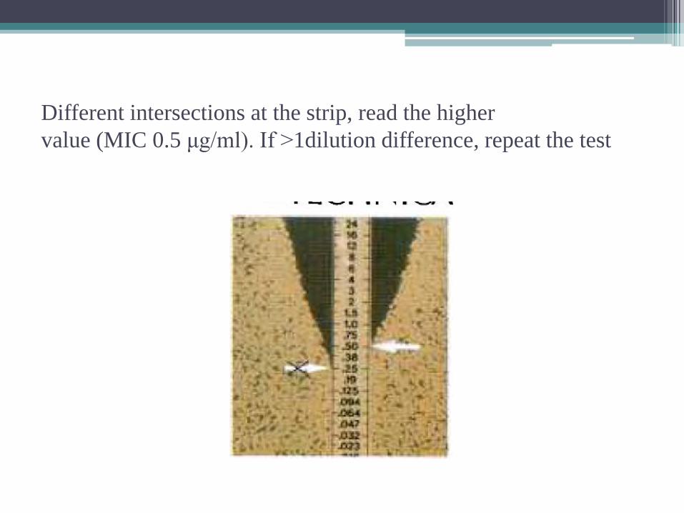
Different intersections at the strip, read the higher
value (MIC 0.5 μg/ml). If >1dilution difference, repeat the test
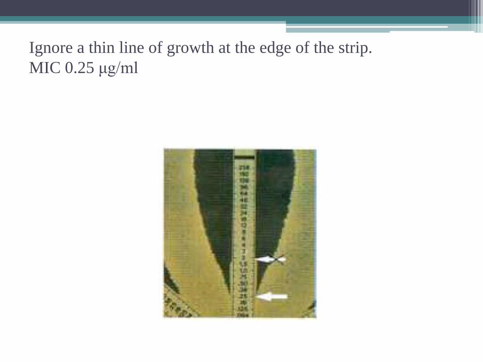
Ignore a thin line of growth at the edge of the strip.
MIC 0.25 μg/ml

Special Applications
• ESBLs
• Anaerobes
• Helicobacter and Campylobacter
• Haemophilus
• Mycobacteria and Actinomycetes
• ……………..
Susceptibility testing of Anaerobes
• Susceptibility testing of anaerobes can be
problematic.
• Disc diffusion is unreliable and not
recommended.
• Almost 15 to 40% of anaerobes do not grow well
in broth microdilution systems.
• Broth microdilution may be suboptimal.
Susceptibility testing limitations
• The low inoculum used may understimate resistance
• Metronidazole testing is problematic
• Reference agar dilution is cumbersome and expensive
to set up for a few strains.

Susceptibility testing of Anaerobes
• The continuous and stable antibiotic gradient in Etest has been documented to be suitable for testing anaerobes
• The use of MIC data has been shown to give therapy change in up to 56% of clinical cases
• The lack of routine susceptibility testing to guide physicians, leads to the use of expensive broad spectrum antibiotics, in order to cover all potentially significant organisms

Why Etest for anaerobes?
• Agar based growth supporting good growth.
• Stable antibiotic gradient minimally influenced by
varying growth rates.
• Extensively validated for anaerobes(120 studies).
• Can provide 24h MIC for critical situations

E test procedure
• Brucella agar with 5% blood, 5 g/ml hemin and 1g/ml
vitamin K, supports growth.
• A broth suspension of viable colonies with turbidity
equivalent to 1 Mcfarland.
• Anaerobic incubation for 24 to 48 hours, or longer
for slow growers
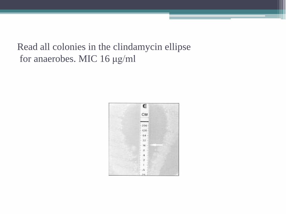
Read all colonies in the clindamycin ellipse
for anaerobes. MIC 16 μg/ml

Susceptibility testing of Helicobacter pylori and
Campylobacter
• Medium
Brucella, Columbia or Mueller Hinton
• An inoculum suspension equivalent to 3 Macfarland
• Incubate the plates for 3 to 5 days.
Etest ESBL
• Enzymes produced by Gram negative aerobic
bacteria mainly in K. pneumoniae and E. coli
• Enzymes generally inhibited by beta-lactamase
inhibitor e.g. clavulanic acid
• Often cross resistance to quinolones,
aminoglycosides and trimethoprim/sulfamethoxazole

Thank you
• To be selected by heavy use of expanded spectrum
cephalosporins (ESC) e.g. ceftazidime
• In nosocomial pathogens from ICUs, oncology, burn and neonatal wards
• In infections associated with indwelling devices
• Increasing prevalence and as outbreaks worldwide
Testing is indicated for:
• Isolates from ICUs and other high risk patients
• Isolates with reduced susceptibility to ESCs i.e. MICs
1 μg/ml or zones 22 mm
• Therapy failure despite in vitro susceptibility
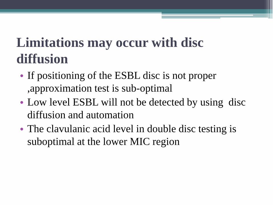
Limitations may occur with disc
diffusion
• If positioning of the ESBL disc is not proper
,approximation test is sub-optimal
• Low level ESBL will not be detected by using disc
diffusion and automation
• The clavulanic acid level in double disc testing is
suboptimal at the lower MIC region

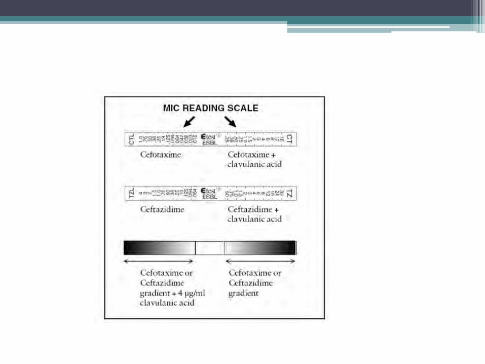
PRINCIPLE
• The Etest ESBL CT/CTL and TZ/TZL strips consist of a
thin, inert and non-porous plastic carrier (5 x 60 mm).
• CT codes for the cefotaxime (0.25-16 μg/ml)gradient
and CTL the cefotaxime (0.016-1 μg/ml) plus 4 μg/ml
clavulanic acid.
• TZ codes for the ceftazidime (0.5-32 μg/ml) gradient
TZL the ceftazidime (0.064-4 μg/ml) plus 4 μg/ml
clavulanic acid.

The presence of ESBL is confirmed by the
appearance of :
• Phantom zone or deformation of the CT or TZ ellipse
• When either CT or TZ MIC is reduced by ≥3 log2
dilutions in the presence of clavulanic acid.
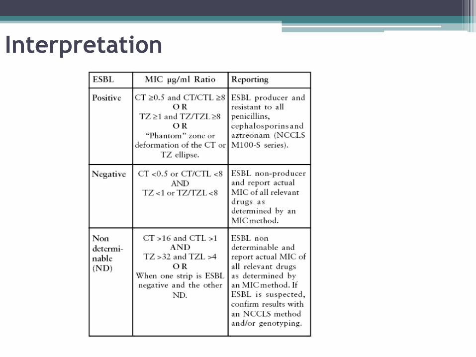
Interpretation
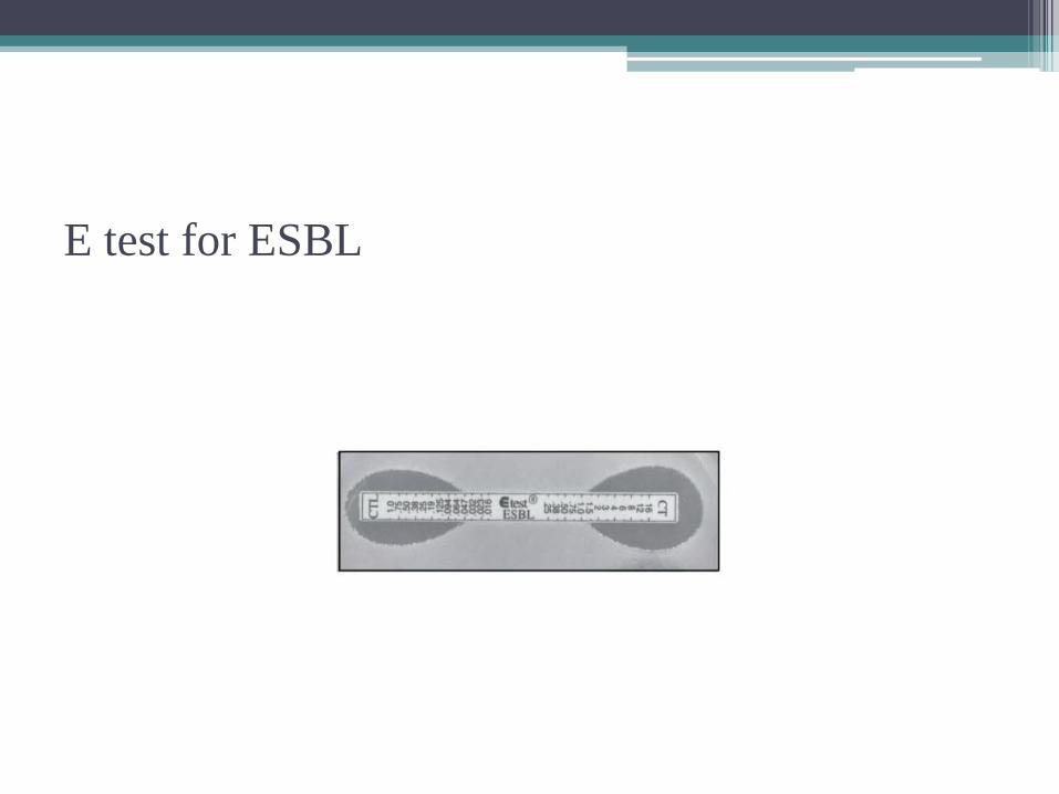
E test for ESBL
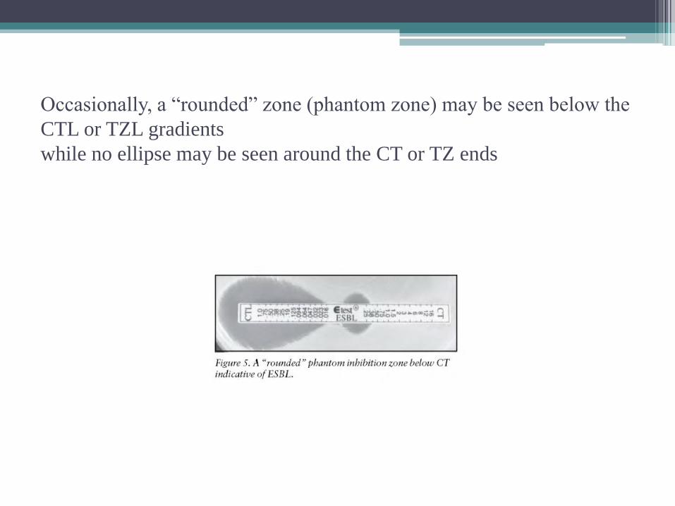
Occasionally, a “rounded” zone (phantom zone) may be seen below the
CTL or TZL gradients
while no ellipse may be seen around the CT or TZ ends
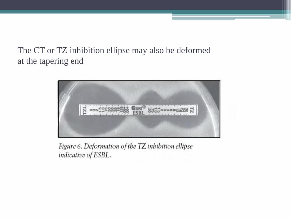
The CT or TZ inhibition ellipse may also be deformed
at the tapering end