Embed Size (px)
Citation preview
1
EE
C24
5
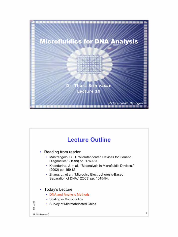
Microfluidics for DNA Analysis
Dr. Thara SrinivasanLecture 19
Picture credit: Nanogen
2U. Srinivasan ©
EE
C24
5
Lecture Outline
• Reading from reader• Mastrangelo, C. H. “Microfabricated Devices for Genetic
Diagnostics,” (1998) pp. 1769-87.• Khandurina, J. et al., “Bioanalysis in Microfluidic Devices,”
(2002) pp. 159-83.• Zhang, L., et al., “Microchip Electrophoresis-Based
Separation of DNA,” (2003) pp. 1645-54.
• Today’s Lecture• DNA and Analysis Methods • Scaling in Microfluidics• Survey of Microfabricated Chips

2
3U. Srinivasan ©
EE
C24
5DNA S
B
P5’ 3’
• Genetic information is stored in chromosomes as long strings of DNA grouped as genes• In humans, 46 chromosomes are 50 - 400 ×106
DNA units long (compared to 4 ×10⁶ for E. coli)
• Units of DNA are nucleotides, consisting of:• A base, a sugar and a phosphate bridge• Sugar linkage has directionality, 5’ and 3’ ends• Four bases: adenine, thymine, guanine, and
cytosine• Bases hydrophobic, backbone hydrophilic• Single-stranded DNA attaches to complementary
strand (G-C, A-T)
4U. Srinivasan ©
EE
C24
5
DNA Analysis• DNA is extracted from cell nucleus and purified
• Break cell membranes using detergent• Remove cell debris, proteins, enzymes
• DNA assays• Detect specific fragments in fingerprint pattern-matching mode• Sequence DNA fragment for base pair order of fragment
• Analysis tools• Chemical amplification• Restriction digestion• Electrophoretic separation• Sanger sequencing process • Hybridization• Fluorescence visualization
3
5U. Srinivasan ©
EE
C24
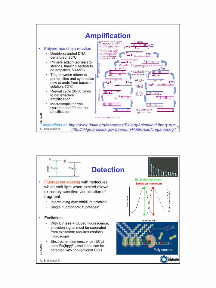
5Amplification
Animations at: http://www.dnalc.org/resources/BiologyAnimationLibrary.htmhttp://bldg6.arsusda.gov/pberkum/Public/sarl/cregan/pcr.gif
• Polymerase chain reaction• Double-stranded DNA
denatured, 95°C• Primers attach (anneal) to
strands, flanking section to be amplified, 50-65°C
• Taq enzymes attach to primer sites and synthesize new strands from bases in solution, 72°C
• Repeat cycle 20-30 times to get effective amplification
• Macroscopic thermalcyclers need 90 min per amplification
6U. Srinivasan ©
EE
C24
5
Excitation maximumEmission maximum
Detection• Fluorescent labeling with molecules
which emit light when excited allows extremely sensitive visualization of fragment• Intercalating dye: ethidium bromide• Single fluorophore: fluorescein
• Excitation• With UV laser-induced fluorescence,
emission signal must be separated from excitation; requires confocal microscope
• Electrochemiluminescence (ECL) uses Ru(bpy)2+
3 end label, can be detected with conventional CCD
4
7U. Srinivasan ©
EE
C24
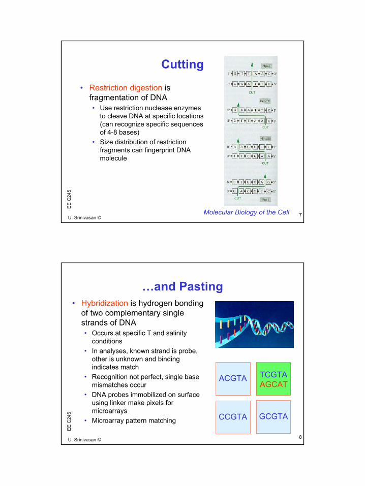
5Cutting
• Restriction digestion is fragmentation of DNA• Use restriction nuclease enzymes
to cleave DNA at specific locations (can recognize specific sequences of 4-8 bases)
• Size distribution of restriction fragments can fingerprint DNA molecule
Molecular Biology of the Cell
8U. Srinivasan ©
EE
C24
5
…and Pasting• Hybridization is hydrogen bonding
of two complementary single strands of DNA• Occurs at specific T and salinity
conditions• In analyses, known strand is probe,
other is unknown and binding indicates match
• Recognition not perfect, single base mismatches occur
• DNA probes immobilized on surface using linker make pixels for microarrays
• Microarray pattern matching
ACGTA
CCGTA GCGTA
TCGTAAGCAT
5
9U. Srinivasan ©
EE
C24
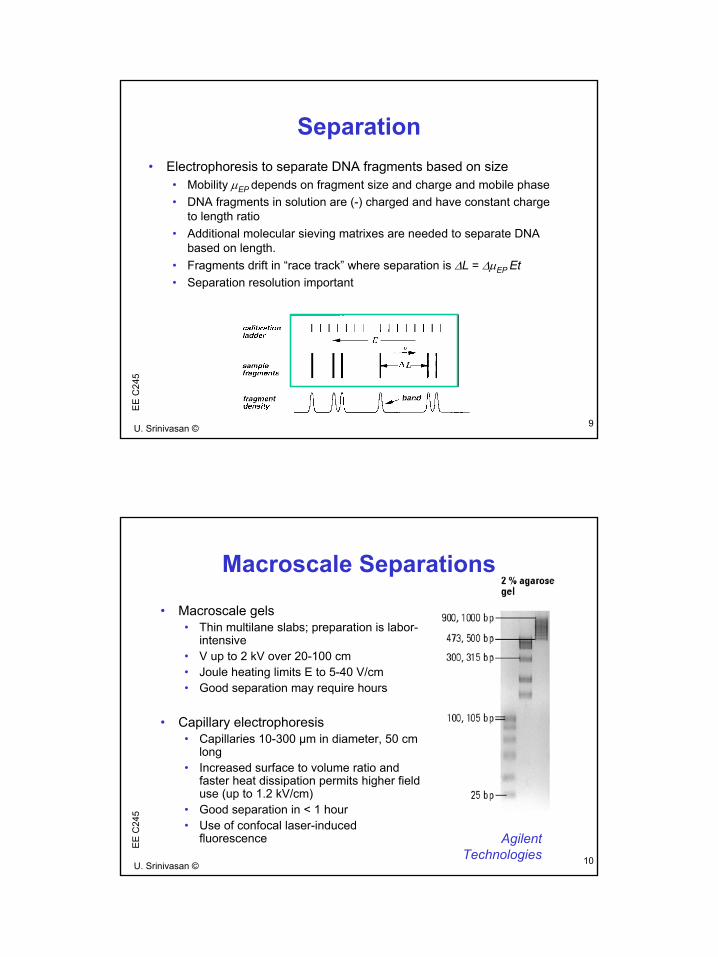
5Separation
• Electrophoresis to separate DNA fragments based on size• Mobility µEP depends on fragment size and charge and mobile phase• DNA fragments in solution are (-) charged and have constant charge
to length ratio• Additional molecular sieving matrixes are needed to separate DNA
based on length.• Fragments drift in “race track” where separation is ∆L = ∆µEP Et• Separation resolution important
10U. Srinivasan ©
EE
C24
5
Macroscale Separations
• Macroscale gels• Thin multilane slabs; preparation is labor-
intensive• V up to 2 kV over 20-100 cm• Joule heating limits E to 5-40 V/cm• Good separation may require hours
• Capillary electrophoresis• Capillaries 10-300 µm in diameter, 50 cm
long• Increased surface to volume ratio and
faster heat dissipation permits higher field use (up to 1.2 kV/cm)
• Good separation in < 1 hour• Use of confocal laser-induced
fluorescence AgilentTechnologies
6
11U. Srinivasan ©
EE
C24
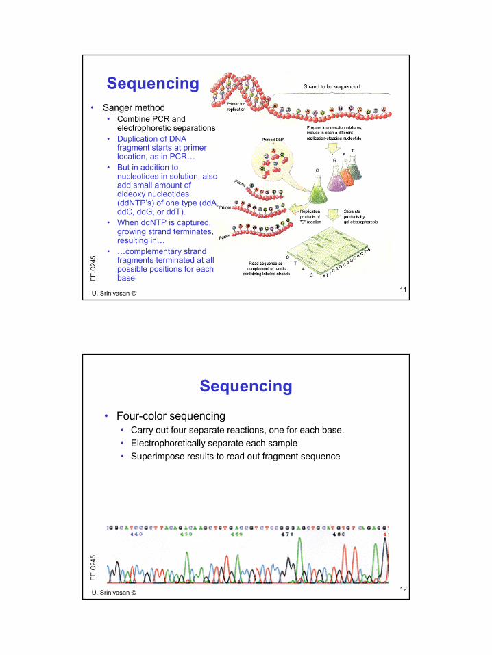
5Sequencing
• Sanger method• Combine PCR and
electrophoretic separations• Duplication of DNA
fragment starts at primer location, as in PCR…
• But in addition to nucleotides in solution, also add small amount ofdideoxy nucleotides (ddNTP’s) of one type (ddA, ddC, ddG, or ddT).
• When ddNTP is captured, growing strand terminates, resulting in…
• …complementary strand fragments terminated at all possible positions for each base
12U. Srinivasan ©
EE
C24
5
Sequencing• Four-color sequencing
• Carry out four separate reactions, one for each base. • Electrophoretically separate each sample• Superimpose results to read out fragment sequence
7
13U. Srinivasan ©
EE
C24
5Today’s Lecture
• DNA and Analysis Methods • Scaling in Microfluidics• Survey of Microfabricated Chips
14U. Srinivasan ©
EE
C24
5
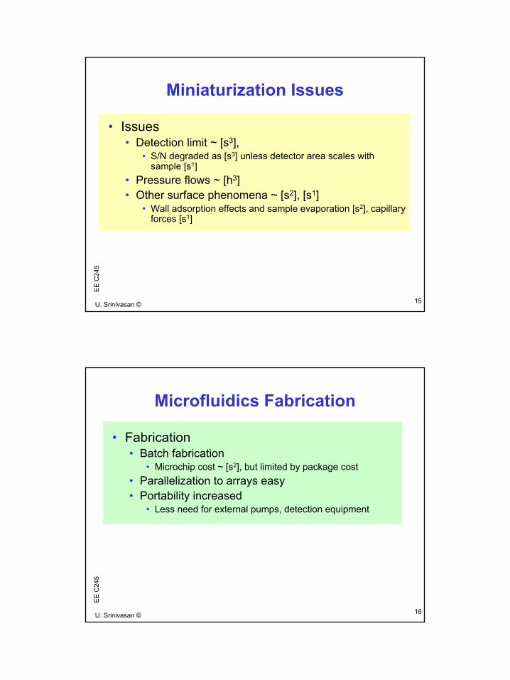
Miniaturization Benefits
• Benefits• Reagent consumption ~ [s3]
• Miniscule reaction volumes reduce reagent cost.• Heat transfer ~ [s2]
• Surface phenomena• Mass transfer ~ [s2]
• Reduced analysis times, with minimum assay time limited by speed of enzyme (30-100 bp/s)
• Flow is laminar• Electroosmotic flow for valveless systems ~ [s2] • Capillary flow ~ [s1]
• Separation efficiency ~ [s-2] • Injection volume well-defined
8
15U. Srinivasan ©
EE
C24
5Miniaturization Issues
• Issues• Detection limit ~ [s3],
• S/N degraded as [s3] unless detector area scales with sample [s1]
• Pressure flows ~ [h3]• Other surface phenomena ~ [s2], [s1]
• Wall adsorption effects and sample evaporation [s2], capillary forces [s1]
16U. Srinivasan ©
EE
C24
5
Microfluidics Fabrication
• Fabrication• Batch fabrication
• Microchip cost ~ [s2], but limited by package cost • Parallelization to arrays easy • Portability increased
• Less need for external pumps, detection equipment
9
17U. Srinivasan ©
EE
C24
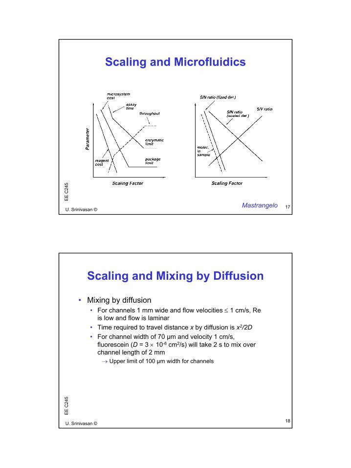
5Scaling and Microfluidics
Mastrangelo
18U. Srinivasan ©
EE
C24
5
Scaling and Mixing by Diffusion
• Mixing by diffusion• For channels 1 mm wide and flow velocities ≤ 1 cm/s, Re
is low and flow is laminar• Time required to travel distance x by diffusion is x2/2D• For channel width of 70 µm and velocity 1 cm/s,
fluorescein (D = 3 × 10⁻⁶ cm2/s) will take 2 s to mix over channel length of 2 mm→ Upper limit of 100 µm width for channels
10
19U. Srinivasan ©
EE
C24
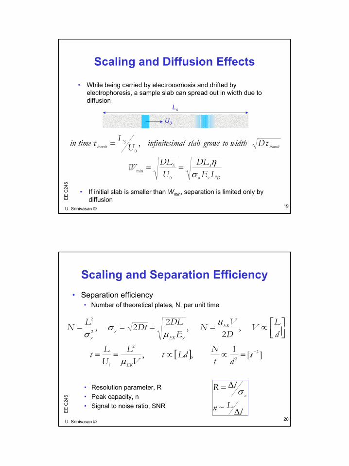
5Scaling and Diffusion Effects
• While being carried by electroosmosis and drifted by electrophoresis, a sample slab can spread out in width due to diffusion
Dxw
SS
transitS
transit
LEDL
UDLW
DwidthtogrowsslabmalinfinitesiULtimein
ση
ττ
==
=
0min
0,
• If initial slab is smaller than Wmin, separation is limited only by diffusion
Ls
U0
20U. Srinivasan ©
EE
C24
5
Scaling and Separation Efficiency• Separation efficiency
• Number of theoretical plates, N, per unit time
[ ] ][1,,
,2
,22,
22
2
2
2
−=∝∝==
∝====
sdt
NLdtVL
ULt
dLV
DVN
EDLDtLN
EKi
EK
xEKx
x
µ
µµ
σσ
• Resolution parameter, R• Peak capacity, n• Signal to noise ratio, SNR
lLn
lRx
∆
∆=
~
σ
11
21U. Srinivasan ©
EE
C24
5Today’s Lecture
• DNA analysis methods • Scaling in microfluidics• Survey of microfabricated devices
22U. Srinivasan ©
EE
C24
5
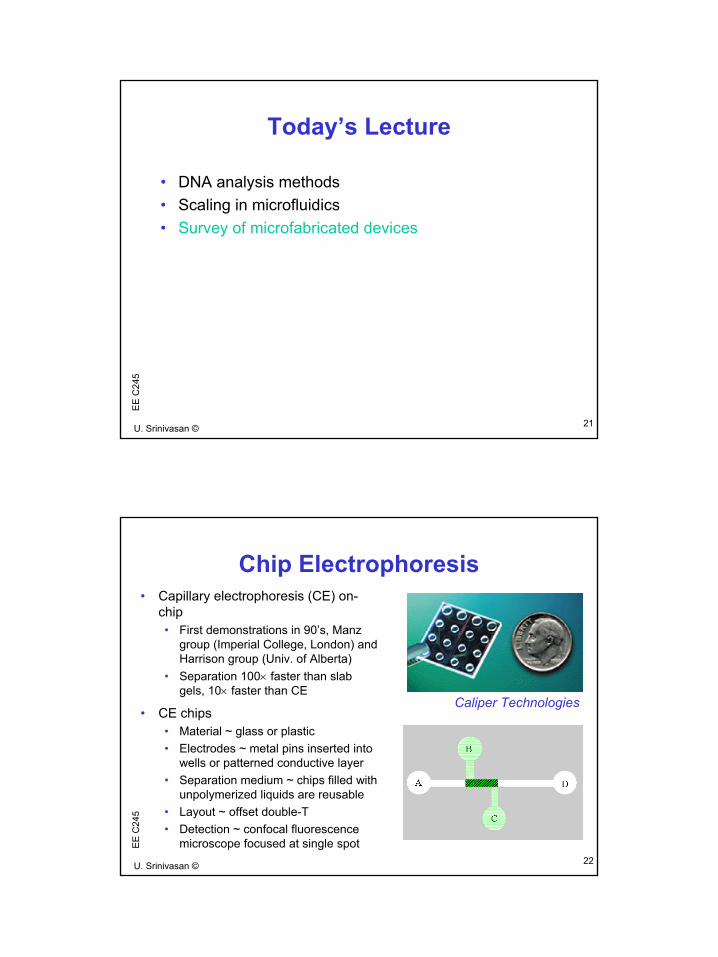
Chip Electrophoresis• Capillary electrophoresis (CE) on-
chip• First demonstrations in 90’s, Manz
group (Imperial College, London) and Harrison group (Univ. of Alberta)
• Separation 100× faster than slab gels, 10× faster than CE
• CE chips• Material ~ glass or plastic• Electrodes ~ metal pins inserted into
wells or patterned conductive layer• Separation medium ~ chips filled with
unpolymerized liquids are reusable• Layout ~ offset double-T • Detection ~ confocal fluorescence
microscope focused at single spot
Caliper Technologies
12
23U. Srinivasan ©
EE
C24
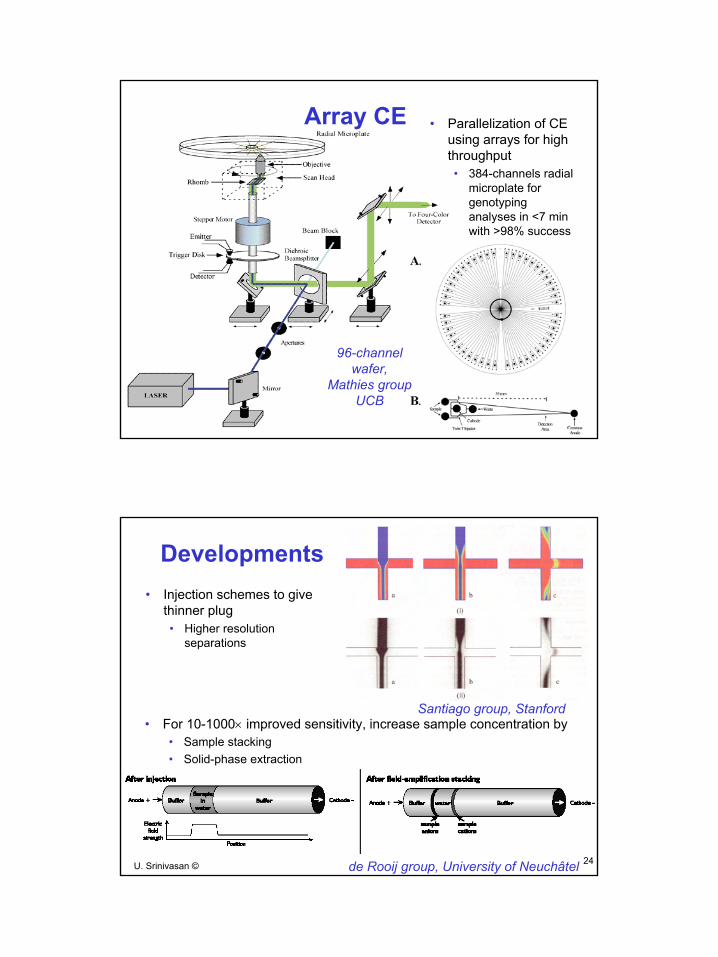
5• Parallelization of CE
using arrays for high throughput• 384-channels radial
microplate for genotyping analyses in <7 min with >98% success
96-channel wafer,
Mathies group UCB
Array CE
24U. Srinivasan ©
EE
C24
5
• For 10-1000× improved sensitivity, increase sample concentration by• Sample stacking• Solid-phase extraction
Developments
Santiago group, Stanford
de Rooij group, University of Neuchâtel
• Injection schemes to give thinner plug• Higher resolution
separations
13
25U. Srinivasan ©
EE
C24
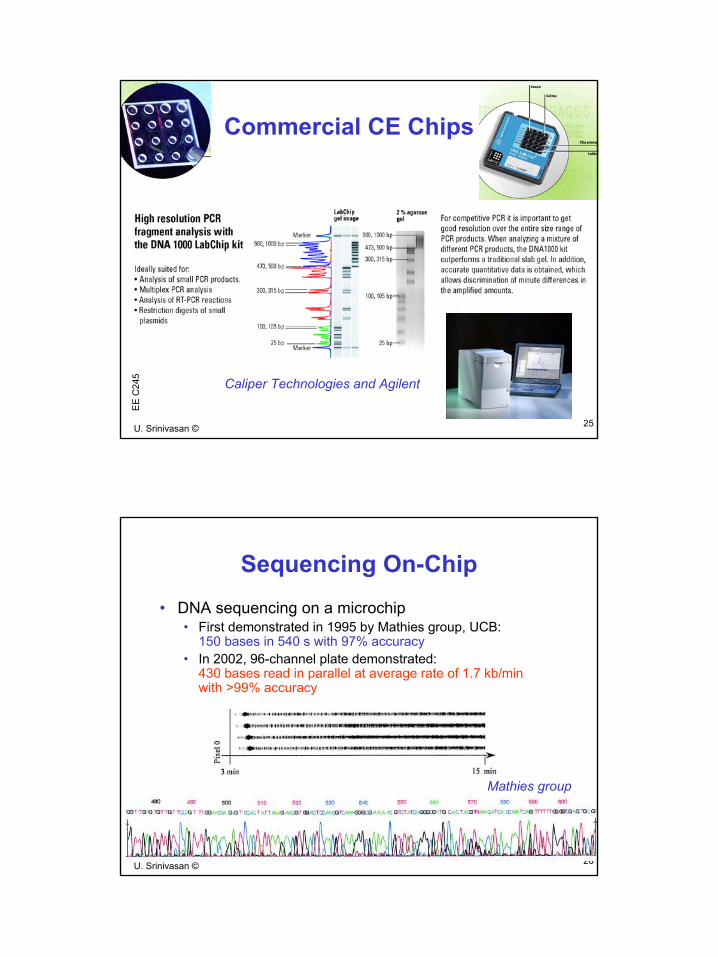
5Commercial CE Chips
Caliper Technologies and Agilent
26U. Srinivasan ©
EE
C24
5
Sequencing On-Chip• DNA sequencing on a microchip
• First demonstrated in 1995 by Mathies group, UCB: 150 bases in 540 s with 97% accuracy
• In 2002, 96-channel plate demonstrated: 430 bases read in parallel at average rate of 1.7 kb/min with >99% accuracy
Mathies group
14
27U. Srinivasan ©
EE
C24
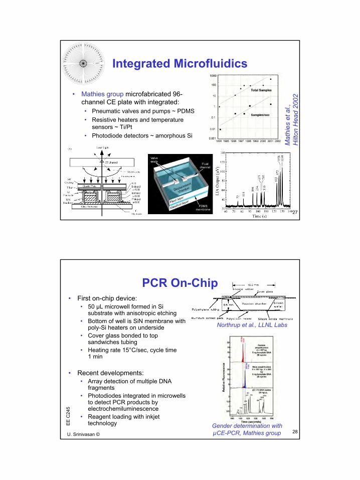
5Integrated Microfluidics
Mat
hies
et a
l.,
Hilt
on H
ead
2002
• Mathies group microfabricated 96-channel CE plate with integrated: • Pneumatic valves and pumps ~ PDMS• Resistive heaters and temperature
sensors ~ Ti/Pt• Photodiode detectors ~ amorphous Si
28U. Srinivasan ©
EE
C24
5
PCR On-Chip• First on-chip device:
• 50 µL microwell formed in Si substrate with anisotropic etching
• Bottom of well is SiN membrane with poly-Si heaters on underside
• Cover glass bonded to top sandwiches tubing
• Heating rate 15°C/sec, cycle time 1 min
• Recent developments:• Array detection of multiple DNA
fragments• Photodiodes integrated in microwells
to detect PCR products by electrochemiluminescence
• Reagent loading with inkjet technology Gender determination with
µCE-PCR, Mathies group
Northrup et al., LLNL Labs

15
29U. Srinivasan ©
EE
C24
5PCR Devices
Cepheid,Sunnyvale CA
Woolley, Mathies and Northrup et al., 1996
30U. Srinivasan ©
EE
C24
5
Microarrays• Fabrication using lithography
and combinatorial chemistry• Fodor et al., 1991
• Glass coated with linker molecule with photoremovable protective group
• UV light through mask removes protective group selectively
• Nucleoside with protected 5’ end bonds to deprotected linkers
• Process repeated one base at a time to give oligonucleotides of arbitary length
• Array of 1024 peptides in 10 steps (210 ), 100 µm probe patches
• McGall et al., 1996, showed technique which uses polyimidephotoresist as protective layer
• Basic microarray today• 50-200 µm patches on 1 cm2 chip
• Up to 40,000 different probes• Possible oligonucleotides for 15-mer is 415
~ 109
• Finished chip in flow-cell package• Detection mainly by fluorescent labeling
Affymetrix process
16
31U. Srinivasan ©
EE
C24
5
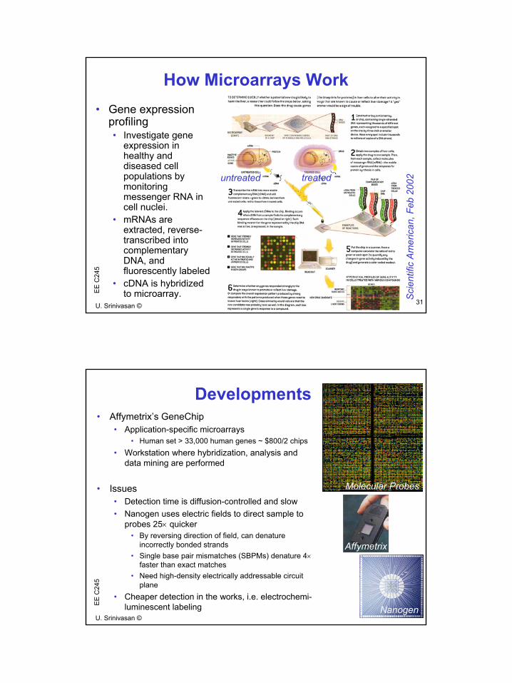
How Microarrays Work
Sci
entif
ic A
mer
ican
, Feb
200
2
• Gene expression profiling• Investigate gene
expression in healthy and diseased cell populations by monitoring messenger RNA in cell nuclei.
• mRNAs are extracted, reverse-transcribed into complementary DNA, and fluorescently labeled
• cDNA is hybridized to microarray.
untreated treated
32U. Srinivasan ©
EE
C24
5
Developments• Affymetrix’s GeneChip
• Application-specific microarrays • Human set > 33,000 human genes ~ $800/2 chips
• Workstation where hybridization, analysis and data mining are performed
• Issues• Detection time is diffusion-controlled and slow• Nanogen uses electric fields to direct sample to
probes 25× quicker• By reversing direction of field, can denature
incorrectly bonded strands• Single base pair mismatches (SBPMs) denature 4×
faster than exact matches• Need high-density electrically addressable circuit
plane• Cheaper detection in the works, i.e. electrochemi-
luminescent labeling
Affymetrix
Nanogen
Molecular Probes