Embed Size (px)
Citation preview
0
Mitochondrial function analysis in diabetes and chronic periodontitis-derived human blood cells: role of resistin
Ana Solange Gomes Costa
Orientadora: Professora Doutora Ana Cristina Rego
Co-orientadora: Professora Doutora Isabel Poiares Baptista
Mestrado Integrado em Medicina Dentária Coimbra, 2017

1
2
Mitochondrial function analysis in diabetes and chronic periodontitis-derived human
blood cells: role of resistin
Costa AS1, Ferreira IL2,3, Baptista IP1,4, Rego AC2,4
1. Área de Medicina Dentária, Faculdade de Medicina da Universidade de Coimbra 2.CNC-Center for Neuroscience and Cell Biology, University of Coimbra, Coimbra, Portugal 3. IIIUC-Institute for Interdisciplinary Research, University of Coimbra, Coimbra, Portugal 4. FMUC-Faculty of Medicine, University of Coimbra, Coimbra, Portugal
Área de Medicina Dentária da Faculdade de Medicina da Universidade de Coimbra
Av. Bissaya Barreto, Bloco de Celas
3000-075 Coimbra
Portugal
Tel: +351 239 484 183
Fax: +351 239 402 910
E-mail: [email protected]
3
Contents Figures and Tables ……..……………………………………………………………………………4
Abbreviations ………………………………………………………………………..………………..5
Resumo .......................………………………………………………………………………..….….6
Abstract …………………………………………………………………………………….………….7
Introduction …………………………………………………………………………………….……...8
Material and methods …………………………………………………………………………..…..12
Results ……………………………………………………………………………………………….17
Discussion ………………………………………………………………...…………………………29
Conclusion ……………………………………………………………………………………...……32
Acknowledgements …………………………………………………………………...…………….33
Bibliography ………………………………………………………………………………………….34
Attachments ………...………………………………………………………………………...……..37
4
Figures and Tables contents
1. Figures
Figure 1. Teeth probing depth ………………………………………………………...……………8
Figure 2. Mitochondria respiratory chain ……………………………………….……………..…15
Figure 3. Representative trace of OCR in isolated PBMCs ……………………………………15
Figure 4. Amplex Red oxidation by H2O2 .….………………………………………...……........16
Figure 5. Clinical parameters for periodontitis evaluation ……………………………………...23
Figure 6. Blood plasma resistin levels and correlation between plasma resistin levels and
body mass index (BMI) ………………..……………………………………………………………24
Figure 7. Correlation between BMI, CAL, BOP and HbA1c with plasmatic resistin levels .....25
Figure 8. H2O2 production by PBMCs ………………………………………………….………...26
Figure 9. Oxygen Consumption Rate (OCR) in PBMCs ……………………………………….27
Figure 10. Oxygen Consumption Rate (OCR) in PBMCs (graphs of parameters
evaluated)…………………………………………………………………………………………….28
2. Tables
Table I. Background groups characteristics ……………………………………………..………18
Table II. Control group medication until the day of blood collection …………………………..19
Table III. Diabetes mellitus type II (DM) patient’s medication until the day of blood collection
…………………………………………………………………………………………………………20
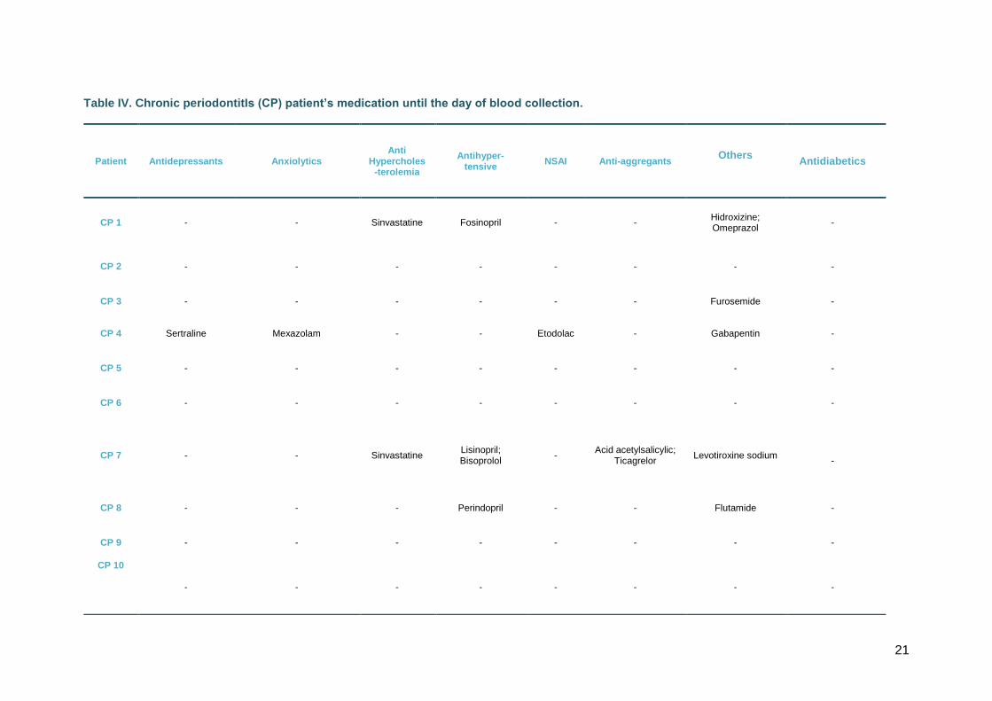
Table IV. Chronic periodontitis (CP) patient’s medication until the day of blood collection ...21
Table V. Chronic periodontitis plus diabetes mellitus type II (DM-CP) patient’s until the day of
blood collection ……………………………………………………………………….……………..22
5
Abbreviations
AAP - American academy of periodontology
ATP - Adenosine triphosphate
BHI - Bioenergetic health index
BMI - Body mass index
BOP - Bleeding on probing
CAL - Clinical attachment level
DM - Diabetes mellitus type II
DMEM - Dulbecco's modified Eagle's medium
EDTA - Ethylenediamine tetraacetic acid
ELISA - Enzyme-linked immunosorbent assay
FBS - Fetal bovine serum
FCCP - Trifluoromethoxy carbonylcyanide phenylhydrazone
HbA1c - Glycated hemoglobin
IL-6 - Interleukin 6
OCR - Oxygen consumption rate
OXPHOS - Oxidative phosphorylation
PBMCs - Peripheral blood mononuclear cells
PBS - Phosphate buffered saline
PD - Probing depth
PI - Plaque index
PGE2 - Prostaglandin E2
ROS - Reactive oxygen species
RPMI 1640 - Roswell park memorial institute 1640 medium
TNF-α - Tumor necrosis factor alfa
6
Resumo
A doença periodontal (DP) é considerada uma doença inflamatória da cavidade oral
onde, por ação de microrganismos patogénicos presentes no biofilme subgengival, o
periodonto é afetado, culminando na reabsorção óssea. A diabetes mellitus tipo II (DM) é
uma doença metabólica inflamatória classificada pelo aumento dos níveis de glicose
sanguínea, existindo uma relação bi-direcional com a DP. Assume-se que a diabetes pode
conduzir ao desenvolvimento de periodontite e que esta última pode influenciar a ocorrência
ou agravamento da diabetes. A resistina, proteína secretada pelo tecido adiposo, é
associada com a resistência à insulina, permitindo relacionar a obesidade com a DM. Estas
doenças inflamatórias têm sido referidas como estando associadas ao stress oxidativo (e.g.
devido ao aumento da produção de espécies reativas de oxigénio) e à disfunção
mitocondrial, dando informação acerca do estado energético celular. As células
mononucleares do sangue periférico (PBMCs, do inglês ‘peripheral blood mononuclear
cells’) constituem modelos úteis para o estudo da função mitocondrial e do stress oxidativo
associado a doenças inflamatórias como a DM e periodontite crónica (PC). Neste trabalho,
procedeu-se à seleção de doentes inseridos na base de dados da consulta de medicina
dentária do Centro Hospitalar da Universidade de Coimbra, onde foram avaliados
parâmetros clínicos para o diagnóstico da saúde periodontal e determinado o índice de
massa corporal. Procedeu-se à recolha de sangue venoso periférico de indivíduos com DM,
PC, DM-PC e em indivíduos controlo, por forma a avaliar os níveis de hemoglobina glicada
níveis plasmáticos de resistina e a produção de peróxido de hidrogénio (H2O2) bem como a
bioenergética mitocondrial no plasma e PBMCs dos diferentes grupos de doentes e
controlos. Os nossos resultados mostram um aumento dos parâmetros clínicos para o
diagnóstico de DP nos doentes com PC e DM-PC e um concomitante aumento da produção
de H2O2. O aumento dos níveis plasmáticos de resisina em indivíduos com DM e DM-PC
foram correlacionados com o índice de massa corporal, ocorrendo um aumento dos níveis
de hemoglobina glicada nos doentes em que se verificou um aumento dos níveis de
resistina (DM-PC). A nível da atividade mitocondrial, verificou-se uma diminuição da
respiração máxima nas PBMCs isoladas dos doentes com PC. Em contraste, observou-se
um aumento da respiração basal, respiração máxima e da produção de ATP mitocondrial
nas PBMCs dos doentes com DM-PC. Este incremento na capacidade respiratória,
associado a um aumento do H+ leak nos doentes com DM-PC poderá explicar a diminuição
do BHI, um índice de avaliação da capacidade bioenergética,nestes doentes.
Palavras-chave: periodontite crónica; diabetes mellitus type II; PBMCs; mitocôndria;
stress oxidativo; taxa de consumo de oxigénio
7
Abstract
Periodontal disease is an oral inflammatory disease due to pathogenic
microorganisms present on subgingival biofilm that affect the periodontium, culminating with
bone resorption. Diabetes mellitus type II (DM) is an inflammatory and metabolic disorder
classified by increased blood glucose levels, fot which a bi-directional relationship with
periodontal disease has been described. It is assumed that diabetes can lead to the
development of periodontitis, and that the latter may influence the occurrence or worse
diabetes condition. Resistin is released by the adipose tissue and related with insulin
resistance, linking obesity and DM. These inflammatory diseases associate with oxidative
stress (e.g. due to increased production of reactive oxygen species) and mitochondrial
dysfunction, giving information about cellular energy status. Peripheral blood mononuclear
cells (PBMCs) are useful models for studying mitochondrial function and oxidative stress
associated with inflammatory diseases such as DM and chronic periodontitis (CP). The
patient’s selection was screened from dentistry consultations at the Hospital Center of
Coimbra University (CHUC) where clinical parameters for the diagnosis of periodontal health
and body mass index were determined. Venous peripheral blood was collected from DM, CP,
DM-CP and control individuals in order to evaluate glycated hemoglobin levels and resistin
plasma levels and H2O2 production and mitochondrial bioenergetics in isolated PBMCs. Our
results show increased clinical parameters for periodontal disease diagnosis in patients with
CP and DM-CP, along with increased production of H2O2. Increased plasma resistin levels in
DM and DM-CP patients correlated with body mass index. Moreover, the increase in
glycated hemoglobin levels also occurred in DM-PC patients. When studying mitochondrial
function, we observed decreased maximal mitochondrial respiration in PBMC isolated from
CP patients. In contrast, an increase in basal and maximal respiration and enhanced
mitochondrial ATP production was detected in PBMCs from DM-CP patients. Such
exacerbated parameters associated with and an increase in H+ leak in DM-CP may explain
the decrease in BHI, an index of bioenergetic health, in these patients.
Key-words: chronic periodontitis; diabetes mellitus type II; oxidative stress; mitochondria;
oxygen consumption rate
8
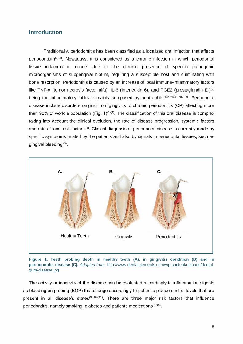
Introduction
Traditionally, periodontitis has been classified as a localized oral infection that affects
periodontium(1)(2). Nowadays, it is considered as a chronic infection in which periodontal
tissue inflammation occurs due to the chronic presence of specific pathogenic
microorganisms of subgengival biofilm, requiring a susceptible host and culminating with
bone resorption. Periodontitis is caused by an increase of local immune-inflammatory factors
like TNF-α (tumor necrosis factor alfa), IL-6 (Interleukin 6), and PGE2 (prostaglandin E2)(3)
being the inflammatory infiltrate mainly composed by neutrophils(1)(4)(5)(6)(7)(2)(8). Periodontal
disease include disorders ranging from gingivitis to chronic periodontitis (CP) affecting more
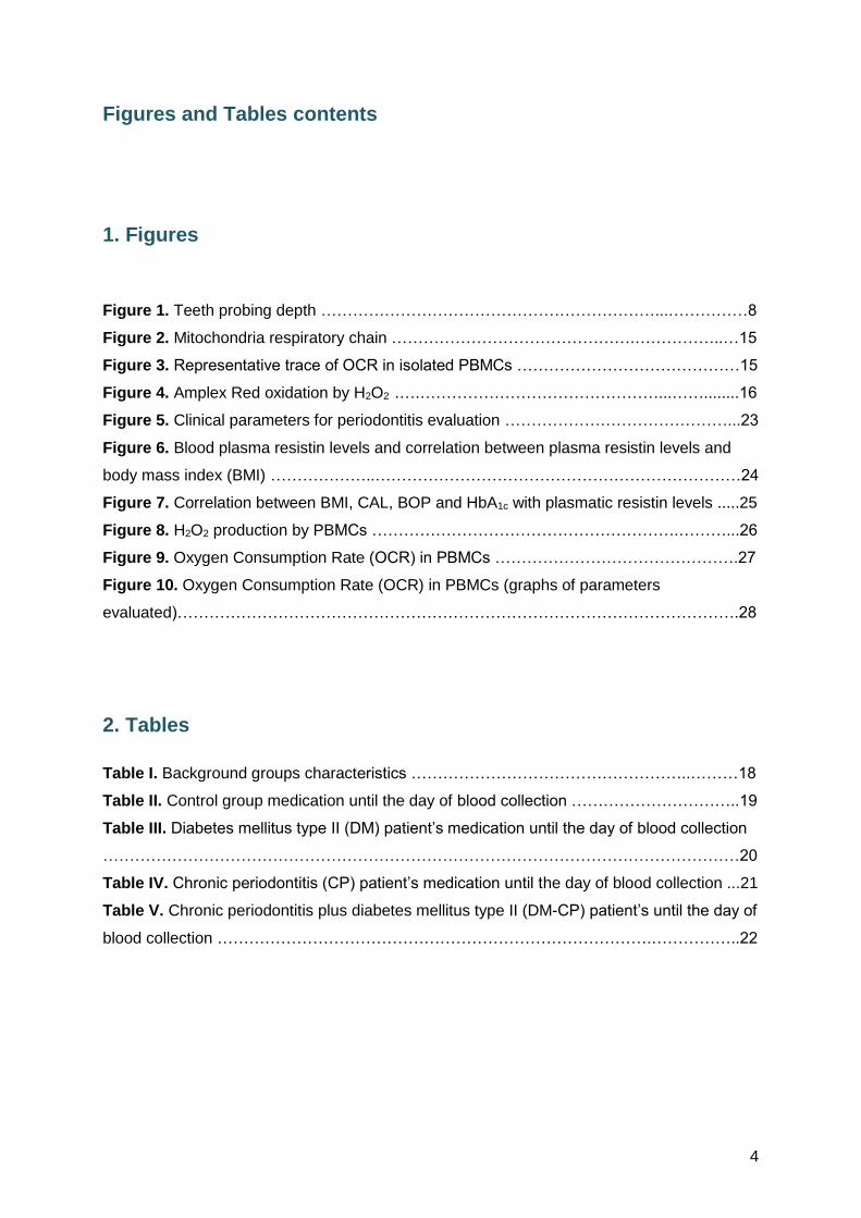
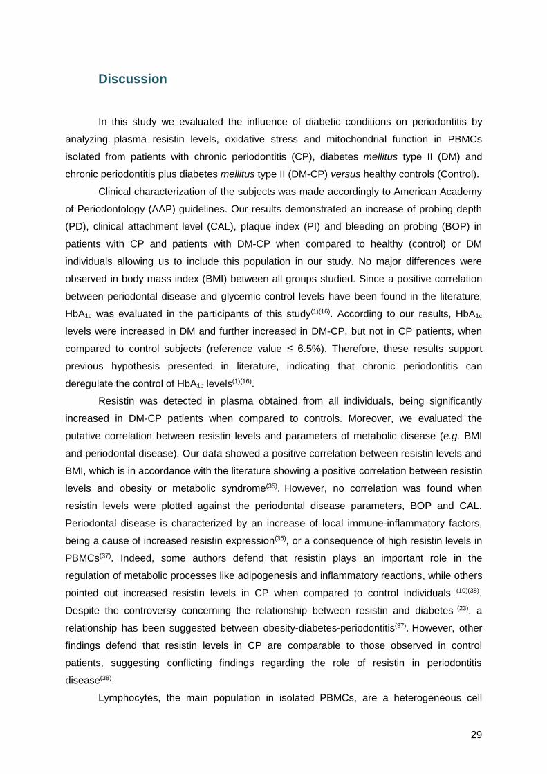
than 90% of world’s population (Fig. 1)(1)(4). The classification of this oral disease is complex
taking into account the clinical evolution, the rate of disease progression, systemic factors
and rate of local risk factors (1). Clinical diagnosis of periodontal disease is currently made by
specific symptoms related by the patients and also by signals in periodontal tissues, such as
gingival bleeding (9).
Figure 1. Teeth probing depth in healthy teeth (A), in gingivitis condition (B) and in
periodontitis disease (C). Adapted from: http://www.dentalelements.com/wp-content/uploads/dental-
gum-disease.jpg
The activity or inactivity of the disease can be evaluated accordingly to inflammation signals
as bleeding on probing (BOP) that change accordingly to patient’s plaque control levels that are
present in all disease’s states(9)(10)(11). There are three major risk factors that influence
periodontitis, namely smoking, diabetes and patients medications (2)(5).
Healthy Teeth Gingivitis Periodontitis
A. B. C.
9
Characterized by hyperglycemia, diabetes mellitus (DM) is defined as a group of
metabolic diseases that result from a defect in insulin secretion, insulin defect action, or
both(2)(12)(13)(14) . DM can be sub-divided in two major classifications: 1) diabetes mellitus type
I, classified as an autoimmune disease where pancreatic β-cells do not secrete insulin - this
type of diabetes is also known as insulin-dependent diabetes, affecting mostly young people;
and 2) diabetes mellitus type II, in which pancreatic β-cells secrete low insulin levels along
with resistance to insulin action. It is characterized by being associated with obesity and
aging. The prevalence of this type of diabetes, when compared to the other types, varies
between 90-95%. Although not so frequent, diabetes is also associated with pregnancy,
diseases of exocrine pancreas and endocrinopathies (8)(12).
Scientific evidence that associate DM with periodontitis had been found. Two-way
relationship between these diseases has been demonstrated, being diabetic individuals two
or three times more susceptible to periodontitis(1)(2)(12)(15)(16)(17). Some authors refer that a poor
glycemic control can influence the periodontitis state(2). Thus, periodontal disease is an
important factor to increase the risk of diabetic complications, being considered the 6th
complication of diabetes (7)(16)(18).
Obesity is considered a risk factor for diabetes mellitus type II, caused by a high caloric
intake in combination with a sedentary lifestyle(5). The body mass index (BMI), an indicator of
obesity, corresponds to the body fat percentage accumulated using a formula that relates
individuals' weight and height, allowing us to classify if someone is within the interval of
normal weight (18.5 – 24.9 kg/m2)(19). Glycated hemoglobin (HbA1c) occurs after binding of
the NH2 terminal of hemoglobin β-chain to excess and continuous glucose, diffused from
plasma into the erythrocytes, along their life cycle. In this way HbA1c is considered a reliable
marker of glucose concentration, indicating hyperglycemia severity in individuals with
diabetes(15)(20).
Adipose tissue abundance, associated to insulin resistance, is responsible for altered
secretion of adipokines that play an important role in pathological and physiological
inflammatory response(6)(21). Currently, there are many different types of adipokines
described on literature and some of them are associated with diabetes mellitus. Apokines,
like resistin, are immune-inflammatory factors secreted by adipose tissue(22). Described as
being associated with insulin resistance and as markers of weight regulation, these
adipokines are involved in conditions such as cardiovascular diseases, diabetes mellitus, and
other inflammatory diseases(6)(7). Resistin, a cysteine-enriched protein, was firstly found in
mice white adipose tissue; however, low resistin levels are produced by adipocytes in
humans being produced by peripheral blood mononuclear cells (PBMCs), like monocytes
and lymphocytes, and also by macrophages(7)(22)(23).
10
Recent findings concerning immunometabolism have focused on the importance of immune
cells in inflammation associated with obesity and diabetes mellitus(24). Derived from the
lymphoid lineage, the uni-nucleated lymphocytes play a very important role in adaptive
immunity, using mitochondria to meet the energetic demands(25). The heterogeneity of these
cells make them ideal to investigate the relationship between bioenergetics and disease
processes related to inflammation(25). Mitochondria are cellular organelles that play a major
role in generating cellular energy in the form of adenosine triphosphate (ATP) via oxidative
phosphorylation (OXPHOS); apart from that they are also involved in the regulation of the
intracellular calcium concentration, intracellular redox signaling, heme synthesisand
apoptosis. Cellular energy requirements are dependent on environment and redox
balance(26)(27)(28). About 80-90% of energy (ATP) required for the process of inflammation is
produced by mitochondria through oxidative processes. It is recognized that alterations in
mitochondrial function can contribute to aging process and to several diseases, like
neurodegenerative diseases, atherosclerosis and cancer(26). In addition, mitochondrial
dysfunction can be a crucial process that can explain the metabolic alterations like insulin
resistance or the etiopathogenic process of other systemic diseases(27)(29). Lymphocytes, the
main population in isolated PBMCs, are a heterogeneous cell population that largely
dependents on mitochondria to meet energetic demands(30). In this way, PBMCs obtained
from human blood have been widely used for mitochondrial function evaluation. In this work,
we assume that PBMCs reproduce systemic changes occurring in inflammatory diseases like
periodontitis. Bioenergetic analysis in blood cells, particularly PBMCs, has suggested that
they may be used to measure oxygen consumption rates (OCR), indicating potential
mitochondrial dysfunction(25)(30).
Reactive oxygen species (ROS), including both oxygen radicals and also non-radical
species, are continuously generated in organisms during mitochondrial oxidative
metabolism(8). However, recent studies suggested that ROS, like hydrogen peroxide (H2O2),
play a role on diabetic and periodontal complications contributing to impairment of the
antioxidant gene expression responsible for ROS degradation and maintenance of vascular
health. Deregulation of these defense mechanisms or high ROS production can lead to
“oxidative stress" state causing tissue damage in individuals with chronic diseases like
diabetes or chronic periodontitis. Moreover, it was shown that increased HbA1c levels lead to
an increase of ROS production(31). Within periodontal tissues, an increase in ROS production
causes deregulation in cellular homeostasis, further inducing tissue damage. In diabetes
mellitus there are several evidences suggesting that oxidative stress can be associated to
pre-diabetic and diabetic states (8), which can also be responsible for diabetic complications
like chronic periodontitis(32). In this way, oxidative stress can act as a link between metabolic

11
syndrome (associated to the combination of obesity, hypertension, hyperinsulinemia and
dyslipidemia associated with diabetes mellitus) and periodontitis(3)(27).
Research on mitochondrial dysfunction and oxidative stress in inflammatory diseases is a
current topic, eliciting special interest in the study of these mechanisms related to diabetes
mellitus and periodontitis, as well as in finding a specific and sensitive biomarker that can be
used for risk and screening assessment of these diseases(6)(26)(22). Thus, the aim of this study
is to investigate the relationship between mitochondrial bioenergetics with the disease
processes associated with individuals with type-II diabetes and periodontitis and also to
evaluate the levels of resistin in serum of the same patients.
12
Material and methods
Material
Resistin kit was from RayBio®. Amplex® Red was obtained from Molecular Probes, Life
Technologies (Eugene, OR, USA). Ficoll-Paque solution was from GE Healthcare (GE
Healthcare Bio-Sciences, PA, USA). RPMI 1640, DMEM, poly-D-lysine, oligomycin,
trifluoromethoxy carbonylcyanide phenylhydrazone (FCCP), antimycin A and rotenone, were
from Sigma Chemical Co. (St Louis, MO, USA). All other reagents were of analytical grade.
1.1. Subject selection
This study was carried out from September 2016 to May 2017. The patient’s selection was
screened from dentistry consultations in Hospital Centre of Coimbra’s University (CHUC).
The study population consisted in patients with 45 to 75 years of age, and balanced
individuals (20 men and 20 women) presenting a minimum of 15 teeth. Informed written
consent was obtained from all subjects that agreed to participate voluntarily (see attachment
1). All the patients were evaluated for medications, weight, height and HbA1c levels.
The individuals satisfying the above criteria were categorized into 3 diseased study
groups (DM group: 10 subjects with no history of periodontal disease, but with clinical
diagnosis for diabetes mellitus type II; CP group: 10 subjects with history of periodontal
disease, with probing depths > 3mm; DM-CP group: 10 subjects with history of periodontal
disease, with probing depths >3mm and clinical diagnosis of diabetes mellitus type II) and a
control group (CONT group: 10 subjects with no history of either periodontal disease or
diabetes mellitus type II). The clinical parameters plaque index (PI), probing depth (PD),
bleeding on probing (BOP) and clinical attachment level (CAL) were clinical parameters
assessed for all the subjects, using a periodontal probe. Periodontitis were classified as:
slight, when PD values are between 3 and 5 mm; moderate, when PD values are between
more or equal to 5 mm and less than 7 mm and severe, when PD values are more or equal
than 7mm; an healthy probing depth is considered between 1-3 mm(10). Moreover,
periodontal disease can be described by CAL which can be characterized in slight (1-2 mm),
moderate (3 to 4 mm) and severe (≥ 5 mm), according to American Academy of
Periodontology Guidelines(11). Body Mass Index (BMI) was also calculated for each patient
according to the formula BMI=weight (kg)/[height (m)2] (Table V).
13
1.2. Blood sample collection and glycated hemoglobin determination
Using a 20-guage needle with 30 ml syringes, about 15 ml of venous peripheral blood was
collected between 8-10 a.m. by venipuncture into commercially available tubes containing
ethylenediamine tetraacetic acid (EDTA) as anticoagulant, at dentistry consultations in
Hospital Centre of Coimbra’s University (CHUC). Collected samples were immediately
transferred to the Center for Neurosciences and Cell Biology (CNC), University of Coimbra,
for PBMCs isolation. An aliquot of 3 ml of blood was used to determinate the level of glycated
hemoglobin (HbA1c) at Clinical Pathology service, CHUC.
1.3 Quantification of resistin levels
The resistin levels were measured by enzyme-linked immunosorbent assay (ELISA kit
from RayBio®) by following the intensity of the fluorescence produced at 450 nm, according
to manufacturer's instructions. All the samples were run in duplicates and resistin
concentration in plasma samples calculated from a standard curve for resistin.
1.4 Isolation and culture of peripheral blood mononuclear cells
After being collected, 12 ml of blood samples were diluted with 36 ml phosphate buffered
saline (PBS) containing (in mM): 137 NaCl, 2.7 KCl, 1.8 KH2PO4, 10 Na2HPO4·2H2O, pH 7.4
and carefully layered onto Ficoll-Paque™ solution in 50 ml Falcon tubes and then centrifuged
at 2500 rpm for 20 minutes at 18ºC in a swing-out rotor without brake. After centrifugation,
the lymphocyte-containing ring was removed with a Pasteur pipette, collected in another 50
ml Falcon tube and further diluted with PBS to a final volume of 45 mL. The tubes were then
centrifuged at 1500 rpm for 10 min at 18°C with maximum braking and acceleration, and
cells resuspended in 10 mL RPMI 1640 (Sigma R4130) culture medium supplemented with:
10 mM Hepes, 12 mM NaHCO3, 2 mM glutamine, 1mM sodium pyruvate plus 10% FBS heat
inactivated. Cells were then counted in a hemocytometer by using trypan blue exclusion test
currently used to determine the number of viable cells in a cell suspension. The method is
based on the principle that live cells with intact cell membranes exclude trypan blue,
presenting a clear cytoplasm, whereas a nonviable cell has a blue cytoplasm under an
inverted brightfield microscope. Cells were plated in 100 μg/mL poly-D-lysine (Sigma P1149;
mol wt: 150,000-300,000) precoated XF24 microplate wells at 0.5 × 10⁶ cells/well in a final
volume of about 180 μL, and let to adhere for 1.5 - 2h in a humidified incubator chamber with
14
95% air and 5% CO2 at 37°C. Cell adherence was checked under an inverted brightfield
microscope with 20X magnification.
1.5 Preparation of XF24 microplate wells for oxygen consumption rate by
Seahorse analyzer
Adhered lymphocytes (in 180μL RPMI 1640, as described above) were carefully rinsed in
order to remove RPMI 1640 culture medium. For this purpose, 100 μL was removed and 100
μL non-buffered DMEM (Sigma 5030) supplemented with 1,85g/L de NaCl, 2g/ L glucose, 1
mM sodium pyruvate and 2 mM glutamine, pH 7.4 at 37ºC were added to the wells. This
procedure was repeated and after a mild washout, 450 μL of DMEM, supplemented as
described above, was added to the wells. The multi-well plate was then placed for 45 min –
1h in a 37ºC-humidified incubator without CO2, containing 95% air.
1.6 Analysis of mitochondrial oxygen consumption rate by Seahorse analyzer
Cellular bioenergetics of the isolated cells was determined using the extracellular flux
analyzer (Seahorse Bioscience), which measures O2 and protons in the extracellular milieu.
This system allows for real time, noninvasive measurement of OCR, which can be correlated
with mitochondrial function/oxidative burst. The Seahorse XF Cell Mito Stress Test Kit
contains a cartridge lid, a sensor cartridge, a hydro booster and XF calibrant apart the utility
plate described above. The day before experiments, seahorse XF24 apparatus was turned
on and seahorse XF calibrant was added to the sensor cartridge in order to hydrate the
boosters at 37°C in a non-CO2 incubator, overnight.
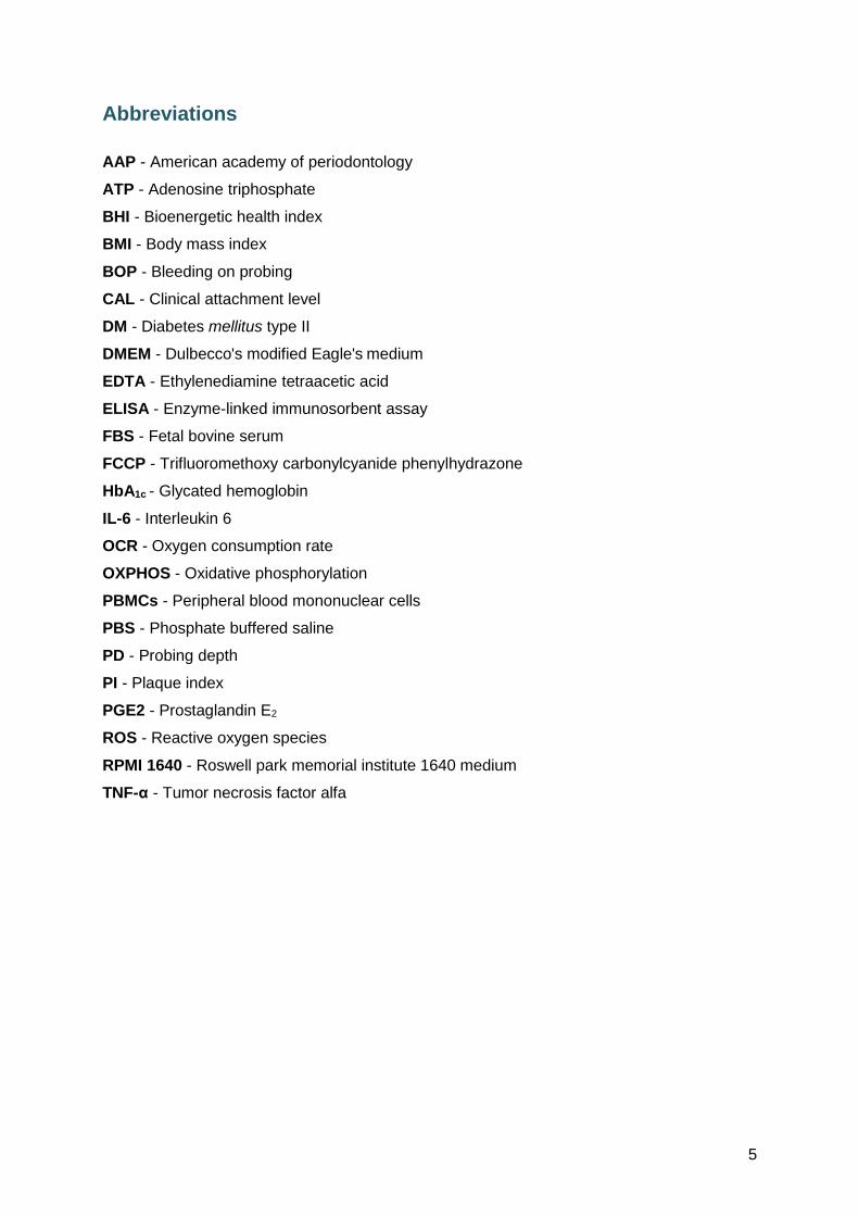
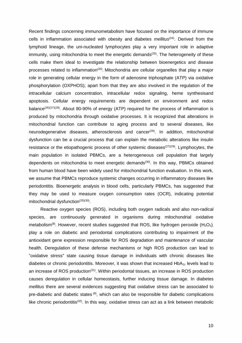
On the day of the experiments, the injection ports attached to the wells that allow for
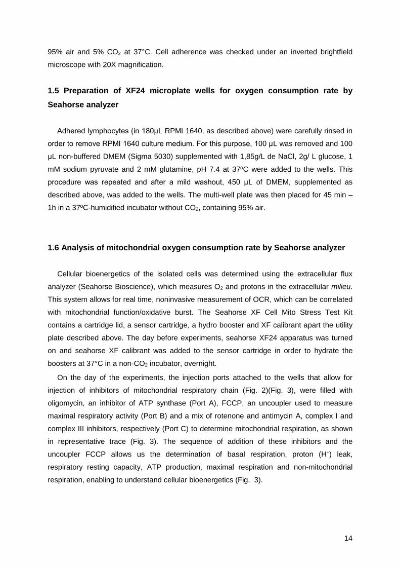
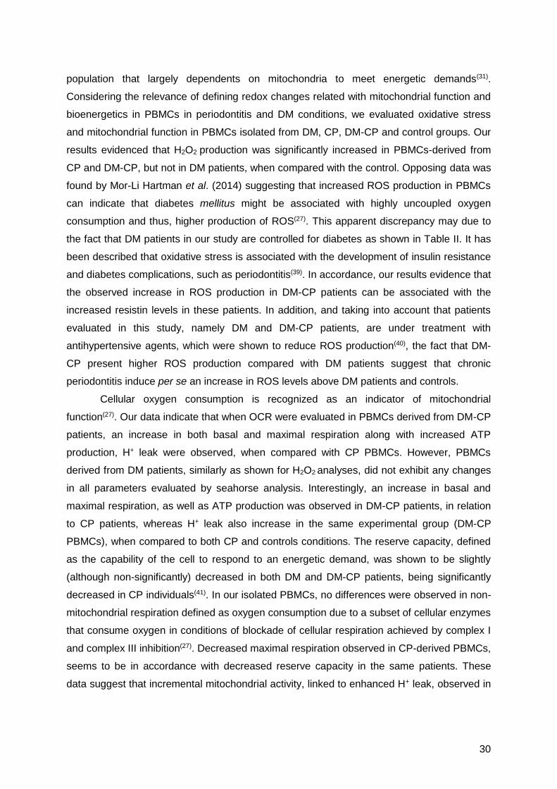
injection of inhibitors of mitochondrial respiratory chain (Fig. 2)(Fig. 3), were filled with
oligomycin, an inhibitor of ATP synthase (Port A), FCCP, an uncoupler used to measure
maximal respiratory activity (Port B) and a mix of rotenone and antimycin A, complex I and
complex III inhibitors, respectively (Port C) to determine mitochondrial respiration, as shown
in representative trace (Fig. 3). The sequence of addition of these inhibitors and the
uncoupler FCCP allows us the determination of basal respiration, proton (H+) leak,
respiratory resting capacity, ATP production, maximal respiration and non-mitochondrial
respiration, enabling to understand cellular bioenergetics (Fig. 3).
15
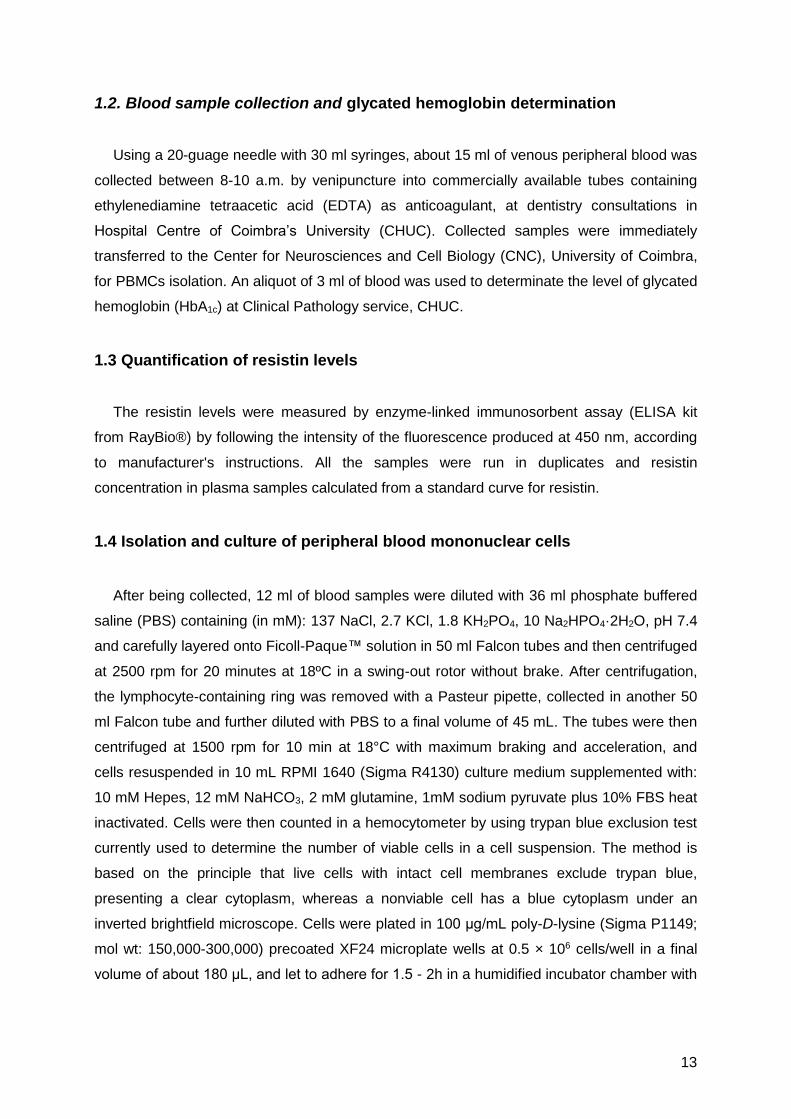
Figure 2. Mitochondria respiratory chain. Complex (Cx) I, II, III, IV and ATP synthase (Cx V)
representation at inner mitochondrial membrane (IMM). H+ flux from matrix to intermembrane space,
through Cx I, III and IV as well as the site of action of inhibitors (rotenone, antimycin A and oligomycin)
and the uncoupler FCCP, are also shown. Adapted from:
https://www.researchgate.net/figure/40034270_fig1_Schematic-representation-of-the-mitochondrial-
respiratory-chain-After-permeabilization
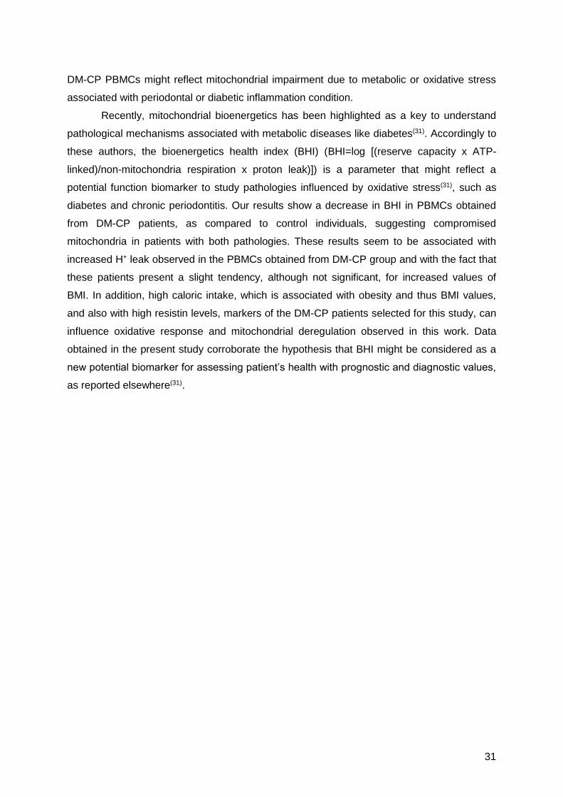
Figure 3. Representative trace of OCR in isolated PBMCs. Parameters of mitochondrial function
include basal respiration, ATP production, proton (H+) leak, non-mitochondrial oxygen consumption,
maximal respiration and reserve capacity parameters, depicted by colored areas.
Rotenone Antimycin A
Oligomycin
FCCP
0
50
100
150
200
Oligomycin(1 mM)
FCCP(1 mM)
Antimycin A (4 mM) +
Basal Respiration
ATP production
Proton leak
Non-mitochondrial Oxygen Consumption
Maximal Respiration
Reserve capacity
Oxygen c
onsum
ptio
n r
ate
(O
CR
)
(nm
ole
s O
2/m
L/m
in/0
.2 x
10
6 c
ells
)
Rotenone (2 mM)
Time (min)
16
1.7 Measurement of H2O2 levels
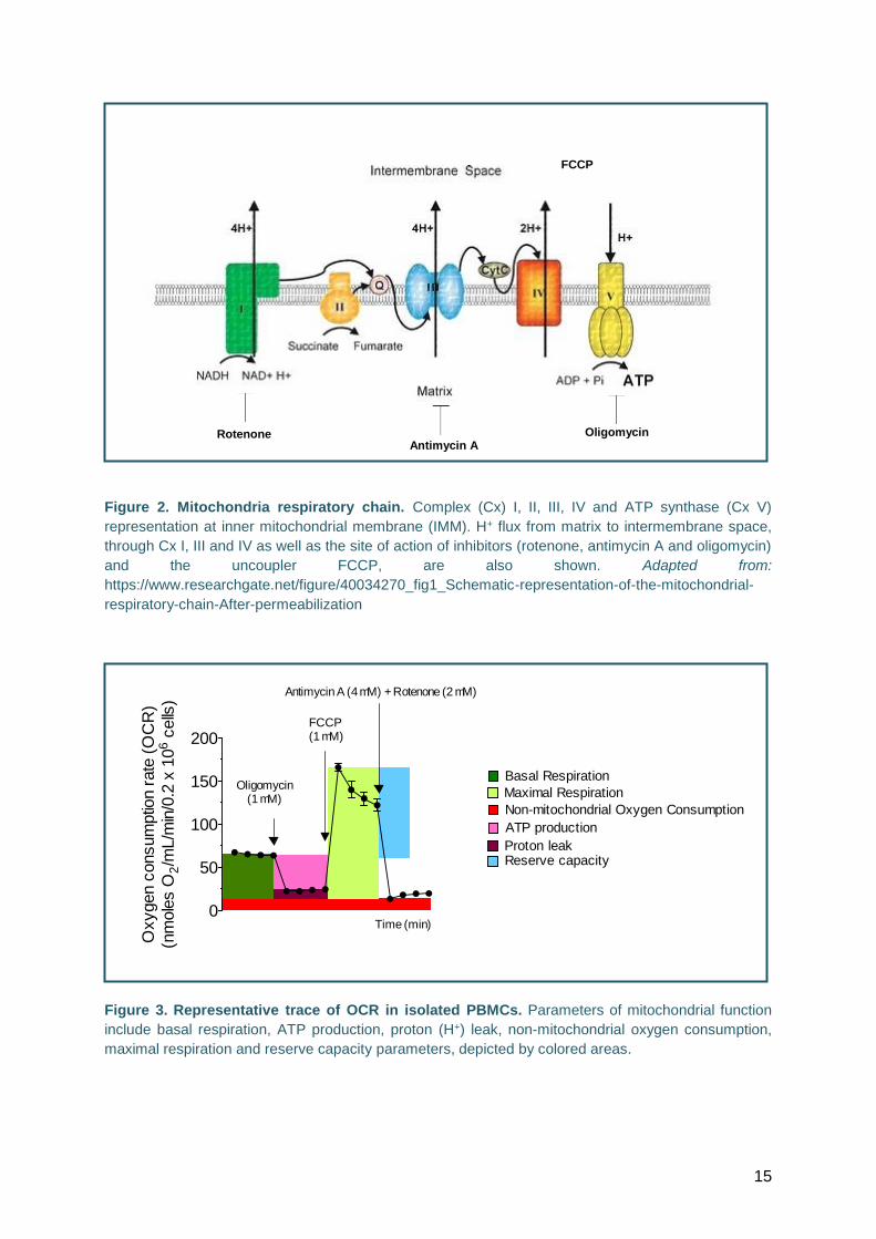
Briefly, 0.5x10⁶ cells were ressuspended in Na+ medium containing (in mM) 140 NaCl, 5
KCl, 1 CaCl2, 1 MgCl2, 10 glucose, 10 Hepes, pH 7.4 plus Amplex® Red reagent (10 μM)
and horseradish peroxidase (0.5 units per mL). The reaction of Amplex® Red (10-acetyl-3.7-
dihydroxyphenoxazin) in the presence of peroxidase, occur in 1:1 stoichiometry, producing
resorufin, a red-fluorescent oxidation product (Fig. 4)(34). Fluorescence was followed at 37°C
for 20 minutes, at an excitation wavelength of 550 nm and an emission wavelength of 580
nm, using a microplate reader Spectrofluorometer Gemini EM (Molecular Devices, USA).
Results were expressed in RFU/minute.
Figure 4. Amplex Red oxidation by H2O2. The Horseradish peroxidase (HRP) uses Amplex Red as
an electron donor to reduce H2O2, resulting in the fluorescent compound resorufin, Changes in
fluorescence intensity are directly proportional to the amount of H2O2 consumed during the reaction
thus reflecting the levels of H2O2. Adapted from:
http://pubs.rsc.org/en/content/articlelanding/2016/lc/c5lc01413a#!divAbstract
Statistical analysis Data were analyzed by using Excel and GraphPad Prism 6 (GraphPad Software, San
Diego, CA, USA) software and results expressed as the mean ± SEM. Resistin levels and
H2O2 production were performed in duplicates or quadruplicates, respectively and OCR in
duplicates or triplicates in 10 individuals per group. Comparison among groups was
performed by one-way ANOVA followed by Tukey’s post-hoc test. Comparison between two
groups, as described in figure legends, was performed by parametric Student t-test.
Significance was defined as p<0.05.
17
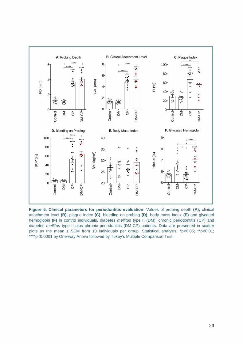
Results
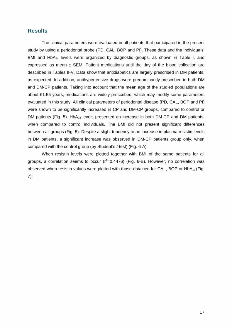
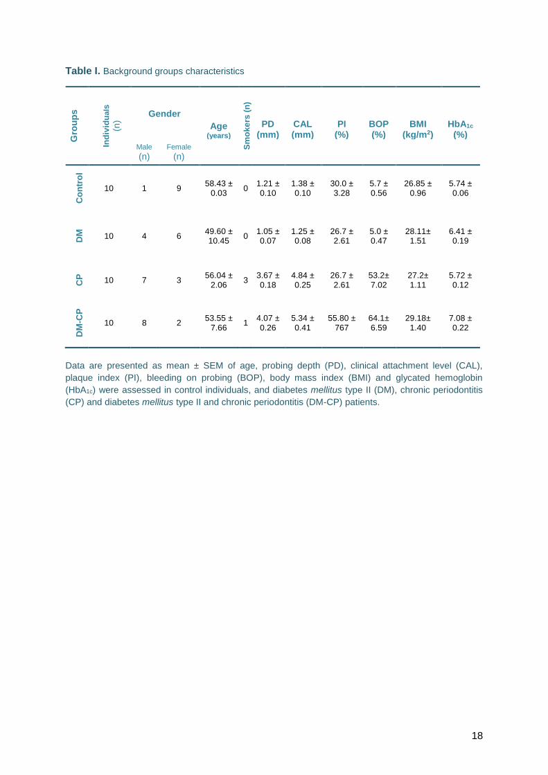
The clinical parameters were evaluated in all patients that participated in the present
study by using a periodontal probe (PD, CAL, BOP and PI). These data and the individuals’
BMI and HbA1c levels were organized by diagnostic groups, as shown in Table I, and
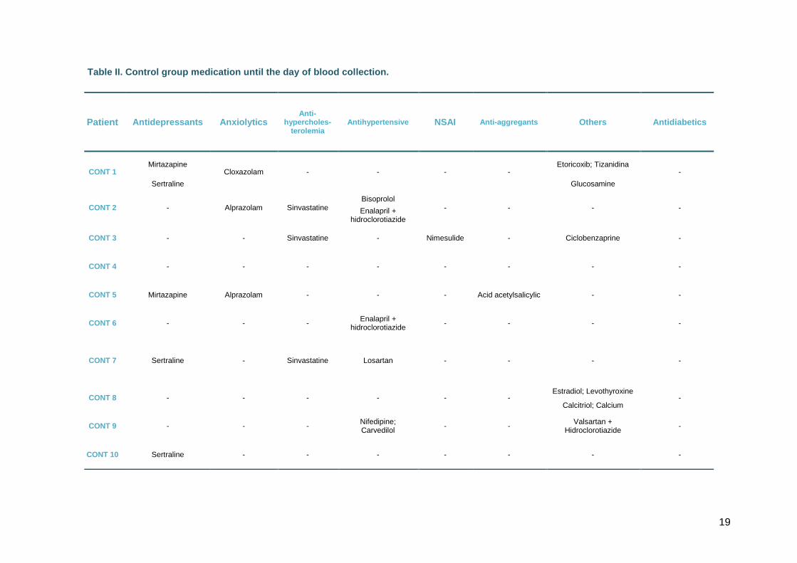
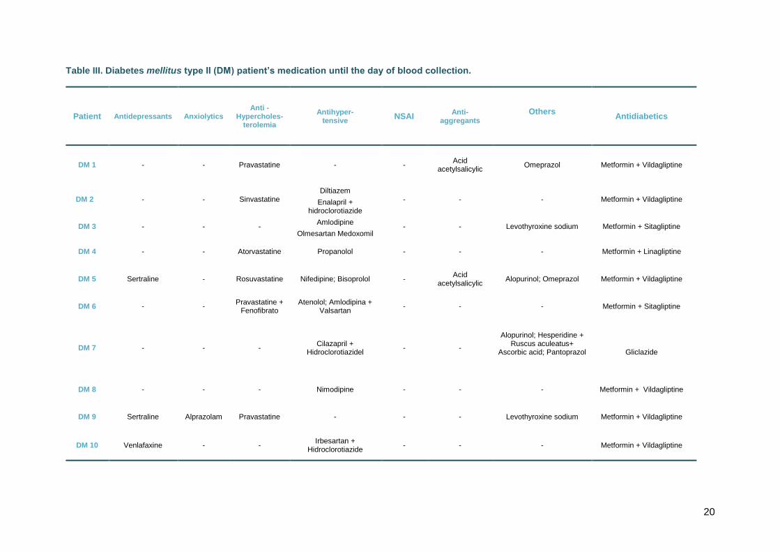
expressed as mean ± SEM. Patient medications until the day of the blood collection are
described in Tables II-V. Data show that antidiabetics are largely prescribed in DM patients,
as expected. In addition, antihypertensive drugs were predominantly prescribed in both DM
and DM-CP patients. Taking into account that the mean age of the studied populations are
about 61.55 years, medications are widely prescribed, which may modify some parameters
evaluated in this study. All clinical parameters of periodontal disease (PD, CAL, BOP and PI)
were shown to be significantly increased in CP and DM-CP groups, compared to control or
DM patients (Fig. 5). HbA1c levels presented an increase in both DM-CP and DM patients,
when compared to control individuals. The BMI did not present significant differences
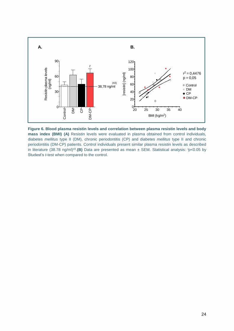
between all groups (Fig. 5). Despite a slight tendency to an increase in plasma resistin levels
in DM patients, a significant increase was observed in DM-CP patients group only, when
compared with the control group (by Student’s t-test) (Fig. 6-A).
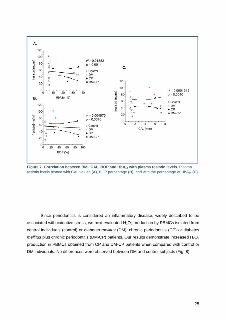
When resistin levels were plotted together with BMI of the same patients for all
groups, a correlation seems to occur (r2=0.4476) (Fig. 6-B). However, no correlation was
observed when resistin values were plotted with those obtained for CAL, BOP or HbA1c (Fig.
7).
18
Table I. Background groups characteristics
Data are presented as mean ± SEM of age, probing depth (PD), clinical attachment level (CAL),
plaque index (PI), bleeding on probing (BOP), body mass index (BMI) and glycated hemoglobin
(HbA1c) were assessed in control individuals, and diabetes mellitus type II (DM), chronic periodontitis
(CP) and diabetes mellitus type II and chronic periodontitis (DM-CP) patients.
Gro
up
s
Ind
ivid
uals
(n
) Gender
Age (years)
Sm
ok
ers
(n
)
PD (mm)
CAL (mm)
PI (%)
BOP (%)
BMI (kg/m2)
HbA1c
(%)
Male
(n) Female
(n)
Co
ntr
ol
10 1 9 58.43 ±
0.03 0
1.21 ± 0.10
1.38 ± 0.10
30.0 ± 3.28
5.7 ± 0.56
26.85 ± 0.96
5.74 ± 0.06
DM
10 4 6 49.60 ± 10.45
0 1.05 ± 0.07
1.25 ± 0.08
26.7 ± 2.61
5.0 ± 0.47
28.11± 1.51
6.41 ± 0.19
CP
10 7 3 56.04 ±
2.06 3
3.67 ± 0.18
4.84 ± 0.25
26.7 ± 2.61
53.2± 7.02
27.2± 1.11
5.72 ± 0.12
DM
-CP
10 8 2 53.55 ±
7.66 1
4.07 ± 0.26
5.34 ± 0.41
55.80 ± 767
64.1± 6.59
29.18± 1.40
7.08 ± 0.22
19
Table II. Control group medication until the day of blood collection.
Patient Antidepressants Anxiolytics Anti-
hypercholes-terolemia
Antihypertensive NSAI Anti-aggregants Others Antidiabetics
CONT 1 Mirtazapine
Cloxazolam - - - - Etoricoxib; Tizanidina
-
Sertraline Glucosamine
CONT 2 - Alprazolam Sinvastatine Bisoprolol
- - - - Enalapril + hidroclorotiazide
CONT 3 - - Sinvastatine - Nimesulide - Ciclobenzaprine -
CONT 4 - - - - - - - -
CONT 5 Mirtazapine Alprazolam - - - Acid acetylsalicylic - -
CONT 6 - - - Enalapril +
hidroclorotiazide - - - -
CONT 7 Sertraline - Sinvastatine Losartan - - - -
CONT 8 - - - - - - Estradiol; Levothyroxine
- Calcitriol; Calcium
CONT 9 - - - Nifedipine; Carvedilol
- - Valsartan +
Hidroclorotiazide -
CONT 10 Sertraline - - - - - - -
20
Table III. Diabetes mellitus type II (DM) patient’s medication until the day of blood collection.
Patient Antidepressants Anxiolytics Anti -
Hypercholes- terolemia
Antihyper- tensive
NSAI Anti-
aggregants
Others
Antidiabetics
DM 1 - - Pravastatine - - Acid
acetylsalicylic Omeprazol Metformin + Vildagliptine
DM 2 - - Sinvastatine Diltiazem
- - - Metformin + Vildagliptine Enalapril + hidroclorotiazide
DM 3 - - - Amlodipine
- - Levothyroxine sodium Metformin + Sitagliptine Olmesartan Medoxomil
DM 4 - - Atorvastatine Propanolol - - - Metformin + Linagliptine
DM 5 Sertraline - Rosuvastatine Nifedipine; Bisoprolol - Acid
acetylsalicylic Alopurinol; Omeprazol Metformin + Vildagliptine
DM 6 - - Pravastatine +
Fenofibrato Atenolol; Amlodipina +
Valsartan - - - Metformin + Sitagliptine
DM 7 - - - Cilazapril +
Hidroclorotiazidel - -
Alopurinol; Hesperidine + Ruscus aculeatus+
Ascorbic acid; Pantoprazol
Gliclazide
DM 8 - - - Nimodipine - - - Metformin + Vildagliptine
DM 9 Sertraline Alprazolam Pravastatine - - - Levothyroxine sodium Metformin + Vildagliptine
DM 10 Venlafaxine - - Irbesartan +
Hidroclorotiazide - - - Metformin + Vildagliptine
21
Table IV. Chronic periodontitIs (CP) patient’s medication until the day of blood collection.
Patient Antidepressants Anxiolytics Anti
Hypercholes -terolemia
Antihyper-tensive
NSAI Anti-aggregants Others
Antidiabetics
CP 1 - - Sinvastatine Fosinopril - - Hidroxizine; Omeprazol
-
CP 2 - - - - - - - -
CP 3 - - - - - - Furosemide -
CP 4 Sertraline Mexazolam - - Etodolac - Gabapentin -
CP 5 - - - - - - - -
CP 6 - - - - - - - -
CP 7 - - Sinvastatine Lisinopril; Bisoprolol
- Acid acetylsalicylic;
Ticagrelor Levotiroxine sodium
-
CP 8 - - - Perindopril - - Flutamide -
CP 9 - - - - - - - -
CP 10
- - - - - - - -
22
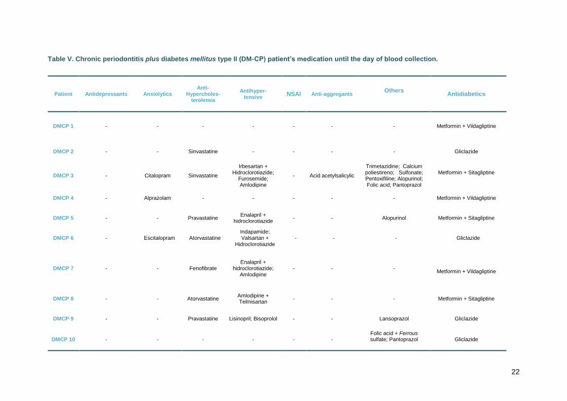
Table V. Chronic periodontitis plus diabetes mellitus type II (DM-CP) patient’s medication until the day of blood collection.
Patient Antidepressants Anxiolytics Anti-
Hypercholes- terolemia
Antihyper- tensive
NSAI Anti-aggregants Others
Antidiabetics
DMCP 1 - - - - - - - Metformin + Vildagliptine
DMCP 2 - - Sinvastatine - - - - Gliclazide
DMCP 3 - Citalopram Sinvastatine
Irbesartan + Hidroclorotiazide;
Furosemide; Amlodipine
- Acid acetylsalicylic
Trimetazidine; Calcium poliestireno; Sulfonate; Pentoxifiline; Alopurinol; Folic acid; Pantoprazol
Metformin + Sitagliptine
DMCP 4 - Alprazolam - - - - - Metformin + Vildagliptine
DMCP 5 - - Pravastatine Enalapril +
hidroclorotiazide - - Alopurinol Metformin + Sitagliptine
DMCP 6 - Escitalopram Atorvastatine Indapamide; Valsartan +
Hidroclorotiazide - - - Gliclazide
DMCP 7 - - Fenofibrate Enalapril +
hidroclorotiazide; Amlodipine
- - -
Metformin + Vildagliptine
DMCP 8 - - Atorvastatine Amlodipine + Telmisartan
- - - Metformin + Sitagliptine
DMCP 9 - - Pravastatine Lisinopril; Bisoprolol - - Lansoprazol Gliclazide
DMCP 10 - - - - - - Folic acid + Ferrous sulfate; Pantoprazol
Gliclazide
23
Figure 5. Clinical parameters for periodontitis evaluation. Values of probing depth (A), clinical
attachment level (B), plaque index (C), bleeding on probing (D), body mass index (E) and glycated
hemoglobin (F) in control individuals, diabetes mellitus type II (DM), chronic periodontitis (CP) and
diabetes mellitus type II plus chronic periodontitis (DM-CP) patients. Data are presented in scatter
plots as the mean ± SEM from 10 individuals per group. Statistical analysis: *p<0.05; **p<0.01;
****p<0.0001 by One-way Anova followed by Tukey’s Multiple Comparison Test.
Contr
ol
DM
CP
DM
-CP
0
2
4
6
PD
(m
m)
A. Probing Depth
************
****
Contr
ol
DM
CP
DM
-CP
0
20
40
60
80
100
PI (%
)
C. Plaque Index
*** *****
**
Contr
ol
DM
CP
DM
-CP
0
20
40
60
80
100
BO
P (
%)
D. Bleeding on Probing
************
****
Contr
ol
DM
CP
DM
-CP
20
25
30
35
40
BM
I (k
g/m
2)
E. Body Mass Index
Contr
ol
DM
CP
DM
-CP
5
6
7
8
9
HbA
1c (
%)
F. Glycated Hemoglobin
******
*
****
24
Figure 6. Blood plasma resistin levels and correlation between plasma resistin levels and body
mass index (BMI) (A) Resistin levels were evaluated in plasma obtained from control individuals,
diabetes mellitus type II (DM), chronic periodontitis (CP) and diabetes mellitus type II and chronic
periodontitis (DM-CP) patients. Control individuals present similar plasma resistin levels as described
in literature (38.78 ng/ml)43.(B) Data are presented as mean ± SEM. Statistical analysis: tp<0.05 by
Studest’s t-test when compared to the control.
20 25 30 35 400
20
40
60
80
100
120
BMI (kg/m2)
[resis
tin] (n
g/m
l) r2 = 0,4476
p = 0,05
CP
DM-CP
Control
DM
B. A.
Contr
ol
DM
CP
DM
-CP
0
30
60
90
Resis
tin p
lasm
a le
vels
(ng/m
l)
38,78 ng/ml
t
(43)
25
FIgure 7. Correlation between BMI, CAL, BOP and HbA1c with plasma resistin levels. Plasma
resistin levels plotted with CAL values (A), BOP percentage (B), and with the percentage of HbA1c (C).
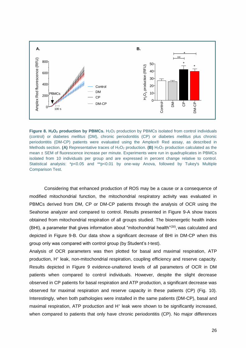
Since periodontitis is considered an inflammatory disease, widely described to be
associated with oxidative stress, we next evaluated H2O2 production by PBMCs isolated from
control individuals (control) or diabetes mellitus (DM), chronic periodontitis (CP) or diabetes
mellitus plus chronic periodontitis (DM-CP) patients. Our results demonstrate increased H2O2
production in PBMCs obtained from CP and DM-CP patients when compared with control or
DM individuals. No differences were observed between DM and control subjects (Fig. 8).
A.
B.
C.
26
Figure 8. H2O2 production by PBMCs. H2O2 production by PBMCs isolated from control individuals
(control) or diabetes mellitus (DM), chronic periodontitis (CP) or diabetes mellitus plus chronic
periodontitis (DM-CP) patients were evaluated using the Amplex® Red assay, as described in
Methods section. (A) Representative traces of H2O2 production. (B) H2O2 production calculated as the
mean ± SEM of fluorescence increase per minute. Experiments were run in quadruplicates in PBMCs
isolated from 10 individuals per group and are expressed in percent change relative to control.
Statistical analysis: *p<0.05 and **p<0.01 by one-way Anova, followed by Tukey's Multiple
Comparison Test.
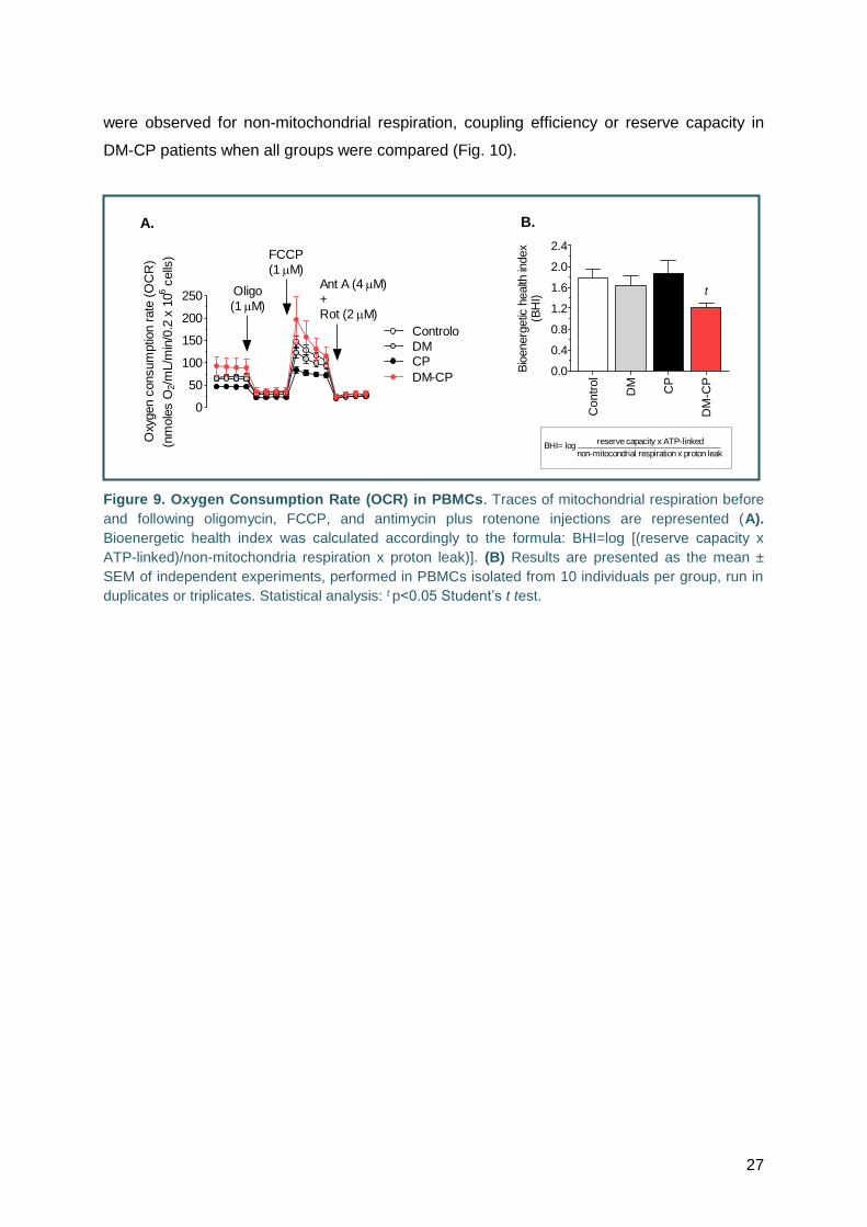
Considering that enhanced production of ROS may be a cause or a consequence of
modified mitochondrial function, the mitochondrial respiratory activity was evaluated in
PBMCs derived from DM, CP or DM-CP patients through the analysis of OCR using the
Seahorse analyzer and compared to control. Results presented in Figure 9-A show traces
obtained from mitochondrial respiration of all groups studied. The bioenergetic health index
(BHI), a parameter that gives information about “mitochondrial health”(30), was calculated and
depicted in Figure 9-B. Our data show a significant decrease of BHI in DM-CP when this
group only was compared with control group (by Student’s t-test).
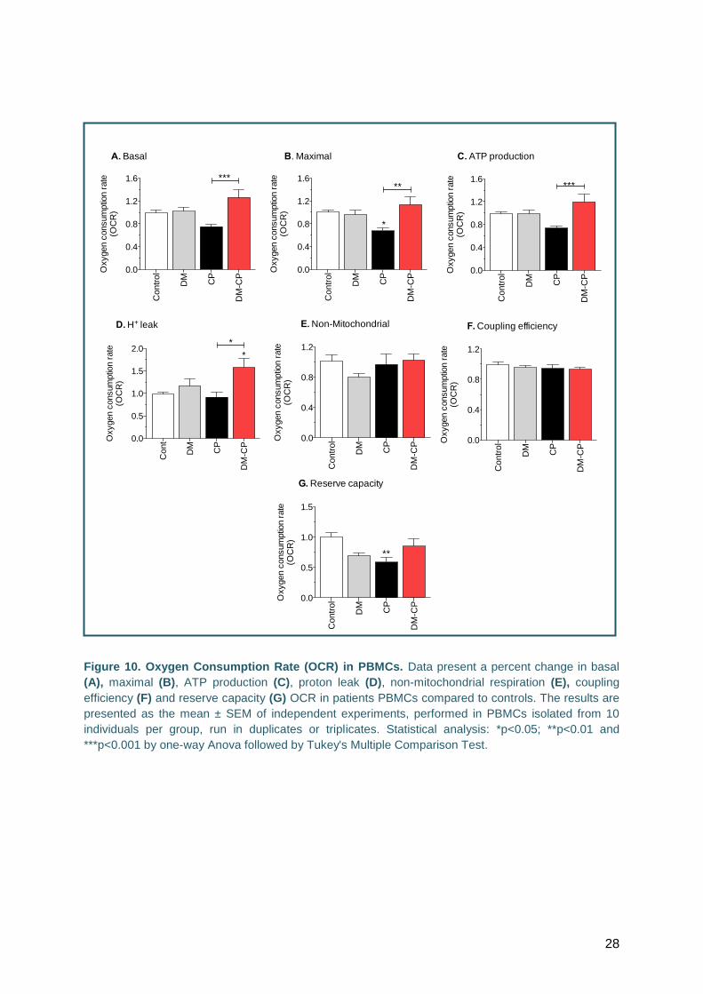
Analysis of OCR parameters was then plotted for basal and maximal respiration, ATP
production, H+ leak, non-mitochondrial respiration, coupling efficiency and reserve capacity.
Results depicted in Figure 9 evidence-unaltered levels of all parameters of OCR in DM
patients when compared to control individuals. However, despite the slight decrease
observed in CP patients for basal respiration and ATP production, a significant decrease was
observed for maximal respiration and reserve capacity in these patients (CP) (Fig. 10).
Interestingly, when both pathologies were installed in the same patients (DM-CP), basal and
maximal respiration, ATP production and H+ leak were shown to be significantly increased,
when compared to patients that only have chronic periodontitis (CP). No major differences
0
200
400
600
800
Am
ple
x R
ed
flu
ore
sce
nce
(R
FU
)
PBMCsCP
DM-CP
Control
DM
100 s
A. B.
Contr
ol
DM
CP
DM
-CP
0
10
20
30
40
50
**
**
*
H2O
2 p
roductio
n (
RF
U)
27
were observed for non-mitochondrial respiration, coupling efficiency or reserve capacity in
DM-CP patients when all groups were compared (Fig. 10).
Figure 9. Oxygen Consumption Rate (OCR) in PBMCs. Traces of mitochondrial respiration before
and following oligomycin, FCCP, and antimycin plus rotenone injections are represented (A).
Bioenergetic health index was calculated accordingly to the formula: BHI=log [(reserve capacity x
ATP-linked)/non-mitochondria respiration x proton leak)]. (B) Results are presented as the mean ±
SEM of independent experiments, performed in PBMCs isolated from 10 individuals per group, run in
duplicates or triplicates. Statistical analysis: t p<0.05 Student’s t test.
0
50
100
150
200
250
Controlo
DM
CP
DM-CP
Oligo
(1 M)
FCCP
(1 M)Ant A (4 M)
+
Rot (2 M)
Oxy
gen c
onsum
ptio
n r
ate
(O
CR
)
(nm
ole
s O
2/m
L/m
in/0
.2 x
106
cells
)
A.
Contr
ol
DM
CP
DM
-CP
0.0
0.4
0.8
1.2
1.6
2.0
2.4
Bio
energ
etic
health
index
(BH
I)
t
BHI= logreserve capacity x ATP-linked
non-mitocondrial respiration x proton leak
B.B.
28
Figure 10. Oxygen Consumption Rate (OCR) in PBMCs. Data present a percent change in basal
(A), maximal (B), ATP production (C), proton leak (D), non-mitochondrial respiration (E), coupling
efficiency (F) and reserve capacity (G) OCR in patients PBMCs compared to controls. The results are
presented as the mean ± SEM of independent experiments, performed in PBMCs isolated from 10
individuals per group, run in duplicates or triplicates. Statistical analysis: *p<0.05; **p<0.01 and
***p<0.001 by one-way Anova followed by Tukey's Multiple Comparison Test.
Contr
ol
DM
CP
DM
-CP
0.0
0.4
0.8
1.2
1.6 ***
A. Basal
Oxygen c
onsum
ptio
n r
ate
(OC
R)
Contr
ol
DM
CP
DM
-CP
0.0
0.4
0.8
1.2
1.6
*
**
B. Maximal
Oxygen c
onsum
ptio
n r
ate
(OC
R)
Contr
ol
DM
CP
DM
-CP
0.0
0.4
0.8
1.2
1.6***
C. ATP production
Oxygen c
onsum
ptio
n r
ate
(OC
R)
Contr
ol
DM
CP
DM
-CP
0.0
0.4
0.8
1.2
ns
F. Coupling efficiency
Oxygen c
onsum
ptio
n r
ate
(OC
R)
Cont
DM
CP
DM
-CP
0.0
0.5
1.0
1.5
2.0*
*
D. H+ leak
Oxygen c
onsum
ptio
n r
ate
(OC
R)
Contr
ol
DM
CP
DM
-CP
0.0
0.4
0.8
1.2
ns
E. Non-Mitochondrial
Oxygen c
onsum
ptio
n r
ate
(OC
R)
Contr
ol
DM
CP
DM
-CP
0.0
0.5
1.0
1.5
Oxygen c
onsum
ptio
n r
ate
(OC
R)
G. Reserve capacity
**
29
Discussion
In this study we evaluated the influence of diabetic conditions on periodontitis by
analyzing plasma resistin levels, oxidative stress and mitochondrial function in PBMCs
isolated from patients with chronic periodontitis (CP), diabetes mellitus type II (DM) and
chronic periodontitis plus diabetes mellitus type II (DM-CP) versus healthy controls (Control).
Clinical characterization of the subjects was made accordingly to American Academy
of Periodontology (AAP) guidelines. Our results demonstrated an increase of probing depth
(PD), clinical attachment level (CAL), plaque index (PI) and bleeding on probing (BOP) in
patients with CP and patients with DM-CP when compared to healthy (control) or DM
individuals allowing us to include this population in our study. No major differences were
observed in body mass index (BMI) between all groups studied. Since a positive correlation
between periodontal disease and glycemic control levels have been found in the literature,
HbA1c was evaluated in the participants of this study(1)(16). According to our results, HbA1c
levels were increased in DM and further increased in DM-CP, but not in CP patients, when
compared to control subjects (reference value ≤ 6.5%). Therefore, these results support
previous hypothesis presented in literature, indicating that chronic periodontitis can
deregulate the control of HbA1c levels(1)(16).
Resistin was detected in plasma obtained from all individuals, being significantly
increased in DM-CP patients when compared to controls. Moreover, we evaluated the
putative correlation between resistin levels and parameters of metabolic disease (e.g. BMI
and periodontal disease). Our data showed a positive correlation between resistin levels and
BMI, which is in accordance with the literature showing a positive correlation between resistin
levels and obesity or metabolic syndrome(35). However, no correlation was found when
resistin levels were plotted against the periodontal disease parameters, BOP and CAL.
Periodontal disease is characterized by an increase of local immune-inflammatory factors,
being a cause of increased resistin expression(36), or a consequence of high resistin levels in
PBMCs(37). Indeed, some authors defend that resistin plays an important role in the
regulation of metabolic processes like adipogenesis and inflammatory reactions, while others
pointed out increased resistin levels in CP when compared to control individuals (10)(38).
Despite the controversy concerning the relationship between resistin and diabetes (23), a
relationship has been suggested between obesity-diabetes-periodontitis(37). However, other
findings defend that resistin levels in CP are comparable to those observed in control
patients, suggesting conflicting findings regarding the role of resistin in periodontitis
disease(38).
Lymphocytes, the main population in isolated PBMCs, are a heterogeneous cell
30
population that largely dependents on mitochondria to meet energetic demands(31).
Considering the relevance of defining redox changes related with mitochondrial function and
bioenergetics in PBMCs in periodontitis and DM conditions, we evaluated oxidative stress
and mitochondrial function in PBMCs isolated from DM, CP, DM-CP and control groups. Our
results evidenced that H2O2 production was significantly increased in PBMCs-derived from
CP and DM-CP, but not in DM patients, when compared with the control. Opposing data was
found by Mor-Li Hartman et al. (2014) suggesting that increased ROS production in PBMCs
can indicate that diabetes mellitus might be associated with highly uncoupled oxygen
consumption and thus, higher production of ROS(27). This apparent discrepancy may due to
the fact that DM patients in our study are controlled for diabetes as shown in Table II. It has
been described that oxidative stress is associated with the development of insulin resistance
and diabetes complications, such as periodontitis(39). In accordance, our results evidence that
the observed increase in ROS production in DM-CP patients can be associated with the
increased resistin levels in these patients. In addition, and taking into account that patients
evaluated in this study, namely DM and DM-CP patients, are under treatment with
antihypertensive agents, which were shown to reduce ROS production(40), the fact that DM-
CP present higher ROS production compared with DM patients suggest that chronic
periodontitis induce per se an increase in ROS levels above DM patients and controls.
Cellular oxygen consumption is recognized as an indicator of mitochondrial
function(27). Our data indicate that when OCR were evaluated in PBMCs derived from DM-CP
patients, an increase in both basal and maximal respiration along with increased ATP
production, H+ leak were observed, when compared with CP PBMCs. However, PBMCs
derived from DM patients, similarly as shown for H2O2 analyses, did not exhibit any changes
in all parameters evaluated by seahorse analysis. Interestingly, an increase in basal and
maximal respiration, as well as ATP production was observed in DM-CP patients, in relation
to CP patients, whereas H+ leak also increase in the same experimental group (DM-CP
PBMCs), when compared to both CP and controls conditions. The reserve capacity, defined
as the capability of the cell to respond to an energetic demand, was shown to be slightly
(although non-significantly) decreased in both DM and DM-CP patients, being significantly
decreased in CP individuals(41). In our isolated PBMCs, no differences were observed in non-
mitochondrial respiration defined as oxygen consumption due to a subset of cellular enzymes
that consume oxygen in conditions of blockade of cellular respiration achieved by complex I
and complex III inhibition(27). Decreased maximal respiration observed in CP-derived PBMCs,
seems to be in accordance with decreased reserve capacity in the same patients. These
data suggest that incremental mitochondrial activity, linked to enhanced H+ leak, observed in
31
DM-CP PBMCs might reflect mitochondrial impairment due to metabolic or oxidative stress
associated with periodontal or diabetic inflammation condition.
Recently, mitochondrial bioenergetics has been highlighted as a key to understand
pathological mechanisms associated with metabolic diseases like diabetes(31). Accordingly to
these authors, the bioenergetics health index (BHI) (BHI=log [(reserve capacity x ATP-
linked)/non-mitochondria respiration x proton leak)]) is a parameter that might reflect a
potential function biomarker to study pathologies influenced by oxidative stress(31), such as
diabetes and chronic periodontitis. Our results show a decrease in BHI in PBMCs obtained
from DM-CP patients, as compared to control individuals, suggesting compromised
mitochondria in patients with both pathologies. These results seem to be associated with
increased H+ leak observed in the PBMCs obtained from DM-CP group and with the fact that
these patients present a slight tendency, although not significant, for increased values of
BMI. In addition, high caloric intake, which is associated with obesity and thus BMI values,
and also with high resistin levels, markers of the DM-CP patients selected for this study, can
influence oxidative response and mitochondrial deregulation observed in this work. Data
obtained in the present study corroborate the hypothesis that BHI might be considered as a
new potential biomarker for assessing patient’s health with prognostic and diagnostic values,
as reported elsewhere(31).
32
Conclusion
Periodontitis has been largely associated with high local inflammation levels and
oxidative stress. On the other hand, diabetes mellitus has been shown to be associated with
obesity and increased resistin levels. Importantly, two-way relationship between these
inflammatory diseases has been described in the literature. Data evidence increased plasma
resistin levels in DM-CP patients that correlate with body mass índex, along with an increase
in glycated hemoglobin. Whether obesity, diabetes mellitus and chronic periodontitis are
related to each other through increased resistin levels and H2O2 production, as well as
mitochondrial dysfunction remains unclear. Our study unveils a novel relationship between
DM and CP by defining that DM-CP patients show exacerbated, but still bioenergetically
abnormal, mitochondrial activity that may impact on increased ROS levels. Indeed, we do not
know whether ROS are being produced by mitochondria and/or if the antioxidant
levels/activity or the activity of mitochondrial respiratory complexes are altered in PBMCs of
these patients. Overall, promoting basic research in this field may help to explain how DM
and CP are interconnected through molecular and cellular events that may culminate in
aggravated disease prognosis.
33
Acknowledgements
À Professora Doutora Ana Cristina Rego, orientadora deste trabalho, por me ter
recebido de braços abertos, por me dar a oportunidade de entrar num projeto como este.
À Professora Doutora Isabel Poiares Baptista, co-orientadora deste trabalho, por me
ajudar, incentivar e apoiar em todas as etapas do trabalho.
À Doutora Luísa Ferreira, a minha “mestre” de laboratório. Obrigada pela paciência,
amizade, apoio incondicional. Obrigada por tudo o que ensinou ao longo de todo o percurso.
Sem si, nada disto seria da mesma forma.
Obrigada às três por me ajudarem a superar todas as dificuldades. Obrigada pela
dedicação. Tudo isto fez com que este projeto se concretizasse.
Ao Doutor Francisco Marques, pela disponibilidade e paciência que mostrou.
Às enfermeiras da Área de Medicina Dentária, pelo carinho, por me ajudarem com o
material e por estarem sempre presentes.
À Isabel Dantas (CNC), pela simpatia que demonstrou ao longo de todas as minhas
visitas e por me ajudar em algumas etapas laboratoriais.
A todos os elementos do CNC com quem contactei, por me receberem de braços
abertos.
A todos os doentes pela autorização na realização da recolha de sangue e análise
da saúde periodontal.
Ao João, pela amizade, paciência, motivação, alegria, amor e apoio. Elementos
essenciais ao longo de todo o meu percurso académico que me ajudaram a crescer e a
tornar-me mais forte.
Aos meus pais e à minha irmã, por me apoiarem em todos os momentos, por serem
os meus pilares. Pessoas que me viram crescer e que estão sempre do meu lado. Obrigada
pela dedicação e pela paciência. Sem o amor incondicional deles, não teria conseguido
conquistar tantas pequenas grandes coisas.
34
Bibliography
1. Preshaw PM, Rcsed FDSR. P e r iodont i t i s Oral Complication of Diabetes. Endocrinol Cetab clin N Am. 2013;42:849–67.
2. Bascones-martínez A, Mu M, Bascones-ilundain J. Diabetes and periodontitis : A bidirectional relationship ଝ. Med Clin (Barc). 2015;145(1):31–5.
3. Hamdy Nassar, Alpdogan Kantarci TEVD. Diabetic periodontitis: a model for activated innate immunity and impaired resolution of inflammation. Periodontol 2000. 2007;6:233–44.
4. Gw T, Rj G, Genco RJ. Effect of periodontal disease on diabetes : systematic review of epidemiologic observational evidence. J Periodontol. 2013;84:135–52.
5. Devanoorkar A, Dwarakanath CD, Gundanavar G, Kathariya R. Evaluation of serum resistin levels in periodontal health and disease and effects of non surgical periodontal therapy on its levels. Dis 32. 2012;32:289–94.
6. Rubim M, Sete C, Júnior RL, Guimarães R, Marcelo C. Serum Adipokine Levels and their Relationship with Fatty Acids in Patients with Chronic Periodontitis. Braz Dent J. 2015;26(2):169–74.
7. Mittal M, Hassan B, Desai K, Duseja S, Santosh K, Sharaschandra R. GCF Resistin As A Novel Marker in Patients with Chronic Periodontitis and Rheumatoid Arthritis. J Clin Diagnostic Res. 2015;9(4):62–4.
8. Allen AA, Med S, Journal C, Link D. Periodontitis and type 2 diabetes : is oxidative stress the. Scott Med J. 2009;54(2):41–6.
9. Manuscript A, Diseases P. Diagnostic Biomarkers for Oral and Periodontal Diseases. Dent Clin North Am. 2008;49(3):1–21.
10. Raju SPP and PA. Gingival crevicular fluid and serum levels of resistin in obese and non-obese subjects with and without periodontitis and association with single nucleotide polymorphism at −420. J Indian Soc Periodontol. 2014;18(5):555–9.
11. Highfield J. Diagnosis and classification of periodontal disease. Aust Dent J. 2009;54(1):11–26.
12. MP G. Dentistry IQ [Internet]. Update from the American Academy of Periodontology. 2015 [cited 2006 Jul 20]. Available from: http://www.dentistryiq.com/articles/2015/08/update-from-the-american-academy-of-periodontology.html
13. MEYLE SKS &JOERG. Local inflammatory reactions in patients with diabetes and periodontitis. Periodontol 2000. 2015;69(2):221–54.
14. Joseph A, Joanisse DR, Baillot RG, Hood DA. Mitochondrial Dysregulation in the Pathogenesis of Diabetes : Potential for Mitochondrial Biogenesis-Mediated Interventions. Exp Diabetes Res. 2012;1–16.
15. Lamster IB, Pagan M. Periodontal disease and the metabolic syndrome. Int Dent J. 2016;1–11.
16. Marigo L, Cerreto R, Giulini M, Somma F LC, M C. Diabetes mellitus : biochemical , histological and microbiological aspects in periodontal disease. Eur Rev Med Pharmacol Sci. 2011;15:751–8.
17. Zhou X, Zhang W, Liu X, Zhang W, Li Y. ScienceDirect Interrelationship between diabetes and periodontitis : Role of hyperlipidemia. Arch Oral Biol. 2015;60:667–74.
18. Bds BC, Park B, Btech BDS, Bds PMB, Hons B, Fracds D. Periodontitis and type II diabetes : a two-way relationship. Int J Evid Based Healthc. 2013;11:317–29.
19. Mary P. C& GJS. Periodontal disease and systemic illness : will the evidence ever be enough ? Periodontol 2000. 2013;62:271–86.
20. Nuttall FQ. Body Mass Index. Nutr Today. 2015;50(3). 21. Chacko BK, Kramer PA, Ravi S, Johnson MS, Hardy RW, Ballinger SW, et al.
Methods for defining distinct bioenergetic profiles in platelets , lymphocytes , monocytes , and neutrophils , and the oxidative burst from human blood. Lab Investig
35
[Internet]. Nature Publishing Group; 2013;93(6):690–700. Available from: http://dx.doi.org/10.1038/labinvest.2013.53
22. Zorzano A, Liesa M, Palacín M. Mitochondrial dynamics as a bridge between mitochondrial dysfunction and insulin resistance. Arch Physiol Biochem. 2009;115(1):1–12.
23. Devanoorkar A, Kathariya R, Guttiganur N, Gopalakrishnan D, Bagchi P. Resistin : A Potential Biomarker for Periodontitis Influenced Diabetes Mellitus and Diabetes Induced Periodontitis. Dis Markers. 2014;2014.
24. Pang S, Le Y. Role of Resistin in Inflammation and Inflammation-Related Diseases. Cell Mol Immunol. 2006;3:29–34.
25. Nicholas D, Proctor EA, Raval FM, Ip BC, Habib C, Ritou E, et al. Advances in the quantification of mitochondrial function in primary human immune cells through extracellular flux analysis. PLoS One. 2017;12(2):1–19.
26. Kramer PA, Ravi S, Chacko B, Johnson MS, Darley-usmar VM. A review of the mitochondrial and glycolytic metabolism in human platelets and leukocytes : Implications for their use as bioenergetic biomarkers. Redox Biol [Internet]. Elsevier; 2014;2:206–10. Available from: http://dx.doi.org/10.1016/j.redox.2013.12.026
27. Hartman M, Shirihai OS, Holbrook M, Kocherla M, Shah A, Fetterman JL, et al. Relation of mitochondrial oxygen consumption in peripheral blood mononuclear cells to vascular function in type 2 diabetes mellitus. Vasc Med. 2014;19(1):67–74.
28. PEDRO BULLON HNN &MAURIZIO B. Obesity , diabetes mellitus , atherosclerosis and chronic periodontitis : a shared pathology via oxidative stress and mitochondrial dysfunction ? Periodontol 2000. 2014;64:139–53.
29. Koliaki C, Roden M. Alterations of Mitochondrial Function and Insulin Sensitivity in Human Obesity and Diabetes Mellitus. Annu Rev Nutr. 2016;337–70.
30. Holmström MH, Iglesias-gutierrez E, Zierath JR, Garcia-roves PM. Tissue-specific control of mitochondrial respiration in obesity-related insulin resistance and diabetes. Am J Physiol Endocrinol Metab. 2012;731–9.
31. Ferrick D, Singal AK, Ballinger SW, Bailey SM. The Bioenergetic Health Index : a new concept in mitochondrial translational research. Clin Sci. 2014;127:367–73.
32. Ritchie CS. Mechanistic links between type 2 diabetes and periodontitis Atheromatous vascular disease and ischaemic stroke in the UK. J Dent. 2009;37:578–9.
33. Syndrome M, Syndrome IR. Risks for All-Cause Mortality , Cardiovascular Disease , and Diabetes. Diabetes Care. 2005;28(7).
34. Votyakova T V, Reynolds IJ. Detection of hydrogen peroxide with Amplex Red: interference by NADH and reduced glutathione auto-oxidation. Arch Biochem Biophys. 2004;431:138–44.
35. Handzlik-orlik RKG. The role of adipokines in connective tissue diseases. Eur J Nutr. 2012;51:513–28.
36. Ghosh S, Ehtesham NZ. Human resistin stimulates the pro-inflammatory cytokines TNF- a and IL-12 in macrophages by NF- j B-dependent pathway. Biochem Biophys Res Commun. 2005;334:1092–101.
37. Kaser S, Kaser A, Sandhofer A, Ebenbichler CF, Tilg H, Patsch JR. Resistin messenger-RNA expression is increased by proinflammatory cytokines in vitro. Biochem Biophys Res Commun. 2003;309:286–90.
38. Furugen R, Hayashida H, Yamaguchi N, Yoshihara A, Ogawa H, Miyazaki H. The relationship between periodontal condition and serum levels of resistin and adiponectin in elderly Japanese. J Periodont Res. 2008;43:556–62.
39. Gorudko I V, Kostevich VA, Sokolov A V, Shamova E V, Buko I V, Konstantinova EE, et al. Functional Activity of Neutrophils in Diabetes Mellitus and Coronary Heart Disease : Role of Myeloperoxidase in the Development of Oxidative Stress. Bull Exp Biol Med. 2012;154(1):23–6.
40. Azar Baradaran, Hamid Nasri and MR-K. Oxidative stress and hypertension: Possibility of hypertension therapy with antioxidants. J Res Med Sci. 2014;19(4):358–

36
67. 41. Agilent Technologies. Agilent Seahorse XFp Cell Mito Stress Test Kit [Internet]. 2017.
p. 8. Available from: http://www.agilent.com/cs/library/usermanuals/public/XFp_Cell_Mito_Stress_Test_Kit_User_Guide.pdf

37
Attachments
Attachment 1. Informed Consent
38
FORMULÁRIO DE INFORMAÇÃO E
CONSENTIMENTO INFORMADO
TÍTULO DO PROJECTO DE INVESTIGAÇÃO: Caracterização sanguínea em doentes com periodontite e diabetes mellitus.
PROTOCOLO Nº não aplicável
PROMOTOR (Entidade ou pessoa(s)
que propõe(m) o estudo):
Mestrado Integrado em Medicina Dentária
INVESTIGADOR COORDENADOR Prof.ª Ana Cristina Rego, Prof.ª Dr.ª Isabel Poiares
Baptista
CENTRO DE ESTUDO
INVESTIGADOR PRINCIPAL Ana Solange Gomes Costa
MORADA Av. Bissaya Barreto e Praceta Prof. Mota Pinto
3000-075 Coimbra
CONTACTO TELEFÓNICO B. central/ Bloco de Celas: Tel: 239 400 400
NOME DO DOENTE
(LETRA DE IMPRENSA) ___________________________________________
É convidado(a) a participar voluntariamente neste estudo no âmbito da identificação de um
marcador em situações de inflamação relacionadas com a diabetes mellitus e doença
periodontal.
Este procedimento é chamado consentimento informado e descreve a finalidade do estudo,
os procedimentos, os possíveis benefícios e riscos. A sua participação poderá
39
contribuir para melhorar o conhecimento sobre a identificação de marcadores no controlo da
hiperglicémia e influência na doença periodontal e relação com a diabetes.
Receberá uma cópia deste Consentimento Informado para rever e solicitar aconselhamento
de familiares e amigos. O Investigador ou outro membro da sua equipa irá esclarecer
qualquer dúvida
que tenha sobre o termo de consentimento e também alguma palavra ou informação que
não possa entender.
Depois de compreender o estudo e de não ter qualquer dúvida acerca do mesmo, deverá
tomar a decisão de participar ou não. Caso queira participar, ser-lhe-á solicitado que assine
e date este formulário. Após a sua assinatura e a do Investigador, ser-lhe-á entregue uma
cópia. Caso não queira participar, não haverá qualquer penalização nos cuidados que irá
receber.
1. INFORMAÇÃO GERAL E OBJECTIVOS DO ESTUDO
Desde há alguns anos, têm sido desenvolvidos estudos que relacionam a diabetes mellitus e
a doença periodontal. Entende-se que a doença periodontal é uma patologia que implica a
perda de osso alveolar e toda a afetação dos tecidos de suporte dentários por ação de
diferentes microorganismos.
A diabetes mellitus pode ser subdividida em dois tipo: diabetes tipo I e diabetes tipo II. Os
diabéticos tipo I são normalmente doentes mais jovens em que a função do pâncreas para
produção de insulina foi perdida na totalidade, não estando associados especificamente a
casos de obesidade, mas sim com a genética. Os diabéticos do tipo II são normalmente
mais velhos e com obesidade ou com tendência para tal. Sendo uma doença que cada vez
mais é frequente na população mundial, reveste-se de grande interesse a investigação
sobre biomarcadores que possam relacionar a diabetes com a periodontite. Neste estudo,
em específico, será abordada a diabetes mellitus tipo II.
40
A nível do plasma, componente sanguíneo, a resistina é um polipéptido de sinalização
derivado dos adipócitos (células que constituem o tecido adiposo/ gordura) e que, tal como o
nome indica, está relacionado com a resistência à insulina.
Através da colheita de sangue, é possível, com recurso a testes bioquímicos, detetar os
níveis desta proteína. Pretende-se então, com este projeto de investigação, verificar se os
níveis da proteína estão alterados nesta amostra de doentes e se a presença ou ausência
de doença periodontal é um fator que influencia os mesmos.
Este estudo irá decorrer na área de Medicina Dentária em colaboração com o serviço de
Bioquímica da Faculdade de Medicina da Universidade de Coimbra, com o objetivo de
explorar a correlação entre os níveis plasmásticos de proteínas (como a resistina) em
doentes com doença periodontal e diabetes.
Trata-se de um estudo observacional, pelo que não será feita nenhuma alteração na sua
medicação ou tratamentos habituais.
Este estudo foi aprovado pela Comissão de Ética da Faculdade Medicina da Universidade
de Coimbra (FMUC) de modo a garantir a proteção dos direitos, segurança e bem-estar de
todos os doentes ou outros participantes incluídos e garantir prova pública dessa proteção.
Como participante neste estudo, beneficiará da vigilância e apoio do seu médico, garantindo
assim a sua segurança.
Serão incluídos 20 doentes e 10 participantes saudáveis.
41
2. PROCEDIMENTOS E CONDUÇÃO DO ESTUDO
2.1. Procedimentos
O doente será sujeito a uma atualização da história clínica médica geral e oral. Em todos os
doentes intervenientes no estudo será identificada a medicação e patologias. Especialmente
dedicada à caracterização da doença periodontal, será preenchido um periodontograma.
Será feita uma recolha analítica e não invasiva de sangue em jejum.
(Colheita de sangue)
As colheitas de sangue serão feitas de acordo com os processos habituais para este tipo de
análises, sendo as mesmas realizadas pela Enfermeira destacada para o serviço no dia em
que o doente comparecer à consulta.
2.2. Calendário das visitas/ Duração (exemplo)
Os doentes necessitarão de comparecer a duas consultas, em dias diferentes, sendo que
na primeira necessitarão jejuar, efetuando-se a colheita de sangue e atualização da história
clínica médica geral. Nesta primeira fase, está prevista uma duração máxima de 30 minutos;
na segunda consulta será feita uma análise clínica mais detalhada que demorará no máximo
2 horas. No âmbito da medicina dentária, existe um interesse em relacionar algumas
patologias sistémicas com patologias específicas da cavidade oral. Como já descrito na
literatura, a doença periodontal ocupa a sexta complicação da diabetes mellitus.
Descrição dos Procedimento (exemplo):
Serão realizados os seguintes procedimentos/exames:
1ª consulta:
• Atualização da história clínica do paciente
• Recolha de sangue
42
2ª consulta:
• Preenchimento de um periodontograma
• No laboratório, efetuar-se-á o isolamento de células sanguíneas e análise
• bioquímica, de acordo com os métodos convencionais determinados.
Após obtenção de todos os dados:
• Tratamento dos resultados e análise estatística
2.3. Tratamento de dados/ Randomização
Foi selecionada uma amostra de 5 doentes para cada grupo. Todos os parâmetros clínicos e
bioquímicos serão calculados de acordo com os testes estatísticos mais indicados para o
estudo.
3. RISCOS E POTENCIAIS INCONVENIENTES PARA O DOENTE
O preenchimento de um periodontograma consiste na medição da profundidade de
sondagem, hemorragia à sondagem, recessão gengival, mobilidade e verificação do
envolvimento das furcas de todos os dentes da cavidade oral do doente, a fim de se fazer
um diagnóstico. Este procedimento não implica quaisquer riscos nem inconvenientes para o
doente.
A recolha de sangue será efetuada por enfermeiras que respeitarão os manuais de boas
práticas para a recolha de fluidos.
4. POTENCIAIS BENEFÍCIOS
Este estudo permitirá confirmar a relação entre as duas patologias , doença periodontal e
diabetes, através da identificação de vários marcadores sanguíneos. Assim, permitirar-se-á
alargar conhecimentos acerca destas patologias, tratando-se de uma temática recente na
bibliografia.
43
5. NOVAS INFORMAÇÕES
Ser-lhe-á dado conhecimento de qualquer nova informação que possa ser relevante para a
sua condição ou que possa influenciar a sua vontade de continuar a participar no estudo.
6. TRATAMENTOS ALTERNATIVOS NÃO APLICÁVEL
7. SEGURANÇA Não aplicável
8. PARTICIPAÇÃO/ ABANDONO VOLUNTÁRIO
O participante é inteiramente livre de aceitar ou recusar participar neste estudo. Pode retirar
o seu consentimento em qualquer altura sem que haja qualquer consequência, não
necessitando de explicar razões. Assim, o doente não será sujeito a nenhum tipo penalidade
ou perda de benefícios, não comprometendo a relação com o Investigador que lhe propõe a
participação neste estudo. Ser-lhe-á pedido para informar o Investigador se decidir retirar o
seu consentimento.
O Investigador do estudo pode decidir terminar a sua participação neste estudo se entender
que não é do melhor interesse para a sua saúde continuar nele. A sua participação pode ser
também terminada se não estiver a seguir o plano do estudo, por decisão administrativa ou
decisão da Comissão de Ética. O médico do estudo notificá-lo-á se surgir uma dessas
circunstâncias, e falará consigo a respeito da mesma.
9. CONFIDENCIALIDADE
De acordo com as leis e regulamentos aplicáveis e, não violando normas de
confidencialidade, será atribuído o acesso aos registos médicos a auditores e autoridades
reguladoras para verificação dos procedimentos realizados e informação obtida no estudo.
De acordo com os regulamentos e leis aplicáveis, todos os registos manter-se-ão
44
confidenciais e anonimizados. Se os resultados deste estudo forem publicados a sua
identidade manter-se-á confidencial.
Ao assinar este Consentimento Informado autoriza este acesso condicionado e restrito.
Pode ainda em qualquer altura exercer o seu direito de acesso à informação. Pode ter
também acesso à sua informação médica diretamente ou através do seu médico neste
estudo. Tem também o direito de se opor à transmissão de dados que sejam cobertos pela
confidencialidade profissional.
Os registos médicos que o identificarem e o formulário de consentimento informado que
assinar serão verificados para fins do estudo pelo promotor e/ou por representantes do
promotor, e para fins regulamentares pelo promotor e/ou pelos representantes do promotor
e agências reguladoras noutros países. A Comissão de Ética responsável pelo estudo pode
solicitar o acesso aos seus registos médicos para assegurar-se que o estudo está a ser
realizado de acordo com o protocolo. Não pode ser garantida confidencialidade absoluta
devido à necessidade de passar a informação a essas partes.
Ao assinar este termo de consentimento informado, permite que as suas informações
médicas neste estudo sejam verificadas, processadas e relatadas conforme for necessário
para finalidades científicas legítimas.
Confidencialidade e tratamento de dados pessoais
Os dados pessoais dos participantes no estudo, incluindo a informação médica ou de saúde
recolhida ou criada como parte do estudo (tais como registos médicos ou resultados de
45
testes), serão utilizados para condução do estudo, designadamente para fins de
investigação científica relacionados com a patologia em estudo.
Ao dar o seu consentimento à participação no estudo, a informação a si respeitante,
designadamente a informação clínica, será utilizada da seguinte forma:
1. O promotor, os investigadores e as outras pessoas envolvidas no estudo recolherão e
utilizarão os seus dados pessoais para as finalidades acima descritas.
2. Os dados do estudo, associados às suas iniciais ou a outro código que não o (a)
identifica diretamente (e não ao seu nome) serão comunicados pelos investigadores e
outras pessoas envolvidas no estudo ao promotor do estudo, que os utilizará para as
finalidades acima descritas.
3. Os dados do estudo, associados às suas iniciais ou a outro código que não permita
identificá-lo(a) diretamente, poderão ser comunicados a autoridades de saúde
nacionais e internacionais.
4. A sua identidade não será revelada em quaisquer relatórios ou publicações
resultantes deste estudo.
5. Todas as pessoas ou entidades com acesso aos seus dados pessoais estão sujeitas a
sigilo profissional.
6. Ao dar o seu consentimento para participar no estudo autoriza o promotor ou
empresas de monitorização de estudos/estudos especificamente contratadas para o
efeito e seus colaboradores e/ou autoridades de saúde, a aceder aos dados
constantes do seu processo
clínico, para conferir a informação recolhida e registada pelos investigadores,
designadamente para assegurar o rigor dos dados que lhe dizem respeito e para
garantir que o estudo se encontra a ser desenvolvido corretamente e que os dados
obtidos são fiáveis.
46
7. Nos termos da lei, tem o direito de, através de um dos médicos envolvidos no
estudo/estudo, solicitar o acesso aos dados que lhe digam respeito, bem como de
solicitar a rectificação dos seus dados de identificação.
8. Tem ainda o direito de retirar este consentimento em qualquer altura através da
notificação ao investigador, o que implicará que deixe de participar no estudo/estudo.
No entanto, os dados recolhidos ou criados como parte do estudo até essa altura que
não o(a) identifiquem poderão continuar a ser utilizados para o propósito de
estudo/estudo, ; de forma a manter a integridade científica do estudo, a sua
informação médica não será removida do arquivo do estudo.
9. Se não der o seu consentimento, assinando este documento, não poderá participar
neste estudo. Se o consentimento agora prestado não for retirado e até que o faça,
este será válido e manter-se-á em vigor.
10. COMPENSAÇÃO
Este estudo é da iniciativa do investigador e, por isso, se solicita a sua participação sem
uma compensação financeira para a sua execução, tal como também acontece com os
investigadores e o Centro de Estudo. O Centro de Estudo suportará todos os custos
inerentes aos procedimentos laboratoriais. Não haverá portanto qualquer custo para o
participante pela sua participação neste estudo, nomeadamente, não lhe será cobrada
nenhuma taxa sobre as análises efetuadas. Como compensação, ser-lhe-á efetuado um
tratamento periodontal.
47
11. CONTACTOS
Se tiver perguntas relativas aos seus direitos como participante deste estudo, deve
contactar:
Presidente da Comissão de Ética da FMUC,
Azinhaga de Santa Comba, Celas – 3000-548 Coimbra
Telefone: 239 857 707
e-mail: [email protected]
Se tiver questões sobre este estudo deve contactar:
(Nome, morada e contactos do Investigador Principal)
NÃO ASSINE ESTE FORMULÁRIO DE CONSENTIMENTO INFORMADO A MENOS QUE
TENHA TIDO A OPORTUNIDADE DE PERGUNTAR E TER RECEBIDO
RESPOSTAS SATISFATÓRIAS A TODAS AS SUAS PERGUNTAS.
CONSENTIMENTO INFORMADO
De acordo com a Declaração de Helsínquia da Associação Médica Mundial e suas
actualizações:
1. Declaro ter lido este formulário e aceito de forma voluntária participar neste estudo.
2. Fui devidamente informado(a) da natureza, objectivos, riscos, duração provável do
estudo, bem como do que é esperado da minha parte.
3. Tive a oportunidade de fazer perguntas sobre o estudo e percebi as respostas e as
informações que me foram dadas.
48
A qualquer momento posso fazer mais perguntas ao médico responsável do estudo.
Durante o estudo e sempre que quiser, posso receber informação sobre o seu
desenvolvimento. O médico responsável dará toda a informação importante que surja
durante o estudo que possa alterar a minha vontade de continuar a participar.
4. Aceito que utilizem a informação relativa à minha história clínica e os meus
tratamentos no estrito respeito do segredo médico e anonimato. Os meus dados
serão mantidos estritamente confidenciais. Autorizo a consulta dos meus dados
apenas por pessoas designadas pelo promotor e por representantes das autoridades
reguladoras.
5. Aceito seguir todas as instruções que me forem dadas durante o estudo. Aceito em
colaborar com o médico e informá-lo(a) imediatamente das alterações do meu
estado de saúde e bem-estar e de todos os sintomas inesperados e não usuais que
ocorram.
6. Autorizo o uso dos resultados do estudo para fins exclusivamente científicos e, em
particular, aceito que esses resultados sejam divulgados às autoridades sanitárias
competentes.
7. Aceito que os dados gerados durante o estudo sejam informatizados pelo promotor
ou outrem por si designado.
Eu posso exercer o meu direito de rectificação e/ ou oposição.
8. Tenho conhecimento que sou livre de desistir do estudo a qualquer momento, sem ter
de justificar a minha decisão e sem comprometer a qualidade dos meus cuidados
médicos. Eu tenho conhecimento que o médico tem o direito de decidir sobre a
minha saída prematura do estudo e que me informará da causa da mesma.
9. Fui informado que o estudo pode ser interrompido por decisão do investigador, do
promotor ou das autoridades reguladoras.
49
Nome do
Participante_____________________________________________________________
Assinatura : _________________________________________
Data:________/_____/____
Nome de Testemunha / Representante
Legal:__________________________________________
Assinatura: ___________________________________________
Data:_______/_____/____
Confirmo que expliquei ao participante acima mencionado a natureza, os objectivos e os
potenciais riscos do Estudo acima mencionado.
Nome do
Investigador:____________________________________________________________
Assinatura: ___________________________________________
Data:_______/_____/____