Embed Size (px)
Citation preview
1
Mizoribine suppresses the progression of experimental
peritoneal fibrosis in a rat model
Shunsuke Takahashi1, Yoshihiko Taniguchi2, Ayumu Nakashima3, Tetsuji Arakawa1, Toru Kawai1,
Shigehiro Doi1, Takafumi Ito4, Takao Masaki3, Nobuoki Kohno1 and Noriaki Yorioka3
Affiliations: 1Department of Molecular and Internal Medicine, Graduate School of Biomedical Sciences, Hiroshima University,
Hiroshima, Japan. 2Division of Clinical Pharmacotherapeutics Department Pharmaceutical Science, Hiroshima International
University, Kure, Japan. 3Department of Advanced Nephrology, Graduate School of Biomedical Sciences, Hiroshima University,
Hiroshima, Japan. 4Section of Nephrology, Department of Internal Medicine, Shimane University Faculty of Medicine, Shimane,
Japan.
Correspondence and requests for reprints to: Noriaki Yorioka, M.D., Ph.D., Department of Advanced Nephrology, Graduate
School of Biomedical Sciences, Hiroshima University, 1-2-3 Kasumi Minami-Ku, Hiroshima City 734-8551, JAPAN.
Abstract Background/Aims: Peritoneal fibrosis is a serious
complication of peritoneal dialysis (PD). It has been
reported that administration of mizoribine, an effective
immunosuppressant, ameliorated renal fibrosis in a rat
model of unilateral ureteral obstruction. We therefore
examined the effects of mizoribine in an experimental
model of peritoneal fibrosis.
Methods: Twenty-four rats were given a daily
intraperitoneal injection of chlorhexidine gluconate and
ethanol dissolved in saline. The rats were divided into
three groups (n=8 per group) that received either vehicle
or mizoribine at a dose of 2 mg/kg or 8 mg/kg once a day.
Twenty-eight days after the start of the treatments the
rats were sacrificed and peritoneal tissue samples
collected. Macrophage infiltration (ED1), myofibroblast
accumulation (α-smooth muscle actin: SMA) and
expression of type III collagen, transforming growth
factor (TGF)-β and monocyte chemotactic protein-1
(MCP-1) were examined by immunohistochemistry.
Results: Mizoribine significantly suppressed
submesothelial zone thickening and reduced macrophage
infiltration. Mizoribine also reduced collagen III+ area
and decreased the number of α-SMA+, TGF-β+ and
MCP-1+ cells. The magnitude of the changes observed
was dose-dependent.
Conclusion: The administration of mizoribine
prevented the progression of peritoneal fibrosis in this
rat model. Mizoribine may represent a novel therapy
for peritoneal sclerosis in patients undergoing
long-term PD.
Key words: macrophage; mizoribine; peritoneal
dialysis; peritoneal fibrosis; rat
Introduction Peritoneal dialysis (PD) has been used for more than
two decades as an attractive treatment for end-stage
kidney disease. However, long-term PD treatment is
associated with histopathological alterations in the
2
peritoneum [1]. Continuous exposure to bioincompatible
dialysis fluids and repeated episodes of bacterial
peritonitis play a major role in the alteration of peritoneal
function and structure that occurs with time [2, 3]. The
characteristic pathologic findings in the peritoneum of
patients on long-term continuous ambulatory peritoneal
dialysis (CAPD) therapy include marked peritoneal
fibrosis with marked accumulation of collagen and loss
of peritoneal mesothelial cells [4]. Indeed, a minority of
patients may develop the serious complication of
encapsulating peritoneal sclerosis (EPS) that is
associated with a high mortality and characterized by
severe progressive peritoneal fibrosis [5]. Therapeutic
strategies for EPS are limited and include the appropriate
use of steroids [6]. However, there is no experimental
model for peritoneal fibrosis that is similar to the fibrosis
that develops in patients on CAPD.
Suga et al. developed an experimental model of
peritoneal fibrosis in rats induced by the peritoneal
injection of chlorhexidine gluconate (CG) [7].
CG-induced peritoneal fibrosis in rats is very similar to
that seen in patients with EPS as many of the pathologic
findings in the peritoneum of CAPD patients including
the increased expression of type III collagen, α-smooth
muscle actin (α-SMA) and macrophage infiltration were
also observed in the peritoneum of animals injected with
CG [8].
Mizoribine is an imidazole nucleoside isolated from
Eupenicillium brefeldianum and is an orally administered
immunosuppressive agent. Mizoribine inhibits the
conversion of inosine 5'-nucleotide to guanosine
5'-nucleotide in the purine nucleotide biosynthetic
pathway and has similar immunosuppressive effects
upon both humoral and cellular immunity to
mycophenolate mofetil (MMF) [9, 10, 11]. The efficacy
of this agent has been demonstrated in patients with
diverse conditions including renal transplant recipients
[12] and patients with rheumatoid arthritis [13], Sjögren's
syndrome [14], lupus nephritis [15] and primary
nephrotic syndrome [16]. Moreover, the incidence of
adverse effects including myelosuppression,
hepatotoxicity and nephrotoxicity is lower with this drug
than other immunosuppressive agents. Furthermore,
recent studies have demonstrated that mizoribine
improves renal tubulointerstitial fibrosis in unilateral
ureteral obstruction (UUO) in the rat [17] in a
dose-dependent manner [18].
In this study, we examined the therapeutic efficacy of
mizoribine in a rat model of peritoneal fibrosis induced
by the administration of CG.
Methods Animals
Fifty-two Wistar rats weighing 190 to 200 g were
obtained from Charles River Laboratories Japan
(Yokosuka, Japan). The animals were housed in the
animal facility of Hiroshima University with free access
to food and water. The Institutional Animal Care and Use
Committee at Hiroshima University (Hiroshima, Japan)
approved all the animal protocols and the experiments
were performed in accordance with the National
Institutes of Health Guidelines on the use of Laboratory
Animals.
Experimental protocol
Peritoneal fibrosis was induced by the intraperitoneal
injection of 0.1% chlorhexidine gluconate (CG) in 15%
ethanol dissolved in saline as described previously [19].
Briefly, rats received a daily intraperitoneal injection of
0.1% CG in 15% ethanol dissolved in 2 mL of saline for
a period of 28 days. The intraperitoneal injection of CG
was performed under anesthesia with isoflurane in order
to ensure the accuracy of the injection. Intraperitoneal
injections of CG were performed in the left portion of the
abdomen, whereas the right portion of the peritoneum
3
was processed for histological evaluation in order to
avoid mechanical damage of the peritoneum caused by
repeated injections confounding the findings.
The rats were divided into three groups that received
either: (1) vehicle buffer (CG + vehicle group, N=8), (2)
mizoribine at 2 mg/kg body weight (CG + 2 mg
mizoribine group, N=8), (3) mizoribine at 8 mg/kg body
weight (CG + 8 mg mizoribine group, N=8).
Mizoribine (Asahi Kasei Pharma Corporation, Tokyo,
Japan) was dissolved in 0.5 ml of distilled water and
administered by daily oral gavage just before the CG
injection. Control rats received a daily intraperitoneal
injection of vehicle only (15% ethanol dissolved in 2 ml
of saline) for 28 days (control group, N=8). Rats were
killed 28 days after starting CG injection and the parietal
peritoneal tissues were carefully dissected prior to
fixation in 10% formalin and embedding in paraffin.
Histological analysis
Formalin-fixed, paraffin-embedded sections (4 μm)
were stained with hematoxylin and eosin (H&E) for light
microscopic observation. Cross-sections of the
abdominal wall were examined and the thickness of the
submesothelial collagenous zone above the abdominal
muscle layer was defined as the peritoneal thickness [20].
The extent of peritoneal thickening was determined by
analysis of digitized images using image analysis
software NIS-Elements D (Nikon Corporation, Tokyo,
Japan). The image was transformed into a matrix of 1280
× 960 pixels and viewed at ×100 magnification. We
selected a width of 840 μm in the examined field under
the microscope and measured the area of the
submesothelial layer within this selected width. For each
sample, eight such areas were selected and the average
area of the submesothelial layer was determined.
Immunohistochemistry analysis
Immunohistochemical analyses were performed using 4
μm tissue sections as described previously [21, 22]. The
following primary antibodies were used: (1) mouse
monoclonal anti-rat ED1 antibody as a macrophage
marker (1:200 dilution, MCA341R; Serotec, Oxford,
UK ); (2) polyclonal rabbit anti-rat type III collagen
antibody (1:500 dilution, AB757P; Chemicon
International Inc., Temecula, CA, USA); (3) mouse
monoclonal anti-α-smooth muscle actin (α-SMA)
(1:1000 dilution, A2547; Sigma, St Louis, MO, USA);
(4) polyclonal rabbit anti-mouse TGF-β1 antibody
(1:1000 dilution, sc-146; Santa Cruz, CA, USA); (5)
polyclonal rabbit anti-rat monocyte chemotactic
protein-1 (MCP-1) antibody (1:250 dilution, FL-148;
Santa Cruz, CA, USA). Tissue sections were placed in
0.01M citrate buffer (pH 6.0) and heated for 10 minutes
in a microwave oven. This treatment was used for ED1,
TGF-β1, and MCP-1 staining. Sections were blocked in
5% fetal calf serum (FCS), 5% bovine serum albumin
(BSA) and 10% normal goat serum in
phosphate-buffered saline (PBS) for 60 minutes and then
incubated overnight at 40C with primary antibody diluted
in 10% normal goat serum and 5% normal rat serum.
After washing, endogenous peroxidase activity was
blocked by incubating tissue sections in 0.6% H2O2 in
methanol for 20 min. For ED1 and α-SMA
immunostaining, tissue sections were incubated with
goat anti-mouse immunoglobulin-G (IgG) conjugated
with horseradish peroxidase (HRP, P0447; DAKO,
Glostrup, Denmark, diluted 1/50) for 45 minutes at room
temperature followed by a complex of HRP-conjugated
mouse anti-HRP IgG (P0850; DAKO, diluted 1/50) for
45 minutes at room temperature. For type III collagen
and MCP-1 immunostaining, tissue sections were
incubated with goat anti-rabbit IgG conjugated with HRP
(P0448; DAKO, diluted 1/50) for 45 minutes at room
temperature followed by a complex of HRP-conjugated
rabbit anti-HRP IgG (Z0113; DAKO diluted 1/50) for 45
minutes at room temperature. Immunostaining for
4
TGF-β1 was conducted using the Vectastain ABC Elite
reagent kit (Vector Labs, Burlingame, CA, USA)
according to the manufacturer’s protocol. Goat
anti-rabbit IgG (1:250 dilution, 65-6140; ZYMED,
Carlsbad, CA, USA) was used as a secondary antibody.
Specific antibody binding was detected by color
development following reaction with H2O2 and 3-3
diaminobenzidine tetrahydrochloride. In each peritoneal
sample, the numbers of ED1 positive cells, α-SMA
positive cells, TGF-β positive cells and MCP-1 positive
cells were counted in 10 fields (×400 magnification). In
order to assess the area positive for type III collagen
immunostaining, the image files (1280 × 960) at ×200
magnification were analyzed using Lumina Vision
software (Mitani, Fukui, Japan). The positive area was
shown as the mean of diaminobenzidine positive pixel
values obtained from five image files in each section.
Two color immunostaining was used to detect
colocalisation of ED1 and TGF-β. After staining for
TGF-β and development with diaminobenzidine to give a
brown color, the sections were placed in 0.01M citrate
buffer (pH 6.0) and heated for 10 minutes in a
microwave oven. The sections were then blocked as
described above, incubated overnight at 4°C with the
monoclonal anti-ED1 antibody and incubated further
with goat anti-mouse HRP for 45 minutes at room
temperature. This was followed by incubation for 45
minutes at room temperature in a complex of
HRP-conjugated mouse anti-HRP IgG and development
with Vector SG to give a blue/gray color.
Pharmacokinetic analysis
Twenty female Wistar rats weighing 190 to 200g were
used for the pharmacokinetic analysis. Rats were divided
into two groups with 10 rats receiving mizoribine at 2
mg/kg body weight by oral gavage (2 mg mizoribine
group) and the remaining 10 rats receiving mizoribine at
8 mg/kg body weight by oral gavage (8 mg mizoribine
group). In each group, blood samples were collected
from 5 rats at 0.5, 2 and 4 hours after administering
mizoribine whilst the blood samples of the remaining 5
rats were collected 1, 3 and 6 hours after administering
mizoribine. To minimize the influence of blood loss upon
the pharmacokinetic analysis, we collected blood three
times per rat. The serum concentration of mizoribine
was determined by high-performance liquid
chromatography (HPLC) [23]. The simulation values of
pharmacokinetics analysis were performed with the
statistical software WinNonlin Ver5.2 (Pharsight
Corporation, USA) and fitted to the one-compartment
model.
Statistical analysis
Results are expressed as means ± standard error (SE) for
each group. Statistical analysis was performed with
analysis of variance by Tukey’s post-hoc test. Data
differences were deemed significant at P<0.05.
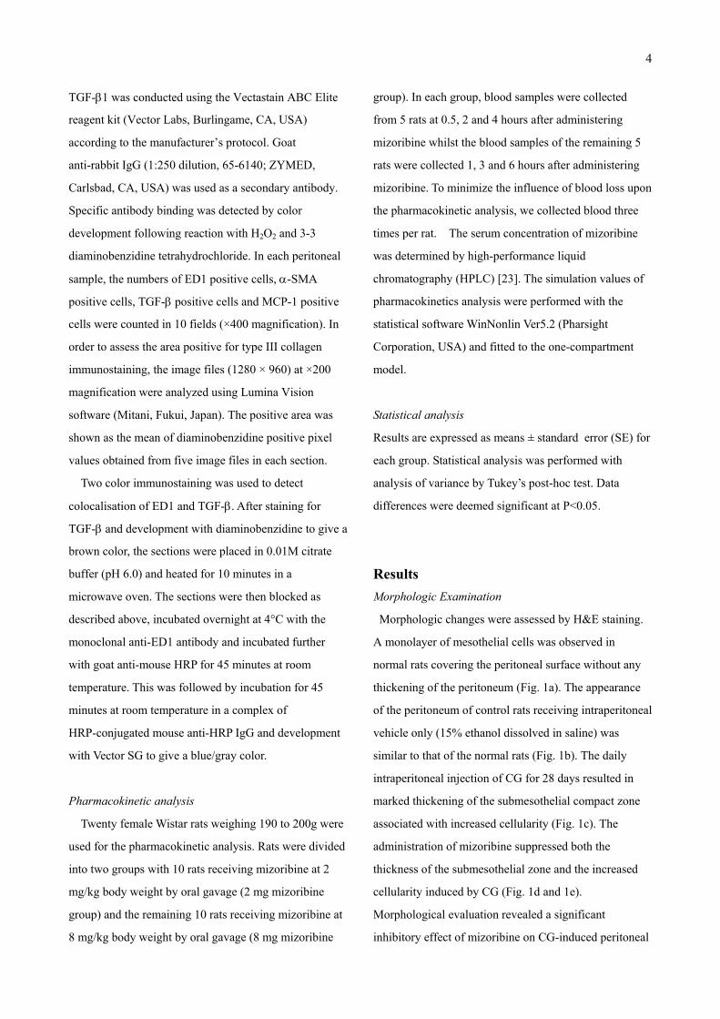
Results Morphologic Examination
Morphologic changes were assessed by H&E staining.
A monolayer of mesothelial cells was observed in
normal rats covering the peritoneal surface without any
thickening of the peritoneum (Fig. 1a). The appearance
of the peritoneum of control rats receiving intraperitoneal
vehicle only (15% ethanol dissolved in saline) was
similar to that of the normal rats (Fig. 1b). The daily
intraperitoneal injection of CG for 28 days resulted in
marked thickening of the submesothelial compact zone
associated with increased cellularity (Fig. 1c). The
administration of mizoribine suppressed both the
thickness of the submesothelial zone and the increased
cellularity induced by CG (Fig. 1d and 1e).
Morphological evaluation revealed a significant
inhibitory effect of mizoribine on CG-induced peritoneal
5
thickening with this anti-fibrotic effect being
dose-dependent (control group 17.4 ± 1.7×103μm2; CG +
vehicle group 222.7 ± 8.5×103μm2; CG + mizoribine 2
mg group 128.5 ± 4.1×103μm2; CG + mizoribine 8 mg
group 85.8 ± 3.3×103μm2, Fig. 1f ).
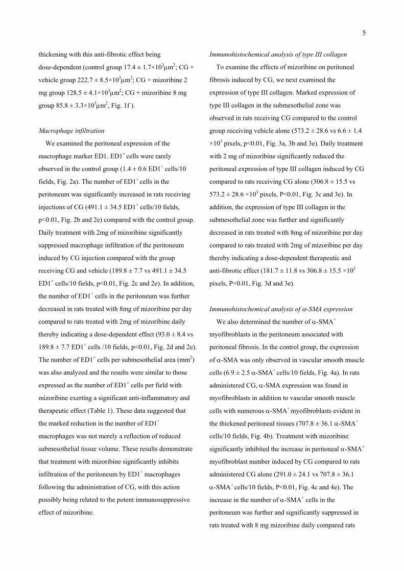
Macrophage infiltration
We examined the peritoneal expression of the
macrophage marker ED1. ED1+ cells were rarely
observed in the control group (1.4 ± 0.6 ED1+ cells/10
fields, Fig. 2a). The number of ED1+ cells in the
peritoneum was significantly increased in rats receiving
injections of CG (491.1 ± 34.5 ED1+ cells/10 fields,
p<0.01, Fig. 2b and 2e) compared with the control group.
Daily treatment with 2mg of mizoribine significantly
suppressed macrophage infiltration of the peritoneum
induced by CG injection compared with the group
receiving CG and vehicle (189.8 ± 7.7 vs 491.1 ± 34.5
ED1+ cells/10 fields, p<0.01, Fig. 2c and 2e). In addition,
the number of ED1+ cells in the peritoneum was further
decreased in rats treated with 8mg of mizoribine per day
compared to rats treated with 2mg of mizoribine daily
thereby indicating a dose-dependent effect (93.0 ± 8.4 vs
189.8 ± 7.7 ED1+ cells /10 fields, p<0.01, Fig. 2d and 2e).
The number of ED1+ cells per submesothelial area (mm2)
was also analyzed and the results were similar to those
expressed as the number of ED1+ cells per field with
mizoribine exerting a significant anti-inflammatory and
therapeutic effect (Table 1). These data suggested that
the marked reduction in the number of ED1+
macrophages was not merely a reflection of reduced
submesothelial tissue volume. These results demonstrate
that treatment with mizoribine significantly inhibits
infiltration of the peritoneum by ED1+ macrophages
following the administration of CG, with this action
possibly being related to the potent immunosuppressive
effect of mizoribine.
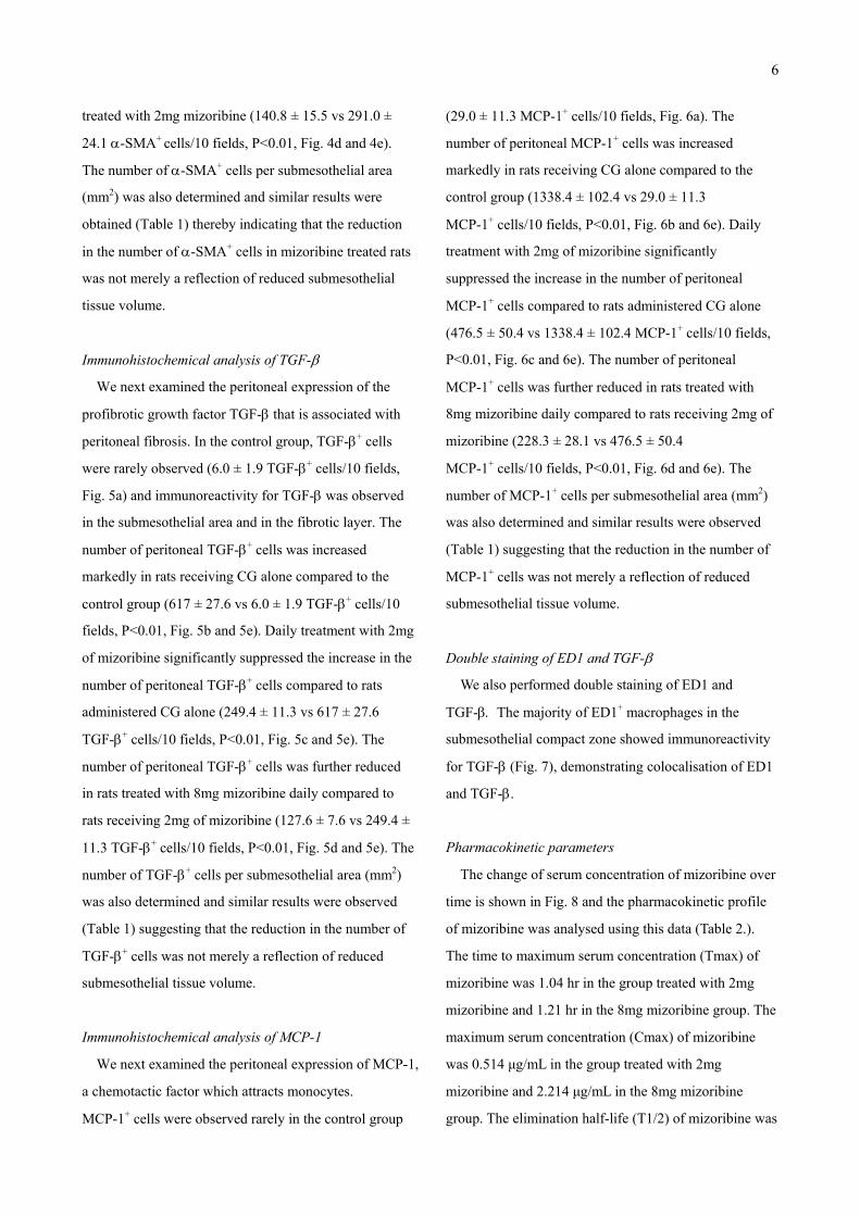
Immunohistochemical analysis of type III collagen
To examine the effects of mizoribine on peritoneal
fibrosis induced by CG, we next examined the
expression of type III collagen. Marked expression of
type III collagen in the submesothelial zone was
observed in rats receiving CG compared to the control
group receiving vehicle alone (573.2 ± 28.6 vs 6.6 ± 1.4
×103 pixels, p<0.01, Fig. 3a, 3b and 3e). Daily treatment
with 2 mg of mizoribine significantly reduced the
peritoneal expression of type III collagen induced by CG
compared to rats receiving CG alone (306.8 ± 15.5 vs
573.2 ± 28.6 ×103 pixels, P<0.01, Fig. 3c and 3e). In
addition, the expression of type III collagen in the
submesothelial zone was further and significantly
decreased in rats treated with 8mg of mizoribine per day
compared to rats treated with 2mg of mizoribine per day
thereby indicating a dose-dependent therapeutic and
anti-fibrotic effect (181.7 ± 11.8 vs 306.8 ± 15.5 ×10³
pixels, P<0.01, Fig. 3d and 3e).
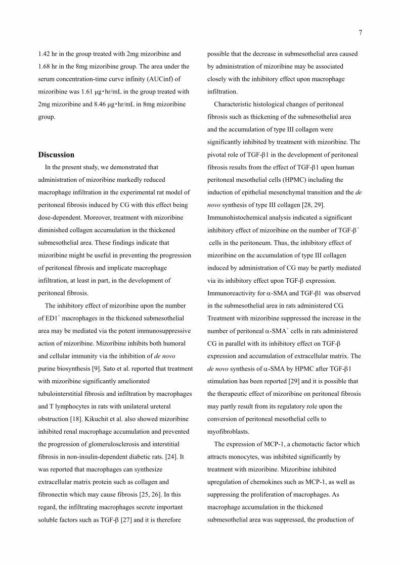
Immunohistochemical analysis of α-SMA expression
We also determined the number of α-SMA+
myofibroblasts in the peritoneum associated with
peritoneal fibrosis. In the control group, the expression
of α-SMA was only observed in vascular smooth muscle
cells (6.9 ± 2.5 α-SMA+ cells/10 fields, Fig. 4a). In rats
administered CG, α-SMA expression was found in
myofibroblasts in addition to vascular smooth muscle
cells with numerous α-SMA+ myofibroblasts evident in
the thickened peritoneal tissues (707.8 ± 36.1 α-SMA+
cells/10 fields, Fig. 4b). Treatment with mizoribine
significantly inhibited the increase in peritoneal α-SMA+
myofibroblast number induced by CG compared to rats
administered CG alone (291.0 ± 24.1 vs 707.8 ± 36.1
α-SMA+ cells/10 fields, P<0.01, Fig. 4c and 4e). The
increase in the number of α-SMA+ cells in the
peritoneum was further and significantly suppressed in
rats treated with 8 mg mizoribine daily compared rats
6
treated with 2mg mizoribine (140.8 ± 15.5 vs 291.0 ±
24.1 α-SMA+ cells/10 fields, P<0.01, Fig. 4d and 4e).
The number of α-SMA+ cells per submesothelial area
(mm2) was also determined and similar results were
obtained (Table 1) thereby indicating that the reduction
in the number of α-SMA+ cells in mizoribine treated rats
was not merely a reflection of reduced submesothelial
tissue volume.
Immunohistochemical analysis of TGF-β
We next examined the peritoneal expression of the
profibrotic growth factor TGF-β that is associated with
peritoneal fibrosis. In the control group, TGF-β+ cells
were rarely observed (6.0 ± 1.9 TGF-β+ cells/10 fields,
Fig. 5a) and immunoreactivity for TGF-β was observed
in the submesothelial area and in the fibrotic layer. The
number of peritoneal TGF-β+ cells was increased
markedly in rats receiving CG alone compared to the
control group (617 ± 27.6 vs 6.0 ± 1.9 TGF-β+ cells/10
fields, P<0.01, Fig. 5b and 5e). Daily treatment with 2mg
of mizoribine significantly suppressed the increase in the
number of peritoneal TGF-β+ cells compared to rats
administered CG alone (249.4 ± 11.3 vs 617 ± 27.6
TGF-β+ cells/10 fields, P<0.01, Fig. 5c and 5e). The
number of peritoneal TGF-β+ cells was further reduced
in rats treated with 8mg mizoribine daily compared to
rats receiving 2mg of mizoribine (127.6 ± 7.6 vs 249.4 ±
11.3 TGF-β+ cells/10 fields, P<0.01, Fig. 5d and 5e). The
number of TGF-β+ cells per submesothelial area (mm2)
was also determined and similar results were observed
(Table 1) suggesting that the reduction in the number of
TGF-β+ cells was not merely a reflection of reduced
submesothelial tissue volume.
Immunohistochemical analysis of MCP-1
We next examined the peritoneal expression of MCP-1,
a chemotactic factor which attracts monocytes.
MCP-1+ cells were observed rarely in the control group
(29.0 ± 11.3 MCP-1+ cells/10 fields, Fig. 6a). The
number of peritoneal MCP-1+ cells was increased
markedly in rats receiving CG alone compared to the
control group (1338.4 ± 102.4 vs 29.0 ± 11.3
MCP-1+ cells/10 fields, P<0.01, Fig. 6b and 6e). Daily
treatment with 2mg of mizoribine significantly
suppressed the increase in the number of peritoneal
MCP-1+ cells compared to rats administered CG alone
(476.5 ± 50.4 vs 1338.4 ± 102.4 MCP-1+ cells/10 fields,
P<0.01, Fig. 6c and 6e). The number of peritoneal
MCP-1+ cells was further reduced in rats treated with
8mg mizoribine daily compared to rats receiving 2mg of
mizoribine (228.3 ± 28.1 vs 476.5 ± 50.4
MCP-1+ cells/10 fields, P<0.01, Fig. 6d and 6e). The
number of MCP-1+ cells per submesothelial area (mm2)
was also determined and similar results were observed
(Table 1) suggesting that the reduction in the number of
MCP-1+ cells was not merely a reflection of reduced
submesothelial tissue volume.
Double staining of ED1 and TGF-β
We also performed double staining of ED1 and
TGF-β. The majority of ED1+ macrophages in the
submesothelial compact zone showed immunoreactivity
for TGF-β (Fig. 7), demonstrating colocalisation of ED1
and TGF-β.
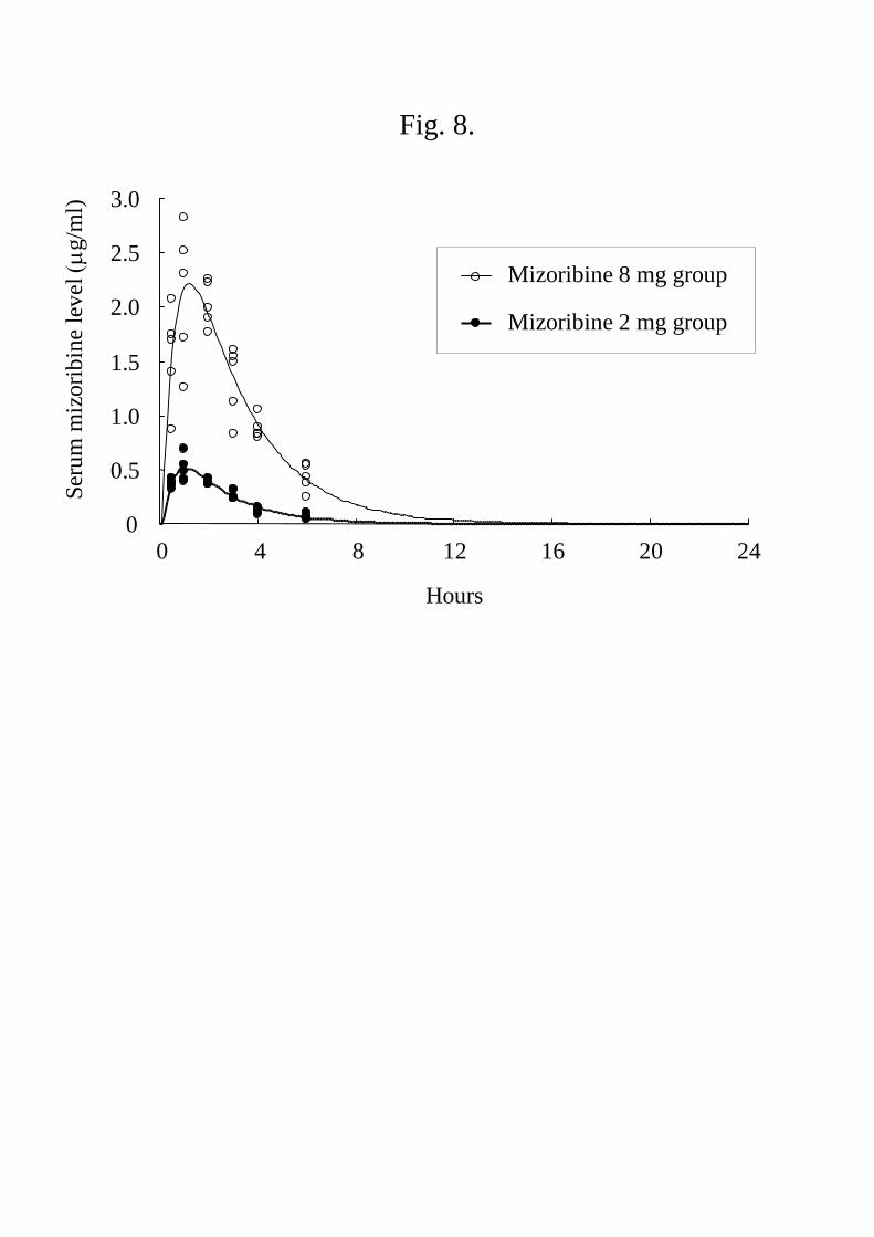
Pharmacokinetic parameters
The change of serum concentration of mizoribine over
time is shown in Fig. 8 and the pharmacokinetic profile
of mizoribine was analysed using this data (Table 2.).
The time to maximum serum concentration (Tmax) of
mizoribine was 1.04 hr in the group treated with 2mg
mizoribine and 1.21 hr in the 8mg mizoribine group. The
maximum serum concentration (Cmax) of mizoribine
was 0.514 μg/mL in the group treated with 2mg
mizoribine and 2.214 μg/mL in the 8mg mizoribine
group. The elimination half-life (T1/2) of mizoribine was
7
1.42 hr in the group treated with 2mg mizoribine and
1.68 hr in the 8mg mizoribine group. The area under the
serum concentration-time curve infinity (AUCinf) of
mizoribine was 1.61 μg・hr/mL in the group treated with
2mg mizoribine and 8.46 μg・hr/mL in 8mg mizoribine
group.
Discussion In the present study, we demonstrated that
administration of mizoribine markedly reduced
macrophage infiltration in the experimental rat model of
peritoneal fibrosis induced by CG with this effect being
dose-dependent. Moreover, treatment with mizoribine
diminished collagen accumulation in the thickened
submesothelial area. These findings indicate that
mizoribine might be useful in preventing the progression
of peritoneal fibrosis and implicate macrophage
infiltration, at least in part, in the development of
peritoneal fibrosis.
The inhibitory effect of mizoribine upon the number
of ED1+ macrophages in the thickened submesothelial
area may be mediated via the potent immunosuppressive
action of mizoribine. Mizoribine inhibits both humoral
and cellular immunity via the inhibition of de novo
purine biosynthesis [9]. Sato et al. reported that treatment
with mizoribine significantly ameliorated
tubulointerstitial fibrosis and infiltration by macrophages
and T lymphocytes in rats with unilateral ureteral
obstruction [18]. Kikuchit et al. also showed mizoribine
inhibited renal macrophage accumulation and prevented
the progression of glomerulosclerosis and interstitial
fibrosis in non-insulin-dependent diabetic rats. [24]. It
was reported that macrophages can synthesize
extracellular matrix protein such as collagen and
fibronectin which may cause fibrosis [25, 26]. In this
regard, the infiltrating macrophages secrete important
soluble factors such as TGF-β [27] and it is therefore
possible that the decrease in submesothelial area caused
by administration of mizoribine may be associated
closely with the inhibitory effect upon macrophage
infiltration.
Characteristic histological changes of peritoneal
fibrosis such as thickening of the submesothelial area
and the accumulation of type III collagen were
significantly inhibited by treatment with mizoribine. The
pivotal role of TGF-β1 in the development of peritoneal
fibrosis results from the effect of TGF-β1 upon human
peritoneal mesothelial cells (HPMC) including the
induction of epithelial mesenchymal transition and the de
novo synthesis of type III collagen [28, 29].
Immunohistochemical analysis indicated a significant
inhibitory effect of mizoribine on the number of TGF-β+
cells in the peritoneum. Thus, the inhibitory effect of
mizoribine on the accumulation of type III collagen
induced by administration of CG may be partly mediated
via its inhibitory effect upon TGF-β expression.
Immunoreactivity for α-SMA and TGF-β1 was observed
in the submesothelial area in rats administered CG.
Treatment with mizoribine suppressed the increase in the
number of peritoneal α-SMA+ cells in rats administered
CG in parallel with its inhibitory effect on TGF-β
expression and accumulation of extracellular matrix. The
de novo synthesis of α-SMA by HPMC after TGF-β1
stimulation has been reported [29] and it is possible that
the therapeutic effect of mizoribine on peritoneal fibrosis
may partly result from its regulatory role upon the
conversion of peritoneal mesothelial cells to
myofibroblasts.
The expression of MCP-1, a chemotactic factor which
attracts monocytes, was inhibited significantly by
treatment with mizoribine. Mizoribine inhibited
upregulation of chemokines such as MCP-1, as well as
suppressing the proliferation of macrophages. As
macrophage accumulation in the thickened
submesothelial area was suppressed, the production of
8
TGF-β may also have been inhibited, resulting in
prevention of peritoneal fibrosis. The colocalisation of
ED1 and TGF-β observed with double immunostaining
supports this hypothesis.
For the analysis of immunohistochemical findings, we
also determined the number of ED1+, α-SMA+ ,TGF-β+
and MCP-1+ cells per submesothelial area (mm2). The
results were similar to the measurement of cell number
per field thereby demonstrating that the significant
therapeutic effects of mizoribine did not merely reflect
the differences in submesothelial thickening between
experimental groups.
No adverse events were observed in this study despite
the fact that the higher dose of mizoribine used was
greater than the conventional dose of mizoribine used
(up to 5 mg/kg body weight). Stypinski et al. reported
that the daily administration of mizoribine up to 12mg/kg
body weight caused no significant adverse events in
healthy male volunteers except for a slight elevation in
serum uric acid [30]. Tanaka et al. reported that a peak
serum level of mizoribine of at least 2.5-3.0 μg/ml is
necessary to achieve satisfactory clinical efficacy of the
drug in the treatment of lupus nephritis [31].
Pharmacokinetic analysis of the rats in this study
indicated that the Cmax was lower than 2.5-3.0 μg/ml
when the dose of mizoribine administered was 8mg/kg
body weight. Moreover, rats receiving 8mg mizoribine
daily exhibited a more marked amelioration in peritoneal
fibrosis than rats receiving 2mg mizoribine. Therefore,
the administration of mizoribine at a dose of 8mg/kg
body weight is optimal for preventing the progression of
peritoneal fibrosis in rats.
In the present study, we induced peritoneal fibrosis by
the intraperitoneal injection of CG. It is true that
CG-induced peritoneal fibrosis does not fully replicate
the peritoneal sclerosis or encapsulating peritoneal
sclerosis observed in patients on long-term PD. However,
there is not an ideal experimental model that simulates
long-term PD and CG-induced peritoneal fibrosis
undoubtedly exhibits key features of peritoneal fibrosis.
In conclusion, the administration of mizoribine
prevented the progression of peritoneal fibrosis in a rat
model. Mizoribine is a potentially useful therapy for
peritoneal sclerosis in patients undergoing long-term PD.
As mizoribine has lower nephrotoxicity than other
immunosuppressive agents, its benefit may be in
preserving residual renal function of PD patients in
comparison with other immunosuppressive agents.
Acknowledgements
The authors would like to thank Asahi-Kasei Pharm,
Tokyo, for conducting the measurements of the blood
levels of mizoribine.
9
References
1. Selgas R, Fernandez-Reyes MJ, Bosque E, Bajo MA,
Borrego F, Jimenez C, Del Peso G, De Alvaro F:
Functional longevity of the human peritoneum: how long
is continuous peritoneal dialysis possible? Results of a
prospective medium long-term study. Am J Kidney Dis
1994; 23: 64-73.
2. Davies SJ, Bryan J, Phillips L, Russell GI: Longitudinal
changes in peritoneal kinetics: the effects of peritoneal
dialysis and peritonitis. Nephrol Dial Transplant 1996;
11: 498-506.
3. Ito T, Yorioka N: Peritoneal damage by peritoneal dialysis
solutions. Clin Exp Nephrol 2008; 12: 243-249.
4. Williams JD, Craig KJ, Topley N, Von Ruhland C, Fallon
M, Newman GR, Mackenzie RK, Williams GT:
Morphologic changes in the peritoneal membrane of
patients with renal disease. J Am Soc Nephrol 2002; 13:
470-479.
5. Gandhi VC, Humayun HM, Ing TS, Daugirdas JT,
Jablokow VR, Iwatsuki S, Geis WP, Hano JE: Sclerotic
thickening of the peritoneal membrane in maintenance
peritoneal dialysis patients. Arch Intern Med 1980; 140:
1201-1203.
6. Kawanishi H, Moriishi M: Encapsulating peritoneal
sclerosis: prevention and treatment. Perit Dial Int 2007;
27 Suppl 2: S289-92.
7. Suga H, Teraoka S, Ota K, Komemushi S, Furutani S,
Yamauchi S, Margolin S: Preventive effect of pirfenidone
against experimental sclerosing peritonitis on rats. Exp
Toxicol Pathol 1995; 47: 287-291.
8. Mishima Y, Miyazaki M, Abe K, Ozono Y, Shioshita K,
Xia Z, Harada T, Taguchi T, Koji T, Kohno S: Enhanced
expression of heat shock protein 47 in rat model of
peritoneal fibrosis. Perit Dial Int 2003; 23: 14-22.
9. Koyama H, Tsuji M: Genetic and biochemical studies on
the activation and cytotoxic mechanism of bredinin, a
potent inhibitor of purine biosynthesis in mammalian cells.
Biochem Pharmacol 1983; 32: 3547-3553
10. Kusumi T, Tsuda M, Katsunuma T, Yamamura M: Dual
inhibitory effect of bredinin. Cell Biochem Funct 1989; 7:
201-204.
11. Ishikawa H: Mizoribine and mycophenolate mofetil. Curr
Med Chem 1999; 6: 575-597.
12. Akiyama T, Okazaki H, Takahashi K, Hasegawa A,
Tanabe K, Uchida K, Takahara S, Toma H: Links
Mizoribine in combination therapy with tacrolimus for
living donor renal transplantation: analysis of a
nationwide study in Japan. Transplant Proc 2005; 37:
843-845.
13. Tanaka E, Inoue E, Kawaguchi Y, Tomatsu T, Yamanaka
H, Hara M, Kamatani N: Acceptability and usefulness of
mizoribine in the management of rheumatoid arthritis in
methotrexate-refractory patients and elderly patients,
based on analysis of data from a large-scale observational
cohort study. Mod Rheumatol 2006; 16: 214-219.
14. Nakayamada S, Saito K, Umehara H, Ogawa N, Sumida T,
Ito S, Minota S, Nara H, Kondo H, Okada J, Mimori T,
Yoshifuji H, Sano H, Hashimoto N. Sugai S, Tanaka Y:
Efficacy and safety of mizoribine for the treatment of
Sjögren's syndrome: a multicenter open-label clinical trial.
Mod Rheumatol 2007; 17: 464-469.
15. Tanaka H, Tsugawa K, Tsuruga K, Suzuki K, Nakahata T,
Ito E, Waga S: Mizoribine for the treatment of lupus
nephritis in children and adolescents. Clin Nephrol 2004;
62: 412-417.
16. Honda M: Nephrotic syndrome and mizoribine in children.
Pediatr Int 2002; 44: 210-216.
17. Sakai T, Kawamura T, Shirasawa T: Mizoribine improves
renal tubulointerstitial fibrosis in unilateral ureteral
obstruction (UUO)-treated rat by inhibiting the infiltration
of macrophages and the expression of alpha-smooth
muscle actin. J Urol 1997; 158: 2316-2322.
18. Sato N, Shiraiwa K, Kai K, Watanabe A, Ogawa S,
Kobayashi Y, Yamagishi-Imai H, Utsunomiya Y, Mitarai
T: Mizoribine ameliorates the tubulointerstitial fibrosis of
10
obstructive nephropathy. Nephron 2001; 89: 177-185.
19. Nishino T, Miyazaki M, Abe K, Furusu A, Mishima Y,
Harada T, Ozono Y, Koji T, Kohno S: Antisense
oligonucleotides against collagen-binding stress protein
HSP47 suppress peritoneal fibrosis in rats. Kidney Int
2003; 64: 887-896.
20. Yoshio Y, Miyazaki M, Abe K, Nishino T, Furusu A,
Mizuta Y, Harada T, Ozono Y, Koji T, Kohno S:
TNP-470, an angiogenesis inhibitor, suppresses the
progression of peritoneal fibrosis in mouse experimental
model. Kidney Int 2004; 66: 1677-1685.
21. Doi S, Masaki T, Arakawa T, Takahashi S, Kawai T,
Nakashima A, Naito T, Kohno N, Yorioka N: Protective
effects of peroxisome proliferator-activated receptor
gamma ligand on apoptosis and hepatocyte growth factor
induction in renal ischemia-reperfusion injury.
Transplantation 2007; 84: 207-213.
22. Hirai T, Masaki T, Kuratsune M, Yorioka N, Kohno N:
PDGF receptor tyrosine kinase inhibitor suppresses
mesangial cell proliferation involving STAT3 activation.
Clin Exp Immunol 2006; 144: 353-361.
23. Hosotsubo H, Takahara S, Taenaka N: Simplified
high-performance liquid chromatographic method for
determination of mizoribine in human serum. J
Chromatography 1988; 432: 340-345
24. Kikuchi Y, Imakiire T, Yamada M, Saigusa T, Hyodo T,
Hyodo N, Suzuki S, Miura S: Mizoribine reduces renal
injury and macrophage infiltration in
non-insulin-dependent diabetic rats. Nephrol Dial
Transplant 2005; 20: 1573-81
25. Vaage J, Lindblad WJ: Production of collagen type I by
mouse peritoneal macrophages. J Leukoc Biol 1990; 48:
274-280.
26. Nathan CF: Secretory products of macrophages. J Clin
Invest 1987; 79: 319-326.
27. Diamond JR, Kees-Folts D, Ding G, Frye JE, Restrepo
NC: Macrophages, monocyte chemoattractant peptide-1,
and TGF-beta 1 in experimental hydronephrosis. Am J
Physiol 1994; 266: F926-933.
28. Margetts PJ, Bonniaud P, Liu L, Hoff CM, Holmes CJ,
West-Mays JA, Kelly MM: Transient overexpression of
TGF-β1 induces epithelial mesenchymal transition in the
rodent peritoneum. J Am Soc Nephrol 2005; 16: 425–436.
29. Yang AH, Chen JY, Lin JK: Myofibroblastic conversion of
mesothelial cells. Kidney Int 2003; 63: 1530–1539.
30. Stypinski D, Obaidi M, Combs M, Weber M, Stewart AJ,
Ishikawa H: Safety, tolerability and pharmacokinetics of
higher-dose mizoribine in healthy male volunteers. Br J
Clin Pharmacol 2007; 63: 459-468.
31. Tanaka H, Tsugawa K, Nakahata T, Kudo M, Suzuki K,
Ito E: Implication of the peak serum level of mizoribine
for control of the serum anti-dsDNA antibody titer in
patients with lupus nephritis. Clin Nephrol 2005; 63:
417-422.
11
Tables
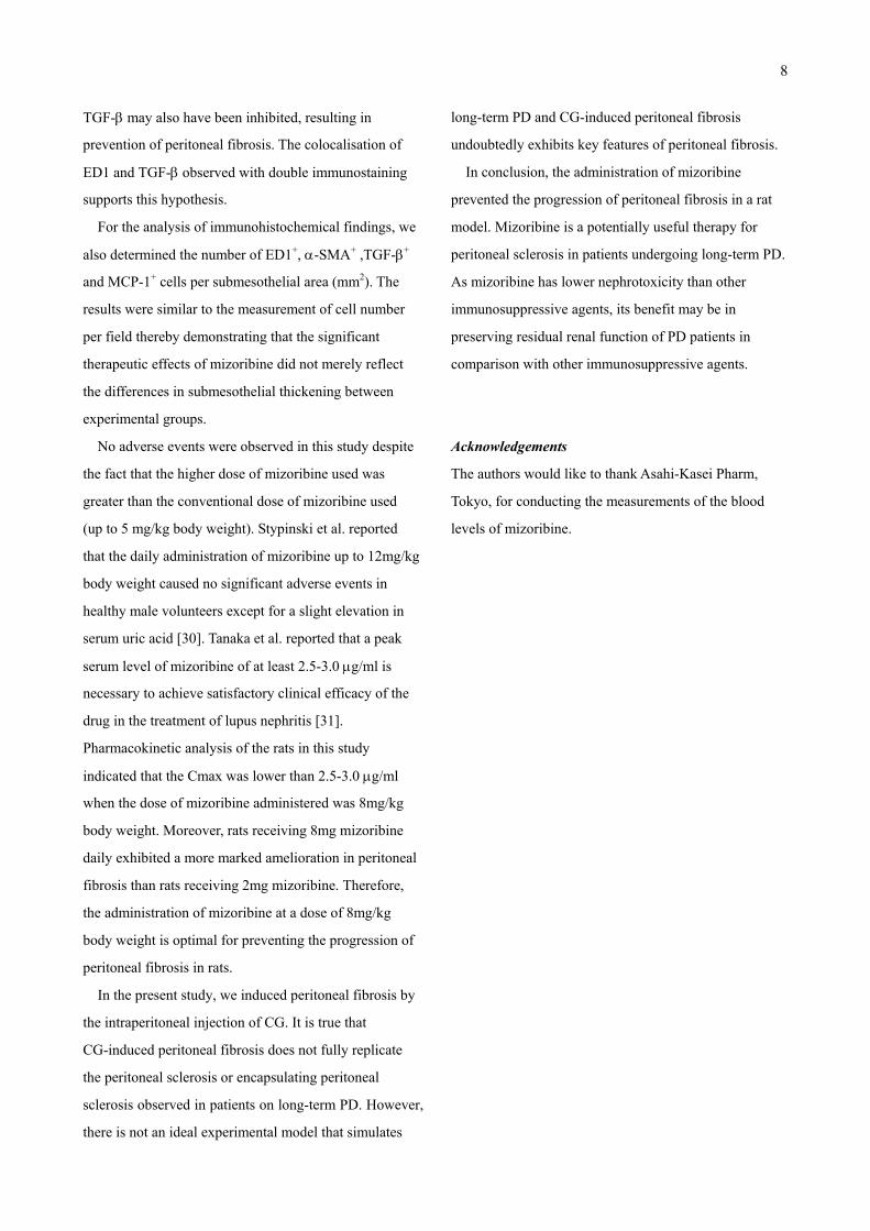
Table 1. Morphometric analysis of immunohistochemical findings
CG + vehicle CG + 2 mg mizoribine CG + 8 mg mizoribine
ED1+ cells (/mm2) 88.3±5.1 59.0±1.3a 43.1±3.1a,b
α-SMA+ cells (/ mm2) 128.0±7.2 89.9±5.6a 65.6±6.7a,b
TGF-β+ cells (/ mm2) 112.6±8.0 78.2±4.8a 59.5±2.2a,b
MCP-1+ cells (/ mm2) 239.5±15.0 151.1±11.5a 119±19.7a,b
All values are mean ± SE. n=8 in each group. The number of positive cells was counted in 10 fields and represented as per mm2 of
submesothelial fibrotic tissue by measuring the fibrotic area using NIS-Elements D software. aP<0.05 vs CG + vehicle group. bP<0.05 vs CG + 2 mg mizoribine group.
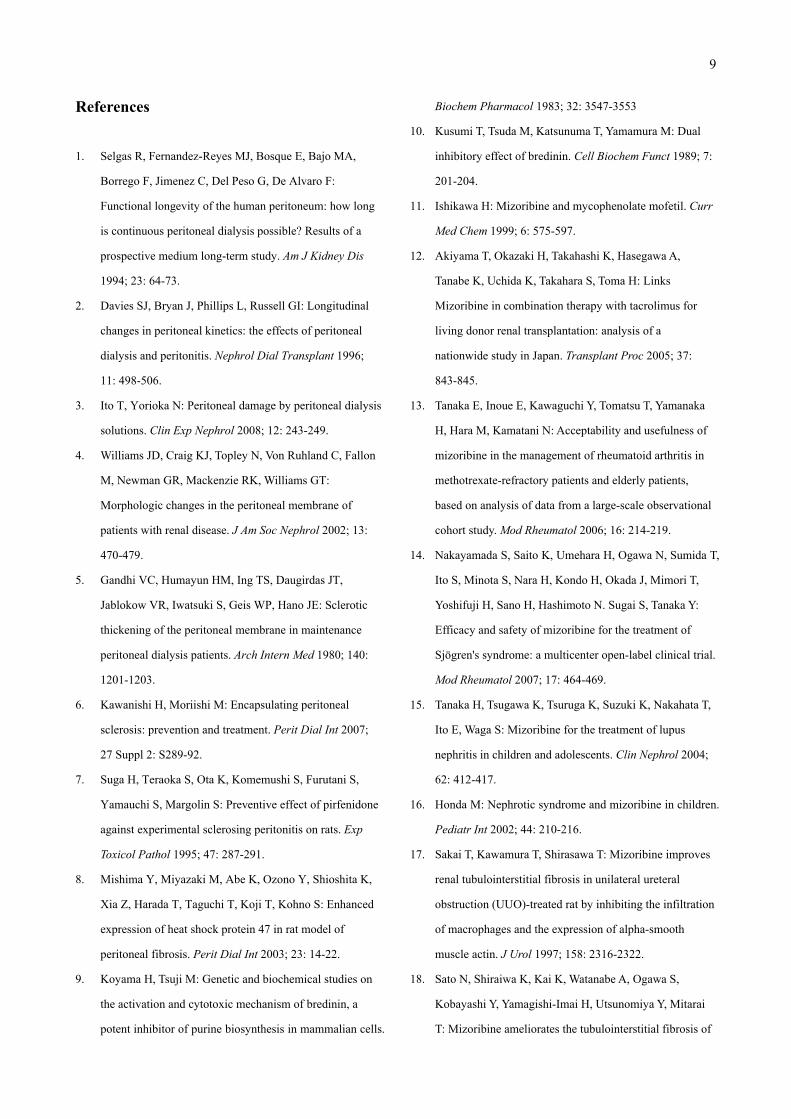
Table 2. Pharmacokinetic parameter (one compartment model).
Mizoribine 2mg/kg Mizoribine 8mg/kg
Tmax (hr) 1.04 1.21
Cmax (μg/mL) 0.514 2.214
T1/2 (hr) 1.42 1.68
AUCinf (μg・hr/mL) 1.61 8.46
Pharmacokinetics analysis was performed with the statistical software WinNonlin Ver5.2 (Pharsight Corporation, USA). Cmax, the
maximum concentration of mizoribine in serum; Tmax, the time to maximum concentration of mizoribine in serum; T1/2,
elimination half-life of mizoribine; AUCinf, the area under the serum concentration-time curve infinity of mizoribine.
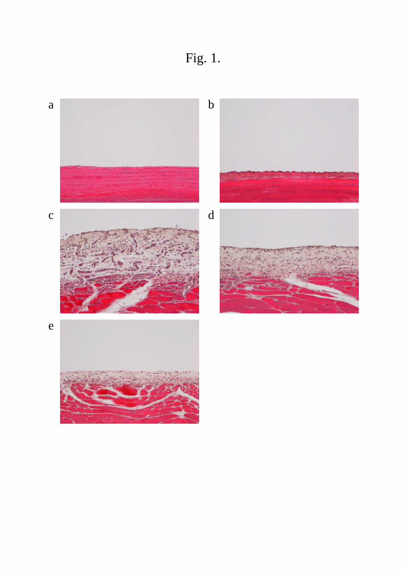
Fig. 1.
a b
c d
e

Fig. 1.
f
0
50
100
150
200
250
C V M2 M8
Per
iton
eal
thic
knes
s(×
103
m2
) *
†
*#
*#
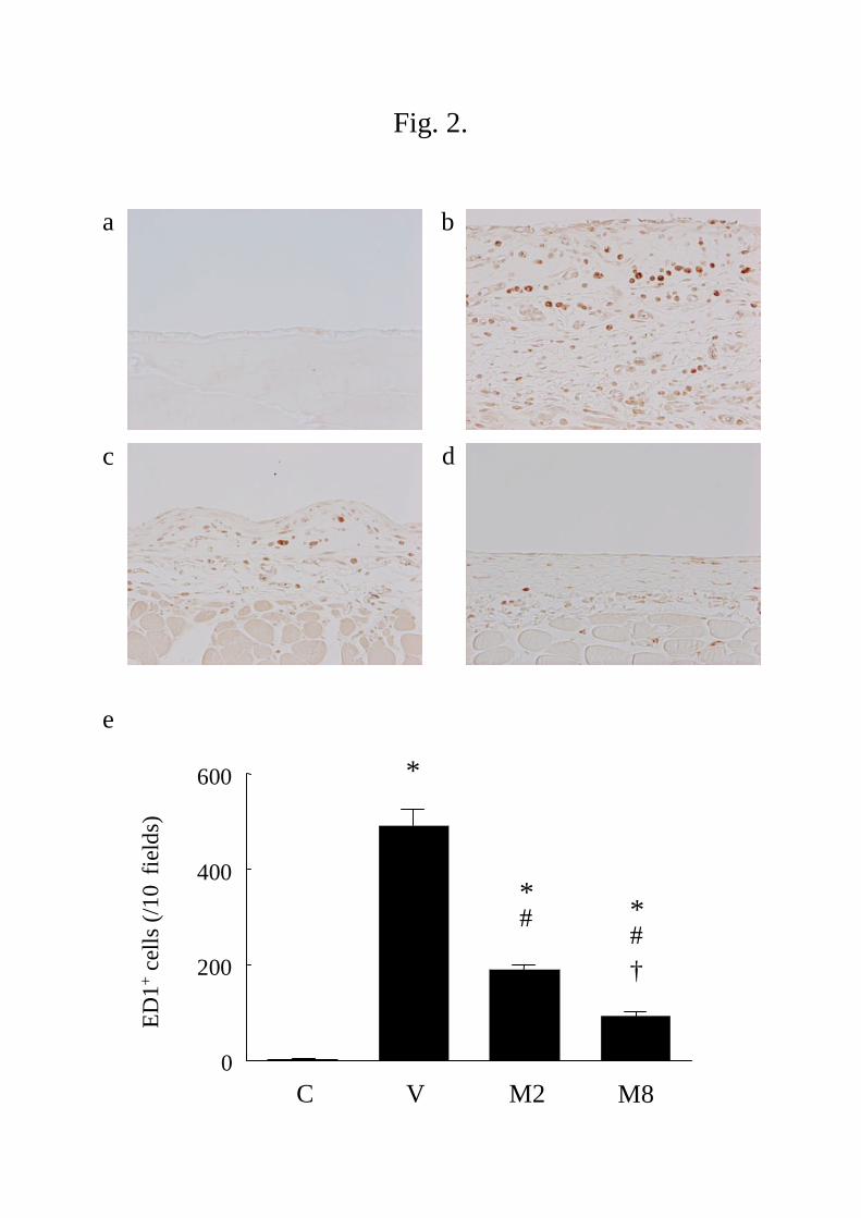
Fig. 2.
a b
c
e
0
200
400
600
ED
1+
cell
s(/
10
fiel
ds)
C V M2 M8
*
†
*#
*#
d
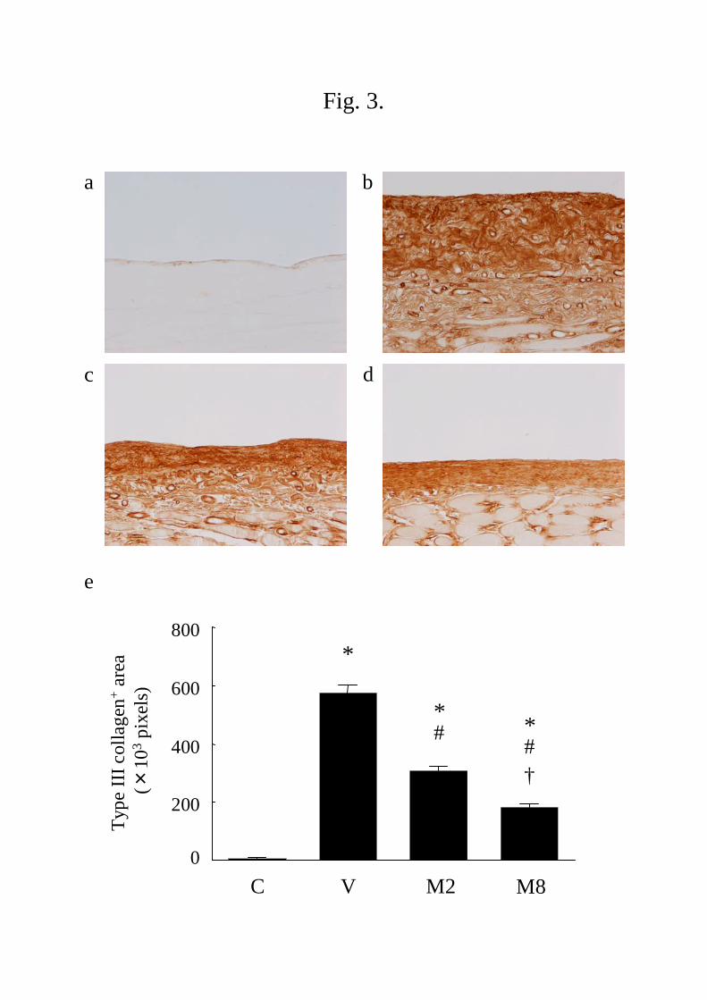
Fig. 3.
a b
c
0
200
400
600
800
Typ
eII
Ico
llag
en+
area
(×10
3p
ixel
s)
e
*
†
*#
*#
C V M2 M8
d
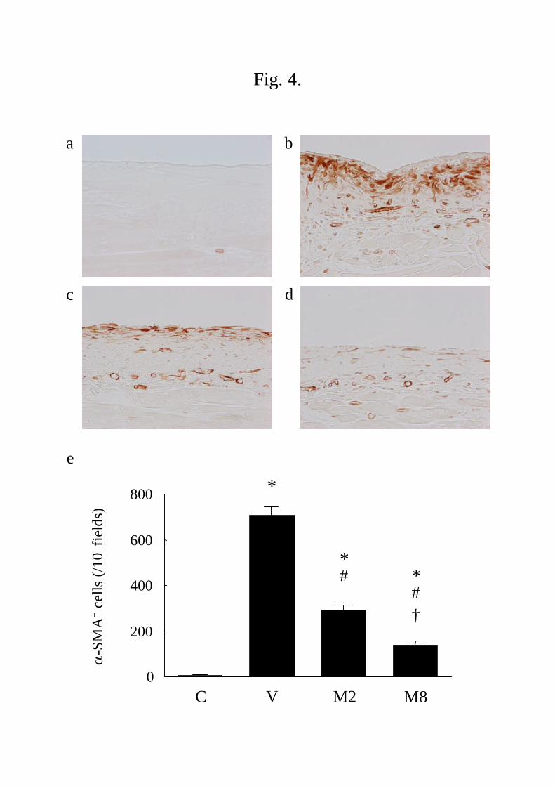
Fig. 4.
a b
c
e
0
200
400
600
800
-S
MA
+ce
lls
(/1
0fi
eld
s)
C V M2 M8
*
†
*#
*#
d
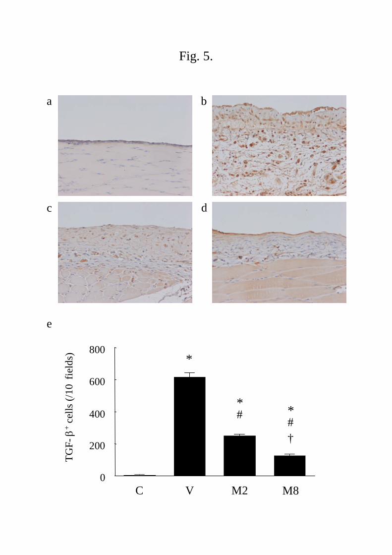
Fig. 5.
a b
c
0
200
400
600
800
TG
F-
+ce
lls
(/1
0fi
eld
s)
e
C V M2 M8
*
†
*#
*#
d
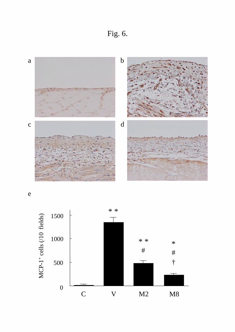
Fig. 6.
500
1000
1500
0
MC
P-1
+ce
lls
(/1
0fi
eld
s)
*#
†
#
C V M2 M8
e
a b
c d
* *
* *
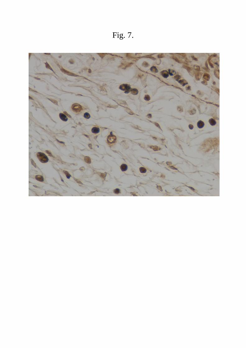
Fig. 7.
0
0.5
1.0
1.5
2.0
2.5
3.0
0 4 8 12 16 20 24
Hours
Ser
um
miz
orib
ine
lev
el
g/m
l)
Fig. 8.
Mizoribine 2 mg group
Mizoribine 8 mg group