Embed Size (px)
DESCRIPTION
Astm
Citation preview
MOL #96800
1
Salmeterol Efficacy and Bias in the Activation and Kinase-Mediated Desensitization of β2-
Adrenergic Receptors
Luis E. Gimenez, Faiza Baameur, Sharat J. Vayttaden and Richard B. Clark
Department of Pharmacology, Vanderbilt University Medical Center, Nashville, TN 37221 (LEG)
Department of Symptom Research, Division of Internal Medicine, The University of Texas MD Anderson
Cancer Center, Houston, TX 77030 (FB)
Department of Integrative Biology and Pharmacology, University of Texas Health Science Center,
Houston, TX 77225 (SJV, RBC)
This article has not been copyedited and formatted. The final version may differ from this version.Molecular Pharmacology Fast Forward. Published on March 17, 2015 as DOI: 10.1124/mol.114.096800
at ASPE
T Journals on M
arch 21, 2015m
olpharm.aspetjournals.org
Dow
nloaded from
MOL #96800
2
Running title section:
Running title: Efficacy of Salmeterol in β2AR desensitization
Corresponding author: Richard B. Clark; Department of Integrative Biology and Pharmacology,
University of Texas Health Science Center, 431 Fannin St. Houston, TX 77030,
Tel: +1-713-500-7490, Fax: +1-713-500 -7444, e-mail address:
Number of text pages: 31
Word count: Abstract: 249
Introduction: 1021
Discussion: 2136
References: 46
Tables: 0
Figures: 7
Abbreviations:
AC, adenylyl cyclase; Arr, arrestin; β2AR, β2 adrenergic receptor; BRET, bioluminescence resonance
energy transfer; cAMP, 3'-5'-cyclic adenosine monophosphate; Cara, carazolol; COPD, chronic
obstructive pulmonary disease; GRK, G protein-coupled receptor kinase; HASM, human airway smooth
muscle cells; ICI, ICI-118.551; Iso, isoproterenol; LABA, Long acting β-adrenoceptor agonists; PDE,
phosphodiesterases; Salm, salmeterol.
This article has not been copyedited and formatted. The final version may differ from this version.Molecular Pharmacology Fast Forward. Published on March 17, 2015 as DOI: 10.1124/mol.114.096800
at ASPE
T Journals on M
arch 21, 2015m
olpharm.aspetjournals.org
Dow
nloaded from
MOL #96800
3
Abstract
Salmeterol is a long acting β2-adrenergic receptor (β2AR) agonist widely used as a bronchodilator for the
treatment of persistent asthma and COPD in conjunction with steroids. Previous studies demonstrated that
salmeterol showed weak efficacy for activation of adenylyl cyclase; however, its efficacy in the complex
desensitization of β2AR remains poorly understood. In the present work we provide insights into the roles
played by GRK/arrestin and PKA in salmeterol-mediated desensitization through BRET studies of
liganded-β2AR binding to arrestin, and kinetic studies of cAMP turnover. First, BRET demonstrated a
much-reduced efficacy for salmeterol recruitment of arrestin to the β2AR relative to isoproterenol. The
ratio of BRETISO/BRETSALM after 5 min stimulation was 20, decreasing to 5 after 35 min, reflecting a
progressive decline in BRETISO, and a stable BRETSALM. Second, to assess salmeterol efficacy for func-
tional desensitization, we examined the kinetics of salmeterol-induced cAMP accumulation (0-30 min) in
human airway smooth muscle cells (HASM) in the presence and absence of PDE inhibition. Analysis of
shaping of cAMP turnover for both agonists demonstrated significant salmeterol desensitization, although
reduced relative to isoproterenol. Using an isoproterenol rescue protocol following either short- (10 min)
or long-term (2 and 14 hr) salmeterol pretreatments, we found that salmeterol progressively depressed
isoproterenol stimulation, but did not prevent subsequent rescue by isoproterenol and additional isopro-
terenol-mediated desensitization. Our findings reveal a complex efficacy for functional desensitization
demonstrating that while salmeterol shows weak efficacy for adenylyl cyclase activation and
GRK/arrestin-mediated desensitization, it acts as a strong agonist in highly amplified PKA-mediated
events.
This article has not been copyedited and formatted. The final version may differ from this version.Molecular Pharmacology Fast Forward. Published on March 17, 2015 as DOI: 10.1124/mol.114.096800
at ASPE
T Journals on M
arch 21, 2015m
olpharm.aspetjournals.org
Dow
nloaded from
MOL #96800
4
Introduction:
Long acting β-adrenoceptor agonists (LABAs) such as salmeterol, properly used only in combination
with inhaled corticosteroids, have shown proven efficacy as a maintenance therapy to avoid recurrence of
asthmatic episodes (Cazzola et al., 2013), although controversy remains primarily based on increased
morbidity and mortality for LABA use alone (Khianey and Oppenheimer, 2011). In spite of its clinical
efficacy and extended action (10-12 hr), the characterization of the mechanisms of salmeterol activation
and desensitization of the β2 adrenergic receptor (β2AR) has proven difficult, primarily because of its
weak efficacy for β2AR stimulation of adenylyl cyclase (AC), high affinity for the β2AR (Kd = 1-2 nM),
and extreme hydrophobicity (Clark et al., 1996; January et al., 1998; January et al., 1997; Rhodes et al.,
1992). Its rapid partitioning into plasma membranes renders it highly resistant to washout confounding
facile determination of its efficacy for desensitization. Thus, many problems remain unresolved, in partic-
ular the multi-faceted nature of efficacy for the complex desensitization process (Clark et al., 1999;
Kenakin and Christopoulos, 2013; van der Westhuizen et al., 2014).
Agonist-mediated desensitization of the β2AR involves a highly amplified rapid G-protein-dependent
cAMP-dependent protein kinase (PKA)-mediated phosphorylation of the receptor and activation of cy-
clic-nucleotide phosphodiesterases (PDE), coupled with the G-protein-independent pathway (January et
al., 1997; Tran et al., 2004; Vayttaden et al., 2010; Xin et al., 2008). The latter is initiated by agonist-
dependent G protein-coupled receptor kinase (GRK)-mediated phosphorylation of the β2AR that in turn
leads to arrestin binding to GRK-phosphorylated active β2AR and internalization (Clark et al., 1999; Tran
et al., 2004). It should be noted that the GRK pathway is not strictly G protein-independent since to the
extent that GRK 2/3 are involved, their membrane localization requires βγ subunit binding (Lodowski et
al., 2003). In extended treatment time, an additional component, downregulation (t1/2 = 3-4 hr), still in-
completely understood, becomes important (Liang et al., 2008; Williams et al., 2000). The consequence
of agonist efficacy on these processes is an additional confounding factor since each interaction of the
agonist-β2AR in protein/protein interactions (Gs, GRKs and arrestin) must exhibit differential efficacy.
This article has not been copyedited and formatted. The final version may differ from this version.Molecular Pharmacology Fast Forward. Published on March 17, 2015 as DOI: 10.1124/mol.114.096800
at ASPE
T Journals on M
arch 21, 2015m
olpharm.aspetjournals.org
Dow
nloaded from
MOL #96800
5
When assessing agonist efficacy for β2AR desensitization, it is important to distinguish direct molecular
measures of β2AR desensitization (e.g., efficacy of AC activation and GRK phosphorylation) from what
we refer to as functional desensitization, the measure of downstream actions (e.g., cAMP turnover by
PDE, and bronchodilation). Lack of distinction of these processes understandably has led to some confu-
sion in defining salmeterol efficacy.
Modeling and simulations based on quantitative assessment of the rates of key parameters have provided
important insights into the complexity of β2AR desensitization, both with endogenous and β2AR overex-
pression systems (Vayttaden et al., 2010; Violin et al., 2008; Xin et al., 2008). In our modeling of isopro-
terenol stimulation and desensitization of the GRK pathway, we found excellent agreement with six pa-
rameters measured following strong (isoproterenol) or weak (salmeterol) agonist stimulation; the rates of
GRK site phosphorylation, dephosphorylation, internalization, recycling, desensitization and
resensitization (Vayttaden et al., 2010). For salmeterol, we found optimum validation of experimental
data when we assumed that the affinity of salmeterol-β2AR for arrestin was much reduced relative to iso-
proterenol. This assumption was based on our prior evidence that salmeterol only weakly drives internali-
zation and arrestin translocation (Moore et al., 2007). Simulations demonstrated that salmeterol caused
decreased GRK pathway-mediated desensitization over the 0-30 min time period of stimulation compared
to isoproterenol, consistent with our prior experimental measurements (Clark et al., 1996; January et al.,
1998; Tran et al., 2004).
In contrast to quantitative analysis of β2AR-level desensitization, defining functional desensitization
based on cAMP turnover by PDE or bronchodilation is more problematic. The role of PDE in isopro-
terenol stimulation of cAMP accumulation in HEK293 cells expressing only endogenous receptor has
also been modeled and simulated (Violin et al., 2008; Xin et al., 2008). These findings highlighted the
important role of PKA activation of PDE, which in terms of its effect on the rapid phase of functional de-
sensitization (to be distinguished from β2AR desensitization), was nearly equivalent to the GRK pathway.
Because of the amplification and sensitivity of the cAMP-PKA-mediated events, weak partial agonists
This article has not been copyedited and formatted. The final version may differ from this version.Molecular Pharmacology Fast Forward. Published on March 17, 2015 as DOI: 10.1124/mol.114.096800
at ASPE
T Journals on M
arch 21, 2015m
olpharm.aspetjournals.org
Dow
nloaded from
MOL #96800
6
such as salmeterol will fully activate these components of desensitization (Tran et al., 2004) even at con-
centrations well below their EC50s, and thus act as strong agonists, while having minimal effects on the
occupancy-dependent GRK pathway. It was shown that β2AR levels are another major factor to consider
in measuring agonist efficacy (Charlton, 2009; Clark and Knoll, 2002; Clark et al., 1999; January et al.,
1998; Whaley et al., 1994; Xin et al., 2008). With high β2AR expression, salmeterol appears as a strong
agonist (Emax = 90-100 % of isoproterenol) whereas with low endogenous β2AR levels, the Emax for
salmeterol stimulation of adenylyl cyclase is much reduced (efficacy 10-20 % of isoproterenol). Further,
salmeterol’s rapid uptake and sequestration into plasma membranes has presented a major obstacle to
quantitative assessment of desensitization by the classic washout and restimulation protocols. Whether in
measurements of either cell-free membrane preparations of AC (Clark et al., 1996; January et al., 1998) or
functional desensitization in intact cells (Cooper et al., 2011; Duringer et al., 2009; Nino et al., 2009),
membrane-sequestered salmeterol has a profoundly confounding effect. Our studies demonstrated that
short-term treatment of the β2AR with salmeterol caused a much-reduced desensitization compared to
strong agonists in membrane assays (Clark et al., 1996; January et al., 1998). Based on studies comparing
equieffective concentrations of salmeterol and strong agonists on functional desensitization in human air-
way smooth muscle (HASM) cells by measure of either cAMP accumulation or bronchodilation and
bronchoprotection, it was concluded that salmeterol was a strong agonist (Cooper et al., 2011; Duringer et
al., 2009).
The goal of the present study was to clarify these issues concerning the efficacy of salmeterol activation
and desensitization mediated by PKA regulation of PDE and GRK pathway. To that end, we report first
on studies testing our proposal that salmeterol binding to the β2AR results in reduced efficacy for its
complex with arrestin. Second, we have further investigated salmeterol activation and functional desensi-
tization in human airway smooth muscle cells. This was accomplished through analysis of both the kinet-
ics of isoproterenol and salmeterol stimulation of cAMP turnover ± PDE inhibition, and the effects of
short and long-term salmeterol pretreatment on subsequent “rescue” by isoproterenol stimulation to facili-
This article has not been copyedited and formatted. The final version may differ from this version.Molecular Pharmacology Fast Forward. Published on March 17, 2015 as DOI: 10.1124/mol.114.096800
at ASPE
T Journals on M
arch 21, 2015m
olpharm.aspetjournals.org
Dow
nloaded from
MOL #96800
7
tate parsing of the contribution of receptor level desensitization and PDE to the overall desensitization
process.
Materials and Methods
Materials:
[2,8-3H]-adenine was purchased from Perkin Elmer. Cell culture medium was purchased from Gibco-Life
Technologies (Grand Island, NY) or Corning (Manassas VA). Salmeterol, ICI-118.551, carazolol, and
isoproterenol were purchased from Sigma-Aldrich Co (St Louis, MO). Salmeterol and carazolol were dis-
solved in DMSO and stored at 10 mM. Isoproterenol was stored at 10 mM with AT (100 mM ascorbate-
10 mM thiourea, pH 7.4; AT was diluted 1/100 for assay). ICI 188.551 was dissolved in H2O as a 10 mM
solution. RR-formoterol was obtained from Sunovion Pharmaceuticals Inc. (Marlborough, MA) and dis-
solved in DMSO at 10 mM. Restriction endonucleases and other DNA-modifying enzymes were obtained
from New England Biolabs (Ipswich, MA). The Renilla luciferase substrate coelenterazine-h was ac-
quired from Prolume-NanoLight Technology (Pinetop, AZ). Coelenterazine-h was dissolved in 100%
ethanol at a concentration of 5 mM (1000 times the final concentration used in BRET assays).
Plasmid constructs:
The plasmid g3NVE-1 (Vsevolod V. Gurevich, Vanderbilt University, Nashville, TN) containing the se-
quence of bovine β-arrestin2 fused to Venus (enhanced variant of yellow-shifted fluorescent protein
(Nagai et al., 2002)) at its N-terminus was constructed using a modified version of the pGEM-2 in-vitro
transcription vector (Promega, Madison, WI) as previously described (Gimenez et al., 2012). In brief, Ve-
nus was amplified by PCR using the 5’…agtcagaattcgcgatcgcggccacgatggtgagcaagggcga…3’ forward
primer that adds EcoRI and AsiSI restriction enzyme recognition sites upstream of the start codon and the
5’…tctcccccatggagtcgagcgctcgccgagacttaagtccggaggtggcct…3’ reverse primer that codes for a short
spacer with the “SGLKSRRALDS” sequence and an in-frame NcoI site. Venus was subcloned into the
This article has not been copyedited and formatted. The final version may differ from this version.Molecular Pharmacology Fast Forward. Published on March 17, 2015 as DOI: 10.1124/mol.114.096800
at ASPE
T Journals on M
arch 21, 2015m
olpharm.aspetjournals.org
Dow
nloaded from
MOL #96800
8
EcoRI – NcoI sites of pGEM-2. The β-arrestin2 sequence was then subcloned in frame with the Venus-
spacer sequence using NcoI and HindIII. The Venus- β-arrestin2 fused sequence was finally ligated into a
modified version of the pcDNA3 mammalian expression vector (Life Technologies; Carlsbad, CA) using
EcoRI and HindIII to generate P3VEA3-1.
Renilla luciferase variant 8 (RLuc8, (Loening et al., 2007; Loening et al., 2006)) was fused in frame at the
C-terminus of the sequence of triple HA-tagged (N-terminus) human β2AR (Genebank accession #
AF203386.1, Missouri S&T cDNA Resource Center, Rolla, MO). To generate the resulting P3HB2ALuc-
2 plasmid (Gimenez et al., 2012), the sequence of β2AR was amplified by PCR using the forward primer
5’…gctagaattctgcgatcgcaccaccatggcgtacccatacgatgttcca…3’ that introduces EcoRI and AsiSI restriction
sites upstream of the receptor sequence start codon and the reverse primer
5’…agcggaagcttctagcctgcaggtgccagcagtgagtcatttg...3’ that adds a SbfI restriction site upstream of the re-
ceptor coding sequence. The amplified sequence was subcloned into a pCDNA3-based plasmid that in-
cluded the Rluc8 sequence with a linker with the EcoRI, AsiSI and SbfI sites.
Bioluminescence resonance energy transfer experiments:
Arrestin recruitment after agonist stimulation was assessed by bioluminescence resonance energy transfer
(BRET)-based assays. To characterize the binding profile of β-arrestin2 to β2AR, COS-7 cells (ATCC
Cat. # CRL-1651, Manassas, VA) seeded onto 60 mm cell culture dishes (1.5x10⁶ cells/dish) were transi-
ently transfected with a fixed amount of β2AR-RLuc construct (0.5 µg) and varying amounts of Venus
arrestin (0 to 12 µg) using Lipofectamine® 2000 (Life Technologies). Transfected levels of β2AR-RLuc
are roughly equivalent to endogenous levels (Gimenez et al., 2012). Further, it was shown that manipula-
tion of very few receptor discriminator residues greatly enhances receptor specificity for arrestins indicat-
ing that there is unlikely significant competition of expression vectors with endogenous receptors that are
at about the same level in COS-7 (Mundell et al., 1999). Maximum arrestin expression in our BRET stud-
ies was estimated to be ten-fold over endogenous levels. The following day, cells were transferred to
This article has not been copyedited and formatted. The final version may differ from this version.Molecular Pharmacology Fast Forward. Published on March 17, 2015 as DOI: 10.1124/mol.114.096800
at ASPE
T Journals on M
arch 21, 2015m
olpharm.aspetjournals.org
Dow
nloaded from
MOL #96800
9
white 96-well plates (5x10⁴ cells/well). Luminescence at 460 and 535 nm was measured 48 hr post- trans-
fection after incubating 5 µM coelenterazine-h and the indicated amount of adrenoceptor ligand (or the
corresponding vehicle for unstimulated cells) for 5 to 35 min. Net BRET after drug treatment was plotted
as a function of the acceptor (Venus-arrestin) fluorescence over donor (receptor-RLuc) luminescence
(F/L) ratio. Acceptor (Venus arrestin) fluorescence was determined from cells plated onto a black 96-well
plate (excitation/emission: 485/535 nm). F/L ratios were used as an indirect measure of arrestin to recep-
tor expression. Arrestin recruitment data (mean ± SEM; 8 replicates per experiment) were fit by non-
linear regression to a three parameter, variable slope hyperbola equation (varying arrestin expression satu-
ration curves) using Prism 6.05 (GraphPad Software, San Diego, CA). Maximum BRET signal
(BRETMAX) was obtained from the curve fits. BRET signals corresponding to different F/L ratios were
obtained by interpolating the indicated ratio (e.g. 0.25 or 1) into the resulting curves from the fitted data.
Intact cell cAMP measurement:
Human airway smooth muscle cells (HASM), 6-10 passages (kindly provided by Dr. Robert Moore, Bay-
lor College of Medicine, Houston, TX), were grown to confluence in F12-HAM medium in 12-well
plates. While β2AR levels were not routinely measured, they were at the lower limit of detection by [125-
ICYP] binding, and in the range of 10-30 fmol/mg. Prior to experiments the growth medium was removed
and replaced with 0.3 ml of NaHCO3-free, 25 mM HEPES-buffered medium (pH 7.4) containing 12 µCi
of [2,8-3H]-adenine at 37°C. For all experiments, the adenine prelabeling time was 3 hr. For protocols
including PDE inhibition, 1.0 mM IBMX was added 30 min prior to agonist treatment. For measuring the
time course of agonist stimulation of cAMP, cells were stimulated with either vehicle (AT,1/100 and–
DMSO (0.5% for controls), 1.0 μM isoproterenol (Iso), or 50 nM salmeterol (Salm) for the indicated
times. To stop the reaction, incubation medium was removed, and solution containing 5% TCA, 1.0 mM
ATP, and 0.1 mM cAMP was added, and cAMP purified by a two-stage column (Dowex 50/alumina)
procedure (Baameur et al., 2014; Salomon et al., 1974).
This article has not been copyedited and formatted. The final version may differ from this version.Molecular Pharmacology Fast Forward. Published on March 17, 2015 as DOI: 10.1124/mol.114.096800
at ASPE
T Journals on M
arch 21, 2015m
olpharm.aspetjournals.org
Dow
nloaded from
MOL #96800
10
cAMP accumulation was also measured by immunoassay; however, this assay was not as sensitive (high-
er baseline) as the 3H-adenine assay, similar to that reported previously (Rosethorne et al., 2010). Other-
wise shaping of the cAMP accumulation was identical for both assays. 3H-adenine uptake into the ATP
pool is completed by 2 hours and remains stable thereafter for prolonged agonist treatment times. The
level of ATP labeling by 3H-adenine is not diminished by 12-hour salmeterol pretreatment.
Two-step isoproterenol rescue protocol:
For measurement of the effects of salmeterol pretreatment on subsequent isoproterenol stimulation of
cAMP (rescue), cells were pretreated with 20 nM salmeterol for either 10 min, 2 hr or 14 hr. Prior to ex-
periments, cells were prelabeled with [2,8-3H]-adenine ± addition of IBMX as described above. Follow-
ing salmeterol pretreatments cells were stimulated with 100 μM isoproterenol (± 1.0 mM IBMX) for 0-30
min and cAMP measured. All assay points were performed in triplicate at each time of treatment. The
average values of triplicates for multiple experiments were expressed as the mean ± SEM.
This article has not been copyedited and formatted. The final version may differ from this version.Molecular Pharmacology Fast Forward. Published on March 17, 2015 as DOI: 10.1124/mol.114.096800
at ASPE
T Journals on M
arch 21, 2015m
olpharm.aspetjournals.org
Dow
nloaded from
MOL #96800
11
Results
Comparison of Salmeterol- and Isoproterenol-liganded β2AR Interactions with β-arrestin2 by BRET:
Prior biochemical and modeling studies demonstrated reduced salmeterol-mediated translocation and in-
ternalization of the β2AR relative to isoproterenol. Based on this, we proposed that the affinity of the
salmeterol-β2AR complex for arrestin was much reduced (Vayttaden et al., 2010). To address this pro-
posal we initiated a BRET study to determine the effects of ligands on the recruitment of arrestin to the
liganded-β2AR using transient expression of Venus N-terminus tagged β-arrestin2 and the β2AR-RLuc8
fusion proteins. In each experiment, arrestin levels (directly proportional to fluorescence or “F”) were
varied at a constant β2AR-Rluc (Luminescence or “L”) expression level, giving six F/L ratios that varied
from 0.25 to 3.0 (Fig 1 A-D). BRET was assessed from 5 to 35 min following addition of either 100 nM
salmeterol (Salm), 10 μM isoproterenol (Iso), or the inverse agonists ICI-118.551 (ICI, 1.0 μM) and
carazolol (Cara; 0.1 μM). Fig 1 (D) shows a comparison of the effects of challenges with high β2AR oc-
cupancy by these compounds on BRETMAX (F/L = 3.0) signals. The data showed a much-reduced
BRETMAX (F/L = 3.0) with salmeterol relative to isoproterenol. Further, while BRET with isoproterenol
steadily diminished over 10-35 min, salmeterol signal was relatively constant. ICI-118.551 and carazolol
initially showed diminished BRET relative to control that in time approached baseline consistent with
their inverse agonism.
To investigate the interaction at reduced agonist occupancy, we performed the experiments shown in Fig
2 (A-C) with the following treatments: (i) 30 and 100 nM isoproterenol, since previous studies had shown
that these concentrations were either at or slightly under the Kd for isoproterenol, but produced maximal
GRK phosphorylation in time (Tran et al., 2004); and (ii) 10 and 30 nM salmeterol, that while exceeding
the Kd (1-2 nM), were used to compensate for the much diminished BRET signal. The results from the
data shown at three F/L ratios (3.0, 1.0, and 0.25) demonstrated first that salmeterol at either concentra-
tion caused a very weak but significant signal that increased slightly in time, with BRETMAX similar to
This article has not been copyedited and formatted. The final version may differ from this version.Molecular Pharmacology Fast Forward. Published on March 17, 2015 as DOI: 10.1124/mol.114.096800
at ASPE
T Journals on M
arch 21, 2015m
olpharm.aspetjournals.org
Dow
nloaded from
MOL #96800
12
that with 100 nM salmeterol (Fig 1 D). Second, isoproterenol at either 30 or 100 nM caused a BRET sig-
nal that diminished significantly over the 10-35 time of assay. BRETMAX with 100 nM isoproterenol was
equivalent to that found with 10 μM isoproterenol (Fig 1), while at 30 nM the signal was significantly
decreased in agreement with the GRK phosphorylation profile (Tran et al., 2004).
To assess the effect of varied arrestin levels and the time of assay we calculated the ratios of BRET sig-
nals (BRET-ISO/BRET-Salm) from the data of Fig 2. The results shown in Fig 3 (A-C) demonstrate a
striking effect of the time of assay of the Arr/β2AR-ligand complex at all three F/L levels. With F/L ratios
of 3.0 and 1.0, the isoproterenol-mediated signal at 5 min was a ≈ 20-fold higher than for salmeterol. For
the F/L ratio of 0.25, the isoproterenol/salmeterol signal was ≈ 35, likely reflecting greater error caused
by the reduced BRET signal at low arrestin levels.
The time-dependence of the BRET results for isoproterenol agree well with the kinetics of its rapid stimu-
lation of GRK phosphorylation, arrestin binding and internalization of the β2AR (t1/2 of internalization ~ 3
min), and that upon internalization, isoproterenol and arrestin dissociate from the receptor consistent with
the transient nature of the arrestin-receptor complex under these circumstances (Tran et al., 2004;
Vayttaden et al., 2010). In contrast, salmeterol-induced BRET increased slightly in time (5-10 min) and
stabilized, demonstrating salmeterol’s reduced efficacy for recruitment of the arrestin. These findings
show that while low efficacy limits the level of the salmeterol-β2AR-arrestin complex, the stability of the
complex is sufficient to cause limited internalization. The results are consistent with the previously re-
ported very limited salmeterol-induced translocation and internalization data (Moore et al., 2007), and
with the simulations of the GRK-pathway for isoproterenol versus salmeterol (Vayttaden et al., 2010).
The Role of PDE in Isoproterenol and Salmeterol Stimulation of cAMP Accumulation and Functional
Desensitization:
The goal of this aspect of our study was to examine and contrast isoproterenol and salmeterol stimulation
of cAMP turnover in primary cultured HASM cells, to determine the effects of salmeterol pretreatment on
This article has not been copyedited and formatted. The final version may differ from this version.Molecular Pharmacology Fast Forward. Published on March 17, 2015 as DOI: 10.1124/mol.114.096800
at ASPE
T Journals on M
arch 21, 2015m
olpharm.aspetjournals.org
Dow
nloaded from
MOL #96800
13
subsequent isoproterenol stimulation, and to allow comparisons of the extent of functional desensitization
(cAMP levels) with and without PDE inhibition. To that end, we first evaluated the effect of the presence
and absence of IBMX (1.0 mM) on cAMP accumulation in HASM cells in response to agonist as shown
in Fig 4A. We found that the efficacy of salmeterol stimulation in the presence or absence of PDE inhibi-
tion was much reduced relative to isoproterenol and formoterol over the entire time course consistent with
previous findings with low endogenous levels of β2AR (Clark et al., 1996; Rosethorne et al., 2010). All
agonists demonstrated functional desensitization shown by the rapidly diminishing rates of cAMP accu-
mulation in time (- IBMX), with cAMP levels peaking at 5 min and subsequently declining. In the pres-
ence of IBMX to moderate the confounding effects of PDE (added 30 min prior to agonists), cAMP levels
at zero time were slightly augmented relative to controls showing the effect of the pretreatment with
IBMX. With agonist stimulation, IBMX eliminated the rapid rise and fall of cAMP and levels increased
to plateau values. The progressive increase in levels of cAMP caused by IBMX demonstrates the time-
dependency of the downstream functional desensitization by PDE, although inhibition of PDE was not
complete (see discussion below). Nonetheless, the shaping of the cAMP levels reflects primarily receptor-
level desensitization by all agonists.
To more clearly compare initial rates (0-2 min) of cAMP accumulation with isoproterenol and salmeterol
± IBMX a separate series of experiments were performed (Fig 4B). While the low levels of stimulation
(minus IBMX) generate significant noise it was clear that the efficacy for salmeterol is reduced approxi-
mately 80% relative to isoproterenol (note values at 1.0 and 2.0 min). In the presence of IBMX, the initial
rates were unmodified; however, after 2 min stimulation there was expectedly a stimulatory effect of PDE
inhibition with isoproterenol as levels of cAMP approached levels such that PDE hydrolysis was signifi-
cant relative to rates of agonist activation.
In preliminary studies we found that 1.0 mM IBMX, 50 mM rolipram, or the combination, resulted in
similar levels of cAMP augmentation, and maximum effects of IBMX were achieved at 250 µM (data not
shown). We also examined the extent of PDE inhibition by observation of the rates of decay of cAMP
This article has not been copyedited and formatted. The final version may differ from this version.Molecular Pharmacology Fast Forward. Published on March 17, 2015 as DOI: 10.1124/mol.114.096800
at ASPE
T Journals on M
arch 21, 2015m
olpharm.aspetjournals.org
Dow
nloaded from
MOL #96800
14
after 10 min isoproterenol stimulation with addition of propranolol. The rates we observed (t1/2 = 3.98 ±
0.68 min and 1.85 ± 0.28 plus and minus propranolol respectively; n = 4) showed 54% inhibition. Given
the affinity of IBMX for PDE, full inhibition of PDE activity is theoretically unattainable as previously
discussed (Xin et al., 2008). However, it is important to realize that removal of isoproterenol activation
will diminish PKA activation of PDE in time, and it is likely that its inhibition is greater than that meas-
ured by following decay of cAMP.
The important conclusion from the agonist shaping of cAMP levels is that salmeterol causes an apparent
desensitization that approaches that of isoproterenol and formoterol. A more quantitative estimate of their
relative extents of functional desensitization after 15 min was performed by comparison of the initial and
final rates of cAMP accumulation in the presence of IBMX (Fig 4). At any point in time, the following
equation describes cAMP levels:
d[cAMP]/dt = kact (t) – kd [cAMP] (t) (Eq. 1)
Where kact is the rate of cAMP formation, and kd is the rate of hydrolysis of cAMP at time t.
First, Fig 4 shows that the initial rate of salmeterol stimulation was just 20% that of the strong agonists.
Second, we can compare the relative activities of agonists at the point at which d[cAMP]/dt is approxi-
mately = 0, where kact = kd [cAMP]. If we assume that at the plateau values of cAMP accumulation, the
rate of hydrolysis by PDE (kd) is equivalent for all agonists (Nino et al., 2009), then we can compare kact
for the two agonists. Substituting the cAMP values at 15 min (+ IBMX conditions) for salmeterol (≈
5000) and isoproterenol (≈ 15000) into the equation kact = kd [cAMP] for each agonist, demonstrates that
kact for isoproterenol is ≈ 3-fold greater than for salmeterol, whereas based on initial rates it was 5-fold
greater. Thus, the relatively greater loss of isoproterenol activity reflects greater desensitization. An im-
portant further consideration is the extent to which spare receptors in HASM cells on desensitization by
strong versus weak agonists. The effects of spare receptors in a variety of non-HASM cells has been pre-
viously well characterized (Clark and Knoll, 2002; January et al., 1998; Whaley et al., 1994; Xin et al.,
This article has not been copyedited and formatted. The final version may differ from this version.Molecular Pharmacology Fast Forward. Published on March 17, 2015 as DOI: 10.1124/mol.114.096800
at ASPE
T Journals on M
arch 21, 2015m
olpharm.aspetjournals.org
Dow
nloaded from
MOL #96800
15
2008). Using human bronchial preparations and measurement of bronchodilation, it was recently shown
they have sufficient receptor reserve (Giembycz, 2009) such that for the strong agonist formoterol the
EC50 for bronchodilation was left shifted ≈ 120 fold from the Kd, and to a much greater extent than for
weak agonists. While our primary cultures of HASM cells likely have much lower levels (10-30 fmol/mg)
we have found that the EC50 for isoproterenol is ≈ 8-fold left-shifted relative to the Kd (EC50 = 30 nM; Kd
for isoproterenol = 250 nM). The result is that salmeterol desensitization will cause immediate decreases
in Emax, while isoproterenol will likely initially display increases in EC50 with limited change in Emax, thus
further overestimating the desensitization caused by salmeterol relative to isoproterenol. The most parsi-
monious explanation for the reduced salmeterol functional desensitization consistent with the BRET re-
sult is the much reduced salmeterol recruitment of arrestin, that in turn causes reduced internalization,
even while GRK phosphorylation eventually reaches a level (10-15 min) similar to isoproterenol (Tran et
al., 2004).
Effect of Pretreatment with Salmeterol on Subsequent Stimulation by Isoproterenol:
As discussed in the Introduction, studies of salmeterol-mediated desensitization are confounded by its
rapid partition into membranes precluding washout studies with subsequent restimulation (Clark et al.,
1996; January et al., 1998; Lombardi et al., 2009; Rhodes et al., 1992). To overcome this obstacle we
used a two-stage protocol in which HASM cells were first stimulated with 20 nM salmeterol for either 10
min (Tran et al., 2004), 2 hr or 14 hr after which a high concentration of isoproterenol (100 µM) was add-
ed to overcome salmeterol stimulation, importantly replacing the need for washout. All assays were per-
formed with 1.0 mM IBMX added 30 min prior to isoproterenol. Since the Kd for salmeterol is 1-2 nM
while that for isoproterenol is ~ 200 nM, occupancy by isoproterenol, (500 fold over the Kd) is sufficient
to displace salmeterol. The results shown in Fig 5 for prestimulation by salmeterol for either 10 min
(square symbols) or 2 hr (closed triangles), or as controls, isoproterenol and salmeterol with no
prestimulation, demonstrated several important features. First, the effect of isoproterenol stimulation after
each of the salmeterol pretreatment times showed a slight lag (1 min) in initial rates of isoproterenol. Af-
This article has not been copyedited and formatted. The final version may differ from this version.Molecular Pharmacology Fast Forward. Published on March 17, 2015 as DOI: 10.1124/mol.114.096800
at ASPE
T Journals on M
arch 21, 2015m
olpharm.aspetjournals.org
Dow
nloaded from
MOL #96800
16
ter the lag, isoproterenol caused profound further stimulation, although it was much reduced in compari-
son with the control isoproterenol stimulation alone, with the inhibitory effect of a 2 hr pretreatment
greater than that after 10 min of pretreatment. The lag precluded estimates of desensitization from initial
rates. This lag may be attributed both to the slow off-rate of salmeterol binding, and a memory of GRK
phosphorylation by salmeterol “priming the system” for subsequent isoproterenol stimulation of arrestin
binding (Vayttaden et al., 2010). A second feature of the shaping of the cAMP accumulation is that iso-
proterenol produced further desensitization after salmeterol prestimulation; indicating that salmeterol-
induced functional desensitization of the isoproterenol response (≈ 30-50% at the 10 min points) was not
as extensive as that subsequently caused by isoproterenol even after 2 h of salmeterol. Also with
salmeterol pretreatment, cAMP levels approached the control isoproterenol stimulation after 30 min, a
further indication of the lesser desensitization caused by salmeterol relative to isoproterenol. As a further
control, co-stimulation by 20 nM salmeterol and 100 μM isoproterenol with no pretreatment (data not
shown) was not significantly different from that of isoproterenol alone.
Effect of a 14 hr Pretreatment with Salmeterol on Isoproterenol stimulation of cAMP accumulation:
The salient clinical efficacy of salmeterol is derived from its prolonged bronchodilation (10-12 hr). To
assess characteristics of its long action, we examined the effects of a 14 hr pretreatment with salmeterol
on subsequent isoproterenol stimulation of cAMP accumulation in HASM cells. Cells were pretreated
with or without 20 nM salmeterol for 14 hr, and then stimulated with 100 μM isoproterenol for 0-30 min
in the presence or absence of IBMX (Fig 6). This allowed observation of the salmeterol-mediated desen-
sitization of the initial rates of isoproterenol stimulation (see insert), as well as the subsequent further de-
sensitization induced by isoproterenol relative to controls. In the absence of IBMX (closed symbols),
salmeterol pretreatment caused a three-fold increase in basal values (0-time). After 2 min, isoproterenol
stimulated levels were not significantly elevated over basal values, and after 10-30 min, isoproterenol
stimulated levels were 3-fold over basal. While the extent of desensitization appeared to be > 95% based
on the initial rate of cAMP accumulation relative to isoproterenol stimulation in controls (no salmeterol),
This article has not been copyedited and formatted. The final version may differ from this version.Molecular Pharmacology Fast Forward. Published on March 17, 2015 as DOI: 10.1124/mol.114.096800
at ASPE
T Journals on M
arch 21, 2015m
olpharm.aspetjournals.org
Dow
nloaded from
MOL #96800
17
it rather likely reflects the slow off rate of salmeterol. Therefore to estimate functional desensitization we
compared the fold activation of isoproterenol over basal values at the 10 min points (3-fold with
salmeterol pretreatment versus ≈ 40 fold without) that indicated > 90% salmeterol-induced desensitiza-
tion. However, this overestimates the desensitization since the salmeterol pretreatment increases the basal
cAMP value.
In the presence of IBMX, the basal levels (0-time) were increased further after salmeterol pretreatment,
followed by an insignificant elevation of cAMP by isoproterenol over 0-2 min (Fig 6 inset). After 10 min
isoproterenol stimulation, cAMP levels with and without salmeterol pretreatment were ≈ 3.5 fold and 100
fold over basal respectively, indicating that the salmeterol-induced receptor-level desensitization was ex-
tensive (≈ 95% after 10-30 min stimulation). Importantly isoproterenol stimulated additional desensitiza-
tion of the β2AR, consistent with the conclusion that as extensive as salmeterol desensitization was from
the pretreatment, it was not complete relative to isoproterenol. Further, cAMP levels with the salmeterol
pretreatment (+ IBMX) were in fact 20-30% of those without pretreatment at the 10-30 min time points.
By these measures, the apparent salmeterol-induced desensitization of isoproterenol cAMP accumulation
was 70-80%. Regardless of the methods for estimating functional desensitization, it was significantly
greater than that from either the 10 min or the 2 hr pretreatments shown in Fig 5.
Discussion
Salmeterol used in combination with steroids has been very effective in the treatment of asthma and
chronic obstructive pulmonary disease (COPD). Providing an explanation for the clinical and molecular
efficacy of salmeterol has been challenging because of its unique chemical properties, and the complexity
of agonist-induced desensitization and downregulation of the β2AR. Early studies clearly demonstrated
that salmeterol was a weak partial agonist for β2AR activation of AC, and caused less receptor-level de-
sensitization relative to strong agonists attributable to weak efficacy for stimulation of GRK phosphoryla-
tion and sequelae. Based on studies of functional desensitization using equieffective concentrations of
This article has not been copyedited and formatted. The final version may differ from this version.Molecular Pharmacology Fast Forward. Published on March 17, 2015 as DOI: 10.1124/mol.114.096800
at ASPE
T Journals on M
arch 21, 2015m
olpharm.aspetjournals.org
Dow
nloaded from
MOL #96800
18
salmeterol and isoproterenol, it was shown that salmeterol exhibited properties of a strong agonist. In the
present study we resolve these paradoxical results by both providing further molecular evidence for
salmeterol’s diminished efficacy for stimulation of the GRK pathway, and yet demonstrating through
careful analysis of cAMP turnover and the downstream PKA regulation of PDE, that salmeterol actions
on the G protein-dependent/PKA pathway at high occupancy show strong efficacy.
Considering first the GRK pathway, Vayttaden et al. (2010) modeled and simulated the short-term isopro-
terenol and salmeterol stimulated GRK/arrestin/internalization pathway of desensitization in HEK293
cells. We concluded that at least for short-term stimulation, salmeterol exhibits low efficacy for this
pathway. A key assumption in the modeling was that the affinity of the salmeterol-β2AR complex for
arrestin was greatly reduced relative to isoproterenol. In the present study, this assumption was addressed
through the use of BRET. We report that salmeterol shows much reduced recruitment of arrestin to the
receptor, consistent with our proposal. At the earliest time measured (5 min), isoproterenol- stimulated
BRET was 20-35 fold greater than salmeterol (Fig 3) at F/L ratios of 3.0 and 0.25 respectively. Between
15-20 min after stimulation, the ratio stabilized with isoproterenol-induced BRET approximately five-fold
elevated over salmeterol. Thus from BRET, the affinity of the salmeterol-β2AR complex for arrestin is in
the range of 5-20 % that of isoproterenol. Our prior simulations had shown that the best agreement to ex-
perimental data required a 100-fold reduction in arrestin affinity (Vayttaden et al., 2010).
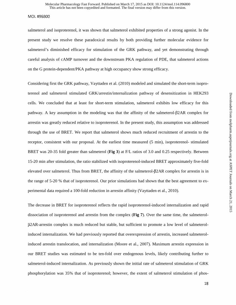
The decrease in BRET for isoproterenol reflects the rapid isoproterenol-induced internalization and rapid
dissociation of isoproterenol and arrestin from the complex (Fig 7). Over the same time, the salmeterol-
β2AR-arrestin complex is much reduced but stable, but sufficient to promote a low level of salmeterol-
induced internalization. We had previously reported that overexpression of arrestin, increased salmeterol-
induced arrestin translocation, and internalization (Moore et al., 2007). Maximum arrestin expression in
our BRET studies was estimated to be ten-fold over endogenous levels, likely contributing further to
salmeterol-induced internalization. As previously shown the initial rate of salmeterol stimulation of GRK
phosphorylation was 35% that of isoproterenol; however, the extent of salmeterol stimulation of phos-
This article has not been copyedited and formatted. The final version may differ from this version.Molecular Pharmacology Fast Forward. Published on March 17, 2015 as DOI: 10.1124/mol.114.096800
at ASPE
T Journals on M
arch 21, 2015m
olpharm.aspetjournals.org
Dow
nloaded from
MOL #96800
19
phorylation matched that of isoproterenol after 10-15 min (Moore et al., 2007; Tran et al., 2004). Thus in
time the level of the salmeterol-β2AR-arrestin complex is the rate-limiting step for internalization and
desensitization. Interestingly, salmeterol with about 200 fold higher affinity relative to isoproterenol cou-
pled with its membrane sequestration (as a source of leaked salmeterol), likely stays bound to the small
fraction of the receptor post internalization, perhaps further explaining the stabilization of the complex
with time (Fig 7). That is, the BRET signal for salmeterol (in contrast to isoproterenol) would not distin-
guish between surface and internalized receptor. In summary, our BRET findings strongly support the
conclusion that short-term salmeterol desensitization via the GRK pathway is diminished by its reduced
efficacy for arrestin binding. In support of this conclusion, the observation of a deficit in salmeterol-
induced internalization via GRK/arrestin was also demonstrated in HAMC cells by immunolocalization of
YFP-tagged β2AR following salmeterol stimulation (Cooper et al., 2011), in mouse myoblast C2C12
cells using a galactosidase complementation assay (Carter and Hill, 2005), and in HEK 293 cells by
arrestin translocation (van der Westhuizen et al., 2014) and FRET (Drake et al., 2008). Further, it was
also shown that knockdown of arrestin reduces functional desensitization (Deshpande et al., 2008).
In the second aspect of these studies, we attempted to parse out the relative contributions of receptor level
desensitization and downstream PDE activity to β2AR desensitization observed in HASM cells. This was
accomplished by two approaches, direct observation of the rapid kinetics of cAMP turnover, and a simple
two stage rescue protocol in which cells were first stimulated with salmeterol, and then challenged by a
second, high occupancy stimulation with isoproterenol to counter the inability to washout sequestered
salmeterol. With regard to the kinetics of salmeterol stimulation of cAMP turnover in the absence of
IBMX, the rapid decline in the rate of cAMP accumulation following peak levels, demonstrated signifi-
cant β2AR desensitization. Inclusion of IBMX demonstrated the large effects of PDE inhibition, (≈ 70-
80% augmentation of cAMP at the 15 min time points), and hence the major role of PKA phosphorylation
of PDE in desensitization. Estimation of the efficacy for salmeterol-mediated desensitization with much
reduced contribution from PDE activity was accomplished by analysis of the shaping of cAMP accumula-
This article has not been copyedited and formatted. The final version may differ from this version.Molecular Pharmacology Fast Forward. Published on March 17, 2015 as DOI: 10.1124/mol.114.096800
at ASPE
T Journals on M
arch 21, 2015m
olpharm.aspetjournals.org
Dow
nloaded from
MOL #96800
20
tion in the presence of IBMX by all agonists (Fig 4). Several observations indicated that salmeterol de-
sensitization was reduced relative to isoproterenol. First, calculations of the initial versus final rates of
cAMP accumulation indicated less salmeterol desensitization. Second, we used high occupancy of ago-
nists, and that in conjunction with significant but limited receptor reserve in HASM cells relative to that
of human bronchial preparations (Giembycz, 2009) predicts that the reduction in cAMP levels Emax by
salmeterol relative to isoproterenol are likely overestimated. That is, with receptor reserve the effects of
strong agonists on concentration-response curves after stimulation are to increase EC50 until receptor lev-
els are much reduced; whereas salmeterol will cause decreases in Emax (Clark et al., 1999; Whaley et al.,
1994). From simulations of salmeterol desensitization via the GRK pathway, we estimated that β2AR ac-
tivity remaining with either a 100-fold, 10-fold or no reduction in arrestin affinity for the salmeterol-
β2AR complex was 65, 35, and 20% respectively after 30 min (Vayttaden et al., 2010). Prior biochemical
evidence of salmeterol desensitization (effects on AC stimulation cell-free) showed it was just 20% of
that caused by high efficacy agonists over the short term in several cell systems (Clark et al., 1996;
January et al., 1998; Vayttaden et al., 2010). In summary, the kinetics of cAMP turnover combined with
the BRET findings demonstrate that salmeterol causes much reduced desensitization attributable to the
GRK pathway, but nonetheless causes substantial desensitization by acting as a strong agonist in the
cAMP/PKA pathway.
The rescue protocol demonstrated that salmeterol-induced desensitization, estimated by cAMP levels in
the presence of IBMX at the 10 min time points, progressed from a moderate extent after 10 min or 2 hr
stimulation (≈ 30- 50%) to 75-80% after 14 hr. Further, the data indicate that even with the 14 hr pre-
treatment, isoproterenol was still able to stimulate the receptor after the lag, and cause further desensitiza-
tion as indicated by the turnover of cAMP reaching a plateau value between 10-30 min. Since down-
stream PKA activation of PDE is both rapid and prolonged and accompanied by PDE induction as well
(Nino et al., 2009; Nino et al., 2012; Xin et al., 2008), it is likely that the contribution of PDE was rela-
tively constant over the time of isoproterenol stimulation regardless of the time of salmeterol pretreat-
This article has not been copyedited and formatted. The final version may differ from this version.Molecular Pharmacology Fast Forward. Published on March 17, 2015 as DOI: 10.1124/mol.114.096800
at ASPE
T Journals on M
arch 21, 2015m
olpharm.aspetjournals.org
Dow
nloaded from
MOL #96800
21
ment. This is further supported by the similar IBMX-induced increase in cAMP levels following isopro-
terenol (10-30 min). In addition, receptor phosphorylation by PKA should be equivalent for salmeterol
and isoproterenol as it is occupancy independent. To summarize our findings with the rescue protocol, it
is clear that while salmeterol causes significant and progressive desensitization of isoproterenol stimula-
tion with pretreatments, further desensitization by isoproterenol occurred. The most parsimonious expla-
nation for the reduced salmeterol desensitization relative to isoproterenol is its low efficacy for activation
and arrestin binding. In addition, salmeterol pre-stimulation primes the receptor by the “memory” of
salmeterol stimulated GRK phosphorylation such that addition of isoproterenol leads to rapid arrestin
binding (Vayttaden et al., 2010).
Since salmeterol-mediated desensitization is caused by both PKA and GRK phosphorylation of the β2AR,
and PDE activation by cAMP/PKA, it is not surprising that there has been ambiguity in defining the over-
all efficacy for salmeterol functional desensitization. It has also been suggested that there may be a 30-
35% reduction in AC activity (types 5 and 6) from PKA phosphorylation in HASM cells further contrib-
uting to PKA regulated events (Horvat et al., 2012; Xu et al., 2001). Therefore, our findings using high
occupancy by agonists, are actually consistent with of the results from several groups in which
equieffective concentration of agonist were used to assess the functional endpoint; that is, desensitization
of bronchodilation, and bronchoprotection caused by long-term treatments with salmeterol relative to
strong by agonists (Cooper et al., 2011; Szczuka et al., 2009). Equieffective concentrations means that
comparisons were made using very low occupancy of strong agonists with high-occupancy salmeterol to
achieve similar cAMP stimulated levels. Under these conditions, salmeterol-induced functional desensiti-
zation would be expected to be approximately equivalent to that of isoproterenol or formoterol, since the
occupancy-dependent GRK pathway for strong agonists would be minimized.
Another important aspect in the assessment of salmeterol’s long-term effects is the very slow
downregulation process. Interestingly we had previously reported that dobutamine, a very low efficacy
agonist for AC stimulation and GRK phosphorylation, showed a level of downregulation in the normal
This article has not been copyedited and formatted. The final version may differ from this version.Molecular Pharmacology Fast Forward. Published on March 17, 2015 as DOI: 10.1124/mol.114.096800
at ASPE
T Journals on M
arch 21, 2015m
olpharm.aspetjournals.org
Dow
nloaded from
MOL #96800
22
human lung epithelial cell line, BEAS-2B, equivalent to isoproterenol (Williams et al., 2000). While some
inroads into the mechanism of the shunting of the β2AR either to recycling pathway or degradation have
been gained (Liang et al., 2008), salmeterol-mediated downregulation in HASM cells has not been ex-
plored, primarily due to the problems of its sequestration in membranes and the limitations of receptor
antibodies at endogenous levels of β2AR. Given the fact that downregulation is a very slow process it is
possible that there is a logjam of a key intermediate in downregulation such that weak agonists like
dobutamine, and possibly salmeterol, cause downregulation equivalent to epinephrine, acting as a strong
agonist in this scenario.
It is interesting to speculate on the implications of our findings to the clinical use of salmeterol with the
obvious caveat that primary cultures of HASM cells, while a very useful model, differ quite considerably
from in vivo use of the drug. Additionally our studies focused on the GRK pathway and the role of only
one downstream action of PKA, PDE, and it is appreciated that the downstream actions caused by β-
adrenergic agonists are complex and not directly correlated with cAMP levels. However, several recent
studies have supported the primacy of PKA in the regulation of bronchodilation. In particular a recent
paper by Penn’s group (Morgan et al., 2014) concluded that PKA is the primary mechanism by which β-
agonists relax airway smooth muscle, and specifically discounts Epac’s role. However, PKA activity was
not directly measured. Penn’s group investigated for the first time a potential role of membrane localiza-
tion by AKAPs in HASM cells (Horvat et al., 2012). While they provided good evidence for a minor role
of AKAPs in prolonging transient signaling, they found that AKAP disruption had “minimal effects on
whole-cell cAMP accumulation”. Further, IP Hall’s group (Billington et al., 2008) reported that “eleva-
tion of cAMP within the cytoplasm after β2AR stimulation is rapid and shows no distinct spatial com-
partmentalization in HASM cells”. In terms of downstream actions Newton’s group (BinMahfouz et al.,
2015) thoroughly investigated a spectrum of important downstream actions of LABAs using BEAS2B
cells, in particular the roles of PDE3 and 4 inhibitors on the time-dependency of PKA activation of CREB
and glucocorticoid regulation of GRE. However, they did not investigate any of the efficacy and desensi-
This article has not been copyedited and formatted. The final version may differ from this version.Molecular Pharmacology Fast Forward. Published on March 17, 2015 as DOI: 10.1124/mol.114.096800
at ASPE
T Journals on M
arch 21, 2015m
olpharm.aspetjournals.org
Dow
nloaded from
MOL #96800
23
tization parameters we examined in our study, nonetheless the studies are remarkably complementary alt-
hough they used the LABA formoterol, not salmeterol.
As a further consideration, in the rescue protocol, we used an overwhelming concentration of isopro-
terenol to obviate the problem of sequestered salmeterol after pretreatments. In vivo, it is unlikely that
salmeterol would be retained at these high levels due to gradual diffusion from its site of activation (hence
the 12 hr limit of salmeterol for effective bronchodilation); thus, our protocol may exaggerate desensitiza-
tion. Similarly, endogenous levels of epinephrine could never be this high. However, in the instance
where high levels of strong agonist are used as rescue therapy in the clinic, our protocol closely mimics
what occurs in vivo since levels of salmeterol would be diminished greatly after 12 hr, and rescue agonist
is used at high concentrations. Further, the use of salmeterol in the clinic is now strictly indicated for use
as an add-on to therapy with steroids. It has been appreciated that use of PDE4 inhibitors in treatment of
asthma and COPD may be clinically important and synergize with glucocorticoids and LABA treatment
to reduce the frequency of exacerbations as recently discussed (BinMahfouz et al., 2015; Cooper et al.,
2011; Dekkers et al., 2012; Giembycz and Newton, 2014; Holden et al., 2011; Kaur et al., 2008;
Manetsch et al., 2013; Moodley et al., 2013; Nino et al., 2010; Theron et al., 2013) Our findings showing
the dramatic effects of PDE inhibition on cAMP levels after either long-term or short-term salmeterol
treatment supports the importance of developing multi-drug therapy that includes PDE inhibitors,
LABAs, and steroids. Finally, because of salmeterol’s low efficacy, it will act as a competitive antagonist
of epinephrine binding in vivo and thereby possibly reduce epinephrine desensitization in vivo.
In summary, our results suggest that the beneficial effects of salmeterol relative to strong agonists for
long-term protection in the presence of steroids derives primarily from its reduced efficacy for AC activa-
tion and the GRK pathway and its remarkable stability, a combination of its high affinity and membrane
sequestration. This coupled with the highly amplified PKA/PDE-mediated desensitization better explains
the significant functional desensitization we and others observe with salmeterol, since even with its low
efficacy for AC activation, salmeterol activation of PKA will be equivalent to strong agonists, thus main-
This article has not been copyedited and formatted. The final version may differ from this version.Molecular Pharmacology Fast Forward. Published on March 17, 2015 as DOI: 10.1124/mol.114.096800
at ASPE
T Journals on M
arch 21, 2015m
olpharm.aspetjournals.org
Dow
nloaded from
MOL #96800
24
taining a significant level of bronchodilation. Finally, it important to put these effects in context with the
clinical use of salmeterol with steroids as discussed above, and with recent evidence demonstrating that
arrestin also mediates downstream G protein-independent, pro-inflammatory effects in animal models of
airway disease (Walker and DeFea, 2014; Walker et al., 2003). Thus, the beneficial effects of salmeterol
may derive not only from the combination of salmeterol’s reduced efficacy for activation, GRK phos-
phorylation and arrestin binding, but also from its attenuation of arrestin-mediated inflammation provid-
ing additional rationale for its clinical efficacy.
This article has not been copyedited and formatted. The final version may differ from this version.Molecular Pharmacology Fast Forward. Published on March 17, 2015 as DOI: 10.1124/mol.114.096800
at ASPE
T Journals on M
arch 21, 2015m
olpharm.aspetjournals.org
Dow
nloaded from

MOL #96800
25
Acknowledgments
The authors would like to thank Ms. Jackie Friedman for assistance in performing the cAMP assays and
Vsevolod V. Gurevich from Vanderbilt University for providing the plasmid construct coding bovine β-
arrestin2.
This article has not been copyedited and formatted. The final version may differ from this version.Molecular Pharmacology Fast Forward. Published on March 17, 2015 as DOI: 10.1124/mol.114.096800
at ASPE
T Journals on M
arch 21, 2015m
olpharm.aspetjournals.org
Dow
nloaded from
MOL #96800
26
Authorship Contributions
Participated in research design: Gimenez, Baameur, Vayttaden, Clark.
Conducted experiments: Gimenez, Baameur, Vayttaden.
Contributed new reagents or analytic tools: Gimenez.
Performed data analysis: Gimenez, Baameur, Vayttaden, Clark.
Wrote or contributed to the writing of the manuscript: Gimenez, Baameur, Vayttaden, Clark.
This article has not been copyedited and formatted. The final version may differ from this version.Molecular Pharmacology Fast Forward. Published on March 17, 2015 as DOI: 10.1124/mol.114.096800
at ASPE
T Journals on M
arch 21, 2015m
olpharm.aspetjournals.org
Dow
nloaded from
MOL #96800
27
References
Baameur F, Hammitt RA, Friedman J, McMurray JS and Clark RB (2014) Biochemical and Cellular
Specificity of Peptide Inhibitors of G Protein-Coupled Receptor Kinases. International Journal of
Peptide Research and Therapeutics 20(1): 1-12.
Billington CK, Le Jeune IR, Young KW and Hall IP (2008) A major functional role for
phosphodiesterase 4D5 in human airway smooth muscle cells. Am J Respir Cell Mol Biol 38(1): 1-7.
BinMahfouz H, Borthakur B, Yan D, George T, Giembycz MA and Newton R (2015) Superiority of
combined phosphodiesterase PDE3/PDE4 inhibition over PDE4 inhibition alone on glucocorticoid-
and long-acting β2-adrenoceptor agonist-induced gene expression in human airway epithelial cells.
Mol Pharmacol 87(1): 64-76.
Carter AA and Hill SJ (2005) Characterization of isoprenaline- and salmeterol-stimulated interactions
between β2-adrenoceptors and β-arrestin 2 using β-galactosidase complementation in C2C12 cells. J
Pharmacol Exp Ther 315(2): 839-848.
Cazzola M, Page CP, Rogliani P and Matera MG (2013) β2-agonist therapy in lung disease. Am J Respir
Crit Care Med 187(7): 690-696.
Charlton SJ (2009) Agonist efficacy and receptor desensitization: from partial truths to a fuller picture. Br
J Pharmacol 158(1): 165-168.
Clark RB, Allal C, Friedman J, Johnson M and Barber R (1996) Stable activation and desensitization of
β2-adrenergic receptor stimulation of adenylyl cyclase by salmeterol: evidence for quasi-irreversible
binding to an exosite. Mol Pharmacol 49(1): 182-189.
Clark RB and Knoll BJ (2002) Measurement of receptor desensitization and internalization in intact cells.
Methods Enzymol 343: 506-529.
Clark RB, Knoll BJ and Barber R (1999) Partial agonists and G protein-coupled receptor desensitization.
Trends Pharmacol Sci 20(7): 279-286.
This article has not been copyedited and formatted. The final version may differ from this version.Molecular Pharmacology Fast Forward. Published on March 17, 2015 as DOI: 10.1124/mol.114.096800
at ASPE
T Journals on M
arch 21, 2015m
olpharm.aspetjournals.org
Dow
nloaded from
MOL #96800
28
Cooper PR, Kurten RC, Zhang J, Nicholls DJ, Dainty IA and Panettieri RA (2011) Formoterol and
salmeterol induce a similar degree of β2-adrenoceptor tolerance in human small airways but via
different mechanisms. Br J Pharmacol 163(3): 521-532.
Dekkers BG, Pehlic A, Mariani R, Bos IS, Meurs H and Zaagsma J (2012) Glucocorticosteroids and β2-
adrenoceptor agonists synergize to inhibit airway smooth muscle remodeling. J Pharmacol Exp Ther
342(3): 780-787.
Deshpande DA, Theriot BS, Penn RB and Walker JK (2008) β-arrestins specifically constrain β2-
adrenergic receptor signaling and function in airway smooth muscle. FASEB J 22(7): 2134-2141.
Drake MT, Violin JD, Whalen EJ, Wisler JW, Shenoy SK and Lefkowitz RJ (2008) β-arrestin-biased
agonism at the β2-adrenergic receptor. J Biol Chem 283(9): 5669-5676.
Duringer C, Grundstrom G, Gurcan E, Dainty IA, Lawson M, Korn SH, Jerre A, Hakansson HF,
Wieslander E, Fredriksson K, Skold CM, Lofdahl M, Lofdahl CG, Nicholls DJ and Silberstein DS
(2009) Agonist-specific patterns of β2-adrenoceptor responses in human airway cells during prolonged
exposure. Br J Pharmacol 158(1): 169-179.
Giembycz MA (2009) An estimation of β2-adrenoceptor reserve on human bronchial smooth muscle for
some sympathomimetic bronchodilators. Br J Pharmacol 158(1): 287-299.
Giembycz MA and Newton R (2014) How phosphodiesterase 4 inhibitors work in patients with chronic
obstructive pulmonary disease of the severe, bronchitic, frequent exacerbator phenotype. Clin Chest
Med 35(1): 203-217.
Gimenez LE, Vishnivetskiy SA, Baameur F and Gurevich VV (2012) Manipulation of very few receptor
discriminator residues greatly enhances receptor specificity of non-visual arrestins. J Biol Chem
287(35): 29495-29505.
Holden NS, Bell MJ, Rider CF, King EM, Gaunt DD, Leigh R, Johnson M, Siderovski DP, Heximer SP,
Giembycz MA and Newton R (2011) β2-Adrenoceptor agonist-induced RGS2 expression is a genomic
mechanism of bronchoprotection that is enhanced by glucocorticoids. Proc Natl Acad Sci U S A
108(49): 19713-19718.
This article has not been copyedited and formatted. The final version may differ from this version.Molecular Pharmacology Fast Forward. Published on March 17, 2015 as DOI: 10.1124/mol.114.096800
at ASPE
T Journals on M
arch 21, 2015m
olpharm.aspetjournals.org
Dow
nloaded from
MOL #96800
29
Horvat SJ, Deshpande DA, Yan H, Panettieri RA, Codina J, DuBose TD, Jr., Xin W, Rich TC and Penn
RB (2012) A-kinase anchoring proteins regulate compartmentalized cAMP signaling in airway smooth
muscle. FASEB J 26(9): 3670-3679.
January B, Seibold A, Allal C, Whaley BS, Knoll BJ, Moore RH, Dickey BF, Barber R and Clark RB
(1998) Salmeterol-induced desensitization, internalization and phosphorylation of the human β2-
adrenoceptor. Br J Pharmacol 123(4): 701-711.
January B, Seibold A, Whaley B, Hipkin RW, Lin D, Schonbrunn A, Barber R and Clark RB (1997) β2-
adrenergic receptor desensitization, internalization, and phosphorylation in response to full and partial
agonists. J Biol Chem 272(38): 23871-23879.
Kaur M, Chivers JE, Giembycz MA and Newton R (2008) Long-acting β2-adrenoceptor agonists
synergistically enhance glucocorticoid-dependent transcription in human airway epithelial and smooth
muscle cells. Mol Pharmacol 73(1): 203-214.
Kenakin T and Christopoulos A (2013) Signalling bias in new drug discovery: detection, quantification
and therapeutic impact. Nat Rev Drug Discov 12(3): 205-216.
Khianey R and Oppenheimer J (2011) Controversies regarding long-acting β2-agonists. Curr Opin
Allergy Clin Immunol 11(4): 345-354.
Liang W, Hoang Q, Clark RB and Fishman PH (2008) Accelerated dephosphorylation of the β2-
adrenergic receptor by mutation of the C-terminal lysines: effects on ubiquitination, intracellular
trafficking, and degradation. Biochemistry 47(45): 11750-11762.
Lodowski DT, Pitcher JA, Capel WD, Lefkowitz RJ and Tesmer JJ (2003) Keeping G proteins at bay: a
complex between G protein-coupled receptor kinase 2 and Gβγ. Science 300(5623): 1256-1262.
Loening AM, Fenn TD and Gambhir SS (2007) Crystal structures of the luciferase and green fluorescent
protein from Renilla reniformis. J Mol Biol 374(4): 1017-1028.
Loening AM, Fenn TD, Wu AM and Gambhir SS (2006) Consensus guided mutagenesis of Renilla
luciferase yields enhanced stability and light output. Protein Eng Des Sel 19(9): 391-400.
This article has not been copyedited and formatted. The final version may differ from this version.Molecular Pharmacology Fast Forward. Published on March 17, 2015 as DOI: 10.1124/mol.114.096800
at ASPE
T Journals on M
arch 21, 2015m
olpharm.aspetjournals.org
Dow
nloaded from
MOL #96800
30
Lombardi D, Cuenoud B and Kramer SD (2009) Lipid membrane interactions of indacaterol and
salmeterol: do they influence their pharmacological properties? Eur J Pharm Sci 38(5): 533-547.
Manetsch M, Rahman MM, Patel BS, Ramsay EE, Rumzhum NN, Alkhouri H, Ge Q and Ammit AJ
(2013) Long-acting β2-agonists increase fluticasone propionate-induced mitogen-activated protein
kinase phosphatase 1 (MKP-1) in airway smooth muscle cells. PLoS One 8(3): e59635.
Moodley T, Wilson SM, Joshi T, Rider CF, Sharma P, Yan D, Newton R and Giembycz MA (2013)
Phosphodiesterase 4 inhibitors augment the ability of formoterol to enhance glucocorticoid-dependent
gene transcription in human airway epithelial cells: a novel mechanism for the clinical efficacy of
roflumilast in severe chronic obstructive pulmonary disease. Mol Pharmacol 83(4): 894-906.
Moore RH, Millman EE, Godines V, Hanania NA, Tran TM, Peng H, Dickey BF, Knoll BJ and Clark RB
(2007) Salmeterol stimulation dissociates β2-adrenergic receptor phosphorylation and internalization.
Am J Respir Cell Mol Biol 36(2): 254-261.
Morgan SJ, Deshpande DA, Tiegs BC, Misior AM, Yan H, Hershfeld AV, Rich TC, Panettieri RA, An
SS and Penn RB (2014) β-Agonist-mediated relaxation of airway smooth muscle is protein kinase A-
dependent. J Biol Chem 289(33): 23065-23074.
Mundell SJ, Loudon RP and Benovic JL (1999) Characterization of G protein-coupled receptor regulation
in antisense mRNA-expressing cells with reduced arrestin levels. Biochemistry 38(27): 8723-8732.
Nagai T, Ibata K, Park ES, Kubota M, Mikoshiba K and Miyawaki A (2002) A variant of yellow
fluorescent protein with fast and efficient maturation for cell-biological applications. Nat Biotechnol
20(1): 87-90.
Nino G, Hu A, Grunstein JS and Grunstein MM (2009) Mechanism regulating proasthmatic effects of
prolonged homologous β2-adrenergic receptor desensitization in airway smooth muscle. Am J Physiol
Lung Cell Mol Physiol 297(4): L746-757.
Nino G, Hu A, Grunstein JS and Grunstein MM (2010) Mechanism of glucocorticoid protection of airway
smooth muscle from proasthmatic effects of long-acting β2-adrenoceptor agonist exposure. J Allergy
Clin Immunol 125(5): 1020-1027.
This article has not been copyedited and formatted. The final version may differ from this version.Molecular Pharmacology Fast Forward. Published on March 17, 2015 as DOI: 10.1124/mol.114.096800
at ASPE
T Journals on M
arch 21, 2015m
olpharm.aspetjournals.org
Dow
nloaded from
MOL #96800
31
Nino G, Hu A, Grunstein JS, McDonough J, Kreiger PA, Josephson MB, Choi JK and Grunstein MM
(2012) G Protein betagamma-subunit signaling mediates airway hyperresponsiveness and
inflammation in allergic asthma. PLoS One 7(2): e32078.
Rhodes DG, Newton R, Butler R and Herbette L (1992) Equilibrium and kinetic studies of the
interactions of salmeterol with membrane bilayers. Mol Pharmacol 42(4): 596-602.
Rosethorne EM, Turner RJ, Fairhurst RA and Charlton SJ (2010) Efficacy is a contributing factor to the
clinical onset of bronchodilation of inhaled β2-adrenoceptor agonists. Naunyn Schmiedebergs Arch
Pharmacol 382(3): 255-263.
Salomon Y, Londos C and Rodbell M (1974) A highly sensitive adenylate cyclase assay. Anal Biochem
58(2): 541-548.
Szczuka A, Wennerberg M, Packeu A and Vauquelin G (2009) Molecular mechanisms for the persistent
bronchodilatory effect of the β2-adrenoceptor agonist salmeterol. Br J Pharmacol 158(1): 183-194.
Theron AJ, Steel HC, Tintinger GR, Feldman C and Anderson R (2013) Can the anti-inflammatory
activities of β2-agonists be harnessed in the clinical setting? Drug Des Devel Ther 7: 1387-1398.
Tran TM, Friedman J, Qunaibi E, Baameur F, Moore RH and Clark RB (2004) Characterization of
agonist stimulation of cAMP-dependent protein kinase and G protein-coupled receptor kinase
phosphorylation of the β2-adrenergic receptor using phosphoserine-specific antibodies. Mol
Pharmacol 65(1): 196-206.
van der Westhuizen ET, Breton B, Christopoulos A and Bouvier M (2014) Quantification of ligand bias
for clinically relevant β2-adrenergic receptor ligands: implications for drug taxonomy. Mol Pharmacol
85(3): 492-509.
Vayttaden SJ, Friedman J, Tran TM, Rich TC, Dessauer CW and Clark RB (2010) Quantitative modeling
of GRK-mediated β2AR regulation. PLoS Comput Biol 6(1): e1000647.
Violin JD, DiPilato LM, Yildirim N, Elston TC, Zhang J and Lefkowitz RJ (2008) β2-adrenergic receptor
signaling and desensitization elucidated by quantitative modeling of real time cAMP dynamics. J Biol
Chem 283(5): 2949-2961.
This article has not been copyedited and formatted. The final version may differ from this version.Molecular Pharmacology Fast Forward. Published on March 17, 2015 as DOI: 10.1124/mol.114.096800
at ASPE
T Journals on M
arch 21, 2015m
olpharm.aspetjournals.org
Dow
nloaded from
MOL #96800
32
Walker JK and DeFea KA (2014) Role for β-arrestin in mediating paradoxical β2AR and PAR2 signaling
in asthma. Curr Opin Pharmacol 16: 142-147.
Walker JK, Fong AM, Lawson BL, Savov JD, Patel DD, Schwartz DA and Lefkowitz RJ (2003) β-
arrestin-2 regulates the development of allergic asthma. J Clin Invest 112(4): 566-574.
Whaley BS, Yuan N, Birnbaumer L, Clark RB and Barber R (1994) Differential expression of the β-
adrenergic receptor modifies agonist stimulation of adenylyl cyclase: a quantitative evaluation. Mol
Pharmacol 45(3): 481-489.
Williams BR, Barber R and Clark RB (2000) Kinetic analysis of agonist-induced down-regulation of the
β2-adrenergic receptor in BEAS-2B cells reveals high- and low-affinity components. Mol Pharmacol
58(2): 421-430.
Xin W, Tran TM, Richter W, Clark RB and Rich TC (2008) Roles of GRK and PDE4 activities in the
regulation of β2 adrenergic signaling. J Gen Physiol 131(4): 349-364.
Xu D, Isaacs C, Hall IP and Emala CW (2001) Human airway smooth muscle expresses 7 isoforms of
adenylyl cyclase: a dominant role for isoform V. Am J Physiol Lung Cell Mol Physiol 281(4): L832-
843.
This article has not been copyedited and formatted. The final version may differ from this version.Molecular Pharmacology Fast Forward. Published on March 17, 2015 as DOI: 10.1124/mol.114.096800
at ASPE
T Journals on M
arch 21, 2015m
olpharm.aspetjournals.org
Dow
nloaded from
MOL #96800
33
Footnote:
This work was supported by the National Institutes of Health General Medical Sciences Grant ARRA
[GM31208] to Richard B. Clark.
SJV current address: Signaling Systems Unit, Laboratory of Systems Biology, National Institute of Aller-
gy and Infectious Diseases, Bethesda, MD 20814
This article has not been copyedited and formatted. The final version may differ from this version.Molecular Pharmacology Fast Forward. Published on March 17, 2015 as DOI: 10.1124/mol.114.096800
at ASPE
T Journals on M
arch 21, 2015m
olpharm.aspetjournals.org
Dow
nloaded from
MOL #96800
34
Figure legends
Fig. 1. Arrestin recruitment by salmeterol is much reduced compared to full agonists such as isopro-
terenol. Maximum arrestin recruitment (BRETMAX) by ligand-occupied β2ARs was determined by type 1
bioluminescence resonance energy transfer (BRET)-based assays. A-D- Recruitment of various expres-
sion levels of N-terminus-fused Venus β-arrstin2 by a fixed amount of C-terminus-fused β2AR-RLuc af-
ter incubation for the indicated time points with A- 10 μM isoproterenol (Iso, open triangles and solid
lines), B- 0.1 μM salmeterol (Salm, filled circles, solid line), C- 1 μM ICI-118.551 (ICI, open diamonds,
dashed line), and D- 0.1 μM carazolol (Cara, filled squares, dashed line). Data represents the Net BRET
after subtraction of BRET ratios obtained in the absence of ligand (y-axis) plotted as a function of (x-axis)
the fluorescence (F) over luminescence (L) ratio (F/L). BRETMAX was determined after fitting the data
with a three-parameter (variable slope) hyperbolic function. Data points represent the mean ± S.E.M.
(n=8) from a typical experiment. E- Net BRETMAX (F/L ≈ 3) for the different ligands used in A-D at the
indicated (x-axis) time points.
Fig. 2. Reduction in salmeterol-induced arrestin recruitment is independent of the arrestin to receptor
(F/L) expression level. β-arrestin2 recruitment was assessed by BRET at three different arrestin to recep-
tor expression-level (F/L) ratios: ≈3 (BRETMAX, panel A), F/L=1 (B), and F/L=0.25 (C). The assays were
performed in the presence of 10 nM (filled circles, dashed lines) and 30 nM (open circles, dashed lines)
salmeterol (Salm), or 30 nM (filled triangles, solid line) and 100 nM (open triangles, solid line) isopro-
terenol (Iso). Experiments were performed as in Fig. 1 and Net BRET values at the different F/L ratios
were obtained after interpolating the BRET values for the respective F/L ratios after fitting the data to
three-parameter variable slope hyperbolas (raw data not shown). Values represent the mean ± SEM of an
n=8 replicates, two independent experiments.
Fig. 3. Arrestin recruitment by isoproterenol significantly diminishes over time in contrast to salmeterol’s
weaker albeit persistent effect. To highlight the differences in the arrestin recruitment time-course (5-35
This article has not been copyedited and formatted. The final version may differ from this version.Molecular Pharmacology Fast Forward. Published on March 17, 2015 as DOI: 10.1124/mol.114.096800
at ASPE
T Journals on M
arch 21, 2015m
olpharm.aspetjournals.org
Dow
nloaded from
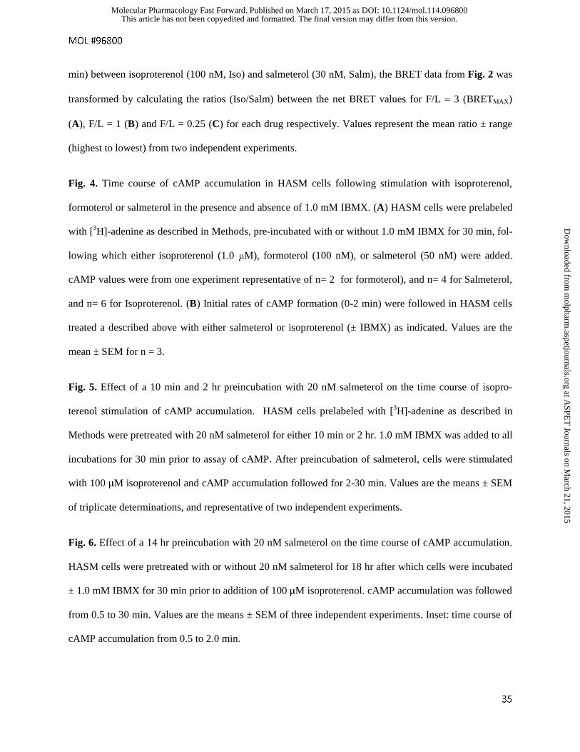
MOL #96800
35
min) between isoproterenol (100 nM, Iso) and salmeterol (30 nM, Salm), the BRET data from Fig. 2 was
transformed by calculating the ratios (Iso/Salm) between the net BRET values for F/L ≈ 3 (BRETMAX)
(A), F/L = 1 (B) and F/L = 0.25 (C) for each drug respectively. Values represent the mean ratio ± range
(highest to lowest) from two independent experiments.
Fig. 4. Time course of cAMP accumulation in HASM cells following stimulation with isoproterenol,
formoterol or salmeterol in the presence and absence of 1.0 mM IBMX. (A) HASM cells were prelabeled
with [3H]-adenine as described in Methods, pre-incubated with or without 1.0 mM IBMX for 30 min, fol-
lowing which either isoproterenol (1.0 μM), formoterol (100 nM), or salmeterol (50 nM) were added.
cAMP values were from one experiment representative of n= 2 for formoterol), and n= 4 for Salmeterol,
and n= 6 for Isoproterenol. (B) Initial rates of cAMP formation (0-2 min) were followed in HASM cells
treated a described above with either salmeterol or isoproterenol (± IBMX) as indicated. Values are the
mean ± SEM for n = 3.
Fig. 5. Effect of a 10 min and 2 hr preincubation with 20 nM salmeterol on the time course of isopro-
terenol stimulation of cAMP accumulation. HASM cells prelabeled with [3H]-adenine as described in
Methods were pretreated with 20 nM salmeterol for either 10 min or 2 hr. 1.0 mM IBMX was added to all
incubations for 30 min prior to assay of cAMP. After preincubation of salmeterol, cells were stimulated
with 100 μM isoproterenol and cAMP accumulation followed for 2-30 min. Values are the means ± SEM
of triplicate determinations, and representative of two independent experiments.
Fig. 6. Effect of a 14 hr preincubation with 20 nM salmeterol on the time course of cAMP accumulation.
HASM cells were pretreated with or without 20 nM salmeterol for 18 hr after which cells were incubated
± 1.0 mM IBMX for 30 min prior to addition of 100 μM isoproterenol. cAMP accumulation was followed
from 0.5 to 30 min. Values are the means ± SEM of three independent experiments. Inset: time course of
cAMP accumulation from 0.5 to 2.0 min.
This article has not been copyedited and formatted. The final version may differ from this version.Molecular Pharmacology Fast Forward. Published on March 17, 2015 as DOI: 10.1124/mol.114.096800
at ASPE
T Journals on M
arch 21, 2015m
olpharm.aspetjournals.org
Dow
nloaded from
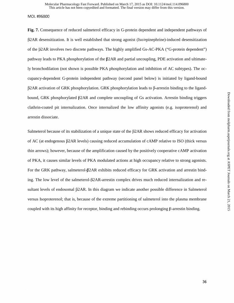
MOL #96800
36
Fig. 7. Consequence of reduced salmeterol efficacy in G-protein dependent and independent pathways of
β2AR desensitization. It is well established that strong agonist (Iso/epinephrine)-induced desensitization
of the β2AR involves two discrete pathways. The highly amplified Gs-AC-PKA (“G-protein dependent”)
pathway leads to PKA phosphorylation of the β2AR and partial uncoupling, PDE activation and ultimate-
ly bronchodilation (not shown is possible PKA phosphorylation and inhibition of AC subtypes). The oc-
cupancy-dependent G-protein independent pathway (second panel below) is initiated by ligand-bound
β2AR activation of GRK phosphorylation. GRK phosphorylation leads to β-arrestin binding to the ligand-
bound, GRK phosphorylated β2AR and complete uncoupling of Gs activation. Arrestin binding triggers
clathrin-coated pit internalization. Once internalized the low affinity agonists (e.g. isoproterenol) and
arrestin dissociate.
Salmeterol because of its stabilization of a unique state of the β2AR shows reduced efficacy for activation
of AC (at endogenous β2AR levels) causing reduced accumulation of cAMP relative to ISO (thick versus
thin arrows); however, because of the amplification caused by the positively cooperative cAMP activation
of PKA, it causes similar levels of PKA modulated actions at high occupancy relative to strong agonists.
For the GRK pathway, salmeterol-β2AR exhibits reduced efficacy for GRK activation and arrestin bind-
ing. The low level of the salmeterol-β2AR-arrestin complex drives much reduced internalization and re-
sultant levels of endosomal β2AR. In this diagram we indicate another possible difference in Salmeterol
versus Isoproterenol; that is, because of the extreme partitioning of salmeterol into the plasma membrane
coupled with its high affinity for receptor, binding and rebinding occurs prolonging β-arrestin binding.
This article has not been copyedited and formatted. The final version may differ from this version.Molecular Pharmacology Fast Forward. Published on March 17, 2015 as DOI: 10.1124/mol.114.096800
at ASPE
T Journals on M
arch 21, 2015m
olpharm.aspetjournals.org
Dow
nloaded from
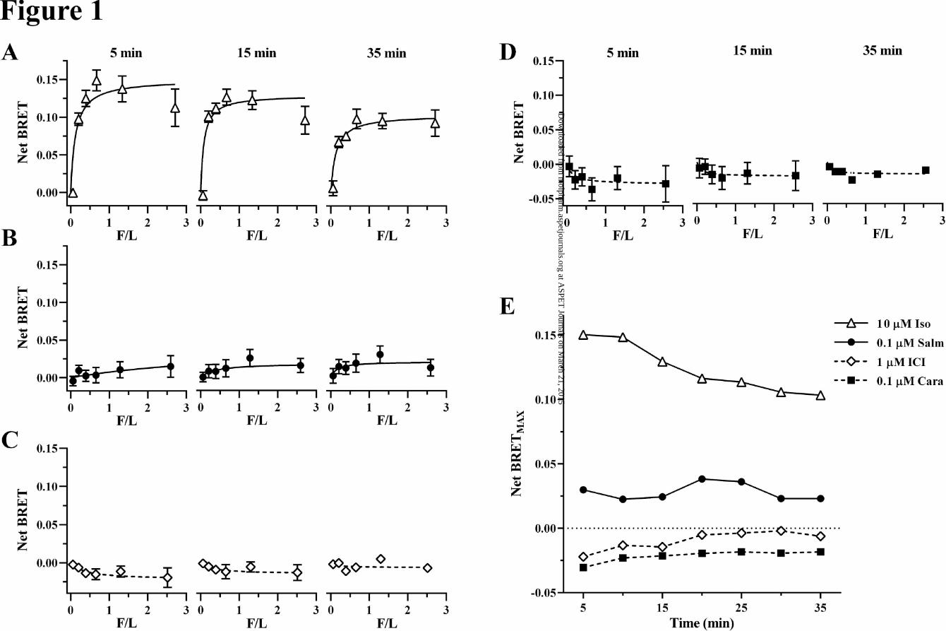
This article has not been copyedited and formatted. The final version may differ from this version.Molecular Pharmacology Fast Forward. Published on March 17, 2015 as DOI: 10.1124/mol.114.096800
at ASPE
T Journals on M
arch 21, 2015m
olpharm.aspetjournals.org
Dow
nloaded from
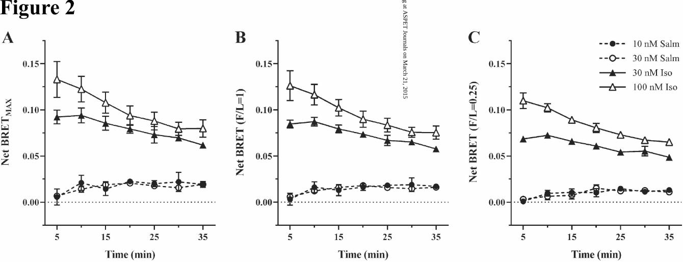
This article has not been copyedited and formatted. The final version may differ from this version.Molecular Pharmacology Fast Forward. Published on March 17, 2015 as DOI: 10.1124/mol.114.096800
at ASPE
T Journals on M
arch 21, 2015m
olpharm.aspetjournals.org
Dow
nloaded from
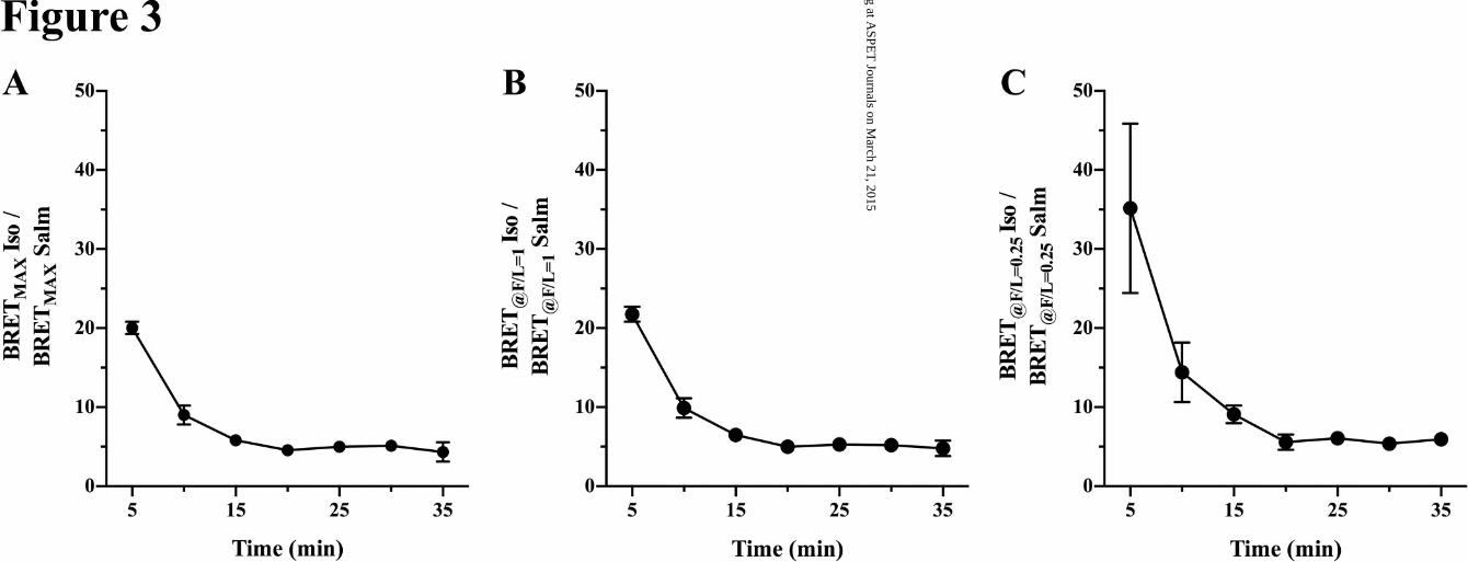
This article has not been copyedited and formatted. The final version may differ from this version.Molecular Pharmacology Fast Forward. Published on March 17, 2015 as DOI: 10.1124/mol.114.096800
at ASPE
T Journals on M
arch 21, 2015m
olpharm.aspetjournals.org
Dow
nloaded from
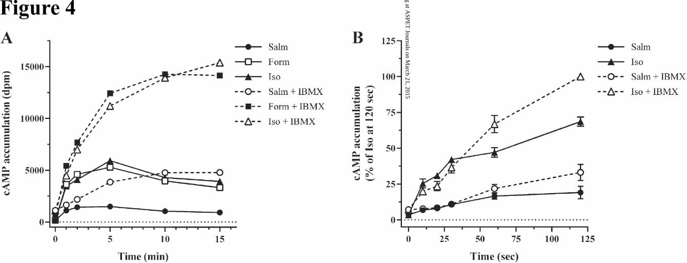
This article has not been copyedited and formatted. The final version may differ from this version.Molecular Pharmacology Fast Forward. Published on March 17, 2015 as DOI: 10.1124/mol.114.096800
at ASPE
T Journals on M
arch 21, 2015m
olpharm.aspetjournals.org
Dow
nloaded from
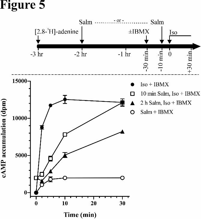
This article has not been copyedited and formatted. The final version may differ from this version.Molecular Pharmacology Fast Forward. Published on March 17, 2015 as DOI: 10.1124/mol.114.096800
at ASPE
T Journals on M
arch 21, 2015m
olpharm.aspetjournals.org
Dow
nloaded from
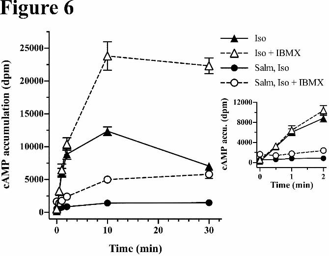
This article has not been copyedited and formatted. The final version may differ from this version.Molecular Pharmacology Fast Forward. Published on March 17, 2015 as DOI: 10.1124/mol.114.096800
at ASPE
T Journals on M
arch 21, 2015m
olpharm.aspetjournals.org
Dow
nloaded from
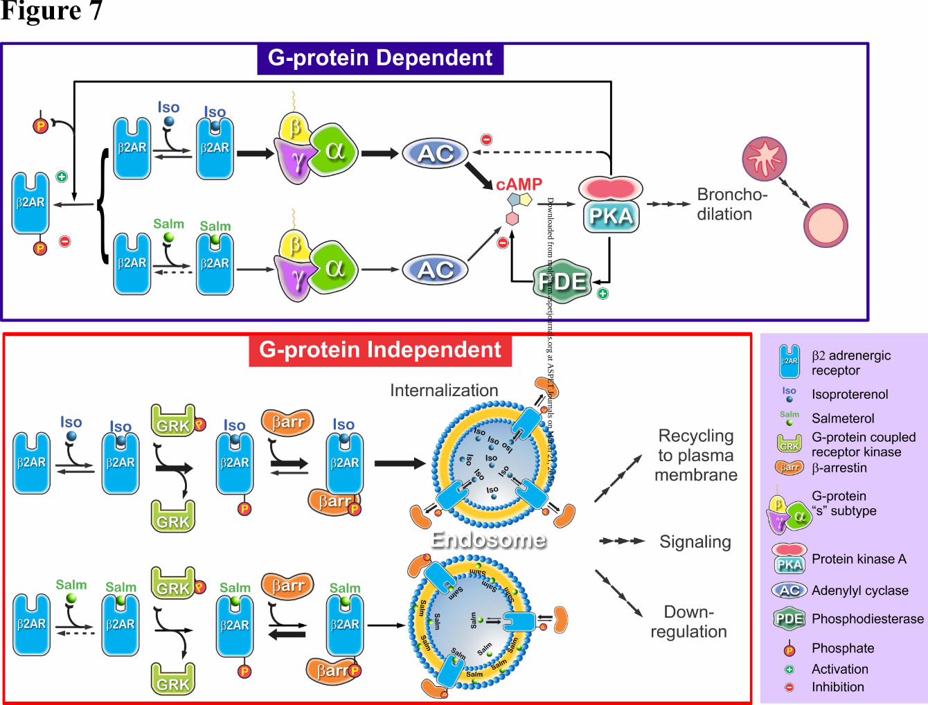
This article has not been copyedited and formatted. The final version may differ from this version.Molecular Pharmacology Fast Forward. Published on March 17, 2015 as DOI: 10.1124/mol.114.096800
at ASPE
T Journals on M
arch 21, 2015m
olpharm.aspetjournals.org
Dow
nloaded from