Embed Size (px)
Citation preview
BIOCHEMICAL AND BIOPHYSICAL RESEARCH COMMUNICATIONS 235, 407–412 (1997)ARTICLE NO. RC976798
Molecular Cloning of a 1a,25-DihydroxyvitaminD3-Inducible Transcript (DDVit 1)1 in HumanBlood Monocytes
Jana Fritsche, Michael Rehli, Stefan W. Krause, Reinhard Andreesen, and Marina Kreutz2
Department of Hematology and Oncology, University of Regensburg, 93042 Regensburg, Germany
Received May 13, 1997
lymphocytes it decreases the production of immuno-The differentiation and activation of monocytes globulins (18) and in T-lymphocytes the production of
(MO) and monocytic cells is modulated by 1a,25-dihy- interleukin-2 and GM-CSF is inhibited after incubationdroxyvitamin D3 (Vitamin D3). In order to investigate with Vitamin D3 (24, 23). Vitamin D3 also modulatesearly effects on the differentiation process of MO, we the differentiation process of various cell types like theused the mRNA Differential Display technology to monocytic cell lines HL-60 and U937 (4, 1), humanidentify genes that are induced in freshly isolated hu-
blood MO (17) and normal or leukemic bone marrowman blood MO cultured for 4 hours with Vitamin D3.progenitors (14). Most of the Vitamin D3 effects areA cDNA fragment was isolated and Northern analysismediated by the Vitamin D receptor. This receptor is aconfirmed a low expression of this cDNA at about 1,4member of the superfamily of nuclear steroid-, thyroid-kb in MO which was increased by the addition of Vita-and retinoic acid-receptors, acting as transcription fac-min D3. Using the rapid amplification of cDNA Endstors (3, 25). The Vitamin D receptor binds either as a(RACE)-PCR we got a transcript (DDVit 1) of a lengthhomodimer or as a heterodimer with thyroid or retinoicof 1251 bp containing an open reading frame that en-acid receptors to response elements in target genes (7).codes a putative 16,5 kD protein. Database search re-The distribution of the Vitamin D receptor is ubiqui-vealed an identity with a possible enterocyte differen-
tiation promoting factor with a length of 1177 bp that tous, e.g. in MO, activated T-lymphocytes and fibro-has not been further characterized. Therefore DDVit blasts (21, 26, 5). Until now many genes and gene prod-1 may be a differentiation promoting factor for the ucts are known to be induced by Vitamin D3 in severalmonocytic lineage. Further investigations will clarify target tissues and cell lines, e.g. murine osteopontinthe role of this protein in the differentiation process and human osteocalcin (20, 13). In MO/macrophagesof MO. q 1997 Academic Press Vitamin D3 induces the expression of CD 14, a mono-
cytic differentiation marker, and M-CSF, a survivalfactor of MO (27, 6, 12). However, most of the regula-tory effects were investigated after a long-term incuba-
The active form of the fat-soluble 1a,25-dihydroxyvi- tion with Vitamin D3 suggesting intermediate genetamin D3 (Vitamin D3) exerts its effects not only in products involved in the regulation. In this report wemineral homeostasis and bone resorption, but also describe the isolation and characterization of a mRNAplays an important role as an immunoregulatory hor- transcript (DDVit 1) induced by short-term incubationmone. Vitamin D3 influences the activity of several with Vitamin D3 in human blood MO.types of hematopoetic cells. In MO Vitamin D3 inducessecretion of prostaglandine E2 and H2O2 (15, 10), in B- MATERIALS AND METHODS
Chemicals. All chemical reagents used were purchased from1 GenBank Accession No. is X98091. Sigma (Deisenhofen, Germany) unless otherwise noted. 1a,25-Dihy-2 To whom correspondence and reprint requests should be ad- droxyvitamin D3 (Vitamin D3) was kindly provided by Hoffmann-dressed at the University of Regensburg, Department of Hematology LaRoche, Basel, Switzerland.and Oncology, 93042 Regensburg, Germany. Fax: **49-941-944-7131. E-mail: [email protected]. Cell separation and culture. Peripheral human blood mononu-
clear cells were isolated by leukapheresis of healthy donors followedAbbreviations used: MO, monocytes; RACE-PCR, rapid amplifica-tion of cDNA ends–PCR; Vitamin D3, 1a,25-dihydroxyvitamin D3; by density gradient centrifugation over Ficoll/Hypaque. Monocytes
(MO) were obtained by countercurrent centrifugation in a J6M-EPMA, phorbol 12-myristate 13-acetate.
0006-291X/97 $25.00Copyright q 1997 by Academic PressAll rights of reproduction in any form reserved.
407
AID BBRC 6798 / 692f$$$561 05-30-97 11:24:22 bbrcgs AP: BBRC

Vol. 235, No. 2, 1997 BIOCHEMICAL AND BIOPHYSICAL RESEARCH COMMUNICATIONS
centrifuge (Beckmann, Munchen, Germany), as described previously Westborough, MA) and UV cross-linked. The cDNA fragment ofDDVit 1 obtained by Differential Display was labeled by random(2). MO were ú 90% pure as determined by morphology and expres-
sion of CD 14 antigen. After isolation MO were cultured at a density priming method (Boehringer, Mannheim, Germany). Hybridizationwas performed over night at 657C (16, 9). In control experimentsof 106 cells/ml in petri dishes with RPMI-medium (Biochrom, Berlin,
Germany) supplemented with mercaptoethanol, antibiotics, pyr- membranes were rehybridized with an 18S rRNA-oligonucleotide ora Glycerinealdehyde-3-phosphatedehydrogenase (GAPDH)-oligonu-uvate, non-essential amino acids, vitamins and glutamin (Gibco BRL,
Eggenstein, Germany). cleotide labeled by T4 Kinase (5*-end labeling kit, Amersham, Buck-inghamshire, UK).All human cell lines were cultured in RPMI-medium supplemented
with the additives described above and 10% fetal calf serum (c.c.pro,mRNA differential display technology (20). This method was per-Karlsruhe, Germany). Mono/Mac 6 (28) was grown in RPMI medium
formed using the RNAmap-system (GenHunter, Nashville, USA). To-containing the same additives, 15% fetal calf serum and OPI mediatal RNA (30mg) of MO cultured with or without 1007 M Vitamin D3supplement (Sigma, Deisenhofen, Germany).was digested with DNAse I (Boehringer, Mannheim, Germany) as
RNA preparation. Total RNA was isolated from MO cultured described previously (22). 0,3mg RNA was reverse transcribed withwith or without 1007 M Vitamin D3 or from cell lines or tissues by the two base-anchored oligo-dT primer T12MC and amplified by poly-the method of Chomczynski and Sacchi (8). The monocytic cell lines, merase chain reaction using AmpliTaq (Perkin Elmer, Weiterstadt,U937 and Mono/Mac 6, and the colon carcinoma cell line Caco-2 were Germany) and the primer combination T12MC and AP2 (5*-GACCGC-cultured for 3 days with or without 1007 M Vitamin D3 or 1008 M TTGT-3 *) in the presence of (33P)deoxy cytidine triphosphate. ThePMA before isolation of total RNA. amplified cDNA was separated on a 6% polyacrylamide gel. After
autoradiography bands of interest were excised and the cDNA frag-Poly(A)/ RNA isolation. Cells were lysed and incubated withments were isolated, amplified, cloned by inserting into the EcoRImagnetic oligo-dT beads (mRNA Direct kit, Dynal AS, Oslo, Norway).site of the plasmid vector pCR 2.1 (TA cloning kit, Invitrogen, NVAfter binding the beads were washed three times and poly(A)/RNALeek, Netherlands) and sequenced.was eluted with water at 657C.
Northern blot analysis. Total RNA (10mg/lane) or poly(A)/RNA 3 *- and 5*-rapid amplification of cDNA ends (RACE)-PCR. 3 *-and 5*-RACE-PCR was performed using the Marathon cDNA Ampli-(3mg/lane) was electrophoretically separated on an 1% agarose form-
aldehyde gel, transferred to nylon membranes (Magna NT, MSI, fication kit (Clontech, Palo Alto, MA) according to the manufacturers
FIG. 1. Expression of DDVit 1 in human blood MO. MO were cultured without any additive (0) or with 1007 M vitamin D3 (VD3) for 4hours. Poly(A)/RNA was isolated and Northern blot analysis was performed according to material and methods. As a RNA loading controlthe membrane was rehybridized with an GAPDH-oligonucleotide (1A) and normalized signals are shown in Figure 1B.
408
AID BBRC 6798 / 692f$$$561 05-30-97 11:24:22 bbrcgs AP: BBRC
Vol. 235, No. 2, 1997 BIOCHEMICAL AND BIOPHYSICAL RESEARCH COMMUNICATIONS
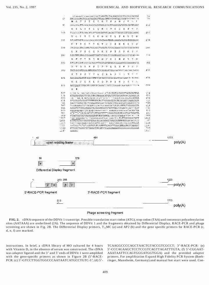
FIG. 2. cDNA sequence of the DDVit 1 transcript. Possible translation start codon (ATG), stop codon (TAA) and consensus polyadenylationsites (AATAAA) are underlined (2A). The sequence of DDVit 1 and the fragments obtained by Differential Display, RACE-PCR and phagescreening are shown in Fig. 2B. The Differential Display primers, T12MC (a) and AP2 (b) and the gene specific primers for RACE-PCR (c,d, e, f) are marked.
instructions. In brief, a cDNA library of MO cultured for 4 hours TCAAGGCCCCAGCTAACTGTACCGTCGCCT; 3*-RACE-PCR: (e)5*-CCCAGAAGCTCCTCCGTCAGTTAGATTTGTA; (f) 5*-CGGAAT-with Vitamin D3 in the absence of serum was constructed. The cDNA
was adapter ligated and the 3* and 5* ends of DDVit 1 were amplified AAATAATTCCAGTGGGATGGTGGA) and the provided adapterprimers. For amplification Expand High Fidelity PCR System (Boeh-with the gene-specific primers as shown in Figure 2B (5*-RACE-
PCR: (c) 5*-GTCCTTGGTGGCCCAATAATCATGCCTGTC-3 *; (d) 5*- ringer, Mannheim, Germany) and manual hot start were used. Con-
409
AID BBRC 6798 / 692f$$$561 05-30-97 11:24:22 bbrcgs AP: BBRC

Vol. 235, No. 2, 1997 BIOCHEMICAL AND BIOPHYSICAL RESEARCH COMMUNICATIONS
FIG. 3. Expression of DDVit 1 in human tissues (A), cells and cell lines (B). RNA of human tissues, cells, freshly isolated MO, in vitroderived macrophages and lymphocytes, and cell lines was isolated and Northern blot analysis was performed as described in material andmethods. As a RNA loading control membranes were rehybridized with an 18S rRNA-oligonucleotide.
ditions were 947C 2 min, 727C 3 min, adding the enzyme, 947C 30 ential Display cDNA fragment revealed a weak signalsec, 657C 30 sec, 727C 2 min for 10 cycles and 947C 30 sec, 657C 30 at about 1,4 kb in MO cultured for 4 hours withoutsec, 727C 2 min (with an extension step of 20 sec for each cycle) for Vitamin D3, but the addition of Vitamin D3 led to a20 cycles.
reproducible increase of this mRNA. For reduction ofScreening of the cDNA library. Plaque screening was performed the exposure time we decided to use poly(A)/RNA in-as previously described (22). In brief, 51105 plaque forming units of a
stead of total RNA. The results are shown in Fig. 1.macrophage derived cDNA library were plated. Phage DNA was trans-ferred to NitroPlus membranes (MSI). Filters were hybridized with a By the method of 3 *-and 5*-rapid amplification of32P-labeled cDNA probe of DDVit 1 cDNA and positive plaques was cDNA ends (RACE)-PCR we synthesized the full lengthsubjected to a second round of screening. Lambda ZAP II phages were sequence of the mRNA transcript (DDVit 1) andconverted into pBluescript SK plasmid vector by in vivo excision ac- thereby obtained a combined sequence of 1251 bp con-cording to the manufacturers instructions (Stratagene, LaJolla; CA).
taining an putative open reading frame of 438 bp and aSequence analysis. The cDNA sequencing was done by Dye Deoxy possible polyadenylation site (AATAAA) 17 nucleotidesTerminator Cycle Sequencing (Applied Biosystems, Weiterstadt,
upstream of the poly(A)/tail (Fig. 2A). As DDVit 1Germany) according to the manufacturer’s instructions and se-quences were analyzed on the Applied Biosystems DNA Sequencing mRNA was also expressed in macrophages (Fig. 3B)System (model 373 A). we screened a cDNA macrophage library (MO cultured
for 7 days in the presence of serum) with the Differen-tial Display cDNA fragment of DDVit 1 to confirm theRESULTS AND DISCUSSIONsequence. The obtained clones were in parts (position/213 to /1251) identical with the DDVit 1 nucleotideIn order to identify 1a,25-dihydroxyvitamin D3 (Vita-sequence (Fig. 2B).min D3) responsive genes, we cultured freshly isolated
In further investigations we analyzed the distribu-human blood monocytes (MO) with or without 1007 Mtion of DDVit 1 in various human tissues, cells and cellVitamin D3 for 4 hours in the absence of serum. Usinglines by Northern blot analysis. DDVit 1 was detectedthe mRNA Differential Display technology we detectedin all tissues (Fig. 3A) and cells (Fig. 3B) examined,a cDNA fragment in MO cultured with Vitamin D3
but a strong expression of DDVit 1 was found in skin(data not shown). The cDNA fragment was excised, re-and colon which are typical target tissues of Vitaminamplified, cloned into a plasmid vector and sequenced.
Northern blot analsis with the cloned 290 bp Differ- D3, whereas in placenta and liver only a weak signal
FIG. 4. Hydrophobicity profile of DDVit 1 obtained by the method of Engelman, Steitz and Goldman. Theoretical calculation of theamino acid sequences of DDVit 1 revealed no hydrophobic regions (values õ 0).
410
AID BBRC 6798 / 692f$$$561 05-30-97 11:24:22 bbrcgs AP: BBRC

Vol. 235, No. 2, 1997 BIOCHEMICAL AND BIOPHYSICAL RESEARCH COMMUNICATIONS
was observed. Furthermore DDVit 1 was strongly ex-pressed in Daudi, a B-lymphoma cell line, and in K562,a chronic myloid cell line. A weak signal of DDVit 1was found in lymphocytes, in vitro derived macro-phages and in freshly isolated monocytes.
It is known that Vitamin D3 induces differentiationin many cell types especially in monocytic cell lines.Therefore we cultured different monocytic cell lines,HL-60, U937, THP-1 and Mono/Mac6 for 3 days eitherwith or without 1007 M Vitamin D3 in the presence of15% fetal calf serum. The expression of DDVit 1 wasanalyzed by Northern blot analysis. In the monocyticcell lines a strong constitutive expression of DDVit 1was found that was only in U937 and Mono/Mac 6slightly increased by culture with Vitamin D3 (data notshown).
In order to clarify whether DDVit 1 is a cytosolicprotein or a protein anchored in the membrane of thecells we investigated the putative amino acid sequenceof DDVit 1. We analyzed the longest open reading FIG. 5. Expression of DDVit 1 in the colon carcinoma cell lineframe of the sequence of DDVit 1 which encodes a pro- Caco-2. RNA of unstimulated cells (0) or of cells stimulated with
1007 M Vitamin D3 (VD3) or 1008 M PMA was extracted and Northerntein with a length of 146 amino acids. Theoretical struc-blot analysis was performed as described in material and methods.ture analysis of these putative protein by the methodAs a RNA loading control membranes were rehybridized with an 18Sof Engelman, Steitz and Goldman (11) revealed no hy- rRNA-oligonucleotide.
drophobic regions. This indicates that DDVit 1 may bea soluble protein in the cytoplasm or nucleus of MO(Fig. 4).
ACKNOWLEDGMENTSAn analysis of consensus sequences resulted in threeprotein kinase C phosphorylation sites at amino acids The authors thank Silvia Seegers for performing the cDNA se-67, 82 and 116, one casein kinase II phosphorylation quencing. The work was supported by DFG.site at amino acid 59, one tyrosine kinase phosphoryla-tion site at amino acid 58 and in two N-myristoylation REFERENCESsites at amino acids 28 and 92.
1. Amento, E. P., Bhalla, A. K., Kurnick, J. T., Kradin, R. L., Clem-Database search revealed an identity of the nucleo-ens, T. L., Holick, S. A., Holick, M. F., and Krane, S. M. (1984)tide sequence of DDVit 1 with a putative enterocyteJ. Clin. Invest. 73, 731–739.differentiation promoting factor (EMBL/GenBank/
2. Andreesen, R., Brugger, W., Scheibenbogen, C., Kreutz, M.,DDBJ databases; accession number: U62136), but no Leser, H.-G., Rehm, A., and Lohr, G. W. (1990) J. Leuk. Biol. 47,further information can be obtained. As this factor was 490–497.isolated in the colon carcinoma cell line Caco-2, we in- 3. Baker, A. R., McDonnell, D. P., Hughes, M., Crisp, T. M.,
Mangelsdorf, D. J., Haussler, M. R., Pike, J. W., Shine, J., andvestigated the expression of DDVit 1 in this cell line.O’Malley, B. W. (1988) Proc. Natl. Acad. Sci. USA 85, 3294–In unstimulated cells only a weak expression of DDVit3298.1 was found, but culture with Vitamin D3 for 3 days
4. Bar-Shavit, Z., Teitelbaum, S. T., Reitsma, P., Hall, A., Pegg,up-regulated the expression of DDVit 1. Another differ- L. E., Trial, J., and Kahn, A. J. (1983) Proc. Natl. Acad. Sci. USAentiation inducing agent, PMA; had the same effect 80, 5907–5911.(Fig. 5). In addition, a strong homology to two proteins 5. Barsony, J., and Marx, S. J. (1991) Proc. Natl. Acad. Sci. USA
88, 1436–1440.(86% and 84% in 146 amino acids) with DNA-binding6. Brugger, W., Kreutz, M., and Andreesen, R. (1991) J. Leuk. Biol.properties was found (EMBL/GenBank/DDBJ data-
49, 483–488.bases; accession numbers: U39360 and U39361).7. Carlberg, C., Bendik, I., Wyss, A., Meier, E., Sturzenbecker, L. J.,In conclusion, we isolated a Vitamin D inducible
Grippo, J. F., and Hunziker, W. (1993) Nature 361, 657–660.mRNA transcript (DDVit 1) in MO. Regarding the Gen-8. Chomczynski, P., and Sacchi, N. (1987) Anal. Biochem. 162, 156–Bank data and the analysis of the amino acid sequence 159.
of the putative protein, we suggest a possible role of 9. Church, G. M., and Gilbert, W. (1984) Proc. Natl. Acad. Sci. USADDVit 1 in intracellular signal transduction of the Vi- 81, 1991–1994.tamin D3 induced differentiation process of MO and 10. Cohen, M. S., Mesler, D. E., Snipes, R. G., and Gray, T. K. (1986)
J. Immunol. 136, 1049–1053.enterocytes.
411
AID BBRC 6798 / 692f$$$561 05-30-97 11:24:22 bbrcgs AP: BBRC

Vol. 235, No. 2, 1997 BIOCHEMICAL AND BIOPHYSICAL RESEARCH COMMUNICATIONS
11. Engelman, D. M., Steitz, T. A., and Goldman, A. (1986) Ann. Rev. and Denhardt, D. T. (1990) Proc. Natl. Acad. Sci. USA 87, 9995–9999.Biophys. Biophys. Chem. 15, 321–353.
21. Provvedini, D. M., Tsoukas, C. D., Deftos, L. J., and Manolagas,12. Kaneki, M., Inoue, S., Hosoi, T., Mizuno, Y., Akedo, Y., Ikegami,S. C. (1983) Science 221, 1181–1182.A., Nakamura, T., Shiraki, M., Ito, H., Suzu, S., Motoyoshi, K.,
Ouchi, Y., and Orimo, H. (1994) Blood 83, 2285–2293. 22. Rehli, M., Krause, S. W., Schwarzfischer, L., Kreutz, M., andAndreesen, R. (1995) Biochem. Biophys. Res. Commun. 217,13. Kerner, S. A., Scott, R. A., and Pike, J. W. (1989) Proc. Natl.661–667.Acad. Sci. USA 86, 4455–4459.
23. Tobler, A., Gasson, J., Reichel, H., Norman, A. W., and Koeffler14. Koeffler, H. P., Amatruda, T., Ikekawa, N., Kobayashi, Y., andH. P. (1987) J. Clin. Invest. 79, 1700–1705.DeLuca, H. F. (1984) Cancer Res. 44, 5624–5628.
24. Tsoukas, C. D., Provvedini, D. M., and Manolagas, S. C. (1984)15. Koren, R., Ravid, A., Rotem, C., Shohami, E., Libermann, U. A.,Science 224, 1438–1440.and Novogrodsky, A. (1986) FEBS 205, 113–116.
25. Umesono, K., Murakami, K. K., Thompson, C. C., and Evans,16. Krause, S. W., Rehli, M., Kreutz, M., Schwarzfischer, L., Pau-R. M. (1991) Cell 65, 1255–1266.lauskis, J., D., and Andreesen, R. (1996) J. Leuk. Biol. 60, 540–
26. Yu, X.-P., Mocharla, H., Hustmyer, F. G., and Manolagas, S. C.545.(1990) J. Biol. Chem. 266, 7588–7595.
17. Kreutz, M., and Andreesen, R. (1990) Blood 76, 2457–2461.27. Zhang, D.-E., Hetherington, C. J., Gonzales, D. A., Chen, H.-M.,
18. Lemire, J. M., Adams, J. S., Sakei, R., and Jordan, S. C. (1984) and Tenen, D. G. (1994) J. Immunol. 153, 3276–3284.J. Clin. Invest. 74, 657–665.
28. Ziegler-Heitbrock, H. W., Thiel, E., Futterer, A., Herzog, V.,19. Liang, P., and Pardee, A. B. (1992) Science 257, 967–971. Wirtz, A., and Riethmuller, G. (1988) Int. J. Cancer 41, 456–
461.20. Noda, M., Vogel, R. L., Craig, A. M., Prahl, J., DeLuca, H. F.,
412
AID BBRC 6798 / 692f$$$561 05-30-97 11:24:22 bbrcgs AP: BBRC