Embed Size (px)
Citation preview
0
Regular Article, Biochim. Biophys. Acta
Molecular mechanisms of action of different concentrations of ethanol in water on
ordered stuctures of intercellular lipids and soft keratin in the stratum corneum
Daisuke Horita,a, d Ichiro Hatta,c, e Masato Yoshimoto,a Yuki Kitao,a Hiroaki Todo,a and Kenji
Sugibayashi*, a, b
aFaculty of Pharmaceutical Sciences and bLife Science Research Center, Josai University, 1-1
Keyakidai, Sakado, Saitama 350-0295, Japan; cDepartment of Research, Nagoya Industrial
Science Research Institute, 1-13 Yotsuyadori, Chikusa-ku, Nagoya 464-0819, Japan;
dResearch Laboratories, Ikeda Mohando Co., Ltd., 16 Jinden, Kamiichi, Nakaniikawa,
Toyama 930-0394, Japan; and eAichi Synchrotron Radiation Center, Aichi Science &
Technology Foundation, 250-3 Minamiyamaguchi-cho, Seto, Aichi 489-0965, Japan
*To whom correspondence should be addressed. e-mail: [email protected]
1
Abstract
Ethanol (EtOH) is one of the bases in topically applied medicines that promotes the
skin permeation of drugs. Although the effects of EtOH have been attributed to structural
modifications in the stratum corneum, the underlying mechanisms, especially the influence of
different concentrations of EtOH, have not been examined extensively. Structural
modifications in the stratum corneum of hairless mouse due to the application of EtOH/water
mixture were herein investigated at the molecular level using synchrotron X-ray diffraction.
The results revealed that all EtOH concentrations examined greatly modified the short
lamellar structures containing the aqueous layer in intercellular lipids and the structure of
keratin fibrils in corneocytes, which can take up hydrophilic compounds. However, the long
lamellar and the hydrocarbon-chain packing structures were unaffected by EtOH. Changes to
the short lamellar structures were not proportional to the concentration of EtOH. However,
the keratin fibril structures changed gradually with increasing EtOH concentration. The X-ray
diffraction experiments enabled the effects of different EtOH concentrations on the
morphology of the stratum corneum to be assessed by using a number of experimental
samples to avoid variations due to individual differences. The results indicated that alterations
to the short lamellar structures appeared to be related to the skin permeability of drugs with
the application of EtOH/water mixture, and monotonous structural changes in the keratin
fibrils with an increase in EtOH concentration may contribute to this permeation as
2
supplement. These results will be useful for the development of new drug formulations
containing EtOH.
Key words ethanol; stratum corneum; intercellular lipids; corneocyte; X-ray diffraction;
hairless mouse
3
1. Introduction
Ethanol (EtOH) is widely used as a skin penetration enhancer as well as a skin
disinfectant and tonic. In order for drugs to be effective at their target sites, the active
ingredients in topical formulations must penetrate the skin barrier, which tightly regulates the
entry of external substances. This skin barrier is mainly comprised of the stratum corneum,
the outermost layer of skin, which markedly restricts the penetration of drugs. EtOH has been
shown to enhance the skin permeation of drugs in topical formulations markedly. Estradiol
and fentanyl dermal patches are typical examples of formulations that contain EtOH in order
to enhance their absorption into the skin [1‒3]. The enhancements reported in the skin
permeation of drugs with the application of topical formulations containing EtOH have been
attributed to structural modifications in the stratum corneum. The various mechanisms
underlying the penetration-enhancing effects of EtOH on the stratum corneum barrier include
lipid extraction, increase in lipid fluidity, enhancement of drug solubility in the lipids of the
stratum corneum, change in skin hydration, an altered putative pore pathway, alteration in
keratinized proteins, and its effects on solvent drugs [4,5]. However, the effects of EtOH on
the structures of soft keratin and intercellular lipids in the stratum corneum have not been
extensively examined at the molecular level.
In the present study, we thus utilized synchrotron X-ray diffraction to examine the
structure of the stratum corneum at the molecular level as well as the effects of EtOH/water
4
mixture on the structures of intercellular lipids and fibrils in the soft keratin. We previously
demonstrated that EtOH/water mixture affected the in vitro permeability of drugs through pig
skin in an EtOH concentration-dependent manner [6]. The skin permeability of hydrophilic
drugs was increased but conversely decreased by low and high concentrations of EtOH,
respectively, suggesting that the effects of EtOH/water mixture on the structure of the stratum
corneum may depend on its volume ratio. In the present study, we measured X-ray diffraction
in the stratum corneum of mouse skin as a function of EtOH concentration. Previous studies
have already used X-ray diffraction in hairless mouse skin [7,8], hairless rat skin [9], and pig
skin [10,11]. Although there are slight differences in the constituents of intercellular lipids
among mammalian species, the lamellae have almost the same periodical structure among the
mammals. As for the skin permeation of chemical compounds, the skin of rodents, such as rat
and mouse, has higher permeability than pig or human skin, whereas the structures of the
stratum corneum are similar among these species. Among these mammals, the X-ray
diffraction profiles of the stratum corneum in hairless mice showed the most distinct
diffraction peaks, and represented the typical characteristics of the stratum corneum in
mammals. Therefore, we employed hairless mouse skin in the present study to detect minute
changes in the structure of the stratum corneum upon treatment with EtOH/water mixture.
The diffraction profile of the stratum corneum has also been investigated in humans [12,13],
and has been found to have similar characteristics to that in hairless mice.
5
In this study, we investigated the molecular mechanisms of action of various volume
ratios in EtOH/water mixture on intercellular lipids and soft keratin based on a structural
analysis using synchrotron X-ray diffraction experiments in stratum corneum treated with the
mixture.
2. Materials and methods
2.1. Materials
EtOH (99.5%, HPLC grade), sodium chloride, disodium hydrogen phosphate, and
potassium dihydrogen phosphate were obtained from Wako Pure Chemical Ind., Ltd. (Osaka,
Japan). Trypsin and trypsin inhibitor were obtained from Sigma-Aldrich Co., Ltd. (St. Louis,
MO, U.S.A.). All other reagents and solvents were of reagent grade or HPLC grade, and used
without further purification.
2.2 Animals
Male hairless mice (HR-1, 8‒9 weeks old) were obtained from Saitama Experimental
Animals Supply Co., Ltd. (Sugito, Saitama, Japan). All animal studies were conducted
according to the recommendations of the Institutional Board for Animal Studies, Josai
University (Sakado, Saitama, Japan).
2.3. Sample preparation process
Hairless mouse skin was separated from the abdominal and dorsal region. After the
6
removal of excess fat, the skin was soaked with 0.1% (w/v) trypsin in pH 7.4
phosphate-buffered saline (PBS) at 4˚C for 16 h. After being incubated for another 4 h at
37˚C, the stratum corneum was separated from the skin. It was then treated with 0.1% (w/v)
trypsin inhibitor and rinsed in distilled water three times. Following this, EtOH/water mixture
of 0/100, 20/80, 40/60, 60/40, 80/20 or 100/0 v/v% was applied to the dried stratum corneum
for 2 h. By reducing the content of EtOH/water mixture from stratum corneum treated by the
mixture the EtOH/water mixture content in the stratum corneum was adjusted to be 20 wt%.
An approximately 5-mg piece of the sample was placed into a capillary glass tube and
immediately sealed for the X-ray diffraction experiment. We used 25 mice in the experiment
and obtained four samples per mouse. In addition, 13‒14 samples were used for each
experiment; the samples treated by EtOH/water mixture were randomly selected in order to
avoid excessive use of a specific region or specific individual.
2.4. X-ray diffraction study
The X-ray diffraction study was performed at the beamline BL40B2 (Structural
Biology II Beamline) of SPring-8 (Harima, Hyogo, Japan). X-ray diffraction profiles were
recorded using an imaging plate system (R-AXIS IV; Rigaku Corporation, Tokyo, Japan)
with a 30 cm × 30 cm area. The X-ray wavelength was 0.0709 nm and sample-to-detector
distance was approximately 500 mm. Reciprocal spacing [S = (2 / λ) × sin θ] was calibrated
from the lattice spacing (d = 5.838 nm, where d is the lamellar repeat distance) of silver
7
behenate at room temperature, where 2θ is the scattering angle. The exposure time was 15 s.
The diffraction pattern was circularly averaged in order to obtain the radial intensity profile.
The profile was obtained from samples with almost the same weight of stratum corneum.
Photon counting for the profile was performed at 1152 pixels in the range of S = 0.05‒3.0
nm-1, and we counted the photon number at each pixel.
3. Results
3.1. Effects of various volume ratios in EtOH/water mixture on the ordered structure of
intercellular lipids
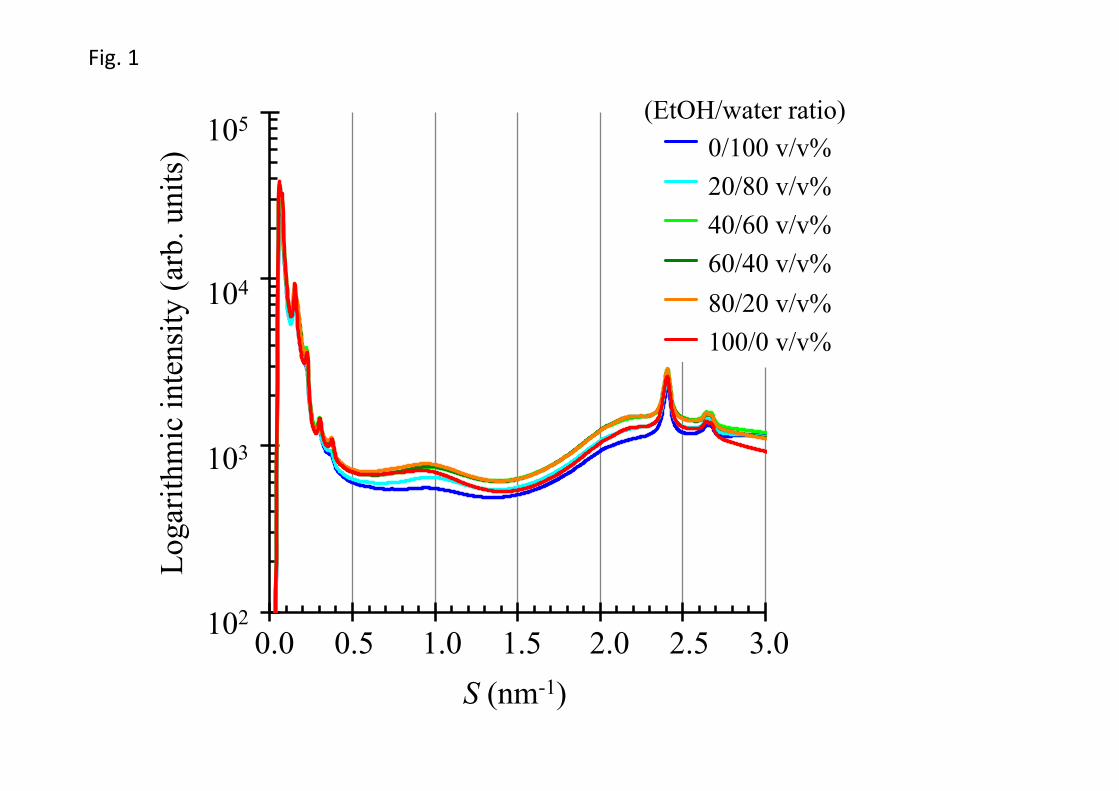
Figure 1 shows the X-ray diffraction profiles (S = 0.05‒3.0 nm-1) of the stratum
corneum of hairless mice following treatments with various volume ratios in EtOH/water
mixture. We determined the X-ray diffraction pattern from approximately one dozen stratum
corneum samples with almost the same volume after the application of EtOH/water mixture at
each volume ratio. Diffraction peaks were due to the repeat distances of the lamellar
structures in the small-angle region as well as the lattice constants of the lateral
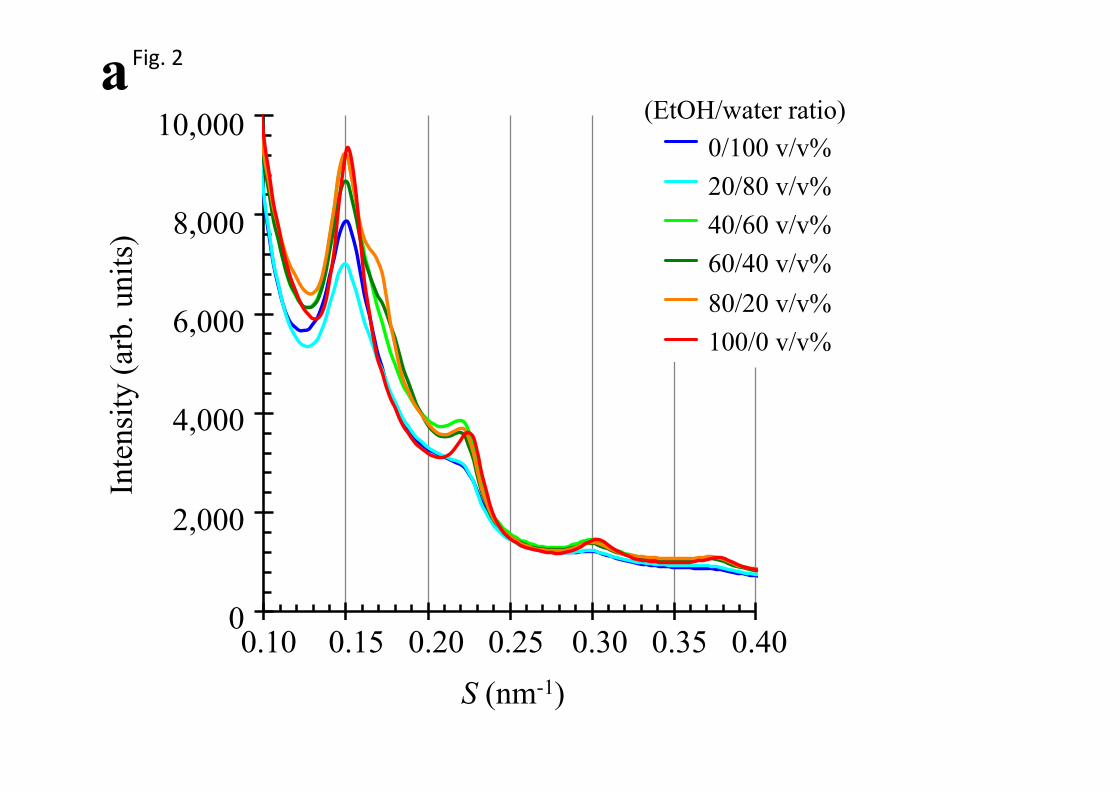
hydrocarbon-chain packing structures in the wide-angle region. Figure 2a shows the X-ray
diffraction profiles in the small-angle region within the range of S = 0.10‒0.40 nm-1. Three
partially overlapping peaks derived from the short and long lamellar structures were detected
within the range of S = 0.12‒0.25 nm-1, and these profiles changed according to the volume
8
ratio. In addition, the single diffraction peaks derived from the long and short lamellar
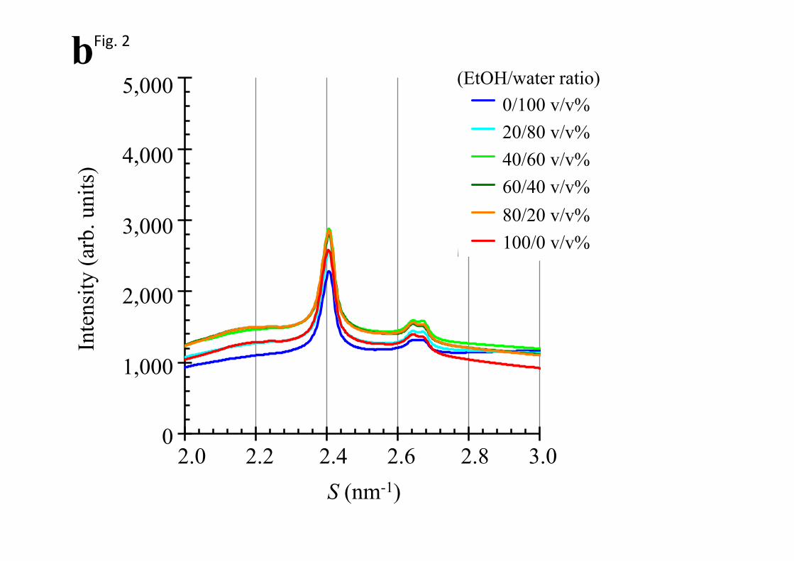
structures were observed near S = 0.30 and S = 0.37, respectively. Figure 2b shows the X-ray
diffraction profiles in the wide-angle region within the range of S = 2.0‒3.0 nm-1. Two
diffraction peaks near S = 2.4 nm-1 and S = 2.7 nm-1 corresponded to the hexagonal and
orthorhombic hydrocarbon-chain packing structures and only the orthorhombic
hydrocarbon-chain packing structures, respectively. In this experiment, the stratum corneum
sample from hairless mice showed split peaks for the orthorhombic hydrocarbon-chain
packing structures near S = 2.7 nm-1.
Figs. 1 and 2
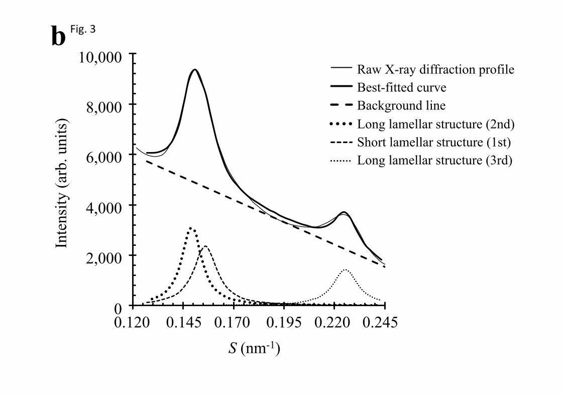
3.2. Analysis of multiple diffraction peaks in the small-angle region
Multiple partially overlapping peaks in the small-angle region in the range of S =
0.12‒0.25 nm-1 in Figure 2a were analyzed by fitting to Gaussian curves. Figures 3a and 3b
show the results obtained by the Gaussian curve fitting analysis for the small-angle X-ray
diffraction profiles of the stratum corneum in hairless mice after treatments with EtOH/water
mixture of 60/40 and 100/0 v/v%, respectively. The 2nd order diffraction of the long lamellar
structure, the 1st order diffraction of the short lamellar structure, and the 3rd order diffraction
of the long lamellar structure occurred in the region from S = 0.12 to 0.25 nm-1 [12,14]. In
9
order to present typical examples of the fitting analysis, Figures 3a and 3b show those for
EtOH/water mixture of 60/40 and 100/0 v/v%, respectively, where the diffraction profiles fit
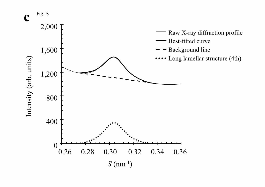
well to the sum of three Gaussian curves and a linear background. Figure 3c shows the results
obtained by the Gaussian curve fitting analysis for 4th order diffraction peak of the long
lamellar structures at EtOH/water mixture of 100/0 v/v%. Although the 2nd and 3rd order
diffraction peaks of the long lamellar structures overlap with other peaks, the 4th order
diffraction peak was obtained as a single peak with a flat background and has adequate
strength for fitting analysis. Therefore, the 4th diffraction peak was used for the analysis to
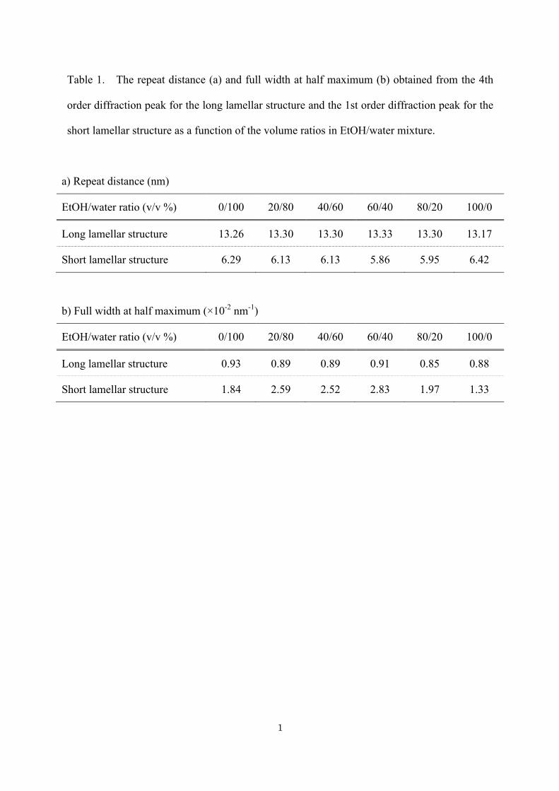
show the change of the long lamellar structures more precisely. Table 1 shows the repeat
distances of the long and short lamellar structures and the full widths at half maximum of the
diffraction peaks as a function of the volume ratios in EtOH/water mixture. In order to
compare variations in the short and long lamellar structures, the normalized repeat distance
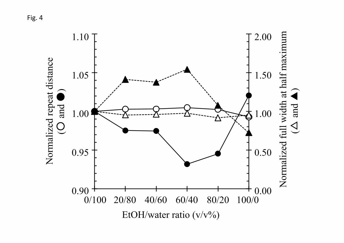
and normalized full width at half maximum are shown in Figure 4.
The application of EtOH/water mixture did not significantly affect the long lamellar
spacing and the peak width. On the other hand, the short lamellar spacing was minimal and
the peak width was maximal after treatment with EtOH/water mixture of 60/40 v/v%. Thus,
EtOH/water mixture was involved in both regularly and irregularly alteration on the periodic
pattern of the short lamellar structure in an EtOH concentration-dependent manner.
10
Fig. 3, Table 1, and Fig. 4
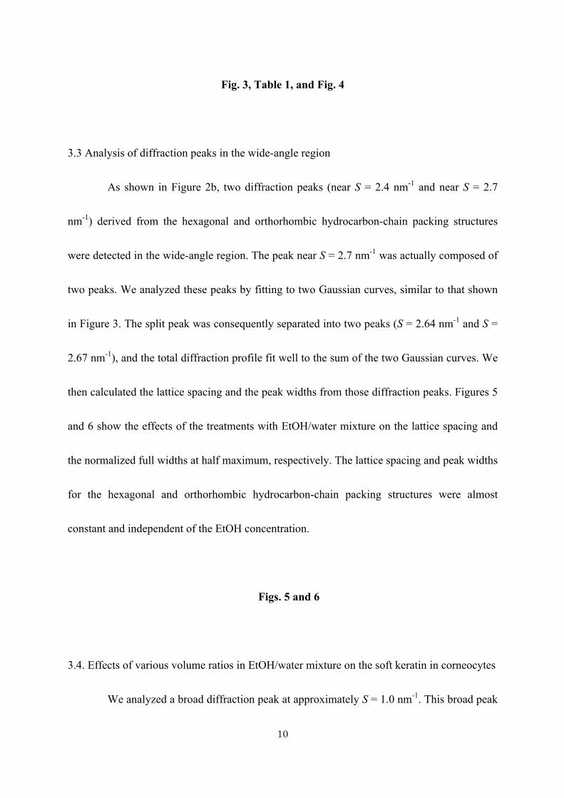
3.3 Analysis of diffraction peaks in the wide-angle region
As shown in Figure 2b, two diffraction peaks (near S = 2.4 nm-1 and near S = 2.7
nm-1) derived from the hexagonal and orthorhombic hydrocarbon-chain packing structures
were detected in the wide-angle region. The peak near S = 2.7 nm-1 was actually composed of
two peaks. We analyzed these peaks by fitting to two Gaussian curves, similar to that shown
in Figure 3. The split peak was consequently separated into two peaks (S = 2.64 nm-1 and S =
2.67 nm-1), and the total diffraction profile fit well to the sum of the two Gaussian curves. We
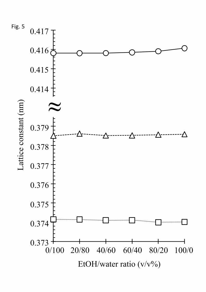
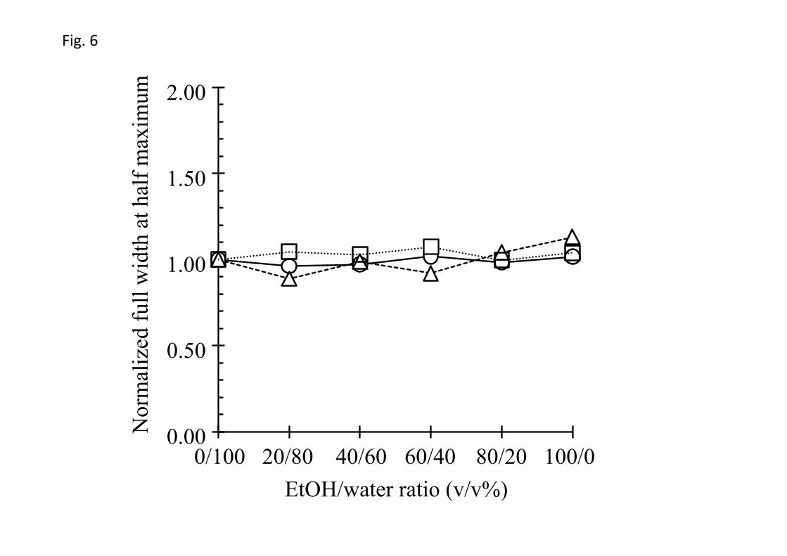
then calculated the lattice spacing and the peak widths from those diffraction peaks. Figures 5
and 6 show the effects of the treatments with EtOH/water mixture on the lattice spacing and
the normalized full widths at half maximum, respectively. The lattice spacing and peak widths
for the hexagonal and orthorhombic hydrocarbon-chain packing structures were almost
constant and independent of the EtOH concentration.
Figs. 5 and 6
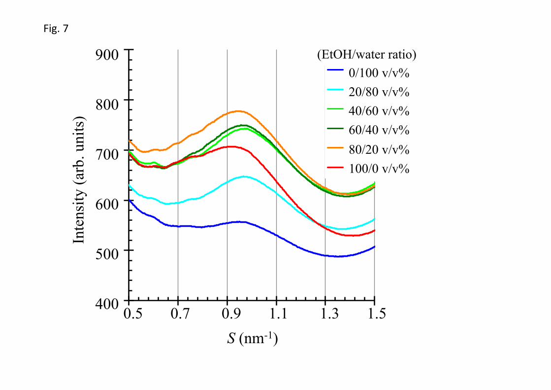
3.4. Effects of various volume ratios in EtOH/water mixture on the soft keratin in corneocytes
We analyzed a broad diffraction peak at approximately S = 1.0 nm-1. This broad peak
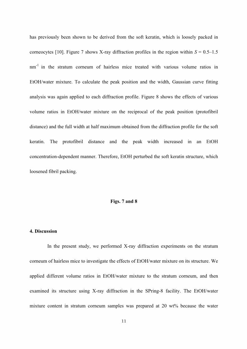
11
has previously been shown to be derived from the soft keratin, which is loosely packed in
corneocytes [10]. Figure 7 shows X-ray diffraction profiles in the region within S = 0.5‒1.5
nm-1 in the stratum corneum of hairless mice treated with various volume ratios in
EtOH/water mixture. To calculate the peak position and the width, Gaussian curve fitting
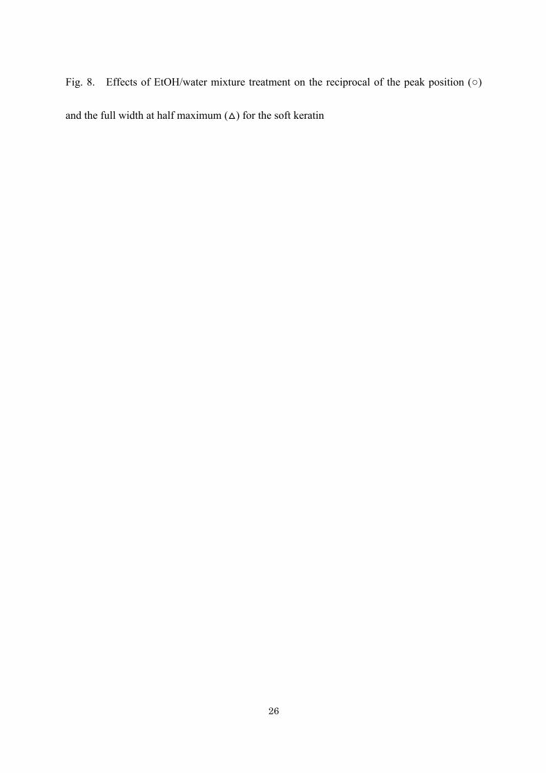
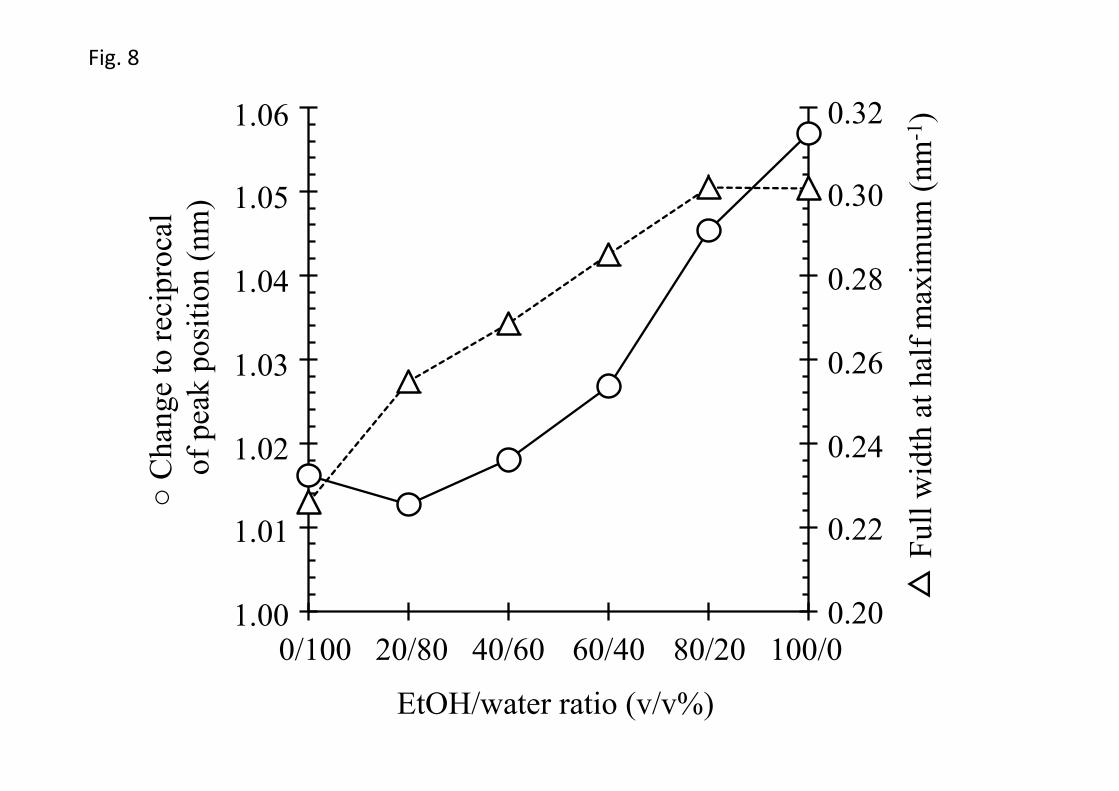
analysis was again applied to each diffraction profile. Figure 8 shows the effects of various
volume ratios in EtOH/water mixture on the reciprocal of the peak position (protofibril
distance) and the full width at half maximum obtained from the diffraction profile for the soft
keratin. The protofibril distance and the peak width increased in an EtOH
concentration-dependent manner. Therefore, EtOH perturbed the soft keratin structure, which
loosened fibril packing.
Figs. 7 and 8
4. Discussion
In the present study, we performed X-ray diffraction experiments on the stratum
corneum of hairless mice to investigate the effects of EtOH/water mixture on its structure. We
applied different volume ratios in EtOH/water mixture to the stratum corneum, and then
examined its structure using X-ray diffraction in the SPring-8 facility. The EtOH/water
mixture content in stratum corneum samples was prepared at 20 wt% because the water
12
content in the skin surface was estimated to be 20‒30% [15] and mostly stabilizes the lamellar
structure at this level [16]. There have been difficulties associated with comparing the results
obtained from X-ray diffraction experiments on multiple samples under different conditions
because the diffraction profiles of the stratum corneum are greatly affected by individual
variations and/or skin sites. Few X-ray diffraction studies have closely examined the effects
of EtOH/water mixture on the structure of the stratum corneum. In this study, we prepared
approximately one dozen skin samples per concentration of EtOH and the X-ray diffraction
patterns obtained; therefore, differences due to individual variations and/or skin sites were
avoided and the influence of skin treatments with any volume ratio in EtOH/water mixture on
the structure of the stratum corneum could be compared clearly.
Ordered lamellar structures, such as the long and short lamellar structures, have
periodic structures in the direction of the long axis of the hydrocarbon chain. Previous studies
reported that the repeat distances of the long and short lamellar structures were approximately
13 nm and 6 nm, respectively [12,14]. To detect minute variations in the lamellar structures,
the 1st order diffraction peak for the short lamellar structures, in particular, must be carefully
isolated since this peak was weaker than that for the long lamellar structures. Gaussian curve
fitting analysis was performed to isolate individual peaks from each other. As a result, the
repeat distance and the full width at half maximum were obtained for the short lamellar
structures.
13
Diffraction peak analyses revealed that EtOH/water mixture influenced the short
lamellar structures more than the long ones. The observation of the short lamellar structure in
hydrated condition was first reported experimentally by Bouwstra et al. [7,10,12] who have
been careful for proposing the swelling behavior. They have pointed out that the X-ray
diffraction peak for the short lamellar structure becomes sharp near the water content of 20
wt% but the swelling effect on the short lamellar structure was not detectable in human [12]
and also hairless mouse [7] stratum corneum. On the other hand, in pig stratum corneum they
have observed a weak swelling effect [10], and from a careful study on much attention to the
effect of water in hairless mouse stratum corneum Ohta et al. have found the swelling
behavior with increasing the water content [16]. So far in various studies, the diffraction peak
of short lamellar structure is sometimes undetectable [12,14], sometimes is only observed
only as a shoulder [7,10] and sometimes gives rise to a clear peak [16]. In addition to these
studies much clearly the swelling behavior in short lamellar structure has been observed from
the neutron diffraction by Charalambopoulou et al. [17]. The neutron diffraction experiment
by using heavy water is a powerful tool to observe the aqueous layer in stratum corneum since
the layer of heavy water can be observed dominantly. It is worthwhile to point out that the
increment of the spacing of short lamellar structure with increasing the water content in the
previous X-ray diffraction study [16] is consistent with the neutron diffraction study [17].
Furthermore, recently Nakazawa et al. have proposed that based upon the X-ray diffraction
14
study on the swelling behavior of short lamellar structure in human stratum corneum it could
involve in adjustment of the water content in stratum corneum [18]. The above fact indicates
that a uniform swelling of short lamellar structure takes place in stratum corneum since X-ray
and neutron diffraction measurement provide evidence for coherent structural alteration and
resultantly uniform swelling of the aqueous layer. The latter fact could be rationally supposed
from the swelling behavior under water obtained from the X-ray diffraction study on egg
lecithin bilayers which are composed of amphiphilic molecules [19] since intercellular lipids
in stratum corneum are composed of amphiphilic molecules such as ceramides and fatty acids.
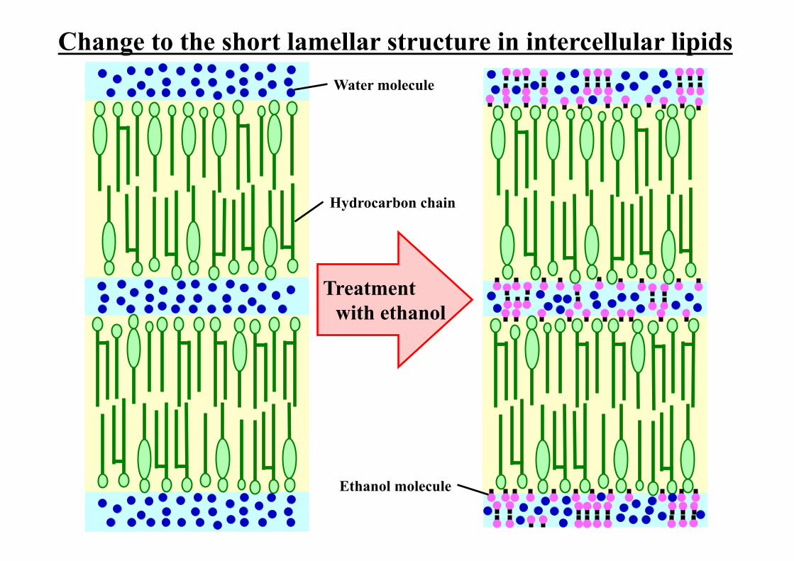
Therefore the aqueous layers in the short lamellar structure could take place in the
face-to-face arrangement of the polar head groups via the aqueous layer. Hence, applied water
or EtOH on the stratum corneum might penetrate the aqueous layer, and the behavior of the
aqueous layer following the application of various volume ratios in EtOH/water mixture may
depend on differences in the interactions of a water molecule and an EtOH molecule with the
polar head groups of ceramides and fatty acids. The applied EtOH seems to combine with the
interface between the aqueous layer and the polar head groups of ceramides and fatty acids by
the hydroxyl group of an EtOH molecule facing the aqueous layer [20]. As a result, the repeat
distance of the short lamellar structures decreased with an increase in the EtOH concentration
in the range of EtOH/water ratio from 0/100 v/v% to 60/40 v/v%, which then conversely
increased when the concentration of EtOH was elevated above 60/40 v/v%. The diffraction
15
peak width increased with an increase in the EtOH concentration in the range of EtOH/water
ratio from 0/100 v/v% to 60/40 v/v%, but decreased with further increases in the EtOH
concentration above 60/40%. These results indicate that treatment with a low concentration of
EtOH disturbs the short lamellar structures, but on the other hand, treatment with a high
concentration of it causes an aligned structure. In the case of treatment with mostly water or
EtOH, water and EtOH molecules may exist as structured molecules in highly concentrated
water and upon high EtOH, respectively. At intermediate concentrations, water and EtOH
may not form structured molecules, so the layer structure may be disrupted. In addition, Kwak
et al. reported [21] the effect of various concentrations of EtOH on model membranes of
stratum corneum lipids composed of different lipid classes. They pointed out that lipid model
membranes including ceramides were disrupted by the action of EtOH compared with those
with free fatty acids. This might indicate that there is less ceramide in the short lamellar
structures, being susceptible to EtOH, than in the long lamellar structures.
On the other hand, the hexagonal and orthorhombic hydrocarbon-chain packing
structures observed on a plane perpendicular to the long axis of the hydrocarbon chain were
previously reported to have lattice constants of 0.42 nm, 0.42 nm, and 0.37 nm [22]. Since the
diffraction profile with split peaks near S = 2.7 nm-1 fits well to the sum of the two Gaussian
curves and a background line, there may be at least two types of orthorhombic
hydrocarbon-chain packing structure in the intercellular lipids in the stratum corneum of the
16
hairless mice used in the present study. The lattice constants and the peak widths derived from
the hexagonal and the orthorhombic hydrocarbon-chain packing structures were not
influenced by the treatment with any volume ratio in EtOH/water mixture.
Corneocytes, which are surrounded by intercellular lipids, are crucially involved in
maintaining the form of skin tissue. Cross-sectional observations of the stratum corneum
using electron microscopy indicated that it is composed of a higher proportion of corneocytes
than intercellular lipids [23]. The interior of corneocytes is filled with agglutinated-keratin
fibers. A diffraction peak of loosely packed soft keratin fibers was previously detected near S
= 0.96 nm-1 in the middle-angle region [10]. Thus, the reciprocal of the peak position
corresponded to the protofibril-to-protofibril distance. This diffraction peak was also observed
and analyzed in the present study. The protofibril distance and peak width increased in an
EtOH concentration-dependent manner. This result indicates that EtOH penetrated the
corneocytes [24] and disturbed the soft keratin structure by loosening fibril packing in the
corneocytes in an EtOH concentration-dependent manner. This phenomenon may be involved
in the denaturation effects of EtOH on keratin proteins.
The results of the present study reveal the molecular mechanisms of action of
different volume ratios in EtOH/water mixture on the ordered structures of intercellular lipids
and soft keratin in the stratum corneum. All volume ratios in EtOH/water mixture greatly
affected the short lamellar structure in the intercellular lipids and the fibril structures of the
17
soft keratin, but had a negligible effect on the long lamellar structures in the intercellular
lipids and the hydrocarbon-chain packing structures. The structural alteration in the stratum
corneum due to the application of EtOH/water mixture suggests that it is heavily involved in
the skin permeability of topically applied drugs in formulations containing both EtOH and
water. We previously reported the effects of EtOH/water mixture on the skin permeability of
drugs using an in vitro pig skin permeation test [6], and demonstrated that the skin
permeability of hydrophilic drugs was increased by a low concentration of EtOH and, on the
other hand, was decreased by a high concentration of it. Although changes in the diffusivity
and partition coefficient of penetrants were expected with the EtOH/water mixture treatment,
the molecular mechanisms underlying the skin permeability of drugs have not yet been fully
elucidated. In the present study, the periodic structures in short lamellar became irregular at
low to medium concentrations of EtOH, but return to regular at higher concentrations of
EtOH. However in the orthogonal direction to the periodic structure the hydrocarbon-chain
packing structure does not change. Therefore not only water but also EtOH does not affect the
hydrocarbon-chain packing and EtOH/water mixture causes disarrangement of the regular
short lamellar structure with the aqueous layers. The structure of the soft keratin was altered
gradually by increases in the EtOH concentration. Therefore, these changes may be strongly
associated with the skin permeability of chemical compounds. The skin penetration of
chemical compounds through the stratum corneum depends on drug partition to the stratum
18
corneum and drug diffusivity in drug-distributed regions. Lipophilic compounds have been
considered to be distributed to the hydrophobic region in intercellular lipids and then diffuse
in the lipid pathway, and as a result, the compounds penetrate the skin. In contrast,
hydrophilic compounds are considered to be distributed to the aqueous regions in intercellular
lipids or corneocytes and diffusion is observed in the aqueous pathway. Accordingly, the
changes to the short lamellar and soft keratin structures due to the application of EtOH/water
mixture imply that the distribution to aqueous regions and the diffusivity of hydrophilic
compounds may be altered. Since EtOH/water mixture did not significantly influence the long
lamellar structures with hydrophobic characteristics, the distribution and the diffusivity of
lipophilic compounds may be slightly changed compared with those of hydrophilic
compounds. Thus, EtOH treatment can greatly affect the skin permeation of hydrophilic
compounds. Although hairless mouse skin was selected for the collection of stratum corneum
samples in the present study, the influence of EtOH/water mixture was previously shown to
be similar on the skin of other animals (pig and rat) including human. The above results are
consistent with the previous findings of an in vitro skin permeation study using pig skin [6].
Further studies are needed to clarify the relationship between the structure of the stratum
corneum and skin permeability of chemical compounds following EtOH/water mixture
treatment. In particular, research that reveals the relative amounts of short lamellar structures
and long lamellar structures appears to be important, since determination of their ratio has not
19
been accomplished yet. For the quantitative consideration we need statistical analysis of the
present data. For this purpose additionally there is room for further research into statistical
analysis.
EtOH is an ingredient in vehicles for topically applied medicines. In the present
study, the effect of EtOH/water mixture on the structure of the stratum corneum was not
directly proportional to its volume ratio. Although EtOH/water mixture changed the structure
of the stratum corneum, the type of change was largely dependent on its volume ratio.
Various concentrations of EtOH are currently selected for topically applied medicines.
Therefore, the effects of EtOH concentrations on the structure of the stratum corneum as well
as drug efficacy and safety must be considered when developing preparations containing both
EtOH and water.
Acknowledgment
We would like to thank Dr. Noboru Ohta for help during the data collection at
BL40B2 in the SPring-8 facility. The experiment was conducted under the approval of the
SPring-8 Proposal Review Committee (proposal number 2012B1255).
References
[1] L. K. Pershing, L. D Lambert., K. Knutson, Mechanism of ethanol-enhanced estradiol
20
permeation across human skin in vivo, Pharm. Res. 7 (1990) 170‒175.
[2] W. R. Good, M. S. Powers, P. Campbell, L. Schenkel, A new transdermal delivery system
for estradiol, J. Control. Release 2 (1985) 89‒97.
[3] B. Berner., G. C. Mazzenga, J. H. Otte, R. J. Steffens, R. H. Juang, C. D. Ebert, Ethanol:
water mutually enhanced transdermal therapeutic system II: skin permeation of ethanol
and nitroglycerin, J. Pharm. Sci. 78 (1989) 402‒407.
[4] A. C. Williams, B. W. Barry, Penetration enhancers, Adv. Drug Deliv. Rev. 56 (2004)
603‒618.
[5] D. Van der Merwe, J. E. Riviere, Comparative studies on the effects of water, ethanol and
water/ethanol mixtures on chemical partitioning into porcine stratum corneum and silastic
membrane, Toxicol. In vitro. 19 (2005) 69‒77.
[6] D. Horita, H. Todo, K. Sugibayashi. Effect of ethanol pretreatment on skin permeation
drugs, Biol. Pharm. Bull. 35 (2012) 1343‒1348.
[7] J. A. Bouwstra, G. S. Gooris, J. A. Van der Spek, S. Lavrijsen, W. Bras, The lipid and
protein structure of mouse stratum corneum: a wide and small angle diffraction study,
Biochim. Biophys. Acta 1212 (1994) 183‒192.
[8] I. Hatta, N. Ohta, S. Ban, H. Tanaka, S. Nakata, X-ray diffraction study on ordered,
disordered and reconstituted intercellular lipid lamellar structure in stratum corneum,
Biophys. Chem. 89 (2001) 239‒242.
21
[9] Y. Obata, I. Hatta, N. Ohta, N. Kunizawa, N. Yagi, K. Takayama, Combined effects of
ethanol and l-menthol on hairless rat stratum corneum investigated by synchrotron X-ray
diffraction, J. Control. Release 115 (2006) 275‒279.
[10] J. A. Bouwstra, G. S. Gooris, W. Bras, D. T. Downing, Lipid organization in pig stratum
corneum, J. Lipid Res. 36 (1995) 685‒695.
[11] M. Fujii, Y. Takeda, M. Yoshida, N. Utoguchi, M. Matsumoto, Y. Watanabe, Comparison
of skin permeation enhancement by 3-l-menthoxypropane-1,2-diol and l-menthol: the
permeation of indomethacin and antipyrine through Yucatan micropig skin and changes in
infrared spectra and X-ray diffraction patterns of stratum corneum, Int. J. Pharm. 258
(2003) 217‒223.
[12] J. A. Bouwstra, G. S. Gooris, J. A. Van der Spek, W. Bras, Structural investigations of
human stratum corneum by small-angle X-ray scattering, J. Invest. Dermatol. 97 (1991)
1005‒1012.
[13] J. A. Bouwstra, G. S. Gooris, M. A. Salomons-de Vries, J. A. Van der Spek, W. Bras,
Structure of human stratum corneum as a function of temperature and hydration: A
wide-angle X-ray diffraction study, Int. J. Pharm. 85 (1992) 205‒216.
[14] I. Hatta, N. Ohta, K. Inoue, N. Yagi, Coexistence of two domains in intercellular lipid
matrix of stratum corneum, Biochim. Biophys. Acta 1758 (2006) 1830‒1836.
[15] I. H. Blank, Factors which influence the water content of the stratum corneum, J. Invest.
22
Dermatol. 18 (1952) 443‒440.
[16] N. Ohta, S. Ban, H. Tanaka, S. Nakata, I. Hatta, Swelling of intercellular lipid lamellar
structure with short repeat distance in hairless mouse stratum corneum as studied by X-ray
diffraction, Chem. Phys. Lipids 123 (2003) 1‒8.
[17] G. Ch. Charalambopoulou, Th. A. Steriotis, Th. Hauss, A. K. Stubos, N. K. Kanellopoulos,
Structural alterations of fully hydrated human stratum corneum, Physica B 350 (2004)
603‒606.
[18] H. Nakazawa, N. Ohta, I. Hatta, A possible regulation mechanism of water content in
human stratum corneum via intercellular lipid matrix, Chem. Phys. Lipids, 165 (2012)
238‒243.
[19] Y. K. Levine, M. H. F. Wilkins, Structure of oriented lipid bilayers, Nature New Biology,
230 (1971) 69‒72.
[20] T. Adachi, H. Takahashi, K. Ohki, I. Hatta, Interdigitated structure of
phospholipid-alcohol systems studied by X-ray diffraction, Biophys. J. 68 (1995) 1850‒
1855.
[21] S. Kwak, E. Brief, D. Langlais, N. Kitson, M. Lafleur, J. Thewalt, Ethanol perturbs lipid
organization in models of stratum corneum membranes: An investigation combining
differential scanning calorimetry, infrared and 2H NMR spectroscopy, Biochim. Biophys.
Acta 1818 (2012) 1410‒1419.
23
[22] G. S. Pilgram, A. M. Engelsma-van Pelt, J. A. Bouwstra, H. K. Koerten, Electron
diffraction provides new information on human stratum corneum lipid organization
studied in relation to depth and temperature, J. Invest. Dermatol. 113 (1999) 403‒409.
[23] L. Norlén, A. Al-Amoudi, Stratum corneum keratin structure, function, and formation: the
cubic rod-packing and membrane templating model, J. Invest. Dermatol. 123 (2004) 715‒
732.
[24] I. Hatta, H. Nakazawa, Y. Obata, N. Ohta, K. Inoue, N. Yagi, Novel method to observe
subtle structural modulation of stratum corneum on applying chemical agents, Chem.
Phys. Lipids 163 (2010) 381‒389.
24
Figure Legends
Fig. 1. X-ray diffraction profiles in the hairless mouse stratum corneum as a function of the
volume ratios in EtOH/water mixture, where n is the number of stratum corneum samples.
Fig. 2. Small-angle X-ray diffraction profiles (a) and wide-angle X-ray diffraction profiles
(b) in the hairless mouse stratum corneum as a function of the volume ratios in EtOH/water
mixture, where n is the number of stratum corneum samples.
Fig. 3. Typical curve fitting analysis to small-angle X-ray diffraction profiles of the stratum
corneum of hairless mice treated with EtOH/water mixture of 60/40 v/v% (a) and 100/0 v/v%
(b, c). The thin solid line shows the mean of raw small-angle X-ray diffraction profiles.
The thick solid line indicates the best-fitted curve composed of the sum of Gaussian dotted
curves and a linear background dotted line.
Fig. 4. Effects of EtOH/water mixture treatment on the repeat distances (○ or ●) and the full
widths at half maximum (△ or ▲) for the 4th order diffraction of the long lamellar structures
and the 1st order diffraction of the short lamellar structures. The normalized data were
calculated by dividing the values of repeat distances and full width at half maximum with
EtOH/water mixture treatments by those for 0/100 v/v%. The open circle or open triangle
25
and the closed circle or closed triangle show the long lamellar structures and the short
lamellar structures, respectively.
Fig. 5. Effects of EtOH/water mixture treatment on the lattice constants for the diffraction
peaks at S = 2.4 nm-1, S = 2.64 nm-1, and S = 2.67 nm-1. The open circle, open triangle, and
open square show the diffraction peaks at S = 2.4 nm-1, S = 2.64 nm-1, and S = 2.67 nm-1,
respectively.
Fig. 6. Effects of EtOH/water mixture treatment on the normalized full widths at half
maximum for the diffraction peaks at S = 2.4 nm-1, S = 2.64 nm-1, and S = 2.67 nm-1. The
normalized data were calculated by dividing the values of full width at half maximum with
EtOH/water mixture treatments by those for 0/100 v/v%. The open circle, open triangle, and
open square show the diffraction peak at S = 2.4 nm-1, S = 2.64 nm-1, and S = 2.67 nm-1,
respectively.
Fig. 7. Medium-angle X-ray diffraction profiles in the hairless mouse stratum corneum as a
function of the volume ratios in EtOH/water mixture, where n is the number of stratum
corneum samples.
26
Fig. 8. Effects of EtOH/water mixture treatment on the reciprocal of the peak position (○)
and the full width at half maximum (△) for the soft keratin
Change to the short lamellar structure in intercellular lipids
Treatment with ethanol
Water molecule
Hydrocarbon chain
Ethanol molecule
1
Table 1. The repeat distance (a) and full width at half maximum (b) obtained from the 4th
order diffraction peak for the long lamellar structure and the 1st order diffraction peak for the
short lamellar structure as a function of the volume ratios in EtOH/water mixture.
a) Repeat distance (nm)
EtOH/water ratio (v/v %) 0/100 20/80 40/60 60/40 80/20 100/0
Long lamellar structure 13.26 13.30 13.30 13.33 13.30 13.17
Short lamellar structure 6.29 6.13 6.13 5.86 5.95 6.42
b) Full width at half maximum (×10-2 nm-1)
EtOH/water ratio (v/v %) 0/100 20/80 40/60 60/40 80/20 100/0
Long lamellar structure 0.93 0.89 0.89 0.91 0.85 0.88
Short lamellar structure 1.84 2.59 2.52 2.83 1.97 1.33
S (nm-1)
Loga
rithm
ic in
tens
ity (a
rb. u
nits
) 105
104
103
102 0.0 0.5 1.0 1.5 2.0 2.5 3.0
0
1,000
2,000
3,000
4,000
5,000
2.0 2.2 2.4 2.6 2.8 3.0
Inte
nsity
(arb
. uni
ts)
S (nm-1)
0% EtOH(n=13)20% EtOH(n=14)40% EtOH(n=14)60% EtOH(n=13)80% EtOH(n=14)
100% EtOH(n=14)
0/100 v/v% 20/80 v/v% 40/60 v/v% 60/40 v/v% 80/20 v/v% 100/0 v/v%
(EtOH/water ratio)
Fig. 1
S (nm-1)
Inte
nsity
(arb
. uni
ts)
10,000
8,000
6,000
4,000
2,000
0 0.10 0.15 0.20 0.25 0.30 0.35 0.40
a
0
1,000
2,000
3,000
4,000
5,000
2.0 2.2 2.4 2.6 2.8 3.0
Inte
nsity
(arb
. uni
ts)
S (nm-1)
0% EtOH(n=13)20% EtOH(n=14)40% EtOH(n=14)60% EtOH(n=13)80% EtOH(n=14)
100% EtOH(n=14)
0/100 v/v% 20/80 v/v% 40/60 v/v% 60/40 v/v% 80/20 v/v% 100/0 v/v%
(EtOH/water ratio)
Fig. 2
S (nm-1)
Inte
nsity
(arb
. uni
ts)
5,000
4,000
3,000
2,000
1,000
0 2.0 2.2 2.4 2.6 2.8 3.0
b
0
1,000
2,000
3,000
4,000
5,000
2.0 2.2 2.4 2.6 2.8 3.0
Inte
nsity
(arb
. uni
ts)
S (nm-1)
0% EtOH(n=13)20% EtOH(n=14)40% EtOH(n=14)60% EtOH(n=13)80% EtOH(n=14)
100% EtOH(n=14)
0/100 v/v% 20/80 v/v% 40/60 v/v% 60/40 v/v% 80/20 v/v% 100/0 v/v%
(EtOH/water ratio)
Fig. 2

S (nm-1)
Inte
nsity
(arb
. uni
ts)
10,000
8,000
6,000
4,000
2,000
0 0.120 0.145 0.170 0.195 0.220 0.245
a Raw X-ray diffraction profile Best-fitted curve Background line Long lamellar structure (2nd) Short lamellar structure (1st) Long lamellar structure (3rd)
実測値
解析値
バックグラウンド
長周期2次
短周期1次
長周期3次
Fig. 3
S (nm-1)
Inte
nsity
(arb
. uni
ts)
10,000
8,000
6,000
4,000
2,000
0 0.120 0.145 0.170 0.195 0.220 0.245
b Raw X-ray diffraction profile Best-fitted curve Background line Long lamellar structure (2nd) Short lamellar structure (1st) Long lamellar structure (3rd)
実測値
解析値
バックグラウンド
長周期2次
短周期1次
長周期3次
Fig. 3
S (nm-1)
Inte
nsity
(arb
. uni
ts)
2,000
1,600
1,200
800
400
0 0.26 0.28 0.30 0.32 0.34 0.36
c Raw X-ray diffraction profile Best-fitted curve Background line Long lamellar structure (4th)
実測値
解析値
バックグラウンド
長周期2次
短周期1次
長周期3次
Fig. 3
0/100 20/80 40/60 60/40 80/20 100/0
EtOH/water ratio (v/v%)
Nor
mal
ized
repe
at d
ista
nce
(○ a
nd ●
)
Nor
mal
ized
full
wid
th a
t hal
f max
imum
(△
and
▲)
1.10
1.05
1.00
0.95
0.90
2.00
1.50
1.00
0.50
0.00
Fig. 4
0/100 20/80 40/60 60/40 80/20 100/0
EtOH/water ratio (v/v%)
~ ~ 0.379
0.378
0.377
0.376
0.375
0.374
0.373
0.417
0.416
0.415
0.414
Latti
ce c
onst
ant (
nm)
Fig. 5
0/100 20/80 40/60 60/40 80/20 100/0 EtOH/water ratio (v/v%)
Nor
mal
ized
full
wid
th a
t hal
f max
imum
2.00
1.50
1.00
0.50
0.00
Fig. 6
S (nm-1)
Inte
nsity
(arb
. uni
ts)
900
800
700
600
500
400 0.5 0.7 0.9 1.1 1.3 1.5
0
1,000
2,000
3,000
4,000
5,000
2.0 2.2 2.4 2.6 2.8 3.0
Inte
nsity
(arb
. uni
ts)
S (nm-1)
0% EtOH(n=13)20% EtOH(n=14)40% EtOH(n=14)60% EtOH(n=13)80% EtOH(n=14)
100% EtOH(n=14)
0/100 v/v% 20/80 v/v% 40/60 v/v% 60/40 v/v% 80/20 v/v% 100/0 v/v%
(EtOH/water ratio)
Fig. 7
0/100 20/80 40/60 60/40 80/20 100/0
EtOH/water ratio (v/v%)
○ C
hang
e to
reci
proc
al
of p
eak
posi
tion
(nm
)
△ F
ull w
idth
at h
alf m
axim
um (n
m-1
) 1.06
1.05
1.04
1.03
1.02
1.01
1.00
0.32
0.30
0.28
0.26
0.24
0.22
0.20
Fig. 8


![Review Potential Mechanisms of Action of Curcumin for ... · angiogenesis and metastasis [26]. The molecular basis of the anticancer activities of curcumin is mainly attributed to](https://img.pdfslide.tips/doc/110x75/5ed770259ab714631510fda8/review-potential-mechanisms-of-action-of-curcumin-for-angiogenesis-and-metastasis.jpg)