Embed Size (px)
Citation preview
Aus dem Institut für Physiologie und Pathophysiolog ie
Geschäftsführender Direktor: Prof. Dr. Dr. Jürgen D aut
des Fachbereichs Medizin der Philipps-Universität M arburg
Molecular Properties and Pathophysiological Relevan ce of the
Predominant K + Conductance in Cochlear Outer Hair Cells
Inaugural-Dissertation zur Erlangung des Doktorgrades der Naturwissenschaften
dem Fachbereich Medizin der Philipps-Universität Marburg
vorgelegt von
Michael Georg Leitner
(geb. in St. Johann in Tirol, Österreich)
Marburg, 2012

i
Angenommen vom Fachbereich Medizin der Philipps-Universität Marburg am: 9. August 2012 (Tag der Disputation) Gedruckt mit Genehmigung des Fachbereichs Dekan: Prof. Dr. Matthias Rothmund
Referent: Prof. Dr. Dominik Oliver
Korreferent: Prof. Dr. Joachim Hoyer
A N M E R K U N G
ii
Diese Doktorarbeit macht von der Möglichkeit Gebrauch, gesammelte Publikationen als
Dissertationsleistung anzuerkennen (kumulative Dissertation) wie in der „Promotionsordnung der
Mathematisch-Naturwissenschaftlichen Fachbereiche und des Medizinischen Fachbereiches für
seine mathematisch-naturwissenschaftlichen Fächer der Philipps-Universität Marburg vom
15.7.2009 (§9)“ erläutert. Demnach besteht diese Arbeit aus einer gemeinsamen Einleitung, einer
Zusammenfassung der folgenden, im Anhang angeführten Publikationen und einer Diskussion.
Michael G. Leitner , Christian R. Halaszovich and Dominik Oliver. Aminoglycosides inhibit
KCNQ4 channels in cochlear outer hair cells via depletion of
phosphatidylinositol(4,5)bisphosphate. Molecular Pharmacology. 2011 Jan:79(1):51-60
Michael G. Leitner , Anja Feuer, Olga Ebers, Daniela N. Schreiber, Christian R. Halaszovich and
Dominik Oliver. Restoration of ion channel function in deafness-causing KCNQ4 mutations by
chemical openers. British Journal of Pharmacology (2012) 165 2244-2259
Moritz Lindner, Michael G. Leitner , Christian R. Halaszovich, Gerald R.V. Hammond and
Dominik Oliver. Probing the regulation of TASK potassium channels by PI(4,5)P2 with switchable
phosphoinositide phosphatases. Journal of Physiology. 2011 Jul 1:589 (Pt 13):3149-62
Jérôme Lacroix, Christian R. Halaszovich, Daniela N. Schreiber, Michael G. Leitner , Francisco
Bezanilla, Dominic Oliver and Carlos A. Villalba-Galea. Controlling the activity of PTEN by
membrane potential. Journal of Biological Chemistry. 2011 May 20:286(20):17945-53
Z U S A M M E N F A S S U N G
iii
Äußere Haarsinneszellen (ÄHZ) des Corti´schen Organs im Innenohr werden biophysikalisch
charakterisiert durch den K+ Strom IK,n, dessen molekulare Grundlage der spannungsabhängige
K+ Kanal KCNQ4 (Kv7.4) ist. IK,n/KCNQ4 dominiert die elektrischen Eigenschaften der ÄHZ
Zellmembran und ist darüber hinaus essentiell für das Überleben der Zellen. Im knock-out
Tiermodell führte der Verlust der KCNQ4 Kanalaktivität zum Untergang der ÄHZ und zu Taubheit.
Dies ist klinisch bedeutend, da im Menschen KCNQ4 Mutationen die Ursache für die erbliche
Taubheit DFNA2 sind. Der Mechanismus, der zum Haarzelluntergang führt, ist noch unklar, doch
korreliert das Überleben von ÄHZ direkt mit der Funktion von KCNQ4. Obwohl der durch
Giftstoffe oder Lärm bewirkte Haarzelluntergang (erworbener Hörverlust) dem KCNQ4-bedingten
ÄHZ Verlust ähnelt, wurde eine Verbindung zwischen IK,n/KCNQ4 und erworbenem Hörverlust
bisher noch nicht experimentell erfasst. In der vorliegenden Arbeit habe ich mich mit der
pathophysiologischen Rolle von IK,n/KCNQ4 in klinisch relevantem Haarzelluntergang beschäftigt,
der durch Aminoglykosid (AG) Antibiotika hervorgerufen wird. Des Weiteren wurde untersucht, ob
chemische KCNQ Kanalöffner die Funktion von IK,n trotz pathophysiologisch relevanter Inhibition
wiederherstellen können, um eventuell Protektion der ÄHZ zu ermöglichen.
Ich konnte erstmalig zeigen, dass AG Antibiotika schnell in Haarzellen eindringen und IK,n
inhibieren. Die Inhibition war darauf zurückzuführen, dass AG über elektrostatische
Wechselwirkungen negativ geladene Phospholipide funktionell depletieren, die für die Funktion
von IK,n essentiell sind. Verschiedene AG zeigen unterschiedliches ototoxisches Potential:
Neomycin führt zum Untergang von ÄHZ, während Gentamycin vestibuläre Haarzellen schädigt.
Das Ausmaß der IK,n Kanalinhibition korrelierte (Neomycin > Gentamycin) mit dem Grad der
Phospholipid Bindung und mit dem ototoxischen Potential der AG. Dies legt nahe, dass die hohe
Anfälligkeit der ÄHZ gegenüber Neomycin auf die Inhibition von IK,n zurückzuführen ist. Des
Weiteren konnte gezeigt werden, dass die Aktivität von IK,n durch chemische KCNQ Kanalöffner
verstärkt wird. Die AG-induzierte Hemmung von IK,n konnte aufgehoben und die Kanalaktivität
vollkommen wiederhergestellt werden.
In heterozygoten DFNA2 Patienten werden IK,n Ströme durch einen dominant-negativen Effekt
mutierter KCNQ4 Kanaluntereinheiten verringert, was den ÄHZ Untergang bewirkt. Es ist mir im
heterologen System gelungen, mittels chemischer KCNQ Kanalöffner KCNQ4 von dieser
dominant-negativen Inhibition zu befreien. Die Ströme in Gegenwart der Kanalöffner waren
vergleichbar mit Strömen unter Kontrollbedingungen, was gleichbedeutend mit der vollständigen
Wiederherstellung der Kanalfunktion war.
Zusammenfassend kann gesagt werden, dass ich in dieser Arbeit erstmals eine Rolle der
Kaliumleitfähigkeit IK,n in erworbenem Hörverlust demonstrieren konnte. Dies kann die verstärkte
Anfälligkeit von ÄHZ gegenüber ototoxischen Einflüssen erklären. Darüber hinaus stabilisieren
chemische Kanalöffner IK,n in Anwesenheit von AG und stellen die Funktion von rekombinanten
KCNQ4 Kanälen trotz dominant-negativer Inhibition wieder her. Dies könnte Haarzellen vor
KCNQ4-bedingtem Untergang schützen. Es bleibt allerdings offen, ob die Substanzen den
Verlust der ÄHZ verzögern und Taubheit abwenden können.
S U M M A R Y
iv
Cochlear outer hair cells (OHCs) are characterised by the voltage-dependent K+ conductance IK,n
that previously was shown to be mediated by KCNQ4 (Kv7) channel subunits. IK,n/KCNQ4
dominates the electrical properties of the OHC cell membrane and furthermore is essential for
OHC survival. Genetic deletion of KCNQ4 causes progressive degeneration of OHCs and
deafness. Similarly, KCNQ4 loss-of-function mutations cause the progressive form of hereditary
deafness DFNA2. The molecular mechanism leading to OHC degeneration remains elusive, but
the survival of OHCs has been linked directly to KCNQ4 channel function. Strikingly, the loss of
OHCs is phenotypically similar to OHC degeneration caused by ototoxic substances and noise
damage (acquired hearing loss), but a role of IK,n/KCNQ4 in acquired hearing loss has never been
investigated so far. In the present study I investigated the pathophysiological relevance of IK,n for
OHC degeneration caused by aminoglycoside (AG) antibiotics. Since KCNQ4 channel function is
essential for OHC survival, chemical current augmentation may provide a protective strategy
against KCNQ4-related hearing loss. Thus, I analysed whether chemical KCNQ channel openers
rescued IK,n currents from pathological inhibition.
In brief, I found that AGs rapidly entered OHCs and that entry was necessary for IK,n current
inhibition. The inhibition was caused by functional depletion of phospholipids by the AGs that are
essential for the function of IK,n/KCNQ4. Various AGs exhibit different ototoxic potential, i.e.
neomycin causes OHC degeneration whereas gentamicin damages vestibular hair cells.
Strikingly, the degree of IK,n inhibition (neomycin > gentamicin) correlated with the phospholipid
binding efficiency and with the ototoxic potential of the respective AG. Given the dependence of
OHCs on IK,n, the ototoxic potential of AGs thus may be determined by their chemical nature and
by their inhibitory impact on IK,n. Furthermore, I showed that IK,n was sensitive to current
augmentation by chemical KCNQ channel openers. A combination of openers rescued IK,n from
AG-induced inhibition to wild-type levels indicating full restoration of IK,n activity.
Most DFNA2 patients are heterozygous carriers of KCNQ4 mutations that reduce IK,n through a
dominant-negative effect. This reduction of IK,n activity causes OHC degeneration. Residual
currents in the dominant-negative situation were essentially rescued to wild-type levels by the
application of KCNQ channel openers, at least in a heterologous expression system. The current
rescue indicated that KCNQ channel openers might be used to stabilise IK,n in heterozygous
DFNA2 patients.
In summary, the present work demonstrated for the first time a role of the essential OHC K+
conductance IK,n in acquired hearing loss. The dependence of OHCs on IK,n may explain the high
vulnerability of OHCs towards ototoxic influences. IK,n current augmentation by chemical KCNQ
openers may be used to stabilise IK,n in OHCs and protect the sensory cells from KCNQ4-related
degeneration. KCNQ openers rescued IK,n from AG-induced inhibition in OHCs and recombinant
KCNQ4 from dominant-negative inhibition by mutant subunits. However, it remains elusive
whether chemical KCNQ agonists alleviate OHC degeneration and protect from hearing loss.
T A B L E O F C O N T E N T S
v
1 INTRODUCTION .................................................................................................................. - 1 -
1.1 Auditory Hair Cells Mediate the Transduction of Sound ............................................... - 1 -
1.2 Outer Hair Cells and the Cochlear Amplifier ................................................................. - 3 -
1.2.1 The Electrophysiology of Outer Hair Cells ............................................................. - 4 -
1.2.2 IK,n is the Predominant K+ Current of Outer Hair Cells ........................................... - 5 -
1.2.3 KCNQ4 Mediates IK,n, But Both Channels Show Biophysical Differences ............ - 6 -
1.2.4 IK,n/KCNQ4 is Essential for the Survival of Outer Hair Cells .................................. - 7 -
1.2.5 Other Outer Hair Cell Pathologies May Be Related to KCNQ4 Dysfunction ......... - 9 -
2 AIMS AND CONTRIBUTIONS ........................................................................................... - 11 -
2.1 Objectives .................................................................................................................... - 11 -
2.2 My Contributions to the Articles Presented ................................................................. - 12 -
3 RESULTS ........................................................................................................................... - 14 -
3.1 The Outer Hair Cell K+ Current IK,n Requires PI(4,5)P2 for Activation ......................... - 14 -
3.2 Inhibition of IK,n by Aminoglycosides is Pathophysiologically Relevant ...................... - 15 -
3.3 IK,n is Sensitive to Chemical KCNQ Openers .............................................................. - 16 -
3.4 KCNQ Channel Openers Rescue IK,n from AG-induced Inhibition and Reconstitute Channel Function of Deafness-Causing KCNQ4 Mutants .......................................... - 16 -
3.5 The Biophysical Properties and the Molecular Nature of IK,n ...................................... - 17 -
3.6 Development of Novel Tools to Experimentally Alter PI Levels in Living Cells .......... - 19 -
4 DISCUSSION ...................................................................................................................... - 20 -
4.1 Biophysical and Pharmacological Differences of IK,n and Recombinant KCNQ4: Implications for the Molecular Identity of IK,n ............................................................... - 20 -
4.2 The Role of IK,n Inhibition in AG-induced Hair Cell Loss ............................................. - 21 -
4.3 Potential Use of KCNQ Openers to Protect from KCNQ4-related Hearing Loss ....... - 22 -
4.4 Outlook ......................................................................................................................... - 23 -
5 REFERENCES ................................................................................................................... - 25 -
6 SUBMITTED PUBLICATIONS ............................................................................................ - 31-
7 APPENDIX ............................................................................................................................... vii
A B B R E V I A T I O N S
vi
AG Aminoglycoside
Akt Protein kinase B
BM Basilar membrane
CFP Cyan fluorescent protein
CHO Chinese hamster ovary
Ci-VSP Ciona intestinalis voltage-sensing phosphatase
DAG Diacylglycerol
DFNA2 Deafness-associated autosomal dominant locus 2
GFP Green fluorescent protein
IHC Inner hair cell
IP3 Inositol-(3,4,5)-trisphosphate
KCNQ Voltage-gated potassium channel family 7 (Kv7)
M1R Muscarinic acetylcholine receptor type 1
MET Mechano-electrical transduction
OHC Outer hair cell
PBM Phosphatidylinositol binding motif
PD Phosphatase domain
PH Pleckstrin homology
PI Phosphoinositide
PI(4)P Phosphatidylinositol-(4)-phosphate
PI(4,5)P2 Phosphatidylinositol-(4,5)-bisphosphate
PI(3,4)P2 Phosphatidylinositol-(3,4)-bisphosphate
PI(3,4,5)P3 Phosphatidylinositol-(3,4,5)-trisphosphate
PI3K Phosphatidylinositol-3 kinase
PLC Phospholipase C
PTEN Phosphatase and tensin homolog deleted from chromosome 10
TASK TWIK-related acid sensing potassium channel
TIRF Total internal reflection fluorescence
TEA Tetraethylammonium
VSD Voltage sensor domain
VSP Voltage-sensing phosphatase
Wt Wild-type
ZnP/Ret Zinc pyrithione plus retigabine
I N T R O D U C T I O N
- 1 -
1 Introduction
Hearing is a complex process that involves the transduction of mechanical sound stimuli into
electrical neuronal signals and the processing of the information in higher auditory brain areas
(for review see Fettiplace and Hackney, 2006; Schwander et al., 2010). Malfunction at any of
these hierarchic steps causes hearing impairment and deafness in humans (Smith et al.,
2008). Hearing impairment can only be mitigated by the use of hearing aids or cochlear
implants, but hereditary hearing loss cannot be averted successfully (Smith et al., 2008). The
voltage-gated potassium channel KCNQ4 (Kv7.4) constitutes the predominant K+
conductance, IK,n, of outer hair cells (OHCs) in the organ of Corti and was shown to be
mutated in hereditary progressive hearing loss, DFNA2 (deafness-associated autosomal
dominant locus 2) (Housley and Ashmore, 1992; Kubisch et al., 1999; Kharkovets et al.,
2006). Loss of KCNQ4 channel function causes progressive loss of OHCs and profound
deafness in affected individuals (Kharkovets et al., 2006; Nie, 2008). OHC degeneration was
linked to the dysfunction of IK,n/KCNQ4, but the mechanism leading to the loss of the sensory
cells remain unclear (Kharkovets et al., 2006). Given the necessity of KCNQ4 channel function
for OHC survival, the potential benefits of KCNQ4 current rescue by chemical channel
openers are obvious. Interestingly, KCNQ channel agonists are already used successfully
against KCNQ-linked epilepsies (Wulff et al., 2009), but have never been tested in the
treatment of KCNQ4-related deafness.
In this thesis fundamental biophysical and pharmacological properties of IK,n were analysed to
evaluate the potencies of chemical KCNQ agonists in the treatment of KCNQ4-related hearing
impairment. In the following section the role of OHCs in the transduction of sound and the
importance of IK,n for OHCs will be discussed.
1.1 Auditory Hair Cells Mediate the Transduction of Sound
The human inner ear detects mechanical displacements below one nanometre (Sellick et al.,
1982), displays an amazing frequency range of perception from 20 Hz to 20 kHz, and
discriminates frequency differences of only 0.2% (Dallos, 1992; Fettiplace and Hackney,
2006). This performance is achieved by anatomical and physiological specialisations: First,
mechanical vibrations are represented on the basilar membrane (BM) as travelling waves with
local frequency-specific maxima along the cochlear axis. These maxima are facilitated by the
mechanical properties of the BM that is stiffer with smaller diameter at the base than at the
apex (Von Békésy, 1960). This produces a frequency map along the cochlea where high
frequencies are represented at the base and low frequencies at the apex (see Figure 1A).
Complex sounds are dispersed into distinct frequency components and lead to multiple
amplitude maxima on the BM (Von Békésy, 1960). Second, the relative movement of the BM
towards the tectorial membrane and local fluid acceleration displace apical stereocilia of inner
(IHCs) and outer hair cells (OHCs) which opens or closes mechano-sensitive ion channels
(mechano-electrical transduction channels, MET) (Schwander et al., 2010). Accordingly, hair
I N T R O D U C T I O N
- 2 -
cells at different positions along the cochlea are excited by different frequencies, i.e. the
characteristic frequency of the hair cell (Figure 1A and B). Third, locally activated OHCs
increase the frequency tuning of the cochlea by active amplification of BM motion (Figure 1B
and C) (Sellick et al., 1982; Dallos, 1992; Ashmore et al., 2010).
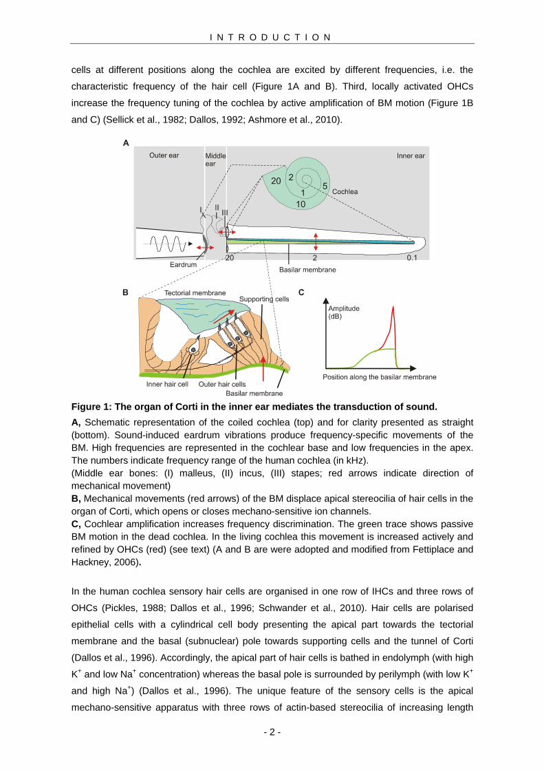
Figure 1: The organ of Corti in the inner ear media tes the transduction of sound.
A, Schematic representation of the coiled cochlea (top) and for clarity presented as straight (bottom). Sound-induced eardrum vibrations produce frequency-specific movements of the BM. High frequencies are represented in the cochlear base and low frequencies in the apex. The numbers indicate frequency range of the human cochlea (in kHz). (Middle ear bones: (I) malleus, (II) incus, (III) stapes; red arrows indicate direction of mechanical movement) B, Mechanical movements (red arrows) of the BM displace apical stereocilia of hair cells in the organ of Corti, which opens or closes mechano-sensitive ion channels. C, Cochlear amplification increases frequency discrimination. The green trace shows passive BM motion in the dead cochlea. In the living cochlea this movement is increased actively and refined by OHCs (red) (see text) (A and B are were adopted and modified from Fettiplace and Hackney, 2006).
In the human cochlea sensory hair cells are organised in one row of IHCs and three rows of
OHCs (Pickles, 1988; Dallos et al., 1996; Schwander et al., 2010). Hair cells are polarised
epithelial cells with a cylindrical cell body presenting the apical part towards the tectorial
membrane and the basal (subnuclear) pole towards supporting cells and the tunnel of Corti
(Dallos et al., 1996). Accordingly, the apical part of hair cells is bathed in endolymph (with high
K+ and low Na+ concentration) whereas the basal pole is surrounded by perilymph (with low K+
and high Na+) (Dallos et al., 1996). The unique feature of the sensory cells is the apical
mechano-sensitive apparatus with three rows of actin-based stereocilia of increasing length
I N T R O D U C T I O N
- 3 -
(Lim, 1980). The stereocilia of IHCs and OHCs are made-up by the same molecular
components and resemble each other in size, geometry and mechanics (Russell et al., 1986b;
Russell et al., 1986a; Russell et al., 1992), but only the OHC hair bundle is embedded into the
tectorial membrane (Lim, 1980) (see Figure 1B). Thus, the bundle of OHCs is believed to be
displaced by movements of the tectorial membrane relative to the BM, whereas the bundle of
IHCs is displaced by motions of the surrounding fluid induced by BM acceleration (Dallos,
1986; Fettiplace and Hackney, 2006). Displacement of the stereocilia towards the longest row
opens (gates) MET channels, whereas deflection into the opposing direction closes the
channels (Corey and Hudspeth, 1983; Russell and Richardson, 1987). It is believed that MET
channel gating is mediated via elastic "gating springs", but neither the molecular nature of the
gating spring nor the molecular components of MET channels have been identified so far
(Gillespie and Muller, 2009; Schwander et al., 2010). K+-carried currents through the MET
channels (transduction currents) depolarise IHCs and OHCs. In IHCs depolarisation stimulates
Ca2+-dependent glutamate release from the basal pole that increases the action potential
frequency of postsynaptic neurons (Moser et al., 2006; Meyer and Moser, 2010). In contrast,
voltage changes drive active length changes of the OHC's cell body, which is the mechanism
underlying cochlear amplification.
1.2 Outer Hair Cells and the Cochlear Amplifier
In the dead cochlea the low sensitivity, low frequency selectivity and linear dependence of BM
movements from stimulus intensity are determined by the mechanical properties of the BM
and can be explained by passive movement of the involved structures (Figure 1C, green
trace). (Von Békésy, 1960; Pickles, 1988; Dallos, 1992). The structure of the BM, however,
does not explain transduction at low stimulus levels, when viscous dampening of the BM and
additional passive filtering by involved structures should cause total loss of the energy (Von
Békésy, 1960). In contrast, in the living cochlea even low stimulus levels produce robust BM
movements that are maximal and sharpest at the characteristic frequency of the respective
BM portion (Figure 1C, red trace) (Sellick et al., 1982). Such frequency tuning indicates active
amplification of BM movements. Indeed, OHCs were shown to generate additional mechanical
output in response to BM displacement which increases BM movement and sharpens cochlear
frequency selectivity (Ryan and Dallos, 1975; Dallos and Harris, 1978; Dallos, 1992; Fettiplace
and Hackney, 2006). This active enhancement of BM movements by OHCs is referred to as
cochlear amplification for which two mechanisms have been proposed: active hair bundle
movement and somatic electromotility. Hair bundle movements originate from Ca2+-dependent
MET channel desensitisation that actively moves the hair bundle back to its resting position
(Fettiplace and Hackney, 2006; Ashmore et al., 2010). Such movement was identified in lower
vertebrates (e.g. turtle and frog) and may also exist in mammals, but it remains controversial
whether the generated force is sufficient to contribute to the amplification of BM movements
(Benser et al., 1996; Martin and Hudspeth, 1999; Ricci et al., 2000; Chan and Hudspeth, 2005;
I N T R O D U C T I O N
- 4 -
Kennedy et al., 2005). The more accepted mechanism of cochlear amplification is length
changes of the OHC's cell body driven by the membrane potential, also referred to as somatic
electromotility. Depolarisation (by gating of MET channels) results in contraction and
hyperpolarisation in elongation of the cells. The length changes are mediated by voltage-
dependent conformational changes of prestin, a protein in the lateral membrane of OHCs
(Brownell et al., 1985; Ashmore, 1987; Zheng et al., 2000; Fettiplace and Hackney, 2006).
Since acoustic stimulation changes the membrane potential of OHCs (see 1.2.1), length
changes follow audio frequencies and produce a "cycle-by-cycle" force on the BM and the
tectorial membrane driven by depolarisation followed by hyperpolarisation (Dallos et al., 1996;
Johnson et al., 2011). This mechanical output increases the amplitude of BM movements in
response to acoustic stimulation and is the cellular mechanism underlying cochlear
amplification (Liberman et al., 2002; Dallos et al., 2008).
In summary, OHCs mediate cochlear amplification through two specialisations: First,
mechanical movement of the BM produces a receptor potential that, second, generates
mechanical force and enhances BM movement. This requires membrane potential changes as
well as conformational responses of prestin and consequently somatic length changes of
OHCs. The electrical properties of OHCs enabling cochlear amplification will be discussed in
the following section.
1.2.1 The Electrophysiology of Outer Hair Cells
The membrane potential of OHCs is determined by depolarising K+ currents through the MET
channels and by hyperpolarising currents through K+ channels in the hair cell's basolateral
membrane (in the nuclear region of the cell). At rest (without displacement of the hair bundle)
approximately 50% of the MET channels are active, setting the membrane potential to around
-40 mV (Johnson et al., 2011). Additional gating of MET channels causes K+-dependent
depolarisation, whereas closing of the MET channels hyperpolarises the membrane potential
(Corey and Hudspeth, 1983; Johnson et al., 2011). Accordingly, mechanical displacement of
the hair bundle by acoustic stimulation causes sinusoidal variations around the resting
potential, i.e. an alternating current (AC) receptor potential (Dallos et al., 1996; Hille, 2001).
The AC receptor potential will be attenuated above a certain stimulus frequency due to low-
pass filtering of biological membranes. In other words, above this cut-off frequency the
receptor potential cannot follow the stimulus anymore and its amplitude is attenuated (Hille,
2001). This is relevant for prestin-driven somatic electromotility. Active amplification of BM
movements via positive feedback requires "cycle-by-cycle" length changes in response to
alternating depolarisation and hyperpolarisation (Johnson et al., 2011). Thus, OHCs amplify
BM movement only if they respond to acoustic stimulation with somatic length changes. If the
stimulus frequency exceeds the cut-off frequency of the OHC, attenuation of the receptor
potential attenuates prestin-mediated electromotility. This truncates positive feedback and
I N T R O D U C T I O N
- 5 -
theoretically limits cochlear amplification to the low frequencies where the receptor potential is
not attenuated, since the stimulus frequency is lower than the cut-off frequency.
The cut-off frequency of the circuit is determined by the time constant (tau) that describes the
time needed to recharge a capacitor through a resistor (tau = R x C where R and C are the
input resistance and membrane capacitance, respectively). Accordingly, the cut-off frequency
increases with faster time constants of the membrane (Hille, 2001). The input resistance (R) of
biological membranes depends on the amount of functional ion channels, i.e. the resistance
decreases with increasing number of activated ion channels. In OHCs voltage-dependent K+
channels are located in the basolateral membrane that dominate the OHC-membrane time
constant and shape the receptor potential (Hille, 2001; Johnson et al., 2011). The K+ channels
are activated completely at rest (around -40 mV), which lowers the membrane resistance and
speeds the membrane time constant substantially (Johnson et al., 2011). In fact, in OHCs K+
and MET current amplitudes are bigger in the cochlear base than in the apex, resulting in a
high cut-off frequency in high frequency OHCs (Dallos et al., 1982; Dallos, 1985; Housley and
Ashmore, 1992; Johnson et al., 2011). Accordingly, complete activation of K+ channels at the
basolateral OHC membrane enables fast prestin-driven electromotility and cochlear
amplification even at the highest frequencies in the cochlea. The following chapter describes
the K+ channels of OHCs.
1.2.2 IK,n is the Predominant K + Current of Outer Hair Cells
The predominant voltage-dependent K+ conductance in OHCs is IK,n that shows an unusually
negative voltage range of activation. IK,n currents activate at -110 mV and current amplitudes
saturate around -40 mV. Thus, IK,n is fully activated at the OHC resting membrane potential,
i.e. IK,n is a physiologically constitutively open current ("background current") (Housley and
Ashmore, 1992; Mammano and Ashmore, 1996; Nenov et al., 1997; Marcotti and Kros, 1999;
Johnson et al., 2011). Thus, as IK,n provides the main K+ conductance at all potentials it
dominates the membrane time constant of OHCs, presumably enabling cochlear amplification
at high frequencies (see 1.2.1) (Housley and Ashmore, 1992; Johnson et al., 2011). In murine
OHCs the developmental appearance of IK,n concurs with the onset of somatic electromotility
around postnatal days eight to ten, implicating the involvement of the conductance in the
biophysical and structural maturation of OHCs around the onset of hearing (days 10 to 12)
(Marcotti and Kros, 1999; Oliver and Fakler, 1999).
Additional basolateral K+ currents in OHCs are IK and SK. IK was identified as large
conductance voltage- and calcium-activated K+ current (BKCa; Maxi-K) (Housley and
Ashmore, 1992; Mammano et al., 1995; Mammano and Ashmore, 1996; Ruttiger et al., 2004).
It resembles a conductance in IHCs and contributes to the fine-tuning of the receptor potential
(Ruttiger et al., 2004; Thurm et al., 2005; Oliver et al., 2006). OHCs also express small
conductance calcium-activated K+ channels (SK) at the basal cell pole that mediate fast
efferent inhibition (Dallos et al., 1996). Acetylcholine increases Ca2+ levels in OHCs via
I N T R O D U C T I O N
- 6 -
postsynaptic nicotinic acetylcholine receptors. Elevated Ca2+ levels in turn activate SK
channels that through K+ currents hyperpolarise the hair cell (Elgoyhen et al., 1994; Oliver et
al., 2000).
Taken together, the physiology of OHCs largely depends on K+ channels at the basolateral
membrane. The electrical properties of the OHC membrane are dominated by IK,n that exhibits
unusual biophysical properties. The following section discusses these characteristics also
considering the voltage-dependent K+ channel KCNQ4 (Kv7.4) that was shown to constitutes
IK,n in OHCs (Kharkovets et al., 2006).
1.2.3 KCNQ4 Mediates I K,n, but Both Channels Show Biophysical Differences
KCNQ4 (Kv7.4) is a member of the KCNQ family (KCNQ1 - KCNQ5, Kv7) of voltage-
dependent potassium channels and was shown to mediate IK,n (Kharkovets et al., 2006; Holt et
al., 2007). Whereas other KCNQ isoforms (e.g. KCNQ2/KCNQ3) are expressed throughout
the nervous system (reviewed in Jentsch, 2000), the expression profile of KCNQ4 seems to be
restricted to hair cells of the inner ear, neurons of the auditory brainstem, mechano-receptors
in the skin and vascular smooth muscle cells (Heidenreich et al.; Kharkovets et al., 2000;
Chambard and Ashmore, 2005; Yeung et al., 2007). KCNQ4 was identified as the molecular
correlate of IK,n based on various findings: First and most convincing, genetic disruption of
KCNQ4 eliminates IK,n in OHCs (Kharkovets et al., 2006). Second, co-expression of KCNQ4
subunits carrying loss-of-function mutations reduces IK,n by a dominant-negative effect, i.e. the
function of the native channel complex is disrupted by mutant KCNQ4 subunits (Holt et al.,
2007). Third, IK,n is sensitive to specific KCNQ channel inhibitors, XE991 and linopirdine,
identifying the conductance as carried by KCNQ subunits (Marcotti and Kros, 1999). Fourth,
KCNQ4 was shown to be expressed in the basolateral membrane of OHCs. In addition, the
developmental expression of KCNQ4 around days eight to ten matches well with the
appearance of IK,n (Kubisch et al., 1999; Kharkovets et al., 2000). Taken together, compelling
evidence points to KCNQ4 as the molecular correlate of IK,n. However, native IK,n shows
biophysical characteristics that are not reproduced by recombinant KCNQ4 (Housley and
Ashmore, 1992; Kubisch et al., 1999). Most obviously, IK,n activates at hyperpolarised
potentials (half-maximal voltages of activation, Vh, -80 mV) compared to recombinant KCNQ4
(Vh between around -20 mV) (Mammano and Ashmore, 1996; Kubisch et al., 1999; Marcotti
and Kros, 1999). The gating kinetics of IK,n are substantially faster than of recombinant KCNQ4
(Housley and Ashmore, 1992; Mammano and Ashmore, 1996). In addition, the
pharmacological properties of IK,n differ from heterologously expressed KCNQ4. IK,n displays
higher sensitivity to inhibition by KCNQ antagonists linopirdine and XE991 (Sogaard et al.,
2001; for IHCs see Oliver et al., 2003; Xu et al., 2007). Moreover, IK,n is largely insensitive to
the K+ channel inhibitor tetraethylammonium (TEA) which potently blocks recombinant KCNQ4
(Housley and Ashmore, 1992; Marcotti and Kros, 1999; Hadley et al., 2000).
I N T R O D U C T I O N
- 7 -
The extraordinary biophysical properties of IK,n cannot be explained by KCNQ4 channel
subunits alone. Since heterologous expression systems normally reproduce biophysical
properties of ion channels quite well, these findings imply an OHC-specific mechanism
responsible for the negative voltages of activation of IK,n. Attractive hypothesis arise from the
presence of hitherto unidentified accessory channel subunits, posttranslational modification or
a yet unknown mechanism. In any case, an OHC-specific mechanism seems to be the most
likely explanation, but its molecular mechanism still needs to be elucidated. One possibility
may be the presence of additional KCNQ subunits (e.g. KCNQ3) in the native channel
complex. Co-expression of KCNQ4 together with KCNQ3 produced currents with amplitude
and gating kinetics similar to IK,n, but failed to reproduce the negative voltage range of
activation (Kubisch et al., 1999; Bal et al., 2008). Also the low TEA sensitivity of IK,n might be
explained by the co-expression of KCNQ4 with KCNQ subunits with low TEA sensitivity (e.g.
KCNQ3; Hadley et al., 2000). However, it remains controversial whether KCNQ3 is expressed
functionally in OHCs (Kubisch et al., 1999; Kharkovets et al., 2000). Another possibility
emerges from the presence of accessory KCNE β-subunits that have been shown to alter the
biophysical and pharmacological properties of the KCNQ α-subunit (McCrossan and Abbott,
2004). All KCNE isoforms were identified in the cochlea and all isoforms seem to co-assemble
with recombinant KCNQ4 (Strutz-Seebohm et al., 2006). However, none of the KCNE
isoforms produced biophysical characteristics of KCNQ4 explaining the properties of IK,n
(Strutz-Seebohm et al., 2006).
Taken together, an OHC-specific mechanism produces the biophysical properties of IK,n, but
the molecular nature of this mechanism remains elusive. Since the biophysical and
pharmacological properties of KCNQ channels depend on the subunit composition of the
channel tetramer, the pharmacological characterisation of IK,n may help to identify channel
subunits apart from KCNQ4 in the native channel complex. Thus, one aspect of this thesis
was the analysis of fundamental biophysical and pharmacological "KCNQ-like" characteristics
of IK,n and the comparison of these properties to recombinant KCNQ channels.
1.2.4 IK,n/KCNQ4 is Essential for the Survival of Outer Hair Cells
It is well established that IK,n/KCNQ4 is essential for the survival of OHCs (Kharkovets et al.,
2000; Winter et al., 2006). Pharmacological inhibition, genetic ablation and loss of KCNQ4
channel function in human hereditary deafness DFNA2 lead to progressive OHC degeneration
that starts at the cochlear base and proceeds to the apex (reviewed in Jentsch, 2000; Nouvian
et al., 2003; Kharkovets et al., 2006; Nie, 2008). Basal OHCs with highest KCNQ4 expression
and biggest IK,n current amplitudes are most susceptible to degeneration whereas apical
OHCs with smallest IK,n currents remain unaffected (Housley and Ashmore, 1992; Kubisch et
al., 1999; Beisel et al., 2000; Nouvian et al., 2003; Kharkovets et al., 2006). The base-to-apex
loss of OHCs causes progressive hearing impairment that starts at high and proceeds to low
frequencies (Kharkovets et al., 2006; Smith and Hildebrand, 2008). In affected humans,
I N T R O D U C T I O N
- 8 -
hearing loss starts in the second or third decade of life and culminates in profound deafness at
later ages (Nie, 2008; Smith and Hildebrand, 2008). The disease phenotype was recapitulated
by the KCNQ4 knock-out mouse demonstrating that profound hearing impairment was caused
by loss of OHCs and absent cochlear amplification (Kharkovets et al., 2006; Smith and
Hildebrand, 2008). Although KCNQ4 was reported to be expressed also in IHCs (Marcotti et
al., 2003; Oliver et al., 2003), vestibular type I hair cells (Rusch and Eatock, 1996; Holt et al.,
2007) and neurons of the auditory brainstem (Kubisch et al., 1999; Kharkovets et al., 2000)
the degree of hearing impairment does not implicate the involvement of IHCs or neuronal
deficits (Kharkovets et al., 2006). However, profound deafness in elderly DFNA2 patients does
not totally rule out additional loss of IHCs at later disease stages (Oliver et al., 2003).
Vestibular defects have not been reported consistently from affected humans or from the
KCNQ4 knock-out mouse (Kharkovets et al., 2006; Smith and Hildebrand, 2008). Hearing loss
was attributed to the loss of KCNQ4 channel function, but the molecular mechanism causing
hair cell degeneration remains elusive. Since KCNQ4 is expressed in the basolateral
membrane of OHCs, the channel was proposed to serve as the basolateral exit path of K+ ions
to the tunnel of Corti (see Figure 1B) (Housley and Ashmore, 1992; Jentsch, 2000; Jentsch et
al., 2000). Thus, IK,n/KCNQ4 may regulate K+ levels in OHCs serving as a key element for the
recycling of K+ ions back to the endolymph via a system of gap junctions and the stria
vascularis (Mistrik and Ashmore, 2009). From this follows that loss of KCNQ4 channel function
disturbs the K+ homeostasis of OHCs together with persistent K+ influx through the MET
channels. Most probably, sustained K+ overload causes prolonged Ca+2 influx through voltage-
dependent Ca2+ channels and Ca2+-dependent OHC degeneration (Zenner et al., 1994;
Jentsch, 2000; Jentsch et al., 2000).
KCNQ4 loss-of-function causes hair cell degeneration and deafness in DFNA2 patients
(Kubisch et al., 1999; Kharkovets et al., 2006). Several DFNA2-causing KCNQ4 mutations
have been identified that disturb channel function through disruption of ion permeation or by a
reduction of channels expressed at the cell surface (summarised in Smith and Hildebrand,
2008). Accordingly, KCNQ4 mutations reduce IK,n, which initiates OHC degeneration
(Kharkovets et al., 2006; Holt et al., 2007). A variety of chemical KCNQ channel openers are
available, and some of these substances are already in clinical use for the treatment of
neurological disorders (Wulff et al., 2009). Importantly, some KCNQ channel agonists (e.g.
retigabine, zinc pyrithione) were shown to rescue channel function of epileptogenic KCNQ2
mutants (Biervert et al., 1998; Schroeder et al., 1998; Xiong et al., 2007; Xiong et al., 2008). In
analogy, chemical KCNQ openers may rescue IK,n/KCNQ4-mediated currents in DFNA2
patients offering a protective strategy against OHC degeneration. Despite the obvious benefits
of such current augmentation, it has never been tested so far whether the substances rescue
channel function of DFNA2 relevant KCNQ4 mutants or whether IK,n is sensitive to chemical
current potentiation. Thus, this thesis aims to evaluate the potency of KCNQ channel openers
in the treatment of KCNQ4-related hearing loss, DFNA2.
I N T R O D U C T I O N
- 9 -
1.2.5 Other Outer Hair Cell Pathologies May Be Rela ted to KCNQ4 Dysfunction
Ototoxic agents, noise exposure or aging cause the irreversible loss of OHCs and acquired
hearing impairment. Basal OHCs are far more vulnerable to degeneration than apical whereas
IHCs are not affected (Smith et al., 2008). The different hair cell susceptibility cannot be
explained so far. However, the loss of OHCs correlates directly with the dependence of OHCs
on IK,n/KCNQ4 (Nouvian et al., 2003; Kharkovets et al., 2006). Although a potential role of
KCNQ4 in age-related deafness has been proposed previously, the involvement of IK,n/KCNQ4
in acquired hearing loss has never been investigated (Van Eyken et al., 2006).
A clinically relevant form of acquired hearing loss is caused by aminoglycosides (AGs): AG-
antibiotics (e.g. neomycin) exhibit high efficiency in the treatment of bacterial infections, but
their use is limited, since most treated patients develop profound hearing impairment (Forge
and Schacht, 2000; Rybak and Ramkumar, 2007). Hearing loss is induced by irreversible
degeneration of OHCs that starts in the cochlear base and proceeds to the apex. Yet, this
base-to-apex loss of OHCs cannot be explained (Forge and Schacht, 2000). Noteworthy, it
resembles KCNQ4-related OHC degeneration (Kharkovets et al., 2006). AGs induce hair cell
death following entry into the cell from the endolymph via a not fully resolved entry pathway,
possibly via the MET channels (Alharazneh et al., 2011). The mechanisms leading to OHC
degeneration may involve the disruption of mitochondrial function (Dehne et al., 2002), the
production of reactive oxygen species (Rybak and Ramkumar, 2007), and the disturbance of
phosphoinositide (PI) metabolism (Jiang et al., 2006a; Goodyear et al., 2008). AGs have been
shown to chelate membrane-resident PIs via electrostatic interactions (Gabev et al., 1989).
This links AG-induced OHC loss to IK,n/KCNQ4, since KCNQ channels essentially require
membrane-resident phosphatidylinositol-(4,5)-bisphosphate (PI(4,5)P2) for channel activation
(Li et al., 2005; Suh et al., 2006). Accordingly, reduction of PI(4,5)P2 inhibits the channel (Suh
and Hille, 2002).
The dependence of OHC survival on IK,n, the PI(4,5)P2 dependence of KCNQ channels and
the chelation of PIs by AGs strongly indicate a contribution of AG-induced IK,n/KCNQ4
inhibition to AG-mediated OHC loss. The role of IK,n in ototoxic hair cell insult has never been
investigated so far, but IK,n inhibition by AGs may explain the high vulnerability of OHCs.
Positive evaluation of this hypothesis makes KCNQ4 a promising target in the treatment of
acquired hearing loss. Accordingly, this thesis evaluates the relevance of IK,n/KCNQ4 inhibition
for AG ototoxicity and the use of KCNQ channel openers to rescue IK,n function from AG-
induced inhibition.
AGs bind to PIs, which reduces their availability to ion channels. However, the degree of
current inhibition depends on the PI affinity and dependence of the respective channel.
Although the dependence on membrane-resident PI(4,5)P2 is the characteristic feature of the
KCNQ channel family, the phospholipid requirement for channel activation of IK,n has never
been investigated so far. The PI dependence of cellular processes has been investigated
previously using voltage-sensing phosphatases (VSPs) that, in contrast to unspecific PI
I N T R O D U C T I O N
- 10 -
chelation by AGs, specifically deplete certain PI species (Murata et al., 2005). VSPs are
modular enzymes that consist of a voltage-sensing domain (VSD) with high homology to
voltage-dependent ion channels coupled to a phosphatase domain (PD) (Murata et al., 2005).
Typically VSPs respond to membrane depolarisations with 5-phosphatase activity towards
PI(4,5)P2 and PI(3,4,5)P3 (Halaszovich et al., 2009). Strikingly, the homology of the PD to the
3-phosphatase PTEN (phosphatase and tensin homolog deleted from chromosome 10) may
enable the design of engineered VSPs with altered substrate specificity (Iwasaki et al., 2008;
Halaszovich et al., 2009). Once transfected into cells these enzymes can be used to analyse
the PI dependence and affinity of ion channels in straightforward electrophysiological
experiments. Even though VSPs are available, the difficile handling of OHCs in culture and
insufficient transfection efficiencies prevented these experiments so far. Thus, technically
refined tools and experimental settings are needed to analyse the PI dependence of IK,n.
Detailed knowledge of the PI dependence and specificity of IK,n allows for a more precise
evaluation of the impact of AGs on the native channel complex in OHCs. Thus, one aspect of
this thesis is the design and the characterisation of a novel VSP that contains the VSD of
prototypic Ci-VSP together with the 3-phosphatase PTEN. Native and designed VSP will be
used in combination to study the PI dependence of IK,n in future studies.
A I M S A N D C O N T R I B U T I O N
- 11 -
2 Aims and Contributions
2.1 Objectives
IK,n dominates the electrical properties of cochlear OHCs, enables prestin-mediated cochlear
amplification and is essential for the survival of OHCs. Nothing is known about the OHC-
specific molecular mechanism that determines the characteristic properties of IK,n. The
involvement of IK,n/KCNQ4 in acquired hearing loss is likely, but has not been investigated so
far. The work presented in this thesis aims to analyse the molecular properties of the
conductance, to evaluate the pathophysiological relevance and mechanisms of IK,n in AG
ototoxicity and to estimate potential benefits of chemical channel openers in the treatment of
KCNQ4-related hearing impairment.
I. The role of IK,n /KCNQ4 in AG-induced hair cell loss
The PI-dependence of ion channels has been investigated repeatedly by intracellular
application of AGs that functionally deplete PIs (Gabev et al., 1989; e.g. Schulze et al., 2003).
In the present work, AGs were used to estimate the PI affinity of IK,n allowing for the
comparisons with heterologously expressed KCNQ isoforms. Given the clinical relevance of
AG-mediated hair cell loss (see 1.2.5), in these experiments the physiological role of AG-
induced IK,n inhibition was evaluated.
II. The sensitivity of IK,n to chemical KCNQ channel openers and their potential for the treatment of KCNQ4-related hearing impairment
Loss of KCNQ4 channel function causes progressive OHC degeneration and hearing loss
(Nouvian et al., 2003; Kharkovets et al., 2006; Holt et al., 2007). Dysfunction of other KCNQ
isoforms (e.g. KCNQ2) causes epileptic seizures that are treated successfully with chemical
KCNQ channel openers (Biervert et al., 1998; Schroeder et al., 1998; Wulff et al., 2009). In
analogy, IK,n/KCNQ4 current potentiation by the openers may be used as protective strategy
for DFNA2 patients and against AG-induced hair cell loss (see (I)). The benefits of chemical
augmentation depend on the sensitivity of IK,n to channel openers. First, IK,n was characterised
pharmacologically and the sensitivity of IK,n towards chemical KCNQ openers was determined.
Second, the reconstitution of IK,n from AG-induced inhibition was assessed. Third, rescue of
DFNA2-causing KCNQ4 mutations by chemical KCNQ openers was investigated (Nie, 2008;
Smith and Hildebrand, 2008).
III. Analysis of the KCNQ subunit composition of the native channel complex in OHCs
The contribution of KCNQ channel subunits apart from KCNQ4 to native IK,n is still
controversial. The pharmacological properties and biophysical characteristics of KCNQ
channels depend on the subunit composition of the channel tetramer (Hadley et al., 2000;
Hernandez et al., 2008; Xiong et al., 2008; Hernandez et al., 2009; Zhang et al., 2011). The
sensitivity towards chemical current augmentation and the PI-dependence of activation of IK,n
were analysed and compared to heterologously expressed KCNQ channel subunits to draw
A I M S A N D C O N T R I B U T I O N
- 12 -
conclusions about the subunit composition of the native channel complex. Furthermore,
recombinant KCNQ4 was established as a sensor for physiologically relevant of PI(4,5)P2 level
changes.
IV. Technically-refined tools to investigate PI-dependent processes
AGs chelate PIs without any preference for a certain PI species. However, the impact of AGs
on channel function depends on the PI specificity and affinity of the channel. Accordingly,
knowledge of the PI affinity allows more detailed estimation of AG-induced channel inhibition.
The PI affinity of ion channels can be determined using stepwise activation of a VSP by
stepwise depolarisation. Since prototypic VSPs are PI(4,5)P2- and PI(3,4,5)P3-specific 5-
phosphatases, the enzymes can be used to determine the affinity of an ion channel towards
these PI species. Voltage-controlled PI(3,4,5)P3 3-phosphatases are not yet available, but the
cytoplasmic 3-phosphatase PTEN shares high sequence homology to the PD of prototypic
VSPs (Okamura and Dixon, 2011). This homology presumably enables the design of a
voltage-dependent 3-phosphatase consisting of the VSD of Ci-VSP coupled to PTEN. In the
work presented, the enzymatic activity and substrate specificity of this chimera were analysed
and the potential of designed VSPs in the analysis of the PI-dependence of ion channels was
evaluated. Further studies will make use of this work for detailed analysis of the PI
dependence of IK,n in OHCs.
2.2 My Contributions to the Articles Presented
In this section, I state my contributions to the publications presented in this thesis.
(1) Michael G. Leitner , Christian R. Halaszovich and Dominik Oliver. Aminoglycosides
inhibit KCNQ4 channels in cochlear outer hair cells via depletion of
phosphatidylinositol(4,5)bisphosphate. Molecular Pharmacology. 2011 Jan:79(1):51-60
(Leitner et al., 2011)
I designed the research together with Prof. Dr. Dominik Oliver, performed all
electrophysiological and imaging experiments including the data analysis and wrote the paper
together with Prof. Dr. Dominik Oliver.
(2) Michael G. Leitner , Anja Feuer, Olga Ebers, Daniela N. Schreiber, Christian R.
Halaszovich and Dominik Oliver. Restoration of ion channel function in deafness
causing KCNQ4 mutations by chemical openers. British Journal of Pharmacology (2012)
165 2244-2259 (Leitner et al., 2012)
I conceived the project, performed the electrophysiological experiments with data analysis and
wrote the paper together with Prof. Dr. Dominik Oliver.
A I M S A N D C O N T R I B U T I O N
- 13 -
(3) Moritz Lindner, Michael G. Leitner , Christian R. Halaszovich, Gerald R. V.
Hammond and Dominik Oliver. Probing the regulation of TASK potassium channels
by PI(4,5)P2. Journal of Physiology. 2011 Jul 1; 589 (Pt 13):3149-62 (Lindner et al.,
2011)
This study is part of the doctoral thesis of Moritz Lindner in the group of Prof. Dr. Dominik
Oliver. I planned, performed and analysed experiments presented in Figure 1 (Receptor-
mediated inhibition of TASK channels and concomitant depletion of PI(4,5)P2) and Figure 2B
(TASK-currents are insensitive to depletion of PI(4,5)P2 by Ci-VSP).
(4) Jérôme Lacroix, Christian R. Halaszovich, Daniela N. Schreiber, Michael G. Leitner ,
Francisco Bezanilla, Dominik Oliver and Carlos A. Villalba-Galea. Controlling the
activity of PTEN by membrane potential. Journal of Biological Chemistry. 2011 May
20; 286(20): 17945-53 (Lacroix et al., 2011)
This project was realised in collaboration with Prof. Dr. Francisco Bezanilla (Chicago, Illinois,
USA) and Carlos A. Villalba-Galea (Richmond, Virginia, USA). In summary, my contributions
to this study are experiments and data analysis shown in Figure 2 (Ci-VSPTEN displays
voltage-activated lipid phosphatase activity; all panels), Figure 5 (Binding of the PBM is
essential for phosphatase activation by the VSD) and Figure 6A (Experimental control of Ci-
VSPTEN activity in intact cells without use of electrophysiological instrumentation).
Hiermit bestätige ich die Richtigkeit der unter 2.2 gemachten Angaben bezüglich des
Eigenanteiles von Michael Leitner an den aufgeführten Publikationen.
Marburg, März 2012
Michael Leitner Prof. Dr. Dominik Oliver
(Autor) (Betreuer)
A I M S A N D C O N T R I B U T I O N
- 14 -
3 Results
3.1 The Outer Hair Cell K + Current I K,n Requires PI(4,5)P 2 for Activation
The PI dependence of ion channels has been investigated repeatedly by intracellular application
of AGs. The positively charged substances bind to the polyanionic PIs, thereby functionally
depleting PIs and reducing their availability to the ion channel (Gabev et al., 1989; Oliver et al.,
2004). In these experiments AGs were applied via a whole cell patch pipette and IK,n or
recombinant KCNQ4 were recorded (Leitner et al., 2011) . Introduction of AGs into OHCs
inhibited IK,n. The time course of inhibition was rapid suggesting direct action of the chelators on
the channels rather than inhibition via intracellular messenger systems (Leitner et al., 2011;
Figure 1) . The degree of current inhibition correlated with the amount of positive charges in the
descending order of neomycin > gentamicin > kanamycin. Intracellular neomycin dose-
dependently inhibited IK,n, whereas ampicillin, a structurally not related antibiotic, was ineffective.
These findings indicated that IK,n inhibition depended on the positive charges of the substances.
Analogous experiments with KCNQ4 heterologously expressed in Chinese hamster ovary (CHO)
cells revealed that the same substances also robustly inhibited recombinant KCNQ4 (Leitner et
al., 2011; Figure 2) . The rank order of KCNQ4 current inhibition was similar to native IK,n, albeit
current inhibition by the chelators was more pronounced compared to IK,n. These findings
indicated higher sensitivity to AG-mediated inhibition of heterologously expressed KCNQ4. Given
the well known binding of AGs to phospholipids and the dependence of KCNQ channel activation
on PI(4,5)P2, these findings strongly suggested inhibition of IK,n/KCNQ4 via the depletion of PIs
(Gabev et al., 1989; Suh and Hille, 2002). I monitored the availability of free PIs upon intracellular
dialysis of neomycin and kanamycin using the PI(4,5)P2 sensor tubby-Cterm-GFP and total
internal reflection fluorescence (TIRF) microscopy (Halaszovich et al., 2009; Leitner et al., 2011;
Figure 3) . In brief, the membrane-association of tubby-Cterm is a direct measure for membrane-
resident PI(4,5)P2. Since in TIRF microscopy only membrane-bound fluorophores are excited, the
fluorescence intensity of GFP-labelled tubby-Cterm directly reports on the amount of PI(4,5)P2 at
the membrane (Santagata et al., 2001; Halaszovich et al., 2009). Introduction of neomycin or
kanamycin via the patch pipette into CHO cells expressing tubby-Cterm-GFP caused rapid
decline of membrane-associated fluorescence indicating the functional depletion of PI(4,5)P2
(Leitner et al., 2011; Figure 3) . The degree of PI(4,5)P2 chelation was stronger for neomycin
than for kanamycin, i.e. neomycin with more positive charges bound PI(4,5)P2 more efficiently
than kanamycin. Of special note, these findings are in agreement with stronger current inhibition
of IK,n and recombinant KCNQ4 by neomycin than by kanamycin. In addition, the time course of
PI(4,5)P2 depletion matched the electrophysiological experiments (compare to Leitner et al.,
2011 Figure 1 and 2) . To see whether KCNQ4 current reduction by AGs depended on the
availability of free PI(4,5)P2 I increased basal PI(4,5)P2 levels in CHO cells by over-expression of
R E S U L T S
- 15 -
a PI(4)-5-kinase (e.g. Li et al., 2005). In these cells KCNQ4 current inhibition was significantly
reduced compared to control cells (Leitner et al., 2011, Figure 3) .
Taken together, these findings strongly suggested inhibition of IK,n and recombinant KCNQ4 by
functional sequestration of PI(4,5)P2 through AGs. Additionally, I demonstrated the PI-
dependence of IK,n, which indicated a role of IK,n current inhibition in AG ototoxicity. Furthermore, I
was able to show that KCNQ4 currents can be used along with fluorescent PI sensors to monitor
PI(4,5)P2 changes in living cells (Lindner et al., 2011) .
3.2 Inhibition of I K,n by Aminoglycosides is Pathophysiologically Relevan t
Given the inhibition of IK,n by AGs and the degeneration of OHCs caused by KCNQ4 dysfunction,
I investigated the relevance of IK,n in AG ototoxicity (Kharkovets et al., 2006; Rybak and
Ramkumar, 2007). In the experiments presented in 3.1 AGs were introduced into the cell.
However, ototoxic degeneration of OHCs occurs subsequent to entry of AGs into hair cells from
the endolymph (Forge and Schacht, 2000; Marcotti et al., 2005; Dai et al., 2006; Wang and
Steyger, 2009). Thus, I examined whether AG entry into OHCs was sufficient to cause relevant
IK,n current inhibition (Leitner et al., 2011; Figure 4) . I applied AGs from the extracellular side
and recorded IK,n currents in OHCs. When applied from the extracellular side, AGs inhibited IK,n
with the same rank order of current inhibition as upon intracellular application (neomycin >
kanamycin) (compare to Leitner et al., 2011; Figure 1) . Noteworthy, the potency of the
substances matched the clinical ototoxic potential with neomycin having the most detrimental
impact on OHCs (Forge and Schacht, 2000). Extracellularly applied AGs robustly depolarised
OHCs highlighting the physiological relevance of AG-induced IK,n inhibition (Leitner et al., 2011;
Figure 6) . Analogous experiments in CHO cells showed that AGs did not inhibit recombinant
KCNQ4 when applied from the extracellular side (Leitner et al., 2011; Figure 6) . These findings
strongly suggested OHC specific inhibition of IK,n. Since AGs were shown to enter hair cells
specifically, we hypothesised that AGs rapidly entered OHCs and inhibited IK,n via intracellular
depletion of PI(4,5)P2. To see whether the AG entry was reasonably fast to explain IK,n inhibition, I
investigated the entry of AGs into hair cells with fluorescently labelled neomycin and confocal
microscopy. Indeed, confocal imaging with neomycin conjugated to Texas Red revealed fast
accumulation of AGs in hair cells as detected by rapid increase of cellular fluorescence (Leitner
et al., 2011; Figure 5) . AGs specifically entered hair cells and the time course was rapid enough
to explain IK,n inhibition (Leitner et al., 2011; Figure 5) . In contrast, CHO cells did not take up
AGs in agreement with absent current inhibition of recombinant KCNQ4 by AGs applied from the
extracellular side (Leitner et al., 2011; Supplemental Figure 2) .
Taken together, I demonstrated rapid entry of AGs into OHCs from the extracellular
(endolymphatic) side and AG-induced inhibition of IK,n via functional depletion of PI(4,5)P2. These
findings strongly suggest a role of IK,n inhibition in AG ototoxicity.
R E S U L T S
- 16 -
3.3 IK,n is Sensitive to Chemical KCNQ Openers
Loss of KCNQ4 channel function causes OHC degeneration and deafness (Kharkovets et al.,
2006). Since OHC survival was directly attributed to IK,n/KCNQ4 function, it can be hypothesised
that augmentation of IK,n by previously described channel openers may alleviate or even avert this
pathology. However, the benefit depends on the sensitivity of native IK,n to the channel openers.
Thus, I characterised the sensitivity of recombinant KCNQ4 and IK,n towards KCNQ agonists
(Leitner et al., 2011; Leitner et al., 2012) . The substances robustly potentiated recombinant
KCNQ4 currents and shifted the voltage-dependence of activation to hyperpolarised voltages
(Leitner et al., 2012; Figure 1) . The rank order of current potentiation was flupirtine < retigabine
< BMS-204352 < zinc pyrithione << zinc pyrithione/retigabine (ZnP/Ret) in line with reports from
other KCNQ isoforms (Tatulian et al., 2001; Xiong et al., 2007; Xiong et al., 2008). The
combination of ZnP/Ret was most effective and shifted the voltage dependence of activation of
heterologously expressed KCNQ4 by -40 mV (Leitner et al., 2012; Supplemental Figure 1) .
Similarly, KCNQ openers robustly augmented IK,n in OHCs and shifted the voltage-dependence of
activation to hyperpolarised voltages (Leitner et al., 2012; Figure 7) . The rank order of current
enhancement was similar, but current potentiation was less pronounced for IK,n than for
recombinant KCNQ4 (Leitner et al., 2012, Figures 1 and 7) . Of special interest, retigabine that is
clinically used as antiepileptic drug also increased IK,n (Leitner et al., 2012, Figure 7) .
In conclusion, IK,n was sensitive to current potentiation by chemical KCNQ openers, albeit the
degree of current potentiation was lower than for recombinant KCNQ4. The sensitivity of IK,n to
KCNQ channel openers, especially to retigabine, may be used to rescue IK,n in case of KCNQ4
dysfunction.
3.4 KCNQ Channel Openers Rescue I K,n from AG-induced Inhibition and
Reconstitute Channel Function of Deafness-Causing K CNQ4 Mutants
Next I tested whether KCNQ channel agonists rescued IK,n from pathophysiologically relevant loss
of function. I started with AG-induced inhibition of IK,n (Leitner et al., 2011) . In brief, neomycin
inhibited IK,n and substantially depolarised OHCs (Leitner et al., 2011; Figures 6 and 7) .
ZnP/Ret rescued IK,n and fully reversed AG-induced depolarisation to control levels before AG
application (Leitner et al., 2011; Figure 7) . IK,n current amplitudes and the membrane potential of
the cells treated with ZnP/Ret in presence of AGs were indistinguishable from normal control
cells. These findings showed that KCNQ agonists could be used to stabilise the KCNQ
conductance in OHCs despite the presence of AGs. KCNQ channel openers thus might offer a
protective strategy against AG-mediated OHC degeneration through augmentation of IK,n (Leitner
et al., 2011; Figure 7) .
Several KCNQ4 loss-of-function mutations have been shown to cause hereditary hearing loss in
humans (Nie, 2008; Smith and Hildebrand, 2008; Kim et al., 2011). I tested whether KCNQ
openers rescued channel function of DNFA2 relevant KCNQ4 mutations (Leitner et al., 2012) . I
over-expressed mutant KCNQ4 subunits in CHO cells and performed whole cell patch clamp
R E S U L T S
- 17 -
experiments. Recombinant KCNQ4 channels carrying a mutation in the pore region could not be
rescued by KCNQ agonists, which suggested total loss of function in these mutants (Leitner et
al., 2012; Figure 3 ). A mutation localised close to the C-terminus of KCNQ4 produced small
currents under control conditions. Application of ZnP/Ret uncovered voltage-dependent KCNQ4
currents mediated by the mutant subunits. The ZnP/Ret-induced currents through the mutant
channels were slightly smaller than KCNQ wild-type (wt) control currents, i.e. the mutant was
rescued partially to wt levels (Leitner et al., 2012; Figure 4) . Most affected individuals are
heterozygous carriers of KCNQ4 mutations, which reduces IK,n by a dominant-negative effect, i.e.
a single mutant subunit disrupts the function of the tetrameric channel (Kubisch et al., 1999; Holt
et al., 2007; Kim et al., 2011). Accordingly, heterozygous patients produce the same amount of wt
and mutant KCNQ4 subunits, which leaves behind only 6.25% functional homomeric wt channels
at the cell surface. This reduction of functional channels causes the decrease of overall currents.
Co-expression of recombinant wt KCNQ4 with mutants in CHO cells reproduced the dominant-
negative situation (Leitner et al., 2012; Figure 5) . Whole cell currents were strongly reduced as
predicted from co-assembly of wt and mutant subunits and disruption of channel function by the
mutants. Strikingly, application of ZnP/Ret rescued the minute residual currents to wt levels, i.e.
currents were indistinguishable from KCNQ4 control currents (Leitner et al., 2012; Figure 5) .
This demonstrates full rescue of KCNQ4 currents from dominant-negative inhibition by KCNQ
channel openers. In the dominant-negative situation, the channel population at the cell membrane
comprises homomeric wt channels, homomeric mutant channels and heteromeric channels of
both, wt and mutant subunits. At this point, it was not clear whether also otherwise dysfunctional
heteromeric channels and with homomeric mutant channels contributed to ZnP/Ret-mediated
current rescue. We constructed concatamers of wt and mutant channel subunits to obtain
homogenous channel populations in the cell membrane and tested whether KCNQ openers
rescued the function of the resulting channels. Using the protein concatamers, I showed that
channel function of tetramers containing mutant subunits was not restored by the channel
openers. Current rescue resulted solely from potentiation of currents through remaining
homomeric wt channels. However, this current potentiation was sufficient to account for the
complete rescue of KCNQ4 from dominant-negative inhibition (Leitner et al., 2012; Figure 6) .
Taken together, these findings showed that KCNQ channel openers rescued KCNQ4 from
dominant-negative inhibition. Since native IK,n (see 3.3) was robustly augmented by KCNQ
channel agonists as well, these substances may be used to stabilise the conductance in OHCs.
This may postpone profound hearing loss or even protect OHCs from degeneration in DFNA2
patients.
3.5 The Biophysical Properties and the Molecular Na ture of I K,n
The subunit composition determines the biophysical and pharmacological properties of tetrameric
KCNQ channels (Hadley et al., 2000; Hernandez et al., 2009). Accordingly, it may be possible to
draw conclusions on the subunit composition from the biophysical properties. In brief, I found that
R E S U L T S
- 18 -
IK,n was inhibited by depletion of PIs via introduction of AGs. This is expected, since PI(4,5)P2-
dependent KCNQ4 subunits contribute to the native channel complex (Leitner et al., 2011) .
However, the IC50 for neomycin of IK,n was significantly higher than for recombinant KCNQ4 (IK,n
0.59 mM; KCNQ4 0.13 mM), indicating lower sensitivity to neomycin-induced current inhibition for
IK,n than of recombinant KCNQ4. Since current inhibition was caused by PI depletion (see 3.1),
the degree of current decrease allowed to estimate the PI affinity of the respective channel.
Accordingly, these results indicated higher PI affinity of IK,n than of recombinant KCNQ4 (Leitner
et al., 2011; Figures 1 and 2) . Equal neomycin concentrations (500 µM) inside the patch pipette
inhibited recombinant KCNQ4 more pronounced (approximately 80% inhibition) than IK,n, (30%
inhibition) albeit the time course of current inhibition was similar. Noteworthy, currents through
recombinant KCNQ3 with high PI(4,5)P2 affinity were reduced by only 10% upon dialysis of the
cells with 500 µM neomycin (Hernandez et al., 2009; Leitner et al., 2011; Figures 1, 2 and 3) .
These differences strongly suggested intermediate PI affinity of IK,n between recombinant KCNQ4
and KCNQ3. Similarly, co-expression of KCNQ2 with low PI(4,5)P2 affinity and KCNQ3 resulted
in heteromeric channels with intermediate PI(4,5)P2 affinity in previous reports (Hernandez et al.,
2009). In analogy, my findings suggested the contribution of additional subunits with high PI
affinity to IK,n in OHCs. Additionally, these experiments established recombinant KCNQ4 as a
perfect read-out for physiologically relevant PI(4,5)P2 changes. This makes KCNQ4 a bona fide
PI(4,5)P2 sensor that can be used to study the PI dependence of cellular processes (Lindner et
al., 2011).
Also the pharmacological profile of IK,n suggested the presence of additional subunits in OHCs.
Current potentiation by KCNQ openers was less pronounced for IK,n than for recombinant KCNQ4
(Leitner et al., 2012; Figure 1 and 7) . Similarly, also the agonist-mediated shift of the voltages of
activation to hyperpolarised potentials was smaller for the native channel complex. Since KCNQ3
shows lowest sensitivities towards chemical current potentiation of all KCNQ isoforms, co-
expression of KCNQ3 may be responsible for the special pharmacological profile of IK,n. Thus, I
co-expressed KCNQ4 together with KCNQ3 and analysed the sensitivity towards chemical
current potentiation of heteromeric channels (Leitner et al., 2012; Supplemental Figure 5) . As
previously reported, current amplitudes through heteromeric channels were increased and
activation kinetics were accelerated compared to homomeric KCNQ4 wt channels (Kubisch et al.,
1999). However, the voltage-dependence of the heteromers was not altered (Leitner et al., 2012;
Supplemental Figure 5) . Also the sensitivities towards chemical KCNQ channel openers was not
different from KCNQ4 wt channels, indicating that co-assembly of KCNQ4 together with KCNQ3
does not explain the unusual pharmacological properties of IK,n (Leitner et al., 2012) .
Taken together, although the PI sensitivity of IK,n suggested the co-expression of KCNQ3
subunits, the pharmacological properties of IK,n argued against the presence of KCNQ3 subunits
in OHCs. Thus, the molecular nature of IK,n remains unknown.
R E S U L T S
- 19 -
3.6 Development of Novel Tools to Experimentally Al ter PI Levels in Living Cells
Ciona intestinalis VSP (Ci-VSP) is a voltage-sensing phosphatase that exhibits PI(3,4,5)P3 and
PI(4,5)P2 5-phosphatase activity upon membrane depolarisation (Murata et al., 2005; Iwasaki et
al., 2008; Halaszovich et al., 2009). The PD of Ci-VSP shows high homology to the tumour
suppressor PTEN, a 3-phosphatase (Maehama and Dixon, 1998; Okamura and Dixon, 2011). We
constructed a chimera of PTEN coupled to the voltage sensor of Ci-VSP, termed Ci-VSPTEN.
The substrate specificity of the chimera was characterised in whole cell voltage clamp
experiments together with TIRF microscopy utilising fluorescent PI sensors (Lacroix et al., 2011) .
I found PI(3,4,5)P3 and PI(3,4)P2 3-phosphatase activity of Ci-VSPTEN fully reproducing the
substrate specificity of wt PTEN (Lacroix et al., 2011; Figure 1) . Voltage-dependent enzymatic
activity was conferred by a phosphoinositide binding motif in the linker between the VSD and PD,
as shown by mutations inside the motif that functionally uncoupled the VSD from phosphatase
activity (Lacroix et al., 2011; Figure 5) .
Taken together, we engineered a novel voltage-dependent 3-phosphatase by conferring voltage-
sensitivity to an otherwise cytoplasmic enzyme. These findings promise future design of voltage-
dependent phosphatases with desired substrate specificity. VSPs can be used to elucidate the PI
dependence of ion channels in their native environment. Thus, future studies will utilise designed
and native VSPs to analyse the PI-dependence of IK,n/KCNQ4 channel gating in OHCs.
D I S C U S S I O N
- 20 -
4 Discussion
4.1 Biophysical and Pharmacological Differences of IK,n and Recombinant KCNQ4:
Implications for the Molecular Identity of I K,n
Strong evidence indicates that KCNQ4 channel subunits mediate IK,n in OHCs (Kharkovets et al.,
2006). However, native and recombinant currents show different biophysical characteristics. Most
obviously IK,n, activates at more negative potentials than recombinant KCNQ4, the activation
kinetics of IK,n are faster and IK,n displays less sensitivity to chemical current inhibition (Mammano
and Ashmore, 1996; Kubisch et al., 1999; Marcotti and Kros, 1999). The molecular explanation
for these unique properties remains elusive. Here, I extend the knowledge to the PI dependence
and to the sensitivity towards current augmentation by chemical KCNQ openers (Leitner et al.,
2011; Leitner et al., 2012).
Intracellular application of AGs inhibited IK,n and heterologous KCNQ4 currents by depletion of
PI(4,5)P2 (Leitner et al., 2011). The sensitivity of IK,n to AG-induced current inhibition was
intermediate between recombinant KCNQ4 and KCNQ3. These findings suggested intermediate
PI(4,5)P2 dependence of IK,n between KCNQ4 with low affinity and KCNQ3 with high affinity (Li et
al., 2005). Since co-assembly of KCNQ channel subunits with low and high PI(4,5)P2 affinity (e.g.
KCNQ2/3) produced tetrameric channels with intermediate affinity (Hernandez et al., 2008;
Hernandez et al., 2009), the PI affinity of IK,n suggests the co-assembly of KCNQ4 with KCNQ3 or
another yet unknown K+ channel subunit with high PI(4,5)P2 affinity. Indeed, KCNQ3 has been
identified on transcript level in OHCs and has been reported to co-assemble with KCNQ4, but
evidence for functional KCNQ3 protein is still missing (Kubisch et al., 1999; Beisel et al., 2000;
Kharkovets et al., 2000; Kharkovets et al., 2006). Although KCNQ3/KCNQ4 may explain the
PI(4,5)P2 dependence of IK,n, such heteromers failed to reproduce the voltages of activation of IK,n
(Kubisch et al., 1999; Leitner et al., 2012). Further work may include the specific knock-down of
KCNQ3 channel subunits in OHCs to elucidate the presence of functional KCNQ3 in the native
hair cell channel complex.
My findings also showed that the pharmacological profile of IK,n differs from recombinant KCNQ4
(Leitner et al, 2012). Thus, current potentiation and the activation shift to hyperpolarised voltages
by ZnP/Ret were less pronounced for IK,n than for heterologously expressed KCNQ4 (Leitner et
al., 2012). Most strikingly, BMS-204352 failed to augment IK,n despite robust increase of currents
through recombinant KCNQ4. The reason for the lower sensitivity of IK,n may be one of the
following: First, OHC-specific mechanisms produce an IK,n channel complex by posttranslational
modification or special subunit composition that is less sensitive to current augmentation by
KCNQ openers than homomeric KCNQ4. The OHC-specific mechanism and KCNQ channel
openers may change the biophysical properties of KCNQ4 via a similar molecular mechanism,
which occludes further current potentiation by the openers. It has been reported that the zinc
pyrithione sensitivity of KCNQ1 in complex with the accessory β-subunit KCNE1 was strongly
reduced, since the molecular determinant of current potentiation was already occupied by the β-
D I S C U S S I O N
- 21 -
subunit (Gao et al., 2008). Similarly, in OHCs the sensitivity of IK,n may be reduced by additional
yet not identified interaction partners (Kubisch et al., 1999; Kharkovets et al., 2006). Since
KCNQ3 shows lowest sensitivity towards chemical current potentiation, co-assembly of KCNQ3
with KCNQ4 may also explain the low sensitivity of IK,n. However, KCNQ3/4 heterotetramers
failed to reproduce the sensitivity of IK,n again indicating that KCNQ3 may rather not be present in
OHCs (Leitner et al., 2012). Second, one explanation for the different sensitivities may be derived
from the molecular mechanism of current augmentation by the openers. KCNQ agonists have
been shown to increase KCNQ-mediated currents via an increase of the channel open probability
(Xiong et al., 2007). The low open probability of recombinant KCNQ4 under control conditions
thus allows for the dramatic current potentiation by the openers (Tatulian et al., 2001; Xiong et al.,
2008). Indeed, KCNQ4 in presence of ZnP/Ret closely resembles native IK,n (Leitner et al., 2012).
This strongly suggests high resting open probability of IK,n that limits maximal possible current
potentiation by the openers. However, the open probability of IK,n at saturating voltages is not
known, but once elucidated it will allow the evaluation of the maximal current augmentation that
can be achieved by KCNQ openers.
Taken together, my findings add to the biophysical differences of IK,n and KCNQ4. These findings
showed that "KCNQ-like" features such as the PI dependence and chemical current
augmentation of IK,n are markedly different from recombinant KCNQ4, but the OHC-specific
mechanism remains elusive and requires further work. An interesting aspect is whether this
mechanism is specific for KCNQ4, i.e. OHC-specific interaction partners specifically alter the
voltage range of activation of KCNQ4 in OHCs. Further work will thus focus on the presence of
specific interaction KCNQ4 partners in OHCs (see 4.4: Outlook).
Since IK,n exhibits larger current amplitudes and markedly different voltage sensitivity, it is likely
that the pore structure of IK,n is altered compared to KCNQ4. Tetraethylammonium (TEA) potently
inhibits K+ channels via subunit specific binding to the pore domain of the tetrameric channel.
Accordingly, TEA and chemically related substances were used to test the pore structure of K+
channels repeatedly (e.g. Piechotta et al., 2011). Strikingly, decreased TEA sensitivity of IK,n was
reported compared to recombinant KCNQ4 (Marcotti and Kros, 1999), strongly suggesting altered
architecture of the channel's pore in OHCs. Since in the tetrameric channel all subunits contribute
to the structure of the pore, lower TEA sensitivity of IK,n thus suggests the presence of additional
K+ channel subunits in the native channel complex. Accordingly, future studies will investigate the
TEA sensitivity of IK,n.
4.2 The Role of I K,n Inhibition in AG-induced Hair Cell Loss
I showed for the first time that AG antibiotics rapidly entered into hair cells from the extracellular,
endolymphatic side and inhibited the predominant K+ conductance in OHCs (Leitner et al., 2011).
Given robust depolarisation caused by this inhibition and the established importance of IK,n for the
survival of OHCs, it seems likely that AG-induced inhibition of IK,n contributes to AG-induced OHC
degeneration (Nouvian et al., 2003; Kharkovets et al., 2006). This idea is well supported by the
D I S C U S S I O N
- 22 -
fact that hair cells exhibit the same vulnerability towards AGs as to KCNQ4 dysfunction,
highlighted by the phenotypical similarities of AG-induced OHC degeneration with KCNQ4-related
hair cell loss (Kharkovets et al., 2006; Rybak and Ramkumar, 2007). Among different hair cell
types in the inner ear IK,n/KCNQ4 provides the predominant K+ conductance only in OHCs and
this cell type is most susceptible to AG-induced degeneration (Housley and Ashmore, 1992;
Forge and Schacht, 2000; Kharkovets et al., 2006). Loss of KCNQ4 channel function causes
specific OHC degeneration, whereas IHCs and vestibular hair cells remain widely unaffected
(Kharkovets et al., 2006). Moreover, the varying vulnerability of OHCs along the cochlear axis
also matches well with the appearance of KCNQ4 with higher current densities and expression
levels in basal, highly susceptible OHCs than in apical OHCs (Beisel et al., 2000; Beisel et al.,
2005; Kharkovets et al., 2006). Since the involvement of reactive oxygen species or mitochondrial
dysfunction in AG-induced hair cell loss is well established (Jiang et al., 2006b; Rybak and
Ramkumar, 2007), we do not believe that the mechanism presented here is the major cause for
hair cell loss. However, it is plausible that AG-induced IK,n inhibition contributes to hair cell
degeneration. We hypothesise that the dependence of OHC survival from KCNQ4 and the
inhibition of the conductance by AGs may explain the high susceptibility of OHCs towards
ototoxic insult (Leitner et al., 2011). Additionally, our findings may explain differences in the
ototoxic potential of various AGs. Neomycin is more ototoxic for hair cells in the organ of Corti
than kanamycin, whereas gentamicin mainly affects vestibular hair cells (Forge and Schacht,
2000). Indeed, the detrimental potential correlated well with the PI chelating efficiency of each AG
and the potency of IK,n current inhibition (Leitner et al., 2011). Thus, the chemical nature of the
substance and the resulting degree of IK,n current inhibition may determine the ototoxic potential
of AGs. However, the mechanisms leading to hair cell degeneration may be different for OHCs
and vestibular hair cells, without the involvement of the KCNQ conductance in the vestibular
system.
4.3 Potential Use of KCNQ Openers to Protect from K CNQ4-related Hearing Loss
Since KCNQ4 channel dysfunction causes OHC degeneration, rescue of IK,n by chemical openers
may protect from OHC loss and thus from deafness (Kharkovets et al., 2006). We showed that
IK,n was sensitive to chemical augmentation by KCNQ agonists (Leitner et al., 2012). Finally, the
most important question remains: Can KCNQ channel openers protect OHCs from KCNQ4-
related degeneration?
KCNQ4 knock-out mice suffer from progressive degeneration of OHCs and thus progressive
hearing loss, suggesting a similar phenotype in DFNA2 patients with KCNQ4 loss-of-function
mutations (Kharkovets et al., 2006). Interestingly, small residual IK,n currents in a dominant-
negative mouse model with a heterozygous KCNQ4 mutation were sufficient to delay hair cell
degeneration and hearing loss significantly (Kharkovets et al., 2006). Thus, it is likely that even
moderate current potentiation by chemical channel openers alleviates KCNQ4-related OHC
D I S C U S S I O N
- 23 -
degeneration. However, the benefits of IK,n potentiation may be different for patients treated with
AGs or humans carrying a DFNA2 mutation.
Due to slow turn-over AGs accumulate in hair cells which initiates OHC degeneration even
months after the treatment with the drugs (Forge and Schacht, 2000; Rybak and Ramkumar,
2007). Thus, the critical period for OHCs comprises the time during the treatment with the drugs
and some months afterwards. Accordingly, stabilisation of IK,n may be needed during this time
only. Since retigabine is already in clinical use and no side-effects have been reported, such
treatment does not bear any risk for the patient. Thus, we propose concomitant treatment of
retigabine with AGs over the critical period until hair cells are cleared from the AGs. This may
serve as a protective strategy to alleviate OHC degeneration AGs by activation of IK,n. Similarly,
the up-regulation of neuronal K+ channels is believed to alleviate neuronal damage under
pathological conditions (Czuczwar et al., 2011). Of course, such treatment needs to be tested in
adequate cell culture and animal models first, but we think that it may provide a yet unknown
protective strategy against KCNQ-related OHC degeneration (see 4.4).
In DFNA2-affected humans the situation is somewhat different. To our knowledge, most DFNA2
patients are heterozygous carriers of KCNQ4 mutations (Smith and Hildebrand, 2008). In these
patients IK,n currents are reduced by a dominant-negative effect, which drastically decreases the
amount of functional channels at the membrane (Kubisch et al., 1999; Holt et al., 2007). For
recombinant KCNQ4 we essentially rescued the remaining currents back to control levels, and we
expect current potentiation of remaining channels also in OHCs (Leitner et al., 2012). We believe
that even slight IK,n current increase by chemical openers may significantly postpone hearing loss
in DFNA2 patients. However, such treatment may require life-long treatment with KCNQ openers
(e.g. retigabine) and it is not yet known to what extend the substances prevent KCNQ4-related
OHC degeneration.
4.4 Outlook
The work presented here offers new insight into the physiology and pathophysiology of the
predominant K+ conductance of OHCs, IK,n. My findings raise a couple of interesting questions:
The first concerns the actual PI(4,5)P2 dependence of the native conductance. So far, we only
estimated the PI dependence of IK,n by intracellular dialysis of AGs that globally deplete cellular
PIs. Graded PI depletion using voltage-dependent phosphatases with specific PI phosphatase
activity should allow for a substantially improved determination of the PI affinity of the channel
(Halaszovich et al., 2009). Such experiments require the over-expression of voltage-dependent
phosphatases (e.g. Ci-VSP, Ci-VSPTEN) in OHCs. Low transfection efficiency can be overcome
with viral transduction that already has been used successfully on OHCs (Holt et al., 2007;
Schaechinger et al., 2011). Accordingly, future work may include the design of viral constructs
and the production of virus particles for the transduction of OHCs with genetically-encoded PI-
manipulating enzymes and subsequent electrophysiological recordings from infected OHCs.
D I S C U S S I O N
- 24 -
Second, as shown for other members of the KCNQ channel family, IK,n may be modulated by
synaptic input (e.g. from the olivo-cochlear cholinergic system) (Brown and Adams, 1980; Wang
et al., 1998; Shapiro et al., 2000; Suh and Hille, 2002). This may include IK,n current inhibition
through G-protein coupled receptors and phospholipase C-mediated PI(4,5)P2 depletion.
Muscarinic receptors have been shown to be expressed in OHCs (Khan et al., 2002), but the
efferent modulation of IK,n has never been investigated. This work will substantially improve the
understanding of the physiological regulation of IK,n in OHCs
Third, it remains elusive whether KCNQ agonists rescue OHCs from KCNQ4-related
degeneration also in vivo. We showed that KCNQ agonists rescued KCNQ4 from dominant-
negative inhibition by mutant subunits at least in an expression system (Leitner et al., 2012).
Although these results seemed promising, evidence for in vivo protection of OHCs is still missing.
Future work will investigate whether chemical KCNQ openers alleviate hearing loss in a
dominant-negative KCNQ4 mouse model and whether the substances rescue OHCs isolated
from these and from KCNQ4 knock-out mice (Kharkovets et al., 2006). Additionally, it will be
interesting to see whether KCNQ agonists reduce OHC degeneration in cultured organs of Corti
treated with AG antibiotics.
Fourth, the molecular nature of IK,n still needs to be determined. Because other KCNQ isoforms
differ from KCNQ4 in terms of sequence homology and possible interaction partners, it is likely
that the OHC-specific environment does not affect these subunits. Thus, a promising strategy to
identify OHC-specific interaction partners might be the transfection of KCNQ subunits into OHCs
of KCNQ4 knock-out mice. This will produce currents either resembling IK,n or heterologously
expressed KCNQ channels. Transfection of chimeric proteins of KCNQ4 with different parts of a
KCNQ subunit that is not affected by the OHC environment may identify amino acid sequences
necessary for the restoration of IK,n. These protein stretches will be used to identify interaction
partners of KCNQ4 in OHCs. In any case, further work is needed to unravel the OHC-specific
mechanism that determines the biophysical properties of IK,n.
R E F E R E N C E S
- 25 -
5 References
Alharazneh A, Luk L, Huth M, Monfared A, Steyger PS, Cheng AG, Ricci AJ (2011) Functional hair cell mechanotransducer channels are required for aminoglycoside ototoxicity. PloS one 6:e22347. Ashmore J, Avan P, Brownell WE, Dallos P, Dierkes K, Fettiplace R, Grosh K, Hackney CM, Hudspeth AJ, Julicher F, Lindner B, Martin P, Meaud J, Petit C, Sacchi JR, Canlon B (2010) The remarkable cochlear amplifier. Hear Res 266:1-17. Ashmore JF (1987) A fast motile response in guinea-pig outer hair cells: the cellular basis of the cochlear amplifier. The Journal of physiology 388:323-347. Bal M, Zhang J, Zaika O, Hernandez CC, Shapiro MS (2008) Homomeric and Heteromeric Assembly of KCNQ (Kv7) K+ Channels Assayed by Total Internal Reflection Fluorescence/Fluorescence Resonance Energy Transfer and Patch Clamp Analysis. The Journal of biological chemistry 283:30668-30676. Beisel KW, Nelson NC, Delimont DC, Fritzsch B (2000) Longitudinal gradients of KCNQ4 expression in spiral ganglion and cochlear hair cells correlate with progressive hearing loss in DFNA2. Brain Res Mol Brain Res 82:137-149. Beisel KW, Rocha-Sanchez SM, Morris KA, Nie L, Feng F, Kachar B, Yamoah EN, Fritzsch B (2005) Differential expression of KCNQ4 in inner hair cells and sensory neurons is the basis of progressive high-frequency hearing loss. J Neurosci 25:9285-9293. Benser ME, Marquis RE, Hudspeth AJ (1996) Rapid, active hair bundle movements in hair cells from the bullfrog's sacculus. J Neurosci 16:5629-5643. Biervert C, Schroeder BC, Kubisch C, Berkovic SF, Propping P, Jentsch TJ, Steinlein OK (1998) A potassium channel mutation in neonatal human epilepsy. Science 279:403-406. Brown DA, Adams PR (1980) Muscarinic suppression of a novel voltage-sensitive K+ current in a vertebrate neurone. Nature 283:673-676. Brownell WE, Bader CR, Bertrand D, de Ribaupierre Y (1985) Evoked mechanical responses of isolated cochlear outer hair cells. Science 227:194-196. Chambard JM, Ashmore JF (2005) Regulation of the voltage-gated potassium channel KCNQ4 in the auditory pathway. Pflugers Arch 450:34-44. Chan DK, Hudspeth AJ (2005) Ca2+ current-driven nonlinear amplification by the mammalian cochlea in vitro. Nat Neurosci 8:149-155. Corey DP, Hudspeth AJ (1983) Kinetics of the receptor current in bullfrog saccular hair cells. J Neurosci 3:962-976. Czuczwar P, Wojtak A, Cioczek-Czuczwar A, Parada-Turska J, Maciejewski R, Czuczwar SJ (2011) Retigabine: the newer potential antiepileptic drug. Pharmacol Rep 62:211-219. Dai CF, Mangiardi D, Cotanche DA, Steyger PS (2006) Uptake of fluorescent gentamicin by vertebrate sensory cells in vivo. Hear Res 213:64-78. Dallos P (1985) Membrane potential and response changes in mammalian cochlear hair cells during intracellular recording. J Neurosci 5:1609-1615. Dallos P (1986) Neurobiology of cochlear inner and outer hair cells: intracellular recordings. Hear Res 22:185-198. Dallos P (1992) The active cochlea. J Neurosci 12:4575-4585. Dallos P, Harris D (1978) Properties of auditory nerve responses in absence of outer hair cells. J Neurophysiol 41:365-383.
R E F E R E N C E S
- 26 -
Dallos P, Santos-Sacchi J, Flock A (1982) Intracellular recordings from cochlear outer hair cells. Science 218:582-584. Dallos P, Popper AN, Fay RR (1996) The Cochlea. New York: Springer. Dallos P, Wu X, Cheatham MA, Gao J, Zheng J, Anderson CT, Jia S, Wang X, Cheng WH, Sengupta S, He DZ, Zuo J (2008) Prestin-based outer hair cell motility is necessary for mammalian cochlear amplification. Neuron 58:333-339. Dehne N, Rauen U, de Groot H, Lautermann J (2002) Involvement of the mitochondrial permeability transition in gentamicin ototoxicity. Hear Res 169:47-55. Fettiplace R, Hackney CM (2006) The sensory and motor roles of auditory hair cells. Nat Rev Neurosci 7:19-29. Forge A, Schacht J (2000) Aminoglycoside antibiotics. Audiol Neurootol 5:3-22. Gabev E, Kasianowicz J, Abbott T, McLaughlin S (1989) Binding of neomycin to phosphatidylinositol 4,5-bisphosphate (PIP2). Biochim Biophys Acta 979:105-112. Gao Z, Xiong Q, Sun H, Li M (2008) Desensitization of chemical activation by auxiliary subunits: convergence of molecular determinants critical for augmenting KCNQ1 potassium channels. The Journal of biological chemistry 283:22649-22658. Gillespie PG, Muller U (2009) Mechanotransduction by hair cells: models, molecules, and mechanisms. Cell 139:33-44. Goodyear RJ, Gale JE, Ranatunga KM, Kros CJ, Richardson GP (2008) Aminoglycoside-induced phosphatidylserine externalization in sensory hair cells is regionally restricted, rapid, and reversible. J Neurosci 28:9939-9952. Hadley JK, Noda M, Selyanko AA, Wood IC, Abogadie FC, Brown DA (2000) Differential tetraethylammonium sensitivity of KCNQ1-4 potassium channels. British journal of pharmacology 129:413-415. Halaszovich CR, Schreiber DN, Oliver D (2009) Ci-VSP is a depolarization-activated phosphatidylinositol-4,5-bisphosphate and phosphatidylinositol-3,4,5-trisphosphate 5'-phosphatase. The Journal of biological chemistry 284:2106-2113. Heidenreich M, Lechner SG, Vardanyan V, Wetzel C, Cremers CW, De Leenheer EM, Aranguez G, Moreno-Pelayo MA, Jentsch TJ, Lewin GR KCNQ4 K(+) channels tune mechanoreceptors for normal touch sensation in mouse and man. Nat Neurosci 15:138-145. Hernandez CC, Zaika O, Shapiro MS (2008) A carboxy-terminal inter-helix linker as the site of phosphatidylinositol 4,5-bisphosphate action on Kv7 (M-type) K+ channels. J Gen Physiol 132:361-381. Hernandez CC, Falkenburger B, Shapiro MS (2009) Affinity for phosphatidylinositol 4,5-bisphosphate determines muscarinic agonist sensitivity of Kv7 K+ channels. J Gen Physiol 134:437-448. Hille B (2001) Ion Channels of Excitable Membranes, 3rd edition Edition. Sunderland, USA: Sinauer Associates, Inc. Holt JR, Stauffer EA, Abraham D, Geleoc GS (2007) Dominant-negative inhibition of M-like potassium conductances in hair cells of the mouse inner ear. J Neurosci 27:8940-8951. Housley GD, Ashmore JF (1992) Ionic currents of outer hair cells isolated from the guinea-pig cochlea. The Journal of physiology 448:73-98. Iwasaki H, Murata Y, Kim Y, Hossain MI, Worby CA, Dixon JE, McCormack T, Sasaki T, Okamura Y (2008) A voltage-sensing phosphatase, Ci-VSP, which shares sequence identity with PTEN, dephosphorylates phosphatidylinositol 4,5-bisphosphate. Proc Natl Acad Sci U S A 105:7970-7975.
R E F E R E N C E S
- 27 -
Jentsch TJ (2000) Neuronal KCNQ potassium channels: physiology and role in disease. Nat Rev Neurosci 1:21-30. Jentsch TJ, Schroeder BC, Kubisch C, Friedrich T, Stein V (2000) Pathophysiology of KCNQ channels: neonatal epilepsy and progressive deafness. Epilepsia 41:1068-1069. Jiang H, Sha SH, Schacht J (2006a) Kanamycin alters cytoplasmic and nuclear phosphoinositide signaling in the organ of Corti in vivo. J Neurochem 99:269-276. Jiang H, Sha SH, Forge A, Schacht J (2006b) Caspase-independent pathways of hair cell death induced by kanamycin in vivo. Cell Death Differ 13:20-30. Johnson SL, Beurg M, Marcotti W, Fettiplace R (2011) Prestin-driven cochlear amplification is not limited by the outer hair cell membrane time constant. Neuron 70:1143-1154. Kennedy HJ, Crawford AC, Fettiplace R (2005) Force generation by mammalian hair bundles supports a role in cochlear amplification. Nature 433:880-883. Khan KM, Drescher MJ, Hatfield JS, Khan AM, Drescher DG (2002) Muscarinic receptor subtypes are differentially distributed in the rat cochlea. Neuroscience 111:291-302. Kharkovets T, Hardelin JP, Safieddine S, Schweizer M, El-Amraoui A, Petit C, Jentsch TJ (2000) KCNQ4, a K+ channel mutated in a form of dominant deafness, is expressed in the inner ear and the central auditory pathway. Proc Natl Acad Sci U S A 97:4333-4338. Kharkovets T, Dedek K, Maier H, Schweizer M, Khimich D, Nouvian R, Vardanyan V, Leuwer R, Moser T, Jentsch TJ (2006) Mice with altered KCNQ4 K+ channels implicate sensory outer hair cells in human progressive deafness. EMBO J 25:642-652. Kim HJ, Lv P, Sihn CR, Yamoah EN (2011) Cellular and Molecular Mechanisms of Autosomal Dominant Form of Progressive Hearing Loss, DFNA2. The Journal of biological chemistry 286:1517-1527. Kubisch C, Schroeder BC, Friedrich T, Lutjohann B, El-Amraoui A, Marlin S, Petit C, Jentsch TJ (1999) KCNQ4, a novel potassium channel expressed in sensory outer hair cells, is mutated in dominant deafness. Cell 96:437-446. Lacroix J, Halaszovich CR, Schreiber DN, Leitner MG, Bezanilla F, Oliver D, Villalba-Galea CA (2011) Controlling the activity of PTEN by membrane potential. The Journal of biological chemistry. Leitner MG, Halaszovich CR, Oliver D (2011) Aminoglycosides inhibit KCNQ4 channels in cochlear outer hair cells via depletion of phosphatidylinositol(4,5)bisphosphate. Mol Pharmacol 79:51-60. Leitner MG, Feuer A, Ebers O, Schreiber DN, Halaszovich CR, Oliver D (2012) Restoration of ion channel function in deafness-causing KCNQ4 mutants by synthetic channel openers. British journal of pharmacology 165:2244-2259. Li Y, Gamper N, Hilgemann DW, Shapiro MS (2005) Regulation of Kv7 (KCNQ) K+ channel open probability by phosphatidylinositol 4,5-bisphosphate. J Neurosci 25:9825-9835. Liberman MC, Gao J, He DZ, Wu X, Jia S, Zuo J (2002) Prestin is required for electromotility of the outer hair cell and for the cochlear amplifier. Nature 419:300-304. Lim DJ (1980) Cochlear anatomy related to cochlear micromechanics. A review. J Acoust Soc Am 67:1686-1695. Maehama T, Dixon JE (1998) The tumor suppressor, PTEN/MMAC1, dephosphorylates the lipid second messenger, phosphatidylinositol 3,4,5-trisphosphate. The Journal of biological chemistry 273:13375-13378. Mammano F, Ashmore JF (1996) Differential expression of outer hair cell potassium currents in the isolated cochlea of the guinea-pig. The Journal of physiology 496 ( Pt 3):639-646.
R E F E R E N C E S
- 28 -
Marcotti W, Kros CJ (1999) Developmental expression of the potassium current IK,n contributes to maturation of mouse outer hair cells. The Journal of physiology 520 Pt 3:653-660. Marcotti W, van Netten SM, Kros CJ (2005) The aminoglycoside antibiotic dihydrostreptomycin rapidly enters mouse outer hair cells through the mechano-electrical transducer channels. The Journal of physiology 567:505-521. Marcotti W, Johnson SL, Holley MC, Kros CJ (2003) Developmental changes in the expression of potassium currents of embryonic, neonatal and mature mouse inner hair cells. The Journal of physiology 548:383-400. Martin P, Hudspeth AJ (1999) Active hair-bundle movements can amplify a hair cell's response to oscillatory mechanical stimuli. Proc Natl Acad Sci U S A 96:14306-14311. McCrossan ZA, Abbott GW (2004) The MinK-related peptides. Neuropharmacology 47:787-821. Meyer AC, Moser T (2010) Structure and function of cochlear afferent innervation. Curr Opin Otolaryngol Head Neck Surg 18:441-446. Mistrik P, Ashmore J (2009) The role of potassium recirculation in cochlear amplification. Curr Opin Otolaryngol Head Neck Surg 17:394-399. Moser T, Brandt A, Lysakowski A (2006) Hair cell ribbon synapses. Cell Tissue Res 326:347-359. Murata Y, Iwasaki H, Sasaki M, Inaba K, Okamura Y (2005) Phosphoinositide phosphatase activity coupled to an intrinsic voltage sensor. Nature 435:1239-1243. Nenov AP, Norris C, Bobbin RP (1997) Outwardly rectifying currents in guinea pig outer hair cells. Hear Res 105:146-158. Nie L (2008) KCNQ4 mutations associated with nonsyndromic progressive sensorineural hearing loss. Curr Opin Otolaryngol Head Neck Surg 16:441-444. Nouvian R, Ruel J, Wang J, Guitton MJ, Pujol R, Puel JL (2003) Degeneration of sensory outer hair cells following pharmacological blockade of cochlear KCNQ channels in the adult guinea pig. Eur J Neurosci 17:2553-2562. Okamura Y, Dixon JE (2011) Voltage-sensing phosphatase: its molecular relationship with PTEN. Physiology (Bethesda) 26:6-13. Oliver D, Fakler B (1999) Expression density and functional characteristics of the outer hair cell motor protein are regulated during postnatal development in rat. The Journal of physiology 519 Pt 3:791-800. Oliver D, Knipper M, Derst C, Fakler B (2003) Resting potential and submembrane calcium concentration of inner hair cells in the isolated mouse cochlea are set by KCNQ-type potassium channels. J Neurosci 23:2141-2149. Oliver D, Lien CC, Soom M, Baukrowitz T, Jonas P, Fakler B (2004) Functional conversion between A-type and delayed rectifier K+ channels by membrane lipids. Science 304:265-270. Pickles JO (1988) An Introduction to the Physiology of Hearing. London: Academic Press. Piechotta PL, Rapedius M, Stansfeld PJ, Bollepalli MK, Ehrlich G, Andres-Enguix I, Fritzenschaft H, Decher N, Sansom MS, Tucker SJ, Baukrowitz T (2011) The pore structure and gating mechanism of K2P channels. EMBO J 30:3607-3619. Ricci AJ, Crawford AC, Fettiplace R (2000) Active hair bundle motion linked to fast transducer adaptation in auditory hair cells. J Neurosci 20:7131-7142. Rusch A, Eatock RA (1996) A delayed rectifier conductance in type I hair cells of the mouse utricle. J Neurophysiol 76:995-1004.
R E F E R E N C E S
- 29 -
Russell IJ, Richardson GP (1987) The morphology and physiology of hair cells in organotypic cultures of the mouse cochlea. Hear Res 31:9-24. Russell IJ, Richardson GP, Cody AR (1986a) Mechanosensitivity of mammalian auditory hair cells in vitro. Nature 321:517-519. Russell IJ, Cody AR, Richardson GP (1986b) The responses of inner and outer hair cells in the basal turn of the guinea-pig cochlea and in the mouse cochlea grown in vitro. Hear Res 22:199-216. Russell IJ, Kossl M, Richardson GP (1992) Nonlinear mechanical responses of mouse cochlear hair bundles. Proc Biol Sci 250:217-227. Ryan A, Dallos P (1975) Effect of absence of cochlear outer hair cells on behavioural auditory threshold. Nature 253:44-46. Rybak LP, Ramkumar V (2007) Ototoxicity. Kidney Int 72:931-935. Santagata S, Boggon TJ, Baird CL, Gomez CA, Zhao J, Shan WS, Myszka DG, Shapiro L (2001) G-protein signaling through tubby proteins. Science 292:2041-2050. Schaechinger TJ, Gorbunov D, Halaszovich CR, Moser T, Kugler S, Fakler B, Oliver D (2011) A synthetic prestin reveals protein domains and molecular operation of outer hair cell piezoelectricity. EMBO J 30:2793-2804. Schroeder BC, Kubisch C, Stein V, Jentsch TJ (1998) Moderate loss of function of cyclic-AMP-modulated KCNQ2/KCNQ3 K+ channels causes epilepsy. Nature 396:687-690. Schulze D, Krauter T, Fritzenschaft H, Soom M, Baukrowitz T (2003) Phosphatidylinositol 4,5-bisphosphate (PIP2) modulation of ATP and pH sensitivity in Kir channels. A tale of an active and a silent PIP2 site in the N terminus. The Journal of biological chemistry 278:10500-10505. Schwander M, Kachar B, Muller U (2010) Review series: The cell biology of hearing. J Cell Biol 190:9-20. Sellick PM, Patuzzi R, Johnstone BM (1982) Measurement of basilar membrane motion in the guinea pig using the Mossbauer technique. J Acoust Soc Am 72:131-141. Shapiro MS, Roche JP, Kaftan EJ, Cruzblanca H, Mackie K, Hille B (2000) Reconstitution of muscarinic modulation of the KCNQ2/KCNQ3 K(+) channels that underlie the neuronal M current. J Neurosci 20:1710-1721. Smith RJH, Hildebrand M (2008) DFNA2 Nonsyndromic Hearing Loss. In: Gene Reviews [Internet] (Pagon RA, Bird TC, Dolan CR, Stephens K, eds). Seattle (WA): University of Washington. Smith RJH, Hildebrand MS, Van Camp G (2008) Deafness and Hereditary Hearing Loss Overview. Sogaard R, Ljungstrom T, Pedersen KA, Olesen SP, Jensen BS (2001) KCNQ4 channels expressed in mammalian cells: functional characteristics and pharmacology. Am J Physiol Cell Physiol 280:C859-866. Strutz-Seebohm N, Seebohm G, Fedorenko O, Baltaev R, Engel J, Knirsch M, Lang F (2006) Functional coassembly of KCNQ4 with KCNE-beta- subunits in Xenopus oocytes. Cell Physiol Biochem 18:57-66. Suh BC, Hille B (2002) Recovery from muscarinic modulation of M current channels requires phosphatidylinositol 4,5-bisphosphate synthesis. Neuron 35:507-520. Suh BC, Inoue T, Meyer T, Hille B (2006) Rapid chemically induced changes of PtdIns(4,5)P2 gate KCNQ ion channels. Science 314:1454-1457.
R E F E R E N C E S
- 30 -
Tatulian L, Delmas P, Abogadie FC, Brown DA (2001) Activation of expressed KCNQ potassium currents and native neuronal M-type potassium currents by the anti-convulsant drug retigabine. J Neurosci 21:5535-5545. Van Eyken E, Van Laer L, Fransen E, Topsakal V, Lemkens N, Laureys W, Nelissen N, Vandevelde A, Wienker T, Van De Heyning P, Van Camp G (2006) KCNQ4: a gene for age-related hearing impairment? Hum Mutat 27:1007-1016. Von Békésy G (1960) Experiments in Hearing. New York. Wang HS, Pan Z, Shi W, Brown BS, Wymore RS, Cohen IS, Dixon JE, McKinnon D (1998) KCNQ2 and KCNQ3 potassium channel subunits: molecular correlates of the M-channel. Science 282:1890-1893. Wang Q, Steyger PS (2009) Trafficking of systemic fluorescent gentamicin into the cochlea and hair cells. J Assoc Res Otolaryngol 10:205-219. Winter H, Braig C, Zimmermann U, Geisler HS, Franzer JT, Weber T, Ley M, Engel J, Knirsch M, Bauer K, Christ S, Walsh EJ, McGee J, Kopschall I, Rohbock K, Knipper M (2006) Thyroid hormone receptors TRalpha1 and TRbeta differentially regulate gene expression of Kcnq4 and prestin during final differentiation of outer hair cells. J Cell Sci 119:2975-2984. Wulff H, Castle NA, Pardo LA (2009) Voltage-gated potassium channels as therapeutic targets. Nat Rev Drug Discov 8:982-1001. Xiong Q, Sun H, Li M (2007) Zinc pyrithione-mediated activation of voltage-gated KCNQ potassium channels rescues epileptogenic mutants. Nat Chem Biol 3:287-296. Xiong Q, Sun H, Zhang Y, Nan F, Li M (2008) Combinatorial augmentation of voltage-gated KCNQ potassium channels by chemical openers. Proc Natl Acad Sci U S A 105:3128-3133. Xu T, Nie L, Zhang Y, Mo J, Feng W, Wei D, Petrov E, Calisto LE, Kachar B, Beisel KW, Vazquez AE, Yamoah EN (2007) Roles of alternative splicing in the functional properties of inner ear-specific KCNQ4 channels. The Journal of biological chemistry 282:23899-23909. Yeung SY, Pucovsky V, Moffatt JD, Saldanha L, Schwake M, Ohya S, Greenwood IA (2007) Molecular expression and pharmacological identification of a role for K(v)7 channels in murine vascular reactivity. British journal of pharmacology 151:758-770. Zenner HP, Reuter G, Zimmermann U, Gitter AH, Fermin C, LePage EL (1994) Transitory endolymph leakage induced hearing loss and tinnitus: depolarization, biphasic shortening and loss of electromotility of outer hair cells. Eur Arch Otorhinolaryngol 251:143-153. Zhang D, Thimmapaya R, Zhang XF, Anderson DJ, Baranowski JL, Scanio M, Perez-Medrano A, Peddi S, Wang Z, Patel JR, Degoey DA, Gopalakrishnan M, Honore P, Yao BB, Surowy CS (2011) KCNQ2/3 openers show differential selectivity and site of action across multiple KCNQ channels. J Neurosci Methods 200:54-62. Zheng J, Shen W, He DZ, Long KB, Madison LD, Dallos P (2000) Prestin is the motor protein of cochlear outer hair cells. Nature 405:149-155.
P U B L I C A T I O N S
- 31 -
6 Publications
A. Aminoglycosides inhibit KCNQ4 channels in cochlear outer hair cells via depletion of phosphatidylinositol(4,5)bisphosphate. Molecular Pharmacology. 2011 Jan:79(1):51-60
B. Restitution of ion channel function in deafness-causing KCNQ4 mutants by synthetic channel openers. British Journal of Pharmacology (2012) 165 2244-2259
C. Probing the regulation of TASK potassium channels by PI(4,5)P2 with switchable phosphoinositide phosphatases. Journal of Physiology. 2011 Jul 1:589 (Pt 13):3149-62
D. Controlling the activity of PTEN by membrane potential. Journal of Biological Chemistry. 2011 May 20:286(20):17945-53
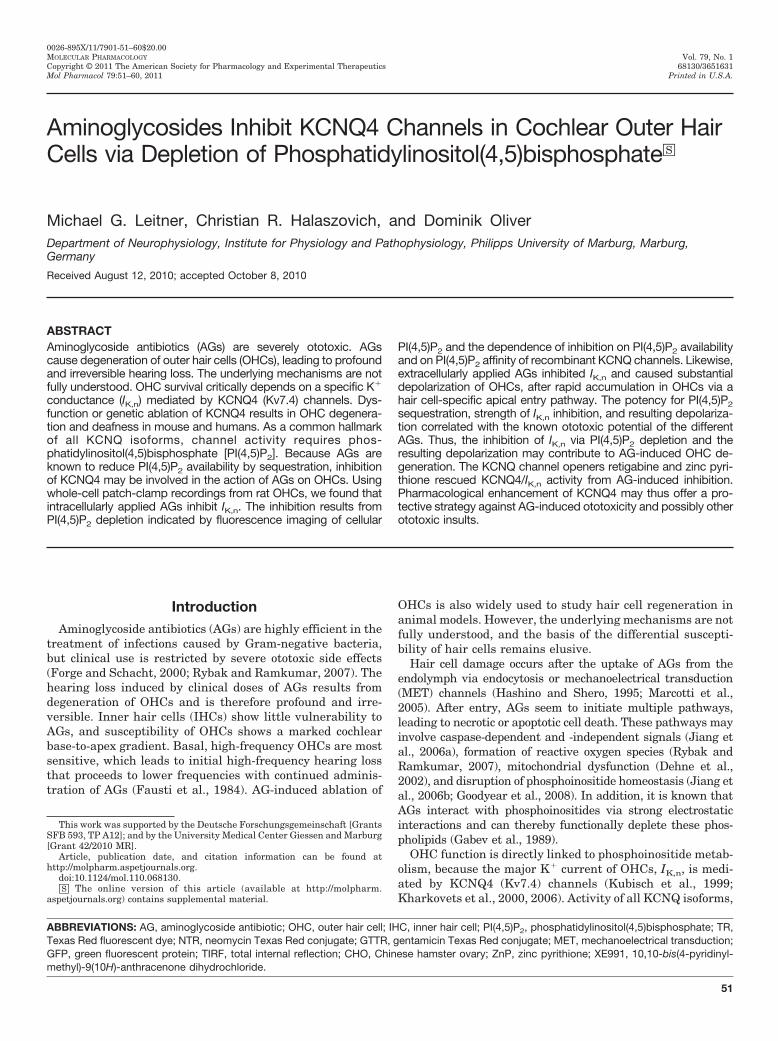
Aminoglycosides Inhibit KCNQ4 Channels in Cochlear Outer HairCells via Depletion of Phosphatidylinositol(4,5)bisphosphate□S
Michael G. Leitner, Christian R. Halaszovich, and Dominik OliverDepartment of Neurophysiology, Institute for Physiology and Pathophysiology, Philipps University of Marburg, Marburg,Germany
Received August 12, 2010; accepted October 8, 2010
ABSTRACTAminoglycoside antibiotics (AGs) are severely ototoxic. AGscause degeneration of outer hair cells (OHCs), leading to profoundand irreversible hearing loss. The underlying mechanisms are notfully understood. OHC survival critically depends on a specific K�
conductance (IK,n) mediated by KCNQ4 (Kv7.4) channels. Dys-function or genetic ablation of KCNQ4 results in OHC degenera-tion and deafness in mouse and humans. As a common hallmarkof all KCNQ isoforms, channel activity requires phos-phatidylinositol(4,5)bisphosphate [PI(4,5)P2]. Because AGs areknown to reduce PI(4,5)P2 availability by sequestration, inhibitionof KCNQ4 may be involved in the action of AGs on OHCs. Usingwhole-cell patch-clamp recordings from rat OHCs, we found thatintracellularly applied AGs inhibit IK,n. The inhibition results fromPI(4,5)P2 depletion indicated by fluorescence imaging of cellular
PI(4,5)P2 and the dependence of inhibition on PI(4,5)P2 availabilityand on PI(4,5)P2 affinity of recombinant KCNQ channels. Likewise,extracellularly applied AGs inhibited IK,n and caused substantialdepolarization of OHCs, after rapid accumulation in OHCs via ahair cell-specific apical entry pathway. The potency for PI(4,5)P2sequestration, strength of IK,n inhibition, and resulting depolariza-tion correlated with the known ototoxic potential of the differentAGs. Thus, the inhibition of IK,n via PI(4,5)P2 depletion and theresulting depolarization may contribute to AG-induced OHC de-generation. The KCNQ channel openers retigabine and zinc pyri-thione rescued KCNQ4/IK,n activity from AG-induced inhibition.Pharmacological enhancement of KCNQ4 may thus offer a pro-tective strategy against AG-induced ototoxicity and possibly otherototoxic insults.
IntroductionAminoglycoside antibiotics (AGs) are highly efficient in the
treatment of infections caused by Gram-negative bacteria,but clinical use is restricted by severe ototoxic side effects(Forge and Schacht, 2000; Rybak and Ramkumar, 2007). Thehearing loss induced by clinical doses of AGs results fromdegeneration of OHCs and is therefore profound and irre-versible. Inner hair cells (IHCs) show little vulnerability toAGs, and susceptibility of OHCs shows a marked cochlearbase-to-apex gradient. Basal, high-frequency OHCs are mostsensitive, which leads to initial high-frequency hearing lossthat proceeds to lower frequencies with continued adminis-tration of AGs (Fausti et al., 1984). AG-induced ablation of
OHCs is also widely used to study hair cell regeneration inanimal models. However, the underlying mechanisms are notfully understood, and the basis of the differential suscepti-bility of hair cells remains elusive.
Hair cell damage occurs after the uptake of AGs from theendolymph via endocytosis or mechanoelectrical transduction(MET) channels (Hashino and Shero, 1995; Marcotti et al.,2005). After entry, AGs seem to initiate multiple pathways,leading to necrotic or apoptotic cell death. These pathways mayinvolve caspase-dependent and -independent signals (Jiang etal., 2006a), formation of reactive oxygen species (Rybak andRamkumar, 2007), mitochondrial dysfunction (Dehne et al.,2002), and disruption of phosphoinositide homeostasis (Jiang etal., 2006b; Goodyear et al., 2008). In addition, it is known thatAGs interact with phosphoinositides via strong electrostaticinteractions and can thereby functionally deplete these phos-pholipids (Gabev et al., 1989).
OHC function is directly linked to phosphoinositide metab-olism, because the major K� current of OHCs, IK,n, is medi-ated by KCNQ4 (Kv7.4) channels (Kubisch et al., 1999;Kharkovets et al., 2000, 2006). Activity of all KCNQ isoforms,
This work was supported by the Deutsche Forschungsgemeinschaft [GrantsSFB 593, TP A12]; and by the University Medical Center Giessen and Marburg[Grant 42/2010 MR].
Article, publication date, and citation information can be found athttp://molpharm.aspetjournals.org.
doi:10.1124/mol.110.068130.□S The online version of this article (available at http://molpharm.
aspetjournals.org) contains supplemental material.
ABBREVIATIONS: AG, aminoglycoside antibiotic; OHC, outer hair cell; IHC, inner hair cell; PI(4,5)P2, phosphatidylinositol(4,5)bisphosphate; TR,Texas Red fluorescent dye; NTR, neomycin Texas Red conjugate; GTTR, gentamicin Texas Red conjugate; MET, mechanoelectrical transduction;GFP, green fluorescent protein; TIRF, total internal reflection; CHO, Chinese hamster ovary; ZnP, zinc pyrithione; XE991, 10,10-bis(4-pyridinyl-methyl)-9(10H)-anthracenone dihydrochloride.
0026-895X/11/7901-51–60$20.00MOLECULAR PHARMACOLOGY Vol. 79, No. 1Copyright © 2011 The American Society for Pharmacology and Experimental Therapeutics 68130/3651631Mol Pharmacol 79:51–60, 2011 Printed in U.S.A.
51
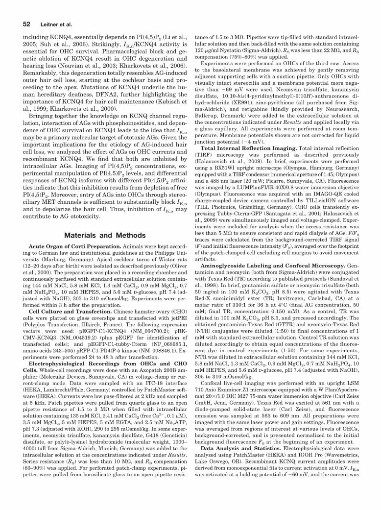
including KCNQ4, essentially depends on PI(4,5)P2 (Li et al.,2005; Suh et al., 2006). Strikingly, IK,n/KCNQ4 activity isessential for OHC survival. Pharmacological block and ge-netic ablation of KCNQ4 result in OHC degeneration andhearing loss (Nouvian et al., 2003; Kharkovets et al., 2006).Remarkably, this degeneration totally resembles AG-inducedouter hair cell loss, starting at the cochlear basis and pro-ceeding to the apex. Mutations of KCNQ4 underlie the hu-man hereditary deafness, DFNA2, further highlighting theimportance of KCNQ4 for hair cell maintenance (Kubisch etal., 1999; Kharkovets et al., 2000).
Bringing together the knowledge on KCNQ channel regu-lation, interaction of AGs with phosphoinositides, and depen-dence of OHC survival on KCNQ4 leads to the idea that IK,n
may be a primary molecular target of ototoxic AGs. Given theimportant implications for the etiology of AG-induced haircell loss, we analyzed the effect of AGs on OHC currents andrecombinant KCNQ4. We find that both are inhibited byintracellular AGs. Imaging of PI(4,5)P2 concentrations, ex-perimental manipulation of PI(4,5)P2 levels, and differentialresponses of KCNQ isoforms with different PI(4,5)P2 affini-ties indicate that this inhibition results from depletion of freePI(4,5)P2. Moreover, entry of AGs into OHCs through stereo-ciliary MET channels is sufficient to substantially block IK,n
and to depolarize the hair cell. Thus, inhibition of IK,n maycontribute to AG ototoxicity.
Materials and MethodsAcute Organ of Corti Preparation. Animals were kept accord-
ing to German law and institutional guidelines at the Philipps Uni-versity (Marburg, Germany). Apical cochlear turns of Wistar rats(12–20 days after birth) were isolated as described previously (Oliveret al., 2000). The preparation was placed in a recording chamber andcontinuously perfused with standard extracellular solution contain-ing 144 mM NaCl, 5.8 mM KCl, 1.3 mM CaCl2, 0.9 mM MgCl2, 0.7mM NaH2PO4, 10 mM HEPES, and 5.6 mM D-glucose, pH 7.4 (ad-justed with NaOH), 305 to 310 mOsmol/kg. Experiments were per-formed within 3 h after the preparation.
Cell Culture and Transfection. Chinese hamster ovary (CHO)cells were platted on glass coverslips and transfected with jetPEI(Polyplus Transfection, Illkirch, France). The following expressionvectors were used: pEGFP-C1-KCNQ4 (NM_004700.2); pBK-CMV-KCNQ3 (NM_004519.2) (plus pEGFP for identification oftransfected cells); and pEGFP-C1-tubby-Cterm (NP_068685.1,amino acids 243–505) pRFP-C1-PI(4)P-5 kinase (NM_008846.1). Ex-periments were performed 24 to 48 h after transfection.
Electrophysiological Recordings from OHCs and CHOCells. Whole-cell recordings were done with an Axopatch 200B am-plifier (Molecular Devices, Sunnyvale, CA) in voltage-clamp or cur-rent-clamp mode. Data were sampled with an ITC-18 interface(HEKA, Lambrecht/Pfalz, Germany) controlled by PatchMaster soft-ware (HEKA). Currents were low pass-filtered at 2 kHz and sampledat 5 kHz. Patch pipettes were pulled from quartz glass to an openpipette resistance of 1.5 to 3 M� when filled with intracellularsolution containing 135 mM KCl, 2.41 mM CaCl2 (free Ca2�, 0.1 �M),3.5 mM MgCl2, 5 mM HEPES, 5 mM EGTA, and 2.5 mM Na2ATP,pH 7.3 (adjusted with KOH), 290 to 295 mOsmol/kg. In some exper-iments, neomycin trisulfate, kanamycin disulfate, G418 (Geneticin)disulfate, or poly(D-lysine) hydrobromide (molecular weight, 1000–4000) (all from Sigma-Aldrich, Munich, Germany) was added to theintracellular solution at the concentrations indicated under Results.Series resistance (RS) was less than 10 M�, and RS compensation(80–90%) was applied. For perforated patch-clamp experiments, pi-pettes were pulled from borosilicate glass to an open pipette resis-
tance of 1.5 to 3 M�. Pipettes were tip-filled with standard intracel-lular solution and then back-filled with the same solution containing120 �g/ml Nystatin (Sigma-Aldrich). RS was less than 22 M�, and RS
compensation (75%–80%) was applied.Experiments were performed on OHCs of the third row. Access
to the basolateral membrane was achieved by gently removingadjacent supporting cells with a suction pipette. Only OHCs withvisually intact stereocilia and a membrane potential more nega-tive than �69 mV were used. Neomycin trisulfate, kanamycindisulfate, 10,10-bis(4-pyridinylmethyl)-9(10H)-anthracenone di-hydrochloride (XE991), zinc-pyrithione (all purchased from Sig-ma-Aldrich), and retigabine (kindly provided by Neurosearch,Ballerup, Denmark) were added to the extracellular solution atthe concentrations indicated under Results and applied locally viaa glass capillary. All experiments were performed at room tem-perature. Membrane potentials shown are not corrected for liquidjunction potential (�4 mV).
Total Internal Reflection Imaging. Total internal reflection(TIRF) microscopy was performed as described previously(Halaszovich et al., 2009). In brief, experiments were performedusing a BX51WI upright microscope (Olympus, Hamburg, Germany)equipped with a TIRF condensor (numerical aperture of 1.45; Olympus)and a 488 nm laser (20 mW; Picarro, Sunnyvale, CA). Fluorescencewas imaged by a LUMPlanFI/IR 40X/0.8 water immersion objective(Olympus). Fluorescence was acquired with an IMAGO-QE cooledcharge-coupled device camera controlled by TILLvisION software(TILL Photonics, Grafelfing, Germany). CHO cells transiently ex-pressing Tubby-Cterm-GFP (Santagata et al., 2001; Halaszovich etal., 2009) were simultaneously imaged and voltage-clamped. Exper-iments were included for analysis when the access resistance wasless than 5 M� to ensure consistent and rapid dialysis of AGs. F/F0
traces were calculated from the background-corrected TIRF signal(F) and initial fluorescence intensity (F0), averaged over the footprintof the patch-clamped cell excluding cell margins to avoid movementartifacts.
Aminoglycoside Labeling and Confocal Microscopy. Gen-tamicin and neomycin (both from Sigma-Aldrich) were conjugatedwith Texas Red (TR) according to published protocols (Sandoval etal., 1998). In brief, gentamicin sulfate or neomycin trisulfate (both50 mg/ml in 100 mM K2CO3, pH 8.5) were agitated with TexasRed-X succinimidyl ester (TR; Invitrogen, Carlsbad, CA) at amolar ratio of 330:1 for 36 h at 4°C (final AG concentration, 50mM; final TR, concentration 0.150 mM). As a control, TR wasdiluted in 100 mM K2CO3, pH 8.5, and processed accordingly. Theobtained gentamicin-Texas Red (GTTR) and neomycin-Texas Red(NTR) conjugates were diluted (1:50) to final concentrations of 1mM with standard extracellular solution. Control TR solution wasdiluted accordingly to obtain equal concentrations of the fluores-cent dye in control experiments (1:50). For some experiments,NTR was diluted in extracellular solution containing 144 mM KCl,5.8 mM NaCl, 1.3 mM CaCl2, 0.9 mM MgCl2, 0.7 mM NaH2PO4, 10mM HEPES, and 5.6 mM D-glucose, pH 7.4 (adjusted with NaOH),305 to 310 mOsmol/kg.
Confocal live-cell imaging was performed with an upright LSM710 Axio Examiner.Z1 microscope equipped with a W Plan/Apochro-mat 20�/1.0 DIC M27 75-mm water immersion objective (Carl ZeissGmbH, Jena, Germany). Texas Red was excited at 561 nm with adiode-pumped solid-state laser (Carl Zeiss), and fluorescenceemission was sampled at 565 to 609 nm. All preparations wereimaged with the same laser power and gain settings. Fluorescencewas averaged from regions of interest at various levels of OHCs,background-corrected, and is presented normalized to the initialbackground fluorescence F0 at the beginning of an experiment.
Data Analysis and Statistics. Electrophysiological data wereanalyzed using PatchMaster (HEKA) and IGOR Pro (Wavemetrics,Lake Oswego, OR). Recombinant KCNQ current amplitudes werederived from monoexponential fits to current activation at 0 mV. IK,n
was activated at a holding potential of �60 mV, and the current was
52 Leitner et al.
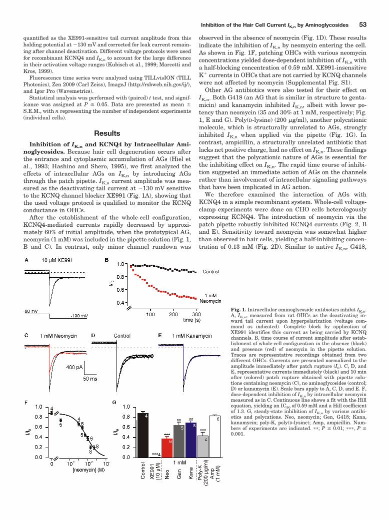
quantified as the XE991-sensitive tail current amplitude from thisholding potential at �130 mV and corrected for leak current remain-ing after channel deactivation. Different voltage protocols were usedfor recombinant KCNQ4 and IK,n to account for the large differencein their activation voltage ranges (Kubisch et al., 1999; Marcotti andKros, 1999).
Fluorescence time series were analyzed using TILLvisION (TILLPhotonics), Zen 2009 (Carl Zeiss), ImageJ (http://rsbweb.nih.gov/ij/),and Igor Pro (Wavemetrics).
Statistical analysis was performed with (paired) t test, and signif-icance was assigned at P � 0.05. Data are presented as mean �S.E.M., with n representing the number of independent experiments(individual cells).
ResultsInhibition of IK,n and KCNQ4 by Intracellular Ami-
noglycosides. Because hair cell degeneration occurs afterthe entrance and cytoplasmic accumulation of AGs (Hiel etal., 1993; Hashino and Shero, 1995), we first analyzed theeffects of intracellular AGs on IK,n by introducing AGsthrough the patch pipette. IK,n current amplitude was mea-sured as the deactivating tail current at �130 mV sensitiveto the KCNQ channel blocker XE991 (Fig. 1A), showing thatthe used voltage protocol is qualified to monitor the KCNQconductance in OHCs.
After the establishment of the whole-cell configuration,KCNQ4-mediated currents rapidly decreased by approxi-mately 60% of initial amplitude, when the prototypical AG,neomycin (1 mM) was included in the pipette solution (Fig. 1,B and C). In contrast, only minor channel rundown was
observed in the absence of neomycin (Fig. 1D). These resultsindicate the inhibition of IK,n by neomycin entering the cell.As shown in Fig. 1F, patching OHCs with various neomycinconcentrations yielded dose-dependent inhibition of IK,n witha half-blocking concentration of 0.59 mM. XE991-insensitiveK� currents in OHCs that are not carried by KCNQ channelswere not affected by neomycin (Supplemental Fig. S1).
Other AG antibiotics were also tested for their effect onIK,n. Both G418 (an AG that is similar in structure to genta-micin) and kanamycin inhibited IK,n, albeit with lower po-tency than neomycin (35 and 30% at 1 mM, respectively; Fig.1, E and G). Poly(D-lysine) (200 �g/ml), another polycationicmolecule, which is structurally unrelated to AGs, stronglyinhibited IK,n when applied via the pipette (Fig. 1G). Incontrast, ampicillin, a structurally unrelated antibiotic thatlacks net positive charge, had no effect on IK,n. These findingssuggest that the polycationic nature of AGs is essential forthe inhibiting effect on IK,n. The rapid time course of inhibi-tion suggested an immediate action of AGs on the channelsrather than involvement of intracellular signaling pathwaysthat have been implicated in AG action.
We therefore examined the interaction of AGs withKCNQ4 in a simple recombinant system. Whole-cell voltage-clamp experiments were done on CHO cells heterologouslyexpressing KCNQ4. The introduction of neomycin via thepatch pipette robustly inhibited KCNQ4 currents (Fig. 2, Band E). Sensitivity toward neomycin was somewhat higherthan observed in hair cells, yielding a half-inhibiting concen-tration of 0.13 mM (Fig. 2D). Similar to native IK,n, G418,
Fig. 1. Intracellular aminoglycoside antibiotics inhibit IK,n.A, IK,n, measured from rat OHCs as the deactivating in-ward tail current upon hyperpolarization (voltage com-mand as indicated). Complete block by application ofXE991 identifies this current as being carried by KCNQchannels. B, time course of current amplitude after estab-lishment of whole-cell configuration in the absence (black)and presence (red) of neomycin in the pipette solution.Traces are representative recordings obtained from twodifferent OHCs. Currents are presented normalized to theamplitude immediately after patch rupture (I0). C, D, andE, representative currents immediately (black) and 10 minafter (colored) patch rupture obtained with pipette solu-tions containing neomycin (C), no aminoglycosides (control;D) or kanamycin (E). Scale bars apply to A, C, D, and E. F,dose-dependent inhibition of IK,n by intracellular neomycinmeasured as in C. Continuous line shows a fit with the Hillequation, yielding an IC50 of 0.59 mM and a Hill coefficientof 1.3. G, steady-state inhibition of IK,n by various antibi-otics and polycations. Neo, neomycin; Gen, G418; Kana,kanamycin; poly-K, poly(D-lysine); Amp, ampicillin. Num-bers of experiments are indicated. ��; P � 0.01; ���, P �0.001.
Inhibition of the Hair Cell Current IK,n by Aminoglycosides 53
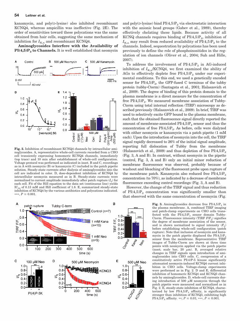
kanamycin, and poly(D-lysine) also inhibited recombinantKCNQ4, whereas ampicillin was ineffective (Fig. 2E). Theorder of sensitivities toward these polycations was the sameobtained from hair cells, suggesting the same mechanism ofinhibition for IK,n and recombinant KCNQ4.
Aminoglycosides Interfere with the Availability ofPI(4,5)P2 to Channels. It is well established that neomycin
and poly(D-lysine) bind PI(4,5)P2 via electrostatic interactionwith the anionic head groups (Gabev et al., 1989), therebyeffectively chelating these lipids. Because activity of allKCNQ channels requires binding of PI(4,5)P2, inhibition ofIK,n may result from reduced availability of PI(4,5)P2 to thechannels. Indeed, sequestration by polycations has been usedpreviously to define the role of phosphoinositides in the reg-ulation of ion channels (Oliver et al., 2004; Suh and Hille,2007).
To address the involvement of PI(4,5)P2 in AG-inducedinhibition of IK,n/KCNQ4, we first examined the ability ofAGs to effectively deplete free PI(4,5)P2 under our experi-mental conditions. To this end, we used a genetically encodedsensor for PI(4,5)P2, the GFP-fused C terminus of the tubbyprotein (tubby-Cterm) (Santagata et al., 2001; Halaszovich etal., 2009). The degree of binding of this protein domain to theplasma membrane is a direct measure for the concentration offree PI(4,5)P2. We measured membrane association of Tubby-Cterm using total internal reflection (TIRF) microscopy as de-scribed previously (Halaszovich et al., 2009). In brief, TIRF wasused to selectively excite GFP bound to the plasma membrane,such that the obtained fluorescence signal directly reported theamount of membrane-associated PI(4,5)P2 sensor and thus theconcentration of free PI(4,5)P2. As before, cells were dialyzedwith either neomycin or kanamycin via a patch pipette (1 mMeach). Upon the introduction of neomycin into the cell, the TIRFsignal rapidly decreased to 26% of the initial signal amplitude,reporting full dislocation of Tubby from the membrane(Halaszovich et al., 2009) and thus depletion of free PI(4,5)P2
(Fig. 3, A and B). In contrast, without neomycin in the pipette(control, Fig. 3, A and B) only an initial minor reduction ofmembrane fluorescence was observed, probably because ofwashout and bleaching of the fluorescent probe upon rupture ofthe membrane patch. Kanamycin also reduced free PI(4,5)P2
concentration (to 70%), as indicated by a decrease of membranefluorescence exceeding control recordings (Fig. 3, A and B).
However, the change of the TIRF signal and thus reductionof PI(4,5)P2 concentration was significantly smaller thanthat observed with the same concentration of neomycin (Fig.
Fig. 2. Inhibition of recombinant KCNQ4 channels by intracellular ami-noglycosides. A, representative whole-cell currents recorded from a CHOcell transiently expressing homomeric KCNQ4 channels, immediately(top trace) and 10 min after establishment of whole-cell configuration.Voltage protocol was performed as indicated in inset. B and C, recordingsas in A with neomycin (B) or kanamycin (C) included in the patch pipettesolution. Steady-state currents after dialysis of aminoglycosides into thecell are indicated in color. D, dose-dependent inhibition of KCNQ4 byintracellular neomycin measured as in B. Steady-state currents werenormalized to current amplitude immediately after patch rupture (I0) foreach cell. Fit of the Hill equation to the data set (continuous line) yieldsIC50 of 0.13 mM and Hill coefficient of 1.8. E, summarized steady-stateinhibition of KCNQ4 by the various antibiotics and polycations indicated.���, P � 0.001.
Fig. 3. Aminoglycosides decrease free PI(4,5)P2 inthe plasma membrane. A, combined TIRF imagingand patch-clamp experiments on CHO cells trans-fected with the PI(4,5)P2 sensor domain Tubby-Cterm. Fluorescence intensity (TIRF F/F0) signifiesthe degree of membrane association of the sensor,and is shown normalized to signal intensity (F0)before establishing whole-cell configuration (patchrupture). Note that inclusion of neomycin and kana-mycin in the patch pipette displaced the PI(4,5)P2sensor from the membrane. Representative TIRFimages of Tubby-Cterm are shown at three timepoints with neomycin applied via the patch pipette(inset; scale bar, 20 �m). B, averaged relativechanges in TIRF signals upon introduction of ami-noglycosides into CHO cells. C, coexpression of aconstitutively active PI(4)P-5 kinase significantlyattenuated neomycin-induced KCNQ4 current inhi-bition in CHO cells. Voltage-clamp experimentswere performed as in Fig. 2. D and E, differentialinhibition of homomeric KCNQ4 and KCNQ3 chan-nels by aminoglycosides. D, whole-cell currents dur-ing introduction of 500 �M neomycin through thepatch pipette were measured and normalized as inFig. 2. E, steady-state inhibition of KCNQ4, charac-terized by low PI(4,5)P2 affinity, is significantlystronger than inhibition of KCNQ3, exhibiting highPI(4,5)P2 affinity. ��; P � 0.01; ���, P � 0.001.
54 Leitner et al.
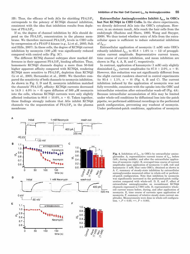
3B). Thus, the efficacy of both AGs for shielding PI(4,5)P2
corresponds to the potency of KCNQ4 channel inhibition,consistent with the idea that inhibition results from deple-tion of PI(4,5)P2.
If so, the degree of channel inhibition by AGs should de-pend on the PI(4,5)P2 concentration in the plasma mem-brane. We therefore increased PI(4,5)P2 levels in CHO cellsby coexpression of a PI(4)P-5 kinase (e.g., Li et al., 2005; Suhand Hille, 2007). In these cells, the degree of KCNQ4 currentinhibition by neomycin (100 �M) was significantly reducedcompared with control cells (Fig. 3C).
The different KCNQ channel subtypes show marked dif-ferences in their apparent PI(4,5)P2 binding affinities. Thus,homomeric KCNQ3 channels display a more than 50-foldhigher apparent affinity compared with KCNQ4, renderingKCNQ4 more sensitive to PI(4,5)P2 depletion than KCNQ3(Li et al., 2005; Hernandez et al., 2009). We therefore com-pared the sensitivity of both channels to neomycin inhibition.As shown in Fig. 3, D and E, neomycin inhibition matchedthe channels’ PI(4,5)P2 affinity: KCNQ4 currents decreasedto 14.9 � 4.0% (n � 6) upon diffusion of 500 �M neomycininto the cells, whereas KCNQ3 currents were only slightlyaffected (reduction to 80.6 � 10.6%, n � 6). Taken together,these findings strongly indicate that AGs inhibit KCNQ4channels via the sequestration of PI(4,5)P2 in the plasmamembrane.
Extracellular Aminoglycosides Inhibit IK,n in OHCsbut Not KCNQ4 in CHO Cells. In the above experiments,we directly delivered AGs into the OHC’s cytoplasm. How-ever, in an ototoxic insult, AGs reach the hair cells from theendolymph (Hashino and Shero, 1995; Wang and Steyger,2009). We thus tested whether entry of AGs from the extra-cellular space is sufficient to induce substantial inhibitionof IK,n.
Extracellular application of neomycin (1 mM) onto OHCsrobustly inhibited IK,n to 65.8 � 1.6% (n � 12) of preappli-cation current amplitude. Representative current traces,time course of current inhibition, and mean inhibition areshown in Fig. 4, A, B, and C, respectively.
In contrast, application of kanamycin (1 mM) only slightlydiminished IK,n current amplitudes (to 87.0 � 2.0%, n � 9).However, this reduction was not significantly different fromthe slight current rundown observed in control experiments(to 93.4 � 1.1%, n � 8) (Fig. 4, B and C). The currentinhibition induced by the application of neomycin was notfully reversible, consistent with the uptake into the OHC andintracellular retention after extracellular wash-off (Fig. 4A).Because intracellular accumulation of AGs may be limitedunder whole-cell conditions by diffusional loss into the patchpipette, we performed additional recordings in the perforatedpatch configuration, preventing any washout of neomycin.Under perforated-patch conditions, application of neomycin
Fig. 4. Inhibition of IK,n in OHCs by extracellular amino-glycosides. A, representative current traces of IK,n before(left), during (middle), and after the extracellular applica-tion of neomycin (right). B, averaged time course of currentamplitudes upon application of neomycin (1 mM, red) andkanamycin (1 mM, blue) onto OHCs, obtained as shown inA. C, summary of steady-state inhibition by extracellularaminoglycosides measured either in whole-cell or perforat-ed-patch configuration. Note that inhibition by neomycinwas significantly increased in the perforated-patch config-uration compared with whole-cell. D, E, and F, effect ofextracellular aminoglycosides on recombinant KCNQ4channels expressed in CHO cells. D, representative whole-cell current traces before, during, and after application ofneomycin. E, time course of currents upon application ofneomycin. F, summary of relative steady state current am-plitudes. Measurements were done in whole-cell configura-tion. �, P � 0.05; ���, P � 0.001.
Inhibition of the Hair Cell Current IK,n by Aminoglycosides 55
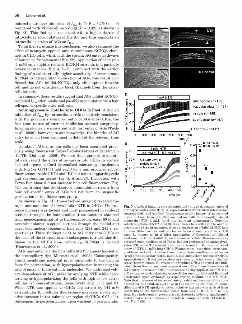
induced a stronger inhibition of IK,n to 54.9 � 3.7% (n � 6)compared with whole-cell recordings (P � 0.02), as shown inFig. 4C. This finding is consistent with a higher degree ofintracellular accumulation of the AG and thus supports anintracellular action of AGs on IK,n.
To further scrutinize this conclusion, we also examined theeffect of neomycin applied onto recombinant KCNQ4 chan-nels in CHO cells, which lack the specific AG entry pathwaysof hair cells (Supplemental Fig. S2). Application of neomycin(1 mM) only slightly reduced KCNQ4 currents in a partiallyreversible manner (Fig. 4, D–F). Combined with the earlierfinding of a substantially higher sensitivity of recombinantKCNQ4 to intracellular application of AGs, this result con-firmed that AGs inhibit KCNQ4 only after uptake into thecell and do not considerably block channels from the extra-cellular side.
In summary, these results suggest that AGs inhibit KCNQ4-mediated IK,n after uptake and possibly accumulation via a haircell-specific specific entry pathway.
Aminoglycoside Uptake into OHCs Is Fast. Althoughinhibition of IK,n by intracellular AGs is entirely consistentwith the previously described entry of AGs into OHCs, thefast time course of current inhibition seemed surprising.Imaging studies are consistent with fast entry of AGs (Tiedeet al., 2009); however, to our knowledge, the kinetics of AGentry have not been examined in detail at the relevant timescale.
Uptake of AGs into hair cells has been monitored previ-ously using fluorescent Texas Red-derivatives of gentamicin(GTTR) (Dai et al., 2006). We used this approach to quanti-tatively record the entry of neomycin into OHCs in acutelyisolated organs of Corti by confocal microscopy. Incubationwith NTR or GTTR (1 mM each) for 5 min produced robustfluorescence inside OHCs and IHC but not in supporting cellsand surrounding tissue (Fig. 5, A and B). Incubation withTexas Red alone did not increase hair cell fluorescence (Fig.5C), confirming that the observed accumulation results fromhair cell-specific entry of AGs but not from an unspecificpermeation of the fluorescent group.
As shown in Fig. 5D, time-resolved imaging revealed therapid accumulation of intracellular NTR in OHCs. Fluores-cence increase was fastest and most pronounced in confocalsections through the hair bundles (time constant obtainedfrom monoexponential fit to fluorescence increase, 69 s) andsomewhat slower in optical sections through the medial andbasal (subnuclear) regions of hair cells (281 and 241 s, re-spectively). These findings point to AG entry into OHCs atthe level of the stereocilia and subsequent intracellular dif-fusion to the OHC’s base, where IK,n/KCNQ4 is located(Kharkovets et al., 2000).
AGs may enter via the hair cell’s MET channels located atthe stereociliary tips (Marcotti et al., 2005). Consequently,apical membrane potential must contribute to the drivingforce for permeation, with hyperpolarization increasing therate of entry of these cationic molecules. We addressed volt-age-dependence of AG uptake by applying NTR while depo-larizing or hyperpolarizing the cells with high or low extra-cellular K� concentrations, respectively (Fig. 5, E and F).When NTR was applied to OHCs depolarized by 144 mMextracellular K�, cellular fluorescence increased slowly (rel-ative increase in the subnuclear region of OHCs, 0.02 s�1).Subsequent hyperpolarization upon washout of extracellular
Fig. 5. Confocal imaging reveals rapid and voltage-dependent entry ofaminoglycosides into OHCs. A, representative differential interferencecontrast (left) and confocal fluorescence (right) images of an isolatedorgan of Corti from rat, after incubation with fluorescently labeledneomycin (NTR; 1 mM) for 5 min (at room temperature). Note theselective uptake of NTR into OHCs and inner hair cells (�). The obliqueorientation of the preparation allows visualization of labeled OHC hairbundles (filled arrow) and cell bodies (open arrow) (scale bars, 100�m). B, images as in A after application of fluorescently labeledgentamicin (GTTR; 1 mM). C, no increase of cellular fluorescence wasdetected upon application of Texas Red not conjugated to aminoglyco-sides (TR; same TR concentration as in A and B). D, time course ofentry of NTR (1 mM) into OHCs. Fluorescence intensities were sam-pled from confocal optical sections through hair bundles, apical region(level of the cuticular plate), middle, and subnuclear regions of OHCs.Application of TR did not produce any detectable increase in fluores-cence (bottom trace). Numbers of individual OHCs analyzed are indi-cated (from five independent preparations). E, voltage dependence ofNTR entry. Increase of OHC fluorescence during application of NTR (1mM) was slow in depolarizing extracellular medium (144 mM KCl) andaccelerated upon exchange by repolarizing medium (5.6 mM KCl).Note that the onset of increased entry is delayed because of the timeneeded for full solution exchange in the recording chamber. F, quan-tification of NTR uptake kinetics. Relative increase was derived fromlinear fits to the fluorescence traces from single OHCs (n � 51–91,from four independent preparations). Asterisks indicate significantlyfaster fluorescence increase at 5.6 mM K� compared with 144 mM K�.���, P � 0.001.
56 Leitner et al.
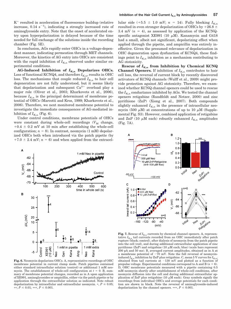
K� resulted in acceleration of fluorescence buildup (relativeincrease, 0.14 s�1), indicating a strongly increased rate ofaminoglycoside entry. Note that the onset of accelerated en-try upon hyperpolarization is delayed because of the timeneeded for full exchange of the solutions inside the recordingchamber (Fig. 5E).
In conclusion, AGs rapidly enter OHCs in a voltage-depen-dent manner, indicating permeation through MET channels.Moreover, the kinetics of AG entry into OHCs are consistentwith the rapid inhibition of IK,n observed under similar ex-perimental conditions.
AG-Induced Inhibition of IK,n Depolarizes OHCs.Loss of functional KCNQ4, and therefore IK,n, results in OHCloss. The mechanisms that couple reduced IK,n to hair celldegeneration are not fully understood, but it seems likelythat depolarization and subsequent Ca2� overload play amajor role (Oliver et al., 2003; Kharkovets et al., 2006),because IK,n is the principal determinant of membrane po-tential of OHCs (Marcotti and Kros, 1999; Kharkovets et al.,2006). Therefore, we next monitored membrane potential toinvestigate the immediate consequences of AG-mediated in-hibition of IK,n (Fig. 6).
Under control conditions, membrane potentials of OHCswere constant during whole-cell recordings (VM change,�0.4 � 0.2 mV at 10 min after establishing the whole-cellconfiguration; n � 8). In contrast, neomycin (1 mM) depolar-ized OHCs both when introduced via the patch pipette (by�7.0 � 2.4 mV; n � 6) and when applied from the extracel-
lular side (�5.5 � 1.0 mV; n � 14). Fully blocking IK,n
resulted in even stronger depolarization of OHCs by �26.8 �3.4 mV (n � 4), as assessed by application of the KCNQ-specific antagonist XE991 (10 �M). Kanamycin and G418had a small, albeit not significant, depolarizing effect whenapplied through the pipette, and ampicillin was entirely in-effective. Given the presumed relevance of depolarization inOHC degeneration upon dysfunction of KCNQ4, these find-ings point to IK,n inhibition as a mechanism contributing toAG ototoxicity.
Rescue of IK,n from Inhibition by Chemical KCNQChannel Openers. If inhibition of IK,n contributes to haircell loss, the reversal of current block by recently discoveredactivators of KCNQ channels (Wulff et al., 2009) might pro-vide protection against AG ototoxicity. Therefore, we exam-ined whether KCNQ channel openers could be used to rescuethe IK,n conductance inhibited by AGs. We tested the channelopeners retigabine (Rundfeldt and Netzer, 2000) and zincpyrithione (ZnP) (Xiong et al., 2007). Both compoundsslightly enhanced IK,n in the presence of intracellular neo-mycin (500 �M) at concentrations of up to 10 �M (Supple-mental Fig. S3). However, combined application of retigabineand ZnP (10 �M each) robustly enhanced IK,n amplitudes(Fig. 7A).
Fig. 6. Neomycin depolarizes OHCs. A, representative recordings of OHCmembrane potential in current clamp mode. Patch pipettes containedeither standard intracellular solution (control) or additional 1 mM neo-mycin. The establishment of whole-cell configuration at t � 0. B, sum-mary of membrane potential changes, recorded as in A upon applicationof XE991, aminoglycosides or ampicillin, either via the patch pipette or byapplication through the extracellular solution as indicated. Note robustdepolarization by intracellular and extracellular neomycin. �, P � 0.05;��; P � 0.01; ���, P � 0.001.
Fig. 7. Rescue of IK,n currents by chemical channel openers. A, represen-tative IK,n tail currents recorded from an OHC immediately after patchrupture (black; control), after dialysis of neomycin from the patch pipetteinto the cell (red), and during additional extracellular application of zincpyrithione (ZnP) and retigabine (10 �M each; blue) (scale bars represent200 pA and 50 ms). B, averaged current amplitudes, obtained as in A ata membrane potential of �70 mV. Note the full reversal of neomycin-induced IK,n inhibition by ZnP plus retigabine. C, mean I-V curves for IK,n,obtained from tail currents at �130 mV and plotted as a function ofprepulse voltage. Experimental conditions correspond to A and B (n � 6).D, OHC membrane potentials measured with a pipette containing 0.5mM neomycin shortly after establishment of whole-cell conditions, afterneomycin diffusion into the cell and during additional extracellular ap-plication of ZnP plus retigabine (10 �M each). Gray symbols signify therecordings from individual OHCs and average potentials for each condi-tion are shown in black. Note the reversal of aminoglycoside-induceddepolarization by the channel openers. ���, P � 0.001.
Inhibition of the Hair Cell Current IK,n by Aminoglycosides 57
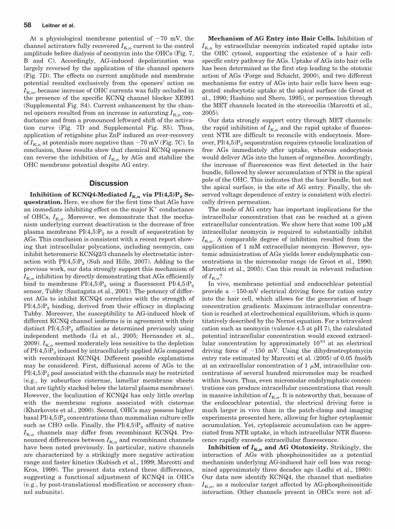
At a physiological membrane potential of �70 mV, thechannel activators fully recovered IK,n current to the controlamplitude before dialysis of neomycin into the OHCs (Fig. 7,B and C). Accordingly, AG-induced depolarization waslargely reversed by the application of the channel openers(Fig. 7D). The effects on current amplitude and membranepotential resulted exclusively from the openers’ action onIK,n, because increase of OHC currents was fully occluded inthe presence of the specific KCNQ channel blocker XE991(Supplemental Fig. S4). Current enhancement by the chan-nel openers resulted from an increase in saturating IK,n con-ductance and from a pronounced leftward shift of the activa-tion curve (Fig. 7D and Supplemental Fig. S5). Thus,application of retigabine plus ZnP induced an over-recoveryof IK,n at potentials more negative than �70 mV (Fig. 7C). Inconclusion, these results show that chemical KCNQ openerscan reverse the inhibition of IK,n by AGs and stabilize theOHC membrane potential despite AG entry.
DiscussionInhibition of KCNQ4-Mediated IK,n via PI(4,5)P2 Se-
questration. Here, we show for the first time that AGs havean immediate inhibiting effect on the major K� conductanceof OHCs, IK,n. Moreover, we demonstrate that the mecha-nism underlying current deactivation is the decrease of freeplasma membrane PI(4,5)P2 as a result of sequestration byAGs. This conclusion is consistent with a recent report show-ing that intracellular polycations, including neomycin, caninhibit heteromeric KCNQ2/3 channels by electrostatic inter-action with PI(4,5)P2 (Suh and Hille, 2007). Adding to theprevious work, our data strongly support this mechanism ofIK,n inhibition by directly demonstrating that AGs efficientlybind to membrane PI(4,5)P2 using a fluorescent PI(4,5)P2
sensor, Tubby (Santagata et al., 2001). The potency of differ-ent AGs to inhibit KCNQ4 correlates with the strength ofPI(4,5)P2 binding, derived from their efficacy in displacingTubby. Moreover, the susceptibility to AG-induced block ofdifferent KCNQ channel isoforms is in agreement with theirdistinct PI(4,5)P2 affinities as determined previously usingindependent methods (Li et al., 2005; Hernandez et al.,2009). IK,n seemed moderately less sensitive to the depletionof PI(4,5)P2 induced by intracellularly applied AGs comparedwith recombinant KCNQ4. Different possible explanationsmay be considered. First, diffusional access of AGs to thePI(4,5)P2 pool associated with the channels may be restricted(e.g., by subsurface cisternae, lamellar membrane sheetsthat are tightly stacked below the lateral plasma membrane).However, the localization of KCNQ4 has only little overlapwith the membrane regions associated with cisternae(Kharkovets et al., 2000). Second, OHCs may possess higherbasal PI(4,5)P2 concentrations than mammalian culture cellssuch as CHO cells. Finally, the PI(4,5)P2 affinity of nativeIK,n channels may differ from recombinant KCNQ4. Pro-nounced differences between IK,n and recombinant channelshave been noted previously. In particular, native channelsare characterized by a strikingly more negative activationrange and faster kinetics (Kubisch et al., 1999; Marcotti andKros, 1999). The present data extend these differences,suggesting a functional adjustment of KCNQ4 in OHCs(e.g., by post-translational modification or accessory chan-nel subunits).
Mechanism of AG Entry into Hair Cells. Inhibition ofIK,n by extracellular neomycin indicated rapid uptake intothe OHC cytosol, supporting the existence of a hair cell-specific entry pathway for AGs. Uptake of AGs into hair cellshas been determined as the first step leading to the ototoxicaction of AGs (Forge and Schacht, 2000), and two differentmechanisms for entry of AGs into hair cells have been sug-gested: endocytotic uptake at the apical surface (de Groot etal., 1990; Hashino and Shero, 1995), or permeation throughthe MET channels located in the stereocilia (Marcotti et al.,2005).
Our data strongly support entry through MET channels:the rapid inhibition of IK,n and the rapid uptake of fluores-cent NTR are difficult to reconcile with endocytosis. More-over, PI(4,5)P2 sequestration requires cytosolic localization offree AGs immediately after uptake, whereas endocytosiswould deliver AGs into the lumen of organelles. Accordingly,the increase of fluorescence was first detected in the hairbundle, followed by slower accumulation of NTR in the apicalpole of the OHC. This indicates that the hair bundle, but notthe apical surface, is the site of AG entry. Finally, the ob-served voltage dependence of entry is consistent with electri-cally driven permeation.
The mode of AG entry has important implications for theintracellular concentration that can be reached at a givenextracellular concentration. We show here that some 100 �Mintracellular neomycin is required to substantially inhibitIK,n. A comparable degree of inhibition resulted from theapplication of 1 mM extracellular neomycin. However, sys-temic administration of AGs yields lower endolymphatic con-centrations in the micromolar range (de Groot et al., 1990;Marcotti et al., 2005). Can this result in relevant reductionof IK,n?
In vivo, membrane potential and endocochlear potentialprovide a �150-mV electrical driving force for cation entryinto the hair cell, which allows for the generation of hugeconcentration gradients. Maximum intracellular concentra-tion is reached at electrochemical equilibrium, which is quan-titatively described by the Nernst equation. For a tetravalentcation such as neomycin (valence 4.5 at pH 7), the calculatedpotential intracellular concentration would exceed extracel-lular concentration by approximately 1010 at an electricaldriving force of �150 mV. Using the dihydrostreptomycinentry rate estimated by Marcotti et al. (2005) of 0.05 fmol/hat an extracellular concentration of 1 �M, intracellular con-centrations of several hundred micromoles may be reachedwithin hours. Thus, even micromolar endolymphatic concen-trations can produce intracellular concentrations that resultin massive inhibition of IK,n. It is noteworthy that, because ofthe endocochlear potential, the electrical driving force ismuch larger in vivo than in the patch-clamp and imagingexperiments presented here, allowing for higher cytoplasmicaccumulation. Yet, cytoplasmic accumulation can be appre-ciated from NTR uptake, in which intracellular NTR fluores-cence rapidly exceeds extracellular fluorescence.
Inhibition of IK,n and AG Ototoxicity. Strikingly, theinteraction of AGs with phosphoinositides as a potentialmechanism underlying AG-induced hair cell loss was recog-nized approximately three decades ago (Lodhi et al., 1980).Our data now identify KCNQ4, the channel that mediatesIK,n, as a molecular target affected by AG-phosphoinositideinteraction. Other channels present in OHCs were not af-
58 Leitner et al.
fected by aminoglycosides (Supplemental Fig. S1), or theirloss does not affect OHC survival (Vetter et al., 1999; Murthyet al., 2009). Because it is well documented that IK,n isessential for the survival of OHCs (Nouvian et al., 2003;Kharkovets et al., 2006), it seems likely that AG-inducedinhibition of this current contributes to ototoxicity.
This idea is supported by a conspicuous correlation be-tween the ototoxic potency of AG antibiotics and their effi-cacy in inhibiting IK,n. Thus, neomycin exhibits higher co-chleotoxicity than gentamicin and kanamycin in vivo and invitro (Lodhi et al., 1980). Likewise, vulnerability to AGs ofthe different hair cell types correlates well with the expres-sion of KCNQ4 and the requirement of KCNQ4 for cell sur-vival. Most obviously, the highest susceptibility to damage byAGs is found in OHCs, in which KCNQ4/IK,n provides essen-tially all of the resting K� conductance (Marcotti and Kros,1999; Kharkovets et al., 2006). Moreover, the higher vulner-ability of basal OHCs that results in the characteristic high-frequency hearing loss upon administration of AGs (Fausti etal., 1984), corresponds to a base-to-apex gradient of KCNQ4expression (Kharkovets et al., 2000) and IK,n conductance(Mammano and Ashmore, 1996). We have shown previouslythat IHC express KCNQ4, in which it contributes to settingthe membrane potential (Oliver et al., 2003). However, IHCfunction and survival do not obviously depend on this current(Nouvian et al., 2003; Kharkovets et al., 2006). In agreementwith this difference to OHCs, IHCs are little, if at all, affectedby AGs (Forge and Schacht, 2000).
A similar correlation is found for vestibular hair cells.Here, KCNQ4 is specifically expressed in type I but not intype II cells (Kharkovets et al., 2000). In analogy to cochlearhair cells, KCNQ4-expressing type I cells are more suscepti-ble to AG-induced hair cell death (Forge and Schacht, 2000).It remains to be shown whether KCNQ-mediated currents investibular type I hair cells are inhibited by AGs and whetherKCNQ4 is required for their survival.
Despite these considerations, it seems unlikely that inhi-bition of IK,n is the unique or predominant cause of AG-induced hair cell loss. Detailed analyses have demonstratedthat multiple intracellular signaling pathways mediate OHCdeath by necrotic and/or apoptotic mechanisms (Jiang et al.,2006a). Considerable evidence, both from in vitro and in vivoexperiments, points to the generation of reactive oxygen spe-cies as a major mechanism leading to AG-induced hair cellloss (Forge and Schacht, 2000). Additional mechanisms suchas phospholipid scrambling triggered by interaction of AGswith phosphoinositides (Goodyear et al., 2008) may contrib-ute as well. At present, it is not known how impairment ofKCNQ4-mediated currents leads to OHC loss. However, cel-lular depolarization may be critical, possibly by initiatingvoltage-dependent Ca2� influx (Oliver et al., 2003; Khark-ovets et al., 2006). Excessive intracellular Ca2� is involved intriggering several signals that promote cell death (Stefanis,2005). It is noteworthy that such Ca2�-sensitive signals havebeen implicated in AG-induced hair cell death, including theactivation of calpain and subsequent release of cathepsins(Jiang et al., 2006a). Moreover, increased intracellular Ca2�
can contribute to mitochondrial dysfunction, which is impli-cated in hair cell degeneration (Dehne et al., 2002).
In conclusion, AG-induced depolarization may add to andact cooperatively with previously identified mechanisms ofhair cell death. We propose that the dependence of outer hair
cells on KCNQ-mediated conductances may be an importantdeterminant of their vulnerability to AGs.
Rescue of IK,n by KCNQ Channel Openers. Given thatinsufficient KCNQ4 activity leads to the degeneration ofOHCs (Nouvian et al., 2003; Kharkovets et al., 2006), strat-egies for the prevention of current inhibition might haveimportant therapeutic potential. Here, we demonstrate thatthe chemical KCNQ openers retigabine and zinc pyrithioneenhance IK,n in OHCs and fully reverse inhibition by AGs.
Retigabine is being evaluated for use as an anticonvulsantdrug in clinical trials (Wulff et al., 2009). In addition, poten-tial clinical applications of retigabine and other recentlyidentified KCNQ channel activators include treatment ofpain and neuropsychiatric conditions (Wulff et al., 2009). Ourdata suggest that KCNQ activators may be evaluated forantagonism of AG ototoxicity. Despite the requirement ofrelatively high concentrations of channel activators (10 �M)for IK,n rescue, the recent discovery and ongoing developmentof further KCNQ openers (Wulff et al., 2009) seems promis-ing with respect to compounds with improved potency for IK,n
enhancement. Moreover, the combinatorial action of differentchannel openers may increase their therapeutic potential ininner ear disease. Additive current enhancement by retigabineand zinc pyrithione was described previously for recombinantneuronal KCNQ2 channels (Xiong et al., 2008). The combina-torial effect seems to result from simultaneous binding to dis-tinct binding sites, and distinct molecular determinants for theaction of each channel opener have been identified (Xiong et al.,2008). Because the determinant amino acid residues are fullyconserved between KCNQ2 and KCNQ4, we presume thatbinding sites and mechanism of action on IK,n are the same asidentified previously for KCNQ2 channels.
We note that KCNQ4 openers might be useful in furtherconditions leading to hearing loss as a result of OHC degen-eration. It will be interesting to see whether the openers canrescue the phenotype of human KCNQ4 mutations underly-ing DFNA2, similar to the rescue of epileptogenic mutationsof KCNQ2 (Xiong et al., 2007). Moreover, correlations be-tween KCNQ4 gene polymorphisms and both age-related andnoise-induced hearing loss have been found (Van Eyken etal., 2006), suggesting that rather subtle changes in KCNQ4expression or function may contribute to deafness. Thus,these frequent conditions might benefit from drugs modulat-ing KCNQ4-mediated currents. In summary, the presentdata suggest that the spectrum of potential therapeutic useof KCNQ channel activators may include hearing loss.
Acknowledgments
cDNA constructs were kindly provided by Drs. Thomas Jentsch(Leibniz Institut fur Molekulare Pharmakologie FMP and Max Del-bruck Centrum fur Molekulare Medizin MDC, Berlin, Germany;KCNQ4 and KCNQ3) and Lawrence Shapiro (Department of Bio-chemistry and Molecular Biophysics, Columbia University, NewYork, NY; tubby-Cterm). We gratefully acknowledge excellent tech-nical assistance by O. Ebers and S. Petzold.
Authorship Contributions
Participated in research design: Leitner, Halaszovich, and Oliver.Conducted experiments: Leitner.Contributed new reagents or analytic tools: Halaszovich.Performed data analysis: Leitner and Oliver.Other: Oliver acquired funding for the research.
Inhibition of the Hair Cell Current IK,n by Aminoglycosides 59
ReferencesDai CF, Mangiardi D, Cotanche DA, and Steyger PS (2006) Uptake of fluorescent
gentamicin by vertebrate sensory cells in vivo. Hear Res 213:64–78.de Groot JC, Meeuwsen F, Ruizendaal WE, and Veldman JE (1990) Ultrastructural
localization of gentamicin in the cochlea. Hear Res 50:35–42.Dehne N, Rauen U, de Groot H, and Lautermann J (2002) Involvement of the
mitochondrial permeability transition in gentamicin ototoxicity. Hear Res 169:47–55.
Fausti SA, Rappaport BZ, Schechter MA, Frey RH, Ward TT, and Brummett RE(1984) Detection of aminoglycoside ototoxicity by high-frequency auditory evalua-tion: selected case studies. Am J Otolaryngol 5:177–182.
Forge A and Schacht J (2000) Aminoglycoside antibiotics. Audiol Neurootol 5:3–22.Gabev E, Kasianowicz J, Abbott T, and McLaughlin S (1989) Binding of neomycin to
phosphatidylinositol 4,5-bisphosphate (PIP2). Biochim Biophys Acta 979:105–112.Goodyear RJ, Gale JE, Ranatunga KM, Kros CJ, and Richardson GP (2008) Amin-
oglycoside-induced phosphatidylserine externalization in sensory hair cells is re-gionally restricted, rapid, and reversible. J Neurosci 28:9939–9952.
Halaszovich CR, Schreiber DN, and Oliver D (2009) Ci-VSP is a depolarization-activated phosphatidylinositol-4,5-bisphosphate and phosphatidylinositol-3,4,5-trisphosphate 5�-phosphatase. J Biol Chem 284:2106–2113.
Hashino E and Shero M (1995) Endocytosis of aminoglycoside antibiotics in sensoryhair cells. Brain Res 704:135–140.
Hernandez CC, Falkenburger B, and Shapiro MS (2009) Affinity for phosphatidyl-inositol 4,5-bisphosphate determines muscarinic agonist sensitivity of Kv7 K�
channels. J Gen Physiol 134:437–448.Hiel H, Erre JP, Aurousseau C, Bouali R, Dulon D, and Aran JM (1993) Gentamicin
uptake by cochlear hair cells precedes hearing impairment during chronic treat-ment. Audiology 32:78–87.
Jiang H, Sha SH, Forge A, and Schacht J (2006a) Caspase-independent pathways ofhair cell death induced by kanamycin in vivo. Cell Death Differ 13:20–30.
Jiang H, Sha SH, and Schacht J (2006b) Kanamycin alters cytoplasmic and nuclearphosphoinositide signaling in the organ of Corti in vivo. J Neurochem 99:269–276.
Kharkovets T, Dedek K, Maier H, Schweizer M, Khimich D, Nouvian R, VardanyanV, Leuwer R, Moser T, and Jentsch TJ (2006) Mice with altered KCNQ4 K�
channels implicate sensory outer hair cells in human progressive deafness. EMBOJ 25:642–652.
Kharkovets T, Hardelin JP, Safieddine S, Schweizer M, El-Amraoui A, Petit C, andJentsch TJ (2000) KCNQ4, a K� channel mutated in a form of dominant deafness,is expressed in the inner ear and the central auditory pathway. Proc Natl Acad SciUSA 97:4333–4338.
Kubisch C, Schroeder BC, Friedrich T, Lutjohann B, El-Amraoui A, Marlin S, PetitC, and Jentsch TJ (1999) KCNQ4, a novel potassium channel expressed in sensoryouter hair cells, is mutated in dominant deafness. Cell 96:437–446.
Li Y, Gamper N, Hilgemann DW, and Shapiro MS (2005) Regulation of Kv7 (KCNQ)K� channel open probability by phosphatidylinositol 4,5-bisphosphate. J Neurosci25:9825–9835.
Lodhi S, Weiner ND, Mechigian I, and Schacht J (1980) Ototoxicity of aminoglyco-sides correlated with their action on monomolecular films of polyphosphoinositi-des. Biochem Pharmacol 29:597–601.
Mammano F and Ashmore JF (1996) Differential expression of outer hair cellpotassium currents in the isolated cochlea of the guinea-pig. J Physiol 496:639–646.
Marcotti W and Kros CJ (1999) Developmental expression of the potassium currentIK,n contributes to maturation of mouse outer hair cells. J Physiol 520:653–660.
Marcotti W, van Netten SM, and Kros CJ (2005) The aminoglycoside antibioticdihydrostreptomycin rapidly enters mouse outer hair cells through the mechano-electrical transducer channels. J Physiol 567:505–521.
Murthy V, Maison SF, Taranda J, Haque N, Bond CT, Elgoyhen AB, Adelman JP,Liberman MC, and Vetter DE (2009) SK2 channels are required for function andlong-term survival of efferent synapses on mammalian outer hair cells. Mol CellNeurosci 40:39–49.
Nouvian R, Ruel J, Wang J, Guitton MJ, Pujol R, and Puel JL (2003) Degenerationof sensory outer hair cells following pharmacological blockade of cochlear KCNQchannels in the adult guinea pig. Eur J Neurosci 17:2553–2562.
Oliver D, Klocker N, Schuck J, Baukrowitz T, Ruppersberg JP, and Fakler B (2000)Gating of Ca2�-activated K� channels controls fast inhibitory synaptic transmis-sion at auditory outer hair cells. Neuron 26:595–601.
Oliver D, Knipper M, Derst C, and Fakler B (2003) Resting potential and submem-brane calcium concentration of inner hair cells in the isolated mouse cochlea areset by KCNQ-type potassium channels. J Neurosci 23:2141–2149.
Oliver D, Lien CC, Soom M, Baukrowitz T, Jonas P, and Fakler B (2004) Functionalconversion between A-type and delayed rectifier K� channels by membrane lipids.Science 304:265–270.
Rundfeldt C and Netzer R (2000) The novel anticonvulsant retigabine activatesM-currents in Chinese hamster ovary-cells transfected with human KCNQ2/3subunits. Neurosci Lett 282:73–76.
Rybak LP and Ramkumar V (2007) Ototoxicity. Kidney Int 72:931–935.Sandoval R, Leiser J, and Molitoris BA (1998) Aminoglycoside antibiotics traffic to
the Golgi complex in LLC-PK1 cells. J Am Soc Nephrol 9:167–174.Santagata S, Boggon TJ, Baird CL, Gomez CA, Zhao J, Shan WS, Myszka DG, and
Shapiro L (2001) G-protein signaling through tubby proteins. Science 292:2041–2050.
Stefanis L (2005) Caspase-dependent and -independent neuronal death: two distinctpathways to neuronal injury. Neuroscientist 11:50–62.
Suh BC and Hille B (2007) Electrostatic interaction of internal Mg2� with membranePIP2 seen with KCNQ K� channels. J Gen Physiol 130:241–256.
Suh BC, Inoue T, Meyer T, and Hille B (2006) Rapid chemically induced changes ofPtdIns(4,5)P2 gate KCNQ ion channels. Science 314:1454–1457.
Tiede L, Steyger PS, Nichols MG, and Hallworth R (2009) Metabolic imaging of theorgan of Corti–a window on cochlea bioenergetics. Brain Res 1277:37–41.
Van Eyken E, Van Laer L, Fransen E, Topsakal V, Lemkens N, Laureys W, NelissenN, Vandevelde A, Wienker T, Van De Heyning P, et al. (2006) KCNQ4: a gene forage-related hearing impairment? Hum Mutat 27:1007–1016.
Vetter DE, Liberman MC, Mann J, Barhanin J, Boulter J, Brown MC, Saffiote-Kolman J, Heinemann SF, and Elgoyhen AB (1999) Role of alpha9 nicotinic AChreceptor subunits in the development and function of cochlear efferent innerva-tion. Neuron 23:93–103.
Wang Q and Steyger PS (2009) Trafficking of systemic fluorescent gentamicin intothe cochlea and hair cells. J Assoc Res Otolaryngol 10:205–219.
Wulff H, Castle NA, and Pardo LA (2009) Voltage-gated potassium channels astherapeutic targets. Nat Rev Drug Discov 8:982–1001.
Xiong Q, Sun H, and Li M (2007) Zinc pyrithione-mediated activation of voltage-gated KCNQ potassium channels rescues epileptogenic mutants. Nat Chem Biol3:287–296.
Xiong Q, Sun H, Zhang Y, Nan F, and Li M (2008) Combinatorial augmentation ofvoltage-gated KCNQ potassium channels by chemical openers. Proc Natl Acad SciUSA 105:3128–3133.
Address correspondence to: Dr. Dominik Oliver, Department of Neuro-physiology, Institute for Physiology and Pathophysiology, Philipps-Univer-sitat Marburg, Deutschhausstr. 2, 35037 Marburg, Germany, E-mail:[email protected]
60 Leitner et al.
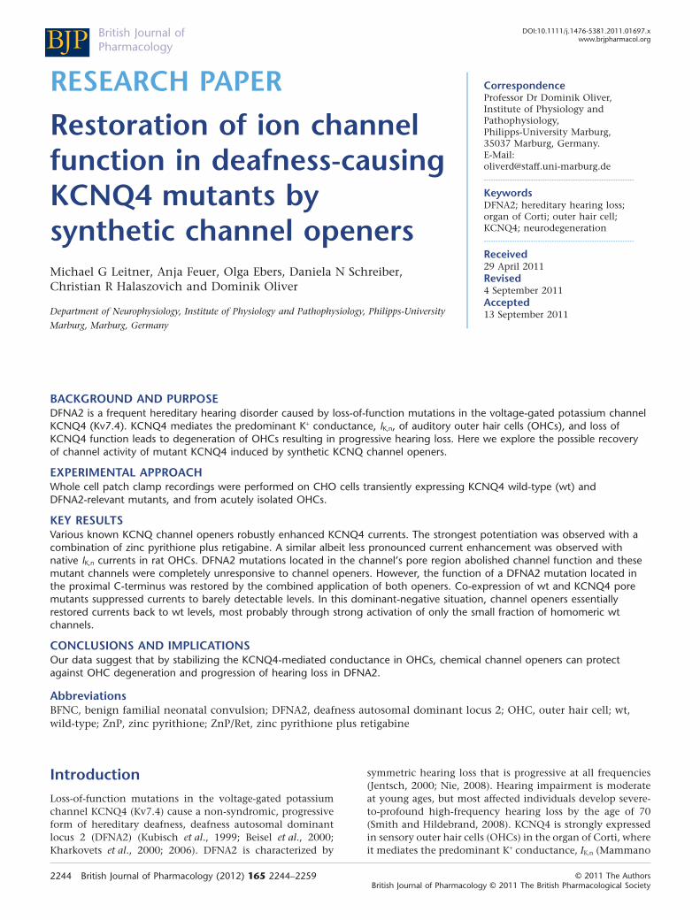
RESEARCH PAPERbph_1697 2244..2259
Restoration of ion channelfunction in deafness-causingKCNQ4 mutants bysynthetic channel openersMichael G Leitner, Anja Feuer, Olga Ebers, Daniela N Schreiber,Christian R Halaszovich and Dominik Oliver
Department of Neurophysiology, Institute of Physiology and Pathophysiology, Philipps-University
Marburg, Marburg, Germany
CorrespondenceProfessor Dr Dominik Oliver,Institute of Physiology andPathophysiology,Philipps-University Marburg,35037 Marburg, Germany.E-Mail:oliverd@staff.uni-marburg.de----------------------------------------------------------------
KeywordsDFNA2; hereditary hearing loss;organ of Corti; outer hair cell;KCNQ4; neurodegeneration----------------------------------------------------------------
Received29 April 2011Revised4 September 2011Accepted13 September 2011
BACKGROUND AND PURPOSEDFNA2 is a frequent hereditary hearing disorder caused by loss-of-function mutations in the voltage-gated potassium channelKCNQ4 (Kv7.4). KCNQ4 mediates the predominant K+ conductance, IK,n, of auditory outer hair cells (OHCs), and loss ofKCNQ4 function leads to degeneration of OHCs resulting in progressive hearing loss. Here we explore the possible recoveryof channel activity of mutant KCNQ4 induced by synthetic KCNQ channel openers.
EXPERIMENTAL APPROACHWhole cell patch clamp recordings were performed on CHO cells transiently expressing KCNQ4 wild-type (wt) andDFNA2-relevant mutants, and from acutely isolated OHCs.
KEY RESULTSVarious known KCNQ channel openers robustly enhanced KCNQ4 currents. The strongest potentiation was observed with acombination of zinc pyrithione plus retigabine. A similar albeit less pronounced current enhancement was observed withnative IK,n currents in rat OHCs. DFNA2 mutations located in the channel’s pore region abolished channel function and thesemutant channels were completely unresponsive to channel openers. However, the function of a DFNA2 mutation located inthe proximal C-terminus was restored by the combined application of both openers. Co-expression of wt and KCNQ4 poremutants suppressed currents to barely detectable levels. In this dominant-negative situation, channel openers essentiallyrestored currents back to wt levels, most probably through strong activation of only the small fraction of homomeric wtchannels.
CONCLUSIONS AND IMPLICATIONSOur data suggest that by stabilizing the KCNQ4-mediated conductance in OHCs, chemical channel openers can protectagainst OHC degeneration and progression of hearing loss in DFNA2.
AbbreviationsBFNC, benign familial neonatal convulsion; DFNA2, deafness autosomal dominant locus 2; OHC, outer hair cell; wt,wild-type; ZnP, zinc pyrithione; ZnP/Ret, zinc pyrithione plus retigabine
IntroductionLoss-of-function mutations in the voltage-gated potassiumchannel KCNQ4 (Kv7.4) cause a non-syndromic, progressiveform of hereditary deafness, deafness autosomal dominantlocus 2 (DFNA2) (Kubisch et al., 1999; Beisel et al., 2000;Kharkovets et al., 2000; 2006). DFNA2 is characterized by
symmetric hearing loss that is progressive at all frequencies(Jentsch, 2000; Nie, 2008). Hearing impairment is moderateat young ages, but most affected individuals develop severe-to-profound high-frequency hearing loss by the age of 70(Smith and Hildebrand, 2008). KCNQ4 is strongly expressedin sensory outer hair cells (OHCs) in the organ of Corti, whereit mediates the predominant K+ conductance, IK,n (Mammano
BJP British Journal ofPharmacology
DOI:10.1111/j.1476-5381.2011.01697.xwww.brjpharmacol.org
2244 British Journal of Pharmacology (2012) 165 2244–2259 © 2011 The AuthorsBritish Journal of Pharmacology © 2011 The British Pharmacological Society
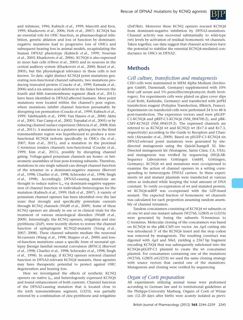
and Ashmore, 1996; Kubisch et al., 1999; Marcotti and Kros,1999; Kharkovets et al., 2006; Holt et al., 2007). KCNQ4 hasan essential role for OHC function, as pharmacological inhi-bition, genetic ablation and loss of function by dominant-negative mutations lead to progressive loss of OHCs andsubsequent hearing loss in animal models, recapitulating thehuman DFNA2 phenotype (Kubisch et al., 1999; Nouvianet al., 2003; Kharkovets et al., 2006). KCNQ4 is also expressedin inner hair cells (Oliver et al., 2003) and in neurons in thecentral auditory system (Kharkovets et al., 2000; Beisel et al.,2005), but the physiological relevance in these cells is notknown. To date, eight distinct KCNQ4 point mutations gen-erating non-functional channel subunits, two mutations pro-ducing truncated protein (Coucke et al., 1999; Kamada et al.,2006) and a six-amino acid deletion in the linker between thefourth and fifth transmembrane segment (Baek et al., 2011)have been identified in DFNA2-affected humans. Most pointmutations were located within the channel’s pore region,where mutations inhibit channel function presumably bydisrupting ion permeation (Coucke et al., 1999; Kubisch et al.,1999; Talebizadeh et al., 1999; Van Hauwe et al., 2000; Akitaet al., 2001; Van Camp et al., 2002; Topsakal et al., 2005) or byreducing channel surface expression (Mencia et al., 2008; Kimet al., 2011). A mutation in a putative splicing site in the thirdtransmembrane region was hypothesized to produce a non-functional KCNQ4 isoform specifically in OHCs (Su et al.,2007; Kim et al., 2011), and a mutation in the proximalC-terminus renders channels non-functional (Coucke et al.,1999; Kim et al., 2011), possibly by disrupting channelgating. Voltage-gated potassium channels are homo- or het-eromeric assemblies of four pore-forming subunits. Therefore,mutations in one single subunit can disrupt channel functionof the tetramer in a dominant-negative manner (Biervertet al., 1998; Charlier et al., 1998; Schroeder et al., 1998; Singhet al., 1998). Accordingly, DFNA2-causing mutations arethought to reduce native IK,n via dominant-negative suppres-sion of channel function in individuals heterozygous for themutation (Kubisch et al., 1999; Holt et al., 2007). A variety ofpotassium channel openers has been identified over the lastyears that strongly and specifically potentiate currentsthrough KCNQ channels (Wulff et al., 2009). Some of theseKCNQ openers are already in use or in clinical trials for thetreatment of various neurological disorders (Wulff et al.,2009). Interestingly, the KCNQ openers, retigabine and zincpyrithione (ZnP), were recently shown to restore the channelfunction of epileptogenic KCNQ2-mutants (Xiong et al.,2007; 2008). These channel subunits mediate the neuronalM-currents (Wang et al., 1998; Shapiro et al., 2000) and loss-of-function mutations cause a specific form of neonatal epi-lepsy [benign familial neonatal convulsion (BFNC)] (Biervertet al., 1998; Charlier et al., 1998; Schroeder et al., 1998; Singhet al., 1998). In analogy, if KCNQ openers restored channelfunction in DFNA2-relevant KCNQ4 mutants, these agonistsmay have therapeutic potential to protect against OHCdegeneration and hearing loss.
Here we investigated the effects of synthetic KCNQopeners on native IK,n and heterologously expressed KCNQ4and found enhancement of both currents. Channel functionof the DFNA2-causing mutation that is located close tothe sixth transmembrane segment, G321S, was partiallyrestored by a combination of zinc-pyrithione and retigabine
(ZnP/Ret). Moreover these KCNQ openers rescued KCNQ4from dominant-negative inhibition by DFNA2-mutations.Channel activity was recovered substantially to wild-type(wt) levels by activation of residual homomeric wt channels.Taken together, our data suggest that channel activators havethe potential to stabilize the essential KCNQ4-mediated con-ductance in OHCs in DFNA2.
Methods
Cell culture, transfection and mutagenesisCHO cells were maintained in MEM Alpha Medium (Invitro-gen GmbH, Darmstadt, Germany) supplemented with 10%fetal calf serum and 1% penicillin/streptomycin (both Invit-rogen). For experiments cells were plated on glass cover slips(Carl Roth, Karlsruhe, Germany) and transfected with jetPEItransfection reagent (Polyplus Transfection, Illkirch, France).Experiments on transfected cells were performed 24 h to 48 hpost-transfection. The expression vectors used were pEGFP-C1-KCNQ4 and pRFP-C1-KCNQ4 (NM_004700.2), and pBK-CMV-KCNQ3 (NM_004519.2). The expressed channels arereferred to as KCNQ4 wt and KCNQ3 wt (Kv7.4 and Kv7.3,respectively) according to the Guide to Receptors and Chan-nels (Alexander et al., 2009). Based on pEGFP-C1-KCNQ4 sixDFNA2-relevant point mutations were generated by site-directed mutagenesis using the QuickChangeII XL Site-Directed mutagenesis kit (Stratagene, Santa Clara, CA, USA),and mutagenesis was verified by sequencing (SEQLAB-Sequence Laboratories Göttingen GmbH, Göttingen,Germany). KCNQ4 wt and mutations were co-expressed toexamine the action of channel openers in a setting corre-sponding to heterozygote DNFA2 carriers. In these experi-ments wt and mutant plasmids were transfected at variousratios as indicated while keeping the total amount of DNAconstant. To verify co-expression of wt and mutated protein,wt KCNQ4-mRFP was co-expressed with the GFP-fusedmutant. The expected fraction of homomeric wt channelswas calculated for each proportion assuming random assem-bly of channel tetramers.
Tandem concatamers consisting of KCNQ4 wt subunits orof one wt and one mutant subunit (W276S, G285S or G321S)were generated by fusing the subunits N-terminus toC-terminus. Molecular cloning of the concatamers was basedon KCNQ4 in the pBK-CMV-rev vector. An AgeI cutting sitewas introduced 3′ of the KCNQ4 insert and the stop codonwas removed by mutagenesis. The resulting construct wasdigested with AgeI and NheI, yielding a 2367 bp fragmentencoding KCNQ4 that was subsequently subcloned into theKCNQ4-pEGFP-C1 plasmid to create the wt concatamerplasmid. For concatamers containing one of the mutations(W276S, G285S orG321S) we used the same cloning strategywith source vectors that carried one of the mutations.Mutagenesis and cloning were verified by sequencing.
Organ of Corti preparationAll experiments utilizing animal tissue were performedaccording to German law and to institutional guidelines atthe Philipps-University Marburg. Organs of Corti of Wistarrats (12–20 days after birth) were acutely isolated as previ-
BJPRescue of DFNA2 mutations by KCNQ agonists
British Journal of Pharmacology (2012) 165 2244–2259 2245
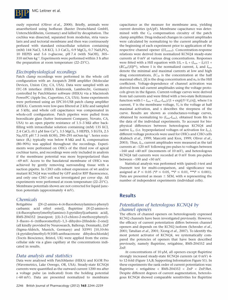
ously reported (Oliver et al., 2000). Briefly, animals wereanaesthetized using Isofluran (Baxter Deutschland GmbH,Unterschleißheim, Germany) and killed by decapitation. Thecochlea was dissected, separated from modiolus, stria vascu-laris and and tectorial membrane and then was continuouslyperfused with standard extracellular solution containing(mM) 144 NaCl, 5.8 KCl, 1.3 CaCl2, 0.9 MgCl2, 0.7 NaH2PO4,10 HEPES and 5.6 D-glucose, pH 7.4 (with NaOH), 305–310 mOsm·kg-1. Experiments were performed within 3 h afterthe preparation at room temperature (22–25°C).
Electrophysiological recordingsPatch clamp recordings were performed in the whole cellconfiguration with an Axopatch 200B amplifier (MolecularDevices, Union City, CA, USA). Data were sampled with anITC-18 interface (HEKA Elektronik, Lambrecht, Germany)controlled by PatchMaster software (HEKA) via a MacintoshPowerPC (Apple Inc, Cupertino, CA, USA). Some experimentswere performed using an EPC10-USB patch clamp amplifier(HEKA). Currents were low-pass filtered at 2 kHz and sampledat 5 kHz, and whole cell capacitance was compensated inwhole-cell configuration. Patch pipettes were pulled fromborosilicate glass (Sutter Instrument Company, Novato, CA,USA) to an open pipette resistance of 1.5–3 MW after back-filling with intracellular solution containing (mM) 135 KCl,2.4 CaCl2 (0.1 mM free Ca2+), 3.5 MgCl2, 5 HEPES, 5 EGTA, 2.5Na2ATP, pH 7.3 (with KOH), 290–295 mOsm·kg-1. Series resis-tance (Rs) typically was below 8 MW and Rs compensation(80–90%) was applied throughout the recordings. Experi-ments were performed on OHCs of the third row of apicalcochlear turns, and recordings were only included in analysisif the membrane potential was more hyperpolarized than-69 mV. Access to the basolateral membrane of OHCs wasachieved by gently removing surrounding tissue with asuction pipette. In CHO cells robust expression of wt and/ormutant KCNQ4 was verified by GFP and/or RFP fluorescence,and only one CHO cell was investigated per cover slip. Allexperiments were performed at room temperature (22–25°C).Membrane potentials shown are not corrected for liquid junc-tion potentials (approximately 4 mV).
ChemicalsRetigabine {[N-(2-amino-4-(4-fluorobenzylamino)-phenyl)carbamic acid ethyl ester]}, flupirtine {N-[2-amino-6-((4-fluorophenyl)methyl)amino)-3-pyridinyl]carbamic acid},BMS-204352 {maxipost; [(S)-3-(5-chloro-2-methoxyphenyl)-3-fluoro-6- (trifluoromethyl) -1,3-dihydro-2Hindole-2-one]}(all kindly provided by Neurosearch, Ballerup, Denmark), ZnP(Sigma-Aldrich, Munich, Germany) and XE991 [10,10-bis(4-pyridinylmethyl)-9(10H)-anthracenone dihydrochloride](Tocris Bioscience, Bristol, UK) were applied from the extra-cellular side via a glass capillary at the concentrations indi-cated in results.
Data analysis and statisticsData were analysed with PatchMaster (HEKA) and IGOR Pro(Wavemetrics, Lake Oswego, OR, USA). Steady-state KCNQ4currents were quantified as the outward current 1200 ms aftera voltage pulse (as indicated) from the holding potential(-60 mV). Data are presented normalized to membrane
capacitance as the measure for membrane area, yieldingcurrent densities (pA/pF). Membrane capacitance was deter-mined with the CM compensation circuitry of the patchclamp amplifier. Drug-induced changes in current amplitudeswere calculated by normalizing to the current amplitude atthe beginning of each experiment prior to application of therespective channel opener (I/Icontrol). Concentration-responserelations were derived from normalized KCNQ4 steady-statecurrents at 0 mV at various drug concentrations. Responseswere fitted with a Hill equation with I/Ib = Ib + (Imax - Ib)/(1 +( )EC S nH
50/[ ] ), where I is the normalized current, Ib and Imax
denote the minimal and maximal currents at low and highdrug concentrations, EC50 is the concentration at the halfmaximal effect, [S] is the drug concentration and nH is the Hillcoefficient. Voltage-dependence of channel activation wasderived from tail current amplitudes using the voltage proto-cols given in the figures. Current-voltage curves were derivedfrom tail currents and were fitted with a two-state Boltzmannfunction with I = Imin + (Imax-Imin)/{1 + exp[(V-Vh)/s]}, where I iscurrent, V is the membrane voltage, Vh is the voltage at halfmaximal activation, and s describes the steepness of thecurve. Results are shown as conductance-voltage curves,obtained by normalizing to (Imax-Imin), obtained from fits tothe data of the individual experiments. To account for bio-physical differences between heterologous KCNQ4 andnative IK,n (i.e. hyperpolarized voltages of activation for IK,n),different voltage protocols were used for OHCs and CHO cells(Kubisch et al., 1999; Marcotti and Kros, 1999; Oliver et al.,2003). Thus, IK,n current amplitudes were measured as the tailcurrents at -120 mV following pre-pulses to voltages between-160 and +40 mV (increments of 10 mV), and heterologousKCNQ4 tail currents were recorded at 0 mV from pre-pulsesbetween -100 and +50 mV.
Statistical analysis was performed with (paired) t-test andDunnett test for multi-comparison, and significance wasassigned at P � 0.05 (*P � 0.05, **P � 0.01, ***P � 0.001).Data are presented as mean � SEM, with n representing thenumber of independent experiments (individual cells).
Results
Potentiation of heterologous KCNQ4 bychannel openersThe effects of channel openers on heterologously expressedKCNQ channels have been investigated previously. However,the efficacy of current potentiation differs between channelopeners and depends on the KCNQ isoform (Schroder et al.,2001; Tatulian et al., 2001; Xiong et al., 2007). To identify themost potent activator of KCNQ4, we systematically com-pared the potencies of openers that have been describedpreviously, namely flupirtine, retigabine, BMS-204352 andZnP.
At concentrations of 10 mM, all openers except flupirtinestrongly increased steady-state KCNQ4 currents (at 0 mV) 4-to 12-fold (Figure 1A,B; Supporting Information Figure S1). Inthese experiments the rank order of current potentiation was:flupirtine < retigabine < BMS-204352 < ZnP < ZnP/Ret.Despite different degrees of current augmentation, heterolo-gous KCNQ4 showed comparable sensitivities for flupirtine
BJP MG Leitner et al.
2246 British Journal of Pharmacology (2012) 165 2244–2259
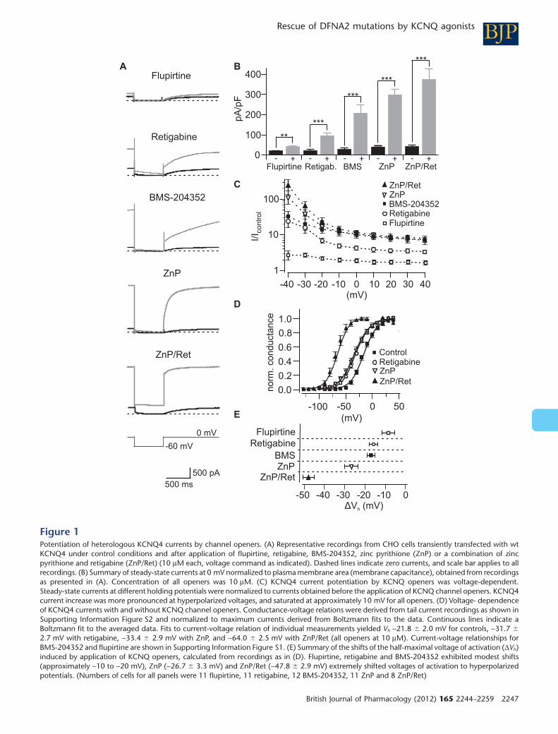
Figure 1Potentiation of heterologous KCNQ4 currents by channel openers. (A) Representative recordings from CHO cells transiently transfected with wtKCNQ4 under control conditions and after application of flupirtine, retigabine, BMS-204352, zinc pyrithione (ZnP) or a combination of zincpyrithione and retigabine (ZnP/Ret) (10 mM each, voltage command as indicated). Dashed lines indicate zero currents, and scale bar applies to allrecordings. (B) Summary of steady-state currents at 0 mV normalized to plasma membrane area (membrane capacitance), obtained from recordingsas presented in (A). Concentration of all openers was 10 mM. (C) KCNQ4 current potentiation by KCNQ openers was voltage-dependent.Steady-state currents at different holding potentials were normalized to currents obtained before the application of KCNQ channel openers. KCNQ4current increase was more pronounced at hyperpolarized voltages, and saturated at approximately 10 mV for all openers. (D) Voltage- dependenceof KCNQ4 currents with and without KCNQ channel openers. Conductance-voltage relations were derived from tail current recordings as shown inSupporting Information Figure S2 and normalized to maximum currents derived from Boltzmann fits to the data. Continuous lines indicate aBoltzmann fit to the averaged data. Fits to current-voltage relation of individual measurements yielded Vh -21.8 � 2.0 mV for controls, -31.7 �
2.7 mV with retigabine, -33.4 � 2.9 mV with ZnP, and -64.0 � 2.5 mV with ZnP/Ret (all openers at 10 mM). Current-voltage relationships forBMS-204352 and flupirtine are shown in Supporting Information Figure S1. (E) Summary of the shifts of the half-maximal voltage of activation (DVh)induced by application of KCNQ openers, calculated from recordings as in (D). Flupirtine, retigabine and BMS-204352 exhibited modest shifts(approximately -10 to -20 mV), ZnP (-26.7 � 3.3 mV) and ZnP/Ret (-47.8 � 2.9 mV) extremely shifted voltages of activation to hyperpolarizedpotentials. (Numbers of cells for all panels were 11 flupirtine, 11 retigabine, 12 BMS-204352, 11 ZnP and 8 ZnP/Ret)
BJPRescue of DFNA2 mutations by KCNQ agonists
British Journal of Pharmacology (2012) 165 2244–2259 2247
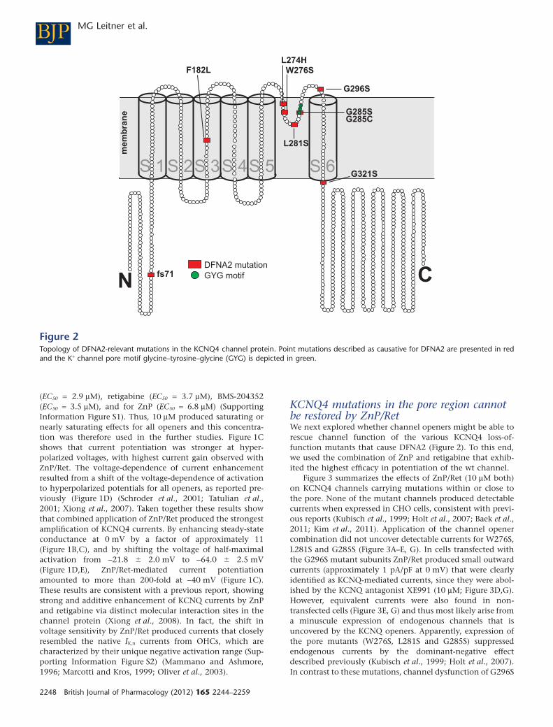
(EC50 = 2.9 mM), retigabine (EC50 = 3.7 mM), BMS-204352(EC50 = 3.5 mM), and for ZnP (EC50 = 6.8 mM) (SupportingInformation Figure S1). Thus, 10 mM produced saturating ornearly saturating effects for all openers and this concentra-tion was therefore used in the further studies. Figure 1Cshows that current potentiation was stronger at hyper-polarized voltages, with highest current gain observed withZnP/Ret. The voltage-dependence of current enhancementresulted from a shift of the voltage-dependence of activationto hyperpolarized potentials for all openers, as reported pre-viously (Figure 1D) (Schroder et al., 2001; Tatulian et al.,2001; Xiong et al., 2007). Taken together these results showthat combined application of ZnP/Ret produced the strongestamplification of KCNQ4 currents. By enhancing steady-stateconductance at 0 mV by a factor of approximately 11(Figure 1B,C), and by shifting the voltage of half-maximalactivation from -21.8 � 2.0 mV to -64.0 � 2.5 mV(Figure 1D,E), ZnP/Ret-mediated current potentiationamounted to more than 200-fold at -40 mV (Figure 1C).These results are consistent with a previous report, showingstrong and additive enhancement of KCNQ currents by ZnPand retigabine via distinct molecular interaction sites in thechannel protein (Xiong et al., 2008). In fact, the shift involtage sensitivity by ZnP/Ret produced currents that closelyresembled the native IK,n currents from OHCs, which arecharacterized by their unique negative activation range (Sup-porting Information Figure S2) (Mammano and Ashmore,1996; Marcotti and Kros, 1999; Oliver et al., 2003).
KCNQ4 mutations in the pore region cannotbe restored by ZnP/RetWe next explored whether channel openers might be able torescue channel function of the various KCNQ4 loss-of-function mutants that cause DFNA2 (Figure 2). To this end,we used the combination of ZnP and retigabine that exhib-ited the highest efficacy in potentiation of the wt channel.
Figure 3 summarizes the effects of ZnP/Ret (10 mM both)on KCNQ4 channels carrying mutations within or close tothe pore. None of the mutant channels produced detectablecurrents when expressed in CHO cells, consistent with previ-ous reports (Kubisch et al., 1999; Holt et al., 2007; Baek et al.,2011; Kim et al., 2011). Application of the channel openercombination did not uncover detectable currents for W276S,L281S and G285S (Figure 3A–E, G). In cells transfected withthe G296S mutant subunits ZnP/Ret produced small outwardcurrents (approximately 1 pA/pF at 0 mV) that were clearlyidentified as KCNQ-mediated currents, since they were abol-ished by the KCNQ antagonist XE991 (10 mM; Figure 3D,G).However, equivalent currents were also found in non-transfected cells (Figure 3E, G) and thus most likely arise froma minuscule expression of endogenous channels that isuncovered by the KCNQ openers. Apparently, expression ofthe pore mutants (W276S, L281S and G285S) suppressedendogenous currents by the dominant-negative effectdescribed previously (Kubisch et al., 1999; Holt et al., 2007).In contrast to these mutations, channel dysfunction of G296S
Figure 2Topology of DFNA2-relevant mutations in the KCNQ4 channel protein. Point mutations described as causative for DFNA2 are presented in redand the K+ channel pore motif glycine–tyrosine–glycine (GYG) is depicted in green.
BJP MG Leitner et al.
2248 British Journal of Pharmacology (2012) 165 2244–2259
appeared not to interfere with the function of the endog-enous channels, in line with a recent report of defectivemembrane targeting of this mutation (Mencia et al., 2008). Itshould be noted that the currents recorded from non-transfected CHO cells or cells transfected with G296S wereextremely small when compared with wt KCNQ4 currents(Figure 3D–G).
Taken together, these experiments show that homomericKCNQ4 channels composed of subunits carrying a mutation
in or close to the pore region cannot be rescued by applica-tion of the most potent KCNQ openers tested in this study,ZnP/Ret.
Partial restoration of channel function of amutation in the proximal C-terminusTwo DFNA2 mutations have been identified within or close totransmembrane regions distinct from the pore structure(F182L and G321S, respectively; see Figure 2) (Coucke et al.,
N
N
C
Figure 3KCNQ4 mutations in the pore region cannot be rescued by ZnP/Ret. (A–E) Representative currents measured from cells expressing the DFNA2mutations W276S (A), L281S (B), G285S (C) and G296S (D), and from non-transfected CHO cells (E) in the presence of ZnP/Ret (10 mM each).Scale bars apply to (A–E). Voltage protocol as indicated in (A). (F) wt KCNQ4 currents in the presence of ZnP/Ret. Voltage protocol as indicatedin (A). Note the different current scaling. (G) Summarized steady-state current densities at 0 mV obtained from experiments as in (A–F), undercontrol conditions, with ZnP/Ret and during subsequent application of the KCNQ antagonist XE991. None of the mutations produced substantialcurrents either in the absence or presence of ZnP/Ret. Application of the openers revealed small currents in non-transfected cells and in cellstransfected with G296S that most likely arose from endogenous expression of KCNQ-like channels in CHO cells (see text). Note the compressedaxis scaling for wt currents.
BJPRescue of DFNA2 mutations by KCNQ agonists
British Journal of Pharmacology (2012) 165 2244–2259 2249
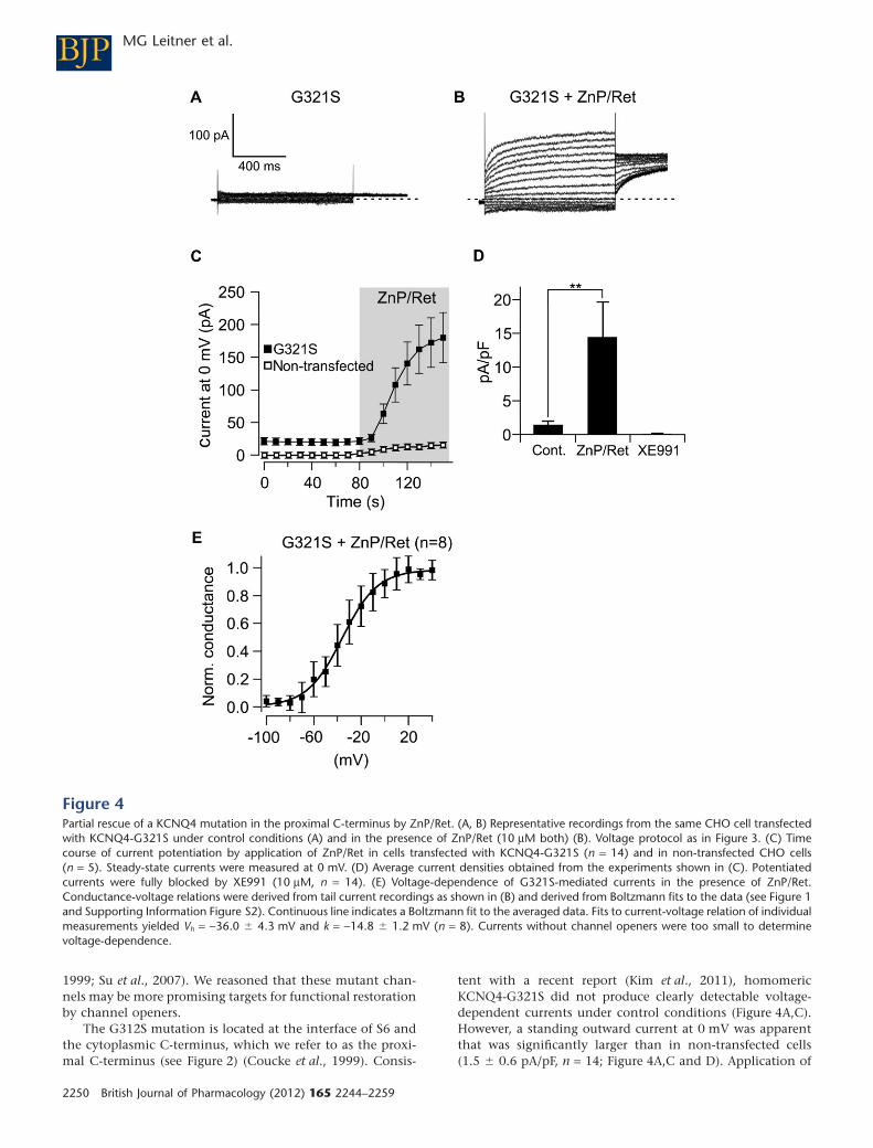
1999; Su et al., 2007). We reasoned that these mutant chan-nels may be more promising targets for functional restorationby channel openers.
The G312S mutation is located at the interface of S6 andthe cytoplasmic C-terminus, which we refer to as the proxi-mal C-terminus (see Figure 2) (Coucke et al., 1999). Consis-
tent with a recent report (Kim et al., 2011), homomericKCNQ4-G321S did not produce clearly detectable voltage-dependent currents under control conditions (Figure 4A,C).However, a standing outward current at 0 mV was apparentthat was significantly larger than in non-transfected cells(1.5 � 0.6 pA/pF, n = 14; Figure 4A,C and D). Application of
Figure 4Partial rescue of a KCNQ4 mutation in the proximal C-terminus by ZnP/Ret. (A, B) Representative recordings from the same CHO cell transfectedwith KCNQ4-G321S under control conditions (A) and in the presence of ZnP/Ret (10 mM both) (B). Voltage protocol as in Figure 3. (C) Timecourse of current potentiation by application of ZnP/Ret in cells transfected with KCNQ4-G321S (n = 14) and in non-transfected CHO cells(n = 5). Steady-state currents were measured at 0 mV. (D) Average current densities obtained from the experiments shown in (C). Potentiatedcurrents were fully blocked by XE991 (10 mM, n = 14). (E) Voltage-dependence of G321S-mediated currents in the presence of ZnP/Ret.Conductance-voltage relations were derived from tail current recordings as shown in (B) and derived from Boltzmann fits to the data (see Figure 1and Supporting Information Figure S2). Continuous line indicates a Boltzmann fit to the averaged data. Fits to current-voltage relation of individualmeasurements yielded Vh = -36.0 � 4.3 mV and k = -14.8 � 1.2 mV (n = 8). Currents without channel openers were too small to determinevoltage-dependence.
BJP MG Leitner et al.
2250 British Journal of Pharmacology (2012) 165 2244–2259
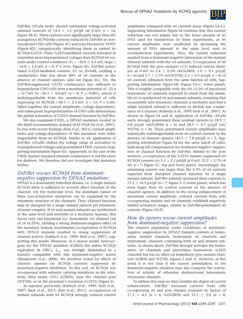
ZnP/Ret (10 mM both) elicited substantial voltage-activatedoutward currents of 14.4 � 5.1 pA/pF (at 0 mV; n = 14;Figure 4B–E). These currents were significantly larger than theendogenous KCNQ-like currents induced by ZnP/Ret in non-transfected CHO cells (Figure 4C) and were blocked by XE991(Figure 4D), unequivocally identifying them as carried byKCNQ4-G321S. These G321S-mediated currents activated atsomewhat more hyperpolarized voltages than KCNQ4 wt cur-rents under control conditions (Vh = -36.0 � 4.3 mV, slope =-14.8 � 1.2 mV, n = 8, P � 0.01; Figure 4E). ZnP/Ret poten-tiated G321S-mediated currents 15- to 20-fold, yielding aconductance that was about 40% of wt currents in theabsence of channel openers (also see Figure 3G). Yet, theZnP/Ret-augmented G321S conductance was sufficient tohyperpolarize CHO cells from a membrane potential of -25.4� 0.7 mV to -50.3 � 4.0 mV (n = 8; P � 0.001), which isindistinguishable from the membrane potential of cellsexpressing wt KCNQ4 (-48.5 � 2.3 mV, n = 12; P = 0.50).Taken together, the current amplitudes, voltage-dependence,and subsequent hyperpolarization of CHO cells demonstratethe partial restoration of G321S channel function by ZnP/Ret.
We also examined F182L, a DFNA2 mutation located inthe third transmembrane domain of KCNQ4 (Su et al., 2007).In line with recent findings (Kim et al., 2011), current ampli-tudes and voltage-dependence of this mutation were indis-tinguishable from wt KCNQ4. Similar to wt, application ofZnP/Ret (10 mM) shifted the voltage range of activation tohyperpolarized voltages and potentiated F182L currents (Sup-porting Information Figure S3). Apparently, in the case of theF182L mutant impaired channel conductance is not the causefor deafness. We therefore did not investigate this mutationfurther.
ZnP/Ret rescues KCNQ4 from dominant-negative suppression by DFNA2 mutationsDFNA2 is a dominantly inherited disease, i.e. a single mutantKCNQ4 allele is sufficient to severely affect function of thechannel. On the molecular level, the dominant nature ofthese loss-of-function mutations can be explained by thetetrameric structure of the channels. Thus, channel functionmay be disrupted by a single mutant subunit per tetramericchannel complex. If wt and mutant subunits are synthesizedat the same level and assemble in a stochastic manner, thisleaves only one functional (i.e. homomeric wt) channel outof 16 (6.25%), yielding a strong dominant-negative effect ofthe mutation. Indeed, stoichimetric co-expression of KCNQ4with DFNA2 mutants resulted in strong suppression ofchannel activity (Kubisch et al., 1999; Holt et al., 2007), sup-porting this model. Moreover, in a mouse model heterozy-gous for the DFNA2 mutation (G285S) the native KCNQ4equivalent in OHCs, IK,n, was strongly diminished in amanner compatible with this dominant-negative action(Kharkovets et al., 2006). We therefore tested for effects ofchannel openers on KCNQ4 currents suppressed bydominant-negative inhibition. To this end, wt KCNQ4 wasco-expressed with subunits carrying mutations in the selec-tivity filter motif, GYG (G285S), near the channel pore(W276S), or in the proximal C-terminus (G321S) (Figure 2).
As reported previously (Kubisch et al., 1999; Holt et al.,2007; Baek et al., 2011; Kim et al., 2011), co-expression ofmutant subunits with wt KCNQ4 strongly reduced current
amplitudes compared with wt currents alone (Figure 5A–C).Supporting Information Figure S4 confirms that this currentreduction was not simply due to the lower amount of wtDNA used for transfection in these experiments, as wtcurrent amplitudes were unaffected by decreasing theamount of DNA amount to the same level used inco-transfection experiments. Thus, the current reductionresulted from a dominant-negative interaction of the mutantchannel subunits with the wt subunits. Co-expression of wtKCNQ4 with the pore mutants (1:1) reduced current densi-ties at 0 mV to 2.4 � 0.3% (wt/G285S; 1.0 � 0.1 pA/pF,n = 6) and 5.7 � 1.7% (wt/W276S; 2.3 � 0.5 pA/pF, n = 8) ofwt currents (obtained from the same batches of cells, Sup-porting Information Figure S4) (Figure 5A–C, lower panel).This is roughly compatible with the 6% (1/16) of functionalhomomeric wt channels expected to result from the assem-bly of co-synthesized wt and mutant subunits, assuming thatco-assembly into tetrameric channels is stochastic and that asingle mutated subunit is sufficient to abolish ion conduc-tance of a channel (Kubisch et al., 1999; Holt et al., 2007). Asshown in Figure 5A and B, application of ZnP/Ret (10 mMeach) strongly potentiated these residual currents to 18.9 �
6.8 pA/pF (wt/G285S; n = 6) and 28.5 � 4.7 pA/pF (wt/W276S; n = 8). These potentiated current amplitudes werestatistically indistinguishable from wt control currents in theabsence of channel openers (43.5 � 7.8 pA/pF, n = 5, Sup-porting Information Figure S4 for the same batch of cells),indicating full compensation for dominant-negative suppres-sion of channel function by ZnP/Ret. Similar to the poremutants, co-expression of the G321S mutant suppressed wtKCNQ4 currents (to 5.3 � 2.1 pA/pF at 0 mV, 12.2 � 2.7% ofwt; n = 7; Figure 5C, top and lower panel). Interestingly, theremaining current was larger than the 6.3% of wt currentsexpected from disrupted channel function by a singlemutant subunit. ZnP/Ret robustly increased these currents to75.2 � 16.5 pA/pF (n = 7; Figure 5, C lower panel), which waseven larger than wt control currents in the absence ofchannel openers. In addition to the strong enhancement ofmaximum current amplitude, restored currents from cellsco-expressing mutant and wt channels exhibited negativelyshifted activation ranges, similar to ZnP/Ret-potentiated wtcurrents (Figure 5D,E).
How do openers rescue current amplitudesfrom dominant-negative suppression?The channel population under conditions of dominant-negative suppression by DFNA2 mutants consists of homo-meric mutant channels, homomeric wt channels andheteromeric channels containing both wt and mutant sub-units. As shown above, ZnP/Ret strongly activates the homo-meric wt channels and potentiates homomeric G321Schannels but has no effect on homomeric pore mutant chan-nels (G285S and W276S; Figures 2 and 3). However, at thispoint it is not clear if the current potentiation, in thedominant-negative situation may also comprise the restora-tion of activity of otherwise dysfunctional heteromericwt/mutant channels.
To address this issue we first consider the degree of currentenhancement. ZnP/Ret increased currents from cellsco-expressing wt and pore mutant channels by factors of17.3 � 4.5 (n = 6; wt/G285S) and 15.1 � 2.8 (n = 8;
BJPRescue of DFNA2 mutations by KCNQ agonists
British Journal of Pharmacology (2012) 165 2244–2259 2251
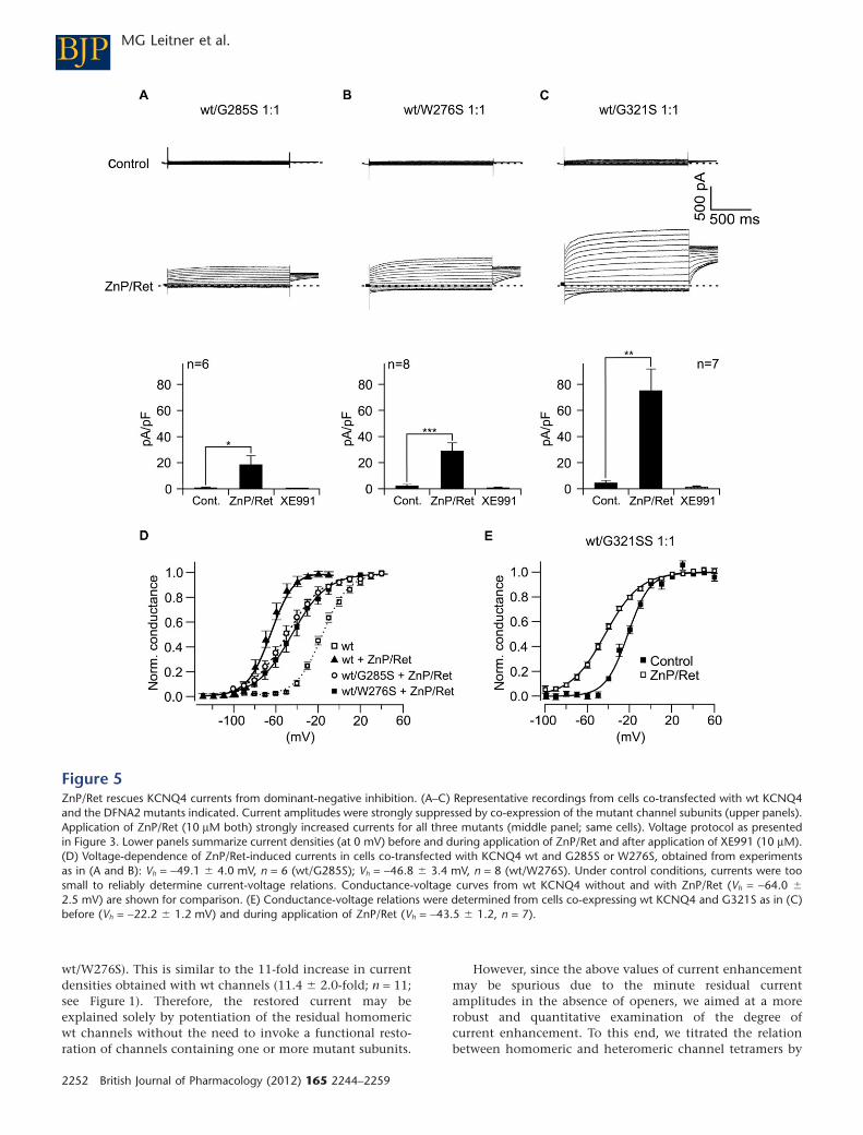
wt/W276S). This is similar to the 11-fold increase in currentdensities obtained with wt channels (11.4 � 2.0-fold; n = 11;see Figure 1). Therefore, the restored current may beexplained solely by potentiation of the residual homomericwt channels without the need to invoke a functional resto-ration of channels containing one or more mutant subunits.
However, since the above values of current enhancementmay be spurious due to the minute residual currentamplitudes in the absence of openers, we aimed at a morerobust and quantitative examination of the degree ofcurrent enhancement. To this end, we titrated the relationbetween homomeric and heteromeric channel tetramers by
Figure 5ZnP/Ret rescues KCNQ4 currents from dominant-negative inhibition. (A–C) Representative recordings from cells co-transfected with wt KCNQ4and the DFNA2 mutants indicated. Current amplitudes were strongly suppressed by co-expression of the mutant channel subunits (upper panels).Application of ZnP/Ret (10 mM both) strongly increased currents for all three mutants (middle panel; same cells). Voltage protocol as presentedin Figure 3. Lower panels summarize current densities (at 0 mV) before and during application of ZnP/Ret and after application of XE991 (10 mM).(D) Voltage-dependence of ZnP/Ret-induced currents in cells co-transfected with KCNQ4 wt and G285S or W276S, obtained from experimentsas in (A and B): Vh = -49.1 � 4.0 mV, n = 6 (wt/G285S); Vh = -46.8 � 3.4 mV, n = 8 (wt/W276S). Under control conditions, currents were toosmall to reliably determine current-voltage relations. Conductance-voltage curves from wt KCNQ4 without and with ZnP/Ret (Vh = -64.0 �
2.5 mV) are shown for comparison. (E) Conductance-voltage relations were determined from cells co-expressing wt KCNQ4 and G321S as in (C)before (Vh = -22.2 � 1.2 mV) and during application of ZnP/Ret (Vh = -43.5 � 1.2, n = 7).
BJP MG Leitner et al.
2252 British Journal of Pharmacology (2012) 165 2244–2259
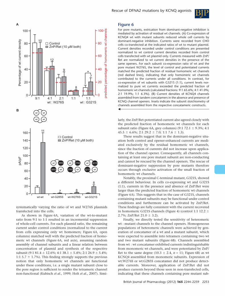
systematically varying the ratio of wt and W276S plasmidstransfected into the cells.
As shown in Figure 6A, variation of the wt-to-mutantratio from 9:1 to 1:1 resulted in an incremental suppressionof whole-cell currents. For each plasmid ratio, the remainingcurrent under control conditions (normalized to the currentfrom cells expressing only wt homomers; Figure 6A, opencolumns) matched well with the predicted fraction of homo-meric wt channels (Figure 6A, red axis), assuming randomassembly of channel subunits and a linear relation betweenconcentration of plasmid and synthesis of the respectivesubunit (9:1 81.4 � 12.6%; 4:1 38.1 � 5.4%; 2:1 26.9 � 5.4%;1:1 5.7 � 1.7%). This finding strongly supports the previousnotion that only homomeric wt channels are functionalunder these conditions, i.e. a single mutant subunit close tothe pore region is sufficient to render the tetrameric channelnon-functional (Kubisch et al., 1999; Holt et al., 2007). Simi-
larly, the ZnP/Ret-potentiated current also agreed closely withthe predicted fraction of homomeric wt channels for eachsubunit ratio (Figure 6A, grey columns) (9:1 72.1 � 9.3%; 4:145.5 � 6.6%; 2:1 29.2 � 7.0; 1:1 7.6 � 1.3).
These results suggest that in the dominant-negative situ-ation both control and opener-enhanced currents are medi-ated exclusively by the residual homomeric wt channels,since the fraction of currents did not increase upon applica-tion of the channel opener. Consequently, all channels con-taining at least one pore mutant subunit are non-conductingand cannot be rescued by the channel openers. The rescue ofdominant-negative suppression by pore mutants thereforeoccurs through exclusive activation of the small fraction ofhomomeric wt channels.
Notably, the proximal C-terminal mutant, G321S, showeda different behaviour. In cells co-expressing wt and G321S(1:1), currents in the presence and absence of ZnP/Ret werelarger than the predicted fraction of homomeric wt channels(Figure 6A). This suggests that in the case of G321S, channelscontaining mutant subunits may be functional under controlconditions and furthermore can be activated by ZnP/Ret.These findings are fully consistent with the current recoveredin homomeric G321S channels (Figure 4) (control 1:1 12.2 �
2.7%; ZnP/Ret 21.0 � 3.2).Finally, we directly tested the sensitivity of heteromeric
wt : mutant channels to the channel openers. Homogeneouspopulations of heteromeric channels were achieved by gen-eration of concatamer of a wt and a mutant subunit, whichwere expected to assemble into tetramers containing two wtand two mutant subunits (Figure 6B). Channels assembledfrom wt : wt concatamer exhibited currents indistinguishablefrom monomeric wt channels, and were potentiated by ZnP/Ret to the same degree (10.3 � 2.4, n = 11; Figure 6B) as wtKCNQ4 assembled from monomeric subunits. Expression ofwt:W276S or wt:G285S concatamer did not produce detect-able currents. Moreover, application of ZnP/Ret did notproduce currents beyond those seen in non-transfected cells,indicating that these channels containing pore mutant sub-
P
Figure 6For pore mutants, extrication from dominant-negative inhibition ismediated by activation of residual wt channels. (A) Co-expression ofKCNQ4 wt with mutant subunits reduced whole cell currents bydominant-negative inhibition. Currents were recorded from CHOcells co-transfected at the indicated ratios of wt to mutant plasmid.Current densities recorded under control conditions are presentednormalized to wt control current densities recorded from controlcells transfected with wt plasmid only. Currents measured with ZnP/Ret are normalized to wt current densities in the presence of thesame openers. For each subunit co-expression ratio of wt and thepore mutant W276S, the level of control and potentiated currentsmatched the predicted fraction of residual homomeric wt channels(red dashed lines), indicating that only homomeric wt channelscontributed to the currents under all conditions. In contrast, forco-expression of wt subunits with G321S (1:1), current levels nor-malized to pure wt currents exceeded the predicted fraction ofhomomeric wt channels (calculated fractions: 9:1 65.6%; 4:1 41.0%;2:1 19.9%; 1:1 6.3%). (B) Current densities of KCNQ4 channelsassembled from tandem concatamers in the absence and presence ofKCNQ channel openers. Insets indicate the subunit stoichiometry ofchannels assembled from the respective concatameric constructs.�
BJPRescue of DFNA2 mutations by KCNQ agonists
British Journal of Pharmacology (2012) 165 2244–2259 2253
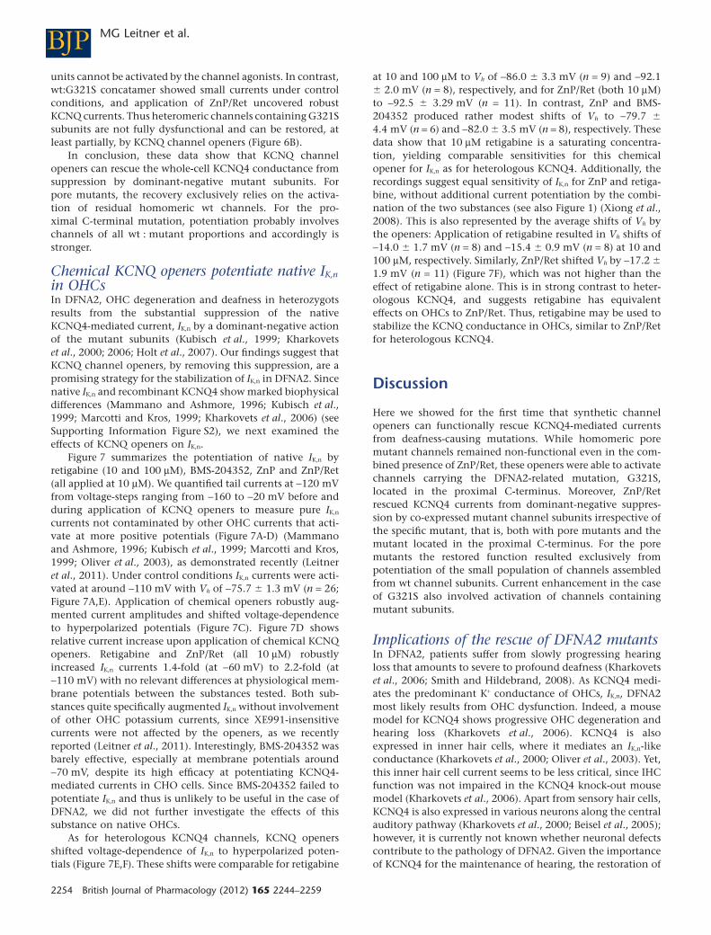
units cannot be activated by the channel agonists. In contrast,wt:G321S concatamer showed small currents under controlconditions, and application of ZnP/Ret uncovered robustKCNQ currents. Thus heteromeric channels containing G321Ssubunits are not fully dysfunctional and can be restored, atleast partially, by KCNQ channel openers (Figure 6B).
In conclusion, these data show that KCNQ channelopeners can rescue the whole-cell KCNQ4 conductance fromsuppression by dominant-negative mutant subunits. Forpore mutants, the recovery exclusively relies on the activa-tion of residual homomeric wt channels. For the pro-ximal C-terminal mutation, potentiation probably involveschannels of all wt : mutant proportions and accordingly isstronger.
Chemical KCNQ openers potentiate native IK,n
in OHCsIn DFNA2, OHC degeneration and deafness in heterozygotsresults from the substantial suppression of the nativeKCNQ4-mediated current, IK,n by a dominant-negative actionof the mutant subunits (Kubisch et al., 1999; Kharkovetset al., 2000; 2006; Holt et al., 2007). Our findings suggest thatKCNQ channel openers, by removing this suppression, are apromising strategy for the stabilization of IK,n in DFNA2. Sincenative IK,n and recombinant KCNQ4 show marked biophysicaldifferences (Mammano and Ashmore, 1996; Kubisch et al.,1999; Marcotti and Kros, 1999; Kharkovets et al., 2006) (seeSupporting Information Figure S2), we next examined theeffects of KCNQ openers on IK,n.
Figure 7 summarizes the potentiation of native IK,n byretigabine (10 and 100 mM), BMS-204352, ZnP and ZnP/Ret(all applied at 10 mM). We quantified tail currents at -120 mVfrom voltage-steps ranging from -160 to -20 mV before andduring application of KCNQ openers to measure pure IK,n
currents not contaminated by other OHC currents that acti-vate at more positive potentials (Figure 7A-D) (Mammanoand Ashmore, 1996; Kubisch et al., 1999; Marcotti and Kros,1999; Oliver et al., 2003), as demonstrated recently (Leitneret al., 2011). Under control conditions IK,n currents were acti-vated at around -110 mV with Vh of -75.7 � 1.3 mV (n = 26;Figure 7A,E). Application of chemical openers robustly aug-mented current amplitudes and shifted voltage-dependenceto hyperpolarized potentials (Figure 7C). Figure 7D showsrelative current increase upon application of chemical KCNQopeners. Retigabine and ZnP/Ret (all 10 mM) robustlyincreased IK,n currents 1.4-fold (at -60 mV) to 2.2-fold (at-110 mV) with no relevant differences at physiological mem-brane potentials between the substances tested. Both sub-stances quite specifically augmented IK,n without involvementof other OHC potassium currents, since XE991-insensitivecurrents were not affected by the openers, as we recentlyreported (Leitner et al., 2011). Interestingly, BMS-204352 wasbarely effective, especially at membrane potentials around-70 mV, despite its high efficacy at potentiating KCNQ4-mediated currents in CHO cells. Since BMS-204352 failed topotentiate IK,n and thus is unlikely to be useful in the case ofDFNA2, we did not further investigate the effects of thissubstance on native OHCs.
As for heterologous KCNQ4 channels, KCNQ openersshifted voltage-dependence of IK,n to hyperpolarized poten-tials (Figure 7E,F). These shifts were comparable for retigabine
at 10 and 100 mM to Vh of -86.0 � 3.3 mV (n = 9) and -92.1� 2.0 mV (n = 8), respectively, and for ZnP/Ret (both 10 mM)to -92.5 � 3.29 mV (n = 11). In contrast, ZnP and BMS-204352 produced rather modest shifts of Vh to -79.7 �
4.4 mV (n = 6) and -82.0 � 3.5 mV (n = 8), respectively. Thesedata show that 10 mM retigabine is a saturating concentra-tion, yielding comparable sensitivities for this chemicalopener for IK,n as for heterologous KCNQ4. Additionally, therecordings suggest equal sensitivity of IK,n for ZnP and retiga-bine, without additional current potentiation by the combi-nation of the two substances (see also Figure 1) (Xiong et al.,2008). This is also represented by the average shifts of Vh bythe openers: Application of retigabine resulted in Vh shifts of-14.0 � 1.7 mV (n = 8) and -15.4 � 0.9 mV (n = 8) at 10 and100 mM, respectively. Similarly, ZnP/Ret shifted Vh by -17.2 �
1.9 mV (n = 11) (Figure 7F), which was not higher than theeffect of retigabine alone. This is in strong contrast to heter-ologous KCNQ4, and suggests retigabine has equivalenteffects on OHCs to ZnP/Ret. Thus, retigabine may be used tostabilize the KCNQ conductance in OHCs, similar to ZnP/Retfor heterologous KCNQ4.
Discussion
Here we showed for the first time that synthetic channelopeners can functionally rescue KCNQ4-mediated currentsfrom deafness-causing mutations. While homomeric poremutant channels remained non-functional even in the com-bined presence of ZnP/Ret, these openers were able to activatechannels carrying the DFNA2-related mutation, G321S,located in the proximal C-terminus. Moreover, ZnP/Retrescued KCNQ4 currents from dominant-negative suppres-sion by co-expressed mutant channel subunits irrespective ofthe specific mutant, that is, both with pore mutants and themutant located in the proximal C-terminus. For the poremutants the restored function resulted exclusively frompotentiation of the small population of channels assembledfrom wt channel subunits. Current enhancement in the caseof G321S also involved activation of channels containingmutant subunits.
Implications of the rescue of DFNA2 mutantsIn DFNA2, patients suffer from slowly progressing hearingloss that amounts to severe to profound deafness (Kharkovetset al., 2006; Smith and Hildebrand, 2008). As KCNQ4 medi-ates the predominant K+ conductance of OHCs, IK,n, DFNA2most likely results from OHC dysfunction. Indeed, a mousemodel for KCNQ4 shows progressive OHC degeneration andhearing loss (Kharkovets et al., 2006). KCNQ4 is alsoexpressed in inner hair cells, where it mediates an IK,n-likeconductance (Kharkovets et al., 2000; Oliver et al., 2003). Yet,this inner hair cell current seems to be less critical, since IHCfunction was not impaired in the KCNQ4 knock-out mousemodel (Kharkovets et al., 2006). Apart from sensory hair cells,KCNQ4 is also expressed in various neurons along the centralauditory pathway (Kharkovets et al., 2000; Beisel et al., 2005);however, it is currently not known whether neuronal defectscontribute to the pathology of DFNA2. Given the importanceof KCNQ4 for the maintenance of hearing, the restoration of
BJP MG Leitner et al.
2254 British Journal of Pharmacology (2012) 165 2244–2259
channel function by chemical channel openers immediatelysuggests that these compounds may be of clinical use tostabilize the KCNQ conductance and prevent or retard OHCloss. It is worth noting that since DNFA2 is a dominantdeafness, most patients present with a heterozygous geno-type. Pathology therefore usually results from dominant-negative suppression of channel function by the mutantchannel subunits transcribed from the mutated allele(Kubisch et al., 1999; Holt et al., 2007; Smith and Hildebrand,2008). Here we demonstrated essentially full restoration ofcurrents in this situation, at least in a heterologous expres-sion system. Thus, enhancement of KCNQ4-mediated cur-
rents by channel openers might be a therapeutic option formost DFNA2 patients irrespective of the particular mutationin the KCNQ4 gene.
Native IK,n currents were potentiated by chemical openerssimilar to heterologously expressed KCNQ4, but current gainwas substantially smaller. Since the action of the openersincludes a substantial shift in the activation voltage range tomore hyperpolarized potentials, the degree of current poten-tiation that can be reached in vivo must also depend on themembrane potential (VM) of OHCs. The VM of OHCs in vivo isnot known precisely. While it has been reported as about-80 mV (Mammano and Ashmore, 1996; Marcotti and Kros,
Figure 7Native IK,n in OHCs is sensitive to chemical KCNQ openers. (A, B) Representative recordings of IK,n currents from an OHC before (A) and duringapplication of ZnP/Ret (10 mM each) (B). Scale bars and voltage protocol apply to (A) and (B). (C) Summary of tail current densities recorded asin (A, B) in presence of retigabine and ZnP/Ret (all 10 mM). Retigabine and ZnP/Ret increased IK,n current amplitudes and shifted voltages ofactivation to hyperpolarized voltages. (D) Potentiation of IK,n by KCNQ openers (all 10 mM). The relative current potentiation at different holdingpotentials is presented, calculated from tail currents as in (A–C). (E) Hyperpolarizing shifts in the voltage-dependence of IK,n currents induced bychemical openers. Voltage-dependence was derived from tail currents as in (C), normalized to the maximal current and fitted to a Boltzmannfunction. Under control conditions Vh was -75.7 � 1.3 mV (n = 26), and was shifted to -82.0 � 3.5 mV by BMS-204352 (10 mM; n = 8), to-86.0 � 3.3 mV by 10 mM retigabine (n = 9), to -92.1 � 2.0 mV by 100 mM retigabine (n = 8), to -79.7 � 4.4 mV by ZnP (10 mM; n = 6), andto -92.5 � 3.29 mV by ZnP/Ret (10 mM; n = 11). (F) Averaged shifts of half-maximal voltages of activation (DVh) upon application of the variousKCNQ openers obtained from the data shown in (E).
BJPRescue of DFNA2 mutations by KCNQ agonists
British Journal of Pharmacology (2012) 165 2244–2259 2255
1999), which is close to the half-activating voltage of IK,n, VM
might be much more depolarized in vivo due to the largedepolarizing transducer conductance that is lost with theusual recording conditions in vitro (He et al., 2004; Johnsonet al., 2011). The lower efficacy of openers on the nativechannel may impose a limitation to any therapeutic use ofchannel openers, unless activators with higher potency arefound. However, it should be noted that in human DFNA2,hearing loss is slowly progressive (Smith and Hildebrand,2008) and its progressive nature was recapitulated in a mousemodel. Hearing loss in mice heterozygous for the dominant-negative pore mutation G285S progressed significantly slowerthan after full deletion of KCNQ4 (Kharkovets et al., 2006).Thus, even the minute current levels retained underdominant-negative suppression of KCNQ-mediated currentsare obviously sufficient to substantially delay hair cell degen-eration. Hence, even a moderate enhancement of native IK,n,as observed with the channel openers, may provide signifi-cant protection against hair cell loss in DFNA2. We expectthat chemical KCNQ openers enhance the current of residualwt channels in the dominant-negative mouse model (Khark-ovets et al., 2006) as also seen for native IK,n in OHCs.However, if this current increase is sufficient to delay theonset of hearing loss or even protect OHCs from degenera-tion, this should be further investigated directly in vivo.
Another issue that deserves attention is the finding thatchannel openers examined here do not only act on KCNQ4channels within the inner ear but also strongly potentiateM-currents mediated by KCNQ2, -3 and -5 subunits in manycentral and peripheral neurons (Xiong et al., 2007; Lv et al.,2010). Of note, ZnP alone showed the highest potency forKCNQ4 as compared with other KCNQ isoforms (Xiong et al.,2007). However, the various openers available to date havedifferent potencies towards KCNQ4 and also differ in theirpotencies towards the different KCNQ isoforms (Schroderet al., 2001; Tatulian et al., 2001; Xiong et al., 2007), suggest-ing that it may be possible to develop openers with higherspecificity for KCNQ4.
Mechanism of current enhancementIt was shown previously that ZnP increases the open prob-ability of KCNQ channels, rather than altering single channelconductance (Xiong et al., 2007). This indicates that ZnP, andmost likely other KCNQ openers, are primarily modifiers ofchannel gating, which is consistent with the shifted voltage-dependence observed here and in previous studies (Schroderet al., 2001; Tatulian et al., 2001; Xiong et al., 2007). DFNA2mutations may abolish channel function via different mecha-nisms: most obviously, mutations in the channel pore areexpected to impair ion permeation, rendering channels con-stitutively non-functional independent of channel gating.This is consistent with our observation that KCNQ openerswere completely ineffective in uncovering any channel activ-ity in the pore mutants. For the mutation in the proximalC-terminus, G321S, there is no obvious reason to expect animpairment of ion permeation, as this region does not con-tribute to the pore structure. It seems more likely that thismutation disrupts channel gating, since in KCNQ channels,the C-terminus mediates channel gating by variousphysiological signals, including phosphatidylinositol(4,5)bisphosphate (Hernandez et al., 2008; 2009), Ca2+-calmodulin
(Gamper et al., 2005) and phosphorylation (Li et al., 2004).Consistent with this hypothesis, we found that the gatingmodifiers, ZnP/Ret, partially restored channel activity inG321S, but not in homomeric or heteromeric pore mutantchannels.
Recently, disruption of targeting to the plasma membranehas been proposed as an alternative explanation for the dys-function of DFNA2 mutants, including the pore mutationsand G321S (Mencia et al., 2008; Kim et al., 2011). Such amechanism would also be consistent with the lack of effect ofchannel openers on the pore mutants. However, the ZnP/Ret-induced currents through KCNQ4-G321S clearly show traf-ficking of this mutant channel to the plasma membrane,although we cannot exclude a reduction of surfaceexpression.
Pharmacological properties and the molecularnature of IK,nThere is strong evidence indicating that the OHC current IK,n
is mediated by KCNQ4 channels (Marcotti and Kros, 1999;Kharkovets et al., 2000; 2006; Holt et al., 2007; Oliver et al.,2003). Nevertheless, the functional properties of IK,n differfrom those of heterologously expressed KCNQ4 channels invarious aspects. Most strikingly, the activation voltage of IK,n
is much more negative and the activation kinetics are faster(Mammano and Ashmore, 1996; Marcotti and Kros, 1999).Here we show that the differences extend to the pharmaco-logical properties. Thus, current potentiation by retigabine,ZnP, and a combination of both openers was much less pro-nounced with IK,n than with heterologously expressedKCNQ4. Similarly, the drug-induced hyperpolarizing shift inthe voltage of activation was smaller for the native channel.The differential sensitivity to channel openers is particularlypronounced for BMS-204352: this opener produced a 10-foldpotentiation of recombinant KCNQ4 at saturating voltages,but was essentially ineffective on native IK,n.
The molecular basis of these differences has remainedelusive so far. An attractive hypothesis, however, is that IK,n
characteristics result from the presence of as yet unknownaccessory subunits within the native ion channel complex.KCNE b-subunits are obvious candidates for such accessorysubunits, as they co-assemble with KCNQ a-subunits and aredeterminants of biophysical and pharmacological channelproperties (McCrossan and Abbott, 2004). KCNE expressionhas been detected in the cochlea and all KCNE isoformsappear to co-assemble with KCNQ4 upon heterologousexpression (Strutz-Seebohm et al., 2006). However, none ofthe KCNE1-5 isoforms alters the biophysical properties ofKCNQ4 currents in a manner consistent with the nativecurrent, IK,n (Strutz-Seebohm et al., 2006). Another possibilityis that native IK,n channels may be heteromeric KCNQ3/KCNQ4 channels (Kubisch et al., 1999). Among the KCNQchannels, KCNQ3 is least sensitive to channel openers (Xionget al., 2007; 2008) and might decrease the sensitivity ofKCNQ4 in the heteromers. In OHCs, the expression ofKCNQ3 cannot be excluded unequivocally (Kubisch et al.,1999; Jentsch, 2000; Kharkovets et al., 2006). Thus, we testedthe properties of heteromeric channels in CHO cellsco-transfected with KCNQ3 and KCNQ4. Voltage- depen-dence and sensitivity towards channel openers of KCNQ3/4heteromers were not different from homomeric KCNQ4
BJP MG Leitner et al.
2256 British Journal of Pharmacology (2012) 165 2244–2259
channels, indicating that co-assembly with KCNQ3 in OHCscannot explain the unusual pharmacological and biophysicalproperties of IK,n (Supporting Information Figure S5). Thus,the molecular underpinnings of the native current remainunknown.
Mechanistically, one explanation for the relative insensi-tivity of IK,n might be that endogenous hair cell-specific modi-fiers of gating increase open probability via the samemechanism that is exploited by the synthetic channelopeners, thereby occluding additive effects. In fact, recombi-nant KCNQ4 in the presence of ZnP/Ret closely resembles thenative current in terms of voltage-dependence and kinetics(Supporting Information Figure S2). One prerequisite for thedramatic current enhancement by channel openers is the lowopen probability of KCNQ channels even at saturating volt-ages (Li et al., 2005). For recombinant KCNQ4, ZnP wasshown to increase the open probability at 0 mV from 0.01 to0.33 (Xiong et al., 2007). The similarity of IK,n with KCNQ4currents potentiated by ZnP/Ret may indicate high basalopen probabilities of the native channels. For pharmacologi-cal restoration of IK,n in DFNA2, a high endogenous openprobability may pose a fundamental obstacle, as it limits theenhancement that can be reached. Therefore, it will be inter-esting to see, if basal open probabilities of native channelsmediating IK,n are indeed higher than those of the correspond-ing channel in an expression system.
In conclusion, we have shown that the dominant-negative suppression of ion channel function underlyinghearing loss in DFNA2 can be overcome using syntheticchannel openers. Beyond hereditary deafness, KCNQ channelopeners may prove useful in forms of deafness that involvehair cell loss, as we have recently shown that ZnP and reti-gabine can also antagonize the suppression of IK,n by ototoxicaminoglycoside antibiotics (Leitner et al., 2011).
Acknowledgements
The authors gratefully acknowledge the kind gift of KCNQ4cDNA constructs from Dr. T.J. Jentsch (Berlin, Germany) andthank S. Petzold for excellent technical assistance, and B.Wilke and A.K. Schlusche for generating KCNQ4 mutants andfor performing preliminary experiments. This work was sup-ported by a research grant of the Medical Center Giessen andMarburg, UKGM (42/2010 MR) to D.O.
Conflict of Interest
The authors have no conflict of interest to declare.
ReferencesAkita J, Abe S, Shinkawa H, Kimberling WJ, Usami S (2001).Clinical and genetic features of nonsyndromic autosomal dominantsensorineural hearing loss: KCNQ4 is a gene responsible inJapanese. J Hum Genet 46: 355–361.
Alexander SP, Mathie A, Peters JA (2009). Guide to Receptors andChannels (GRAC). 4th edn. Br J Pharmacol 158 (Suppl. 1): S1–S254.
Baek JI, Park HJ, Park K, Choi SJ, Lee KY, Yi JH et al. (2011).Pathogenic effects of a novel mutation (c.664_681del) in KCNQ4channels associated with auditory pathology. Biochim Biophys Acta1812: 536–543.
Beisel KW, Nelson NC, Delimont DC, Fritzsch B (2000).Longitudinal gradients of KCNQ4 expression in spiral ganglion andcochlear hair cells correlate with progressive hearing loss in DFNA2.Brain Res Mol Brain Res 82: 137–149.
Beisel KW, Rocha-Sanchez SM, Morris KA, Nie L, Feng F, Kachar Bet al. (2005). Differential expression of KCNQ4 in inner hair cellsand sensory neurons is the basis of progressive high-frequencyhearing loss. J Neurosci 25: 9285–9293.
Biervert C, Schroeder BC, Kubisch C, Berkovic SF, Propping P,Jentsch TJ et al. (1998). A potassium channel mutation in neonatalhuman epilepsy. Science 279: 403–406.
Charlier C, Singh NA, Ryan SG, Lewis TB, Reus BE, Leach RJ et al.(1998). A pore mutation in a novel KQT-like potassium channelgene in an idiopathic epilepsy family. Nat Genet 18: 53–55.
Coucke PJ, Van Hauwe P, Kelley PM, Kunst H, Schatteman I,Van Velzen D et al. (1999). Mutations in the KCNQ4 gene areresponsible for autosomal dominant deafness in four DFNA2families. Hum Mol Genet 8: 1321–1328.
Gamper N, Li Y, Shapiro MS (2005). Structural requirements fordifferential sensitivity of KCNQ K+ channels to modulation byCa2+/calmodulin. Mol Biol Cell 16: 3538–3551.
He DZ, Jia S, Dallos P (2004). Mechanoelectrical transduction ofadult outer hair cells studied in a gerbil hemicochlea. Nature 429:766–770.
Hernandez CC, Zaika O, Shapiro MS (2008). A carboxy-terminalinter-helix linker as the site of phosphatidylinositol4,5-bisphosphate action on Kv7 (M-type) K+ channels. J GenPhysiol 132: 361–381.
Hernandez CC, Falkenburger B, Shapiro MS (2009). Affinity forphosphatidylinositol 4,5-bisphosphate determines muscarinicagonist sensitivity of Kv7 K+ channels. J Gen Physiol 134: 437–448.
Holt JR, Stauffer EA, Abraham D, Geleoc GS (2007).Dominant-negative inhibition of M-like potassium conductances inhair cells of the mouse inner ear. J Neurosci 27: 8940–8951.
Jentsch TJ (2000). Neuronal KCNQ potassium channels: physiologyand role in disease. Nat Rev Neurosci 1: 21–30.
Johnson SL, Beurg M, Marcotti W, Fettiplace R (2011).Prestin-driven cochlear amplification is not limited by the outerhair cell membrane time constant. Neuron 70: 1143–1154.
Kamada F, Kure S, Kudo T, Suzuki Y, Oshima T, Ichinohe A et al.(2006). A novel KCNQ4 one-base deletion in a large pedigree withhearing loss: implication for the genotype-phenotype correlation.J Hum Genet 51: 455–460.
Kharkovets T, Hardelin JP, Safieddine S, Schweizer M, El-Amraoui A,Petit C et al. (2000). KCNQ4, a K+ channel mutated in a form ofdominant deafness, is expressed in the inner ear and the centralauditory pathway. Proc Natl Acad Sci U S A 97: 4333–4338.
Kharkovets T, Dedek K, Maier H, Schweizer M, Khimich D,Nouvian R et al. (2006). Mice with altered KCNQ4 K+ channelsimplicate sensory outer hair cells in human progressive deafness.EMBO J 25: 642–652.
BJPRescue of DFNA2 mutations by KCNQ agonists
British Journal of Pharmacology (2012) 165 2244–2259 2257
Kim HJ, Lv P, Sihn CR, Yamoah EN (2011). Cellular and molecularmechanisms of autosomal dominant form of progressive hearingloss, DFNA2. J Biol Chem 286: 1517–1527.
Kubisch C, Schroeder BC, Friedrich T, Lutjohann B, El-Amraoui A,Marlin S et al. (1999). KCNQ4, a novel potassium channel expressedin sensory outer hair cells, is mutated in dominant deafness. Cell96: 437–446.
Leitner MG, Halaszovich CR, Oliver D (2011). Aminoglycosidesinhibit KCNQ4 channels in cochlear outer hair cells via depletionof phosphatidylinositol(4,5)bisphosphate. Mol Pharmacol 79:51–60.
Li Y, Langlais P, Gamper N, Liu F, Shapiro MS (2004). Dualphosphorylations underlie modulation of unitary KCNQ K(+)channels by Src tyrosine kinase. J Biol Chem 279: 45399–45407.
Li Y, Gamper N, Hilgemann DW, Shapiro MS (2005). Regulation ofKv7 (KCNQ) K+ channel open probability by phosphatidylinositol4,5-bisphosphate. J Neurosci 25: 9825–9835.
Lv P, Wei D, Yamoah EN (2010). Kv7-type channel currents inspiral ganglion neurons: involvement in sensorineural hearing loss.J Biol Chem 285: 34699–34707.
Mammano F, Ashmore JF (1996). Differential expression of outerhair cell potassium currents in the isolated cochlea of theguinea-pig. J Physiol 496: 639–646.
Marcotti W, Kros CJ (1999). Developmental expression of thepotassium current IK,n contributes to maturation of mouse outerhair cells. J Physiol 520: 653–660.
McCrossan ZA, Abbott GW (2004). The MinK-related peptides.Neuropharmacology 47: 787–821.
Mencia A, Gonzalez-Nieto D, Modamio-Hoybjor S, Etxeberria A,Aranguez G, Salvador N et al. (2008). A novel KCNQ4 pore-regionmutation (p.G296S) causes deafness by impairing cell-surfacechannel expression. Hum Genet 123: 41–53.
Nie L (2008). KCNQ4 mutations associated with nonsyndromicprogressive sensorineural hearing loss. Curr Opin Otolaryngol HeadNeck Surg 16: 441–444.
Nouvian R, Ruel J, Wang J, Guitton MJ, Pujol R, Puel JL (2003).Degeneration of sensory outer hair cells following pharmacologicalblockade of cochlear KCNQ channels in the adult guinea pig. Eur JNeurosci 17: 2553–2562.
Oliver D, Klocker N, Schuck J, Baukrowitz T, Ruppersberg JP,Fakler B (2000). Gating of Ca2+-activated K+ channels controls fastinhibitory synaptic transmission at auditory outer hair cells.Neuron 26: 595–601.
Oliver D, Knipper M, Derst C, Fakler B (2003). Resting potentialand submembrane calcium concentration of inner hair cells in theisolated mouse cochlea are set by KCNQ-type potassium channels.J Neurosci 23: 2141–2149.
Schroder RL, Jespersen T, Christophersen P, Strobaek D, Jensen BS,Olesen SP (2001). KCNQ4 channel activation by BMS-204352 andretigabine. Neuropharmacology 40: 888–898.
Schroeder BC, Kubisch C, Stein V, Jentsch TJ (1998). Moderate lossof function of cyclic-AMP-modulated KCNQ2/KCNQ3 K+ channelscauses epilepsy. Nature 396: 687–690.
Shapiro MS, Roche JP, Kaftan EJ, Cruzblanca H, Mackie K, Hille B(2000). Reconstitution of muscarinic modulation of theKCNQ2/KCNQ3 K(+) channels that underlie the neuronal Mcurrent. J Neurosci 20: 1710–1721.
Singh NA, Charlier C, Stauffer D, DuPont BR, Leach RJ, Melis Ret al. (1998). A novel potassium channel gene, KCNQ2, is mutatedin an inherited epilepsy of newborns. Nat Genet 18: 25–29.
Smith RJH, Hildebrand M (2008). DFNA2 nonsyndromic hearingloss. In: Pagon RA, Bird TC, Dolan CR, Stephens K (eds). GeneReviews [Internet]. University of Washington: Seattle, WA.http://www.ncbi.nlm.nih.gov/books/NBK1209/.
Strutz-Seebohm N, Seebohm G, Fedorenko O, Baltaev R, Engel J,Knirsch M et al. (2006). Functional coassembly of KCNQ4 withKCNE-beta- subunits in Xenopus oocytes. Cell Physiol Biochem 18:57–66.
Su CC, Yang JJ, Shieh JC, Su MC, Li SY (2007). Identification ofnovel mutations in the KCNQ4 gene of patients withnonsyndromic deafness from Taiwan. Audiol Neurootol 12: 20–26.
Talebizadeh Z, Kelley PM, Askew JW, Beisel KW, Smith SD (1999).Novel mutation in the KCNQ4 gene in a large kindred withdominant progressive hearing loss. Hum Mutat 14: 493–501.
Tatulian L, Delmas P, Abogadie FC, Brown DA (2001). Activation ofexpressed KCNQ potassium currents and native neuronal M-typepotassium currents by the anti-convulsant drug retigabine.J Neurosci 21: 5535–5545.
Topsakal V, Pennings RJ, te Brinke H, Hamel B, Huygen PL,Kremer H et al. (2005). Phenotype determination guides swiftgenotyping of a DFNA2/KCNQ4 family with a hot spot mutation(W276S). Otol Neurotol 26: 52–58.
Van Camp G, Coucke PJ, Akita J, Fransen E, Abe S, De Leenheer EMet al. (2002). A mutational hot spot in the KCNQ4 gene responsiblefor autosomal dominant hearing impairment. Hum Mutat 20:15–19.
Van Hauwe P, Coucke PJ, Ensink RJ, Huygen P, Cremers CW,Van Camp G (2000). Mutations in the KCNQ4 K+ channel gene,responsible for autosomal dominant hearing loss, cluster in thechannel pore region. Am J Med Genet 93: 184–187.
Wang HS, Pan Z, Shi W, Brown BS, Wymore RS, Cohen IS et al.(1998). KCNQ2 and KCNQ3 potassium channel subunits: molecularcorrelates of the M-channel. Science 282: 1890–1893.
Wulff H, Castle NA, Pardo LA (2009). Voltage-gated potassiumchannels as therapeutic targets. Nat Rev Drug Discov 8: 982–1001.
Xiong Q, Sun H, Li M (2007). Zinc pyrithione-mediated activationof voltage-gated KCNQ potassium channels rescues epileptogenicmutants. Nat Chem Biol 3: 287–296.
Xiong Q, Sun H, Zhang Y, Nan F, Li M (2008). Combinatorialaugmentation of voltage-gated KCNQ potassium channels bychemical openers. Proc Natl Acad Sci U S A 105: 3128–3133.
Supporting information
Additional Supporting Information may be found in theonline version of this article:
Figure S1 Chemical KCNQ openers augment heterologousKCNQ4. (A–D) Dose-response relationships of CHO cellsexpressing wt KCNQ4 for (A) retigabine (B) BMS-204352 (C)flupirtine and (D) zinc pyrithione. Current amplitudes at0 mV were normalized to control currents prior the applica-tion of the KCNQ agonist, and were fitted to a Hill equationto calculate the concentration at half-maximal effect (EC50)
BJP MG Leitner et al.
2258 British Journal of Pharmacology (2012) 165 2244–2259
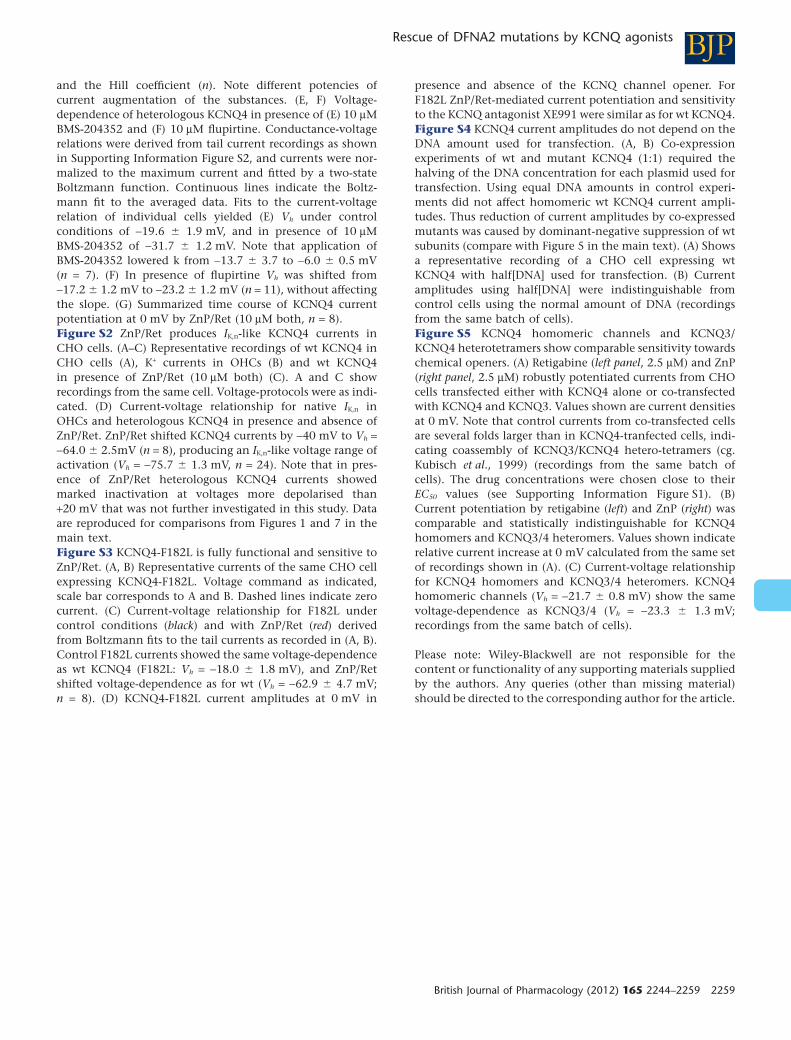
and the Hill coefficient (n). Note different potencies ofcurrent augmentation of the substances. (E, F) Voltage-dependence of heterologous KCNQ4 in presence of (E) 10 mMBMS-204352 and (F) 10 mM flupirtine. Conductance-voltagerelations were derived from tail current recordings as shownin Supporting Information Figure S2, and currents were nor-malized to the maximum current and fitted by a two-stateBoltzmann function. Continuous lines indicate the Boltz-mann fit to the averaged data. Fits to the current-voltagerelation of individual cells yielded (E) Vh under controlconditions of -19.6 � 1.9 mV, and in presence of 10 mMBMS-204352 of -31.7 � 1.2 mV. Note that application ofBMS-204352 lowered k from -13.7 � 3.7 to -6.0 � 0.5 mV(n = 7). (F) In presence of flupirtine Vh was shifted from-17.2 � 1.2 mV to -23.2 � 1.2 mV (n = 11), without affectingthe slope. (G) Summarized time course of KCNQ4 currentpotentiation at 0 mV by ZnP/Ret (10 mM both, n = 8).Figure S2 ZnP/Ret produces IK,n-like KCNQ4 currents inCHO cells. (A–C) Representative recordings of wt KCNQ4 inCHO cells (A), K+ currents in OHCs (B) and wt KCNQ4in presence of ZnP/Ret (10 mM both) (C). A and C showrecordings from the same cell. Voltage-protocols were as indi-cated. (D) Current-voltage relationship for native IK,n inOHCs and heterologous KCNQ4 in presence and absence ofZnP/Ret. ZnP/Ret shifted KCNQ4 currents by -40 mV to Vh =-64.0 � 2.5mV (n = 8), producing an IK,n-like voltage range ofactivation (Vh = -75.7 � 1.3 mV, n = 24). Note that in pres-ence of ZnP/Ret heterologous KCNQ4 currents showedmarked inactivation at voltages more depolarised than+20 mV that was not further investigated in this study. Dataare reproduced for comparisons from Figures 1 and 7 in themain text.Figure S3 KCNQ4-F182L is fully functional and sensitive toZnP/Ret. (A, B) Representative currents of the same CHO cellexpressing KCNQ4-F182L. Voltage command as indicated,scale bar corresponds to A and B. Dashed lines indicate zerocurrent. (C) Current-voltage relationship for F182L undercontrol conditions (black) and with ZnP/Ret (red) derivedfrom Boltzmann fits to the tail currents as recorded in (A, B).Control F182L currents showed the same voltage-dependenceas wt KCNQ4 (F182L: Vh = -18.0 � 1.8 mV), and ZnP/Retshifted voltage-dependence as for wt (Vh = -62.9 � 4.7 mV;n = 8). (D) KCNQ4-F182L current amplitudes at 0 mV in
presence and absence of the KCNQ channel opener. ForF182L ZnP/Ret-mediated current potentiation and sensitivityto the KCNQ antagonist XE991 were similar as for wt KCNQ4.Figure S4 KCNQ4 current amplitudes do not depend on theDNA amount used for transfection. (A, B) Co-expressionexperiments of wt and mutant KCNQ4 (1:1) required thehalving of the DNA concentration for each plasmid used fortransfection. Using equal DNA amounts in control experi-ments did not affect homomeric wt KCNQ4 current ampli-tudes. Thus reduction of current amplitudes by co-expressedmutants was caused by dominant-negative suppression of wtsubunits (compare with Figure 5 in the main text). (A) Showsa representative recording of a CHO cell expressing wtKCNQ4 with half[DNA] used for transfection. (B) Currentamplitudes using half[DNA] were indistinguishable fromcontrol cells using the normal amount of DNA (recordingsfrom the same batch of cells).Figure S5 KCNQ4 homomeric channels and KCNQ3/KCNQ4 heterotetramers show comparable sensitivity towardschemical openers. (A) Retigabine (left panel, 2.5 mM) and ZnP(right panel, 2.5 mM) robustly potentiated currents from CHOcells transfected either with KCNQ4 alone or co-transfectedwith KCNQ4 and KCNQ3. Values shown are current densitiesat 0 mV. Note that control currents from co-transfected cellsare several folds larger than in KCNQ4-tranfected cells, indi-cating coassembly of KCNQ3/KCNQ4 hetero-tetramers (cg.Kubisch et al., 1999) (recordings from the same batch ofcells). The drug concentrations were chosen close to theirEC50 values (see Supporting Information Figure S1). (B)Current potentiation by retigabine (left) and ZnP (right) wascomparable and statistically indistinguishable for KCNQ4homomers and KCNQ3/4 heteromers. Values shown indicaterelative current increase at 0 mV calculated from the same setof recordings shown in (A). (C) Current-voltage relationshipfor KCNQ4 homomers and KCNQ3/4 heteromers. KCNQ4homomeric channels (Vh = -21.7 � 0.8 mV) show the samevoltage-dependence as KCNQ3/4 (Vh = -23.3 � 1.3 mV;recordings from the same batch of cells).
Please note: Wiley-Blackwell are not responsible for thecontent or functionality of any supporting materials suppliedby the authors. Any queries (other than missing material)should be directed to the corresponding author for the article.
BJPRescue of DFNA2 mutations by KCNQ agonists
British Journal of Pharmacology (2012) 165 2244–2259 2259
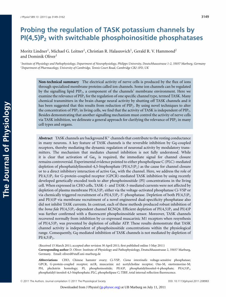
J Physiol 589.13 (2011) pp 3149–3162 3149
The
Jou
rnal
of
Phys
iolo
gy
Probing the regulation of TASK potassium channels byPI(4,5)P2 with switchable phosphoinositide phosphatases
Moritz Lindner1, Michael G. Leitner1, Christian R. Halaszovich1, Gerald R. V. Hammond2
and Dominik Oliver1
1Institute of Physiology and Pathophysiology, Department of Neurophysiology, Philipps University, Deutschhausstrasse 1-2, 35037 Marburg, Germany2Department of Pharmacology, University of Cambridge, Tennis Court Road, Cambridge CB2 1PD, UK
Non-technical summary The electrical activity of nerve cells is produced by the flux of ionsthrough specialized membrane proteins called ion channels. Some ion channels can be regulatedby the signalling lipid PIP2, a component of the channels’ membrane environment. Here weexamine the relevance of PIP2 for the regulation of one specific channel type, termed TASK. Manychemical transmitters in the brain change neural activity by shutting off TASK channels and ithas been suggested that this results from reduction of PIP2. By using novel techniques to alterthe concentration of PIP2 in living cells, we find that the activity of TASK is independent of PIP2.Besides demonstrating that another signalling mechanism must control the activity of nerve cellsvia TASK inhibition, we delineate a general approach for clarifying the relevance of PIP2 in manycell types and organs.
Abstract TASK channels are background K+ channels that contribute to the resting conductancein many neurons. A key feature of TASK channels is the reversible inhibition by Gq-coupledreceptors, thereby mediating the dynamic regulation of neuronal activity by modulatory trans-mitters. The mechanism that mediates channel inhibition is not fully understood. Whileit is clear that activation of Gαq is required, the immediate signal for channel closureremains controversial. Experimental evidence pointed to either phospholipase C (PLC)-mediateddepletion of phosphatidylinositol-4,5-bisphosphate (PI(4,5)P2) as the cause for channel closureor to a direct inhibitory interaction of active Gαq with the channel. Here, we address the role ofPI(4,5)P2 for G-protein-coupled receptor (GPCR)-mediated TASK inhibition by using recentlydeveloped genetically encoded tools to alter phosphoinositide (PI) concentrations in the livingcell. When expressed in CHO cells, TASK-1- and TASK-3-mediated currents were not affected bydepletion of plasma membrane PI(4,5)P2 either via the voltage-activated phosphatase Ci-VSP orvia chemically triggered recruitment of a PI(4,5)P2-5′-phosphatase. Depletion of both PI(4,5)P2
and PI(4)P via membrane recruitment of a novel engineered dual-specificity phosphatase alsodid not inhibit TASK currents. In contrast, each of these methods produced robust inhibition ofthe bona fide PI(4,5)P2-dependent channel KCNQ4. Efficient depletion of PI(4,5)P2 and PI(4)Pwas further confirmed with a fluorescent phosphoinositide sensor. Moreover, TASK channelsrecovered normally from inhibition by co-expressed muscarinic M1 receptors when resynthesisof PI(4,5)P2 was prevented by depletion of cellular ATP. These results demonstrate that TASKchannel activity is independent of phosphoinositide concentrations within the physiologicalrange. Consequently, Gq-mediated inhibition of TASK channels is not mediated by depletion ofPI(4,5)P2.
(Received 15 March 2011; accepted after revision 30 April 2011; first published online 3 May 2011)Corresponding author D. Oliver: Institute of Physiology and Pathophysiology, Deutschhausstrasse 2, 35037 Marburg,Germany. Email: [email protected]
Abbreviations CHO, Chinese hamster ovary; Ci-VSP, Ciona intestinalis voltage-sensitive phosphatase;GPCR, G-protein-coupled receptor; m1R, muscarinic m1 acetylcholine receptor; Oxo-M, oxotremorine-M;PH, pleckstrin homology; PI, phosphoinositide; PI(4)P, phosphatidylinositol-4-phosphate; PI(4,5)P2,phosphatidyl-inositol-4,5-bisphosphate; PLC, phospholipase C; TIRF, total internal reflection fluorescence.
C© 2011 The Authors. Journal compilation C© 2011 The Physiological Society DOI: 10.1113/jphysiol.2011.208983
) at UB Marburg on July 11, 2011jp.physoc.orgDownloaded from J Physiol (
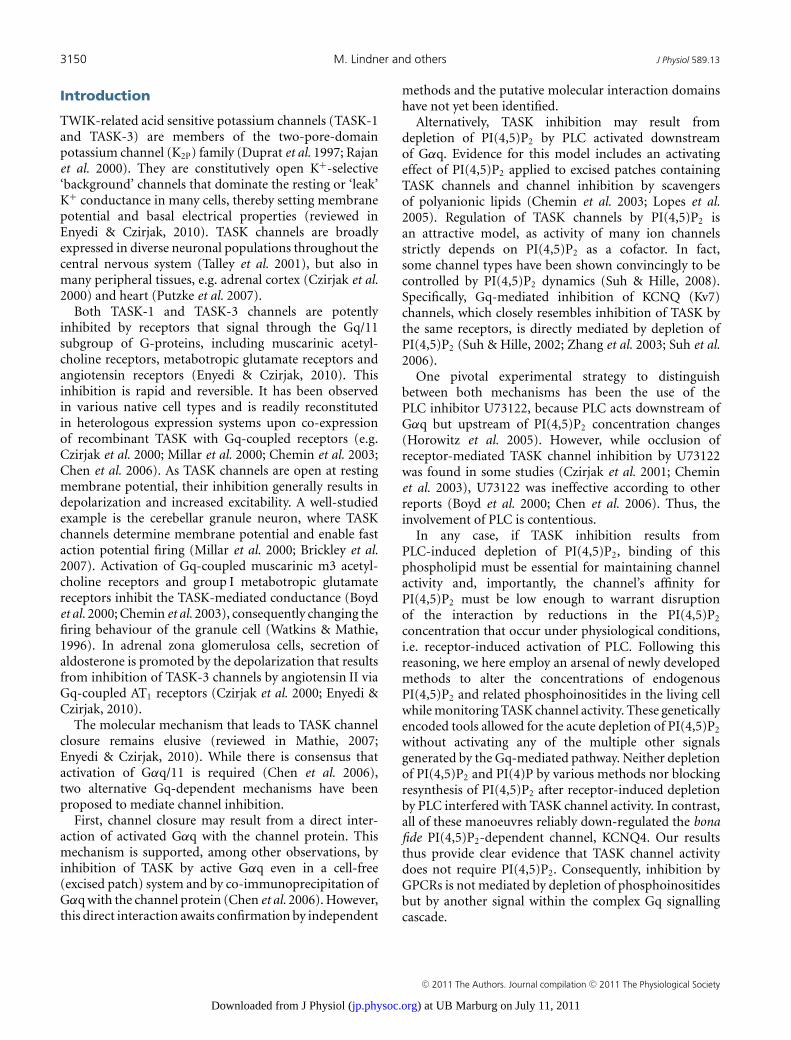
3150 M. Lindner and others J Physiol 589.13
Introduction
TWIK-related acid sensitive potassium channels (TASK-1and TASK-3) are members of the two-pore-domainpotassium channel (K2P) family (Duprat et al. 1997; Rajanet al. 2000). They are constitutively open K+-selective‘background’ channels that dominate the resting or ‘leak’K+ conductance in many cells, thereby setting membranepotential and basal electrical properties (reviewed inEnyedi & Czirjak, 2010). TASK channels are broadlyexpressed in diverse neuronal populations throughout thecentral nervous system (Talley et al. 2001), but also inmany peripheral tissues, e.g. adrenal cortex (Czirjak et al.2000) and heart (Putzke et al. 2007).
Both TASK-1 and TASK-3 channels are potentlyinhibited by receptors that signal through the Gq/11subgroup of G-proteins, including muscarinic acetyl-choline receptors, metabotropic glutamate receptors andangiotensin receptors (Enyedi & Czirjak, 2010). Thisinhibition is rapid and reversible. It has been observedin various native cell types and is readily reconstitutedin heterologous expression systems upon co-expressionof recombinant TASK with Gq-coupled receptors (e.g.Czirjak et al. 2000; Millar et al. 2000; Chemin et al. 2003;Chen et al. 2006). As TASK channels are open at restingmembrane potential, their inhibition generally results indepolarization and increased excitability. A well-studiedexample is the cerebellar granule neuron, where TASKchannels determine membrane potential and enable fastaction potential firing (Millar et al. 2000; Brickley et al.2007). Activation of Gq-coupled muscarinic m3 acetyl-choline receptors and group I metabotropic glutamatereceptors inhibit the TASK-mediated conductance (Boydet al. 2000; Chemin et al. 2003), consequently changing thefiring behaviour of the granule cell (Watkins & Mathie,1996). In adrenal zona glomerulosa cells, secretion ofaldosterone is promoted by the depolarization that resultsfrom inhibition of TASK-3 channels by angiotensin II viaGq-coupled AT1 receptors (Czirjak et al. 2000; Enyedi &Czirjak, 2010).
The molecular mechanism that leads to TASK channelclosure remains elusive (reviewed in Mathie, 2007;Enyedi & Czirjak, 2010). While there is consensus thatactivation of Gαq/11 is required (Chen et al. 2006),two alternative Gq-dependent mechanisms have beenproposed to mediate channel inhibition.
First, channel closure may result from a direct inter-action of activated Gαq with the channel protein. Thismechanism is supported, among other observations, byinhibition of TASK by active Gαq even in a cell-free(excised patch) system and by co-immunoprecipitation ofGαq with the channel protein (Chen et al. 2006). However,this direct interaction awaits confirmation by independent
methods and the putative molecular interaction domainshave not yet been identified.
Alternatively, TASK inhibition may result fromdepletion of PI(4,5)P2 by PLC activated downstreamof Gαq. Evidence for this model includes an activatingeffect of PI(4,5)P2 applied to excised patches containingTASK channels and channel inhibition by scavengersof polyanionic lipids (Chemin et al. 2003; Lopes et al.2005). Regulation of TASK channels by PI(4,5)P2 isan attractive model, as activity of many ion channelsstrictly depends on PI(4,5)P2 as a cofactor. In fact,some channel types have been shown convincingly to becontrolled by PI(4,5)P2 dynamics (Suh & Hille, 2008).Specifically, Gq-mediated inhibition of KCNQ (Kv7)channels, which closely resembles inhibition of TASK bythe same receptors, is directly mediated by depletion ofPI(4,5)P2 (Suh & Hille, 2002; Zhang et al. 2003; Suh et al.2006).
One pivotal experimental strategy to distinguishbetween both mechanisms has been the use of thePLC inhibitor U73122, because PLC acts downstream ofGαq but upstream of PI(4,5)P2 concentration changes(Horowitz et al. 2005). However, while occlusion ofreceptor-mediated TASK channel inhibition by U73122was found in some studies (Czirjak et al. 2001; Cheminet al. 2003), U73122 was ineffective according to otherreports (Boyd et al. 2000; Chen et al. 2006). Thus, theinvolvement of PLC is contentious.
In any case, if TASK inhibition results fromPLC-induced depletion of PI(4,5)P2, binding of thisphospholipid must be essential for maintaining channelactivity and, importantly, the channel’s affinity forPI(4,5)P2 must be low enough to warrant disruptionof the interaction by reductions in the PI(4,5)P2
concentration that occur under physiological conditions,i.e. receptor-induced activation of PLC. Following thisreasoning, we here employ an arsenal of newly developedmethods to alter the concentrations of endogenousPI(4,5)P2 and related phosphoinositides in the living cellwhile monitoring TASK channel activity. These geneticallyencoded tools allowed for the acute depletion of PI(4,5)P2
without activating any of the multiple other signalsgenerated by the Gq-mediated pathway. Neither depletionof PI(4,5)P2 and PI(4)P by various methods nor blockingresynthesis of PI(4,5)P2 after receptor-induced depletionby PLC interfered with TASK channel activity. In contrast,all of these manoeuvres reliably down-regulated the bonafide PI(4,5)P2-dependent channel, KCNQ4. Our resultsthus provide clear evidence that TASK channel activitydoes not require PI(4,5)P2. Consequently, inhibition byGPCRs is not mediated by depletion of phosphoinositidesbut by another signal within the complex Gq signallingcascade.
C© 2011 The Authors. Journal compilation C© 2011 The Physiological Society
) at UB Marburg on July 11, 2011jp.physoc.orgDownloaded from J Physiol (
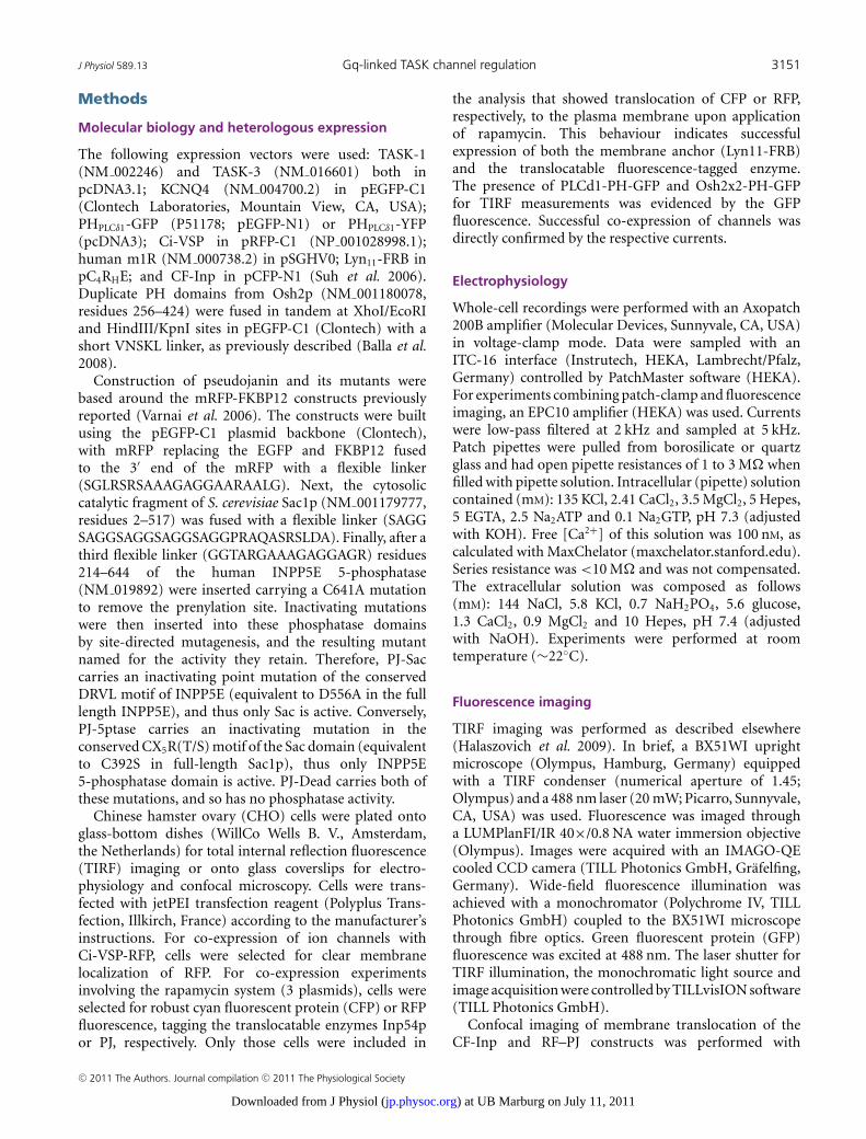
J Physiol 589.13 Gq-linked TASK channel regulation 3151
Methods
Molecular biology and heterologous expression
The following expression vectors were used: TASK-1(NM 002246) and TASK-3 (NM 016601) both inpcDNA3.1; KCNQ4 (NM 004700.2) in pEGFP-C1(Clontech Laboratories, Mountain View, CA, USA);PHPLCδ1-GFP (P51178; pEGFP-N1) or PHPLCδ1-YFP(pcDNA3); Ci-VSP in pRFP-C1 (NP 001028998.1);human m1R (NM 000738.2) in pSGHV0; Lyn11-FRB inpC4RHE; and CF-Inp in pCFP-N1 (Suh et al. 2006).Duplicate PH domains from Osh2p (NM 001180078,residues 256–424) were fused in tandem at XhoI/EcoRIand HindIII/KpnI sites in pEGFP-C1 (Clontech) with ashort VNSKL linker, as previously described (Balla et al.2008).
Construction of pseudojanin and its mutants werebased around the mRFP-FKBP12 constructs previouslyreported (Varnai et al. 2006). The constructs were builtusing the pEGFP-C1 plasmid backbone (Clontech),with mRFP replacing the EGFP and FKBP12 fusedto the 3′ end of the mRFP with a flexible linker(SGLRSRSAAAGAGGAARAALG). Next, the cytosoliccatalytic fragment of S. cerevisiae Sac1p (NM 001179777,residues 2–517) was fused with a flexible linker (SAGGSAGGSAGGSAGGSAGGPRAQASRSLDA). Finally, after athird flexible linker (GGTARGAAAGAGGAGR) residues214–644 of the human INPP5E 5-phosphatase(NM 019892) were inserted carrying a C641A mutationto remove the prenylation site. Inactivating mutationswere then inserted into these phosphatase domainsby site-directed mutagenesis, and the resulting mutantnamed for the activity they retain. Therefore, PJ-Saccarries an inactivating point mutation of the conservedDRVL motif of INPP5E (equivalent to D556A in the fulllength INPP5E), and thus only Sac is active. Conversely,PJ-5ptase carries an inactivating mutation in theconserved CX5R(T/S) motif of the Sac domain (equivalentto C392S in full-length Sac1p), thus only INPP5E5-phosphatase domain is active. PJ-Dead carries both ofthese mutations, and so has no phosphatase activity.
Chinese hamster ovary (CHO) cells were plated ontoglass-bottom dishes (WillCo Wells B. V., Amsterdam,the Netherlands) for total internal reflection fluorescence(TIRF) imaging or onto glass coverslips for electro-physiology and confocal microscopy. Cells were trans-fected with jetPEI transfection reagent (Polyplus Trans-fection, Illkirch, France) according to the manufacturer’sinstructions. For co-expression of ion channels withCi-VSP-RFP, cells were selected for clear membranelocalization of RFP. For co-expression experimentsinvolving the rapamycin system (3 plasmids), cells wereselected for robust cyan fluorescent protein (CFP) or RFPfluorescence, tagging the translocatable enzymes Inp54por PJ, respectively. Only those cells were included in
the analysis that showed translocation of CFP or RFP,respectively, to the plasma membrane upon applicationof rapamycin. This behaviour indicates successfulexpression of both the membrane anchor (Lyn11-FRB)and the translocatable fluorescence-tagged enzyme.The presence of PLCd1-PH-GFP and Osh2x2-PH-GFPfor TIRF measurements was evidenced by the GFPfluorescence. Successful co-expression of channels wasdirectly confirmed by the respective currents.
Electrophysiology
Whole-cell recordings were performed with an Axopatch200B amplifier (Molecular Devices, Sunnyvale, CA, USA)in voltage-clamp mode. Data were sampled with anITC-16 interface (Instrutech, HEKA, Lambrecht/Pfalz,Germany) controlled by PatchMaster software (HEKA).For experiments combining patch-clamp and fluorescenceimaging, an EPC10 amplifier (HEKA) was used. Currentswere low-pass filtered at 2 kHz and sampled at 5 kHz.Patch pipettes were pulled from borosilicate or quartzglass and had open pipette resistances of 1 to 3 M� whenfilled with pipette solution. Intracellular (pipette) solutioncontained (mM): 135 KCl, 2.41 CaCl2, 3.5 MgCl2, 5 Hepes,5 EGTA, 2.5 Na2ATP and 0.1 Na2GTP, pH 7.3 (adjustedwith KOH). Free [Ca2+] of this solution was 100 nM, ascalculated with MaxChelator (maxchelator.stanford.edu).Series resistance was <10 M� and was not compensated.The extracellular solution was composed as follows(mM): 144 NaCl, 5.8 KCl, 0.7 NaH2PO4, 5.6 glucose,1.3 CaCl2, 0.9 MgCl2 and 10 Hepes, pH 7.4 (adjustedwith NaOH). Experiments were performed at roomtemperature (∼22◦C).
Fluorescence imaging
TIRF imaging was performed as described elsewhere(Halaszovich et al. 2009). In brief, a BX51WI uprightmicroscope (Olympus, Hamburg, Germany) equippedwith a TIRF condenser (numerical aperture of 1.45;Olympus) and a 488 nm laser (20 mW; Picarro, Sunnyvale,CA, USA) was used. Fluorescence was imaged througha LUMPlanFI/IR 40×/0.8 NA water immersion objective(Olympus). Images were acquired with an IMAGO-QEcooled CCD camera (TILL Photonics GmbH, Grafelfing,Germany). Wide-field fluorescence illumination wasachieved with a monochromator (Polychrome IV, TILLPhotonics GmbH) coupled to the BX51WI microscopethrough fibre optics. Green fluorescent protein (GFP)fluorescence was excited at 488 nm. The laser shutter forTIRF illumination, the monochromatic light source andimage acquisition were controlled by TILLvisION software(TILL Photonics GmbH).
Confocal imaging of membrane translocation of theCF-Inp and RF–PJ constructs was performed with
C© 2011 The Authors. Journal compilation C© 2011 The Physiological Society
) at UB Marburg on July 11, 2011jp.physoc.orgDownloaded from J Physiol (
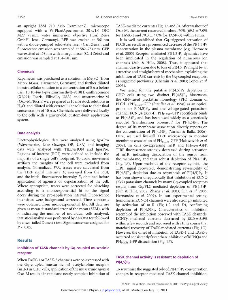
3152 M. Lindner and others J Physiol 589.13
an upright LSM 710 Axio Examiner.Z1 microscopeequipped with a W-Plan/Apochromat 20×/1.0 DICM27 75 mm water immersion objective (Carl ZeissGmbH, Jena, Germany). RFP was excited at 561 nmwith a diode-pumped solid-state laser (Carl Zeiss), andfluorescence emission was sampled at 582–754 nm. CFPwas excited at 458 nm with an argon laser (Carl Zeiss) andemission was sampled at 454–581 nm.
Chemicals
Rapamycin was purchased as a solution in Me2SO (fromMerck KGaA, Darmstadt, Germany) and further dilutedin extracellular solution to a concentration of 5 μM beforeuse. 10,10-bis(4-pyridinylmethyl)-9(10H)-anthracenone(XE991; Tocris, Ellisville, USA) and oxotremorine-M(Oxo-M; Tocris) were prepared as 10 mM stock solutions inH2O, and diluted with extracellular solution to their finalconcentration of 10 μM. Chemicals were delivered locallyto the cells with a gravity-fed, custom-built applicationsystem.
Data analysis
Electrophysiological data were analysed using IgorPro(Wavemetrics, Lake Oswego, OR, USA) and imagingdata were analysed with TILLvisION and IgorPro.Regions of interest (ROI) were defined to include themajority of a single cell’s footprint. To avoid movementartifacts the margins of the cell were excluded fromanalysis. Normalized F/F0 traces were calculated fromthe TIRF signal intensity F , averaged from the ROI,and the initial fluorescence intensity F0 obtained beforeapplication of agonists or depolarization of the cell.Where appropriate, traces were corrected for bleachingaccording to a monoexponential fit to the signaldecay during the pre-application interval. Fluorescenceintensities were background-corrected. Time constantswere obtained from monoexponential fits. All data aregiven as mean ± standard error of the mean (SEM), withn indicating the number of individual cells analysed.Statistical analysis was performed by ANOVA test followedby a two-tailed Dunett t test. Significance was assigned forP < 0.05.
Results
Inhibition of TASK channels by Gq-coupled muscarinicreceptor
When TASK-1 or TASK-3 channels were co-expressed withthe Gq-coupled muscarinic m1 acetylcholine receptor(m1R) in CHO cells, application of the muscarinic agonistOxo-M resulted in rapid and nearly complete inhibition of
TASK-mediated currents (Fig. 1A and B). After washout ofOxo-M, the current recovered to about 70% (69.1 ± 7.0%for TASK-1 and 70.3 ± 3.0% for TASK-3) within 4 min.
It is well established that Gq-triggered activation ofPLCß can result in a pronounced decrease of the PI(4,5)P2
concentration in the plasma membrane (e.g. Horowitzet al. 2005) Receptor-mediated PI(4,5)P2 dynamics havebeen implicated in the regulation of numerous ionchannels (Suh & Hille, 2008). Thus, it appeared thatchannel deactivation due to loss of PI(4,5)P2 might be anattractive and straightforward mechanism explaining theinhibition of TASK currents by the Gq-coupled receptors,as suggested previously (Chemin et al. 2003; Lopes et al.2005).
We tested for the putative PI(4,5)P2 depletion inCHO cells using two distinct PI(4,5)P2 biosensors,the GFP-fused pleckstrin homology (PH) domain ofPLCd1 (PHPLCδ1-GFP (Stauffer et al. 1998) as an opticalprobe for PI(4,5)P2, and the voltage-gated potassiumchannel KCNQ4 (Kv7.4). PHPLCδ1-GFP specifically bindsto PI(4,5)P2 and has been used widely as a geneticallyencoded ‘translocation biosensor’ for PI(4,5)P2. Thedegree of its membrane association directly reports onthe concentration of PI(4,5)P2 (Varnai & Balla, 2006).Here, we used live-cell TIRF microscopy to monitormembrane association of PHPLCδ1-GFP (Halaszovich et al.2009). In cells co-expressing m1R and PHPLCδ1-GFP,TIRF fluorescence strongly decreased during activationof m1R, indicating dissociation of the sensor fromthe membrane, and thus robust depletion of PI(4,5)P2
(Fig. 1E). Upon washout of the receptor agonist, theTIRF signal recovered, demonstrating reversibility ofPI(4,5)P2 depletion due to resynthesis of PI(4,5)P2. Ithas been shown unequivocally that inhibition of KCNQ(Kv7) potassium channels by many Gq-coupled receptorsresults from Gq/PLC-mediated depletion of PI(4,5)P2
(Suh & Hille, 2002; Zhang et al. 2003; Suh et al. 2006;Hernandez et al. 2009). In our experimental setting,homomeric KCNQ4 channels were also strongly inhibitedby activation of m1R (Fig. 1C and D), confirmingdepletion of PI(4,5)P2. Characteristics of inhibitionresembled the inhibition observed with TASK channels:KCNQ4-mediated currents decreased by 88.0 ± 5.5%within a few seconds and recovered with a time course thatmatched recovery of TASK-mediated currents (Fig. 1C).However, the onset of inhibition of TASK-1 and TASK-3occurred consistently faster than inhibition of KCNQ4 andPHPLCδ1-GFP dissociation (Fig. 1E).
TASK channel activity is resistant to depletion ofPI(4,5)P2
To scrutinize the suggested role of PI(4,5)P2 concentrationchanges in receptor-mediated TASK channel inhibition,
C© 2011 The Authors. Journal compilation C© 2011 The Physiological Society
) at UB Marburg on July 11, 2011jp.physoc.orgDownloaded from J Physiol (
J Physiol 589.13 Gq-linked TASK channel regulation 3153
we used different molecular tools for the selectivedepletion of phosphoinositides in the intact cell.
First, we used the voltage-sensitive phosphoinositidephosphatase Ci-VSP (Murata et al. 2005; Murata& Okamura, 2007), a membrane-resident PI(4,5)
P2-5-phosphatase, that is rapidly and reversibly activatedby strong depolarization of the membrane potential.Upon activation, Ci-VSP efficiently removes PI(4,5)P2
from the plasma membrane by dephosphorylation toPI(4)P (Halaszovich et al. 2009; Falkenburger et al. 2010).
Figure 1. Receptor-mediated inhibition of TASK channels and concomitant depletion of PI(4,5)P2
A, averaged time course of TASK currents upon activation of muscarinic m1 receptors. Data were obtained fromCHO cells co-transfected with m1R and either TASK-1 (n = 6) or TASK-3 (n = 12). Application of the muscarinicagonist oxo-M (10 μM, 60 s) inhibited currents to 17.0 ± 2.4% for TASK-1 and 9.7 ± 2.4% for TASK-3. Cells wereclamped at –60 mV and currents were measured in response to voltage ramps every 2 s. Traces show currentsat +50 mV, normalized to current amplitude prior to application for each cell before averaging. B, representativerecordings for the experiments summarized in A. Current traces in response to the voltage ramp shown wereobtained from a cell co-expressing m1R and TASK-3. Dashed line indicates zero current level. C, muscarinicinhibition of KCNQ4 channels measured from CHO cells co-transfected with m1R and KCNQ4 (n = 11). Currentsobtained at 0 mV using the voltage step protocol shown in D at 5 s intervals (holding potential, 0 mV) werenormalized to the current amplitude prior to application of Oxo-M for each cell before averaging. D, representativerecordings and voltage protocol for the experiments summarized in C. E, TIRF recordings from CHO cells expressingm1R and PHPLCδ1-GFP (n = 17 cells from 3 independent experiments). Application of 10 μM Oxo-M triggered astrong and reversible decrease in TIRF, indicative of translocation of the GFP-fused sensor from the membraneto the cytosol. Insets show epifluorescence (upper) and TIRF image (lower panel) of a representative cell beforeand after application of Oxo-M. Scale bar, 10 μm. F, average time constants of the onset of muscarinic currentinhibition and of translocation of PHPLCδ1-GFP. Time constants were obtained from monoexponential fits to thedata shown in (A–E). Asterisks indicate significantly faster time constants of TASK-1 and TASK-3 compared toKCNQ4 (P ≤ 0.05).
C© 2011 The Authors. Journal compilation C© 2011 The Physiological Society
) at UB Marburg on July 11, 2011jp.physoc.orgDownloaded from J Physiol (
3154 M. Lindner and others J Physiol 589.13
As shown in Fig. 2A, TASK channels were completelyinsensitive to the activation of co-expressed Ci-VSP. After50 s of depolarization to +80 mV, TASK-1 and TASK-3current amplitudes were 102.0 ± 3.2% and 107.9 ± 5.8%,respectively, of current amplitude before activation ofCi-VSP. In contrast, KCNQ4 currents were stronglyinhibited by the same voltage protocol when co-expressedwith Ci-VSP (residual current, 32.3 ± 5.8%; Fig. 2A).This indicated efficient depletion of PI(4,5)P2 by Ci-VSP,which was further confirmed by strong translocationof the fluorescent PI(4,5)P2 probe PHPLCδ1-GFP upondepolarization (Fig. 2D).
We next applied an independent approach thatwas developed recently to acutely modify PI(4,5)P2
concentrations (Suh et al. 2006; Varnai et al. 2006). Here,PI(4,5)P2 is depleted by chemically triggered recruitmentof a yeast polyphosphoinositide-5-phosphatase, Inp54p,that specifically dephosphorylates PI(4,5)P2 at the 5′
position. This method is based on the hetero-dimerization
of the FRB (FKBP/rapamycin binding) domain frommTOR and the FKBP (FK506 binding protein) in thepresence of rapamycin. FRB is anchored to the plasmamembrane by fusion to a membrane targeting motif fromLyn (Lyn11-FRB) and Inp54p is fused to CFP-tagged FKBP(CF-Inp) (Suh et al. 2006). Consequently dimerizationinduced by the addition of rapamycin results in massiveand rapid translocation of Inp54p to the membraneand thus consumption of PI(4,5)P2. Recruitment canbe monitored conveniently as the translocation of CFPfluorescence from the cytosol to the membrane (Fig. 3A,inset).
As shown in Fig. 3A, currents mediated by TASK-1 orTASK-3 were not affected by application of rapamycinto cells co-expressing either channel with Lyn11-FRB andCF-Inp. For all recordings, efficient recruitment of Inp54pwas verified by monitoring the translocation of CFPfluorescence to the membrane (insets). As before, wetested for depletion of PI(4,5)P2 by rapamycin-triggered
Figure 2. TASK currents are insensitive to depletion of PI(4,5)P2 by Ci-VSPA, representative whole-cell currents from CHO cells co-transfected with TASK-3 and Ci-VSP. Currents wererecorded in response to brief voltage ramps from a holding potential of –80mV (a). Holding potential was thendepolarised to +80 mV for 40 s to activate Ci-VSP (b), followed by repolarisation to –80 mV (c). Scale bars,0.5 nA and 100 ms. B, currents from cells co-transfected with KCNQ4 and Ci-VSP obtained with the same holdingpotential protocol as in A. Currents were evoked by brief intermittent voltage steps (–80 to 0 mV) as indicated.Scale bars, 0.1 nA and 200 ms. C, mean time course of currents obtained from experiments as in A and B fromcells co-transfected with Ci-VSP and either TASK-1, TASK-3 or KCNQ4. The shaded area indicates depolarisationto +80 mV from an initial holding potential of –80 mV. Depolarisation induced rapid inhibition of KCNQ4 currents(to 34.1 ± 5.8% of initial current at the end of the depolarizing period; n = 12) but not of TASK-1 (102.0 ± 3.2%,n = 6) and TASK-3 (107.9 ± 5.8%, n = 7). Monoexponential fits to the recovery of KCNQ4 currents yielded timeconstants of 14.6 ± 2.2 s. D, mean TIRF intensities from voltage-clamped cells expressing Ci-VSP and PHPLCδ1-GFPobtained as in Fig. 1E (n = 12). Depolarization as in C induced fast and reversible translocation of the PI(4,5)P2
probe. Monoexponential fits to the re-association of the PH domain yielded time constants of 10.2 ± 0.8 s.
C© 2011 The Authors. Journal compilation C© 2011 The Physiological Society
) at UB Marburg on July 11, 2011jp.physoc.orgDownloaded from J Physiol (
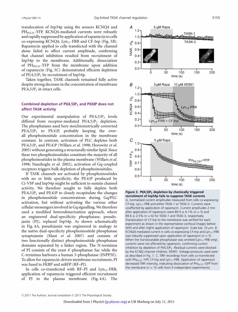
J Physiol 589.13 Gq-linked TASK channel regulation 3155
translocation of Inp54p using the sensors KCNQ4 andPHPLCδ1-YFP. KCNQ4-mediated currents were robustlyand rapidly suppressed by application of rapamycin to cellsco-expressing KCNQ4, Lyn11-FRB and CF-Inp (Fig. 3B).Rapamycin applied to cells transfected with the channelalone failed to affect current amplitude, confirmingthat channel inhibition resulted from recruitment ofInp54p to the membrane. Additionally, dissociationof PHPLCδ1-YFP from the membrane upon additionof rapamycin (Fig. 3C) demonstrated efficient depletionof PI(4,5)P2 by recruitment of Inp54p.
Taken together, TASK channels remained fully activedespite strong decrease in the concentration of membranePI(4,5)P2 in intact cells.
Combined depletion of PI(4,5)P2 and PI(4)P does notaffect TASK activity
Our experimental manipulation of PI(4,5)P2 levelsdiffered from receptor-mediated PI(4,5)P2 depletion.The phosphatases used here stoichiometrically convertedPI(4,5)P2 to PI(4)P, probably keeping the over-all phosphoinositide concentration in the membraneconstant. In contrast, activation of PLC depletes bothPI(4,5)P2 and PI(4)P (Willars et al. 1998; Horowitz et al.2005) without generating a structurally similar lipid. Sincethese two phosphoinositides constitute the majority of allphosphoinositides in the plasma membrane (Willars et al.1998; Nasuhoglu et al. 2002), activation of Gq-coupledreceptors triggers bulk depletion of phosphoinositides.
If TASK channels are activated by phosphoinositideswith no or little specificity, the PI(4)P produced byCi-VSP and Inp54p might be sufficient to sustain channelactivity. We therefore sought to fully deplete bothPI(4,5)P2 and PI(4)P to closely recapitulate the changesin phosphoinositide concentrations during Gq/PLCactivation, but without activating the various othercellular messengers involved in Gq signalling. We thereforeused a modified heterodimerization approach, wherean engineered dual-specificity phosphatase, pseudo-janin (PJ), replaced Inp54p. As shown schematicallyin Fig. 4A, pseudojanin was engineered in analogy tothe native dual-specificity phosphoinositide phosphatasesynaptojanin (Mani et al. 2007) and consists oftwo functionally distinct phosphoinositide phosphatasedomains separated by a linker region. The N-terminusof PJ consists of the yeast 4′-phosphatase Sac while theC-terminus harbours a human 5-phosphatase (INPP5E).To allow for rapamycin-driven membrane recruitment, PJwas fused to FKBP and mRFP (RF–PJ).
In cells co-transfected with RF–PJ and Lyn11-FRB,application of rapamycin triggered efficient recruitmentof PJ to the plasma membrane (Fig. 4A). The
Figure 3. PI(4,5)P2 depletion by chemically triggeredrecruitment of Inp54p fails to suppress TASK currentsA, normalized current amplitudes measured from cells co-expressingCF-Inp, Lyn11-FRB and either TASK-1 or TASK-3. Currents wereunaffected by application of rapamycin. Current amplitudes 2 minafter application of rapamycin were 89.9 ± 4.1% (n = 5) and89.6 ± 2.5% (n = 6) for TASK-1 and TASK-3, respectively.Translocation of CF-Inp to the membrane was verified for eachexperiment as shown in the representative confocal images before(left) and after (right) application of rapamycin. Scale bar, 10 μm. B,KCNQ4-mediated current in cells co-expressing CF-Inp and Lyn11-FRBwas robustly suppressed upon application of rapamycin (n = 5).When the translocatable phosphatase was omitted (Lyn11-FRB only),currents were not affected by rapamycin, confirming currentinhibition by depletion of PI(4,5)P2. Residual currents were blockedby the KCNQ channel inhibitor, XE991. Voltage protocols used wereas described in Fig. 1. C, TIRF recordings from cells co-transfectedwith PHPLCδ1-YFP, CF-Inp and Lyn11-FRB. Application of rapamycindecreased TIRF intensity, indicating dissociation of PHPLCδ1-GFP fromthe membrane (n = 15 cells from 4 independent experiments).
C© 2011 The Authors. Journal compilation C© 2011 The Physiological Society
) at UB Marburg on July 11, 2011jp.physoc.orgDownloaded from J Physiol (
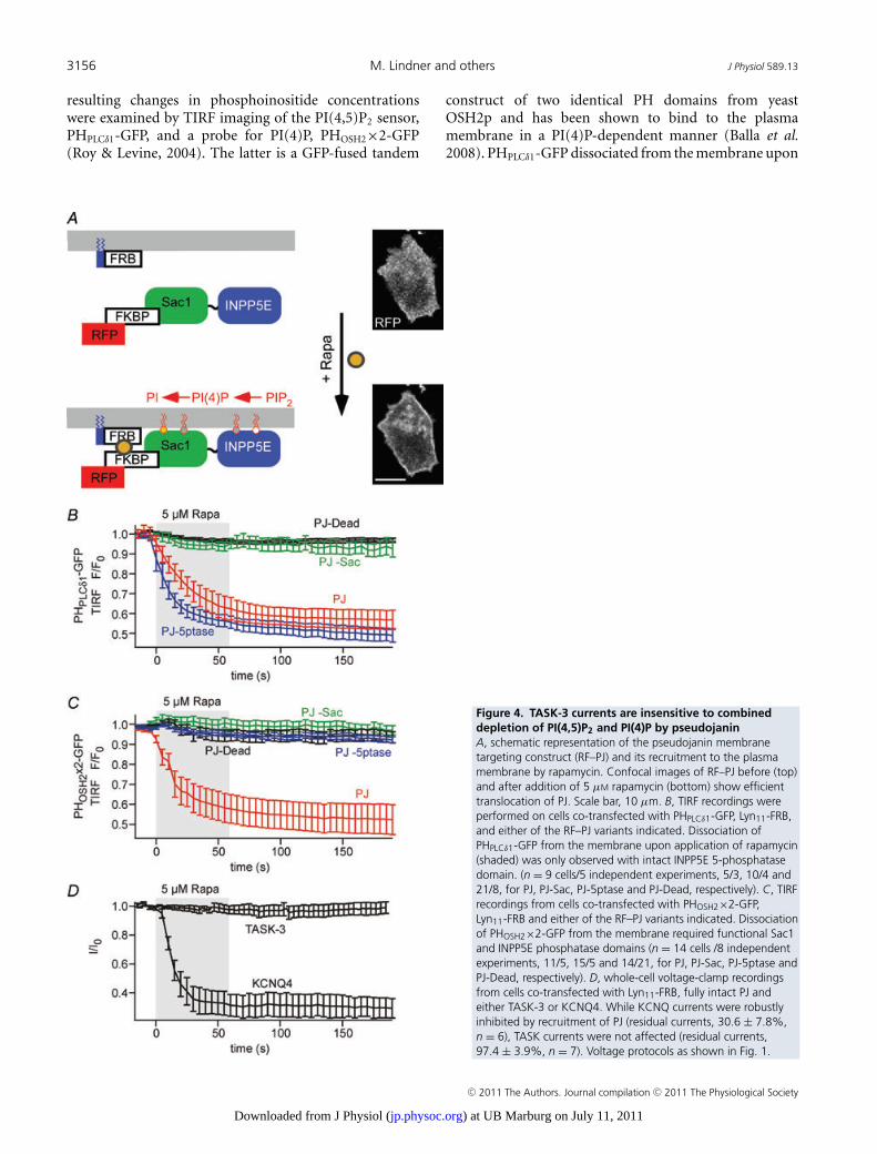
3156 M. Lindner and others J Physiol 589.13
resulting changes in phosphoinositide concentrationswere examined by TIRF imaging of the PI(4,5)P2 sensor,PHPLCδ1-GFP, and a probe for PI(4)P, PHOSH2×2-GFP(Roy & Levine, 2004). The latter is a GFP-fused tandem
construct of two identical PH domains from yeastOSH2p and has been shown to bind to the plasmamembrane in a PI(4)P-dependent manner (Balla et al.2008). PHPLCδ1-GFP dissociated from the membrane upon
Figure 4. TASK-3 currents are insensitive to combineddepletion of PI(4,5)P2 and PI(4)P by pseudojaninA, schematic representation of the pseudojanin membranetargeting construct (RF–PJ) and its recruitment to the plasmamembrane by rapamycin. Confocal images of RF–PJ before (top)and after addition of 5 μM rapamycin (bottom) show efficienttranslocation of PJ. Scale bar, 10 μm. B, TIRF recordings wereperformed on cells co-transfected with PHPLCδ1-GFP, Lyn11-FRB,and either of the RF–PJ variants indicated. Dissociation ofPHPLCδ1-GFP from the membrane upon application of rapamycin(shaded) was only observed with intact INPP5E 5-phosphatasedomain. (n = 9 cells/5 independent experiments, 5/3, 10/4 and21/8, for PJ, PJ-Sac, PJ-5ptase and PJ-Dead, respectively). C, TIRFrecordings from cells co-transfected with PHOSH2×2-GFP,Lyn11-FRB and either of the RF–PJ variants indicated. Dissociationof PHOSH2×2-GFP from the membrane required functional Sac1and INPP5E phosphatase domains (n = 14 cells /8 independentexperiments, 11/5, 15/5 and 14/21, for PJ, PJ-Sac, PJ-5ptase andPJ-Dead, respectively). D, whole-cell voltage-clamp recordingsfrom cells co-transfected with Lyn11-FRB, fully intact PJ andeither TASK-3 or KCNQ4. While KCNQ currents were robustlyinhibited by recruitment of PJ (residual currents, 30.6 ± 7.8%,n = 6), TASK currents were not affected (residual currents,97.4 ± 3.9%, n = 7). Voltage protocols as shown in Fig. 1.
C© 2011 The Authors. Journal compilation C© 2011 The Physiological Society
) at UB Marburg on July 11, 2011jp.physoc.orgDownloaded from J Physiol (
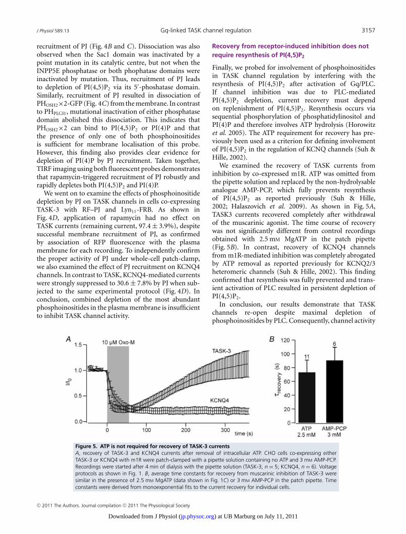
J Physiol 589.13 Gq-linked TASK channel regulation 3157
recruitment of PJ (Fig. 4B and C). Dissociation was alsoobserved when the Sac1 domain was inactivated by apoint mutation in its catalytic centre, but not when theINPP5E phosphatase or both phophatase domains wereinactivated by mutation. Thus, recruitment of PJ leadsto depletion of PI(4,5)P2 via its 5′-phoshatase domain.Similarly, recruitment of PJ resulted in dissociation ofPHOSH2×2-GFP (Fig. 4C) from the membrane. In contrastto PHPLCδ1, mutational inactivation of either phosphatasedomain abolished this dissociation. This indicates thatPHOSH2×2 can bind to PI(4,5)P2 or PI(4)P and thatthe presence of only one of both phosphoinositidesis sufficient for membrane localisation of this probe.However, this finding also provides clear evidence fordepletion of PI(4)P by PJ recruitment. Taken together,TIRF imaging using both fluorescent probes demonstratesthat rapamycin-triggered recruitment of PJ robustly andrapidly depletes both PI(4,5)P2 and PI(4)P.
We went on to examine the effects of phosphoinositidedepletion by PJ on TASK channels in cells co-expressingTASK-3 with RF–PJ and Lyn11-FRB. As shown inFig. 4D, application of rapamycin had no effect onTASK currents (remaining current, 97.4 ± 3.9%), despitesuccessful membrane recruitment of PJ, as confirmedby association of RFP fluorescence with the plasmamembrane for each recording. To independently confirmthe proper activity of PJ under whole-cell patch-clamp,we also examined the effect of PJ recruitment on KCNQ4channels. In contrast to TASK, KCNQ4-mediated currentswere strongly suppressed to 30.6 ± 7.8% by PJ when sub-jected to the same experimental protocol (Fig. 4D). Inconclusion, combined depletion of the most abundantphosphoinositides in the plasma membrane is insufficientto inhibit TASK channel activity.
Recovery from receptor-induced inhibition does notrequire resynthesis of PI(4,5)P2
Finally, we probed for involvement of phosphoinositidesin TASK channel regulation by interfering with theresynthesis of PI(4,5)P2 after activation of Gq/PLC.If channel inhibition was due to PLC-mediatedPI(4,5)P2 depletion, current recovery must dependon replenishment of PI(4,5)P2. Resynthesis occurs viasequential phosphorylation of phosphatidylinositol andPI(4)P and therefore involves ATP hydrolysis (Horowitzet al. 2005). The ATP requirement for recovery has pre-viously been used as a criterion for defining involvementof PI(4,5)P2 in the regulation of KCNQ channels (Suh &Hille, 2002).
We examined the recovery of TASK currents frominhibition by co-expressed m1R. ATP was omitted fromthe pipette solution and replaced by the non-hydrolysableanalogue AMP-PCP, which fully prevents resynthesisof PI(4,5)P2 as reported previously (Suh & Hille,2002; Halaszovich et al. 2009). As shown in Fig. 5A,TASK3 currents recovered completely after withdrawalof the muscarinic agonist. The time course of recoverywas not significantly different from control recordingsobtained with 2.5 mM MgATP in the patch pipette(Fig. 5B). In contrast, recovery of KCNQ4 channelsfrom m1R-mediated inhibition was completely abrogatedby ATP removal as reported previously for KCNQ2/3heteromeric channels (Suh & Hille, 2002). This findingconfirmed that resynthesis was fully prevented and trans-ient activation of PLC resulted in persistent depletion ofPI(4,5)P2.
In conclusion, our results demonstrate that TASKchannels re-open despite maximal depletion ofphosphoinositides by PLC. Consequently, channel activity
Figure 5. ATP is not required for recovery of TASK-3 currentsA, recovery of TASK-3 and KCNQ4 currents after removal of intracellular ATP. CHO cells co-expressing eitherTASK-3 or KCNQ4 with m1R were patch-clamped with a pipette solution containing no ATP and 3 mM AMP-PCP.Recordings were started after 4 min of dialysis with the pipette solution (TASK-3, n = 5; KCNQ4, n = 6). Voltageprotocols as shown in Fig. 1. B, average time constants for recovery from muscarinic inhibition of TASK-3 weresimilar in the presence of 2.5 mM MgATP (data shown in Fig. 1C) or 3 mM AMP-PCP in the patch pipette. Timeconstants were derived from monoexponential fits to the current recovery for individual cells.
C© 2011 The Authors. Journal compilation C© 2011 The Physiological Society
) at UB Marburg on July 11, 2011jp.physoc.orgDownloaded from J Physiol (
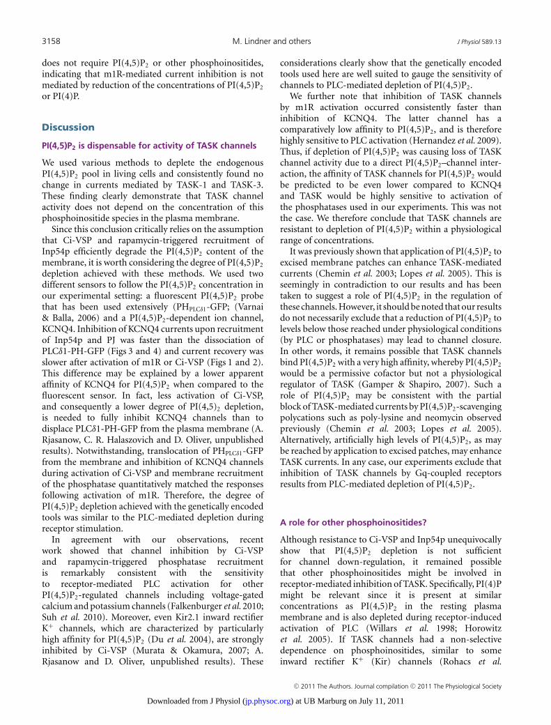
3158 M. Lindner and others J Physiol 589.13
does not require PI(4,5)P2 or other phosphoinositides,indicating that m1R-mediated current inhibition is notmediated by reduction of the concentrations of PI(4,5)P2
or PI(4)P.
Discussion
PI(4,5)P2 is dispensable for activity of TASK channels
We used various methods to deplete the endogenousPI(4,5)P2 pool in living cells and consistently found nochange in currents mediated by TASK-1 and TASK-3.These finding clearly demonstrate that TASK channelactivity does not depend on the concentration of thisphosphoinositide species in the plasma membrane.
Since this conclusion critically relies on the assumptionthat Ci-VSP and rapamycin-triggered recruitment ofInp54p efficiently degrade the PI(4,5)P2 content of themembrane, it is worth considering the degree of PI(4,5)P2
depletion achieved with these methods. We used twodifferent sensors to follow the PI(4,5)P2 concentration inour experimental setting: a fluorescent PI(4,5)P2 probethat has been used extensively (PHPLCδ1-GFP; (Varnai& Balla, 2006) and a PI(4,5)P2-dependent ion channel,KCNQ4. Inhibition of KCNQ4 currents upon recruitmentof Inp54p and PJ was faster than the dissociation ofPLCδ1-PH-GFP (Figs 3 and 4) and current recovery wasslower after activation of m1R or Ci-VSP (Figs 1 and 2).This difference may be explained by a lower apparentaffinity of KCNQ4 for PI(4,5)P2 when compared to thefluorescent sensor. In fact, less activation of Ci-VSP,and consequently a lower degree of PI(4,5)2 depletion,is needed to fully inhibit KCNQ4 channels than todisplace PLCδ1-PH-GFP from the plasma membrane (A.Rjasanow, C. R. Halaszovich and D. Oliver, unpublishedresults). Notwithstanding, translocation of PHPLCδ1-GFPfrom the membrane and inhibition of KCNQ4 channelsduring activation of Ci-VSP and membrane recruitmentof the phosphatase quantitatively matched the responsesfollowing activation of m1R. Therefore, the degree ofPI(4,5)P2 depletion achieved with the genetically encodedtools was similar to the PLC-mediated depletion duringreceptor stimulation.
In agreement with our observations, recentwork showed that channel inhibition by Ci-VSPand rapamycin-triggered phosphatase recruitmentis remarkably consistent with the sensitivityto receptor-mediated PLC activation for otherPI(4,5)P2-regulated channels including voltage-gatedcalcium and potassium channels (Falkenburger et al. 2010;Suh et al. 2010). Moreover, even Kir2.1 inward rectifierK+ channels, which are characterized by particularlyhigh affinity for PI(4,5)P2 (Du et al. 2004), are stronglyinhibited by Ci-VSP (Murata & Okamura, 2007; A.Rjasanow and D. Oliver, unpublished results). These
considerations clearly show that the genetically encodedtools used here are well suited to gauge the sensitivity ofchannels to PLC-mediated depletion of PI(4,5)P2.
We further note that inhibition of TASK channelsby m1R activation occurred consistently faster thaninhibition of KCNQ4. The latter channel has acomparatively low affinity to PI(4,5)P2, and is thereforehighly sensitive to PLC activation (Hernandez et al. 2009).Thus, if depletion of PI(4,5)P2 was causing loss of TASKchannel activity due to a direct PI(4,5)P2–channel inter-action, the affinity of TASK channels for PI(4,5)P2 wouldbe predicted to be even lower compared to KCNQ4and TASK would be highly sensitive to activation ofthe phosphatases used in our experiments. This was notthe case. We therefore conclude that TASK channels areresistant to depletion of PI(4,5)P2 within a physiologicalrange of concentrations.
It was previously shown that application of PI(4,5)P2 toexcised membrane patches can enhance TASK-mediatedcurrents (Chemin et al. 2003; Lopes et al. 2005). This isseemingly in contradiction to our results and has beentaken to suggest a role of PI(4,5)P2 in the regulation ofthese channels. However, it should be noted that our resultsdo not necessarily exclude that a reduction of PI(4,5)P2 tolevels below those reached under physiological conditions(by PLC or phosphatases) may lead to channel closure.In other words, it remains possible that TASK channelsbind PI(4,5)P2 with a very high affinity, whereby PI(4,5)P2
would be a permissive cofactor but not a physiologicalregulator of TASK (Gamper & Shapiro, 2007). Such arole of PI(4,5)P2 may be consistent with the partialblock of TASK-mediated currents by PI(4,5)P2-scavengingpolycations such as poly-lysine and neomycin observedpreviously (Chemin et al. 2003; Lopes et al. 2005).Alternatively, artificially high levels of PI(4,5)P2, as maybe reached by application to excised patches, may enhanceTASK currents. In any case, our experiments exclude thatinhibition of TASK channels by Gq-coupled receptorsresults from PLC-mediated depletion of PI(4,5)P2.
A role for other phosphoinositides?
Although resistance to Ci-VSP and Inp54p unequivocallyshow that PI(4,5)P2 depletion is not sufficientfor channel down-regulation, it remained possiblethat other phosphoinositides might be involved inreceptor-mediated inhibition of TASK. Specifically, PI(4)Pmight be relevant since it is present at similarconcentrations as PI(4,5)P2 in the resting plasmamembrane and is also depleted during receptor-inducedactivation of PLC (Willars et al. 1998; Horowitzet al. 2005). If TASK channels had a non-selectivedependence on phosphoinositides, similar to someinward rectifier K+ (Kir) channels (Rohacs et al.
C© 2011 The Authors. Journal compilation C© 2011 The Physiological Society
) at UB Marburg on July 11, 2011jp.physoc.orgDownloaded from J Physiol (
J Physiol 589.13 Gq-linked TASK channel regulation 3159
1999; Tucker & Baukrowitz, 2008), then activation ofPI(4,5)P2-5-phosphatases might not suffice for channeldeactivation despite strong depletion of PI(4,5)P2. In otherwords, the differential sensitivity of TASK to PLC versusphosphatases might result from the different changes inthe overall concentration of phosphoinositides accordingto the different enzymatic activities.
To examine such a possible role of combined depletionof PI(4,5)P2 and PI(4)P in receptor-induced TASKinhibition, we used PJ, a novel engineered dual-specificityphosphatase for dephosphorylation of PI(4,5)P2 andPI(4)P. Recruitment of PJ to the membrane efficientlydepleted both phosphoinositides as indicated by specificfluorescent sensors. However, the complete insensitivity ofTASK to this manoeuvre indicated that neither PI(4,5)P2
nor PI(4)P concentrations contribute to regulation ofTASK channels by PLC. This conclusion is furtherstrongly supported by the full reactivation of TASKchannels from receptor-induced inhibition observed aftersubstitution of ATP by the non-hydrolysable analogueAMP-PCP. Under this condition, resynthesis of PI(4)P andPI(4,5)P2 is abrogated, resulting in persistent depletionof both phosphoinositides after activation of Gq-coupledreceptors.
A TASK-associated pool of PI(4,5)P2 inaccessible toexogenous phosphatases?
It has been suggested that the plasma membranemay harbour functionally distinct pools of PI(4,5)P2
(Wang et al. 2004; Vasudevan et al. 2009). Such poolsmay correspond to PI(4,5)P2 localized to spatiallyseparated membrane domains, such as lipid rafts orcaveolae versus non-raft membranes (Johnson et al.2008; Cui et al. 2010), although such local domainsare difficult to reconcile with the fast diffusion ofphosphoinositides as determined in native plasmamembranes (Hilgemann, 2007). Experimental findingssuggested that localized PI(4,5)P2 pools resulting fromrestricted diffusion may determine regulation of ionchannels by PI(4,5)P2 in cardiomyocytes (Cho et al.2005). The efficient inhibition of KCNQ2/3 heteromericchannels by Gq-coupled muscarinic receptors in HEKcells required co-localization of channels and receptorsto detergent-resistant membrane fractions (Oldfield et al.2009). In principle, the observed differential action ofreceptor-activated PLC and recombinant phosphatasescould be compatible with PI(4,5)P2-mediated regulationof TASK channels, if the channels interacted with a distinct‘private’ pool of PI(4,5)P2 that is depleted by endogenousPLC but is inaccessible to the phosphatase constructs usedin this study. However, any membrane pool of PI(4,5)P2
must remain exhausted after depletion by PLC whenreplenishment of PI(4,5)P2 is blocked. Therefore, such a
possibility is clearly ruled out by our finding of full currentrecovery in the absence of intracellular ATP.
In summary, our results obtained upon depletionof PI(4,5)P2 and PI(4)P by exogenous phosphatasesand inhibition of their resynthesis after depletion byendogenous PLC rule out a requirement of thesephosphoinositides for TASK channel activity. Specifically,we conclude that the inhibition of TASK channels byGq-coupled receptors is not mediated by depletion ofphosphoinositides.
Possible mechanisms for Gq/PCR-mediated TASKchannel inhibition
Since depletion of PI(4,5)P2 can be ruled out as the signalthat mediates channel closure downstream of Gq/PCRactivation, which other mechanisms of channel inhibitionremain? In principle, several other intermediates withinthe Gq-mediated signalling cascade may interact withTASK channels to induce channel closure.
The currently most complete evidence suggests thata direct interaction of activated Gαq protein (Gαq∗)can deactivate TASK channels (Chen et al. 2006). Thisscenario is supported by elegant experiments includingthe demonstration of channel activation by purified Gαq∗
in excised patches and co-immunoprecipitation of Gαq∗
with TASK channels (Chen et al. 2006). However, itshould be noted that previous studies demonstrated thesuppression of GqPCR-mediated TASK channel inhibitionby the PLC inhibitor U73122 (Czirjak et al. 2001; Cheminet al. 2003), suggesting at least a contribution of signalsdownstream of PLC activation. Among these signals,channel phosphorylation by PKC has been ruled out,since elimination of candidate phosphorylation sites didnot impede Gq/PCR-dependent channel inhibition (Talley& Bayliss, 2002; Veale et al. 2007). This conclusion issupported by our current results showing that inhibitionis unaffected by removal of the intracellular ATP requiredfor phosphorylation.
Given the high physiological relevance of Gq/PCR-mediated inhibition of TASK-1 and TASK-3, further workis needed to unequivocally identify the mechanism ofchannel regulation.
Methods for probing the phosphoinositide regulationof ion channels
The combined methods used here provide a toolbox forprobing the physiological relevance of phosphoinositidesand their concentration dynamics for the regulation ofion channels. Many ion channels have been shown tobe affected by PI(4,5)P2 (Suh & Hille, 2008). However,in many cases the proposal of a functional role forPI(4,5)P2 is solely based on the application of exogenous
C© 2011 The Authors. Journal compilation C© 2011 The Physiological Society
) at UB Marburg on July 11, 2011jp.physoc.orgDownloaded from J Physiol (
3160 M. Lindner and others J Physiol 589.13
phosphoinositides to excised membrane patches. Inaddition to TASK channels, examples for the alterationof channel behaviour by exogenous PI(4,5)P2 includeTREK channels as additional members within the K2P
channel family (Chemin et al. 2005), voltage-gatedK+ (Kv) channels (Oliver et al. 2004), various TRPchannels (Rohacs & Nilius, 2007) and HCN channels(Zolles et al. 2006), among others. As shown here, suchsensitivity to applied phosphoinositides may not alwaysprovide strong evidence for a role in the regulation ofchannels under physiological conditions. Furthermore,pharmacological tools for alterations of phosphoinositideconcentrations are poorly developed, and a control forthe effectiveness of the intended changes is usually notperformed. Thus, genetically encoded tools for the acutemanipulation of endogenous phosphoinositide poolscombined with fluorescence-based sensors for control ofthese manipulations should help substantially in obtaininga clearer picture of the genuine roles of PI(4,5)P2 inion channel regulation. Moreover, these methods are notlimited to the analysis of ion channels but may be usedto address the role of phosphoinositides in other cellularprocesses as well.
References
Balla A, Kim YJ, Varnai P, Szentpetery Z, Knight Z, Shokat KM& Balla T (2008). Maintenance of hormone-sensitivephosphoinositide pools in the plasma membrane requiresphosphatidylinositol 4-kinase IIIα. Mol Biol Cell 19,711–721.
Boyd DF, Millar JA, Watkins CS & Mathie A (2000). The role ofCa2+ stores in the muscarinic inhibition of the K+ currentIK(SO) in neonatal rat cerebellar granule cells. J Physiol 529,321–331.
Brickley SG, Aller MI, Sandu C, Veale EL, Alder FG, Sambi H,Mathie A & Wisden W (2007). TASK-3 two-pore domainpotassium channels enable sustained high-frequency firingin cerebellar granule neurons. J Neurosci 27, 9329–9340.
Chemin J, Girard C, Duprat F, Lesage F, Romey G & LazdunskiM (2003). Mechanisms underlying excitatory effects ofgroup I metabotropic glutamate receptors via inhibition of2P domain K+ channels. EMBO J 22, 5403–5411.
Chemin J, Patel AJ, Duprat F, Lauritzen I, Lazdunski M &Honore E (2005). A phospholipid sensor controlsmechanogating of the K+ channel TREK-1. EMBO J 24,44–53.
Chen X, Talley EM, Patel N, Gomis A, McIntire WE, Dong B,Viana F, Garrison JC & Bayliss DA (2006). Inhibition of abackground potassium channel by Gq protein α-subunits.Proc Natl Acad Sci U S A 103, 3422–3427.
Cho H, Kim YA, Yoon J-Y, Lee D, Kim JH, Lee SH & Ho W-K(2005). Low mobility of phosphatidylinositol4,5-bisphosphate underlies receptor specificity ofGq-mediated ion channel regulation in atrial myocytes. ProcNatl Acad Sci U S A 102, 15241–15246.
Cui S, Ho WK, Kim ST & Cho H (2010). Agonist-inducedlocalization of Gq-coupled receptors and G protein-gatedinwardly rectifying K+ (GIRK) channels to caveolaedetermines receptor specificity of phosphatidylinositol4,5-bisphosphate signaling. J Biol Chem 285, 41732–41739.
Czirjak G, Fischer T, Spat A, Lesage F & Enyedi P (2000). TASK(TWIK-related acid-sensitive K+ channel) is expressed inglomerulosa cells of rat adrenal cortex and inhibited byangiotensin II. Mol Endocrinol 14, 863–874.
Czirjak G, Petheo GL, Spat A & Enyedi P (2001). Inhibition ofTASK-1 potassium channel by phospholipase C. Am J PhysiolCell Physiol 281, C700–C708.
Du X, Zhang H, Lopes C, Mirshahi T, Rohacs T & LogothetisDE (2004). Characteristic interactions withphosphatidylinositol 4,5-bisphosphate determine regulationof Kir channels by diverse modulators. J Biol Chem 279,37271–37281.
Duprat F, Lesage F, Fink M, Reyes R, Heurteaux C & LazdunskiM (1997). TASK, a human background K+ channel to senseexternal pH variations near physiological pH. EMBO J 16,5464–5471.
Enyedi P & Czirjak G (2010). Molecular background of leak K+currents: two-pore domain potassium channels. Physiol Rev90, 559–605.
Falkenburger BH, Jensen JB & Hille B (2010). Kinetics of PIP2metabolism and KCNQ2/3 channel regulation studied with avoltage-sensitive phosphatase in living cells. J Gen Physiol135, 99–114.
Gamper N & Shapiro MS (2007). Regulation of ion transportproteins by membrane phosphoinositides. Nat Rev Neurosci8, 921–934.
Halaszovich CR, Schreiber DN & Oliver D (2009). Ci-VSP is adepolarization-activated phosphatidylinositol-4,5-bisphosphate andphosphatidylinositol-3,4,5-trisphosphate 5′-phosphatase.J Biol Chem 284, 2106–2113.
Hernandez CC, Falkenburger B & Shapiro MS (2009). Affinityfor phosphatidylinositol 4,5-bisphosphate determinesmuscarinic agonist sensitivity of Kv7 K+ channels. J GenPhysiol 134, 437–448.
Hilgemann D (2007). Local PIP2 signals: when, where, andhow? Pflugers Archiv 455, 55–67.
Horowitz LF, Hirdes W, Suh B-C, Hilgemann DW, Mackie K &Hille B (2005). Phospholipase C in living cells: activation,inhibition, Ca2+ requirement, and regulation of M current. JGen Physiol 126, 243–262.
Johnson CM, Chichili GR & Rodgers W (2008).Compartmentalization of phosphatidylinositol4,5-bisphosphate signaling evidenced using targetedphosphatases. J Biol Chem 283, 29920–29928.
Lopes CMB, Rohacs T, Czirjak G, Balla T, Enyedi P &Logothetis DE (2005). PIP2 hydrolysis underliesagonist-induced inhibition and regulates voltage gating oftwo-pore domain K+ channels. J Physiol 564, 117–129.
Mani M, Lee SY, Lucast L, Cremona O, Di Paolo G, De CamilliP & Ryan TA (2007). The dual phosphatase activity ofsynaptojanin1 is required for both efficient synaptic vesicleendocytosis and reavailability at nerve terminals. Neuron 56,1004–1018.
C© 2011 The Authors. Journal compilation C© 2011 The Physiological Society
) at UB Marburg on July 11, 2011jp.physoc.orgDownloaded from J Physiol (
J Physiol 589.13 Gq-linked TASK channel regulation 3161
Mathie A (2007). Neuronal two-pore-domain potassiumchannels and their regulation by G protein-coupledreceptors. J Physiol 578, 377–385.
Millar JA, Barratt L, Southan AP, Page KM, Fyffe RE,Robertson B & Mathie A (2000). A functional role for thetwo-pore domain potassium channel TASK-1 in cerebellargranule neurons. Proc Natl Acad Sci USA 97, 3614–3618.
Murata Y, Iwasaki H, Sasaki M, Inaba K & Okamura Y (2005).Phosphoinositide phosphatase activity coupled to anintrinsic voltage sensor. Nature 435, 1239–1243.
Murata Y & Okamura Y (2007). Depolarization activates thephosphoinositide phosphatase Ci-VSP, as detected inXenopus oocytes coexpressing sensors of PIP2. J Physiol 583,875–889.
Nasuhoglu C, Feng S, Mao J, Yamamoto M, Yin HL, Earnest S,Barylko B, Albanesi JP & Hilgemann DW (2002).Nonradioactive analysis of phosphatidylinositides and otheranionic phospholipids by anion-exchange high-performanceliquid chromatography with suppressed conductivitydetection. Anal Biochem 301, 243–254.
Oldfield S, Hancock J, Mason A, Hobson SA, Wynick D, KellyE, Randall AD & Marrion NV (2009). Receptor-mediatedsuppression of potassium currents requires colocalizationwithin lipid rafts. Mol Pharmacol 76, 1279–1289.
Oliver D, Lien CC, Soom M, Baukrowitz T, Jonas P & Fakler B(2004). Functional conversion between A-type and delayedrectifier K+ channels by membrane lipids. Science 304,265–270.
Putzke C, Wemhoner K, Sachse FB, Rinne S, Schlichthorl G, LiXT, Jae L, Eckhardt I, Wischmeyer E, Wulf H, Preisig-MullerR, Daut J & Decher N (2007). The acid-sensitive potassiumchannel TASK-1 in rat cardiac muscle. Cardiovasc Res 75,59–68.
Rajan S, Wischmeyer E, Xin Liu G, Preisig-Muller R, Daut J,Karschin A & Derst C (2000). TASK-3, a novel tandem poredomain acid-sensitive K+ channel. An extracellular histidingas pH sensor. J Biol Chem 275, 16650–16657.
Rohacs T, Chen J, Prestwich GD & Logothetis DE (1999).Distinct specificities of inwardly rectifying K+ channels forphosphoinositides. J Biol Chem 274, 36065–36072.
Rohacs T & Nilius B (2007). Regulation of transient receptorpotential (TRP) channels by phosphoinositides. PflugersArchiv 455, 157–168.
Roy A & Levine TP (2004). Multiple pools ofphosphatidylinositol 4-phosphate detected using thepleckstrin homology domain of Osh2p. J Biol Chem 279,44683–44689.
Stauffer TP, Ahn S & Meyer T (1998). Receptor-inducedtransient reduction in plasma membrane PtdIns(4,5)P2
concentration monitored in living cells. Curr Biol 8, 343–346.Suh BC & Hille B (2002). Recovery from muscarinic
modulation of M current channels requiresphosphatidylinositol 4,5-bisphosphate synthesis. Neuron 35,507–520.
Suh BC & Hille B (2008). PIP2 is a necessary cofactor for ionchannel function: how and why? Annu Rev Biophys 37,175–195.
Suh B-C, Inoue T, Meyer T & Hille B (2006). Rapid chemicallyinduced changes of PtdIns(4,5)P2 gate KCNQ ion channels.Science 314, 1454–1457.
Suh BC, Leal K & Hille B (2010). Modulation of high-voltageactivated Ca2+ channels by membrane phosphatidylinositol4,5-bisphosphate. Neuron 67, 224–238.
Talley EM & Bayliss DA (2002). Modulation of TASK-1(Kcnk3) and TASK-3 (Kcnk9) potassium channels: volatileanesthetics and neurotransmitters share a molecular site ofaction. J Biol Chem 277, 17733–17742.
Talley EM, Solorzano G, Lei Q, Kim D & Bayliss DA (2001).CNS distribution of members of the two-pore-domain(KCNK) potassium channel family. J Neurosci 21,7491–7505.
Tucker SJ & Baukrowitz T (2008). How highly charged anioniclipids bind and regulate ion channels. J Gen Physiol 131,431–438.
Varnai P & Balla T (2006). Live cell imaging ofphosphoinositide dynamics with fluorescent proteindomains. Biochim Biophys Acta 1761, 957–967.
Varnai P, Thyagarajan B, Rohacs T & Balla T (2006). Rapidlyinducible changes in phosphatidylinositol 4,5-bisphosphatelevels influence multiple regulatory functions of the lipid inintact living cells. J Cell Biol 175, 377–382.
Vasudevan L, Jeromin A, Volpicelli-Daley L, De Camilli P,Holowka D & Baird B (2009). The β- and γ-isoforms of typeI PIP5K regulate distinct stages of Ca2+ signaling in mastcells. J Cell Sci 122, 2567–2574.
Veale EL, Kennard LE, Sutton GL, MacKenzie G, Sandu C &Mathie A (2007). Gαq-mediated regulation of TASK3two-pore domain potassium channels: the role of proteinkinase C. Mol Pharmacol 71, 1666–1675.
Wang YJ, Li WH, Wang J, Xu K, Dong P, Luo X & Yin HL(2004). Critical role of PIP5KIγ87 in InsP3-mediated Ca2+signaling. J Cell Biol 167, 1005–1010.
Watkins CS & Mathie A (1996). A non-inactivating K+ currentsensitive to muscarinic receptor activation in rat culturedcerebellar granule neurons. J Physiol 491, 401–412.
Willars GB, Nahorski SR & Challiss RA (1998). Differentialregulation of muscarinic acetylcholine receptor-sensitivepolyphosphoinositide pools and consequences for signalingin human neuroblastoma cells. J Biol Chem 273, 5037–5046.
Zhang H, Craciun LC, Mirshahi T, Rohacs T, Lopes CM, Jin T& Logothetis DE (2003). PIP2 activates KCNQ channels, andits hydrolysis underlies receptor-mediated inhibition of Mcurrents. Neuron 37, 963–975.
Zolles G, Klocker N, Wenzel D, Weisser-Thomas J,Fleischmann BK, Roeper J & Fakler B (2006). Pacemaking byHCN channels requires interaction with phosphoinositides.Neuron 52, 1027–1036.
Author contributions
M.L. designed and performed most experiments, analyseddata and wrote parts of the manuscript. M.G.L. and C.R.H.performed experiments and contributed to the design ofthe experiments. G.R.V.H. developed pseudojanin, advised onand designed experiments, and revised the manuscript. D.O.conceived the project, designed experiments, analysed dataand wrote the manuscript. All authors approved the finalversion. Experiments were performed at the Philipps UniversityMarburg.
C© 2011 The Authors. Journal compilation C© 2011 The Physiological Society
) at UB Marburg on July 11, 2011jp.physoc.orgDownloaded from J Physiol (
3162 M. Lindner and others J Physiol 589.13
Acknowledgements
We thank Drs Jurgen Daut and Thomas Baukrowitz for insightfuldiscussions. cDNA constructs used in this study were kindlyprovided by J. Daut (TASK-1/3), T. Jentsch (KCNQ4), T. Meyer(PHPLCδ1-GFP, rapamycin recruitment system) and Y. Okamura
(Ci-VSP). We are indebted to S. Petzold, O. Haeckel and G.Fischer for excellent technical assistance and to D. Schreiberfor advice on molecular biological techniques. This work wassupported by the Deutsche Forschungsgemeinschaft (SFB 593,TP A12; D.O.)
C© 2011 The Authors. Journal compilation C© 2011 The Physiological Society
) at UB Marburg on July 11, 2011jp.physoc.orgDownloaded from J Physiol (
Controlling the Activity of a Phosphatase and TensinHomolog (PTEN) by Membrane Potential*
Received for publication, November 8, 2010, and in revised form, February 21, 2011 Published, JBC Papers in Press, March 17, 2011, DOI 10.1074/jbc.M110.201749
Jerome Lacroix‡1, Christian R. Halaszovich§1, Daniela N. Schreiber§, Michael G. Leitner§, Francisco Bezanilla‡,Dominik Oliver§2, and Carlos A. Villalba-Galea¶3
From the ‡Department of Biochemistry and Molecular Biology, The University of Chicago, Chicago, Illinois 60637, the §Institute ofPhysiology and Pathophysiology, Philipps University, 35037 Marburg, Germany, and the ¶Department of Physiology andBiophysics, Virginia Commonwealth University, Richmond, Virginia 23298
The recently discovered voltage-sensitive phosphatases(VSPs) hydrolyze phosphoinositides upon depolarization of themembrane potential, thus representing a novel principle for thetransduction of electrical activity into biochemical signals.Here, we demonstrate the possibility to confer voltage sensitiv-ity to cytosolic enzymes. By fusing the tumor suppressor PTENto the voltage sensor of the prototypic VSP from Ciona intesti-nalis, Ci-VSP, we generated chimeric proteins that are voltage-sensitive and display PTEN-like enzymatic activity in a strictlydepolarization-dependent manner in vivo. Functional couplingof the exogenous enzymatic activity to the voltage sensor ismediated by a phospholipid-binding motif at the interfacebetween voltage sensor and catalytic domains. Our findingsreveal that the main domains of VSPs and related phosphoino-sitide phosphatases are intrinsically modular and define struc-tural requirements for coupling of enzymatic activity to a volt-age sensor domain. A key feature of this prototype of novelengineered voltage-sensitive enzymes, termed Ci-VSPTEN, isthe novel ability to switch enzymatic activity of PTEN rapidlyand reversibly. We demonstrate that experimental control ofCi-VSPTENcanbeobtained either by electrophysiological tech-niques or more general techniques, using potassium-induceddepolarization of intact cells. Thus, Ci-VSPTEN provides anovel approach for studying the complex mechanism of activa-tion, cellular control, and pharmacology of this importanttumor suppressor. Moreover, by inducing temporally preciseperturbation of phosphoinositide concentrations, Ci-VSPTENwill be useful for probing the role and specificity of these mes-sengers in many cellular processes and to analyze the timing ofphosphoinositide signaling.
Many cellular processes, ranging from secretion of hormonesand neurotransmitters to gene transcription, can be triggeredby rapid changes in membrane potential. The canonical princi-ple for transduction of membrane potential changes into intra-
cellular signals involves Ca2� influx by activation of voltage-gated channels, resulting in an increase of the intracellularCa2� concentration, which in turn acts via downstream Ca2�-sensitive proteins as effectors (1–3). The recent discovery ofvoltage-sensitive phosphatases (VSPs)4 (4, 5) has broadenedthis view fundamentally. In response to depolarization of themembrane potential, the prototypic VSP fromCiona intestina-lis, Ci-VSP (5, 6), degrades both phosphatidylinositol 4,5-bis-phosphate (PI(4,5)P2) and phosphatidylinositol 3,4,5-trisphos-phate (PI(3,4,5)P3) by removing the phosphate group inposition 5 of the inositol ring (5–8). These phosphoinositidesare signaling molecules with pivotal roles in the regulation ofvarious cellular processes such as cell proliferation and differ-entiation (9, 10), ion channel activity (11), synaptic exocytosisand endocytosis (12, 13), and neural development (9). Thus,VSPs constitute a novel mechanism of coupling of intracellularpathways to electrical activity at the plasma membrane,although their physiological role remains elusive.VSPs are homologues of the tumor suppressor PTEN (phos-
phatase and tensin homolog deleted from chromosome 10) (14,15), a key regulator of phosphoinositide signaling pathways.PTEN is a cytosolic 3�-phosphoinositide phosphatase, acting asan antagonist of theAkt/PI3Kpathway. Loss-of-functionmuta-tions of PTEN are frequently found in human cancer, rankingthis protein as one of the most important tumor suppressorsknown presently (16). Regulation of PTEN activity is highlycomplex, but in contrast to VSPs, it is independent of mem-brane potential because PTEN is a cytosolic protein. Becausethe membrane lipid PI(3,4,5)P3 is the main substrate of PTEN,binding to the plasmamembrane is a prerequisite for enzymaticactivity. Targeting to the plasmamembrane is mediated by twodistinct domains located in theN andC termini (17–20). The Cterminus harbors a C2 domain that binds phosphatidylserine;its binding affinity is regulated by phosphorylation (19, 21). TheN terminus constitutes a phosphoinositide-binding motif(PBM) that recruits PTEN to the membrane by specificallybinding to PI(4,5)P2, the main catalytic product of PTEN (17,22). Moreover, binding of the PBM to PI(4,5)P2 allostericallyactivates PTEN (17, 22). Thus, the activity of PTEN relies not
* This work was supported, in whole or in part, by National Institutes of HealthGrant GM030376 (to F. B.). This work was also supported by DeutscheForschungsgemeinschaft Grant SFB593 TPA12 (to D. O.).
1 Both authors contributed equally to this work.2 To whom correspondence may be addressed: Institut fur Physiologie und
Pathophysiologie, Deutschhausstr. 1-2, 35037 Marburg, Germany. Fax:49-6421-2862306; E-mail: [email protected].
3 To whom correspondence may be addressed: Department of Physiologyand Biophysics, 1101 E. Marshall St., Richmond, VA 23298. Fax: 1-804-828-7382; E-mail: [email protected].
4 The abbreviations used are: VSP, voltage-sensitive phosphatase; CD, cata-lytic domain; OK, opossum kidney; PBM, phosphoinositide binding motif;PD, phosphatase domain; PH, pleckstrin homology domain; PI(4,5)P2,phosphatidylinositol(4,5)bisphosphate; PI(3,4)P2, phosphatidylinosit-ol(3,4)bisphosphate; PI(3,4,5)P2, phosphatidylinositol(3,4,5)trisphosphate;PI(4)P, phosphatidylinositol 4-phosphate.
THE JOURNAL OF BIOLOGICAL CHEMISTRY VOL. 286, NO. 20, pp. 17945–17953, May 20, 2011© 2011 by The American Society for Biochemistry and Molecular Biology, Inc. Printed in the U.S.A.
MAY 20, 2011 • VOLUME 286 • NUMBER 20 JOURNAL OF BIOLOGICAL CHEMISTRY 17945
at UB
Marburg, on M
ay 18, 2011w
ww
.jbc.orgD
ownloaded from
only on the recognition of its substrate but also on binding tothe membrane and on a membrane-delimited allosteric inter-action with PI(4,5)P2, which acts as an activating ligand. Thiscomplexity poses substantial challenges for further detailedexamination of the mechanism of activation, enzymatic mech-anism, and regulation of PTEN and of itsmultiple cellular func-tions (23). A robust method for experimental control of PTENactivity, analogous to the voltage control of the VSPs, mighttherefore greatly facilitate such examination.In VSPs, voltage sensitivity is conferred by a voltage-sensing
domain (VSD) located in the N terminus, which, in turn, con-trols the activity of the catalytic domain (CD) locatedwithin theC terminus (see Fig. 1A). Both domains are fully functionalwhen expressed individually (5, 24). The modular nature ofboth domains suggested that the VSD initiates the catalyticactivity by operating a molecular switch intrinsic to the CD.The N terminus of the CD of Ci-VSP also contains a PBMhighly homologous to the PBM of PTEN (see Fig. 1B), which iscritical for depolarization-triggered activity. In fact, it has beenproposed that the mechanism of activation of the Ci-VSPinvolves binding of the PBM to the membrane (8, 24); accord-ingly, voltage sensitivity relies on the control of this bindingstep by the VSD. Thus, activation of VSPs resembles the PBM-mediated activation of PTEN. Moreover, the CD of Ci-VSPshares substantial sequence conservation with PTEN (8, 25). Astructural homology model for the CD of Ci-VSP (see Fig. 1C)based upon the crystal structure of PTEN as a template (26)confirmed a high degree of structural similarity between Ci-VSP and PTEN, including an architecture consisting of a phos-phatase domain (PD) and a C2 domain. These considerationssuggested that it may be possible to impose voltage controlupon PTEN if it was connected properly to a VSD. Followingthis idea, we here describe the successful generation and char-acterization of engineered voltage-activated enzymes (VEnz),by conferring voltage sensitivity upon the cytoplasmic signalingenzyme PTEN.
EXPERIMENTAL PROCEDURES
Generation of Ci-VSPTEN Chimera and Mutagenesis—Ci-VSPTEN chimeras were built by swapping the DNA fragmentsencoding the codons 240–576 (Ci-VSPTEN0), 255–576 (Ci-VSPTEN16), and 260–576 (Ci-VSPTEN21) of Ci-VSP by theDNA fragments encoding the codons 1–403 (Ci-VSPTEN0),16–403 (Ci-VSPTEN16), and 21–403 (Ci-VSPTEN21) of themouse PTEN cDNA.5 For this, we employed a PCR-basedprimer extension strategy. Briefly, the PTEN DNA fragmentswere generated by PCR-amplification from a Sport6 plasmidcontaining the full-length PTEN cDNA. The 5� ends of thesense and antisense primers used for these PCR were comple-mentary to the 5� and 3� regions immediately flanking thesequence to swap in the Ci-VSP cDNA. The obtained PCRproducts were purified and used as extended primers for a sec-
ond PCR reaction using a standard QuikChange mutagenesisprotocol (Stratagene). TheDNAtemplate used for these secondPCR was a pBSTA plasmid encoding the Ci-VSP cDNA underthe T7 promoter. For expression in CHO and opossum kidney(OK) cells, Ci-VSPTEN chimeras were subcloned from pBSTAinto pRFP-C1. Ci-VSPTEN mutants were generated by stand-ard site-directedmutagenesis and verified by sequencing. Sens-ing current recordings and voltage clamp fluorometry, wereperformed with Ci-VSPTEN containing the additional muta-tion C363S, equivalent to the catalytically inactive mutantC124S in PTEN.Expression in Xenopus Oocytes—The DNA was linearized
with NotI and transcribed using T7 RNA polymerase. 50 nl of0.5–1 �g/�l RNA was injected per oocyte, followed by incuba-tion at 18 °C in a solution containing 100 mM NaCl, 2 mM KCl,1 mM MgCl2, 1.8 mM CaCl2, 2 mM sodium pyruvate, 50 �M
EDTA, and 10 mM HEPES, pH 7.5 (27).Sensing Currents and Voltage Clamp Fluorometry—Sensing
currents were measured 2–4 days after injection with the cut-open oocyte voltage clamp technique (28) as described by Vil-lalba-Galea et al. (27). Currents were measured in response tovoltage steps (400 ms, 10-s interval) from a holding potential of�80 mV without leak subtraction during acquisition. Capaci-tance transient currents were compensated analogically usingthe amplifier compensation circuit. The external recordingsolutions contained 120 mM NMG-MeSO3 (methanesulfon-ate), 10 mM HEPES, and 2 mM CaCl2, pH 7.4, whereas internalsolutions contained 120mMNMG-MeSO3, 10mMHEPES, and2 mM EGTA, pH 7.4. Labeling of oocytes with tetramethylrho-damine-5-maleimide and fluorometry were done as describedpreviously (29). Briefly, tetramethylrhodamine-5-maleimidefluorescence was measured through a BX51WI microscope(Olympus) equipped with a LUMPlanFl 40� water immersionobjective (numerical aperture, 0.80) and an appropriate filterset. Fluorescence intensity was monitored with a PhotoMax-201-PIN photodiode controlled by a PhotoMax 200 amplifier(Dagan). Electrophysiological and fluorescence data were fil-tered at 2–5 kHz and sampled at 5–20 kHz and recorded andanalyzed with the acquisition system and programs describedpreviously (27).Expression in CHO and OK Cells—CHO and OK cells were
grown as described (7, 30), plated onto glass-bottomed dishes(WillCoWells B. V., Amsterdam, The Netherlands) or glass cov-erslips, respectively, and transfected using jetPEI (CHO cells;Polyplus Transfection, Illkirch, France) or Lipofectamine 2000(OK cells; Invitrogen). Experiments were done 24–48 h post-transfectionon cells selected for expressionofmRFP-Ci-VSPTENand the presence of corresponding sensing currents. Vectors usedfor transfectionwere as follows: PLC�1-PH-pEGFP-N1 (GenBankaccession no. P51178); Akt1-PH-pEGFP-N1 (GenBank accessionno. AAL55732.1); Btk-PH-pEGFP (GenBank accession no.AAC51347.1); OSBP-PH-pEGFP-N1 (GenBank accession no.NP_002547.1); TAPP1-PH-FUGW (GenBank accession no. NP_067635); bovine phosphatidylinositol 3-kinase p110� (constitu-tivelyactivemutantK227E;GenBankaccessionno.NP_776999.1);and hTASK-3-pcDNA3.1 (GenBank accession no. NP_057685).Combined Patch Clamp and TIRF Microscopy—CHO cells
werewhole-cell voltage clampedwith an EPC-10 amplifier con-
5 PTEN from both human and mouse displays 96% identity in their nucleotidesequence, while only differing in a single amino acid in the distal region ofthe C terminus, particularly a Serine in position 398, instead of a Threonine,as in human. To our knowledge, there is no evidence showing any differ-ence between the activities of these two enzymes.
Voltage-controlled PTEN Activity
17946 JOURNAL OF BIOLOGICAL CHEMISTRY VOLUME 286 • NUMBER 20 • MAY 20, 2011
at UB
Marburg, on M
ay 18, 2011w
ww
.jbc.orgD
ownloaded from
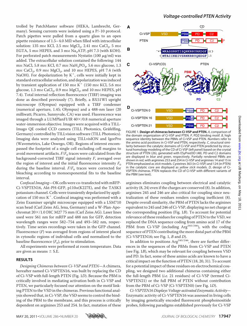
trolled by PatchMaster software (HEKA, Lambrecht, Ger-many). Sensing currents were isolated using a P/-10 protocol.Patch pipettes were pulled from a quartz glass to an openpipette resistance of 1.5–4.0 M� when filled with intracellularsolution: 135 mM KCl, 2.5 mM MgCl2, 2.41 mM CaCl2, 5 mM
EGTA, 5 mM HEPES, and 3 mM Na2ATP, pH 7.3 (with KOH).For perforated patch measurements Nystatin (100 �g/ml) wasadded. The extracellular solution contained the following: 144mM NaCl, 5.8 mM KCl, 0.7 mM NaH2PO4, 5.6 mM glucose, 1.3mM CaCl2, 0.9 mM MgCl2, and 10 mM HEPES, pH 7.4 (withNaOH). For depolarization by K�, cells were initially kept instandard extracellular solution, anddepolarizationwas inducedby transient application of 150 mM K� (150 mM KCl, 5.6 mM
glucose, 1.3 mM CaCl2, 0.9 mM MgCl2, and 10 mM HEPES, pH7.4). Total internal reflection fluorescence (TIRF) imaging wasdone as described previously (7). Briefly, a BX51WI uprightmicroscope (Olympus) equipped with a TIRF condenser(numerical aperture, 1.45; Olympus) and a 488-nm laser (20milliwatt; Picarro, Sunnyvale, CA) was used. Fluorescence wasimaged through a LUMPlanFI/IR 40�/0.8 numerical aperturewater-immersion objective. Images were acquired with a TILL-Imago QE cooled CCD camera (TILL Photonics, Grafelfing,Germany) controlled by TILLvision software (TILL Photonics).Imaging data were analyzed using TILLvisION and IgorPro(Wavemetrics, Lake Oswego, OR). Regions of interest encom-passed the footprint of a single cell excluding cell margins toavoid movement artifacts. F/F0 traces were calculated from thebackground-corrected TIRF signal intensity F, averaged overthe region of interest and the initial fluorescence intensity F0during the baseline interval. F/F0 traces were corrected forbleaching according to monoexponential fits to the baselineinterval.Confocal Imaging—OKcellswere co-transfectedwithmRFP-
Ci-VSPTEN16, Akt-PH-GFP, p110�(K227E), and the TASK3potassium channel. Cells were transiently depolarized by appli-cation of 150 mM K�. Confocal imaging was performed with aZeiss Examiner upright microscope equipped with a LSM710scan head (Carl Zeiss AG, Jena, Germany) and a W-Plan-Apo-chromat 20�/1.0 DICM27 75 mm (Carl Zeiss AG). Laser linesused were 561 nm for mRFP and 488 nm for GFP, detectionwavelength ranges were 582–754 and 493–582 nm, respec-tively. Time series recordings were taken in the GFP channel.Fluorescence (F) was averaged from regions of interest placedin cytosolic regions of individual cells and normalized to thebaseline fluorescence (F0), prior to stimulation.
All experiments were performed at room temperature. Dataare given as means � S.E.
RESULTS
Designing Chimeras between Ci-VSP and PTEN—A chimera,hereafter named Ci-VSPTEN16, was built by replacing the CDof Ci-VSP with full-length PTEN (Fig. 1D). Because the PBM iscritically involved in mediating activation both in Ci-VSP andPTEN, we particularly focused our attention on the motif link-ing PTEN to theVSD in the chimeras. Previous functional anal-ysis showed that, in Ci-VSP, theVSD seems to control the bind-ing of the PBM to the membrane, and this process is criticallydependent on arginines 253 and 254. In fact, mutation of these
arginines eliminates coupling between electrical and catalyticactivity (8, 24) even if the charges are conserved (8). In addition,arginines 245 and 246 are also critical for coupling since neu-tralization of these residues renders coupling inefficient (8).Despite overall similarity, the PBMof PTEN lacks the argininesequivalent to 245 and 246 ofCi-VSP, displaying nonet charge atthe corresponding position (Fig. 1B). To account for potentialrelevance of these residues for coupling of PTEN to theVSD,wereplaced the DNA sequence coding from amino acid 17 of thePBM from Ci-VSP (including Arg245/246), with the codingsequence of PTENcontributing themore distal part of the PBM(Ci-VSPTEN16; see Fig. 1, B and D).In addition to positions Arg245/246, there are further differ-
ences in the sequences of the PBMs from Ci-VSP and PTEN(see Fig. 1B), which may be relevant for coupling between VSDand PD. In fact, some of these amino acids are known to have acritical impact on the function of PTEN (18, 20, 31). To accountfor a potential impact of these residues on electrochemical cou-pling, we designed two additional chimeras containing eitherthe full-length PBM (i.e. 21 residues) of Ci-VSP (termed Ci-VSPTEN21) or the full PBM of PTEN without contributionfrom the PBM of Ci-VSP (Ci-VSPTEN0) (see Fig. 1D).Ci-VSPTEN16DisplaysVoltage-activatedEnzymaticActivity—
Enzymatic activity of Ci-VSPTEN16 was assessed in living cellsby imaging genetically encoded fluorescent phosphoinositideprobes, following paradigms established previously for Ci-VSP
VSD
VSD PBM
VSD PB
Ci-VSPTEN 0
Ci-VSPTEN 16
Ci-VSPTEN 21
PTENCi-VSP(CD)
PBM PD C2VSD
CD
Ci-VSP
PTEN
��� ���
� ��
A
B
C
D
FIGURE 1. Design of chimeras between Ci-VSP and PTEN. A, comparison ofthe domain organization of Ci-VSP and PTEN. P, PDZ-binding motif. B, highsequence identity between the PBMs of Ci-VSP and PTEN. Numbers refer tothe amino acid positions in Ci-VSP and PTEN, respectively. C, structural simi-larity between the catalytic domains of Ci-VSP and PTEN predicted by struc-tural homology modeling of the CD of Ci-VSP (left panel) based on the crystalstructure of PTEN (26), generated with ESyPred3D (48). PD and C2 domainsare displayed in blue and green, respectively. Partially rendered PBMs areshown in red, with arginines 253 and 254 in Ci-VSP and arginines 14 and 15 inPTEN emphasized as stick models. Cysteines 363 (in Ci-VSP) and 124 (in PTEN)in the catalytic core are displayed as yellow stick models. D, design of Ci-VSPTEN chimeras. PTEN replaces the CD of Ci-VSP with different variants ofthe PBM (see text).
Voltage-controlled PTEN Activity
MAY 20, 2011 • VOLUME 286 • NUMBER 20 JOURNAL OF BIOLOGICAL CHEMISTRY 17947
at UB
Marburg, on M
ay 18, 2011w
ww
.jbc.orgD
ownloaded from
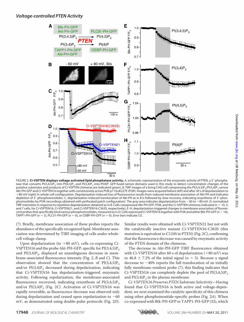
(7). Briefly, membrane association of these probes reports theabundance of the specifically recognized lipid.Membrane asso-ciation was determined by TIRF imaging of cells under whole-cell voltage clamp.Upon depolarization (to �80 mV), cells co-expressing Ci-
VSPTEN16 and the probe Akt-PH-GFP, specific for PI(3,4,5)P3and PI(3,4)P2, displayed an unambiguous decrease in mem-brane-associated fluorescence intensity (Fig. 2, B and C). Thisobservation showed that the concentration of PI(3,4,5)P3and/or PI(3,4)P2 decreased during depolarization, indicatingthat Ci-VSPTEN16 has depolarization-triggered enzymaticactivity. Following repolarization, the membrane-associatedfluorescence recovered, indicating resynthesis of PI(3,4,5)P3and/or PI(3,4)P2 (Fig. 2C). Activation of Ci-VSPTEN16 wasrapidly reversible, as fluorescence decrease was observed onlyduring depolarization and ceased upon repolarization to �60mV, as demonstrated using double-pulse protocols (Fig. 2D).
Similar results were obtained with Ci-VSPTEN21 but not withthe catalytically inactive mutant Ci-VSPTEN16-C363S (thismutation is equivalent to C124S in PTEN) (Fig. 2C), confirmingthat the fluorescence decrease was caused by enzymatic activityof the PTEN domain of the chimeras.The decrease in Akt-PH-GFP TIRF fluorescence obtained
with Ci-VSPTEN16 after 60 s of depolarization (�80 mV) wasto 46.8 � 7.2% of the initial signal (n � 5). Because a signaldecrease to �40% reports the full translocation of an initiallyfully membrane-resident probe (7), this finding indicates thatCi-VSPTEN16 can completely deplete the pool of PI(3,4,5)P3and PI(3,4)P2 in the plasma membrane.Ci-VSPTEN16 Preserves PTENSubstrate Selectivity—Having
found that Ci-VSPTEN16 is both active and voltage-depen-dent, we next examined the catalytic specificity of this chimerausing other phosphoinositide-specific probes (Fig. 2A). Whenco-expressedwithBtk-PH-GFPorTAPP1-PH-GFP (32), which
FIGURE 2. Ci-VSPTEN displays voltage-activated lipid phosphatase activity. A, schematic representation of the enzymatic activity of PTEN, a 3�-phospha-tase that converts PI(3,4,5)P3 into PI(4,5)P2 and PI(3,4)P2 into PI(4)P. GFP-fused sensor domains used in this study to detect concentration changes of theputative substrates and products of Ci-VSPTEN chimeras are indicated (green). B, TIRF images of a living CHO cell coexpressing the PI(3,4,5)P2/PI(3,4)P2 sensorAkt-PH-GFP and Ci-VSPTEN16 together with constitutively active PI3K p110�K227E (PI3K). Images were acquired before (left) and after 30 s of depolarization to�80 mV (right) in whole-cell configuration. Depolarization-induced loss of fluorescence results from reduced membrane association of Akt-PH and indicatesdepletion of 3�-phosphoinositides. C, depolarization-induced translocation of Akt-PH as in B is followed by slow recovery, indicating resynthesis of 3�-phos-phoinositides by PI3K (recordings obtained with perforated patch configuration). The gray area indicates depolarization from �60 to �80 mV. D, normalizedTIRF intensities in response to repetitive depolarization obtained as in B. Cells coexpressed Akt-PH-GFP, PI3K, and the Ci-VSPTEN chimeras indicated (n � 10, 3,and 7 cells, for Ci-VSPTEN16, Ci-VSPTEN21, and Ci-VSPTEN16-C363S, respectively). E–H, depolarization-triggered changes in membrane association of fluores-cent probes that specifically bind various phosphoinositides, measured as in D. Cells expressed Ci-VSPTEN16 together with PI3K and either Btk-PH-GFP (n � 16),TAPP1-PH-GFP (n � 5), PLC�1-PH-GFP (n � 6), or OSBP-PH-GFP (n � 6). Error bars indicate S.E.
Voltage-controlled PTEN Activity
17948 JOURNAL OF BIOLOGICAL CHEMISTRY VOLUME 286 • NUMBER 20 • MAY 20, 2011
at UB
Marburg, on M
ay 18, 2011w
ww
.jbc.orgD
ownloaded from
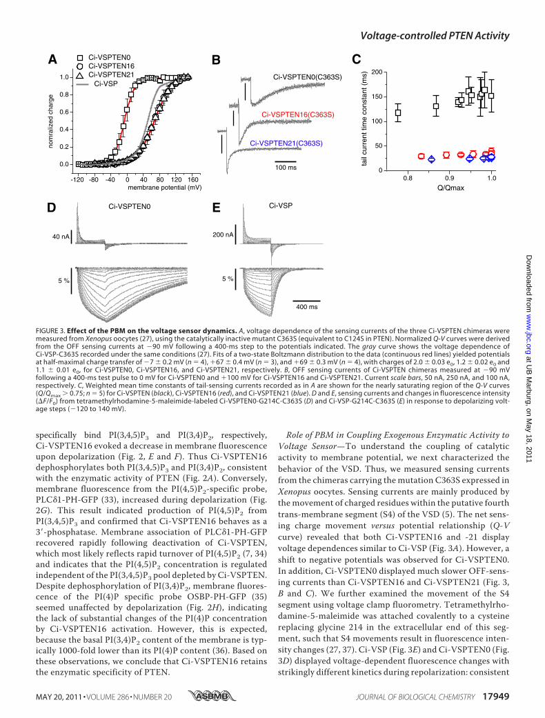
specifically bind PI(3,4,5)P3 and PI(3,4)P2, respectively,Ci-VSPTEN16 evoked a decrease in membrane fluorescenceupon depolarization (Fig. 2, E and F). Thus Ci-VSPTEN16dephosphorylates both PI(3,4,5)P3 and PI(3,4)P2, consistentwith the enzymatic activity of PTEN (Fig. 2A). Conversely,membrane fluorescence from the PI(4,5)P2-specific probe,PLC�1-PH-GFP (33), increased during depolarization (Fig.2G). This result indicated production of PI(4,5)P2 fromPI(3,4,5)P3 and confirmed that Ci-VSPTEN16 behaves as a3�-phosphatase. Membrane association of PLC�1-PH-GFPrecovered rapidly following deactivation of Ci-VSPTEN,which most likely reflects rapid turnover of PI(4,5)P2 (7, 34)and indicates that the PI(4,5)P2 concentration is regulatedindependent of the PI(3,4,5)P3 pool depleted by Ci-VSPTEN.Despite dephosphorylation of PI(3,4)P2, membrane fluores-cence of the PI(4)P specific probe OSBP-PH-GFP (35)seemed unaffected by depolarization (Fig. 2H), indicatingthe lack of substantial changes of the PI(4)P concentrationby Ci-VSPTEN16 activation. However, this is expected,because the basal PI(3,4)P2 content of the membrane is typ-ically 1000-fold lower than its PI(4)P content (36). Based onthese observations, we conclude that Ci-VSPTEN16 retainsthe enzymatic specificity of PTEN.
Role of PBM in Coupling Exogenous Enzymatic Activity toVoltage Sensor—To understand the coupling of catalyticactivity to membrane potential, we next characterized thebehavior of the VSD. Thus, we measured sensing currentsfrom the chimeras carrying the mutation C363S expressed inXenopus oocytes. Sensing currents are mainly produced bythe movement of charged residues within the putative fourthtrans-membrane segment (S4) of the VSD (5). The net sens-ing charge movement versus potential relationship (Q-Vcurve) revealed that both Ci-VSPTEN16 and -21 displayvoltage dependences similar to Ci-VSP (Fig. 3A). However, ashift to negative potentials was observed for Ci-VSPTEN0.In addition, Ci-VSPTEN0 displayed much slower OFF-sens-ing currents than Ci-VSPTEN16 and Ci-VSPTEN21 (Fig. 3,B and C). We further examined the movement of the S4segment using voltage clamp fluorometry. Tetramethylrho-damine-5-maleimide was attached covalently to a cysteinereplacing glycine 214 in the extracellular end of this seg-ment, such that S4 movements result in fluorescence inten-sity changes (27, 37). Ci-VSP (Fig. 3E) and Ci-VSPTEN0 (Fig.3D) displayed voltage-dependent fluorescence changes withstrikingly different kinetics during repolarization: consistent
FIGURE 3. Effect of the PBM on the voltage sensor dynamics. A, voltage dependence of the sensing currents of the three Ci-VSPTEN chimeras weremeasured from Xenopus oocytes (27), using the catalytically inactive mutant C363S (equivalent to C124S in PTEN). Normalized Q-V curves were derivedfrom the OFF sensing currents at �90 mV following a 400-ms step to the potentials indicated. The gray curve shows the voltage dependence ofCi-VSP-C363S recorded under the same conditions (27). Fits of a two-state Boltzmann distribution to the data (continuous red lines) yielded potentialsat half-maximal charge transfer of �7 � 0.2 mV (n � 4), �67 � 0.4 mV (n � 3), and �69 � 0.3 mV (n � 4), with charges of 2.0 � 0.03 e0, 1.2 � 0.02 e0 and1.1 � 0.01 e0, for Ci-VSPTEN0, Ci-VSPTEN16, and Ci-VSPTEN21, respectively. B, OFF sensing currents of Ci-VSPTEN chimeras measured at �90 mVfollowing a 400-ms test pulse to 0 mV for Ci-VSPTEN0 and �100 mV for Ci-VSPTEN16 and Ci-VSPTEN21. Current scale bars, 50 nA, 250 nA, and 100 nA,respectively. C, Weighted mean time constants of tail-sensing currents recorded as in A are shown for the nearly saturating region of the Q-V curves(Q/Qmax 0.75; n � 5) for Ci-VSPTEN (black), Ci-VSPTEN16 (red), and Ci-VSPTEN21 (blue). D and E, sensing currents and changes in fluorescence intensity(F/F0) from tetramethylrhodamine-5-maleimide-labeled Ci-VSPTEN0-G214C-C363S (D) and Ci-VSP-G214C-C363S (E) in response to depolarizing volt-age steps (�120 to 140 mV).
Voltage-controlled PTEN Activity
MAY 20, 2011 • VOLUME 286 • NUMBER 20 JOURNAL OF BIOLOGICAL CHEMISTRY 17949
at UB
Marburg, on M
ay 18, 2011w
ww
.jbc.orgD
ownloaded from
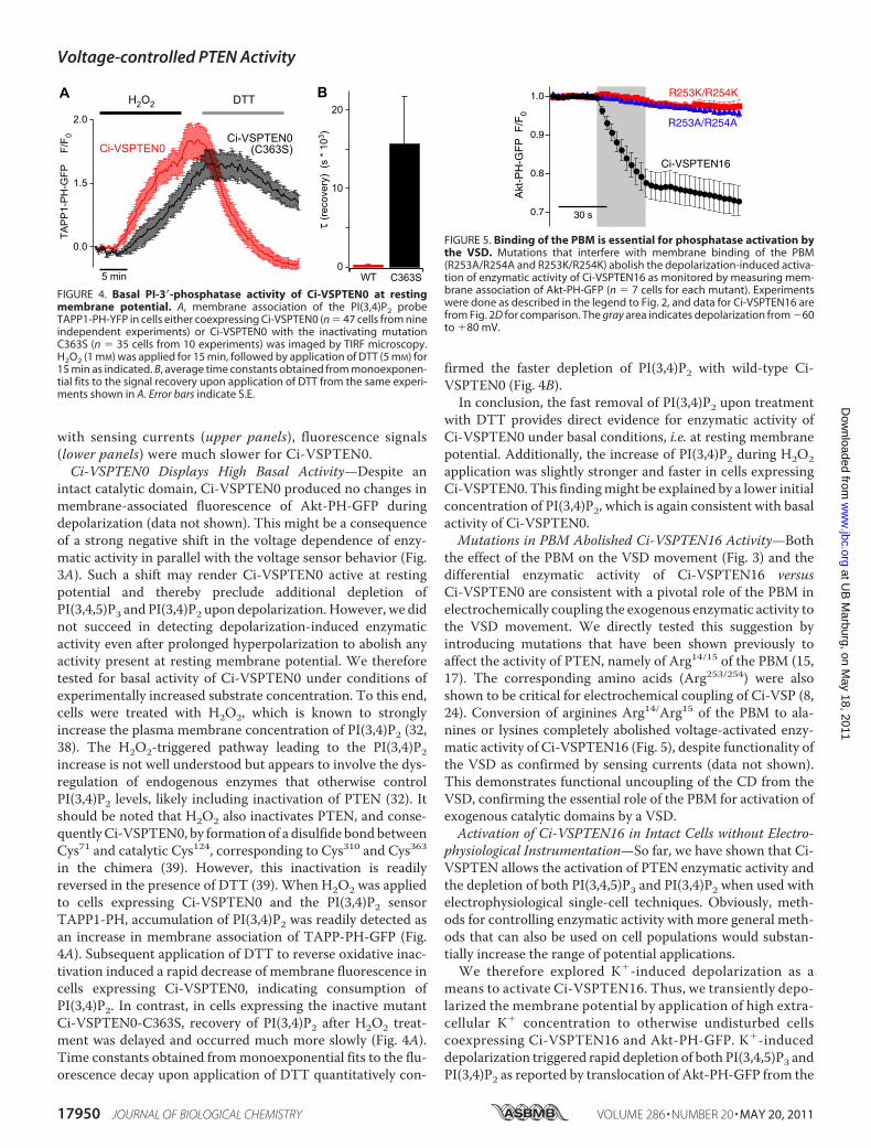
with sensing currents (upper panels), fluorescence signals(lower panels) were much slower for Ci-VSPTEN0.Ci-VSPTEN0 Displays High Basal Activity—Despite an
intact catalytic domain, Ci-VSPTEN0 produced no changes inmembrane-associated fluorescence of Akt-PH-GFP duringdepolarization (data not shown). This might be a consequenceof a strong negative shift in the voltage dependence of enzy-matic activity in parallel with the voltage sensor behavior (Fig.3A). Such a shift may render Ci-VSPTEN0 active at restingpotential and thereby preclude additional depletion ofPI(3,4,5)P3 and PI(3,4)P2 upon depolarization. However, we didnot succeed in detecting depolarization-induced enzymaticactivity even after prolonged hyperpolarization to abolish anyactivity present at resting membrane potential. We thereforetested for basal activity of Ci-VSPTEN0 under conditions ofexperimentally increased substrate concentration. To this end,cells were treated with H2O2, which is known to stronglyincrease the plasma membrane concentration of PI(3,4)P2 (32,38). The H2O2-triggered pathway leading to the PI(3,4)P2increase is not well understood but appears to involve the dys-regulation of endogenous enzymes that otherwise controlPI(3,4)P2 levels, likely including inactivation of PTEN (32). Itshould be noted that H2O2 also inactivates PTEN, and conse-quentlyCi-VSPTEN0, by formation of a disulfide bondbetweenCys71 and catalytic Cys124, corresponding to Cys310 and Cys363in the chimera (39). However, this inactivation is readilyreversed in the presence of DTT (39). When H2O2 was appliedto cells expressing Ci-VSPTEN0 and the PI(3,4)P2 sensorTAPP1-PH, accumulation of PI(3,4)P2 was readily detected asan increase in membrane association of TAPP-PH-GFP (Fig.4A). Subsequent application of DTT to reverse oxidative inac-tivation induced a rapid decrease of membrane fluorescence incells expressing Ci-VSPTEN0, indicating consumption ofPI(3,4)P2. In contrast, in cells expressing the inactive mutantCi-VSPTEN0-C363S, recovery of PI(3,4)P2 after H2O2 treat-ment was delayed and occurred much more slowly (Fig. 4A).Time constants obtained frommonoexponential fits to the flu-orescence decay upon application of DTT quantitatively con-
firmed the faster depletion of PI(3,4)P2 with wild-type Ci-VSPTEN0 (Fig. 4B).In conclusion, the fast removal of PI(3,4)P2 upon treatment
with DTT provides direct evidence for enzymatic activity ofCi-VSPTEN0 under basal conditions, i.e. at resting membranepotential. Additionally, the increase of PI(3,4)P2 during H2O2application was slightly stronger and faster in cells expressingCi-VSPTEN0. This findingmight be explained by a lower initialconcentration of PI(3,4)P2, which is again consistent with basalactivity of Ci-VSPTEN0.Mutations in PBM Abolished Ci-VSPTEN16 Activity—Both
the effect of the PBM on the VSD movement (Fig. 3) and thedifferential enzymatic activity of Ci-VSPTEN16 versusCi-VSPTEN0 are consistent with a pivotal role of the PBM inelectrochemically coupling the exogenous enzymatic activity tothe VSD movement. We directly tested this suggestion byintroducing mutations that have been shown previously toaffect the activity of PTEN, namely of Arg14/15 of the PBM (15,17). The corresponding amino acids (Arg253/254) were alsoshown to be critical for electrochemical coupling of Ci-VSP (8,24). Conversion of arginines Arg14/Arg15 of the PBM to ala-nines or lysines completely abolished voltage-activated enzy-matic activity of Ci-VSPTEN16 (Fig. 5), despite functionality ofthe VSD as confirmed by sensing currents (data not shown).This demonstrates functional uncoupling of the CD from theVSD, confirming the essential role of the PBM for activation ofexogenous catalytic domains by a VSD.Activation of Ci-VSPTEN16 in Intact Cells without Electro-
physiological Instrumentation—So far, we have shown that Ci-VSPTEN allows the activation of PTEN enzymatic activity andthe depletion of both PI(3,4,5)P3 and PI(3,4)P2 when used withelectrophysiological single-cell techniques. Obviously, meth-ods for controlling enzymatic activity with more general meth-ods that can also be used on cell populations would substan-tially increase the range of potential applications.We therefore explored K�-induced depolarization as a
means to activate Ci-VSPTEN16. Thus, we transiently depo-larized the membrane potential by application of high extra-cellular K� concentration to otherwise undisturbed cellscoexpressing Ci-VSPTEN16 and Akt-PH-GFP. K�-induceddepolarization triggered rapid depletion of both PI(3,4,5)P3 andPI(3,4)P2 as reported by translocation of Akt-PH-GFP from the
FIGURE 4. Basal PI-3�-phosphatase activity of Ci-VSPTEN0 at restingmembrane potential. A, membrane association of the PI(3,4)P2 probeTAPP1-PH-YFP in cells either coexpressing Ci-VSPTEN0 (n � 47 cells from nineindependent experiments) or Ci-VSPTEN0 with the inactivating mutationC363S (n � 35 cells from 10 experiments) was imaged by TIRF microscopy.H2O2 (1 mM) was applied for 15 min, followed by application of DTT (5 mM) for15 min as indicated. B, average time constants obtained from monoexponen-tial fits to the signal recovery upon application of DTT from the same experi-ments shown in A. Error bars indicate S.E.
FIGURE 5. Binding of the PBM is essential for phosphatase activation bythe VSD. Mutations that interfere with membrane binding of the PBM(R253A/R254A and R253K/R254K) abolish the depolarization-induced activa-tion of enzymatic activity of Ci-VSPTEN16 as monitored by measuring mem-brane association of Akt-PH-GFP (n � 7 cells for each mutant). Experimentswere done as described in the legend to Fig. 2, and data for Ci-VSPTEN16 arefrom Fig. 2D for comparison. The gray area indicates depolarization from �60to �80 mV.
Voltage-controlled PTEN Activity
17950 JOURNAL OF BIOLOGICAL CHEMISTRY VOLUME 286 • NUMBER 20 • MAY 20, 2011
at UB
Marburg, on M
ay 18, 2011w
ww
.jbc.orgD
ownloaded from
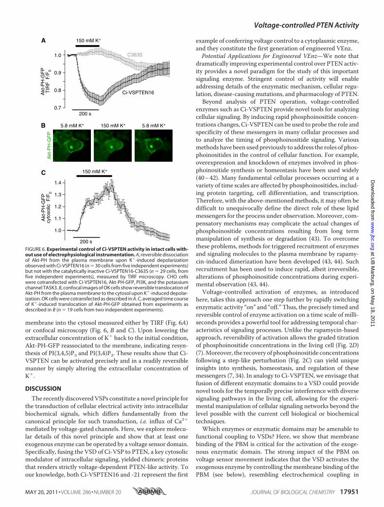
membrane into the cytosol measured either by TIRF (Fig. 6A)or confocal microscopy (Fig. 6, B and C). Upon lowering theextracellular concentration of K� back to the initial condition,Akt-PH-GFP reassociated to the membrane, indicating resyn-thesis of PI(3,4,5)P3 and PI(3,4)P2. These results show that Ci-VSPTEN can be activated precisely and in a readily reversiblemanner by simply altering the extracellular concentration ofK�.
DISCUSSION
The recently discoveredVSPs constitute a novel principle forthe transduction of cellular electrical activity into intracellularbiochemical signals, which differs fundamentally from thecanonical principle for such transduction, i.e. influx of Ca2�
mediated by voltage-gated channels. Here, we explore molecu-lar details of this novel principle and show that at least oneexogenous enzyme can be operated by a voltage sensor domain.Specifically, fusing the VSD of Ci-VSP to PTEN, a key cytosolicmodulator of intracellular signaling, yielded chimeric proteinsthat renders strictly voltage-dependent PTEN-like activity. Toour knowledge, both Ci-VSPTEN16 and -21 represent the first
example of conferring voltage control to a cytoplasmic enzyme,and they constitute the first generation of engineered VEnz.Potential Applications for Engineered VEnz—We note that
dramatically improving experimental control over PTEN activ-ity provides a novel paradigm for the study of this importantsignaling enzyme. Stringent control of activity will enableaddressing details of the enzymatic mechanism, cellular regu-lation, disease-causing mutations, and pharmacology of PTEN.Beyond analysis of PTEN operation, voltage-controlled
enzymes such as Ci-VSPTEN provide novel tools for analyzingcellular signaling. By inducing rapid phosphoinositide concen-trations changes, Ci-VSPTEN can be used to probe the role andspecificity of these messengers in many cellular processes andto analyze the timing of phosphoinositide signaling. Variousmethods have beenusedpreviously to address the roles of phos-phoinositides in the control of cellular function. For example,overexpression and knockdown of enzymes involved in phos-phoinositide synthesis or homeostasis have been used widely(40–42). Many fundamental cellular processes occurring at avariety of time scales are affected by phosphoinositides, includ-ing protein targeting, cell differentiation, and transcription.Therefore, with the above-mentionedmethods, it may often bedifficult to unequivocally define the direct role of these lipidmessengers for the process under observation.Moreover, com-pensatory mechanisms may complicate the actual changes ofphosphoinositide concentrations resulting from long termmanipulation of synthesis or degradation (43). To overcomethese problems, methods for triggered recruitment of enzymesand signaling molecules to the plasma membrane by rapamy-cin-induced dimerization have been developed (43, 44). Suchrecruitment has been used to induce rapid, albeit irreversible,alterations of phosphoinositide concentrations during experi-mental observation (43, 44).Voltage-controlled activation of enzymes, as introduced
here, takes this approach one step further by rapidly switchingenzymatic activity “on” and “off.” Thus, the precisely timed andreversible control of enzyme activation on a time scale of milli-seconds provides a powerful tool for addressing temporal char-acteristics of signaling processes. Unlike the rapamycin-basedapproach, reversibility of activation allows the graded titrationof phosphoinositide concentrations in the living cell (Fig. 2D)(7).Moreover, the recovery of phosphoinositide concentrationsfollowing a step-like perturbation (Fig. 2C) can yield uniqueinsights into synthesis, homeostasis, and regulation of thesemessengers (7, 34). In analogy to Ci-VSPTEN, we envisage thatfusion of different enzymatic domains to a VSD could providenovel tools for the temporally precise interference with diversesignaling pathways in the living cell, allowing for the experi-mental manipulation of cellular signaling networks beyond thelevel possible with the current cell biological or biochemicaltechniques.Which enzymes or enzymatic domains may be amenable to
functional coupling to VSDs? Here, we show that membranebinding of the PBM is critical for the activation of the exoge-nous enzymatic domain. The strong impact of the PBM onvoltage sensor movement indicates that the VSD activates theexogenous enzyme by controlling themembrane binding of thePBM (see below), resembling electrochemical coupling in
FIGURE 6. Experimental control of Ci-VSPTEN activity in intact cells with-out use of electrophysiological instrumentation. A, reversible dissociationof Akt-PH from the plasma membrane upon K�-induced depolarizationobserved with Ci-VSPTEN16 (n � 30 cells from five independent experiments)but not with the catalytically inactive Ci-VSPTEN16-C363S (n � 29 cells, fromfive independent experiments), measured by TIRF microscopy. CHO cellswere cotransfected with Ci-VSPTEN16, Akt-PH-GFP, PI3K, and the potassiumchannel TASK3. B, confocal images of OK cells show reversible translocation ofAkt-PH from the plasma membrane to the cytosol upon K�-induced depolar-ization. OK cells were cotransfected as described in A. C, averaged time courseof K�-induced translocation of Akt-PH-GFP obtained from experiments asdescribed in B (n � 19 cells from two independent experiments).
Voltage-controlled PTEN Activity
MAY 20, 2011 • VOLUME 286 • NUMBER 20 JOURNAL OF BIOLOGICAL CHEMISTRY 17951
at UB
Marburg, on M
ay 18, 2011w
ww
.jbc.orgD
ownloaded from
native VSPs (8, 24). Although it has been shown that binding ofthe PBM to PI(4,5)P2 increases the �-helicity of PTEN (22), itremains unknown whether activation occurs by the reorienta-tion of the PD toward its membrane-resident substrate or bydirect interaction of the PBM with the catalytic site. Althoughfurther work is needed to distinguish between these mecha-nisms, the latter model would suggest that generation of VEnzmay be limited to CDs whose intrinsic activation mechanisminvolves a PBM-like motif. Such candidates may include, with-out being limited to, additional PTEN-related proteins, namelyTPTE (45), TPIP (46), and PLIP (47). In vitro, these moleculesmay possess phosphoinositide phosphatase activities with dis-tinct substrate specificities (46, 47). Generation of VEnz chi-merawith these proteinsmay substantially help to address theirfunction in intact cells, which has been largely unexplored.Mechanism of Coupling in Engineered VEnz—The loss of
electrically triggered enzymatic activity observed with PBMmutants (Fig. 5) clearly demonstrates that electrochemical cou-pling in Ci-VSPTEN16 involves the PBM. Howmight the PBMmediate the interaction of VSD and PD? For PTEN, it has beenshown that the PBM mediates both membrane binding andactivation of catalytic activity (17, 19, 20). Likewise, work onCi-VSP suggested that binding of the PBM to the plasmamem-brane is involved in coupling and that VSD movement modu-lates the PBM binding to control enzymatic activity (8, 24).Thus, sensor movement following depolarization is thought topromote membrane binding of the PBM and thereby activatethe PBM. According to this model, the membrane binding ofthe PBMwill reciprocally affect voltage sensormovement. Spe-cifically, binding of the PBM upon depolarization will restrictsubsequent voltage sensormovement upon repolarization, thusslowing down OFF sensing currents (8, 24).Our present results with Ci-VSPTEN chimeras are consis-
tent with this idea. Thus, mutations in the PBMmost probablyabrogate electrochemical coupling by interfering with mem-brane binding, similar to previous results obtainedwith Ci-VSP(8). We further observed that the full PBM from PTEN drasti-cally slowed down voltage sensor return to the resting stateupon repolarization, when compared with both of the otherchimeras. According to the model outlined above, this obser-vation suggests distinct membrane binding affinities of the dif-ferent chimeric PBMs, with the full PBM of PTEN (i.e.Ci-VSPTEN0) exhibiting the strongest binding.Following this idea, the proposed distinctmembrane binding
affinities may point to a specific interaction between the differ-ent PDs and their associated PBM. Thus, we note that the chi-meric PBMs of Ci-VSPTEN16 and 21 yielded the fastest OFF-sensing currents, which may indicate that their boundconformation is less stable compared with native PBMs of Ci-VSP or Ci-VSPTEN0 associated with their original PDs (Fig. 3and Ref. 8). These combined observations suggest that for opti-mal membrane binding, the PBM must match the catalyticdomain with which they couple. In this view, the PBM in Ci-VSP serves as an adapter for the binding between the mem-brane and the CD. Combinatorial exchange of PBMs in VSPchimeras designed to operate as Ci-VSPTEN should help indirectly addressing such specific interaction of PBM and PD.
One caveat to this model comes from its failure to fullyexplain the apparent lack of control of the VSD over the CD inCi-VSPTEN0. The basal activity at negative potentials mayeither indicate that PTEN is constitutively active at all poten-tials (i.e. uncoupled from the VSD) or that the activation rangeis also shifted to negative potentials, as found for the sensingcurrent. Both scenarios are not mutually exclusive and may beconsistent with a highermembrane binding affinity of the PBMof PTEN. For the first case, it is plausible that the control of theVSD can be overridden by a strong interaction between themembrane and the PBM. For the second case, the shift in volt-age dependence may prevent the sensor from adopting a con-formation in which the PBM cannot bind (as in the case ofCi-VSPTEN16, -21 and other VSPs at negative potentials),resulting in high basal activity.Furthermore, it should be noted that the observed shift in the
voltage dependence of Ci-VSPTEN0 might be explained by analternative scenario, in which the PBM influences the profile ofthe focused electrical field across the voltage sensor. In thiscase, the PBMcould increase the sensitivity of theVSD to depo-larization and therefore self-promote the activation of the cat-alytic activity, rendering a high basal activity. To distinguishbetween these different possibilities, further work is required,which is beyond the scope of the current study.In Ci-VSP, cationic residues Arg245, Arg246, Arg253, and
Arg254 within the PBM are involved in electrochemical cou-pling, presumably by contributing to binding to negativelycharged phospholipids in the membrane (8, 24). It is thereforeremarkable that the PBM from PTEN, although lacking thepositive charge at residue pair 245–246 (R245K-R246E; see Fig.1B), produced slowed OFF-sensing currents and enhancedenzymatic activity at resting potentials, both consistent withenhanced membrane binding according to the coupling mech-anism proposed above. This finding supports the idea that thePBM binds to membrane lipids in a stereo-specific mannerrather than simply by electrostatic interaction with the mem-brane (8). This conclusion also is consistent with the elimina-tion of electrochemical coupling by isocoulombic substitutionof Arg253 and Arg254 to Lys (Fig. 5). Further detailed analysis ofthe role of individual residues within the PBM for membranebinding is required to fully resolve this issue.In conclusion, we have been able to engineer a series of chi-
meric proteins conferring voltage sensitivity to the tumor sup-pressor PTEN in vivo. Thiswork supports the idea that the PBMis a key element in the activation of VSP. In a broader view, thisstudy constitutes a proof-of-concept to a novel approach forcontrolling enzymatic activity using VSDs.
Acknowledgments—We thank Drs. I. S. Ramsey, D. E. Logothetis, andL. J. DeFelice for helpful comments on the manuscript and S. Kriegerand V. Petrou for excellent technical assistance. Constructs used inthis work were kindly provided by Y. Okamura (Ci-VSP), T. Balla(PLC�1-PH, Akt-PH, Btk-PH, OSBP-PH), D. Alessi (TAPP1-PH), J.Downward (PI3K), and J. Daut (TASK3).
REFERENCES1. Finkbeiner, S., and Greenberg, M. E. (1998) J. Neurobiol. 37, 171–1892. Kingsbury, T. J., Bambrick, L. L., Roby, C. D., and Krueger, B. K. (2007)
Voltage-controlled PTEN Activity
17952 JOURNAL OF BIOLOGICAL CHEMISTRY VOLUME 286 • NUMBER 20 • MAY 20, 2011
at UB
Marburg, on M
ay 18, 2011w
ww
.jbc.orgD
ownloaded from
J. Neurochem. 103, 761–7703. Wheeler, D. G., Barrett, C. F., Groth, R. D., Safa, P., andTsien, R.W. (2008)
J. Cell Biol. 183, 849–8634. Hossain, M. I., Iwasaki, H., Okochi, Y., Chahine, M., Higashijima, S., Na-
gayama, K., and Okamura, Y. (2008) J. Biol. Chem. 283, 18248–182595. Murata, Y., Iwasaki, H., Sasaki, M., Inaba, K., and Okamura, Y. (2005)
Nature 435, 1239–12436. Murata, Y., and Okamura, Y. (2007) J. Physiol. 583, 875–8897. Halaszovich, C. R., Schreiber, D. N., and Oliver, D. (2009) J. Biol. Chem.
284, 2106–21138. Villalba-Galea, C. A., Miceli, F., Taglialatela, M., and Bezanilla, F. (2009)
J. Gen. Physiol. 134, 5–149. Leslie, N. R., Batty, I. H., Maccario, H., Davidson, L., and Downes, C. P.
(2008) Oncogene 27, 5464–547610. Ooms, L. M., Horan, K. A., Rahman, P., Seaton, G., Gurung, R.,
Kethesparan, D. S., and Mitchell, C. A. (2009) Biochem. J. 419, 29–4911. Suh, B. C., and Hille, B. (2008) Annu. Rev. Biophys. 37, 175–19512. Cremona, O., Di Paolo, G., Wenk, M. R., Luthi, A., Kim, W. T., Takei, K.,
Daniell, L., Nemoto, Y., Shears, S. B., Flavell, R. A., McCormick, D. A., andDe Camilli, P. (1999) Cell 99, 179–188
13. Haucke, V. (2005) Biochem. Soc. Trans. 33, 1285–128914. Maehama, T., and Dixon, J. E. (1998) J. Biol. Chem. 273, 13375–1337815. Steck, P. A., Pershouse, M. A., Jasser, S. A., Yung, W. K., Lin, H., Ligon,
A. H., Langford, L. A., Baumgard,M. L., Hattier, T., Davis, T., Frye, C., Hu,R., Swedlund, B., Teng, D. H., and Tavtigian, S. V. (1997) Nat. Genet. 15,356–362
16. Wymann, M. P., and Schneiter, R. (2008) Nat. Rev. Mol. Cell Biol. 9,162–176
17. Campbell, R. B., Liu, F., and Ross, A. H. (2003) J. Biol. Chem. 278,33617–33620
18. Das, S., Dixon, J. E., and Cho, W. (2003) Proc. Natl. Acad. Sci. U.S.A. 100,7491–7496
19. Vazquez, F., Matsuoka, S., Sellers, W. R., Yanagida, T., Ueda, M., andDevreotes, P. N. (2006) Proc. Natl. Acad. Sci. U.S.A. 103, 3633–3638
20. Walker, S. M., Leslie, N. R., Perera, N. M., Batty, I. H., and Downes, C. P.(2004) Biochem. J. 379, 301–307
21. Rahdar, M., Inoue, T., Meyer, T., Zhang, J., Vazquez, F., and Devreotes,P. N. (2009) Proc. Natl. Acad. Sci. U.S.A. 106, 480–485
22. Redfern, R. E., Redfern, D., Furgason, M. L., Munson, M., Ross, A. H., andGericke, A. (2008) Biochemistry 47, 2162–2171
23. Liu, Y., and Bankaitis, V. A. (2010) Prog. Lipid Res. 49, 201–21724. Kohout, S. C., Bell, S. C., Liu, L., Xu, Q., Minor, D. L., Jr., and Isacoff, E. Y.
(2010) Nat. Chem. Biol. 6, 369–37525. Iwasaki, H., Murata, Y., Kim, Y., Hossain, M. I., Worby, C. A., Dixon, J. E.,
McCormack, T., Sasaki, T., and Okamura, Y. (2008) Proc. Natl. Acad. Sci.U.S.A. 105, 7970–7975
26. Lee, J. O., Yang, H., Georgescu, M. M., Di Cristofano, A., Maehama, T.,Shi, Y., Dixon, J. E., Pandolfi, P., and Pavletich, N. P. (1999) Cell 99,323–334
27. Villalba-Galea, C. A., Sandtner,W., Starace, D.M., and Bezanilla, F. (2008)Proc. Natl. Acad. Sci. U.S.A. 105, 17600–17607
28. Stefani, E., and Bezanilla, F. (1998)Methods Enzymol. 293, 300–31829. Cha, A., Zerangue, N., Kavanaugh, M., and Bezanilla, F. (1998) Methods
Enzymol. 296, 566–57830. Schaechinger, T. J., and Oliver, D. (2007) Proc. Natl. Acad. Sci. U.S.A. 104,
7693–769831. Denning, G., Jean-Joseph, B., Prince, C., Durden, D. L., and Vogt, P. K.
(2007) Oncogene 26, 3930–394032. Kimber, W. A., Trinkle-Mulcahy, L., Cheung, P. C., Deak, M., Marsden,
L. J., Kieloch, A., Watt, S., Javier, R. T., Gray, A., Downes, C. P., Lucocq,J. M., and Alessi, D. R. (2002) Biochem. J. 361, 525–536
33. Varnai, P., and Balla, T. (1998) J. Cell Biol. 143, 501–51034. Falkenburger, B. H., Jensen, J. B., and Hille, B. (2010) J. Gen. Physiol. 135,
99–11435. Balla, A., Tuymetova, G., Tsiomenko, A., Varnai, P., and Balla, T. (2005)
Mol. Biol. Cell 16, 1282–129536. Leslie, N. R., and Downes, C. P. (2002) Cell Signal 14, 285–29537. Kohout, S. C., Ulbrich, M. H., Bell, S. C., and Isacoff, E. Y. (2008) Nat.
Struct. Mol. Biol. 15, 106–10838. Van derKaay, J., Beck,M., Gray, A., andDownes, C. P. (1999) J. Biol. Chem.
274, 35963–3596839. Lee, S. R., Yang, K. S., Kwon, J., Lee, C., Jeong, W., and Rhee, S. G. (2002)
J. Biol. Chem. 277, 20336–2034240. Chen, X., Talley, E. M., Patel, N., Gomis, A., McIntire, W. E., Dong, B.,
Viana, F., Garrison, J. C., and Bayliss, D. A. (2006) Proc. Natl. Acad. Sci.U.S.A. 103, 3422–3427
41. Milosevic, I., Sørensen, J. B., Lang, T., Krauss, M., Nagy, G., Haucke, V.,Jahn, R., and Neher, E. (2005) J. Neurosci. 25, 2557–2565
42. Wang, Y. J., Li, W. H., Wang, J., Xu, K., Dong, P., Luo, X., and Yin, H. L.(2004) J. Cell Biol. 167, 1005–1010
43. Varnai, P., Thyagarajan, B., Rohacs, T., andBalla, T. (2006) J. Cell Biol. 175,377–382
44. Suh, B. C., Inoue, T., Meyer, T., and Hille, B. (2006) Science 314,1454–1457
45. Tapparel, C., Reymond, A., Girardet, C., Guillou, L., Lyle, R., Lamon, C.,Hutter, P., and Antonarakis, S. E. (2003) Gene 323, 189–199
46. Walker, S. M., Downes, C. P., and Leslie, N. R. (2001) Biochem. J. 360,277–283
47. Pagliarini, D. J., Worby, C. A., and Dixon, J. E. (2004) J. Biol. Chem. 279,38590–38596
48. Lambert, C., Leonard, N., De Bolle, X., and Depiereux, E. (2002) Bioinfor-matics 18, 1250–1256
Voltage-controlled PTEN Activity
MAY 20, 2011 • VOLUME 286 • NUMBER 20 JOURNAL OF BIOLOGICAL CHEMISTRY 17953
at UB
Marburg, on M
ay 18, 2011w
ww
.jbc.orgD
ownloaded from

A P E N D I X
vii
7 Appendix
7.1 Table of Figures
Figure 1: The organ of Corti in the inner ear mediates the transduction of sound......................-2-
C U R R I C U L U M V I T A E
viii
7.2 Curriculum Vitae
Personal Details
Name Michael Georg Leitner Address
Herrmannstraße 69 35037 Marburg (Germany)
Date of Birth 09.05.1980 in St. Johann in Tirol (Austria)
Scholastics
2001 - 2005 Leopold-Franzens-Universität Innsbruck (Austria) - Bachelors Programme Biology (11.4.2005 Bakk.rer.nat.)
2005 -2007 Leopold-Franzens-Universität Innsbruck (Austria) –
Masters Programme Zoology (02.05.2007 Mag. Biol.) Diploma Thesis – Interaction of Nicotine and Menthol in Sensory Neurons (Department of Physiology and Medical Physics, Medical University Innsbruck)
2008 - 2012 Philipps-Universität Marburg (Germany) – Doctoral
thesis in the group of Prof. Dr. Dominik Oliver (Neurophysiology) "Molecular Properties and Pathophysiological Relevance of the Predominant K+ Conductance in Cochlear Outer Hair Cells"
Awards
2004 - 2006 Achievement oriented scholarship of the Leopold-Franzens Universität Innsbruck
2007 Incentive Scholarship of the Medical University Innsbruck
2009 Poster award at the 88th annual meeting of the “Deutsche Physiologische Gesellschaft”
2010 2nd position Dissertationspreis Philips-University Marburg
A P P E N D I X
ix
7.3 Verzeichnis der akademischen Lehrer
Meine Akademischen Lehrer waren die Damen und Herren...
…in Innsbruck (Österreich)
Dallinger, Doppler, Fürst, Haas, Haselwandter, Hobmeyer, Jennings, Kaufmann, Kress,
Krumschnabl, Loidl, Maier, Maly, Meyer, Moser, Pelster, Pfaller, Psenner, Ravasio, Rieger,
Sattler, Schinner, Schwerte, Sommaruga, Sturmbauer, Tappeiner, Thaler, Tuluc
…in Marburg (Deutschland)
Braun, Oberwinkler, Del Rey, Oliver
A P P E N D I X
x
7.4 Acknowledgements
Hiermit möchte ich alle erwähnen, ohne die diese Arbeit wohl niemals in dieser Form
möglich gewesen wäre:
An erster Stelle möchte ich mich bei Dominik (nicht weil ich muss, sondern will) für die
Unterstützung während der Arbeit danken. Besonders auch dafür, dass ich auch messen
durfte, was vielleicht (auf den ersten Blick) keinen Sinn gemacht hat….es hat ja doch ab
und zu funktioniert. Und dafür, dass er die Arbeit dann doch noch gelesen hat ;)
Ich bedanke mich besonders bei allen Mitgliedern der Arbeitsgruppe für den sehr
amüsanten Alltag und die immense Unterstützung bei der Arbeit. Vor allem natürlich Sigrid
und Olga für die besten CHO Zellen des Universums, Christian (Herr Dr.) für die Hilfe und
dafür, dass ich seine Arbeit machen durfte, und Herrn Nies, Moritz, Bettina, Anja und
Angeliki für die kurzweiligen Pausen.
Ohne Ludwig und Simon (sowie natürlich alle anderen Feinmechaniker) wäre das Setup
nicht so funktional, nicht so hübsch und schon gar nicht so abgerundet, wie es jetzt ist.
Ich bedanke mich bei Gisela, Eva und Daniela, die sich rührend um meine Plasmide
gekümmert haben, obwohl ich wohl des Öfteren gar lästig war.
Meiner Familie danke ich dafür, dass sie mir das Studium überhaupt erst ermöglicht haben.
Abseits des Labors wäre es wohl nicht so lustig und nahrhaft gewesen, hätte es nicht Die
Petzoldts, Bettina und Institutskino gegeben.
Abschließend möchte noch besonders Bettina für das Verständnis und die Freundschaft
danken; wer weiß, wo das alles noch hinführt…...
DANKE EUCH ALLEN
Achso, „Die Katze ist aller Laster Anfang“ (http://sprichwortgenerator.de)
A P P E N D I X
xi
7.5 Ehrenwörtliche Erklärung
Ich erkläre ehrenwörtlich, dass ich die dem Fachbereich Medizin Marburg zur
Promotionsprüfung eingereichte Arbeit mit dem Titel
"Molecular Properties and Pathophysiological Relevan ce of the Predominant K +
Conductance in Cochlear Outer Hair Cells"
im Institut für Normale und Pathologische Physiologie unter der Leitung von Prof. Dr. Dr.
Daut mit Unterstützung von Prof. Dr. Oliver ohne sonstige Hilfe selbst durchgeführt und bei
der Abfassung der Arbeit keine anderen als die in der Dissertation aufgeführten Hilfsmittel
benutzt habe. Ich habe bisher an keinem in- oder ausländischen Medizinischen
Fachbereich ein Gesuch um Zulassung zur Promotion eingereicht, noch die vorliegende
oder eine andere Arbeit als Dissertation vorgelegt.
Marburg, am 16.3.2012
Michael Leitner