Embed Size (px)
Citation preview

ČESKÉ VYSOKÉ UČENÍ TECHNICKÉ V PRAZE
TEZE K DISERTAČNÍ PRÁCI

České vysoké učení technické v Praze
Fakulta jaderná a fyzikálně inženýrská
Katedra dozimetrie a aplikace ionizujícího záření
Mgr. Jan Pipek
Monte Carlo Model of Leksell Gamma Knife®4C and Perfexion™
Doktorský studijní program: Aplikace přírodních věd
Studijní obor: Radiologická fyzika
Teze disertace k získání akademického titulu „doktor“, ve zkratce „Ph.D.“
Praha, září 2014
Disertační práce byla vypracována v kombinované formě doktorského studia naKatedře dozimetrie a aplikace ionizujícího záření Fakulty jaderné a fyzikálně in-ženýrské ČVUT v Praze.
Uchazeč: Mgr. Jan PipekKatedra dozimetrie a aplikace ionizujícího zářeníFakulta jaderná a fyzikálně inženýrská ČVUTBřehová 7, 115 19 Praha 1
Školitel: doc. Ing. Josef Novotný, CSc.Oddělení lékařské fyzikyNemocnice na HomolceRoentgenova 2137, 150 30 Praha 5
Oponenti: prof. RNDr. František Cvachovec, CSc.Fakulta vojenských technologiíUniverzita obranyKounicova 65, 662 10 Brno
doc. Ing. Jaroslav Klusoň, CSc.Katedra dozimetrie a aplikace ionizujícího zářeníFakulta jaderná a fyzikálně inženýrská ČVUTBřehová 7, 115 19 Praha 1
Teze byly rozeslány dne: ………………………….
Obhajoba disertace se koná dne ………………………….. v …… hod. před komisí pro ob-hajobu disertační práce ve studijním oboru radiologická fyzika v zasedací místnostič ……. Fakulty jaderné a fyzikálně inženýrské ČVUT v Praze.
S disertací je možno se seznámit na děkanátě Fakulty jaderné a fyzikálně inženýrskéČVUT v Praze, na oddělení pro vědeckou a výzkumnou činnost, Břehová 7, Praha 1.
prof. Ing. Tomáš Čechák, CSc.předseda komise pro obhajobu disertační práce
ve studijním oboruradiologická fyzika
Fakulta jaderná a fyzikálně inženýrská ČVUT, Břehová 7, Praha 1
Contents
1 Introduction 6
2 Goals of this thesis 8
3 Methods 8
4 Results 11
5 Conclusions 13
References 17
List of Publications 19
Resumé 20
5
1 Introduction1.1 Leksell Gamma KnifeLeksell Gamma Knife® is a device for stereotactic intracranial radiosurgery, in-vented by Lars Leksell and Börje Larsson from Karolinska Institutet, Sweden, in1967 and manufactured since then by Elekta Instrument AB. From the functionalpoint of view, it consists of a large number of 60Co sources (there are 201 or 192 ofthem in specific models), massive metal shielding with narrow collimation chan-nels, a means for precise patient positioning, and a specialized soware for treat-ment planning and delivery.
Each collimation channel limits the radiation from its respective source to avery narrow conical beam. ese beams are all targeted at a single point, the isocen-tre, where they create a very small radiation field with steep dose curves at theedges. is field is approximately ellipsoidal, which arises from the symmetric po-sition of sources in layers “above” the patient’s head.
Leksell Gamma Knife 4C contains 201 60Co sources located on a sphere sur-face (i.e. in the same distance from the isocentre) in five rings (i.e. with the sameangle to anterior-posterior axis). e first part of the collimation system as viewedfrom the source (the main shielding) is fixed, the second part (closer to the patient)is formed by one of the four tungsten helmets with collimation channels of dif-ferent widths (labelled according to the approximate field FWHM in isocentre inmillimetres: 4, 8, 14 and 18). e system is almost spherically symmetric with allcollimation channels sharing the same geometric properties.
As a successor of LGK 4C, Leksell Gamma Knife Perfexion was designedand introduced in 2006 to overcome some of the weaknesses of the old model, espe-ciallywith respect to patients’ comfort. ere are 192 sources in five rings (each ringhas a different source-to-isocenter distance) which are organized into 8 sectors of24 sources each. e collimation system (with three sets of channels) is completelyintegrated within the machine and does not move. On the other hand, the sourcesare actively moved in individual sectors to different collimation positions. Each ofthe sectors thus can contribute with different field size (4, 8 or 16 mm) or be blockedentirely.
1.2 Dose calculation in Leksell GammaPlane treatment planning system for Leksell GammaKnife, called Leksell GammaPlan(LGP), as of version 10[1], offers two algorithms for point dose calculation:
• TissueMaximumRatio (TMR):is algorithm is characterized by a straight-forward analytical approach. It is based on inverse square law (due to the
6
divergence of photon beams) and exponential aenuation (as beam passesthrough maer). It does not take into account anatomical details and treatsthe whole head as a uniform water phantom. e skull shape is approxi-mated by cubic splines with measured radii at 24 different angles. ere aretwo variants of this method with slightly different algorithms and differentsets of configuration data—TMR Classic and TMR 10.
• Convolution: is algorithm, described in [2], was designed to enable pre-cise dose estimation in areaswith pronounced dose inhomogeneities. It buildson the collapsed cone convolution and pencil beam convolution methods. Incontrast to the TMR algorithms, the patient anatomy (i.e. tissue material andskull shape) is constructed from computed tomography (CT) data. On theother hand, the computation takes considerably larger amount of time andrequires more careful preparation.
Of the algorithm parameters, two aremost oen examined (experimentally andusing the Monte Carlo method):
• Output factors (OFs) are defined for each collimator as a ratio of dose rate ofthis collimator and the reference one when measured in the machine isocen-tre in the middle of the reference phantom. LGK 4C has 3 output factors (ωc )for each collimator size (4, 8, 14); the reference one is the 18-mm collima-tor. For LGK Perfexion, in total 14 output factors (ωc,r ) are defined for eachcollimator and ring; the reference one is the 16-mm collimator in the secondring. Effective output factors relate summed dose rates of all beams of thesame size in Perfexion (in contrast to ring output factors, effective OFs canbe measured).
• Lateral beam profiles are linear functions describing the relative dose ratein radial direction, perpendicular to beam axis at the isocentre plane using thestandard phantom seing. In LGK 4C, these profiles are azimutally symmet-ric and thus only one function is necessary for each collimator size (Pc (d )).For Perfexion, each collimator and ring combination requires indepent pro-files; furthermore, the 8-mm and 16-mm beams profiles are dependent onazimutal angle (Pc,r (d ,ϕ)). e profiles are used only in the TMR algorithm.
1.3 Monte Carlo models of Leksell Gamma Knifee literature about Monte Carlo modelling of Leksell Gamma Knife 4C is quiterich. ere are published models of this machine in EGS4 (e.g. by Cheung et al.[3]),in PENELOPE (e.g. by Moskvin et al.[4] or by Al-Dweri and Lallena[5]), in MCNP
7
(by Trnka et al.[6]), and in Geant4 (Romano et al.[7]). When present, simulatedoutput factors from these works are included in table 1.
Unlike in the case of the 4C model, there are currently only few publishedworks on Perfexion Monte Carlo modelling: Best in his dissertation[8] used PENE-LOPE, Baistoni et al.[9] used FLUKA. eir calculated output factors are includedin table 2.
Geant4 simulation toolkit[10] is a general-purpose Monte Carlo code whichoriginated in the high energy physics field but has become popular in medicalphysics as well. It is implemented in C++ and it offers huge customizability atmany different levels.
2 Goals of this thesise main goals of the doctoral research were as follows:
• To create Monte Carlo models of Leksell Gamma Knife 4C and Perfexion inGeant4 framework that would provide an alternative description of the sys-tem to models prepared in other Monte Carlo codes;
• to find relative output factor values and other dose distribution characteris-tics using both models and compare them to available published results andtreatment planning soware output;
• to prepare the Perfexion model for the use of voxel phantoms constructedfrom computed tomography (CT) data that would be used for the assessmentof real treatment plans, especially in the presence of inhomogeneities in tissuedensity near the treated volume and/or organs at risk (OAR).
3 Methods3.1 Modular application architectureGeant4, as authors describe it[10], is a toolkit that enables developers (or scientists)to write their own applications, libraries and frameworks. It provides neither asingle application executable nor any tool that would automagically construct thegeometry and physics of the problem of interest and initialize (or even run) the sim-ulation. When dealing with any complex research problems in Geant4, the devel-oper is required to construct their own application structure and programmatically,using C++ language, set up all simulation parameters.
In order to deal with the complexity of the Leksell Gamma Knife Perfexionmodel and the number of different applications it was used in, I developed three
8
libraries that were necessary to complete the research tasks, but are general enoughto be used in a wide range of Geant4 applications:
• g4application: is library provides a plugin-loading system and a generalfunctionality for combining independent components in one simulation.
• g4application-dicom: is library contains a tool for constructing voxelphantoms from computed tomography DICOM files.
• g4hdf5tools: is library adds the possibility of writing scoring files intoHDF5 format and a few utilities to work with this format.
3.2 Geometry model of LGK 4Ce parts of Leksell Gamma Knife 4C that are important for Monte Carlo simula-tions inside the helmet are almost spherically symmetric; all collimation channels ofa given size share same geometric properties. Furthermore, all sources and collima-tion channels are separated from each other by a thick layer of heavymetal—there isno significant cross-channel contribution to the dose near the isocentre. erefore,a simplified geometry was constructed. Only one collimation channel is present (asis common in most other 4C Monte Carlo studies as well as in the treatment plan-ning soware) and the shielding was idealized to a set of concentric sphere shells.Physical dimensions and material information were taken from Trnka’s doctoralthesis[11].
e model of standard phantom was constructed as a sphere with a diameterof 16 cm, made of polystyrene (one of the Geant4 predefined materials, here usedas an approximation of the real material, acrylonitrile butadiene styrene plastic).
3.3 Geometry model of LGK PerfexionUnlike Leksell Gamma Knife 4C, Perfexion lacks the beneficial symmetry. Its de-scription is more complex and the same simplified approach could not be used; acomplete model with all 576 collimator channels had to be created (it is possible tolimit this to one channel to increase computing efficiency). e source is a modelledas a single cylinder volume; however, a more detailed description can be turned onfor the simulation. e standard phantom shares the basic geometric propertieswith the one used for LGK 4C.
All geometric dimensions and source positions were obtained from Elekta ABunder non-disclosure agreement and are kept in data files independent of the sourcecode.
9
3.4 Particle production and physicse initial momenta of photons from cobalt decay (with energies of either 1.1732or 1.3325 MeV) are distributed isotropically. However, only a tiny fraction of thempasses through the collimation system and reaches the inner cavity of Leksell GammaKnife. erefore, a method of variance reduction was employed in which the pho-ton directions were sampled preferrably in a narrow cone symmetrical around thecollimator axis; the particle statistical weight was adjusted accordingly.
For themodel of LGK 4C, half of the photonswas generated in an opening angleof 2.9°, while the remaining half was distributed over the remaining solid angle. Inthe case of LGK Perfexion, the dependence of dose profiles in the isocenter areawas investigated for a range of opening angles. Increasing the angle above 5° didnot bring any difference; eventually, this value was used.
Geant4 provides two sets of electromagnetic processes description in the stud-ied energy range: standard electromagnetic physics and low-energy electromagneticphysics. Aer initial comparison with no significant differences, standard electro-magnetic physics was used.
3.5 Phase space filesAs the traversal of photons through the collimation system is computationally ex-pensive and because I wanted to perform several independent studies on the modelof LGK Perfexion, I split the simulation into two steps:
• In the first step, only the collimation system was present in the simulation.All particles entering the inner volume of the machine were removed fromthe simulation and their properties (event number, momentum, particle type)were wrien to a phase space file.
• For each study, the particles were loaded from the phase space file and in-serted into simulation. When requested, the particle properties were trans-formed as if they originated from another (random) collimator in the samering.
3.6 Voxel phantom constructionevoxel phantomwas constructed from computed tomographyDICOMfiles usingthe g4application-dicom library.
e translation from Hounsfield unit (HU) scale to electron densities was madeusing the calibration curve of the Siemens SOMATOMDefinition Flash scanner (thesame that is used also in the treatment planning system): Voxels withHU< 900were
10
treated as air, voxels with a larger value approximated by so tissue of a varyingdensity.
ree different coordinate systems were used to construct and place the voxelphantom, as well as the scoring volumes: e set of images from computed to-mography is localized in patient coordinates; Frame coordinates are used in thetreatment planning soware to describe points of the patient anatomy. World co-ordinates are used by Geant4 and in their system, the shielding position is fixed.e transformations between these coordinates (defined by relatively arbitrary po-sition of the patient during CT scanning and by the isocentre position and gammaangle of each shot) had to be obtained and applied in the model.
3.7 Gamma indexIn order to assess the difference between two dose distributions (a reference oneand the tested one), several quantitative methods exist. Gamma indexmethod[12]ranks among the most popular. For each point r⃗i , it minimizes the combined dif-ference in dose and physical position to obtain a single number, the gamma indexγ (r⃗i ):
γ (r⃗i ) = min{r⃗ j }
√√√√√(Dtest (r⃗i ) − Dref (r⃗ j )
∆DM
)2+
���r⃗ j − r⃗i ���∆dM
2
(1)
where the two parameters distance-to-agreement (∆dM ) andmaximum dose dif-ference (∆DM ) are criteria that have to be met separately or in combination. I im-plemented routines for optimized comparison of 3D dose distributions in Pythonprogramming language.
4 Results4.1 Leksell Gamma Knife 4CI scored all energy depositions of charged particles in a 15×3×3mm rectangularvolume centered around the isocentre into data files for later analysis. A customsensitive detector class was developed for scoring.
4.1.1 Output factors
Output factor is defined in a single point, the isocentre. However, such a value can-not be obtained neither experimentally nor using Monte Carlo method. erefore,it has to be approximated as a ratio of doses over finite volumes. In the presented
11
analysis, isocentre was surrounded by spherical sensitive volumes of various radii(from 0.05 to 1.45 mm) and thus many approximations of output factor value wereobtained. Table 1 contains the optimum values (different spheres for different col-liamtor sizes) of output factors together with other Monte Carlo and selected ex-perimental results.
4.1.2 Single-beam profiles
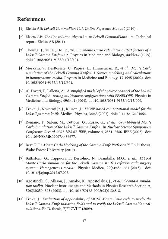
To evaluate lateral beam profiles, I integrated the total deposited energy in 57 cu-bical volumes of 0.5 × 0.5 × 0.5 mm along x-axis with volume centres at x =−14.0, . . . , +14.0mm and normalized them against the central volume. e pro-files for all collimator sizes are ploed in figure 2 together with profiles extractedfrom the treatment planning soware.
4.2 Leksell Gamma Knife PerfexionFor all studies of Perfexion, I used standard command-based scoring of Geant4 toscore deposited energy (and dose in the case of voxel phantoms) in rectangular3D grids. e computing and storage facilities of the National Grid InfrastructureMetaCentrum were used for all Perfexion simulations.
4.2.1 Output factors
e scoring volume was a 1 × 1 × 1 cm cube placed in the isocentre, divided into50 × 50 × 50 voxels. From this volume, different voxel sets were used in order tominimize statistical error while keeping estimated systematic error (arising fromintegration over inhomogeneous dose distribution) reasonably low. Output factorsfor each combination of ring and collimator size were calculated.
Output factors for individual rings cannot be measured under standard con-ditions. erefore, I determined the measurable effective output factor for eachcollimator size—a weighted sum of output factors of individual rings, normalizedto the sum of 16-mm output factors. e results are listed in table 2.
4.2.2 Dose profiles
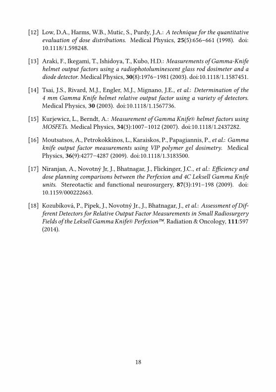
For all collimator sizes, I obtained combined relative dose profiles from all 192 beamsalong the x- and z-axes in “world” coordinates. Also in this case, there is a trade-offbetween dose homogeneity and statistic uncertainty. I investigated different voxelsizes before selecting the optimal ones; these were 0.5, 1.0, and 2.0 mm in length forthe 4-mm, 8-mm, and 16-mm collimators, respectively. e profiles are ploed infigure 2.
12
4.2.3 Dose in voxel phantoms comparison
ree datasets were investigated in this study: a gel phantom, a neuralgia (in bothcases, a plan with a single shot and lile material inhomogeneity), and a more com-plex adenoma case with 17 shots close to bone structures and nasal cavity.
e gamma index method was used to quantitatively assess the match betweenthe dose distribution of voxel phantom Monte Carlo data and the prediction of thetreatment planning soware. For each pair of distributions, the percentage of 57 ×57 × 57 points in a 3D matrix satisfying γ < 1 condition was calculated, for severalvalues of the ∆dM parameter. e ∆DM parameter was kept fixed to 3 %; onlypoint with relative dose over 10 % were considered. For ∆dM ≥ 1mm, more than99,7 % points met the conditions in all studied cases; the differences between thealgorithms were minimal.
5 Conclusionsemain focus of this work was to create a detailed and reliable Monte Carlo modelof both Leksell Gamma Knife 4C and Perfexion stereotactic radiosurgery machines,using the Geant4 toolkit. In the former case, it is the second independent model inGeant4 available, the laer one is as yet the first model in Geant4 and one of thefew published Monte Carlo models in general.
Using these models, I simulated dose distribution inside the reference phantomand determined basic parameters of this distribution that are used in the LeksellGammaPlan treatment planning soware. I have obtained the following results:
• e output factors for Leksell Gamma 4C were obtained for the three smallercollimators: ω4 = 0.89 ± 0.01, ω8 = 0.961 ± 0.002, ω14 = 0.983 ± 0.002. esevalues are very close to those of the TMR 10 algorithm with differences up to1 %.
• e lateral dose profiles of single beams in Leksell Gamma Knife 4C weredetermined for each collimator size. ey show a very good agreement withthe TMR 10 algorithm; the discrepancy observed by previous studies betweenMonte Carlo and TMR Classic algorithm for the 8-mm collimator profile wasconfirmed in this work.
• e output factors for all collimator sizes and rings (14 in total) of LeksellGamma Knife Perfexion were calculated with the effective ones beingω4,eff =0.830 ± 0.004, ω8,eff = 0.921 ± 0.002. ese values differ slightly from bothTMR algorithms by up to 3 %.
• Dose profiles of the composite radiation field created by all beams of eachspecified collimator size (4-mm, 8-mm and 16-mm) were determined along
13
the two main axes of the system. Results were compared to the profiles ob-tained from Leksell GammaPlan. e simulated profiles are slightly sharperthan the LGP ones for 4-mm and 8-mm collimators.
Finally, a method of creating a voxel phantom for Leksell Gamma Knife Per-fexion (but general enough to be used without modification for 4C) was developed.For three clinical cases (i.e. treatment plans exported from Leksell GammaPlan), thedose distribution was simulated and compared (in relative values using the gammaindex method) to the results of all three LGP algorithms. e agreement was good,although some small differences are yet to be explained.
During the development, the complexity of the model and the variety of itsdifferent applications led to creating a modular architecture for Geant4 application:the g4application library and its plug-ins (e.g. g4application-dicom). Although thedevelopment was driven by the needs of this particular research, I believe this codeis general enough to be employed in many other Geant4 projects, especially whenthere is a need of combining multiple components that are developed individually.
In general, the presented Monte Carlo models provide a good description ofboth machines. Long computation times disqualify these models from being rou-tinely used as a verification tool. Nevertheless, they can be used to investigateselected cases and to assist dosimetry of small fields. Provided the remaining issuesare resolved, it can be used to compare the treatment planning algorithm accuracy.
e presented work also opens many opportunities for further research anddevelopment. e possible goals include:
• If the computation demands are met (or methods for increasing simulationefficiency are found), new phase space files can be created in the Perfexionmodel withmore accurate description of source capsules and a wider openingangle for the primary photons. Aer that, the output factors and profilesshould be recalculated.
• A new study on suitable treatment plans with more pronounced material in-homogeneities using the voxel phantom would be suitable to assess the ap-propriateness of the three LGP algorithms in these situations.
• e models created will be used to support the research on gel dosimetry,starting with the voxel model already presented in this work.
• When properly documented and refactored, the source code of the Perfex-ion model can be published with the ambition to be included as an officialexample of Geant4.
14
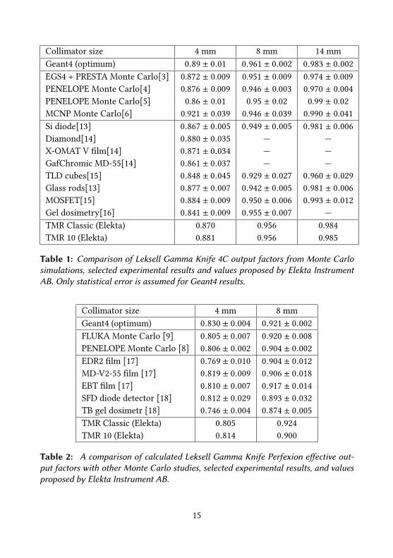
Collimator size 4 mm 8 mm 14 mmGeant4 (optimum) 0.89 ± 0.01 0.961 ± 0.002 0.983 ± 0.002EGS4 + PRESTA Monte Carlo[3] 0.872 ± 0.009 0.951 ± 0.009 0.974 ± 0.009PENELOPE Monte Carlo[4] 0.876 ± 0.009 0.946 ± 0.003 0.970 ± 0.004PENELOPE Monte Carlo[5] 0.86 ± 0.01 0.95 ± 0.02 0.99 ± 0.02MCNP Monte Carlo[6] 0.921 ± 0.039 0.946 ± 0.039 0.990 ± 0.041Si diode[13] 0.867 ± 0.005 0.949 ± 0.005 0.981 ± 0.006Diamond[14] 0.880 ± 0.035 — —X-OMAT V film[14] 0.871 ± 0.034 — —GafChromic MD-55[14] 0.861 ± 0.037 — —TLD cubes[15] 0.848 ± 0.045 0.929 ± 0.027 0.960 ± 0.029Glass rods[13] 0.877 ± 0.007 0.942 ± 0.005 0.981 ± 0.006MOSFET[15] 0.884 ± 0.009 0.950 ± 0.006 0.993 ± 0.012Gel dosimetry[16] 0.841 ± 0.009 0.955 ± 0.007 —TMR Classic (Elekta) 0.870 0.956 0.984TMR 10 (Elekta) 0.881 0.956 0.985
Table 1: Comparison of Leksell Gamma Knife 4C output factors from Monte Carlosimulations, selected experimental results and values proposed by Elekta InstrumentAB. Only statistical error is assumed for Geant4 results.
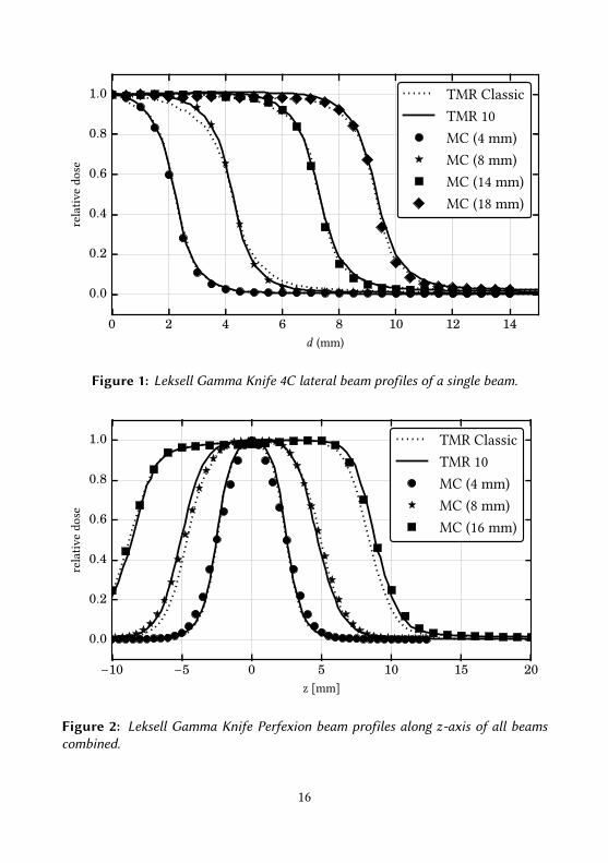
Collimator size 4 mm 8 mmGeant4 (optimum) 0.830 ± 0.004 0.921 ± 0.002FLUKA Monte Carlo [9] 0.805 ± 0.007 0.920 ± 0.008PENELOPE Monte Carlo [8] 0.806 ± 0.002 0.904 ± 0.002EDR2 film [17] 0.769 ± 0.010 0.904 ± 0.012MD-V2-55 film [17] 0.819 ± 0.009 0.906 ± 0.018EBT film [17] 0.810 ± 0.007 0.917 ± 0.014SFD diode detector [18] 0.812 ± 0.029 0.893 ± 0.032TB gel dosimetr [18] 0.746 ± 0.004 0.874 ± 0.005TMR Classic (Elekta) 0.805 0.924TMR 10 (Elekta) 0.814 0.900
Table 2: A comparison of calculated Leksell Gamma Knife Perfexion effective out-put factors with other Monte Carlo studies, selected experimental results, and valuesproposed by Elekta Instrument AB.
15
0 2 4 6 8 10 12 14d (mm)
0.0
0.2
0.4
0.6
0.8
1.0re
lativ
edo
seTMR ClassicTMR 10MC (4 mm)MC (8 mm)MC (14 mm)MC (18 mm)
Figure 1: Leksell Gamma Knife 4C lateral beam profiles of a single beam.
−10 −5 0 5 10 15 20z [mm]
0.0
0.2
0.4
0.6
0.8
1.0
rela
tive
dose
TMR ClassicTMR 10MC (4 mm)MC (8 mm)MC (16 mm)
Figure 2: Leksell Gamma Knife Perfexion beam profiles along z-axis of all beamscombined.
16
References[1] Elekta AB: Leksell GammaPlan 10.1, Online Reference Manual (2010).
[2] Elekta AB: e Convolution algorithm in Leksell GammaPlan® 10. Technicalreport, Elekta AB (2011).
[3] Cheung, J., Yu, K., Ho, R., Yu, C.: Monte Carlo calculated output factors of aLeksell Gamma Knife unit. Physics in Medicine and Biology, 44:N247 (1999).doi:10.1088/0031-9155/44/12/401.
[4] Moskvin, V., DesRosiers, C., Papiez, L., Timmerman, R., et al.: Monte Carlosimulation of the Leksell Gamma Knife®: I. Source modelling and calculationsin homogeneous media. Physics in Medicine and Biology, 47:1995 (2002). doi:10.1088/0031-9155/47/12/301.
[5] Al-Dweri, F., Lallena, A.: A simplified model of the source channel of the LeksellGamma Knife®: testing multisource configurations with PENELOPE. Physics inMedicine and Biology, 49:3441 (2004). doi:10.1088/0031-9155/49/15/009.
[6] Trnka, J., Novotný Jr, J., Klusoň, J.: MCNP-based computational model for theLeksell gamma knife. Medical Physics, 34:63 (2007). doi:10.1118/1.2401054.
[7] Romano, F., Sabini, M., Cuone, G., Russo, G., et al.: Geant4-based MonteCarlo Simulation of the Leksell Gamma Knife®. In Nuclear Science SymposiumConference Record, 2007. NSS’07. IEEE, volume 4, 2581–2586. IEEE (2008). doi:10.1109/NSSMIC.2007.4436677.
[8] Best, R.C.: Monte Carlo Modeling of the Gamma Knife Perfexion™. Ph.D. thesis,Wake Forest University (2010).
[9] Baistoni, G., Cappucci, F., Bertolino, N., Brambilla, M.G., et al.: FLUKAMonte Carlo simulation for the Leksell Gamma Knife Perfexion radiosurgerysystem: Homogeneous media. Physica Medica, 29(6):656–661 (2013). doi:10.1016/j.ejmp.2012.07.005.
[10] Agostinelli, S., Allison, J., Amako, K., Apostolakis, J., et al.: Geant4-a simula-tion toolkit. Nuclear Instruments and Methods in Physics Research Section A,506(3):250–303 (2003). doi:10.1016/S0168-9002(03)01368-8.
[11] Trnka, J.: Evaluation of applicability of MCNP Monte Carlo code to model theLeksell Gamma Knife radiation fields and to verify the Leksell GammaPlan cal-culations. Ph.D. thesis, FJFI ČVUT (2009).
17
[12] Low, D.A., Harms, W.B., Mutic, S., Purdy, J.A.: A technique for the quantitativeevaluation of dose distributions. Medical Physics, 25(5):656–661 (1998). doi:10.1118/1.598248.
[13] Araki, F., Ikegami, T., Ishidoya, T., Kubo, H.D.: Measurements of Gamma-Knifehelmet output factors using a radiophotoluminescent glass rod dosimeter and adiode detector. Medical Physics, 30(8):1976–1981 (2003). doi:10.1118/1.1587451.
[14] Tsai, J.S., Rivard, M.J., Engler, M.J., Mignano, J.E., et al.: Determination of the4 mm Gamma Knife helmet relative output factor using a variety of detectors.Medical Physics, 30 (2003). doi:10.1118/1.1567736.
[15] Kurjewicz, L., Berndt, A.: Measurement of Gamma Knife® helmet factors usingMOSFETs. Medical Physics, 34(3):1007–1012 (2007). doi:10.1118/1.2437282.
[16] Moutsatsos, A., Petrokokkinos, L., Karaiskos, P., Papagiannis, P., et al.: Gammaknife output factor measurements using VIP polymer gel dosimetry. MedicalPhysics, 36(9):4277–4287 (2009). doi:10.1118/1.3183500.
[17] Niranjan, A., Novotný Jr, J., Bhatnagar, J., Flickinger, J.C., et al.: Efficiency anddose planning comparisons between the Perfexion and 4C Leksell Gamma Knifeunits. Stereotactic and functional neurosurgery, 87(3):191–198 (2009). doi:10.1159/000222663.
[18] Kozubíková, P., Pipek, J., Novotný Jr., J., Bhatnagar, J., et al.: Assessment of Dif-ferent Detectors for Relative Output Factor Measurements in Small RadiosurgeryFields of the Leksell GammaKnife® Perfexion™. Radiation &Oncology, 111:597(2014).
18
List of publicationsPublications relevant to the research presented in the thesisand published in foreign journal with impact factor
• Pipek, J., Novotný, J., Novotný Jr., J., Kozubíková, P.: Amodular Geant4modelof Leksell Gamma Knife® Perfexion™. Physics in Medicine and Biology 59:7609-7623 (2014), ISSN 0031-9155.
Other publications• Kozubíková, P., Šolc, J., Novotný Jr., J., Pilařová, K., Pipek, J., Končeková, J.:Assessment of radiochromic gel dosimeter based on Turnbull Blue dye forrelative output factor measurements of the Leksell Gamma Knife® Perfex-ion™. In IC3DDose: 8th International Conference on 3D Radiation Dosime-try (2014).
• Kozubíková, P., Pipek, J., Novotný Jr., J., Bhatnagar, J., et al.: Assessment ofDifferent Detectors for Relative Output Factor Measurements in Small Ra-diosurgery Fields of the Leksell Gamma Knife® PerfexionTM. Radiation &Oncology, 111, Supplement 1:597 (2014), ISSN 0167-8140.
• Pipek, J.: Výpočet faktorů velikosti pole a profilů svazku Leksellova gamanože pomocí Geant4. In: XXXII. dny radiační ochrany : sborník abstraktů:Třeboň, Jižní Čechy, 8.-12. 11. 2010, ČVUT (2010). ISBN 978-80-01-04647-0.
• Janky, F., Havlíček, J., Batista, A.J.N., Kudláček, O., Seidl, J., Neto, A.C., Pipek,J., Hron, M., Mikulín, O., Duarte, A.S., Carvalho, B.B., Stöckel, J., Pánek, R.:Upgrade of the COMPASS tokamak real-time control system. Fusion Engi-neering and Design, 89(3):186-194 (2014), ISSN 0920-3796.
• Hron, M., Janky, F., Pipek, J., Sousa, J., Carvalho, B.B., Fernandes, H., Von-dráček, P., Cahyna, P., Urban, J., Papřok, R., Mikulín, O., Aanas, M., Pánek,R., Havlíček, J., Fortunato, J., Batista, A.J.N., Santos, B., Duarte, A.S., Pereira,T., Valcarel, D.: Overview of the COMPASS CODAC system. Fusion Engi-neering and Design, 89(3):177-185 (2014), ISSN 0920-3796.
• Urban, J., Pipek, J., Hron, M., Janky, F., Papřok, R., Peterka, M., Duarte, A.S.:Integrated data acquisition, storage, retrieval and processing using the COM-PASS DataBase (CDB). In: Proceedings of the 9th IAEA Technical Meeting onControl, Data Acquisition, and Remote Participation for Fusion Research, Fu-sion Engineering and Design, 89(5):712-716 (2014), ISSN 0920-3796.
19
ResuméNejprve byl vytvořen model Leksellova gama nože 4C s použitímMonte Carlo kóduGeant4. Byla ověřena platnost základních parametrů sowaru pro plánování léčby,Leksellova GammaPlanu (LGP), konkrétně faktorů velikosti pole (OF) a příčnýchdávkových profilů jednoho svazku.
Dalším krokembylo vytvoření detailníhomodelu Leksellova gama nože Perfex-ion. Složitost zkoumaného problému si vyžádala pomocnou infrastrukturu, kteránení standardně v Geant4 obsažena. Proto byla vyvinuta modulární knihovna g4-Application a několik rozšiřujících zásuvných modulů (plug-inů) pro ni. Faktoryvelikosti a dávkové profily byly určeny také pro Perfexion.
Nakonec byla rozvinuta metoda pro konstrukci voxelového fantomu z dat počí-tačové tomografie (CT) a jeho správné umístění v rámci modelu Leksellova gamanože Perfexion. Díky této metodě je možné ověřovat klinické ozařovací plány ex-portované z plánovacího sowaru. U tří ukázkových příkladů bylo nasimulovánorozložení dávky a výsledky byly porovnány s výstupem tří různých algoritmů LGP.
20


![jktLFkku ljdkj vkink icU/ku ,o lgk;rk foHkkx vf/ · PDF filejktLFkku ljdkj vkink icU/ku ,o lgk;rk foHkkx dzekd% ,Q 1¼1½¼4½ vk-iz-,o lgk@lkekU;@2014@ t;ij] fnukd vf/klwpuk ftyk](https://img.pdfslide.tips/doc/110x75/5a90c8fe7f8b9a085a8e6d14/jktlfkku-ljdkj-vkink-icuku-o-lgkrk-fohkkx-vf-ljdkj-vkink-icuku-o-lgkrk-fohkkx.jpg)