Embed Size (px)
Citation preview
Journal of Cellular Biochemistry 87:16–28 (2002)
Morphological Changes Induced by Extracellular MatrixAre Correlated with Maturation of Rat Small Hepatocytes
Shinichi Sugimoto,1,3 Toshihiro Mitaka,1* Shinichiro Ikeda,1,2 Keisuke Harada,1,2 Iwao Ikai,3
Yoshio Yamaoka,3 and Yohichi Mochizuki1
1Department of Pathology, Cancer Research Institute, Sapporo Medical University School of Medicine,Sapporo, Japan2Department of Surgery, Sapporo Medical University School of Medicine, Sapporo, Japan3Department of Gastroenterological Surgery, Kyoto University Medical School, Kyoto, Japan
Abstract Small hepatocytes (SHs), which are known to be hepatic progenitor cells, were isolated from an adult ratliver. SHs in a colony sometimes change their shape from small to large and from flat to rising/piled-up. The aim of thepresent study is to clarify whether the alteration of cell shape is correlated with the maturation of SHs and whetherextracellular matrix (ECM) can induce the morphological changes of SHs. We used liver-enriched transcription factors(LETFs) such as hepatocyte nuclear factor (HNF) 4a, HNF6, CCAAT/enhancer binding proteins (C/EBP) a, and C/EBPb,tryptophan 2,3-dioxygenase (TO), and serine dehydratase (SDH) as markers of hepatic maturation. To enrich the numberof SH colonies, the colonies were isolated from dishes and replated. Replated colonies proliferated and the averagenumber of cells per colonywas about five times larger at day9 than at day1.When the cellswere treatedwith laminin, typeIV collagen, amixture of laminin and type IV collagen,MatrigelTMor collagen gel (CG), only the cells treatedwithMatrigeldramatically changed their shapewithin several days andhad reducedgrowthactivity,whereas the cells treatedwith otherECMdidnot.HNF4a,HNF6,C/EBPa, C/EBPb, andTOwerewell expressed in the cells treatedwithMatrigel. Furthermore,addition of both glucagon and dexamethasone dramatically induced the expression of SDHmRNAandprotein in the cellstreated with Matrigel. In conclusion, morphological changes of SHs may be correlated with hepatic maturation andbasement membrane (BM)-like structure may induce the morphological changes of SHs. J. Cell. Biochem. 87: 16–28,2002. � 2002 Wiley-Liss, Inc.
Key words: liver-enriched transcription factors; Matrigel; serum proteins; growth; hepatic nonparenchymal cells
Small hepatocytes (SHs) have been identifiedas proliferating cells with hepatic characteris-tics [Mitaka et al., 1992, 1993; Tateno andYoshizato, 1996]. Recently, we showed that asingle SH could clonally proliferate and form alarge colony [Mitaka et al., 1995, 1999]. Some
SH colonies changed their shapes from flat torising/piled-up cells with time in culture. Therising/piled-up cells were large and tall, pos-sessed many mitochondria, peroxisomes with acrystalline nucleoid, and glycogen granules[Mitaka et al., 1999]. In such colonies nonpar-enchymal cells (NPCs) invadedunder the colonyand an accumulation of extracellular matrix
� 2002 Wiley-Liss, Inc.
Grant sponsor: Ministry of Education, Science, Sports andCulture Japan; Grant numbers: 10670213 (to TM),12670211 (to TM), 11470244 (to II), 1247243 (to YM); Grantsponsor: Health Sciences Research Grant; Grant sponsor:Research on Human Genome, Tissue Engineering FoodBiotechnology.
*Correspondence to: Toshihiro Mitaka, Department ofPathology, Cancer Research Institute, Sapporo MedicalUniversity School of Medicine, Chuo-Ku, S-1, W-17,Sapporo 060-8556, Japan. E-mail: [email protected]
Received 28 May 2002; Accepted 17 June 2002
DOI 10.1002/jcb.10274
Abbreviations used: AP, alkaline phosphatase; Asc2P,ascorbic acid 2-phosphate; BC, bile canaliculi; BM, base-ment membrane; C/EBPa, CCAAT/enhancer bindingprotein a; Cx, connexin; DAB, 30-diaminobenzidine; DAPI,6-diamino-2-phenylindole; DMEM, Dulbecco’s modifiedEagle’s medium; DMSO, dimethylsulfoxide; ECM, extra-cellular matrix; EGF, epidermal growth factor; EHS,Engelbreth–Holm–Swarm; ELISA, enzyme-linked immu-nosorbent assay; FBS, fetal bovine serum; HNF, hepatocytenuclear factor; LECs, liver epithelial cells; LETFs, liver-enriched transcription factors; MHs, mature hepatocytes;NPCs, nonparenchymal cells; PAGE, polyacrylamide gelelectrophoresis; PCNA, proliferating cell nuclear antigen;SDH, serine dehydratase; SHs, small hepatocytes; TEM,transmission electron microscopy; TO, tryptophan 2,3-dioxygenase.
(ECM) between hepatocytyes and NPCs wasobserved. Therefore, we suspected that SHscould differentiate into mature hepatocytes(MHs) that interacted with hepatic NPCs andECM [Mitaka et al., 1999].For the purpose of maintaining the differen-
tiated functions, many researchers have usedvarious substances and changed culture condi-tions through the use of nicotinamide [Inoueet al., 1989; Mitaka et al., 1991], phenobarbital[Miyazaki et al., 1985], dimethylsulfoxide[DMSO; Isom et al., 1985], ECM [Rojkind et al.,1980; Bissell et al., 1987; Ben-Ze’ev et al., 1988;Schetz et al., 1988; Dunn et al., 1992], coculturewith NPCs [Guguen-Guillouzo, 1986], andspheroid formation [Koide et al., 1989; Ingber,1993; Iredale and Arthur, 1994; Rojkind andGreenwel, 1994]. In such experiments, themaintenance of liver-specific functions of thecells was evaluated by expression of mRNAsand/or proteins such as serum proteins, gapjunctional proteins like connexin 32 (Cx32) andCx26, tryptophan 2,3-dioxygenase (TO), andserine dehydratase (SDH). Many genes of thoseliver-specific proteins are known to be mainlyregulated by liver-enriched transcription fac-tors (LETFs) such as CCAAT/enhancer bindingprotein (C/EBP) a and C/EBPb, and hepatocytenuclear factor (HNF) 1a, HNF3a, HNF4a, andHNF6 [Tian and Schibler, 1991; Kuo et al.,1992; Cereghini, 1996; Uzma and Costa, 1996].Primary hepatocytes cultured on MatrigelTM,which is Engelbreth–Holm–Swarm (EHS)sarcoma-derived matrix, maintained some dif-ferentiated functions such as albumin, trans-thyretin, and apolipoprotein A-I production andkept the transcription of HNF1a and HNF4mRNAs [Nagaki et al., 1995; Oda et al., 1995].It has been emphasized that cell shape is a keyfactor to regulate the growth, differentiation,and survival of hepatocytes [Walt, 1986;Maher,1988]. ECM was reported to be able to modu-late the shapes of cultured hepatocytes [Bissellet al., 1987; Maher, 1988; Koide et al., 1989].Ben-Ze’ev et al. [1988] suggested that cell shapemight be a primary regulator of tissue-specificgene expression and that cytoskeletal compo-nents might interact directly with the nuclearmatrix to affect gene transcriptional rates.In the present study, we showed that the
addition of Matrigel could dramatically changethe shapes of the cells as well as the structure ofthe colonies. In addition, the changes of cellshape were correlated with the expression of
hepatic differentiated proteins. To clarify whyMatrigel could induce the morphological chan-ges of SHs, we examined the effects of variousECM and growth factors, which are included inMatrigel, on SHs in the colonies. Not onlylaminin, type IV collagen, a mixture of lamininand type IV collagen, and collagen gel (CG) butalso basic fibroblast growth factor (bFGF),pletelet-derived growth factor (PDGF), nervegrowth factor (NGF), and transforming growthfactor b (TGFb) did not affect the alteration ofcellular morphology. Thus, we hypothesize thatmorphological changes of SHs may be corre-lated with hepatic maturation and that theformation of the basement membrane (BM)-like structure may be responsible in part for thebeneficial effect of those morphological changesof the cells.
MATERIALS AND METHODS
Isolation and Culture of Hepatic Cells
Male Sprague–Dawley rats (Shizuoka Labo-ratory Animal Center, Hamamatsu, Japan),weighing 250–400 g, were used to isolate SHs.All animals received humane care and the ex-perimental protocol was approved by the Com-mittee of Laboratory Animals according toUniversity guidelines. Details of the isolationand culture procedure of the cells were previ-ously described [Mitaka et al., 1999]. Finally,2� 105 viable cells/ml were plated on dishes(1.5 ml/35-mm dish; 4 ml/60-mm dish; 10 ml/100-mm dish; Corning Glass Works, Corning,NY) and cultured inDulbecco’smodifiedEagle’smedium (DMEM; GIBCO Laboratories, GrandIsland, NY) supplementedwith 20mMHEPES,25 mM NaHCO3, 30 mg/L L-proline, 10% fetalbovine serum (HyClone, Logan, UT), 10 mMnicotinamide (Katayama Chemical Co., Osaka,Japan), 1mMascorbic acid 2-phosphate (Asc2P;Wako Pure Chem, Tokyo, Japan), 10 ng/mlepidermal growth factor (EGF; CollaborativeResearch, Inc., Lexington, MA), hormone, andantibiotics. After 4 days of culture, 1% DMSO(Aldrich Chem Co., Milwaukee, WI) was addedto the medium. Medium was replaced everyother day.
Replating of Small Hepatocyte Colonies
When SHs proliferated and formed coloniesconsisting of 15–40 cells (8–12 days afterplating), the colonieswere detached fromdishesand replated on new dishes. Cells were rinsed
Maturation of Rat Small Hepatocytes by ECM 17
with PBS and then treated with 0.02% EDTA/PBS for 1 min. The cells were then treated withcell dissociation solution (Sigma Chem Co.,St. Louis, MO) for 5 min at 378C. After additionof DMEM supplemented with 10% FBS in thedish, SH colonies were collected into conicaltubes and the cell suspension was centrifugedat 50g for 5 min. The pellet was resuspendedin the medium. The number of viable colonieswas counted and the colonies were plated on rattail collagen-coated dishes [Michalopoulos andPitot, 1975]. Four to five hours after plating,the medium was replaced with the serum-freemedium.
Addition of ECM or Growth Factors
At 11 days after replating, the cells weretreatedwith various ECMsuch as laminin, typeIV collagen, a mixture of laminin and type IVcollagen, fibronectin, CG, or growth factor-reduced Matrigel (Becton Dickinson Labware,Bedford, MA). The concentrations of individualECM components used were similar to those inMatrigel (themanufacturer’s data). Forty-eighthours after the treatment, the medium wasreplaced with fresh medium without ECM. Onthe other hand, growth factors such as TGF-b,PDGF (Genzyme/Techne, Minneapolis, MN),bNGF (PeproTech EC Ltd., London, UnitedKingdom), and bFGF (Dainippon Pharm Ltd.,Osaka, Japan) were added to the medium atday 11. The concentrations of the growth fac-tors were maximally 10 times larger than inMatrigel. Fresh growth factors were added tothe medium at the time of medium change.
Photographs of Cells
The same fields of dishes identified by needlemarkswere digitally recorded by using a phase-contrast microscope (Olympus Optical Co.,Tokyo, Japan) equipped with a CCD camera(Roper Scientific, Trenton, NJ).
Immunostaining of Cultured Cells
Cells were fixed with cold absolute ethanol.Mouse anti-proliferating cell nuclear antigen(PCNA;DAKO,Copenhagen,Denmark)andanti-cytokeratin (CK)8antibodies (AmershamCorp.,Buckinghamshire, United Kingdom) were usedas the primary antibodies, followed by theavitin–biotin peroxidase complex method (Vec-tastainABCEliteKit; VectorLaboratories, Inc.,Burlingame, CA). 30-Diaminobenzidine (DAB;
TokyoKasei Industries, Tokyo, Japan)wasusedas a substrate. The cells were then counter-stained with hematoxylin. For counting thenumber of the cells in a colony, immunocyto-chemistry procedures for CK8 and PCNA wereused to identify SHs and to examine the growthactivity of the cells, respectively.
For triple immunofluorescent staining, weused a rabbit anti-C/EBPa, a goat anti-HNF4a,or a goat anti-HNF6 antibody (Santa Cruz Bio-technology, Inc., Santa Cruz, CA) and a mouseanti-E-cadherinantibody (TransductionLabora-tory, Lexington, KY) as the primary antibody.Alexa488-conjugated anti-rabbit and goat IgGantibodies or Alexa594-conjugated anti-mouseIgG (Mol Probe, Eugene, OR) as the secondaryantibody were also used. 6-Diamino-2-pheny-lindole (DAPI) was used as a marker of nuclei.The samples were analyzed using the CELL-Scan system (Scanalytics, Billerica, MA). Thedetails of the procedure were previously des-cribed [Mitaka et al., 1999].
Enzyme-Linked ImmunosorbentAssay (ELISA) for Rat Albumin
The medium was collected every 48 h at thetime of medium replacement and centrifugedat 1� 104g for 10 min. The supernatant waskept at �358C until use. Secreted albumin wasmeasured by ELISA as previously described[Mitaka et al., 1995].
Western Blot Analysis
The dishes were washed with PBS andthen treated with MatriSperseTM Cell ReleaseSolution (Becton Dickinson) for 15 min at 378C.Thereafter, 300 ml of buffer solution (10 mMHEPES [pH 7.2], 0.25 M sucrose, 0.5 mMMgCl2) was added to the dish. The cells werescraped and collected into microcentrifugetubes. After pipetting several times with a mic-rosyringe (Hamilton Com, Reno, NV), homo-genates were centrifuged at 500g for 5 min at48C. The supernatants (microsomal fraction)were kept at �358C until use. The pellets wereresuspended in 50 ml of buffer (20 mM Tris-HCl[pH 7.4], 150 mM NaCl, 0.5% deoxycholate,1 mM EDTA, 2 mM phenylmethylsulfonic acid,1% NP-40, 200 KIU/ml aprotinin, 20% glycerol,and 0.4 M KCl) and gently mixed for 30 min at48C. After centrifugation at 13,000g for 15 min,the supernatant was stored at �358C (nuclearprotein fraction). Concentrations of the protein
18 Sugimoto et al.
were measured using a BCA Protein Assay kit(Pierce, Rockford, IL). Samples (medium: 1 ml/lane; microsomal and nuclear protein fractions:10 or 20 mg/lane) were separated by SDS–polyacrylamide gel electrophoresis (PAGE) andthen transferred electrophoretically to an Im-mobilon-Pmembrane (MilliporeCorp., Bedford,MA) with a semi-dry transfer cell (BioRad,Richmond, CA). Rabbit anti-albumin, anti-transferrin, anti-a1-antitrypsin, anti-fibrinogen(Cappel, Costa Mesa, CA), anti-TO (a gift fromT. Nakamura), anti-SDH (a gift from R.Kanamoto), anti-C/EBPa, anti-C/EBPb, goatanti-HNF1a, anti-HNF3a, anti-HNF4a, anti-HNF6, and mouse anti-PCNA antibodies wereused. Horseradish peroxidase-conjugated anti-rabbit IgG, anti-goat IgG, and anti-mouse IgGantibodies (DAKO) were applied and positivebands were detected by incubation in Super-Signal West Dura Extended Duration substrate(Pierce). To induce the expression of TO andSDH, the cells were treated with both 10�5 Mdexamethasone and 10�7 M glucagon.
Northern Blot Analysis
Total RNA was extracted from the cells usingthe single-step thiocyanate–phenol–chloroformextraction method [Chomczynski and Sacchi,1987] as modified by Xie and Rothblum [1991].Total RNA (20 mg/lane) was loaded on 1%agarose gel containing 0.5 mg/L of ethidiumbromide. Gels were capillary-blotted in 20�SSPE (3 M NaCl, 173 mM NaH2PO4, 25 mMEDTA) onto a nylon membrane (Hybond-N,Amersham) and fixed by ultraviolet light. Forthe detection of TO and SDH mRNAs, alkalinephosphatase (AP)-labeled cDNA probes wereprepared from rat TO cDNA (full 1.7 kb EcoRIfragment; a gift from T. Nakamura), rat SDHcDNA (full 1.45 kb EcoRI fragment; a giftfrom R. Kanamoto) using an AlkPhos DIRECTLabeling and Detection System with CDP-Star(Amersham). The method used followed themanufacturer’s manual (Amersham).
Perpendicular Sections of Cultured Cells
Perpendicular sections of the colony were ex-amined by using semithin sections of the mate-rials in the process of transmission electronmicroscopy (TEM). Details of the procedurewere previously described [Mitaka et al., 1999].
RESULTS
Morphological Changes of SHs
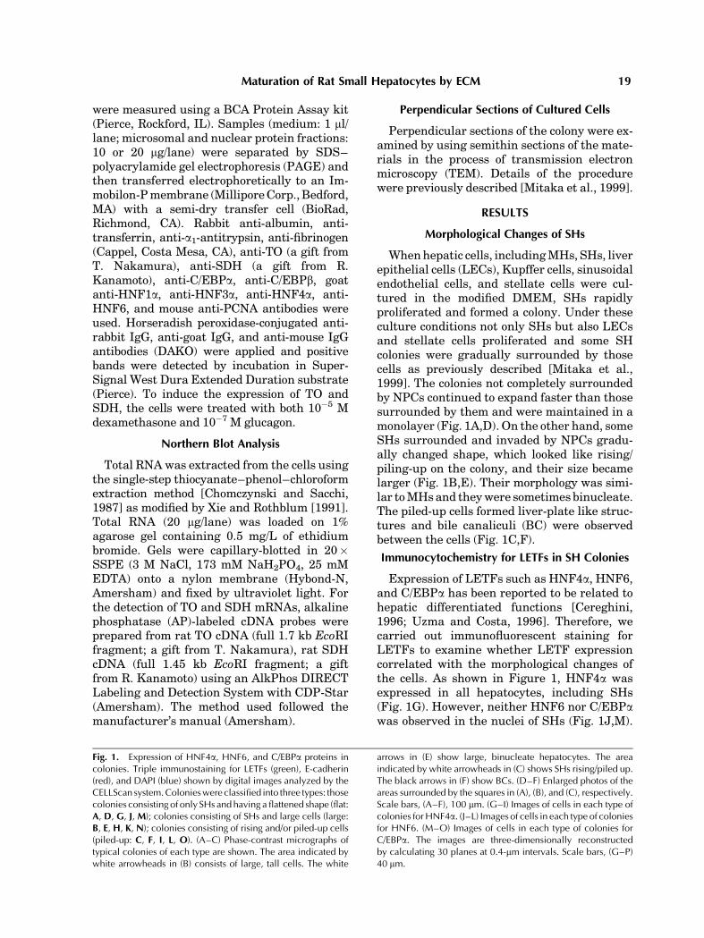
Whenhepatic cells, includingMHs, SHs, liverepithelial cells (LECs), Kupffer cells, sinusoidalendothelial cells, and stellate cells were cul-tured in the modified DMEM, SHs rapidlyproliferated and formed a colony. Under theseculture conditions not only SHs but also LECsand stellate cells proliferated and some SHcolonies were gradually surrounded by thosecells as previously described [Mitaka et al.,1999]. The colonies not completely surroundedby NPCs continued to expand faster than thosesurrounded by them and were maintained in amonolayer (Fig. 1A,D). On the other hand, someSHs surrounded and invaded by NPCs gradu-ally changed shape, which looked like rising/piling-up on the colony, and their size becamelarger (Fig. 1B,E). Their morphology was simi-lar toMHsand theywere sometimes binucleate.The piled-up cells formed liver-plate like struc-tures and bile canaliculi (BC) were observedbetween the cells (Fig. 1C,F).
Immunocytochemistry for LETFs in SH Colonies
Expression of LETFs such as HNF4a, HNF6,and C/EBPa has been reported to be related tohepatic differentiated functions [Cereghini,1996; Uzma and Costa, 1996]. Therefore, wecarried out immunofluorescent staining forLETFs to examine whether LETF expressioncorrelated with the morphological changes ofthe cells. As shown in Figure 1, HNF4a wasexpressed in all hepatocytes, including SHs(Fig. 1G). However, neither HNF6 nor C/EBPawas observed in the nuclei of SHs (Fig. 1J,M).
Fig. 1. Expression of HNF4a, HNF6, and C/EBPa proteins incolonies. Triple immunostaining for LETFs (green), E-cadherin(red), and DAPI (blue) shown by digital images analyzed by theCELLScan system.Colonieswere classified into three types: thosecolonies consisting of only SHs and having a flattened shape (flat:A, D, G, J, M); colonies consisting of SHs and large cells (large:B, E, H, K, N); colonies consisting of rising and/or piled-up cells(piled-up: C, F, I, L, O). (A–C) Phase-contrast micrographs oftypical colonies of each type are shown. The area indicated bywhite arrowheads in (B) consists of large, tall cells. The white
arrows in (E) show large, binucleate hepatocytes. The areaindicated by white arrowheads in (C) shows SHs rising/piled up.The black arrows in (F) show BCs. (D–F) Enlarged photos of theareas surrounded by the squares in (A), (B), and (C), respectively.Scale bars, (A–F), 100 mm. (G–I) Images of cells in each type ofcolonies forHNF4a. (J–L) Images of cells in each type of coloniesfor HNF6. (M–O) Images of cells in each type of colonies forC/EBPa. The images are three-dimensionally reconstructedby calculating 30 planes at 0.4-mm intervals. Scale bars, (G–P)40 mm.
Maturation of Rat Small Hepatocytes by ECM 19
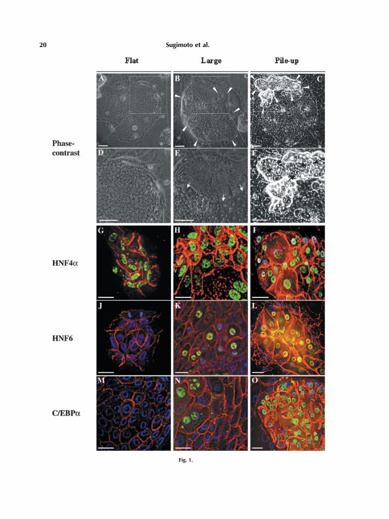
Fig. 1.
20 Sugimoto et al.
Nuclei of large and rising/piled-up cells in thecolonies were positive for HNF6 (Fig. 1K,L) andC/EBPa (Fig. 1N,O).
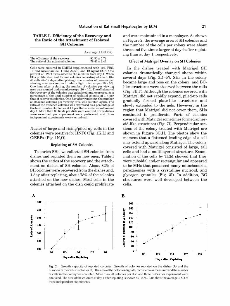
Replating of SH Colonies
To enrich SHs, we collected SH colonies fromdishes and replated them on new ones. Table Ishows the ratios of the recovery and the attach-ment on dishes of SH colonies. About 82% ofSHcolonieswere recovered from the dishes and,1 day after replating, about 78% of the coloniesattached on the new dishes. Most cells in thecolonies attached on the dish could proliferate
and were maintained in a monolayer. As shownin Figure 2, the average area of SH colonies andthe number of the cells per colony were aboutthree and five times larger at day 9 after replat-ing than at day 1, respectively.
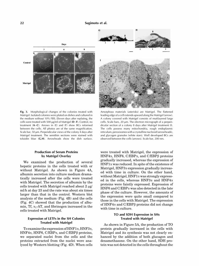
Effect of Matrigel Overlay on SH Colonies
In the dishes treated with Matrigel SHcolonies dramatically changed shape withinseveral days (Fig. 3D–F). SHs in the colonybecame large and rose on the colony, and BC-like structures were observed between the cells(Fig. 3E,F). Although the colonies covered withMatrigel did not rapidly expand, piled-up cellsgradually formed plate-like structures andslowly extended to the gels. However, in theregion that Matrigel did not cover them, SHscontinued to proliferate. Parts of coloniescoveredwithMatrigel sometimes formed spher-oid-like structures (Fig. 7l). Perpendicular sec-tions of the colony treated with Matrigel areshown in Figure 3G,H. The photos show themoment that a flattened leading edge of a cellmay extend upward along Matrigel. The colonycovered with Matrigel consisted of large, tallcells and had a multilayered structure. Exam-ination of the cells by TEM showed that theywere cuboidal and/or rectangular and appearedto be MHs that possessed many mitochondria,peroxisomes with a crystalline nucleoid, andglycogen granules (Fig. 3I). In addition, BCstructures were well developed between thecells.
TABLE I. Efficiency of the Recovery andthe Ratio of the Attachment of Isolated
SH Colonies
Average�SD (%)
The efficiency of the recovery 81.93� 5.78The ratio of the attached colonies 78.41� 2.43
Cells were cultured in DMEM supplemented with 10% FBS,10 mM nicotinamide, 1 mM Asc2P, and 10 ng/ml EGF. Onepercent of DMSO was added to the medium from day 4. WhenSHs proliferated and formed colonies consisting of about 10–40 cells (8–12 days after plating), the number of colonies perviewing area was counted under a light microscope (10� 10).One hour after replating, the number of colonies per viewingareawas counted under amicroscope (10� 10). The efficiency ofthe recovery of the colonies was calculated and expressed as apercentage of the total number of replated colonies at 1 h perthat of recovered colonies. One day after replating, the numberof attached colonies per viewing area was counted again. Theratio of the attached colonies was expressed as a percentage ofthe total number of colonies at 1hper that of attached colonies atday 1. More than 30 fields per dish were counted, three disheswere examined per experiment were performed, and threeindependent experiments were carried out.
Fig. 2. Growth capacity of replated colonies. Growth of colonies replated on the dishes (A) and thenumbers of the cells in colonies (B). The areaof the colonies digitally recordedwasmeasuredand thenumberof cells in the colony was counted. More than 20 colonies per dish and three dishes per experiment wereanalyzed. The area of the colonies at day 1 after replating is shown as 100%. Bars show the average� SD ofthree independent experiments.
Maturation of Rat Small Hepatocytes by ECM 21
Production of Serum Proteinsby Matrigel Overlay
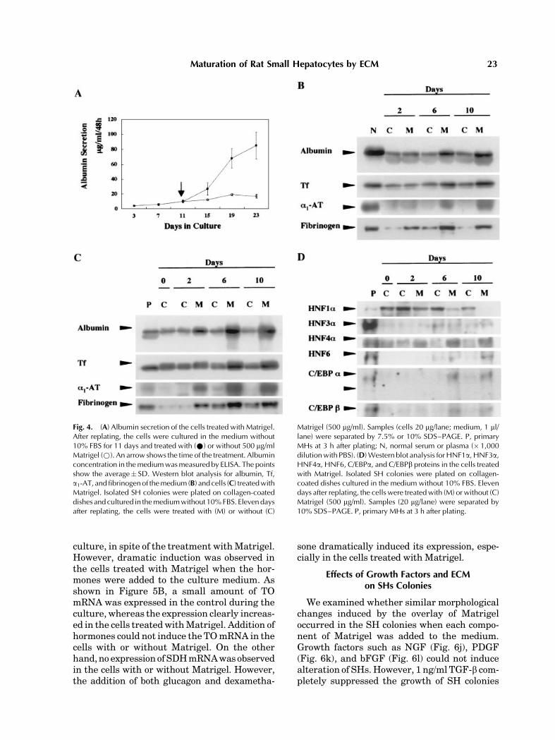
We examined the production of severalhepatic proteins in the cells treated with orwithout Matrigel. As shown in Figure 4A,albumin secretion into culture medium drama-tically increased after the cells were treatedwith Matrigel. The secretion of albumin by thecells treated with Matrigel reached about 2 mg/ml/h at day 23 and the rate was about six timeslarger than that in the control. Western blotanalysis of the medium (Fig. 4B) and the cells(Fig. 4C) showed that the production of albu-min, Tf, a1-AT, and fibrinogen increased in thecells treated with Matrigel.
Expression of LETFs in the SH ColoniesTreated with Matrigel
Toexamine theexpressionofHNF1a,HNF3a,HNF4a, HNF6, C/EBPa, and C/EBPb proteins,we separated nuclei from the cells and theproteins extracted from the nuclei were ana-lyzed by Western blotting (Fig. 4D). When cells
were treated with Matrigel, the expression ofHNF4a, HNF6, C/EBPa, and C/EBPb proteinsgradually increased, whereas the expression ofHNF1awas reduced. In spite of the existence ofMatrigel, HNF3a expression gradually increas-ed with time in culture. On the other hand,withoutMatrigel,HNF1awas strongly express-ed in the cells, whereas HNF3a and HNF4aproteins were faintly expressed. Expression ofHNF6 and C/EBPawas also detected in the latephase of the culture. However, the amounts ofthe expression were quite small compared tothose in the cells with Matrigel. The expressionof HNF4a and C/EBPb proteins did not changewith time in culture.
TO and SDH Expression in SHsTreated with Matrigel
As shown in Figure 5A, the production of TOprotein gradually increased in the cells withMatrigel and its synthesis was not clearly en-hanced by the addition of both glucagon anddexamethasone. On the other hand, SDH pro-teinwas not detected in the cells throughout the
Fig. 3. Morphological changes of the colonies treated withMatrigel. Isolated colonies were plated on dishes and cultured inthe medium without 10% FBS. Eleven days after replating, thecells were treated with 500 mg/ml of Matrigel (D–F). Control, notreatment (A–C). Arrows in (E) and (F) show BCs reformedbetween the cells. All photos are of the same magnification.Scale bar, 50 mm. Perpendicular views of the colony 4 days afterMatrigel treatment. The semithin sections were stained withtoluidin blue (G,H). Arrowheads show the dish surface.
Amorphous materials (asterisks) are Matrigel. The flattenedleading edge of a cell extends upward along theMatrigel (arrow).A colony covered with Matrigel consists of mutilayered largecells. Scale bars, 20 mm. The electron micrograph of a perpen-dicular section of a colony 8 days after Matrigel treatment (I).The cells possess many mitochondria, rough endoplasmicreticulum,peroxisomeswith a crystallinenucleoid (arrowheads),and glycogen granules (white stars). Well developed BCs areobserved between the cells (arrows). Scale bar, 200 nm.
22 Sugimoto et al.
culture, in spite of the treatment withMatrigel.However, dramatic induction was observed inthe cells treated with Matrigel when the hor-mones were added to the culture medium. Asshown in Figure 5B, a small amount of TOmRNA was expressed in the control during theculture, whereas the expression clearly increas-ed in the cells treatedwithMatrigel. Addition ofhormones could not induce the TOmRNA in thecells with or without Matrigel. On the otherhand,no expressionofSDHmRNAwasobservedin the cells with or without Matrigel. However,the addition of both glucagon and dexametha-
sone dramatically induced its expression, espe-cially in the cells treated with Matrigel.
Effects of Growth Factors and ECMon SHs Colonies
We examined whether similar morphologicalchanges induced by the overlay of Matrigeloccurred in the SH colonies when each compo-nent of Matrigel was added to the medium.Growth factors such as NGF (Fig. 6j), PDGF(Fig. 6k), and bFGF (Fig. 6l) could not inducealteration of SHs.However, 1 ng/ml TGF-b com-pletely suppressed the growth of SH colonies
Fig. 4. (A) Albumin secretion of the cells treated with Matrigel.After replating, the cells were cultured in the medium without10% FBS for 11 days and treated with (*) or without 500 mg/mlMatrigel (*). An arrow shows the time of the treatment. Albuminconcentration in themediumwasmeasured by ELISA. The pointsshow the average� SD. Western blot analysis for albumin, Tf,a1-AT, andfibrinogenof themedium (B) and cells (C) treatedwithMatrigel. Isolated SH colonies were plated on collagen-coateddishes andcultured in themediumwithout 10%FBS. Elevendaysafter replating, the cells were treated with (M) or without (C)
Matrigel (500 mg/ml). Samples (cells 20 mg/lane; medium, 1 ml/lane) were separated by 7.5% or 10% SDS–PAGE. P, primaryMHs at 3 h after plating; N, normal serum or plasma (�1,000dilutionwith PBS). (D)Western blot analysis forHNF1a, HNF3a,HNF4a, HNF6, C/EBPa, and C/EBPb proteins in the cells treatedwith Matrigel. Isolated SH colonies were plated on collagen-coated dishes cultured in the medium without 10% FBS. Elevendays after replating, the cells were treatedwith (M) or without (C)Matrigel (500 mg/ml). Samples (20 mg/lane) were separated by10% SDS–PAGE. P, primary MHs at 3 h after plating.
Maturation of Rat Small Hepatocytes by ECM 23
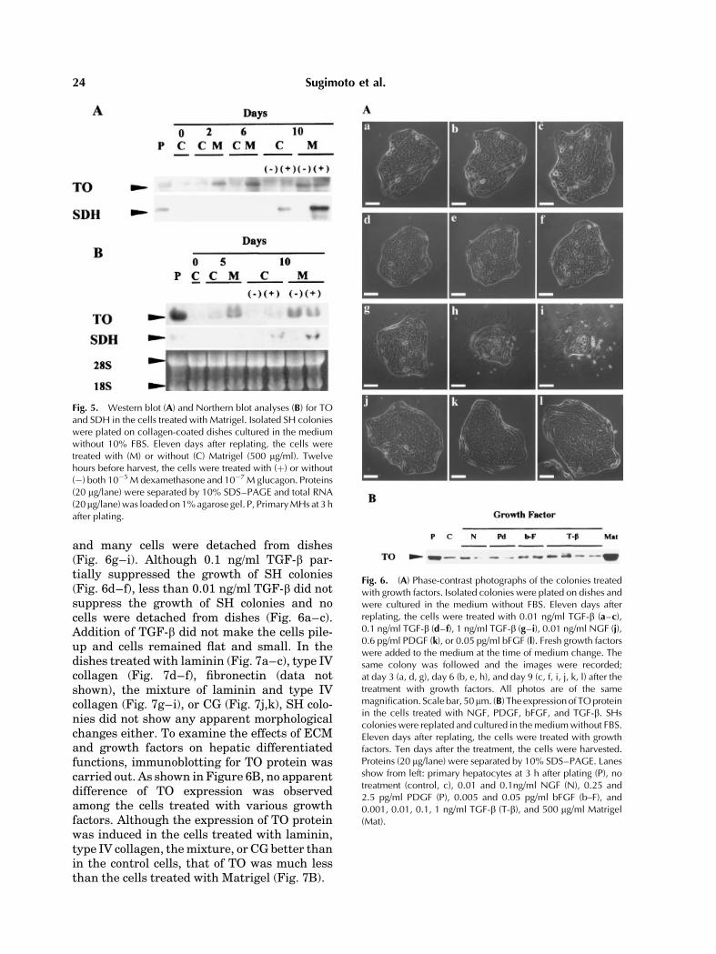
and many cells were detached from dishes(Fig. 6g–i). Although 0.1 ng/ml TGF-b par-tially suppressed the growth of SH colonies(Fig. 6d–f), less than 0.01 ng/ml TGF-b did notsuppress the growth of SH colonies and nocells were detached from dishes (Fig. 6a–c).Addition of TGF-b did not make the cells pile-up and cells remained flat and small. In thedishes treated with laminin (Fig. 7a–c), type IVcollagen (Fig. 7d–f), fibronectin (data notshown), the mixture of laminin and type IVcollagen (Fig. 7g–i), or CG (Fig. 7j,k), SH colo-nies did not show any apparent morphologicalchanges either. To examine the effects of ECMand growth factors on hepatic differentiatedfunctions, immunoblotting for TO protein wascarried out. As shown in Figure 6B, no apparentdifference of TO expression was observedamong the cells treated with various growthfactors. Although the expression of TO proteinwas induced in the cells treated with laminin,type IV collagen, themixture, or CG better thanin the control cells, that of TO was much lessthan the cells treated with Matrigel (Fig. 7B).
Fig. 5. Western blot (A) and Northern blot analyses (B) for TOand SDH in the cells treated with Matrigel. Isolated SH colonieswere plated on collagen-coated dishes cultured in the mediumwithout 10% FBS. Eleven days after replating, the cells weretreated with (M) or without (C) Matrigel (500 mg/ml). Twelvehours before harvest, the cells were treated with (þ) or without(�) both 10�5 M dexamethasone and 10�7 M glucagon. Proteins(20 mg/lane) were separated by 10% SDS–PAGE and total RNA(20 mg/lane)was loaded on1%agarose gel. P, PrimaryMHs at 3 hafter plating.
Fig. 6. (A) Phase-contrast photographs of the colonies treatedwith growth factors. Isolated colonies were plated on dishes andwere cultured in the medium without FBS. Eleven days afterreplating, the cells were treated with 0.01 ng/ml TGF-b (a–c),0.1 ng/ml TGF-b (d–f), 1 ng/ml TGF-b (g–i), 0.01 ng/ml NGF (j),0.6 pg/ml PDGF (k), or 0.05 pg/ml bFGF (l). Fresh growth factorswere added to the medium at the time of medium change. Thesame colony was followed and the images were recorded;at day 3 (a, d, g), day 6 (b, e, h), and day 9 (c, f, i, j, k, l) after thetreatment with growth factors. All photos are of the samemagnification. Scale bar, 50mm. (B) The expressionof TOproteinin the cells treated with NGF, PDGF, bFGF, and TGF-b. SHscolonies were replated and cultured in themediumwithout FBS.Eleven days after replating, the cells were treated with growthfactors. Ten days after the treatment, the cells were harvested.Proteins (20 mg/lane) were separated by 10% SDS–PAGE. Lanesshow from left: primary hepatocytes at 3 h after plating (P), notreatment (control, c), 0.01 and 0.1ng/ml NGF (N), 0.25 and2.5 pg/ml PDGF (P), 0.005 and 0.05 pg/ml bFGF (b–F), and0.001, 0.01, 0.1, 1 ng/ml TGF-b (T-b), and 500 mg/ml Matrigel(Mat).
24 Sugimoto et al.

Growth Activity of SHs Treated with Matrigel
To examine growth activity of SHs treatedwith Matrigel, immunostaining, and Westernblot analysis for PCNA were performed. As
shown in Figure 8a, many replated SHs werepositive for PCNA. This resultmeant thatmanySHs in monolayer colony actively proliferated.However, the ratio of PCNA-positive nuclei toPCNA-negative ones in a colony decreased withthe expansion of the colony at the time whensome cells became large hepatocytes (Fig. 8b).On the other hand, when the cells were coveredwith Matrigel, the number of PCNA-positivecells clearly decreased (Fig. 8d). However, thenuclei of the cells in the process of changingtheir shapes showed the PCNA-positivity,
Fig. 7. (A) Phase-contrast photographs of the colonies treatedwith laminin, type IV collagen, a mixture of laminin and type IVcollagen, and CG. Isolated colonies were plated and cultured inthe medium without FBS. Eleven days after replating, the cellswere treated with 300 mg/ml laminin (a–c), 150 mg/ml type IVcollagen (d–f), a mixture of 300 mg/ml laminin and 150 mg/mltype IV collagen (g–i), CG (j, k), and 500 mg /ml Matrigel (l).The same colony was followed and the images were recorded;at day 3 (a, d, g), day 6 (b, e, h, j), and day 9 (c, f, i, k, l) after thetreatment with ECM. All photos are of the same magnification.Scale bar, 50 mm. (B) The expression of TO protein in the cellstreated with laminin, type IV collagen, a mixture of lamininand type IV collagen, CG, and Matrigel. SHs colonies werereplated and cultured in medium without FBS. Eleven days afterreplating, the cells were treated with 300 mg/ml laminin (L),150 mg/ml typeIV collagen (IV), a mixture of 300 mg/ml lamininand 150 mg/ml type IV collagen (Mix), CG, and 500 mg/mlMatrigel (M). Ten days after the treatment, the cells wereharvested. Proteins (10 mg/lane) were separated by 10% SDS–PAGE. P, primary hepatocytes at 3 h after plating. C, control.
Fig. 8. (A) Immunocytochemistry for PCNA of the cells incolonies. The colonies were treated with (a, b) and withoutMatrigel (c, d). (a) At day 3 and (b) at day 8 after replating, (c) atday 3, and (d) at day 9 after the treatmentwith 500 mg ofMatrigel.Darkened nuclei are positive for PCNA. The cells were counter-stained with hematoxylin. Arrows in (c) show dead cells andarrowheads show the cells that are rising and positive for PCNA.All photographs show the same magnification. Scale bars,100 mm. (B) Western blot analysis for PCNA proteins in thecolonies treated with Matrigel. Nuclear proteins (20 mg/lane)were separated by 10% SDS–PAGE. Isolated SH colonies wereplated on collagen-coated dishes cultured in the mediumwithout 10% FBS. Eleven days after replating, the cells weretreated with (M) or without (C) Matrigel (500 mg/ml). P, primaryhepatocytes at 3 h after plating.
Maturation of Rat Small Hepatocytes by ECM 25
whereas most piled-up cells were not stained(Fig. 8c). As shown in Figure 8B, the expressionof PCNA protein was remarkably inhibited inthe nuclei of the cells treated with Matrigel.In addition, with time after the treatment, theamount of the protein decreased and the expres-sion was scarcely detected at 10 days after thetreatment. This result was similar to that ofthe immunostaining.
DISCUSSION
Morphological Changes and Maturation of SHs
We previously showed that SHs in coloniessometimes changed shape [Mitaka et al., 1999].In such cases NPCs invaded under the colonyand the formation of BM-like structures, whichmight be reconstituted with ECM produced byNPCs, was observed. On the other hand, manygenes of liver-specific proteins are known to bemainly regulated by LETFs and their expres-sion seems to be correlated with hepatic mat-uration [Birkenmeir et al., 1989; Kuo et al.,1990; Cereghini, 1996; Uzma and Costa, 1996].Therefore, to investigatewhether the alterationof cell shapewas correlatedwith thematurationof SHs and whether BM-like structures couldinduce the morphological changes of SHs, wefirstimmunocytochemicallyexaminedtheexpres-sions of LETFs in the primary cultured cellsshowing various morphologies and, second,examined whether Matrigel could induce thesimilarmorphological changesofSHs.As showninFigure 1, the sequential expression pattern ofLETFs accompanyingmorphological changes ofSHs was observed. When SHs were maintainedin a flat monolayer, staining for HNF6, C/EBPaand C/EBPb proteins was negative in theirnuclei, TO expression was quite low, and SDHexpression was not induced. On the other hand,when SHs were covered with Matrigel, thecells rapidly changed shape and increased thesecretion of serum proteins such as albumin,Tf, a1-AT, and fibrinogen. In addition, theexpression of LETFs could be recovered inthe cells and the amounts of HNF4a, HNF6,C/EBPa, and C/EBPb proteins at day 10 afterMatrigel addition were near those of proteinsin MHs, although the expression of HNF1aand HNF3a was not affected with or withoutMatrigel. Furthermore, TO was well expressedand SDH could be induced by hormones. There-fore, not only morphological changes fromsmall to large/piled-up but also the synergistic
induction of LETFs like HNF4, HNF6, C/EBPa,and C/EBPb expressions may be necessary forSHs to differentiate into MHs.
Effects of ECM Components and GrowthFactors in Matrigel
We showed that accumulation of ECM couldresult in morphological changes and the matu-ration of SHs [Mitaka et al., 1999]. In thisexperiment Matrigel could induce a change ofshape of SHs. To investigate whether inductionof those morphological changes was dependenton the individual components of Matrigel ormechanical stress resulting from the overlay ofa gel-formed material, we examined the effectsof major ECM components, a CG, and growthfactors on SH colonies. These individual compo-nents did not influence the shape of the coloniesshown with Matrigel. Although TGFb (morethan 0.1 ng/ml) had the ability to suppress thegrowth of SHs as was the case of Matrigel,no differentiation of the cells was induced. Onthe contrary, the higher concentration of TGFbresulted in cell death. TGFb in Matrigel may benot a soluble form but a form binding to ECM.Such different forms may affect the growth anddeath of SHs. On the other hand, although alarge amount of each ECM might possibly in-duce the morphological changes of SH colonies,the concentration used in the present experi-ment did not affect the cell shapes. However,ECM could enhance the synthesis of TO pro-teins compared to the control. Some hepaticdifferentiated functionsmay be induced by eachECM, though the degree of the expression waslower than in the cells treated with Matrigel.Furthermore, to exclude the possibility that themorphological changes of SHs resulted from themechanical stress caused by the overlay of gel-formed materials such as Matrigel, the colonieswere covered with a thick CG. Although TOexpression was enhanced in the cells, no altera-tion of the cell shape was observed. However,after the cells were cultured for more than1 month, they gradually formed cystic struc-tures under the gel (data not shown). Althoughwe have no clear data, the gradual proliferationof NPCs bound to SH colonies and the produc-tion of ECM might have caused the cellularchanges. These findings suggested that the in-dividual components were insufficient and thecombinationand/or complex ofECMandgrowthfactors might be required for morphologicalchanges of SHs. The ideal form may be natural
26 Sugimoto et al.
BM. Further experiments will be required toformacompatiblebed for the cells similar toBM.
Formation of Plate-like Structures in Matrigel
It is of interest that SHs andSH-derived largehepatocytes could proliferate andmigrate in theMatrigel. As shown in Figure 8, the number ofPCNA-positive cells decreased in those coloniesand the speed of the colony expansion becameslow. However, plate-like structures slowlyelongated in Matrigel. Hepatocytes, as is thecase with many other cell types, can maintaindifferentiated functions but do not proliferate inMatrigel [Bissell et al., 1987; Rana et al., 1994].However, Michalopoulos et al. [1999] showedthat MHs isolated from an adult rat liver couldform plate-like structures withinMatrigel afterthey were cultured on collagen-coated polystyr-enebeads in roller bottle for about2weeks.Theyreported that the epithelial cells on beads hadcharacteristics of small MHs and that, after thecells on beads were implanted in Matrigel, theelongation of liver plate-like protrusions wasobserved. The protrusions consisted of hepato-cytes showing cytoplasmic differentiation com-pared to the cells in the roller bottle. Thosephenomena were quite similar to our observa-tions in this experiment. Block et al. [1996] andMichalopoulos et al. [1999] suggested that MHsonce lost their differentiated functions to becomeepithelial cells (they called them ‘‘hepatoblasts’’)and then could redifferentiate toMHs.However,considering our present results, it may be feasi-ble to think that their ‘‘hepatoblasts’’maybeSHsbecause about 2% of hepatocytes isolated fromthe adult rat liver are estimated to be SHs[Mitaka et al., 1993].In summary, although it is unclear how
Matrigel can induce the maturation of SHs,our present data suggest that a complex gel-form of ECM like Matrigel and the formation ofBM-like structures may result in the morpho-logical changes of SHs that can induce specificLETFs such as HNF4a, HNF6, C/EBPa, andC/EBPb.TheseLETFsmaysynergisticallyworkfor the expression of hepatic differentiated func-tions. Although we used growth factor-reducedMatrigel, and investigated the effects of theindividualmajor components included inMatri-gel, the effects of other possible contaminantscan not be ignored. Further experiments will benecessary to examine the exact mechanisms ofdifferentiation/maturation of SHs.
ACKNOWLEDGMENTS
The authors thank Dr. T. Nakamura (OsakaUniversity, Osaka, Japan) for the rabbit anti-rat TO antibody and TO cDNA, and Dr. R.Kanamoto (KyotoPrefecturalUniversity, Kyoto,Japan) for the rabbit anti-SDH antibody andSDH cDNA. We thank Ms. M. Kuwano, Ms. Y.Tanaka, and Mr. H. Itoh for technical assis-tance. We also thankMr. K. Barrymore for helpwith the manuscript.
REFERENCES
Ben-Ze’ev A, Robinson GS, Bucher NLR, Farmer SR. 1988.Cell–cell and cell–matrix interactions differentiallyregulate the expression of hepatic and cytoskeletal genesin primary culture of rat hepatocytes. Proc Natl Acad SciUSA 85:2162–2165.
Birkenmeir EH, Gwynn B, Howard S, Jerry J, Gordon JI,Landschulz WH, Mckight SL. 1989. Tissue-specificexpression, developmental regulation, and genetic map-ping of the gene encoding CCAAT/enhancer bindingprotein. Genes Dev 3:1146–1156.
Bissell DM, Anderson DM, Maher JJ, Roll FJ. 1987.Support of cultured hepatocytes by a laminin-rich gel.J Clin Invest 79:801–812.
Block GD, Locker J, Bowen WC, Peterson BE, Katyal S,Strom SC, Riley T, Howard TA, Michalopoulos GK. 1996.Population expansion, clonal growth, and specific differ-entiation patterns in primary cultures of hepatocytesinduced by HGF/SF, EGF, and TGFa in a chemicallydefined (HGM) medium. J Cell Biol 132:1133–1149.
Cereghini S. 1996. Liver-enriched transcriptional factorsand hepatocyte differentiation. FASEB J 10:267–282.
Chomczynski P, Sacchi N. 1987. Single-stepmethod of RNAisolation by acid guanidium thiocyanate–phenol–chloro-form extraction. Anal Biochem 162:156–159.
Dunn JCY, Tompkins RG, YarmuchML. 1992. Hepatocytesin collagen sandwich: Evidence for transcriptional andtranslational regulation. J Cell Biol 116:1043–1053.
Guguen-Guillouzo C. 1986. Role of homotypic and hetero-typic cell interactions in expression of specific functionsby cultured heptocytes. In: Guillouzo A, Guguen-Guil-louzo C, editors. Research in isolated and culturedhepatocytes. John Libbey Eurotext Ltd. pp 259–284.
Ingber DE. 1993. Extracellular matrix and the develop-ment of tissue architecture: A mechanochemical perspec-tive. In: ZernMA, Reid LM, editors. Extracellular matrix:Chemistry, biology, and pathobiology with emphasis onthe liver. New York: Marcel Dekker Inc. pp 403–428.
Inoue C, Yamamoto H, Nakamura T, Ichihara A, OkamotoH. 1989. Nicotinamide prolongs survival of primarycultured hepatocytes without involving loss of hepato-cyte-specific functions. J Biol Chem 264:4747–4750.
Iredale JP, Arthur MJP. 1994. Hepatocyte–matrix inter-actions. Gut 35:29–32.
Isom HC, Scott T, Georgoff I, Woodworth C, Mummaw J.1985. Maintenance of differentiated rat hepatocytes inprimary culture. Proc Natl Acad Sci USA 82:3252–3256.
Koide N, Shinji T, Tanabe T, Asano K, Kawaguchi M,Sakaguchi K, Koide Y, Mori M, Tsuji T. 1989. Continuedhigh albumin production by multicellular spheroids of
Maturation of Rat Small Hepatocytes by ECM 27
adult rat hepatocytes formed in the presence of liver-derived proteoglycans. Biochem Biophys Res Commun161:385–391.
Kuo CF, Xanthopoulos KG, Darnell JE, Jr. 1990. Fetal andadultlocalization of C/EBP: Evidence for combinationalaction of transcription factors in cell-specific gene expres-sion. Development 109:473–481.
Kuo CJ, Colonley PB, Chen L, Sladek FM, Darnell JE,Jr., Crabtree GR. 1992. A transcriptional hierarchyinvolved in mammalian cell-type specification. Nature355:457–461.
Maher JJ. 1988. Primary hepatocyte culture: Is it homeaway from home? Hepatology 8:1162–1166.
Michalopoulos GK, Pitot HC. 1975. Primary cultures ofparenchymal liver cells on collagen membranes: Mor-phological and biological observations. Exp Cell Res 94:70–78.
Michalopoulos GK, Bowen WC, Zajac VF, Beer-Stolz D,Watkins S, Kostrubsky V, Strom SC. 1999. Morphoge-netic events in mixed cultures of rat hepatocytes andnonparenchymal cells maintained in biological matricesin the presence of hepatocyte growth factor and epider-mal growth factor. Hepatology 29:90–100.
Mitaka T, Sattler CA, Sattler GL, Sargent LM, Pitot HC.1991. Multiple cell cycles occur in rat hepatocytes cultur-ed in the presence of nicotinamide and epidermal growthfactor. Hepatology 13:21–30.
Mitaka T, Mikami M, Sattler GL, Pitot HC, Mochizuki Y.1992. Small cell colonies appear in the primary culture ofadult rat hepatocytes in the presence of nicotinamide andepidermal growth factor. Hepatology 16:440–447.
Mitaka T, Norioka K, Sattler GL, Pitot HC, Mochizuki Y.1993. Effect of age on the formation of small-cell coloniesin cultures of primary rat hepatocytes. Cancer Res 53:3145–3148.
Mitaka T, Kojima T, Mizuguchi T, Mochizuki Y. 1995.Growth and maturation of small hepatocytes isolatedfrom adult rat liver. Biochem Biophys Res Commun 214:310–317.
Mitaka T, Sato F, Mizuguchi T, Yokono T, Mochizuki Y.1999. Reconstruction of hepatic organoid by rat smallhepatocytes and hepatic nonparenchymal cells. Hepatol-ogy 29:111–125.
Miyazaki M, Handa Y, Oda M, Yabe T, Miyano K, Sato J.1985. Long-term survival of functional hepatocytes from
adult rats in the presence of phenobarbital in primaryculture. Exp Cell Res 159:176–190.
Nagaki M, Shidoji Y, Yamada Y, Sugiyama A, Tanaka M,Akaike T, Ohnishi H, Moriwaki H, Muto Y. 1995.Regulation of hepatic gene and transcription factors inrat hepatocytes by extracellar matrix. Biochem BiophysRes Commun 210:38–43.
Oda H, Nozawa N, Hitomi Y, Kakinuma A. 1995. Laminin-rich extracellular matrix maintains high level of hepato-cyte nuclear factor 4 in rat hepatocyte culture. BiochemBiophys Res Commun 212:800–805.
Rana S, Mischoulon D, Xie Y, Bucher NLR, Famer SR.1994. Cell–extracellular matrix interactions can regu-late the switch between growth and differentiation in rathepatocytes: Reciprocal expression of C/EBP alpha andimmediate-early growth response transcription factors.Mol Cell Biol 14:5858–5869.
Rojkind M, Greenwel P. 1994. The extracellular matrix ofthe liver. In: Arias IM, Boyer JL, Fausto N, Jakoby WB,Schachter DA, Shafritz DA, editors. The liver: Biologyand pathobiology. 3rd edn. New York: Raven Press Ltd.pp 843–868.
Rojkind M, Gatmaintan Z, Mackenson S, Giambrone MA,Ponce P, Reid LM. 1980. Connective tissue biomatrix: Itsisolation and utilization for long-term cultures of normalrat hepatocytes. J Cell Biol 87:255–263.
Schetz EG, Donna LI, Omiecinski CJ, Muller-Eberhand U,Kleinman HK, Elswick B, Guzelian PS. 1988. Regula-tion of gene expression in adult rat hepatocytes culturedon a basement membrane matrix. J Cell Physiol 134:309–323.
Tateno C, Yoshizato K. 1996. Growth and differentiation inculture of clonogenic hepatocytes that express bothphenotypes of hepatocytes and biliary epithelial cells.Am J Pathol 149:1593–1605.
Tian JM, Schibler U. 1991. Tissue-specific expression of thegene encoding hepatocyte nuclear factor 4. Genes Dev 5:2225–2234.
Uzma S, Costa RH. 1996. The transcriptional activatorhepatocyte nuclear factor 6 regulates liver gene expres-sion. Mol Cell Biol 16:6273–6284.
Walt FM. 1986. The extracellular matrix and cell shape.TIBS 11:482–448.
Xie W, Rothblum LI. 1991. Rapid small-scale RNA isolationfrom tissue culture cells. Biotechniques 11:324–327.
28 Sugimoto et al.