Embed Size (px)
Citation preview
MORPHOLOGY, TOXICITY AND TOXIN PROPERTIES OF CULTURED TOXIC CYANOBACTERIA SPECIES ISOLATED FROM
AQUACULTURE POND OF SARAWAK AND SABAH
Jasmina Binti Majit
Master of Science 2012
Pusat Knidmat Maklumat Akadem; t. U1vIVERSTTI MALAYSIA SARAWAK
MORPHOLOGY, TOXICITY AND TOXIN PROPERTIES OF CULTURED TOXIC CYANOBACTERIA SPECIES ISOLATED FROM AQUACULTURE POND OF
SARAWAK AND SABAH
JASMINA BINTI MAJIT
This thesis is submitted in fulfillment of the requirement for the degree for Master of Science (Aquatic Toxicology)
Faculty of Resource Science and Technology
UNIVERSITI MALAYSIA SARAWAK
2012
DECLARATION
No portion of the work referred to this dissertation has been submitted in support of an
application for another degree or qualification of this or any other university or institution of
higher learning.
(JASMINA BINTI MAJIT)
Department of Aquatic Science
Faculty of Resource Science and Technology
Universiti Malaysia Sarawak (UNIMAS)
i

ACKNOWLEDGEMENT
First and foremost, I would like to thank ALLAH for the continuous blessing that I have received
during those challenging and wonderful times that I have gone through from the starting until the
end of this project.
Secondly, I would like to express my deepest gratitude and sincere appreciation to my
supervisor, Dr. Samsur Mohamad and other lecturers for their advices, guidance, supports and
building comments from the very beginning until the completion of this project.
Million of thanks are extended to Indigenous Fisheries Research and Production Centre (IFRPC)
and Babagon Fisheries Center (BFC) for their permission and kind assistance during the sample
collection for this study. Sincere thanks also go to the laboratory staff especially Mr. Nazri Latip
and other aquatic's laboratory staff for their help and companionship throughout this project.
My heartfelt appreciation also goes to Faculty of Resource Science for the facilities and
UNIMAS scholarship for the financial assistances throughout the completion of this study.
Last but not least, my devoted appreciation and greatest thankfulness goes to my loving and
caring family, especially my parents: Haji Majit Bin Bubat and Hajjah Ziniba Bt Hj. Bujang,
brother, sisters and friends for their unconditional love, understanding, supports and prayers.
ii
ABSTRACT
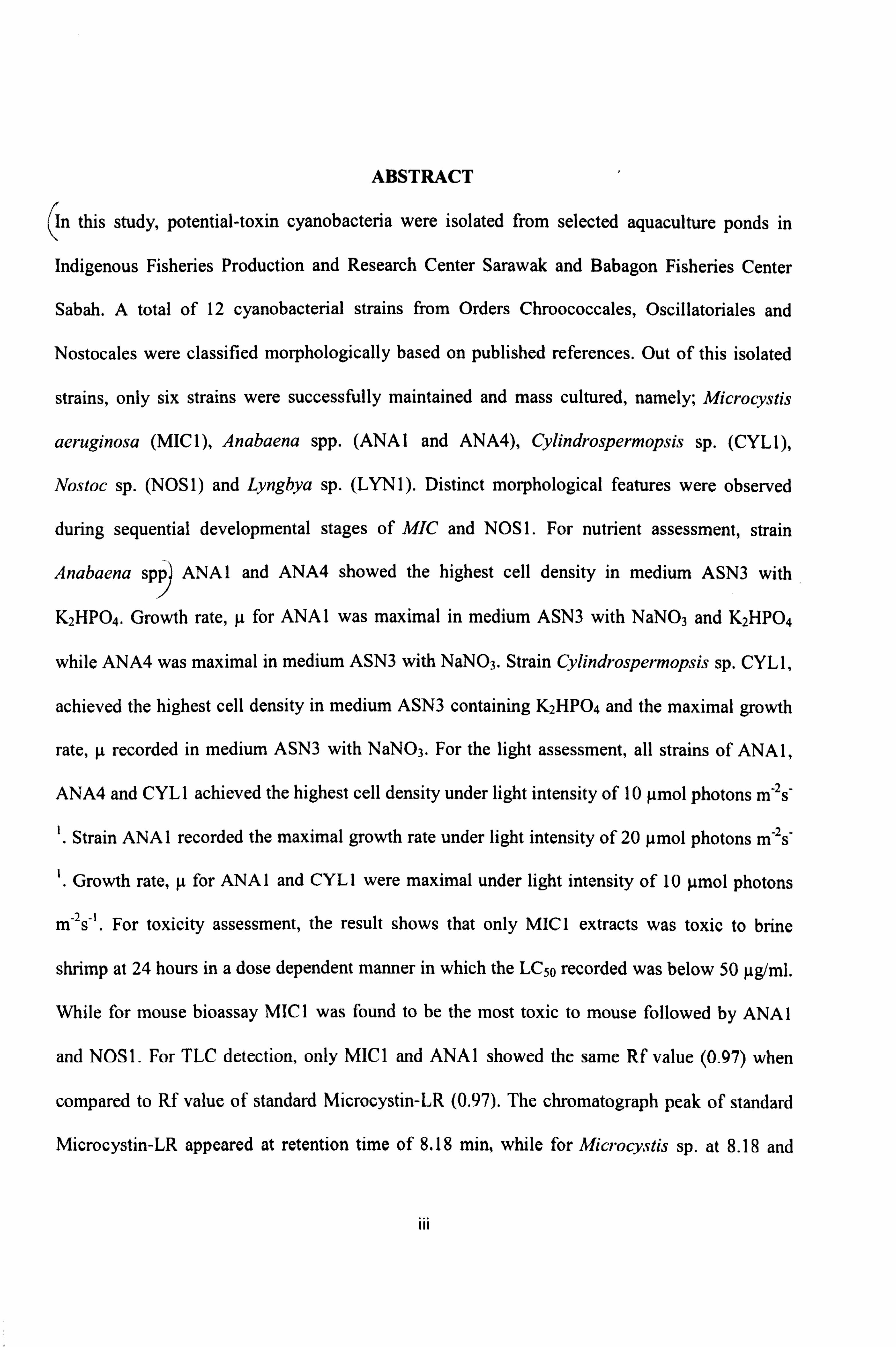
(In this study, potential-toxin cyanobacteria were isolated from selected aquaculture ponds in
Indigenous Fisheries Production and Research Center Sarawak and Babagon Fisheries Center
Sabah. A total of 12 cyanobacterial strains from Orders Chroococcales, Oscillatoriales and
Nostocales were classified morphologically based on published references. Out of this isolated
strains, only six strains were successfully maintained and mass cultured, namely; Microcystis
aeruginosa (MIC 1), Anabaena spp. (ANA I and ANA4), Cylindrospermopsis sp. (CYL 1),
Nostoc sp. (NOS 1) and Lyngbya sp. (LYN 1). Distinct morphological features were observed
during sequential developmental stages of MIC and NOS 1. For nutrient assessment, strain
Anabaena spp) ANA1 and ANA4 showed the highest cell density in medium ASN3 with
K2HPO4. Growth rate, µ for ANA I was maximal in medium ASN3 with NaNO3 and K2HPO4
while ANA4 was maximal in medium ASN3 with NaNO3. Strain Cylindrospermopsis sp. CYL1,
achieved the highest cell density in medium ASN3 containing K2HPO4 and the maximal growth
rate, g recorded in medium ASN3 with NaNO3. For the light assessment, all strains of ANA1,
ANA4 and CYL 1 achieved the highest cell density under light intensity of 10 gmol photons m-2s-
1. Strain ANA1 recorded the maximal growth rate under light intensity of 20 µmol photons m-2s"
. Growth rate, µ for ANA 1 and CYL 1 were maximal under light intensity of 10 gmol photons
m 2s-1. For toxicity assessment, the result shows that only MIC1 extracts was toxic to brine
shrimp at 24 hours in a dose dependent manner in which the LC50 recorded was below 50 µg/ml.
While for mouse bioassay MIC 1 was found to be the most toxic to mouse followed by ANA 1
and NOS 1. For TLC detection, only MIC 1 and ANA I showed the same Rf value (0.97) when
compared to Rf value of standard Microcystin-LR (0.97). The chromatograph peak of standard
Microcystin-LR appeared at retention time of 8.18 min, while for Microcystis sp. at 8.18 and
III

Anabaena sp. at 8.19 min, respectively. The results indicate that MC-LR toxin was found in
Microcystis sp. MIC 1 and Anabaena sp. ANA 1.
Keywords: Cyanobacteria, cultured, brine shrimp, mouse assay, TLC, HPLC
iv
MORFOLOGI, KETOKSIKAN DAN SIFAT TOKSIN BAGI SPESIES
SIANOBAKTERIA YANG DIPENCILKAN DARI KOLAM AKUAKULTUR DI
SARAWAK DAN SABAH
ABSTRAK
Dalam kajian ini, sianobakteria yang berpotensi menghasilkan toksin telah dipencilkan dari
kolam akuakultur terpilih di Pusat Penyelidikan dan Pengeluaran Ikan Tempatan Darat
(Bahagian Perikanan), Sarawak dan Pusat Perikanan Babagon, Sabah. Sebanyak 12 jenis
sianobakteria dari Order Chroococcales, Oscillatoriales dan Nostocales telah diklasifikasikan
mengikut morfologi berdasarkan kekunci dan rujukan. Hanya enam strain berjaya dikultur iaitu:
Microcystis aeru iýnosa (MICl), Anabaena spp. (ANA] dan ANA4), Cylindrospermopsis sp.
(CYL1), Nostoc sp. (NOSI) dan LynQbya sp. (LYN1). Terdapat beberapa beberapa ciri
perbezaan morfologi telah dikenalpasti sepanjang pertumbuhan MICl dan NOS]. Untuk
penilaian nutrien, strain Anabaena spp. ANA] dan ANA4 menunjukkan sel kepadatan tertinggi
dalam medium ASN3 dengan K2HPO4. Kadar pertumbuhan, µ untuk ANA] maksima dalam
medium ASN3 mengandungi NaNO3 dan K2HPO4 manakala ANA4 maksima dalam medium
ASN3 mengandtmgi NaNO3. Strain Cylindrospermopsis sp. CYLI, mencapai sel kepadatan
paling tinggi dalam ASN3 mengandungi K2HPO4 dan kadar pertumbuhan yang maksimal, it
direkodkan dalam medium ASN3 mengandungi NaNO3. Untuk penilaian cahaya, semua strain
ANA1, ANA4 dun CYL1 mencapai sel densiti paling tinggi di bawah keamatan cahaya 10 foton
Nmol m"2s"1. Strain ANA1 mencatatkan kadar pertumbuhan maksima di bawah keamatan cahaya
20 foton µmol m-2s-1. Kadar pertumbuhan, µ untuk ANAI dan CYLI maksima di bawah keamatan
V
cahaya 10 foton l, unol m 2s 1. Penilaian ketoksikan menunjukkan hanya ekstrak MICI adalah
toksik dalam ujian bioesei anak udang dengan catatan LCso di bawah paras 50 µg/ml. Manakala
bagi ujian bioesei tikus, MICI menunjukkan kesan ketoksikan yang paling tinggi diikuti ANA I.
Untuk pengesanan KLT, hanya MICI dan ANA1 menunjukkan nilai Rfyang sama (0.97) dengan
perbandingan kepada nilai Rfpiawai Mikrosistin-LR (0.97). Manakala bagi pengesanan KCPT,
puncak kromatograf piawai Mikrosistin-LR muncul pada minit ke 8.18 manakala bagi
Microcystis sp. dan Anabaena sp. masing-masing pada minit ke 8.18 dan 8.19. Keputusan kajian
menunjukkan bahawa toksin MC-LR telah dikesan pada Microcystis sp. MICI dan Anabaena sp.
ANA].
Kata kunci: Sianobakteria, kultur, anak udang, bioesei tikus, KLT, KCPT
vi
Pusat Khidmat Maklumat Akademik UNIVERSITi MALAYSIA SARAWAK
TABLE OF CONTENT PAGE
i DECLARATION .................................................................................... ACKNOWLEDGEMENT .........................................................................
ii
ABSTRACT .......................................................................................... AB STRAK .......................................................................................... .
vii TABLE OF CONTENT ............................................................................ xi LIST OF TABLES ..................................................................................
LIST OF FIGURES ................................................................................. xii LIST OF ABBREVIATIONS ..................................................................... xvi
1.0 INTRODUCTION
1.1 General Introduction ............................................................... 1
1.2 Cyanobacteria Characteristic ...................................................... 5
1.3 Habitat of Cyanobacteria .......................................................... 6
1.4 Bloom of Cyanobacteria ........................................................... 7
1.5 Cyanobacteria and Public Health Concern ...................................... 9
1.6 Toxin Producer Cyanobacteria ................................................... 10
1.6.1 Cyclic Peptide ............................................................ 12
1.6.1.1 Microcystin ...................................................... 13
1.6.1.2 Nodularin ........................................................ 13 14 1.6.2 Alkaloids ..................................................................
1.6.2.1 Anatoxin ......................................................... 14
1.6.2.2 Saxitoxin ......................................................... 14
1.6.2.3 Cylindrospermopsin ............................................ 15
1.6.3 Lipopolysaccharides..................................................... 1.7 Toxicological assessment of cyanobacterial toxin .............................
16
1.8 Objectives and scope ............................................................... 17
1.9 Thesis Organization ................................................................ 18
2.0 OCCURRENCE AND MORPHOLOGY ASSESSMENT OF POTENTIAL TOXIN
PRODUCER CYANOBACTERIA OF SELECTED AQUACULTURE PONDS
2.1 INTRODUCTION .................................................................. 19 2.2 MATERIALS AND METHODS .................................................
21 2.2.1 Fieldwork .................................................................. 21 2.2.2 Labwork ....................................................................
23 2.2.2.1 Scanning Electron Microscopy ............................... 23
VII
LIST OF TABLE
Table No. TITLE Page
Table 1.1 General features of the cyanotoxins, adapted from WHO (1998). 12
Table 1.2 Hepatotoxins produced by different cyanobacterial genera 13
Table 2.1 Cyanobacterial strain in present study and their origin. 27
Table 3.1 Laboratory experiment on different cyanobacteria genera isolated from 54 different sources
Table 4.1 Separation of toxic fraction of Standard MC-LR, strain MIC 1 and ANA 1 101 by TLC.
XI
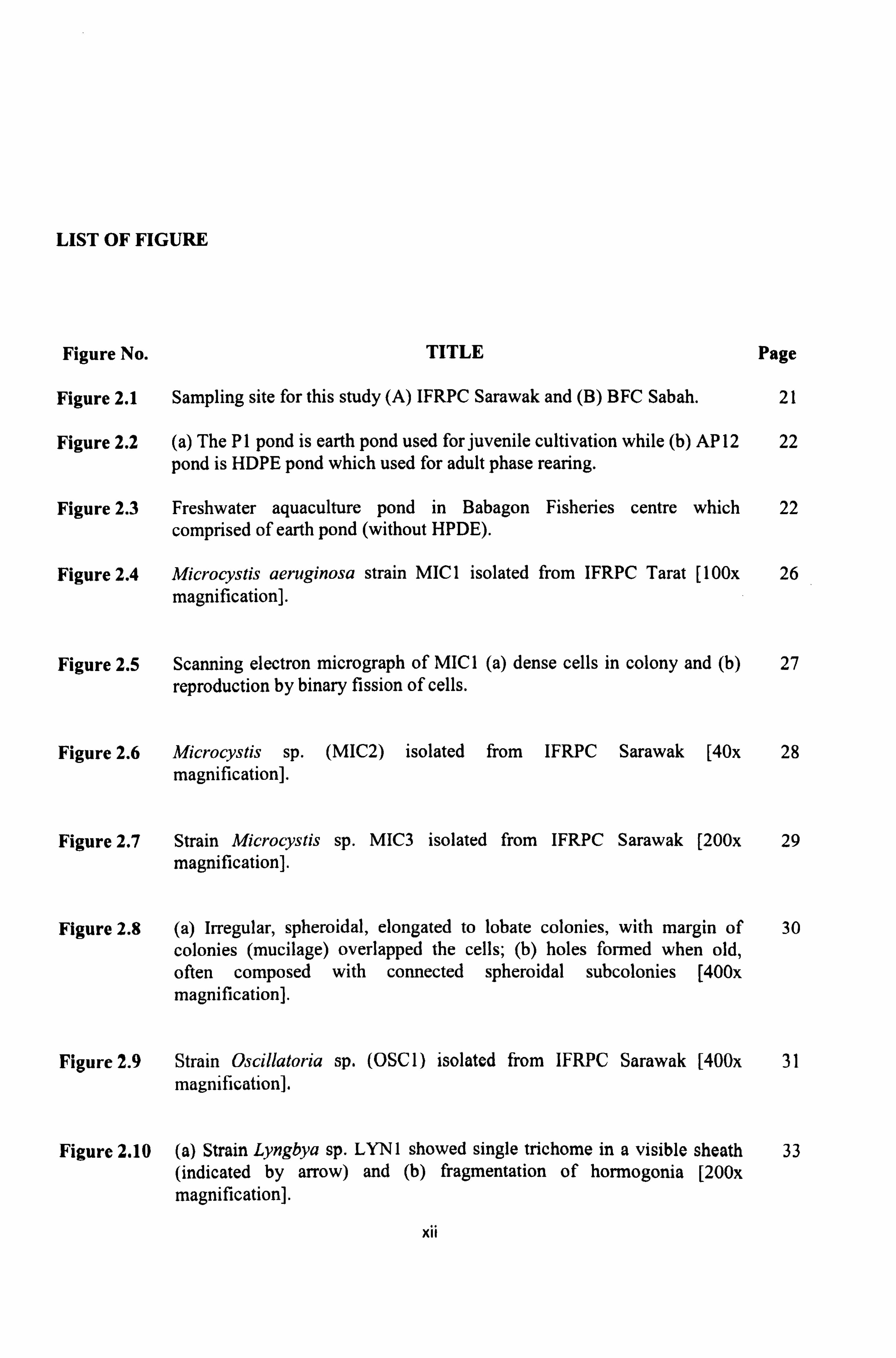
LIST OF FIGURE
Figure No. TITLE Page
Figure 2.1 Sampling site for this study (A) IFRPC Sarawak and (B) BFC Sabah. 21
Figure 2.2 (a) The P1 pond is earth pond used for juvenile cultivation while (b) AP 12 22 pond is HDPE pond which used for adult phase rearing.
Figure 2.3 Freshwater aquaculture pond in Babagon Fisheries centre which 22 comprised of earth pond (without HPDE).
Figure 2.4 Microcystis aeruginosa strain MIC 1 isolated from IFRPC Tarat [100x 26 magnification].
Figure 2.5 Scanning electron micrograph of MIC I (a) dense cells in colony and (b) 27 reproduction by binary fission of cells.
Figure 2.6 Microcystis sp. (MIC2) isolated from IFRPC Sarawak [40x 28 magnification].
Figure 2.7 Strain Microcystis sp. MIC3 isolated from IFRPC Sarawak [200x 29 magnification].
Figure 2.8 (a) Irregular, spheroidal, elongated to lobate colonies, with margin of 30 colonies (mucilage) overlapped the cells; (b) holes formed when old, often composed with connected spheroidal subcolonies [400x magnification].
Figure 2.9 Strain Oscillatoria sp. (OSC 1) isolated from IFRPC Sarawak [400x 31 magnification].
Figure 2.10 (a) Strain Lyngbya sp. LYN 1 showed single trichome in a visible sheath 33 (indicated by arrow) and (b) fragmentation of hormogonia [200x magnification].
XII
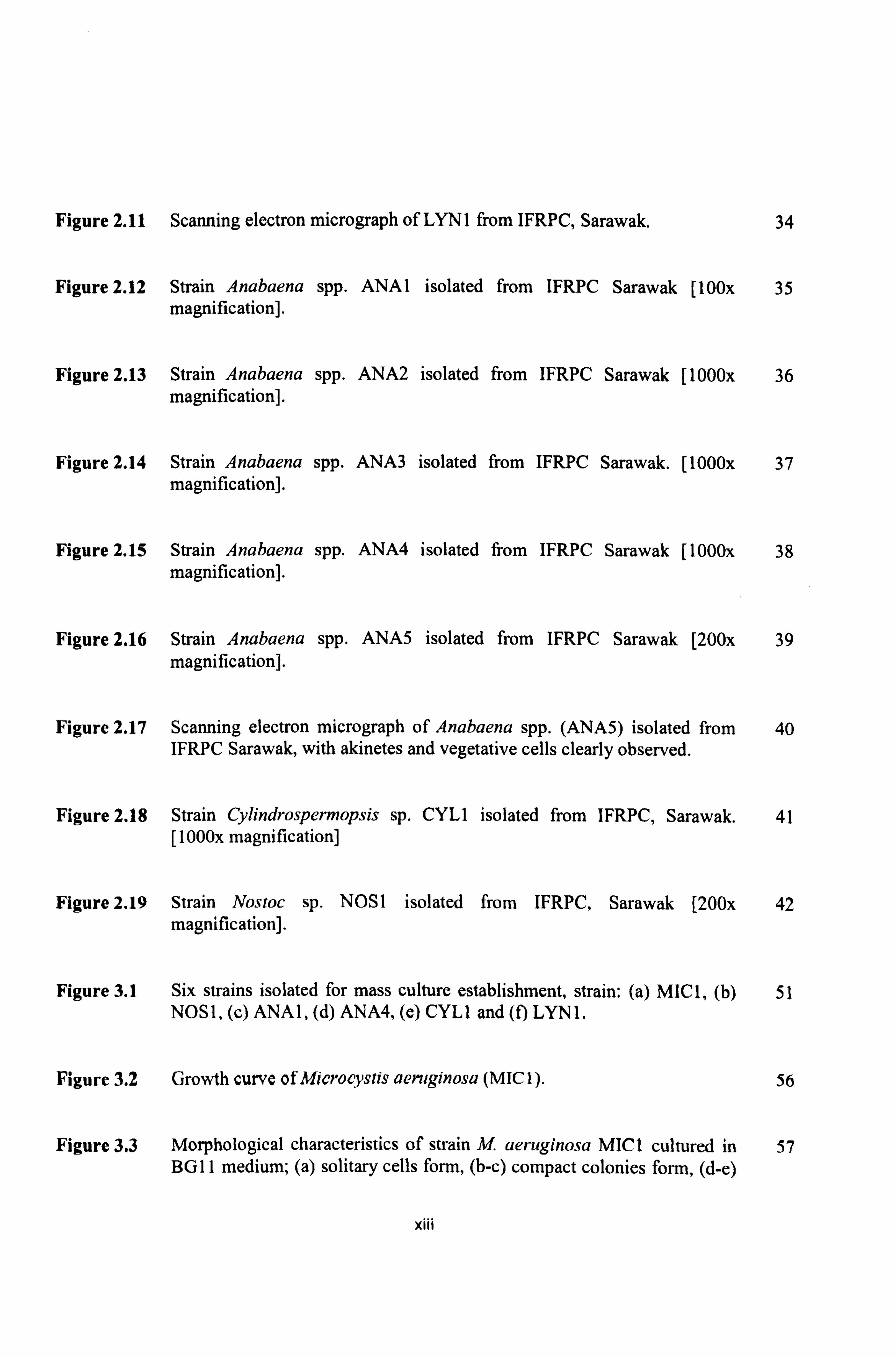
Figure 2.11 Scanning electron micrograph of LYN 1 from IFRPC, Sarawak. 34
Figure 2.12 Strain Anabaena spp. ANA1 isolated from IFRPC Sarawak [100x 35 magnification].
Figure 2.13 Strain Anabaena spp. ANA2 isolated from IFRPC Sarawak [1000x 36 magnification].
Figure 2.14 Strain Anabaena spp. ANA3 isolated from IFRPC Sarawak. [1000x 37 magnification].
Figure 2.15 Strain Anabaena spp. ANA4 isolated from IFRPC Sarawak [1000x 38 magnification].
Figure 2.16 Strain Anabaena spp. ANA5 isolated from IFRPC Sarawak [200x 39 magnification].
Figure 2.17 Scanning electron micrograph of Anabaena spp. (ANA5) isolated from 40 IFRPC Sarawak, with akinetes and vegetative cells clearly observed.
Figure 2.18 Strain Cylindrospermopsis sp. CYL1 isolated from IFRPC, Sarawak. 41 [I 000x magnification]
Figure 2.19 Strain Nostoc sp. NOS 1 isolated from IFRPC, Sarawak [200x 42 magnification].
Figure 3.1 Six strains isolated for mass culture establishment, strain: (a) MIC1, (b) 51 NOS1, (c) ANA1, (d) ANA4, (e) CYL1 and (f) LYN1.
Figure 3.2 Growth curve of Microcystis aeruginosa (MIC 1). 56
Figure 3.3 Morphological characteristics of strain M. aeruginosa MIC 1 cultured in 57 BG 11 medium; (a) solitary cells form, (b-c) compact colonies form, (d-e)
XIII
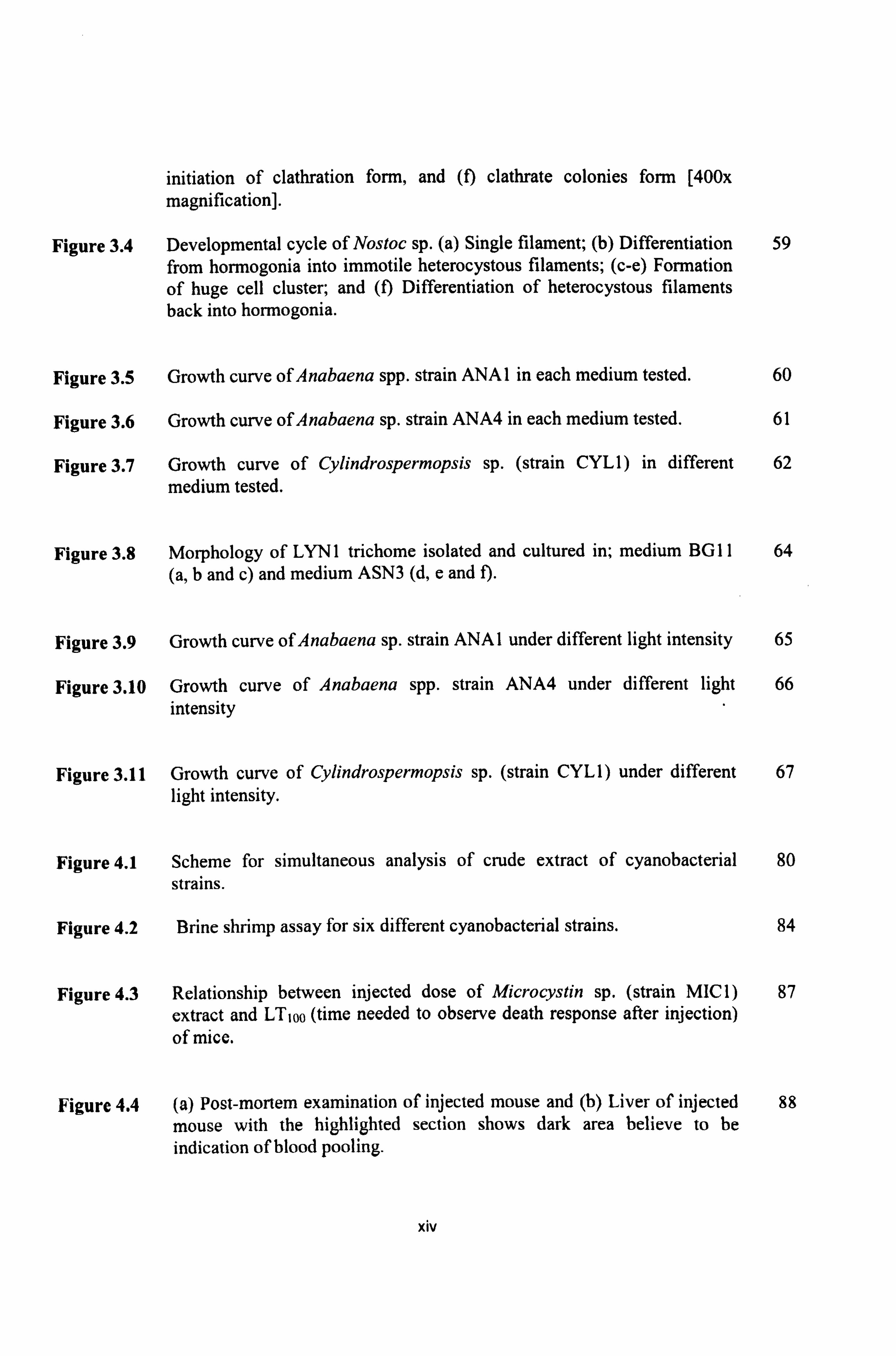
initiation of clathration form, and (f) clathrate colonies form [400x
magnification].
Figure 3.4 Developmental cycle of Nostoc sp. (a) Single filament; (b) Differentiation 59 from hormogonia into immotile heterocystous filaments; (c-e) Formation of huge cell cluster; and (f) Differentiation of heterocystous filaments back into hormogonia.
Figure 3.5 Growth curve of Anabaena spp. strain ANA 1 in each medium tested. 60
Figure 3.6 Growth curve of Anabaena sp. strain ANA4 in each medium tested. 61
Figure 3.7 Growth curve of Cylindrospermopsis sp. (strain CYL1) in different 62 medium tested.
Figure 3.8 Morphology of LYN 1 trichome isolated and cultured in; medium BG 11 64 (a, b and c) and medium ASN3 (d, e and f).
Figure 3.9 Growth curve of Anabaena sp. strain ANA 1 under different light intensity 65
Figure 3.10 Growth curve of Anabaena spp. strain ANA4 under different light 66 intensity
Figure 3.11 Growth curve of Cylindrospermopsis sp. (strain CYLI) under different 67 light intensity.
Figure 4.1 Scheme for simultaneous analysis of crude extract of cyanobacterial 80
strains.
Figure 4.2 Brine shrimp assay for six different cyanobacterial strains. 84
Figure 4.3 Relationship between injected dose of Microcystin sp. (strain MIC I) 87 extract and LT1oo (time needed to observe death response after injection)
of mice.
Figure 4.4 (a) Post-mortem examination of injected mouse and (b) Liver of injected 88 mouse with the highlighted section shows dark area believe to be indication of blood pooling.
XIV
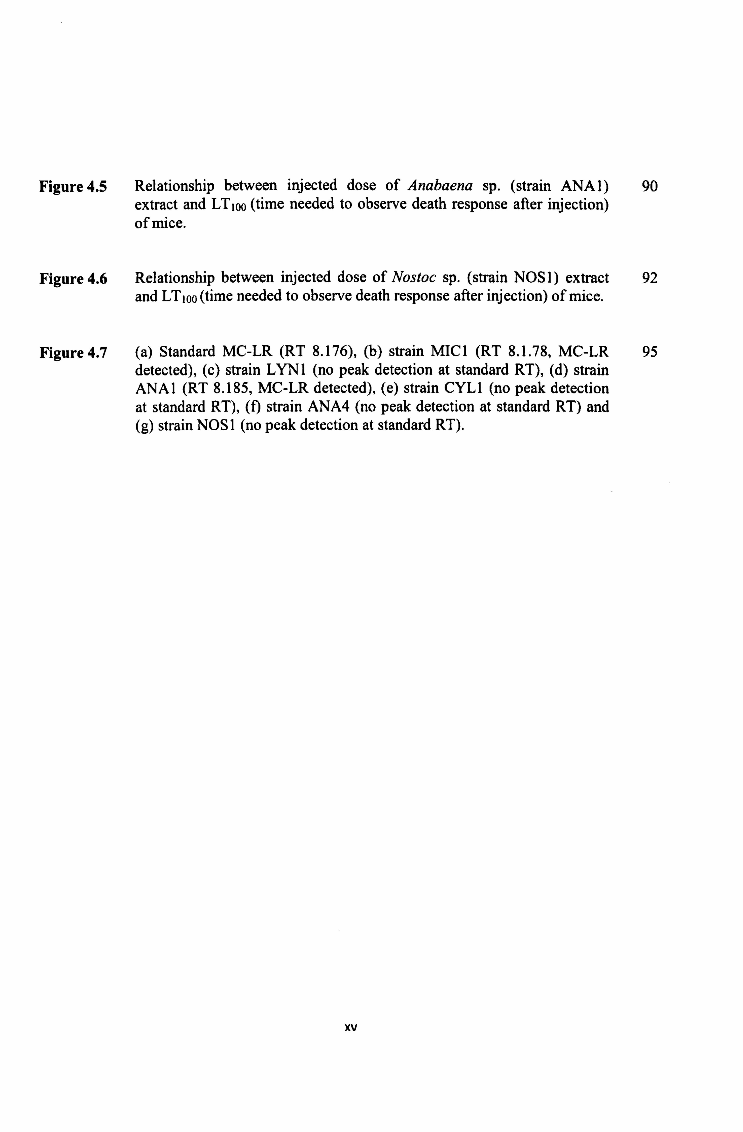
Figure 4.5 Relationship between injected dose of Anabaena sp. (strain ANA l) 90 extract and LTioo (time needed to observe death response after injection) of mice.
Figure 4.6 Relationship between injected dose of Nostoc sp. (strain NOS 1) extract 92 and LT, 00 (time needed to observe death response after injection) of mice.
Figure 4.7 (a) Standard MC-LR (RT 8.176), (b) strain MIC 1 (RT 8.1.78, MC-LR 95 detected), (c) strain LYN 1 (no peak detection at standard RT), (d) strain ANA I (RT 8.185, MC-LR detected), (e) strain CYL 1 (no peak detection at standard RT), (I) strain ANA4 (no peak detection at standard RT) and (g) strain NOS 1 (no peak detection at standard RT).
xv
LIST OF ABBREVIATION
ANA I
ANA2
ANA3
ANA4
ANA5
BFC
CYL 1
HPLC
IFRPC
LYN 1
MIC 1
MIC2
MIC3
NOS I
Rf
RT
TLC
Anabaena spp. strain I
Anabaena spp. strain 2
Anabaena spp. strain 3
Anabaena spp. strain 4
Anabaena spp. strain 5
Babagon Fisheries Centre
Cylindrospermopsis sp. strain 1
High Performance Liquid Chromatography
Indigenous Fisheries Research and Production Center
Lyngbya sp. strain 1
Microcystis sp. strain MIC 1
Microcystis sp. strain 2
Microcystis sp. strain 3
Nostoc sp. strain 1
Retention factor
Retention Time
Thin Layer Chromatography
xvi
CHAPTER I
INTRODUCTION
1.1 Gen eral Introduction
Blue green algae, scientifically known as Cyanobacteria are prokaryotic algae that have
existed for over three billion years (Sze, 1993). It belongs to Kingdom Monera, Class
Cyanophycea, which divided into four Orders namely Chroococcales, Nostocales,
Oscillatoriales and Stigonematales (Euzeby, 2004; Skulberg 1993). Cyanobacteria, which
formerly classified as algae, were later considered to be affiliated to bacteria, owing to their
biology and absence of a differentiated nucleus (Reyssac and Pletikosic, 1990). They have a
relatively simple prokaryotic structure and lack membrane-bound organelles, which are
structurally and physiologically, like other gram-negative bacteria although they conduct
photosynthesis like plants in aquatic systems (Rodgers, 2008).
Cyanobacteria are widely distributed and known as common phytoplankton found in
freshwaters (i. e. lakes, ponds, rivers and reservoirs) and brackishwaters (i. e. seas, estuaries and
lakes) waters (Metcalf and Codd, 2004; Castro et al., 2004). The ability to widespread and
colonize in different environment are due to their special characteristics including cell
buoyancy, nitrogen fixing akinetes and unfavourable condition resistant heterocysts (Sze.
1993; Oberholsteret al., 2004). In addition, the combined effects of environmental factors such
as temperature fluctuations, water movement, grazing and overloading of nutrients, which also
known as eutrophication contributes to their proliferation in freshwater environment (WHO,
1998).
1
Excessive growth of cyanobacteria, known as bloom, which is characterized by the
sudden appearance of large number of cells, represents a serious threat to aquatic ecosystem
(Kroggman et al., 1986). In aquaculture system, cyanobacteria can out-compete algae for
nutrients, thrive with low dissolved oxygen, and photosynthesize more efficiently at low light
levels and produce bloom that contributes to unfavourable condition (Rodgers, 2008).
Aquaculture ponds could be experience cyanobacteria bloom as they provide favorable
condition due to excessive fertilizer (Jewel et al., 2003). The increase demand of protein
source has shifted to intensive aquaculture activities, in which growth of cultured species is
supported by the natural productivity towards intensively operated production system
(Prommana et al., 2006). The intensive aquaculture activity often requires high stocking
densities and supplemental feed to achieve high productivity per unit volume, commonly
resulting in eutrophic to hypereutrophic conditions (Tucker, 1996).
Eutrophication and cyanobacterial bloomin freshwater ecosystem have become a
worldwide problem which can become serious when bloom-forming species release potent
water soluble toxins (WHO, 1998; Falconer, 1993, Metcalf and Codd, 2004, Vasconcelos,
2001). Till date, cyanobacteria known to produce many bioactive compounds that are
structurally and biochemically diverse which are classified as odorous metabolites and
bioactive metabolites (Smith et al., 2008). Furthermore, Rodgers (2008) revealed that algal
toxins can cause problems in the freshwater aquaculture organism by three different conditions
namely as off-flavor, indirect toxicity through changes in water quality and direct toxicity.
Aquaculture species are susceptible to odorous and bioactive secondary metabolites through
the ingestion of cyanobacteria, consumption of contaminated food items and absorption of
dissolved compound from the water column (Smith et al., 2008). Odorous compounds such as
2
geosmin (GSM) and 2-methylisoborneol (2MIB) are metabolites produced by certain species
of cyanobacteria (Phoslock, 2008).
Off-flavour is the most economically significant problems encountered in freshwater
aquaculture which are mainly caused by the absorption of odorous compounds from the water
(Smith et al., 2008; Phoslock, 2008). A number of commercially important species have been
affected by this problem including Nile tilapia, Oreochromis niloticus (Yamparoon and
Noomhorn, 2000), shrimp (Whitfield et al., 1988), Atlantic salmon, Salmo salar (Fanner et
al., 1995) and rainbow trout, Salmo gairdneri (From and Horlyck, 1984).
Moreover, aquaculture species also are vulnerable to bioactive metabolites that can
cause mortality, initiate or promote tumors, or deteriorate the health of cultivated species or
their prey species by affecting feeding, growth, or immune defense (Smith et al., 2008).
Indeed, certain species of cyanobacteria were reported to give hazardous effect on fishes
(Reysacc and Pletikosic, 1990) and massive mortality particularly among carp due to
cyanobacterial intoxicationwas recorded (Prescott, 1948). The cause of mortality was
postulated due to toxic cellular materialsfrom cyanobacteria blooms which been released into
the water during the cell lysis process (Jewel et al., 2003).
Yet, the aquatic organisms (e. g. fish, molluscs, crustacean) may accumulate
cyanobacterial metabolites (Smith et al., 2008) via ingestion of cyanobacteria cells in
contaminate food (Funari and Testai, 2008). Some aquatic organism such as molluscs (Prepas
et al., 1997), crustaceans (Prommana et al., 2006) and fishes (Jewel, 2003; Liqiang et al.,
2005) were identified as a vector and able to transfer the cyanotoxins (e. g. microcystions)
along the food chain that may risk the human.
In Thailand, toxic cyanobacterial blooms found in many water bodies of the regions,
including drinking water sources and fisheries activity area. Prommana et al. (2006) reported
3
that cyanobacteriaspecies (i. e Microcystis sp) and it toxins in prawn cultivation ponds possibly
pose a hazard to aquatic organisms and to humans through food webs. It proven as the
laboratory experiments indicated that prawn hepatopancreas, heart and brain are primary
organs for hepatotoxin bioaccumulation (Prommana et al., 2006).
Likewise, molluscs also reported as potential vectors for cyanotoxins (Funari and
Testai, 2008). High concentration of toxin was found in the visceral mass of some
individual's clams due to increase exposure to selective bioconcentration of
Microcystis(Prepas et al., 1997). In addition, Oberholster et al. (2006) reported
histopathological investigations of toxic cyanobacterial blooms in Sheldon Lake showed that
rainbow trout death due to cyanobacterial blooms, which indicated by damage of gills and
fins. Gill damage, probably caused by the high pH induced by cyanobacterial photosynthesis
activity prior to the bloom collapse, together with the higher level of ammonia arising from the
decomposition of the cyanobacteria (Sivonen and Jones, 1999).
In tropical countries, very few studies have been done on cyanobacteria in comparison
to temperate countries (Gires et al., 2002), even though it poses a high impact to human and
aquatic environment. Specifically in Sarawak, only three published reports on cyanobacteria
studies were conducted in selected freshwater ecosystem that mainly in Kuching District
(Ramlah, 2005; Mardhiah, 2006; Nazriq, 2007). For diversity study done by Ramlah (2005),
nine cyanobacterial genera namely Anabaena, Anacystis, Calothrix, Chamaesiphonales,
Gloeotrichia, Lyngbya, Microcystis, Oscillatoria, and Spirulinawere identified in which some
are classified as toxigenic cyanobacteria based on Skulberg et al. (1993). Mardhiah (2006) and
Nazriq (2007) have successfully isolated and established a pure culture of potentially toxin
producing cyanobacteria namely Microcystis sp. which was collected from aquaculture pond
oflndigenous Fisheries Research and Production Centre (IFRPC), Tarat, Serian, Sarawak.
4
Pusat Khidmat Maklumat Akademik UNIVERSITI MALAYSIA SARAWAK
Consistently, several cyanobacteria monitoring were conducted on respective pond as
part to monitor the bloom occurrence and to identify the reason of fish mortality (pers comm,
2010) in the IFRPC aquaculture pond. Hence, this study were conducted to identify potential
toxic cyanobacteria and it's toxin properties that might be a turning point to help the centre for
better aquaculture ponds management. In brief, IFRPC are consisted of two different types of
freshwater aquaculture ponds which are; earth pond and earth pond that layered with black
HDPE ( High Density Polyethylene). Fishes such as Tor species and catfish are cultured in the
ponds size which are varied from 60 x 60m2 to 120 x 60m2 using natural water system from
the nearest water source. Besides of that, Babagon Fisheries Centre (BFC), which is located in
Penampang Sabah also been selected as study sites, since it is facing a common problem as in
Tarat. BFC had practised basic water system using freshwater source from the highland at the
nearest area and the cultured fishes are common carp and tilapia in various size of earth pond.
1.2 Cyanobacteria characteristics
Cyanobacteria are photosynthetic prokaryotic algae (Oberholster et al., 2006) which belong to
Division Cyanophyta and contain chlorophyll a as their major pigment (Funari and Testai,
2008). They contained accessory pigments such as phycobiliprotein, which comprises of
phycoerythrin, phycocyanin, allophycocyanin and phycoerythrocyanin (Sze, 1993; Briand et
al., 2003). Cyanobacteria reproduce asexually by binary fission, spore production, or
fragmentation, forming singular cells, colonies and filaments (Mur et al� 1999). They are
divided into non-filamentous and filamentous type, with the filamentous species further
subdivided by the presence or absence of specialized cells called heterocyst and akinetes (Sze,
1993).
5
Vegetative cells are the normal photosynthetic cells that are formed under favorable
growing conditions. During stress condition, including anoxic state, heterocysts will act as
converter and fix nitrogen from the air into ammonia (NH3), nitrites (NO2-) or nitrates (NO3")
form, which can be absorbed by plants (Ahern, 2002). In heterocysts, the respiratory pathways
and part of the photosynthesis system function to provide energy in the form of ATP and
reduced compounds to accomplish nitrogen fixation. Meanwhile, akinetes are the climate-
resistant spores that may form under the inappropriate environmental conditions. Thick-walled
akinetes will form at the end of a period of growth and survive in a dormant state until
conditions are again favourable to grow (Sze, 1993).
Blue-green algae are advantageous over other algae because of their ability to control
buoyancy to access areas of increased nutrients and light (Phoslock, 2008). Many species of
cyanobacteria possess numerous gas vacuoles (Oberholster et al., 2006). These are
cytoplasmic inclusions that enable buoyancy regulation and are gas-filled, cylindrical
structures (Mur et al., 1999). Gas vesicles are important device to ensure positive buoyancy so
that they float on the surface and act to regulate the position of cells in the water column (Sze,
1993). Moreover, Mur et al. (1999) stated that cyanobacteria use different environmental
stimuli such as photic gravitational, chemical, thermal to optimize their position, and thus to
find a suitable niche for survival and growth. Therefore, they can tolerate adverse conditions
such as the complete drying of a pond or the cold winter temperatures (Vincent et al., 1993).
1.3 Habitat of cyanobacteria
The ability of cyanobacteria to survive under extreme of environmental stress such as
desiccation, temperature fluctuation, high light intensities and oligotrophic low nutrient
condition make them an ubiquitous organism that can be found virtually in all ecosystem
6
habitats on earth. Habitats occupied range from freshwater lakes and rivers, through to the
oceans, including hot springs, and deserts, ranging from the hottest to the cold dry valleys of
Antarctica (Hitzfeld et al., 2000; Acreman, 1994; Vincent et al., 1993).
Cyanobacteria are found nearly everywhere, occurring in typical aquatic and terrestrial
habitats as well as in such extreme sites as hot springs with temperatures as high as 71 °C and
crevices of desert rocks (Büdel, 1999) and bare soil (Mur et al., 1999). For example,
Synechoccus that can tolerate temperatures up to 74°C while other species of cyanobacteria
such as Phormidium and Lyngbya are found in Antartic lakes (Sze, 1993). Cyanobacteria also
tolerate high salt concentrations, as they may occur in tidepools and lakes when evaporation
concentrates salts. The filamentous cyanobacterium Spirulina commonly occurs in lakes with
a high soda content and high pH (Sze, 1993). Cyanobacteria have a number of special
properties which determine their relative importance in phytoplankton communities. They
tend to favor neutral or basic conditions and are less common at low pH (Sze, 1993). The
optimum temperature for toxin production in cyanobacteria is between 20°C and 25°C
(Watanabe and Oishi, 1985). These optimum temperatures are higher than for green algae and
diatom which cause most cyanobacteria in temperate region bloom during summer (Funari and
Testai, 2008).
1.4 Bloom of cyanobacterin
Cyanobacteria are a common form of algae which are often referred to as "pond scum"
(Acreman, 1994) and form large colonies which indicate by greenish appearance of scum,
foam or mats on the surface of freshwater lakes and ponds (Epperson, 2002). Generally the
blooms are most obvious when the cyanobacterium is one containing gas vacuoles that allow it
to concentrate at the surface of the water to maximize light absorption for photosynthesis
7
(Muret al., 1999). Cyanobacterial blooms are often been relate with the eutrophication and
enrichment of waters with nutrients (Metcalf and Codd, 2004) especially nitrogen and
phosphorus (Sze, 1993).
Experimental data have indicated that the affinity of many cyanobacteria for nitrogen
or phosphorus is higher than for many other photosynthetic organisms (Funari and Testai,
2008). Nitrogen is an essential element for plant growth, required for the synthesis of amino
acids, proteins, nucleotides, nucleic acids, coenzymes, chlorophyll and other photosynthetic
pigments (Bartram et al., 1999; Falconer, 2005 and Sze, 1993). Most cyanobacteria are
nitrogen fixers, converting atmospheric nitrogen to ammonia via the enzyme nitrogenase,
which aid to proliferation in low-nitrogen condition (Phoslock, 2008). Besides nitrogen, Ahern
(2002) included phosphate as the main part in regulating metabolism and ultimately the
growth of cyanobacteria. Bartram et a!. (1999) stated that concentration of phosphorus less
than 0.1 mgl-l is sufficient enough to induce a cyanobacterial bloom.
The phenomena of cyanobacterial bloom thus shifted many lakes from having
phytoplankton communities dominated by diatoms to having phytoplankton communities
dominates by cyanobacteria (Campbell, 2002). Bloom formation often results hazard effect to
the environment. When cyanobacterial blooms occur, light penetration are limited by scum
formation on the water surface, reducing the growth of other benthic producer organism such
as epiphyton, benthic algae and rooted vascular plants (Havens, 2007). Besides that,
decomposition of dead algal bloom may lead to the depletion of dissolved oxygen in the water,
resulting secondary problems such as fish mortality (Bartram et al., 1999). Moreover, during
intense blooms, photosynthetic activity depletes free CO2 from lake water and cause the
elevation of pH which may harmful to certain species of fish (Havens, 2007).
8
Although not all cyanobacteria blooms are toxic, some blooms can produce significant
quantities of natural toxins (Kaebernick and Neilan, 2000). When highly active natural
biotoxins produced, these blue-green algae blooms are considered as "Harmful Algal Bloom
(HAB)" (Sivonen and Jones, 1999; Gireset al., 2002). The effects of algal blooms vary widely.
Some algae are toxic only at very high densities, while others can be toxic at very low
densities (Rodgers, 2008). These toxins can affect aquatic life, humans as well as terrestrial
animals. If the toxins are persistent, they may form a health risk via transfer and accumulation
in the food web (Lehtiniemiet al., 2002). The levels of toxin accumulation sufficient to pose a
risk will depend on levels of human consumption and the severity of toxic blooms in the area
where fish or shellfish are caught or collected (Kozlowskyet al., 2003).
1.5 Cyanobacteria and public health concern
Human may be exposed to cyanotoxins via several routes such as ingestion, inhalation,
intravenous (dialysis) and skin contact (Vasconcelos, 2001). One of most well known
occurrence episode is "Palm Island Mystery Disease" in Australia. In 1979, about 140
indigenous people, mostly children were hospitalized after drinking water where a dense
cyanobacterial bloom on a water supply occurred (Metcalf and Codd, 2004; WHO 1998). In
response to the growing concern about nonlethal acute and chronic effects of microcystins, the
World Health Organization has previously set a new provisional guideline value for
microcystin-LR of 1. Oµg/L drinking water (Hitzfeld et al., 2000).
When surface waters infested by cyanobacteria are used for hemodialysis, they can
represent a remarkable risk for patients; indeed, the paternal route of exposure considerably
increases the internal dose of toxins, directly entering the blood stream (Funari and Testai,
2008). The most remarkable episode of human health resulted from hemodialysis from
9