Embed Size (px)
Citation preview
Hirosaki Med.J. 66:28―37,2015
ORIGINAL ARTICLE
MUC5AC-NEGATIVE PHENOTYPE IS CORRELATED WITH POOR PATIENT PROGNOSIS OF PANCREAS HEAD DUCTAL CARCINOMA.
Shingo Sakuraba1,2),Satoko Morohashi1),Tadashi Yoshizawa1),Shinji Tsutsumi2), Norihisa Kimura2),Daisuke Kudo2),Keinosuke Ishido2),Yoshikazu Toyoki2),
Kenichi Hakamada2), and Hiroshi Kijima1)
Abstract Both pancreas head ductal carcinoma (PHDC) and distal bile duct carcinoma (DBDC) are located within the pancreas head/intra-pancreatic bile duct region, and are the most aggressive malignancies with poor patient prognosis. In the present study, we demonstrated clinicopathological features and patients prognosis of PHDC/DBDC. We examined total 87 surgically resected cases of PHDC (40 cases) and DBDC (47 cases). PHDC showed frequent neural invasion (85.0%) and lymph node metastasis (77.5%), compared with DBDC (57.4% and 40.4% respectively), resulting in the poorer prognosis (P=0.0219) than DBDC. In addition, PHDC expressed MUC2 (10.0%) and MUC6 (25.0%) less frequently, compared with DBDC (36.2% and 55.3%, respectively). MUC5AC-negative PHDC exhibited significantly poorer patient’s prognosis, compared with MUC5AC-positive PHDC (P=0.0111), MUC5AC-positive DBDC (P=0.000162), and MUC5AC-negative DBDC (P=0.00416). In conclusion, MUC5AC-negative PHDC showed significantly poor patient’s prognosis.
Hirosaki Med.J. 66:28―37,2015
Key words: mucin; MUC5AC; pancreas cancer; bile duct cancer; patient prognosis.
1) Department of Pathology and Bioscience, Hirosaki University Graduate School of Medicine
2) Department of Gastroenterological Surgery, Hirosaki University Graduate School of Medicine
Correspondence: H. KijimaReceived for publication, December 12, 2014Accepted for publication, December 25, 2014
Introduction
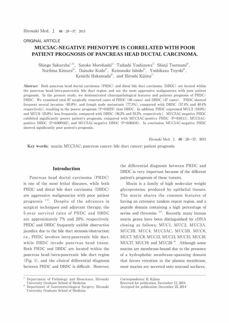
Pancreas head ductal carcinoma (PHDC) is one of the most lethal diseases, while both PHDC and distal bile duct carcinoma (DBDC) are aggressive malignancies with poor patient prognosis 1- 4). Despite of the advances in surgical techniques and adjuvant therapy, the 5 -year survival rates of PHDC and DBDC are approximately 7% and 20%, respectively. PHDC and DBDC frequently exhibit obstructive jaundice due to the bile duct stenosis/obstruction; i.e., PHDC involves intra-pancreatic bile duct, while DBDC invade pancreas head tissue. Both PHDC and DBDC are located within the pancreas head/intra-pancreatic bile duct region
(Fig. 1), and the clinical differential diagnosis between PHDC and DBDC is difficult. However,
the differential diagnosis between PHDC and DBDC is very important because of the different patient’s prognosis of these tumors. Mucin is a family of high molecular weight glycoproteins, produced by epithelial tissues. The mucin shares the common features of having an extensive tandem repeat region, and a peptide domain containing a high percentage of serine and threonine 5-7). Recently, many human mucin genes have been distinguished by cDNA cloning as follows: MUC1, MUC2, MUC3A, MUC3B, MUC4, MUC5AC, MUC5B, MUC6, MUC7, MUC8, MUC12, MUC13, MUC15, MUC16, MUC17, MUC19, and MUC20 8). Although some mucins are membrane-bound due to the presence of a hydrophobic membrane-spanning domain that favors retention in the plasma membrane, most mucins are secreted onto mucosal surfaces.
29MUC5AC-Negative Pancreas Cancer
Table 1 Antibodies for immunohistochemistry
Antigen Monoclonal/polyclonal Clone Dilution SourceMUC1 Monoclonal, mouse Ma695 1 : 50 NovocastraMUC2 Monoclonal, mouse Ccp58 1 : 50 Novocastra
MUC5AC Monoclonal, mouse CLH2 1 : 100 NovocastraMUC6 Monoclonal, mouse CLH5 1 : 100 Novocastra
depth of invasion (T-grade), histological type, lymphatic invasion, venous invasion, neural invasion, and lymph nodal metastasis. Degrees of lymphatic, venous and neural invasions were classified as follows: 0, no invasion; 1, mild invasion; 2, moderate invasion; and 3, severe invasion. These data were evaluated according to our previous study 2, 9) with reference to the World Health Organization classification 1), and staged according to the TMN classification of the International Union Against Cancer (UICC) 10). We also investigated mucin phenotypes of PHDC and DBDC using immunohistochemical procedure described as follows.
Immunohistochemistry For histological examination, PHDC/DBDC specimens were routinely fixed with formalin, embedded in paraffin, thin-sectioned. Four-μm-thick sections were mounted on saline-coated glass slides. Immunohistochemical examination was performed on deparaf f inized sections using the standard avidin-biotin-peroxidase complex method with automated immunostainer
(Benchmark XT; Ventana Medical System, Tucson, AZ, USA) according to our previous study 9, 11). We used MUC1, MUC2, MUC5AC and MUC6 to clarify mucin expression of PHDC and DBDC. The antibodies used are listed in Table 1.
Evaluation of immunohistochemistry Two investigators (SS, HK) simultaneously assessed the immunohistochemical results without any patient’s clinicopathological data. Luminal membranous immunoreactivities of the tumor were judged as positive for MUC1, and
It is necessary to make the pathological dif ferential diagnosis between PHDC and DBDC in order to perform effective therapeutic strategies for these malignant tumors. In this study, we analyzed the mucin phenotype and clinical outcome of 40 surgically-resected cases of PHDC, compared with 47 cases of DBDC.
Materials and MethodsPatients We investigated 87 surgically resected cases of PHDC and DBDC treated between January 2007 and December 2012, after obtaining each patient’s informed consent with to use their clinical records and pathology specimens at Hirosaki University Hospital. Survival data were obtained from hospital medical charts, and median observat ion period was 26 .4 months (87 cases). The series consisted of 47 men and 40 women with a median age of 66.7 years (range 31-83 years). Curative resection
(pancreaticoduodenectomy or pylorus-preserving pancreaticoduodenectomy or subtotal stomach-preserving pancreaticoduodenectomy) and regional lymph node dissection were performed. The present study followed the principles of the World Medical Association Declaration of Helsinki 1964.
Pathological analysis All surgically resected specimens were routinely fixed with 10% formalin, then embedded in paraffin, and stained with hematoxylin and eosin (H&E) for pathological evaluation. The following histological features were assessed:
30 S. Sakuraba, et al.
cytoplasmic immunoreactivities as positive for MUC2, MUC5AC and MUC6. According to the above immunohistochemical expression of each mucin, the cases were divided into two groups; a negative group in which < 10% of tumor cells were stained, and a positive group in which ≧ 10% were stained.
Statistical analysis Statistical comparisons between two groups were analyzed using both Chi square test and Fisher’s exact test. Survival curves were constructed using the Kaplan-Meier method and differences in survival were evaluated using log-rank test. The relative prognostic factors were analyzed with the Cox’s proportional hazards regression model. Differences were considered
Table 2 Clinicopathological factors and mucin expression of PHDC/DBDC
Variables PDAC DBDA P-ValueAge <65 19 17 0.285 ≧65 21 30Gender Male 16 31 0.0155*
Female 24 16Histological differentiation well, mod, pap 33 40 0.97 por, others 7 7Depth of invasion T1 1 6 0.118 T2, 3, 4 39 41Lymphatic invation ly0, 1 6 29 1 ly2, 3 34 18Venous invasion v0, 1 13 25 0.0525 v2, 3 27 22Neural invation ne0, 1 6 20 0.0103*
ne2, 3 34 27Lymph node metastasis pN(-) 9 28 0.00108*
pN(+) 31 19MUC expression MUC1 + 35 43 0.798 - 5 4 MUC2 + 4 17 0.00955*
- 36 30 MUC5AC + 19 30 0.126 - 21 17 MUC6 + 10 26 0.00421*
- 30 2 PHDC, pancreas head ductal carcinoma; DBDC, distal bile duct carcinomawell, well-differentiated adenocarcinoma; mod, moderately differentiated adenocarcinoma; pap, papillary adenocarcinoma; por, poorly differentiated adenocarcinoma. *Statistically significant: P<0.05.
31MUC5AC-Negative Pancreas Cancer
to be significant if the p-value was less than 0.05. All statistical evaluations were performed using R (http://www.r-project.org), and PASW statistics software (version 18.0; SPSS, Inc., Chicago, IL, USA).
ResultsClinicopathological factors and mucin expression of PHDC/DBDC Clinicopathological factors and mucin expres-
sion of PHDC/DBDC are summarized in Tables 2 and 3. Both of PHDC and DBDC were mainly composed differentiated adenocarcinoma, i.e., well-differentiated, moderately differentiated or papillary adenocarcinoma (Fig. 1). The majorities of PHDC/DBDC were pT2 - pT4 advanced cancers, i.e., PHDC more than 2 cm in size, and DBDC beyond the bile duct wall. On the other hand, PHDC significantly showed frequent neural invasion (ne2,3: 34/40, 85.0%) and lymph node metastasis (pN(+): 31/40, 77.5%),
Table 3 Clinicopathogical factors and MUC5AC expression of PHDC/DBDC
Variables MUC5AC(-) MUC5AC(+) P-ValueAge <65 18 18 0.317 ≧65 20 31Gender Male 22 25 0.523 Female 16 24Histological differentiation well, mod, pap 30 43 0.415 por, Other 8 6Depth of invasion T1 2 5 0.461 T2, 3, 4 36 44Lymphatic invation ly0, 1 10 25 0.0197* ly2, 3 28 24Venous invasion v0, 1 13 25 0.116 v2, 3 25 24Neural invation ne0, 1 10 16 0.521 ne2, 3 28 33Lymph node metastasis pN(-) 12 25 0.0688 pN(+) 26 24MUC expression MUC1 + 34 44 1 - 4 5 MUC2 + 1 20 1 - 37 29 MUC6 + 5 31 1 - 33 18 PHDC, pancreas head ductal carcinoma; DBDC, distal bile duct carcinomawell, well-differentiated adenocarcinoma; mod, moderately differentiated adenocarcinoma; pap, papillary adenocarcinoma; por, poorly differentiated adenocarcinoma. *Statistically significant: P<0.05.
32 S. Sakuraba, et al.
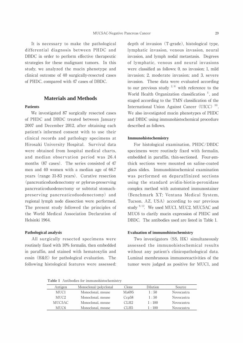
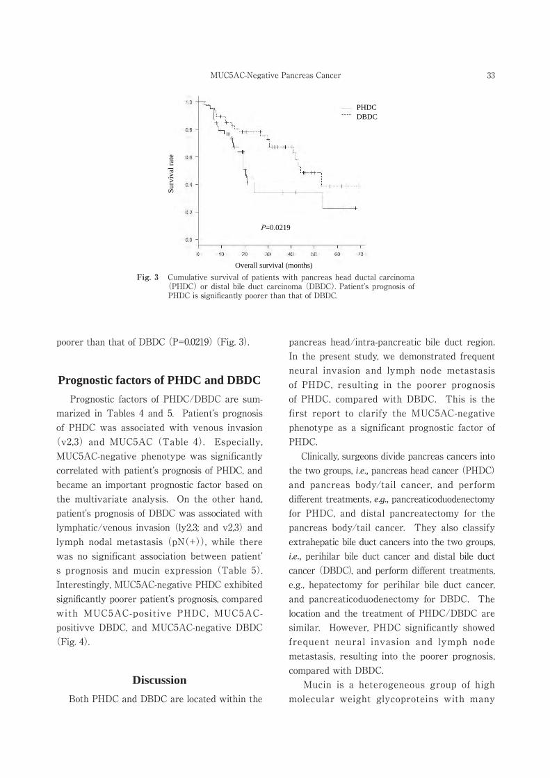
compared with DBDC (ne2,3: 27/47, 57.4%; and pN(+): 19/47, 40.4%). PHDC expressed MUC2 (4/40, 10.0%) and MUC6 (10/40, 25.0%) less frequently, compared with DBDC (MUC2:
17/47, 36.2%; and MUC6: 26/47, 55.3%) (Fig. 2). MUC5AC expression was inversely correlated with lymphatic invasion (P=0.0197) (Table 3). Patient’s prognosis of PHDC was significantly
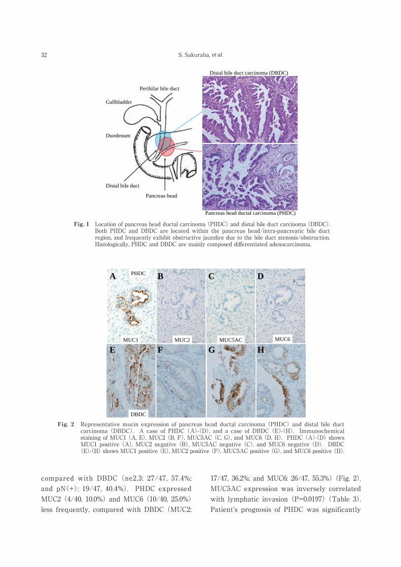
Fig. 1 Location of pancreas head ductal carcinoma (PHDC) and distal bile duct carcinoma (DBDC). Both PHDC and DBDC are located within the pancreas head/intra-pancreatic bile duct region, and frequently exhibit obstructive jaundice due to the bile duct stenosis/obstruction. Histologically, PHDC and DBDC are mainly composed differentiated adenocarcinoma.
Gallbladder
Perihilar bile duct
Distal bile duct
Pancreas head
Duodenum
Distal bile duct carcinoma (DBDC)
Pancreas head ductal carcinoma (PHDC)
Fig. 2 Representative mucin expression of pancreas head ductal carcinoma (PHDC) and distal bile duct carcinoma (DBDC). A case of PHDC (A)-(D), and a case of DBDC (E)-(H). Immunochemical staining of MUC1 (A, E), MUC2 (B, F), MUC5AC (C, G), and MUC6 (D, H). PHDC (A)-(D) shows MUC1 positive (A), MUC2 negative (B), MUC5AC negative (C), and MUC6 negative (D). DBDC
(E)-(H) shows MUC1 positive (E), MUC2 positive (F), MUC5AC positive (G), and MUC6 positive (H).
A B C D
E F G HMUC1 MUC2 MUC5AC MUC6
PHDC
DBDC
33MUC5AC-Negative Pancreas Cancer
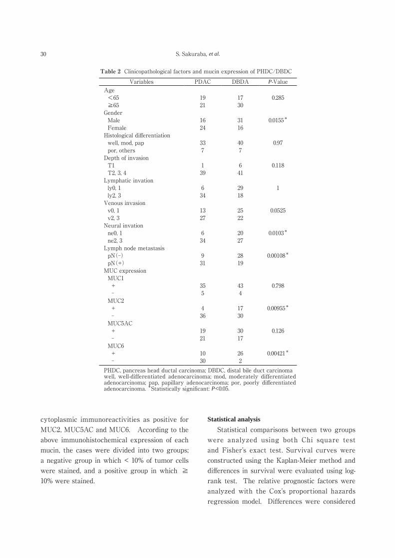
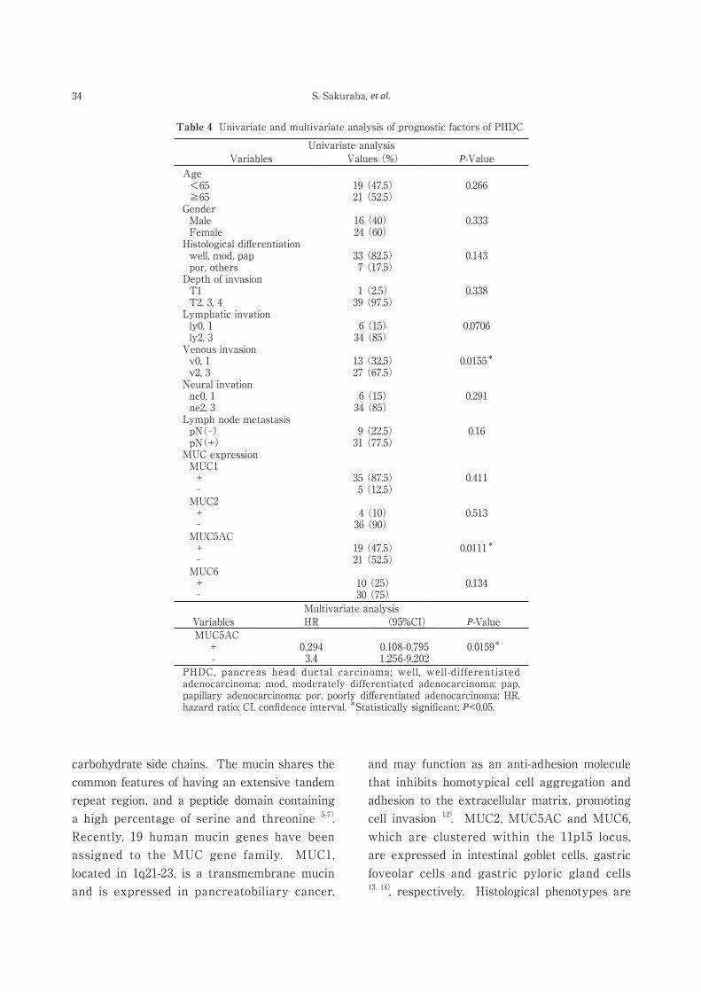
Fig. 3 Cumulative survival of patients with pancreas head ductal carcinoma (PHDC) or distal bile duct carcinoma (DBDC). Patient’s prognosis of PHDC is significantly poorer than that of DBDC.
P=0.0219
Surv
ival
rate
Overall survival (months)
DBDCPHDC
poorer than that of DBDC (P=0.0219) (Fig. 3).
Prognostic factors of PHDC and DBDC Prognostic factors of PHDC/DBDC are sum-marized in Tables 4 and 5. Patient’s prognosis of PHDC was associated with venous invasion
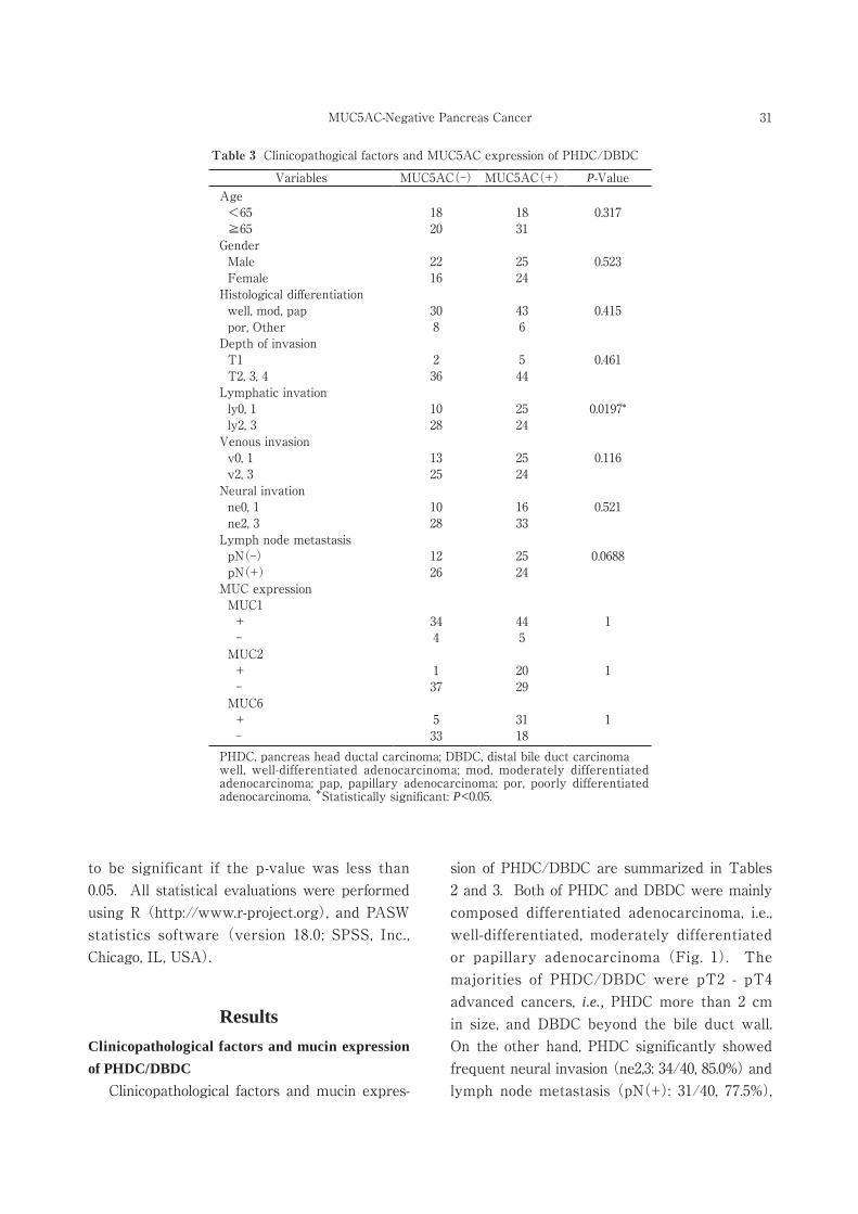
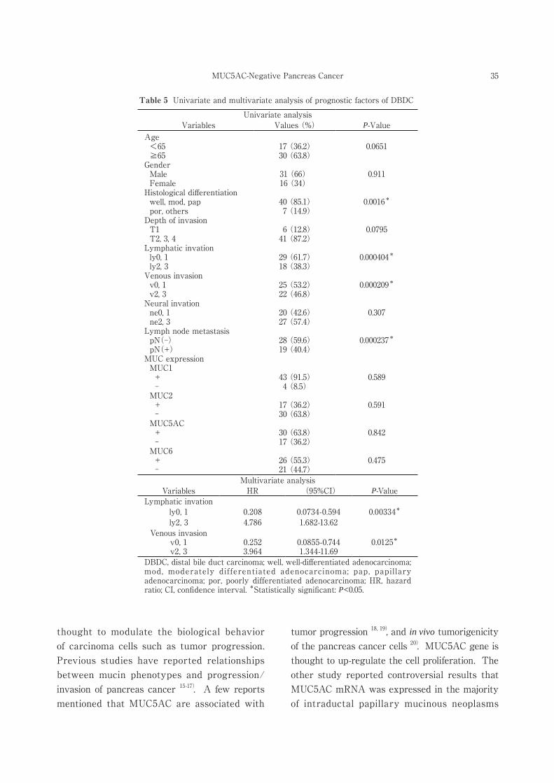
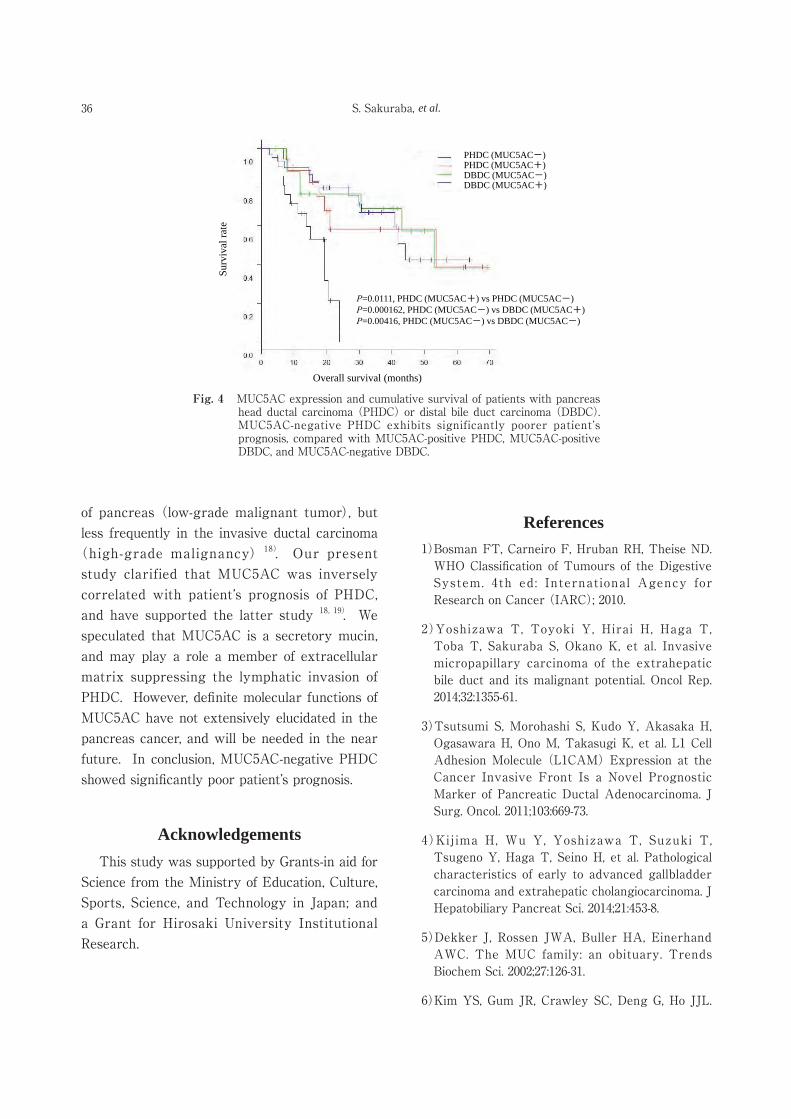
(v2,3) and MUC5AC (Table 4). Especially, MUC5AC-negative phenotype was significantly correlated with patient’s prognosis of PHDC, and became an important prognostic factor based on the multivariate analysis. On the other hand, patient’s prognosis of DBDC was associated with lymphatic/venous invasion (ly2,3; and v2,3) and lymph nodal metastasis (pN(+)), while there was no significant association between patient’s prognosis and mucin expression (Table 5). Interestingly, MUC5AC-negative PHDC exhibited significantly poorer patient’s prognosis, compared with MUC5AC-positive PHDC, MUC5AC-positivve DBDC, and MUC5AC-negative DBDC
(Fig. 4).
Discussion Both PHDC and DBDC are located within the
pancreas head/intra-pancreatic bile duct region. In the present study, we demonstrated frequent neural invasion and lymph node metastasis of PHDC, resulting in the poorer prognosis of PHDC, compared with DBDC. This is the first report to clarify the MUC5AC-negative phenotype as a significant prognostic factor of PHDC. Clinically, surgeons divide pancreas cancers into the two groups, i.e., pancreas head cancer (PHDC) and pancreas body/tail cancer, and perform different treatments, e.g., pancreaticoduodenectomy for PHDC, and distal pancreatectomy for the pancreas body/tail cancer. They also classify extrahepatic bile duct cancers into the two groups, i.e., perihilar bile duct cancer and distal bile duct cancer (DBDC), and perform different treatments, e.g., hepatectomy for perihilar bile duct cancer, and pancreaticoduodenectomy for DBDC. The location and the treatment of PHDC/DBDC are similar. However, PHDC significantly showed frequent neural invasion and lymph node metastasis, resulting into the poorer prognosis, compared with DBDC. Mucin is a heterogeneous group of high molecular weight glycoproteins with many
34 S. Sakuraba, et al.
carbohydrate side chains. The mucin shares the common features of having an extensive tandem repeat region, and a peptide domain containing a high percentage of serine and threonine 5-7). Recently, 19 human mucin genes have been assigned to the MUC gene family. MUC1, located in 1q21-23, is a transmembrane mucin and is expressed in pancreatobiliary cancer,
and may function as an anti-adhesion molecule that inhibits homotypical cell aggregation and adhesion to the extracellular matrix, promoting cell invasion 12). MUC2, MUC5AC and MUC6, which are clustered within the 11p15 locus, are expressed in intestinal goblet cells, gastric foveolar cells and gastric pyloric gland cells 13, 14), respectively. Histological phenotypes are
Table 4 Univariate and multivariate analysis of prognostic factors of PHDC
Univariate analysisVariables Values (%) P-Value
Age <65 19 (47.5) 0.266 ≧65 21 (52.5)Gender Male 16 (40) 0.333 Female 24 (60)Histological differentiation well, mod, pap 33 (82.5) 0.143 por, others 7 (17.5)Depth of invasion T1 1 (2.5) 0.338 T2, 3, 4 39 (97.5)Lymphatic invation ly0, 1 6 (15) 0.0706 ly2, 3 34 (85)Venous invasion v0, 1 13 (32.5) 0.0155*
v2, 3 27 (67.5)Neural invation ne0, 1 6 (15) 0.291 ne2, 3 34 (85)Lymph node metastasis pN(-) 9 (22.5) 0.16 pN(+) 31 (77.5)MUC expression MUC1 + 35 (87.5) 0.411 - 5 (12.5) MUC2 + 4 (10) 0.513 - 36 (90) MUC5AC + 19 (47.5) 0.0111*
- 21 (52.5) MUC6 + 10 (25) 0.134 - 30 (75)
Multivariate analysisVariables HR (95%CI) P-Value
MUC5AC+ 0.294 0.108-0.795 0.0159*
- 3.4 1.256-9.202 PHDC, pancreas head ductal carcinoma; well, well-differentiated adenocarcinoma; mod, moderately differentiated adenocarcinoma; pap, papillary adenocarcinoma; por, poorly differentiated adenocarcinoma; HR, hazard ratio; CI, confidence interval. *Statistically significant: P<0.05.
35MUC5AC-Negative Pancreas Cancer
thought to modulate the biological behavior of carcinoma cells such as tumor progression. Previous studies have reported relationships between mucin phenotypes and progression/invasion of pancreas cancer 15-17). A few reports mentioned that MUC5AC are associated with
tumor progression 18, 19), and in vivo tumorigenicity of the pancreas cancer cells 20). MUC5AC gene is thought to up-regulate the cell proliferation. The other study reported controversial results that MUC5AC mRNA was expressed in the majority of intraductal papillary mucinous neoplasms
Table 5 Univariate and multivariate analysis of prognostic factors of DBDC
Univariate analysisVariables Values (%) P-Value
Age <65 17 (36.2) 0.0651 ≧65 30 (63.8)Gender Male 31 (66) 0.911 Female 16 (34)Histological differentiation well, mod, pap 40 (85.1) 0.0016*
por, others 7 (14.9)Depth of invasion T1 6 (12.8) 0.0795 T2, 3, 4 41 (87.2)Lymphatic invation ly0, 1 29 (61.7) 0.000404*
ly2, 3 18 (38.3)Venous invasion v0, 1 25 (53.2) 0.000209*
v2, 3 22 (46.8)Neural invation ne0, 1 20 (42.6) 0.307 ne2, 3 27 (57.4)Lymph node metastasis pN(-) 28 (59.6) 0.000237*
pN(+) 19 (40.4)MUC expression MUC1 + 43 (91.5) 0.589 - 4 (8.5) MUC2 + 17 (36.2) 0.591 - 30 (63.8) MUC5AC + 30 (63.8) 0.842 - 17 (36.2) MUC6 + 26 (55.3) 0.475 - 21 (44.7)
Multivariate analysisVariables HR (95%CI) P-Value
Lymphatic invationly0, 1 0.208 0.0734-0.594 0.00334*
ly2, 3 4.786 1.682-13.62Venous invasion
v0, 1 0.252 0.0855-0.744 0.0125*
v2, 3 3.964 1.344-11.69 DBDC, distal bile duct carcinoma; well, well-differentiated adenocarcinoma; mod, moderately differentiated adenocarcinoma; pap, papillary adenocarcinoma; por, poorly differentiated adenocarcinoma; HR, hazard ratio; CI, confidence interval. *Statistically significant: P<0.05.
36 S. Sakuraba, et al.
of pancreas (low-grade malignant tumor), but less frequently in the invasive ductal carcinoma
(high-grade malignancy) 18). Our present study clarified that MUC5AC was inversely correlated with patient’s prognosis of PHDC, and have supported the latter study 18, 19). We speculated that MUC5AC is a secretory mucin, and may play a role a member of extracellular matrix suppressing the lymphatic invasion of PHDC. However, definite molecular functions of MUC5AC have not extensively elucidated in the pancreas cancer, and will be needed in the near future. In conclusion, MUC5AC-negative PHDC showed significantly poor patient’s prognosis.
Acknowledgements This study was supported by Grants-in aid for Science from the Ministry of Education, Culture, Sports, Science, and Technology in Japan; and a Grant for Hirosaki University Institutional Research.
References1)Bosman FT, Carneiro F, Hruban RH, Theise ND.
WHO Classification of Tumours of the Digestive System. 4th ed: International Agency for Research on Cancer (IARC); 2010.
2)Yoshizawa T, Toyoki Y, Hirai H, Haga T, Toba T, Sakuraba S, Okano K, et al. Invasive micropapillary carcinoma of the extrahepatic bile duct and its malignant potential. Oncol Rep. 2014;32:1355-61.
3)Tsutsumi S, Morohashi S, Kudo Y, Akasaka H, Ogasawara H, Ono M, Takasugi K, et al. L1 Cell Adhesion Molecule (L1CAM) Expression at the Cancer Invasive Front Is a Novel Prognostic Marker of Pancreatic Ductal Adenocarcinoma. J Surg. Oncol. 2011;103:669-73.
4)Kijima H, Wu Y, Yoshizawa T, Suzuki T, Tsugeno Y, Haga T, Seino H, et al. Pathological characteristics of early to advanced gallbladder carcinoma and extrahepatic cholangiocarcinoma. J Hepatobiliary Pancreat Sci. 2014;21:453-8.
5)Dekker J, Rossen JWA, Buller HA, Einerhand AWC. The MUC family: an obituary. Trends Biochem Sci. 2002;27:126-31.
6)Kim YS, Gum JR, Crawley SC, Deng G, Ho JJL.
Fig. 4 MUC5AC expression and cumulative survival of patients with pancreas head ductal carcinoma (PHDC) or distal bile duct carcinoma (DBDC). MUC5AC-negative PHDC exhibits significantly poorer patient’s prognosis, compared with MUC5AC-positive PHDC, MUC5AC-positive DBDC, and MUC5AC-negative DBDC.
Surv
ival
rate
Overall survival (months)
PHDC (MUC5AC+)
DBDC (MUC5AC+)
PHDC (MUC5AC-)
DBDC (MUC5AC-)
P=0.0111, PHDC (MUC5AC+) vs PHDC (MUC5AC-)P=0.000162, PHDC (MUC5AC-) vs DBDC (MUC5AC+)P=0.00416, PHDC (MUC5AC-) vs DBDC (MUC5AC-)
37MUC5AC-Negative Pancreas Cancer
Mucin gene and antigen expression in biliopan-creatic carcinogenesis. Ann Oncol. 1999;10:51-5.
7)Moniaux N, Escande F, Porchet N, Aubert JP, Batra SK. Structural organization and classification of the human mucin genes. Front Biosci. 2001; 6:D1192-206.
8)Perez -Vi lar J , Hi l l RL . Mucin Fami ly o f Glycoproteins. In: LennarzWJ, Lane MD. editors. Encyclopedia of Biological Chemistry. Vol. 2. Elsevier Academic Press; 2004. p.758-64.
9)Toba T, Kijima H, Hakamada K, Igarashi Y. Histological phenotype is correlated with the wall-invasion pattern of gallbladder adenocarcinoma. Biomed Res. 2014;35:295-302.
10)Sobin LH, Gospodarowicz MK. Wittekind C. TNM classification of Malignant Tumours. 7th ed. Chichester: Wily-Blackwell; 2010.
11)Yonaiyama S, Toyoki Y, Morohashi S, Sakuraba S, Yoshizawa T, Suzuki T, Wu Y, et al. Epithelial cell adhesion molecule (EpCAM) overexpression is correlated with malignant potentials of intraductal papillary mucinous neoplasms (IPMNs) of the pancreas. Biomed Res. 2013;34:87-95.
12)Ligtenberg MJ, Buijs F, Vos HL, Hilkens J. Suppression of cellular aggregation by high levels of episialin. Cancer Res. 1992;52:2318-24.
13)Sasaki M, Yamato T, Nakanuma Y, Ho SB, Kim YS. Expression of MUC2, MUC5AC and MUC6 apomucins in carcinoma, dysplasia and non-dysplastic epithelia of the gallbladder. Pathol Int. 1999;49:38-44.
14)Yamato T, Sasaki M, Watanabe Y, Nakanuma Y. Expression of MUC1 and MUC2 mucin core
proteins and their messenger RNA in gall bladder carcinoma: an immunohistochemical and in situ hybridization study. J Pathol. 1999;188:30-7.
15)Ohuchida K, Mizumoto K, Yamada D, Fujii K, Ishikawa N, Konomi H, Nagai E, et al. Quantitative analysis of MUC1 and MUC5AC mRNA in pancreatic juice for preoperative diagnosis of pancreatic cancer. Int J Cancer. 2006;118:405-11.
16)Nagata K, Horinouchi M, Saitou M, Higashi M, Nomoto M, Goto M, Yonezawa S. Mucin expression profile in pancreatic cancer and the precursor lesions. J Hepatobiliary Pancreat Surg. 2007;14:243-54.
17)Sopha SC, Gopal P, Merchant NB, Revetta FL, Gold DV, Washington K, Shi CJ. Diagnostic and therapeutic implications of a novel immunohis-tochemical panel detecting duodenal mucosal invasion by pancreatic ductal adenocarcinoma. Int J Clin Exp Pathol. 2013;6:2476-86.
18)Yonezawa S, Horinouchi M, Osako M, Kubo M, Takao S, Arimura Y, Nagata K, et al. Gene expression of gastric type mucin (MUC5AC) in pancreatic tumors: Its relationship with the biological behavior of the tumor. Pathol Int. 1999; 49:45-54.
19)Park SY, Roh SJ, Kim YN, Kim SZ, Park HS, Jang KY, Chung MJ, et al. Expression of MUC1, MUC2, MUC5AC and MUC6 in cholangiocarcinoma: Prognostic impact. Oncol Rep. 2009;22:649-57.
20)Hoshi H, Sawada T, Uchida M, Saito H, Iijima H, Toda-Agetsuma M, Wada T, et al. Tumor-associated MUC5AC stimulates in vivo tumori-genicity of human pancreatic cancer. Int J Oncol. 2011;38:619-27.


![[DDBJing33] Japanese Genotype-phenotype Archive の紹介](https://img.pdfslide.tips/doc/110x75/5870c1d21a28ab0b4a8b7447/ddbjing33-japanese-genotype-phenotype-archive-.jpg)