Embed Size (px)
Citation preview
of June 9, 2018.This information is current as
-Defensin, HD5αHuman Multivalent Binding of Carbohydrates by the
Hans J. Gabius and Wuyuan LuRobert I. Lehrer, Grace Jung, Piotr Ruchala, Sabine Andre,
http://www.jimmunol.org/content/183/1/480doi: 10.4049/jimmunol.0900244
2009; 183:480-490; ;J Immunol
Referenceshttp://www.jimmunol.org/content/183/1/480.full#ref-list-1
, 27 of which you can access for free at: cites 85 articlesThis article
average*
4 weeks from acceptance to publicationFast Publication! •
Every submission reviewed by practicing scientistsNo Triage! •
from submission to initial decisionRapid Reviews! 30 days* •
Submit online. ?The JIWhy
Subscriptionhttp://jimmunol.org/subscription
is online at: The Journal of ImmunologyInformation about subscribing to
Permissionshttp://www.aai.org/About/Publications/JI/copyright.htmlSubmit copyright permission requests at:
Email Alertshttp://jimmunol.org/alertsReceive free email-alerts when new articles cite this article. Sign up at:
Print ISSN: 0022-1767 Online ISSN: 1550-6606. Immunologists, Inc. All rights reserved.Copyright © 2009 by The American Association of1451 Rockville Pike, Suite 650, Rockville, MD 20852The American Association of Immunologists, Inc.,
is published twice each month byThe Journal of Immunology
by guest on June 9, 2018http://w
ww
.jimm
unol.org/D
ownloaded from
by guest on June 9, 2018
http://ww
w.jim
munol.org/
Dow
nloaded from
Multivalent Binding of Carbohydrates by the Human�-Defensin, HD51
Robert I. Lehrer,2* Grace Jung,* Piotr Ruchala,* Sabine Andre,† Hans J. Gabius,†
and Wuyuan Lu‡
Four of the six human �-defensins (human neutrophil peptides 1–3 and human �-defensin 5; HD5) have a lectin-like ability to bindglycosylated proteins. Using HD5 as a model, we applied surface plasmon resonance techniques to gain insights into this property.HD5 bound natural glycoproteins > neoglycoproteins based on BSA > nonglycosylated BSA �� free sugars. The affinity of HD5for simple sugars covalently bound to BSA was orders of magnitude greater than its affinity for the same sugars in solution. Theaffinity of HD5 for protein-bound carbohydrates resulted from multivalent interactions which may also involve noncarbohydrateresidues of the proteins. HD5 showed concentration-dependent self-association that began at submicromolar concentrations andproceeded to dimer and tetramer formation at concentrations below 5 �M. The (R9A, R28A) and (R13A, R32A) analogs of HD5showed greatly reduced self-association as well as minimal binding to BSA and to BSA-affixed sugars. From this and otherevidence, we conclude that the extensive binding of HD5 to (neo)glycoproteins results from multivalent nonspecific interactions ofindividual HD5 molecules with carbohydrate and noncarbohydrate moieties of the target molecule and that the primary bindingevents are magnified and enhanced by subsequent in situ assembly and oligomerization of HD5. Self-association and multivalentbinding may play integral roles in the ability of HD5 to protect against infections caused by viruses and other infectiousagents. The Journal of Immunology, 2009, 183: 480–490.
D efensins are small, 2.0- to 4.5-kDa peptides that contrib-ute to host defense against microbial (1–3) and viral(4–7) infections by interacting with the pathogen di-
rectly, or in concert with other components of the immune system(8, 9). The three subfamilies of defensins (�, �, and �) found inmammals evolved from a common ancestral gene (10, 11) whose�-defensin progeny extend back to bony fishes (12), if not beyond(13). All of these defensin peptides contain six cysteines, havethree intramolecular disulfide bonds, and are cationic. To date,�-defensin peptides have been found only in mammals (14) and�-defensins have been identified only in certain nonhuman pri-mates (11, 15, 16). �-Defensins have a largely �-sheet structurecomposed of 29- to 35-aa residues. �-Defensins are cyclic andhave only 18 residues. Uniquely, their cyclic backbone is formedby posttranslational ligation of two precursor peptides (11, 17).
Given the involvement of defensins in antimicrobial and anti-viral host defense, it can be surmised that they possess an abilityto distinguish self from nonself targets. Their preferential bindingto microbial surfaces and membranes is attributable, in part, to thenet positive charge of defensins and the fact that microbial surfacesand membranes are typically enriched in anionic moieties such as
lipoteichoic acid, lipid A, and anionic phospholipids (18). De-fensins also bind to and/or inactivate enveloped viruses, the mem-branes of which are host cell derived. In such instances, carbohy-drate epitopes of glycan chains afford likely biorecognition sites(19, 20). The carbohydrate-binding ability of �-defensins has beendocumented and examined with respect to their antiviral properties(21, 22) and also because �-defensins are among the smallestknown lectins (23–25). Humans express six different �-defensins.Four were first discovered in neutrophils, and are called humanneutrophil peptides (HNP)3 1–4 (16, 26, 27). The other two human�-defensins (HD), HD5 and HD6, are expressed primarily bysmall intestinal Paneth cells (28) and in regions of the female uro-genital tract (26, 29). Murine Paneth cell �-defensins, also calledcryptdins, protect intestinal crypts from microbial incursions andinfluence the resident microbial flora of the small intestine (30).Human HD5 deficiency has been linked to the pathogenesis ofcertain diarrheal infections (31) and to that of ileal Crohn’s disease(32–34), although the latter assertions have been challenged (35).
In recent studies, HNPs 1–3 and HD5 proved to be potent an-tagonists of infection by human papillomaviruses, the primarycauses of cervical cancer (36). HD5 was particularly active againstsexually transmitted papillomaviruses, with IC50 values in the highnanogram per milliliter range. HD5 did not block virion binding orinternalization, instead acting by preventing the escape of virionsfrom their endocytic vesicles (36). HD5 inhibits adenovirus infec-tions by binding to the virus capsid and preventing its subsequent
*David Geffen School of Medicine at University of California at Los Angeles, LosAngeles, CA 90095; †Faculty of Veterinary Medicine, Department of Veterinary Sci-ences, Chair of Physiological Chemistry, Ludwig-Maximilians-University Munich,Munich, Germany; and ‡Institute of Human Virology and Department of Biochem-istry and Molecular Biology, University of Maryland School of Medicine, Baltimore,MD 21201
Received for publication January 26, 2009. Accepted for publication April 20, 2009.
The costs of publication of this article were defrayed in part by the payment of pagecharges. This article must therefore be hereby marked advertisement in accordancewith 18 U.S.C. Section 1734 solely to indicate this fact.1 Supported in part by Grant 5R01AI061482 from the National Institutes of Health (toW.L.) and by the research initiative LMUexcellent.2 Address correspondence and reprint requests to Dr. Robert I. Lehrer, The DavidGeffen School of Medicine at UCLA, Center for Health Sciences CHS 37-062, 10833Le Conte Avenue, Los Angeles, CA 90095-1690. E-mail address: [email protected]
3 Abbreviations used in this paper: HNP, human neutrophil peptide; BSA-�-D-Glc-NAc, a neoglycoprotein containing �-D-N-acetylglucosamine residues covalentlybound to BSA; CM, carboxymethylated; FO0.5, the peptide concentration that binds,on average, one-half of the sugar residues on a neoglycoprotein; gD1, glycoprotein Dof herpes simplex type 1; HD, human defensin; IC0.5, concentration of an inhibitorthat reduces binding of 1 �g/ml HD5 to the binding level shown by 0.5 �g/ml (in thisreport); m/z, mass divided by charge; MR, molar ratio; RU, response (or resonance)units; SPR, surface plasmon resonance.
Copyright © 2009 by The American Association of Immunologists, Inc. 0022-1767/09/$2.00
The Journal of Immunology
www.jimmunol.org/cgi/doi/10.4049/jimmunol.0900244
by guest on June 9, 2018http://w
ww
.jimm
unol.org/D
ownloaded from
uncoating, an event the virus requires to escape the endosomalcompartment and enter the cytosol (37). HD5 and HNP1 also in-hibit infection by BK polyoma virus, a cause of interstitial nephri-tis in immunosuppressed renal transplant recipients, evidently act-ing by binding to the virions and agglutinating them (38).
In this article a systematic approach is used to detect carbohy-drate binding with respect to sugar specificity and density ofepitope presentation. Toward this end, a panel of chemically gly-cosylated molecules was prepared, using BSA as carrier. Theseneoglycoproteins have been useful sensors for lectin sites in pre-vious studies with isolated proteins and different cell systems (39–43). Several natural glycoproteins were also tested to characterizethe carbohydrate-binding properties of HD5. Our detailed exami-nation of sugar binding by HD5 demonstrates that multivalentbinding and the self-association of the peptide play important rolesin this process. Although we did not study viruses directly, ourfindings may explain how HD5 can cross-link intact viruses tocause agglutination or elements of a viral capsid to prevent viraluncoating.
Materials and MethodsGlycoproteins
Recombinant glycosylated glycoprotein D (gD1) from HSV-1, McIntyrestrain) was purchased from Fitzgerald Industries. It contained aa 21–339 ofthe ectodomain and had an estimated nonglycosylated mass of 35,238 Da.HIV-1BAL gp120, immunoaffinity purified from infected HEK 293 cells,was obtained from the National Institutes of Health AIDS Research andReference Reagent Program, Division of AIDS, National Institute of Al-lergy and Infectious Diseases, National Institutes of Health. Its molecularmass was �116,000 by SDS-PAGE, and its nonglycosylated mass was54,157. HIV-1LAV gp120 (Protein Sciences) was a full-length glycosylatedenvelope protein produced in insect cells by a baculovirus system.
Neoglycoproteins
Chemically glycosylated BSA-based neoglycoproteins were prepared bytwo standard procedures. The neoglycoproteins in set 1 were made byreacting diazonium salts derived from p-aminophenyl glycosides obtainedby the catalytic reduction of corresponding p-nitrophenyl glycosides (44).The neoglycoproteins in set 2 were prepared by conjugating p-isothiocya-natophenyl glycosides made by treating p-aminophenyl glycosides withthiophosgene to the carbohydrate-free carrier protein (44). The reagentsused to carry out these procedures were obtained from Sigma-Aldrich.Mass analyses were done on a Voyager System-4299 MALDI-TOF instru-ment (Applied Biosystems), using a matrix of sinapinic acid. The meanextent of glycosylation was estimated by first subtracting the peak mass ofunmodified BSA (66, 650) from the peak mass of the neoglycoprotein andthen dividing this difference by the molecular mass of the carbohydrateplus that of the linker, which was 88 mass units for the neoglycoproteinsin group 1 and 104 for those in group 2. Diazonium-derivatized carbohy-drates will attach primarily to tyrosyl, histidyl, and lysyl residues, andisothiocyanato groups should attach almost exclusively to amino groups,especially those of lysine (44).
Surface plasmon resonance (SPR)
CM5 chips have four compartments (flow cells) with surfaces that consistof a carboxymethylated (CM) dextran matrix covalently attached to a thingold film. When working with neoglycoproteins, we reserved one CM5compartment to measure background binding to the surface matrix, at-tached carbohydrate-free BSA to another, and attached neoglycoproteins tothe remaining two. When working with natural glycoproteins, only theCM-dextran compartment was used as a background control. Proteins andpeptides were immobilized on the CM5 chips by amine coupling per themanufacturer’s instructions, using reagents (N-hydroxysuccinimide, 1-ethyl-3-(3-aminopropyl)carbodiimide hydrochloride, and ethanolamine hy-drochloride) purchased from Biacore. The analyte buffer, HBS-EP, con-tained 0.15 M NaCl, 3 mM EDTA, 0.005% (v/v) surfactant P20, and 0.01M HEPES (pH 7.4). Binding was monitored at 1-s intervals for 5 min withan analyte flow rate of 50 �l/min. Dissociation was monitored at 1-s in-tervals for 2–4 min. The sensor chips were regenerated by washing themwith 10 mM HCl, and (if needed) 10 mM NaOH. Data analysis was per-formed using BIAevaluation 4.1 software from BiaCore, and Sigma Plot 9and Sigma Stat 3.0 software from Systat.
Results of SPR experiments are expressed in resonance units (RU),which sense changes in the refractive index close to the metal surface of abiosensor. According to Biacore and other sources, 1 RU is equivalent to1 pg of protein per mm2 of sensor surface (45–47). Because the sensorsurface of a CM5 chip is close to 1 mm2, the target density (2000 RU), weselected for the neoglycoprotein-presenting biosensors corresponds to �2ng of BSA/mm2. In actuality, the many biosensors used in this study con-tained, on average, 1878.2 � 60.4 RU (mean � SEM; n � 29) and rangedfrom 1392 to 2617 RU, necessitating certain corrections that are describedin the text.
Compensating for background binding to BSA
We assumed that background binding of any given concentration of HD5to carbohydrate-free BSA immobilized on a CM5 biosensor was directlyproportional to the BSA density, in RU. Data supporting this assumption isprovided in the text. Accordingly, if BSA compartment of the biosensorhad a ligand density of 1600 RU and the neoglycoprotein compartment ofthe biosensor had a ligand density of 1950 RU, we multiplied the RUbound to the BSA compartment by 1.219 (1950/1600) before subtracting itfrom the RU bound to the neoglycoprotein compartment to obtain the netbinding attributable to the bound carbohydrate.
Molar ratio and fractional occupancy
Molar ratio (MR; Equation 1) signifies the number of molecules of HD5bound per molecule of neoglycoprotein. MRsa (Equation 2) signifies themolar ratio, corrected for the self-association of HD5. Fractional occu-pancy (FO) signifies the number of HD5 molecules, corrected for self-association, per attached carbohydrate molecule. In Equation 3, the self-association factor, s, corresponds to the molar ratio shown in Fig. 5.
MR � �ab�/�cd� (1)
MRsa � MR/(1 � s) (2)
FO � MRsa/Cmean (3)
In Equation 1, a � net (i.e., background-corrected) binding in RU at 300 s;b � mean neoglycoprotein mass; c � mass of the analyte (HD5 or HD5analog); d � ligand density (RU), of the neoglycoprotein immobilized onthe biosensor. In Equation 3, Cmean represents the mean number of carbo-hydrate residues on each neoglycoprotein, determined by MALDI-MSanalysis as described in the text. Finally, the FO0.5 indicates the concen-tration of HD5 responsible for the net binding of 0.5 HD5 molecules percarbohydrate molecule on the neoglycoprotein. The FO0.5 is determinedgraphically from Equation 3, as illustrated in the text below.
ResultsBinding of HD5 to natural glycoproteins
Fig. 1 shows the binding of HD5 to three glycoproteins: fetuin;gp120LAV/IIIB; and gD1 of HSV-1. At 1 �g/ml, HD5 bound eachglycoprotein extensively but showed little binding to nonglycosy-lated BSA. Increasing the HD5 concentration to 10 �g/ml did notovercome this differential. To estimate the affinity of HD5’s bind-ing, we used BIAevalution 4.1 software, assumed 1:1 binding, andtested six concentrations of HD5 from 50 to 300 ng/ml. In twoexperiments, done on different days, the mean Kd values � SDwere 14.4 � 4.9 and 15.7 � 14.0 nM for fetuin, 23.5 � 14.0 and26.0 � 12.9 nM for gD of HSV-1, and 24.5 � 10.8 and 23.0 �25.7 nM for gp120 from HIV-1LAV/IIIB.
We also evaluated HD5 binding by measuring the ability ofsolution phase glycoproteins to inhibit binding of 1 �g/ml HD5 toan appropriate biosensor. In Fig. 2a, the standard curve shows thebinding of HD5 to a CM5 biosensor containing immobilized fetuinin the absence of any competitor. The open circles show that add-ing different concentrations of fetuin to 1 �g/ml (279 nM) of HD5in the analyte solution caused a concentration-dependent inhibitionof HD5 binding to the biosensor.
Fetuin, an acidic (pI 3.3) protein with sialic acids in the bi- andtriantennary complex-type N-glycans and core 1 O-glycans, has amass of 48,400 Da (48). The horizontal and vertical reference linesshow that 9 �g/ml fetuin (186 nM) reduced binding of 1 �g/ml
481The Journal of Immunology
by guest on June 9, 2018http://w
ww
.jimm
unol.org/D
ownloaded from
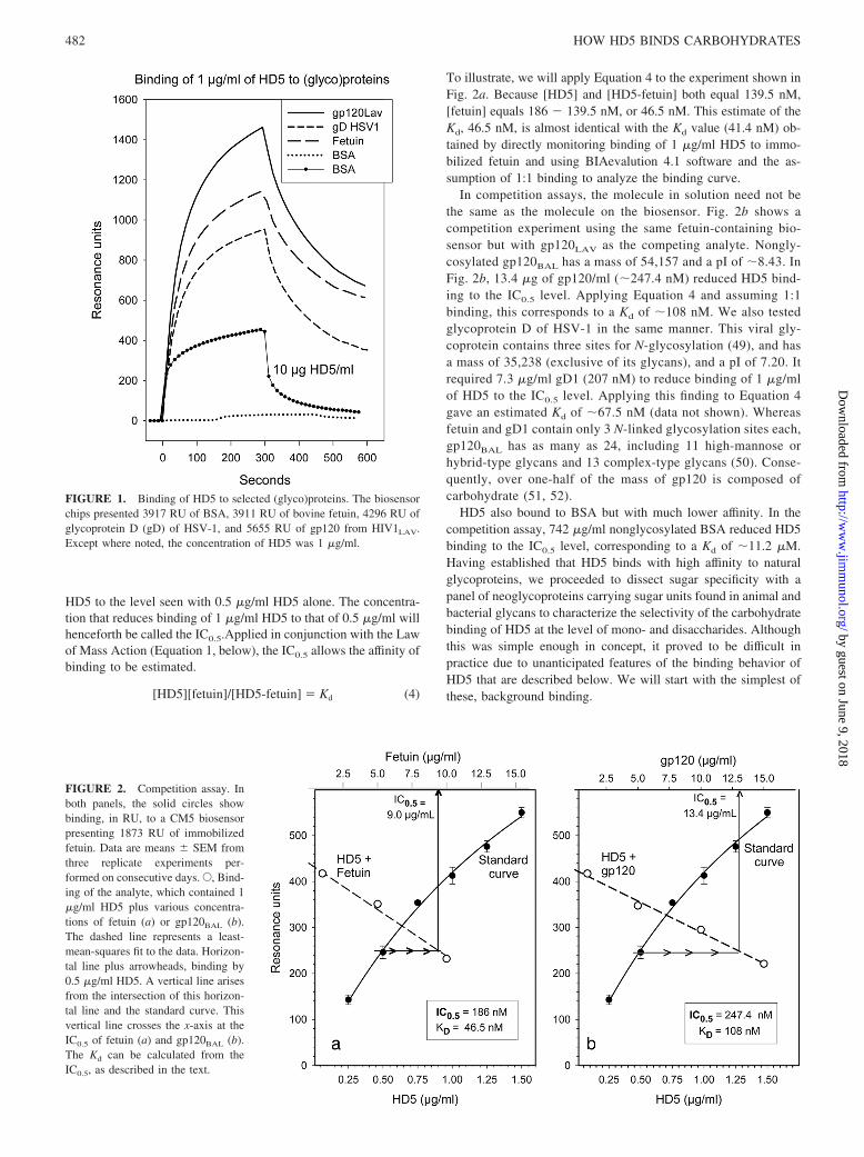
HD5 to the level seen with 0.5 �g/ml HD5 alone. The concentra-tion that reduces binding of 1 �g/ml HD5 to that of 0.5 �g/ml willhenceforth be called the IC0.5.Applied in conjunction with the Lawof Mass Action (Equation 1, below), the IC0.5 allows the affinity ofbinding to be estimated.
[HD5][fetuin]/[HD5-fetuin] � Kd (4)
To illustrate, we will apply Equation 4 to the experiment shown inFig. 2a. Because [HD5] and [HD5-fetuin] both equal 139.5 nM,[fetuin] equals 186 � 139.5 nM, or 46.5 nM. This estimate of theKd, 46.5 nM, is almost identical with the Kd value (41.4 nM) ob-tained by directly monitoring binding of 1 �g/ml HD5 to immo-bilized fetuin and using BIAevalution 4.1 software and the as-sumption of 1:1 binding to analyze the binding curve.
In competition assays, the molecule in solution need not bethe same as the molecule on the biosensor. Fig. 2b shows acompetition experiment using the same fetuin-containing bio-sensor but with gp120LAV as the competing analyte. Nongly-cosylated gp120BAL has a mass of 54,157 and a pI of �8.43. InFig. 2b, 13.4 �g of gp120/ml (�247.4 nM) reduced HD5 bind-ing to the IC0.5 level. Applying Equation 4 and assuming 1:1binding, this corresponds to a Kd of �108 nM. We also testedglycoprotein D of HSV-1 in the same manner. This viral gly-coprotein contains three sites for N-glycosylation (49), and hasa mass of 35,238 (exclusive of its glycans), and a pI of 7.20. Itrequired 7.3 �g/ml gD1 (207 nM) to reduce binding of 1 �g/mlof HD5 to the IC0.5 level. Applying this finding to Equation 4gave an estimated Kd of �67.5 nM (data not shown). Whereasfetuin and gD1 contain only 3 N-linked glycosylation sites each,gp120BAL has as many as 24, including 11 high-mannose orhybrid-type glycans and 13 complex-type glycans (50). Conse-quently, over one-half of the mass of gp120 is composed ofcarbohydrate (51, 52).
HD5 also bound to BSA but with much lower affinity. In thecompetition assay, 742 �g/ml nonglycosylated BSA reduced HD5binding to the IC0.5 level, corresponding to a Kd of �11.2 �M.Having established that HD5 binds with high affinity to naturalglycoproteins, we proceeded to dissect sugar specificity with apanel of neoglycoproteins carrying sugar units found in animal andbacterial glycans to characterize the selectivity of the carbohydratebinding of HD5 at the level of mono- and disaccharides. Althoughthis was simple enough in concept, it proved to be difficult inpractice due to unanticipated features of the binding behavior ofHD5 that are described below. We will start with the simplest ofthese, background binding.
FIGURE 1. Binding of HD5 to selected (glyco)proteins. The biosensorchips presented 3917 RU of BSA, 3911 RU of bovine fetuin, 4296 RU ofglycoprotein D (gD) of HSV-1, and 5655 RU of gp120 from HIV1LAV.Except where noted, the concentration of HD5 was 1 �g/ml.
FIGURE 2. Competition assay. Inboth panels, the solid circles showbinding, in RU, to a CM5 biosensorpresenting 1873 RU of immobilizedfetuin. Data are means � SEM fromthree replicate experiments per-formed on consecutive days. E, Bind-ing of the analyte, which contained 1�g/ml HD5 plus various concentra-tions of fetuin (a) or gp120BAL (b).The dashed line represents a least-mean-squares fit to the data. Horizon-tal line plus arrowheads, binding by0.5 �g/ml HD5. A vertical line arisesfrom the intersection of this horizon-tal line and the standard curve. Thisvertical line crosses the x-axis at theIC0.5 of fetuin (a) and gp120BAL (b).The Kd can be calculated from theIC0.5, as described in the text.
482 HOW HD5 BINDS CARBOHYDRATES
by guest on June 9, 2018http://w
ww
.jimm
unol.org/D
ownloaded from
Background binding
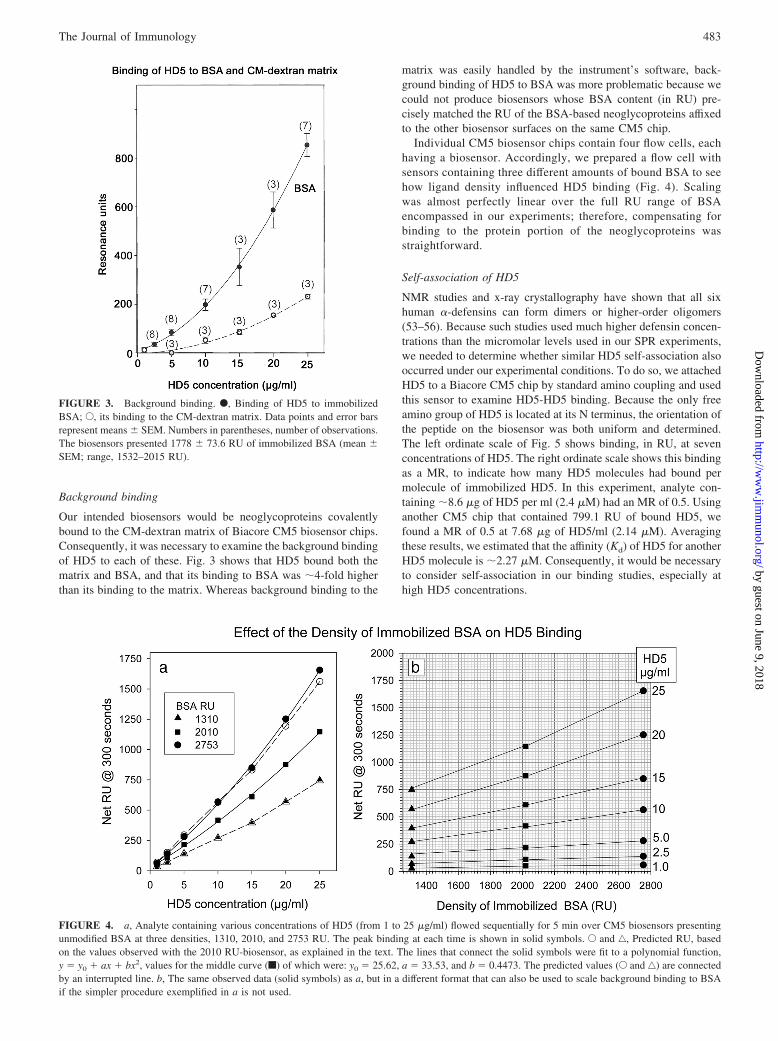
Our intended biosensors would be neoglycoproteins covalentlybound to the CM-dextran matrix of Biacore CM5 biosensor chips.Consequently, it was necessary to examine the background bindingof HD5 to each of these. Fig. 3 shows that HD5 bound both thematrix and BSA, and that its binding to BSA was �4-fold higherthan its binding to the matrix. Whereas background binding to the
matrix was easily handled by the instrument’s software, back-ground binding of HD5 to BSA was more problematic because wecould not produce biosensors whose BSA content (in RU) pre-cisely matched the RU of the BSA-based neoglycoproteins affixedto the other biosensor surfaces on the same CM5 chip.
Individual CM5 biosensor chips contain four flow cells, eachhaving a biosensor. Accordingly, we prepared a flow cell withsensors containing three different amounts of bound BSA to seehow ligand density influenced HD5 binding (Fig. 4). Scalingwas almost perfectly linear over the full RU range of BSAencompassed in our experiments; therefore, compensating forbinding to the protein portion of the neoglycoproteins wasstraightforward.
Self-association of HD5
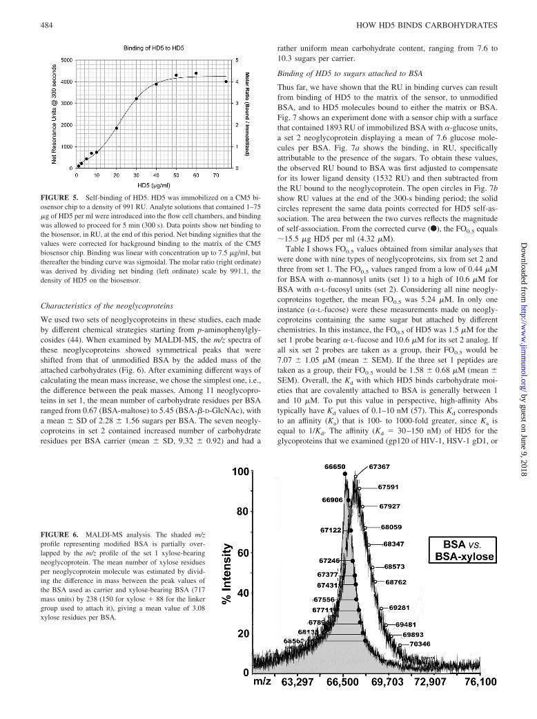
NMR studies and x-ray crystallography have shown that all sixhuman �-defensins can form dimers or higher-order oligomers(53–56). Because such studies used much higher defensin concen-trations than the micromolar levels used in our SPR experiments,we needed to determine whether similar HD5 self-association alsooccurred under our experimental conditions. To do so, we attachedHD5 to a Biacore CM5 chip by standard amino coupling and usedthis sensor to examine HD5-HD5 binding. Because the only freeamino group of HD5 is located at its N terminus, the orientation ofthe peptide on the biosensor was both uniform and determined.The left ordinate scale of Fig. 5 shows binding, in RU, at sevenconcentrations of HD5. The right ordinate scale shows this bindingas a MR, to indicate how many HD5 molecules had bound permolecule of immobilized HD5. In this experiment, analyte con-taining �8.6 �g of HD5 per ml (2.4 �M) had an MR of 0.5. Usinganother CM5 chip that contained 799.1 RU of bound HD5, wefound a MR of 0.5 at 7.68 �g of HD5/ml (2.14 �M). Averagingthese results, we estimated that the affinity (Kd) of HD5 for anotherHD5 molecule is �2.27 �M. Consequently, it would be necessaryto consider self-association in our binding studies, especially athigh HD5 concentrations.
FIGURE 3. Background binding. F, Binding of HD5 to immobilizedBSA; E, its binding to the CM-dextran matrix. Data points and error barsrepresent means � SEM. Numbers in parentheses, number of observations.The biosensors presented 1778 � 73.6 RU of immobilized BSA (mean �SEM; range, 1532–2015 RU).
FIGURE 4. a, Analyte containing various concentrations of HD5 (from 1 to 25 �g/ml) flowed sequentially for 5 min over CM5 biosensors presentingunmodified BSA at three densities, 1310, 2010, and 2753 RU. The peak binding at each time is shown in solid symbols. E and ‚, Predicted RU, basedon the values observed with the 2010 RU-biosensor, as explained in the text. The lines that connect the solid symbols were fit to a polynomial function,y � y0 � ax � bx2, values for the middle curve (f) of which were: y0 � 25.62, a � 33.53, and b � 0.4473. The predicted values (E and ‚) are connectedby an interrupted line. b, The same observed data (solid symbols) as a, but in a different format that can also be used to scale background binding to BSAif the simpler procedure exemplified in a is not used.
483The Journal of Immunology
by guest on June 9, 2018http://w
ww
.jimm
unol.org/D
ownloaded from
Characteristics of the neoglycoproteins
We used two sets of neoglycoproteins in these studies, each madeby different chemical strategies starting from p-aminophenylgly-cosides (44). When examined by MALDI-MS, the m/z spectra ofthese neoglycoproteins showed symmetrical peaks that wereshifted from that of unmodified BSA by the added mass of theattached carbohydrates (Fig. 6). After examining different ways ofcalculating the mean mass increase, we chose the simplest one, i.e.,the difference between the peak masses. Among 11 neoglycopro-teins in set 1, the mean number of carbohydrate residues per BSAranged from 0.67 (BSA-maltose) to 5.45 (BSA-�-D-GlcNAc), witha mean � SD of 2.28 � 1.56 sugars per BSA. The seven neogly-coproteins in set 2 contained increased number of carbohydrateresidues per BSA carrier (mean � SD, 9.32 � 0.92) and had a
rather uniform mean carbohydrate content, ranging from 7.6 to10.3 sugars per carrier.
Binding of HD5 to sugars attached to BSA
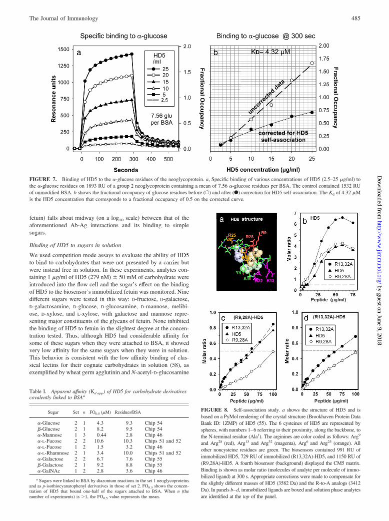
Thus far, we have shown that the RU in binding curves can resultfrom binding of HD5 to the matrix of the sensor, to unmodifiedBSA, and to HD5 molecules bound to either the matrix or BSA.Fig. 7 shows an experiment done with a sensor chip with a surfacethat contained 1893 RU of immobilized BSA with �-glucose units,a set 2 neoglycoprotein displaying a mean of 7.6 glucose mole-cules per BSA. Fig. 7a shows the binding, in RU, specificallyattributable to the presence of the sugars. To obtain these values,the observed RU bound to BSA was first adjusted to compensatefor its lower ligand density (1532 RU) and then subtracted fromthe RU bound to the neoglycoprotein. The open circles in Fig. 7bshow RU values at the end of the 300-s binding period; the solidcircles represent the same data points corrected for HD5 self-as-sociation. The area between the two curves reflects the magnitudeof self-association. From the corrected curve (F), the FO0.5 equals�15.5 �g HD5 per ml (4.32 �M).
Table I shows FO0.5 values obtained from similar analyses thatwere done with nine types of neoglycoproteins, six from set 2 andthree from set 1. The FO0.5 values ranged from a low of 0.44 �Mfor BSA with �-mannosyl units (set 1) to a high of 10.6 �M forBSA with �-L-fucosyl units (set 2). Considering all nine neogly-coproteins together, the mean FO0.5 was 5.24 �M. In only oneinstance (�-L-fucose) were these measurements made on neogly-coproteins containing the same sugar but attached by differentchemistries. In this instance, the FO0.5 of HD5 was 1.5 �M for theset 1 probe bearing �-L-fucose and 10.6 �M for its set 2 analog. Ifall six set 2 probes are taken as a group, their FO0.5 would be7.07 � 1.05 �M (mean � SEM). If the three set 1 peptides aretaken as a group, their FO0.5 would be 1.58 � 0.68 �M (mean �SEM). Overall, the Kd with which HD5 binds carbohydrate moi-eties that are covalently attached to BSA is generally between 1and 10 �M. To put this value in perspective, high-affinity Abstypically have Kd values of 0.1–10 nM (57). This Kd correspondsto an affinity (Ka) that is 100- to 1000-fold greater, since Ka isequal to 1/Kd. The affinity (Kd � 30–150 nM) of HD5 for theglycoproteins that we examined (gp120 of HIV-1, HSV-1 gD1, or
FIGURE 5. Self-binding of HD5. HD5 was immobilized on a CM5 bi-osensor chip to a density of 991 RU. Analyte solutions that contained 1–75�g of HD5 per ml were introduced into the flow cell chambers, and bindingwas allowed to proceed for 5 min (300 s). Data points show net binding tothe biosensor, in RU, at the end of this period. Net binding signifies that thevalues were corrected for background binding to the matrix of the CM5biosensor chip. Binding was linear with concentration up to 7.5 �g/ml, butthereafter the binding curve was sigmoidal. The molar ratio (right ordinate)was derived by dividing net binding (left ordinate) scale by 991.1, thedensity of HD5 on the biosensor.
FIGURE 6. MALDI-MS analysis. The shaded m/zprofile representing modified BSA is partially over-lapped by the m/z profile of the set 1 xylose-bearingneoglycoprotein. The mean number of xylose residuesper neoglycoprotein molecule was estimated by divid-ing the difference in mass between the peak values ofthe BSA used as carrier and xylose-bearing BSA (717mass units) by 238 (150 for xylose � 88 for the linkergroup used to attach it), giving a mean value of 3.08xylose residues per BSA.
484 HOW HD5 BINDS CARBOHYDRATES
by guest on June 9, 2018http://w
ww
.jimm
unol.org/D
ownloaded from
fetuin) falls about midway (on a log10 scale) between that of theaforementioned Ab-Ag interactions and its binding to simplesugars.
Binding of HD5 to sugars in solution
We used competition mode assays to evaluate the ability of HD5to bind to carbohydrates that were not presented by a carrier butwere instead free in solution. In these experiments, analytes con-taining 1 �g/ml of HD5 (279 nM) � 50 mM of carbohydrate wereintroduced into the flow cell and the sugar’s effect on the bindingof HD5 to the biosensor’s immobilized fetuin was monitored. Ninedifferent sugars were tested in this way: D-fructose, D-galactose,D-galactosamine, D-glucose, D-glucosamine, D-mannose, melibi-ose, D-xylose, and L-xylose, with galactose and mannose repre-senting major constituents of the glycans of fetuin. None inhibitedthe binding of HD5 to fetuin in the slightest degree at the concen-tration tested. Thus, although HD5 had considerable affinity forsome of these sugars when they were attached to BSA, it showedvery low affinity for the same sugars when they were in solution.This behavior is consistent with the low affinity binding of clas-sical lectins for their cognate carbohydrates in solution (58), asexemplified by wheat germ agglutinin and N-acetyl-D-glucosamine
FIGURE 7. Binding of HD5 to the �-glucose residues of the neoglycoprotein. a, Specific binding of various concentrations of HD5 (2.5–25 �g/ml) tothe �-glucose residues on 1893 RU of a group 2 neoglycoprotein containing a mean of 7.56 �-glucose residues per BSA. The control contained 1532 RUof unmodified BSA. b shows the fractional occupancy of glucose residues before (E) and after (F) correction for HD5 self-association. The Kd of 4.32 �Mis the HD5 concentration that corresponds to a fractional occupancy of 0.5 on the corrected curve.
FIGURE 8. Self-association study. a shows the structure of HD5 and isbased on a PyMol rendering of the crystal structure (Brookhaven Protein DataBank ID: 1ZMP) of HD5 (55). The 6 cysteines of HD5 are represented byspheres, with numbers 1–6 referring to their proximity, along the backbone, tothe N-terminal residue (Ala1). The arginines are color coded as follows: Arg9
and Arg28 (red), Arg13 and Arg32 (magenta), Arg6 and Arg25 (orange). Allother noncysteine residues are green. The biosensors contained 991 RU ofimmobilized HD5, 729 RU of immobilized (R13,32A)-HD5, and 1150 RU of(R9,28A)-HD5. A fourth biosensor (background) displayed the CM5 matrix.Binding is shown as molar ratio (molecules of analyte per molecule of immo-bilized ligand) at 300 s. Appropriate corrections were made to compensate forthe slightly different masses of HD5 (3582 Da) and the R-to-A analogs (3412Da). In panels b–d, immobilized ligands are boxed and solution phase analytesare identified at the top of the panel.
Table I. Apparent affinity (Kd app) of HD5 for carbohydrate derivativescovalently linked to BSAa
Sugar Set n FO0.5 (�M) Residues/BSA
�-Glucose 2 1 4.3 9.3 Chip 54�-Glucose 2 1 8.2 9.5 Chip 54�-Mannose 1 3 0.44 2.8 Chip 46�-L-Fucose 2 2 10.6 10.3 Chips 51 and 52�-L-Fucose 1 2 1.5 3.2 Chip 46�-L-Rhamnose 2 1 3.4 10.0 Chips 51 and 52�-Galactose 2 2 6.7 7.6 Chip 55�-Galactose 2 1 9.2 8.8 Chip 55�-GalNAc 1 2 2.8 3.6 Chip 46
a Sugars were linked to BSA by diazonium reactions in the set 1 neoglycoproteinsand as p-isothiocyanatophenyl derivatives in those of set 2. FO0.5 shows the concen-tration of HD5 that bound one-half of the sugars attached to BSA. When n (thenumber of experiments) is �1, the FO0.5 value represents the mean.
485The Journal of Immunology
by guest on June 9, 2018http://w
ww
.jimm
unol.org/D
ownloaded from
(Ka � 0.4 10�3 M�1), soy bean agglutinin and N-acetyl-D-galactosamine (Ka � 9 10�3 M�1), or galectin-1 and lactose(Ka � 5.6 10�3 M�1).
Arginine-deficient analogs of HD5
HD5 contains six arginine residues (Fig. 8a), and the side chains ofarginines 13 and 32 and of arginines 9 and 28 extend from thepeptide backbone, in close proximity to each other. We preparedanalogs of HD5 wherein a pair of arginines, either Arg9 and Arg28
or Arg13 and Arg32, were replaced by alanine residues and testedtheir ability to self-associate and to bind HD5. We will refer to theformer analog as (R9,28A)-HD5 and to the latter analog as(R13,32A)-HD5. Fig. 8b shows that at 50 �g/ml, �6.5 moleculesof solution-phase HD5, binds each molecule of immobilized HD5,even higher than the MR of 4.3 shown in Fig. 5. HD5 also bound(R13,32A) and (R9,28A)-HD5, and its subsequent self-associationreached a MR of �4.0 at 50 �g/ml.
Self-binding of the R-to-A analogs and their subsequent self-association were greatly decreased. Only at 100 �g/ml (Fig. 8c)did the self-binding of (R9,28A)-HD5 achieve a MR of 0.5, indi-cating that its affinity for itself (Kd, 29.3 �M) was 13.3-fold lowerthan the affinity of HD5 for itself (Kd, 2.2 �M). It required 60�g/ml (R9,28A)-HD5 to attain a MR of 0.5 when either HD5 or(R13,32A)-HD5 were the immobilized ligands, consistent with aKd of �17 �M.
Fig. 8d shows that the binding and self-association of(R13,32A)-HD5 were also greatly impaired, relative to HD5. Itrequired �60 �g/ml (17.7 �M) for self-binding to reach a MR of0.5, and 100 �g/ml (29.3 �M) for binding to HD5 or (R9,28A)-HD5 to reach a MR of 0.5. We interpret these results as indicationsthat arginines-9,-13, �28 and �32 are intimately involved in oli-gomerization of HD5 at micromolar concentrations.
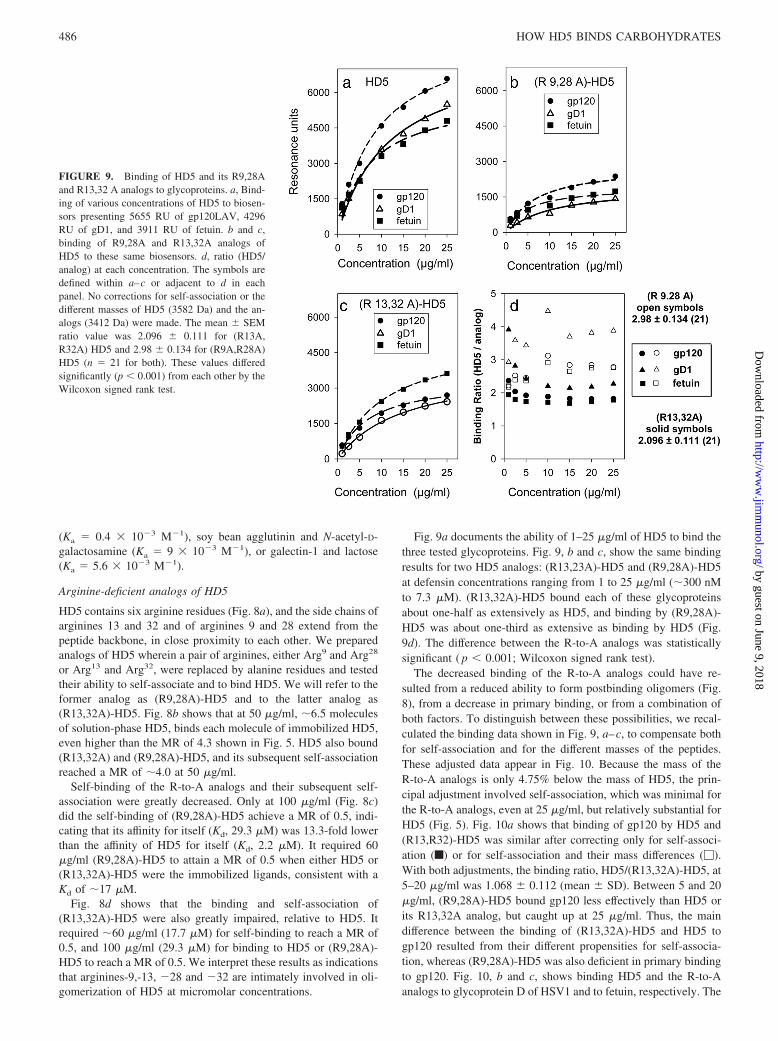
Fig. 9a documents the ability of 1–25 �g/ml of HD5 to bind thethree tested glycoproteins. Fig. 9, b and c, show the same bindingresults for two HD5 analogs: (R13,23A)-HD5 and (R9,28A)-HD5at defensin concentrations ranging from 1 to 25 �g/ml (�300 nMto 7.3 �M). (R13,32A)-HD5 bound each of these glycoproteinsabout one-half as extensively as HD5, and binding by (R9,28A)-HD5 was about one-third as extensive as binding by HD5 (Fig.9d). The difference between the R-to-A analogs was statisticallysignificant ( p 0.001; Wilcoxon signed rank test).
The decreased binding of the R-to-A analogs could have re-sulted from a reduced ability to form postbinding oligomers (Fig.8), from a decrease in primary binding, or from a combination ofboth factors. To distinguish between these possibilities, we recal-culated the binding data shown in Fig. 9, a–c, to compensate bothfor self-association and for the different masses of the peptides.These adjusted data appear in Fig. 10. Because the mass of theR-to-A analogs is only 4.75% below the mass of HD5, the prin-cipal adjustment involved self-association, which was minimal forthe R-to-A analogs, even at 25 �g/ml, but relatively substantial forHD5 (Fig. 5). Fig. 10a shows that binding of gp120 by HD5 and(R13,R32)-HD5 was similar after correcting only for self-associ-ation (f) or for self-association and their mass differences (�).With both adjustments, the binding ratio, HD5/(R13,32A)-HD5, at5–20 �g/ml was 1.068 � 0.112 (mean � SD). Between 5 and 20�g/ml, (R9,28A)-HD5 bound gp120 less effectively than HD5 orits R13,32A analog, but caught up at 25 �g/ml. Thus, the maindifference between the binding of (R13,32A)-HD5 and HD5 togp120 resulted from their different propensities for self-associa-tion, whereas (R9,28A)-HD5 was also deficient in primary bindingto gp120. Fig. 10, b and c, shows binding HD5 and the R-to-Aanalogs to glycoprotein D of HSV1 and to fetuin, respectively. The
FIGURE 9. Binding of HD5 and its R9,28Aand R13,32 A analogs to glycoproteins. a, Bind-ing of various concentrations of HD5 to biosen-sors presenting 5655 RU of gp120LAV, 4296RU of gD1, and 3911 RU of fetuin. b and c,binding of R9,28A and R13,32A analogs ofHD5 to these same biosensors. d, ratio (HD5/analog) at each concentration. The symbols aredefined within a–c or adjacent to d in eachpanel. No corrections for self-association or thedifferent masses of HD5 (3582 Da) and the an-alogs (3412 Da) were made. The mean � SEMratio value was 2.096 � 0.111 for (R13A,R32A) HD5 and 2.98 � 0.134 for (R9A,R28A)HD5 (n � 21 for both). These values differedsignificantly (p 0.001) from each other by theWilcoxon signed rank test.
486 HOW HD5 BINDS CARBOHYDRATES
by guest on June 9, 2018http://w
ww
.jimm
unol.org/D
ownloaded from
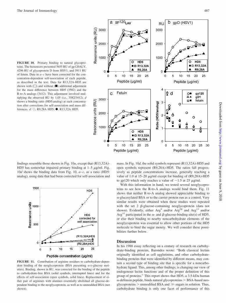
findings resemble those shown in Fig. 10a, except that (R13,32A)-HD5 has somewhat impaired primary binding at 1–5 �g/ml. Fig.10d shows the binding data from Fig. 10, a–c, as a ratio (HD5:analog), using data that had been corrected for self-association and
mass. In Fig. 10d, the solid symbols represent (R13,32A)-HD5 andopen symbols represent (R9,28A)-HD5. The ratios fall progres-sively as peptide concentrations increase, generally reaching avalue of 1.0 at 15–20 �g/ml except for binding of (R9,28A)-HD5to gp120 which only reaches a value of �1.5 at 25 �g/ml.
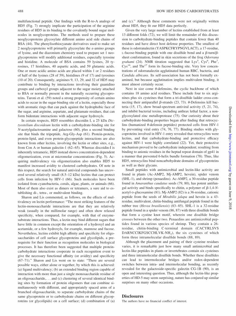
With this information in hand, we tested several neoglycopro-teins to see how the R-to-A analogs would bind them. Fig. 11shows that neither R-to-A analog showed appreciable binding to�-glucosylated BSA or to the carrier protein run as a control. Verysimilar results were obtained when these studies were repeatedwith the set 2 �-glucose-containing neoglycoprotein (data notshown). Evidently, either Arg9 and/or Arg28 and Arg13 and/orArg32 participated in the �- and �-glucose-binding site(s) of HD5,or else their binding to nearby noncarbohydrate elements of theneoglycoprotein was essential to allow other portions of the HD5molecule to bind the sugar moiety. We will consider these possi-bilities further below.
DiscussionIn his 1988 essay reflecting on a century of research on carbohy-drate-binding proteins, Barondes wrote: “Both classical lectinsoriginally identified as cell agglutinins, and other carbohydrate-binding proteins that were identified by different means, may con-tain a second type of binding site that is specific for a noncarbo-hydrate ligand. This, among other findings, is changing our view ofendogenous lectin functions and of the proper definition of thisgroup of proteins.” This report shows that HD5, a 3.5-kDa human�-defensin peptide, binds natural glycoproteins � BSA-based neo-glycoproteins � unmodified BSA and �� sugars in solution. Thus,carbohydrate binding is only one facet of performance of this
FIGURE 10. Primary binding to natural glycopro-teins. The biosensors presented 5655 RU of gp120ALV,4296 RU of glycoprotein D from HSV1, and 3911 RUof fetuin. Data in a–c have been corrected for the con-centration-dependent self-association of each peptide,as described in the text. Data for R13,32A-HD5 areshown with (�) and without (f) additional adjustmentfor the mass difference between HD5 (3582) and theR-to-A analogs (3412). This adjustment involved mul-tiplying the observed RU by 1.05 (i.e., 3582/3412). dshows a binding ratio (HD5:analog) at each concentra-tion after corrections for self-association and mass dif-ferences. d: E, R9,28A HD5; F, R13,32A HD5.
FIGURE 11. Contribution of arginine residues to carbohydrate-depen-dent binding of the neoglycoprotein (BSA presenting �-D-glucose moi-eties). Binding, shown in RU, was corrected for the binding of the peptideto carbohydrate-free BSA (solid symbols, interrupted lines) and for theeffects of self-association (open symbols, solid lines). Replacement of ei-ther pair of arginines with alanines essentially abolished all glucose-de-pendent binding to the neoglycoprotein, as well as to unmodified BSA (notshown).
487The Journal of Immunology
by guest on June 9, 2018http://w
ww
.jimm
unol.org/D
ownloaded from
multifunctional peptide. Our findings with the R-to-A analogs ofHD5 (Fig. 7) strongly implicate the participation of the arginineresidues of HD5 in its binding to the covalently bound sugar mol-ecules in neoglycoproteins. The methods used to prepare theseneoglycoproteins glycosylate different amino acid side chains ofBSA (44). The phenylisothiocyanate derivatives used to make set2 neoglycoproteins will primarily glycosylate the �-amino groupsof lysine, and the diazonium chemistry used to prepare set 1 neo-glycoproteins will modify additional residues, especially tyrosineand histidine. A molecule of BSA contains 59 lysines, 20 ty-rosines, 17 histidines, 40 aspartic acids, and 59 glutamic acids.One or more acidic amino acids are placed within 1 or 2 residuesof half of the lysines (28 of 59), histidines (9 of 17) and tyrosines(10 of 20). Consequently, arginines 9, 13, 29, and 32 of HD5 maycontribute to binding by interactions involving their guanidinogroups and carboxyl groups adjacent to the sugar moiety attachedto BSA or normally present in the naturally occurring glycopro-teins. Taroni et al. (59) noted a strong propensity of certain aminoacids to occur in the sugar-binding site of a lectin, especially thosewith aromatic rings that can pack against the hydrophobic face ofthe sugar, and arginine, aspartate, and glutamate residues, that canform bidentate interactions with adjacent sugar hydroxyls.
In certain respects, HD5 resembles discoidin I, a 25 kDa Dic-tyostelium discoideum lectin with a carbohydrate-binding site forN-acetylgalactosamine and galactose (60), plus a second bindingsite that binds the tripeptide, Arg-Gly-Asp (61). Protein-protein,protein-lipid, and even protein-glycopeptide interactions are alsoknown from other lectins, involving the lectin or other sites, e.g.,from Con A or human galectin-1 (62–65). Whereas discoidin-I isinherently tetrameric, HD5 instead shows concentration-dependentoligomerization, even at micromolar concentrations (Fig. 5). Ac-quiring multivalency via oligomerization also enables HD5 tomanifest increased avidity to clustered carbohydrates. Of note inthis respect, the search for natural antiviral compounds has uncov-ered several relatively small (8.5–12 kDa) lectins that can protectcells from infection by HIV-1 (66). Such molecules have beenisolated from cyanobacteria, corals, algae, plants, or animals (66).Most of them also exist as dimers or tetramers, a sure aid to es-tablishing di-, tetra-, or multivalent binding.
Sharon and Lis commented, as follows, on the effects of mul-tivalency on lectin performance: “The most striking features of thelectin-monosaccharide interactions are that they are relativelyweak (usually in the millimolar range) and often show relaxedspecificity, when compared, for example, with that of enzyme-substrate interactions. Thus, a lectin may bind different sugars thathave little in common except the orientation of a hydroxyl and anacetamide, or a few hydroxyls, for example, mannose and fucose.Nevertheless, lectins exhibit high affinity and specificity for oligo-saccharides of cell surface glycoproteins and glycolipids, a pre-requisite for their function as recognition molecules in biologicalprocesses. It has therefore been suggested that multiple protein-carbohydrate interactions cooperate in each recognition event togive the necessary functional affinity (or avidity) and specificity(67–71).” Sharon and Lis went on to state: “There are severalpossible ways, either alone or together, by which this is achieved:(a) ligand multivalency; (b) an extended binding region capable ofinteraction with more than just a single monosaccharide residue ofan oligosaccharide,. . . and (c) clustering of several identical bind-ing sites by formation of protein oligomers that can combine si-multaneously with different, and appropriately spaced arms of abranched oligosaccharide, to separate carbohydrate chains of thesame glycoprotein or to carbohydrate chains on different glycop-roteins (or glycolipids) on a cell surface; (d) combination of (a)
and (c).” Although these comments were not originally writtenabout HD5, they fit our HD5 data perfectly.
Given the very large number of lectins established from at least13 different folds (72), we will limit the remainder of this discus-sion to carbohydrate-binding peptides that contain fewer than 40residues and have direct host defense properties. The smallest ofthese is odorranalectin (YASPKCFRYPNGVLACT), a 17-residue,L-fucose-binding peptide with one disulfide bond and a �-turn/�-sheet conformation, found in skin secretions of the frog Odorranagrahami (24). NMR titration suggested that Lys5, Cys6, Phe7,Cys16, and Thr17 form its fucose-binding site. Very low concen-trations of odorranalectin agglutinated human RBC, bacteria, andCandida albicans. Its self-association has not been formally ex-amined, but because agglutination implies multivalent binding, itmust almost certainly occur.
Next in size come �-defensins, the cyclic backbone of whichcontains 18 amino acid residues. These include four to six argi-nines plus six cysteines that form a tri-disulfide ladder cross-con-necting their antiparallel �-strands (23, 73). �-Defensins kill bac-teria (15, 17), show broad-spectrum antiviral activity (5, 21, 74),and inhibit bacterial toxins, including anthrax lethal factor, a non-glycosylated zinc metalloprotease (75). Our curiosity about theircarbohydrate-binding properties began after finding that retrocyc-lins (humanized �-defensin peptides) protected cells from HIV-1by preventing viral entry (74, 76, 77). Binding studies with gly-coproteins involved in HIV-1 entry revealed that retrocyclins werelectins, and that carbohydrate-binding and protective efficacyagainst HIV-1 were highly correlated (22). Yet, their protectivemechanism proved to be carbohydrate independent; resulting frombinding to anionic residues in the heptad repeat domain of gp41 ina manner that prevented 6-helix bundle formation (78). Thus, likeHD5, retrocyclins bind noncarbohydrate domains of glycoproteinsas well as their glycans.
Small peptides with antimicrobial and lectin-like activity arefound in plants (Ac-AMP2, Mj-AMP2, hevein), spider venom(SHL-1), and shrimp (penaedins). Ac-AMP2, a 30-residue peptidefound in Amaranthus caudatus seeds (79, 80), has potent antifun-gal activity and binds specifically to chitin, a polymer of �-1,4-N-acetyl-D-glucosamine (81). Mj-AMP2 (82) is a 36-residue, cationicpeptide from the seeds of Mirabilis jalapa and hevein is a 43-residue, multivalent, chitin-binding antifungal peptide found in therubber tree (Hevea brasiliensis) (83–85). SHL-1 is a 32-residuepeptide found in a spider venom (86, 87) with three disulfide bondsthat form a cystine knot motif, wherein one disulfide bridgecrosses between the other two. Penaedins are antimicrobial pep-tides found in various species of shrimp. They contain a 26-residue, chitin-binding C-terminal domain (CACYRLSVSDARNCCIKFGSCCHLVK-NH2), the six cysteines of whichform three intramolecular disulfide bonds (88, 89).
Although the placement and pairing of their cysteine residuesvaries, it is remarkable just how many small antimicrobial andlectin-like peptides in plants or invertebrates contain six cysteinesand three intramolecular disulfide bonds. Whether these disulfidescan lead to intermolecular bridges and/or redox-dependentswitches between intra- and intermolecular bonding, as recentlyrevealed for the galactoside-specific galectin CG-1B (90), is anopen and interesting question. Thus, although the lectin-like prop-erties of HD-5 may seem surprising, nature has conjured up similarsurprises on many other occasions.
DisclosuresThe authors have no financial conflict of interest.
488 HOW HD5 BINDS CARBOHYDRATES
by guest on June 9, 2018http://w
ww
.jimm
unol.org/D
ownloaded from
References1. Ericksen, B., Z. Wu, W. Lu, and R. I. Lehrer. 2005. Antibacterial activity and
specificity of the six human �-defensins. Antimicrob. Agents Chemother. 49:269–275.
2. Lehrer, R. I., A. Barton, K. A. Daher, S. S. Harwig, T. Ganz, and M. E. Selsted.1989. Interaction of human defensins with Escherichia coli: mechanism of bac-tericidal activity. J. Clin. Invest. 84: 553–561.
3. Sahl, H. G., U. Pag, S. Bonness, S. Wagner, N. Antcheva, and A. Tossi. 2005.Mammalian defensins: structures and mechanism of antibiotic activity. J. Leu-kocyte Biol. 77: 466–475.
4. Chang, T. L., and M. E. Klotman. 2004. Defensins: natural anti-HIV peptides.AIDS Rev. 6: 161–168.
5. Hazrati, E., B. Galen, W. Lu, W. Wang, Y. Ouyang, M. J. Keller, R. I. Lehrer,and B. C. Herold. 2006. Human �- and �-defensins block multiple steps in herpessimplex virus infection. J. Immunol. 177: 8658–8666.
6. Klotman, M. E., and T. L. Chang. 2006. Defensins in innate antiviral immunity.Nat. Rev. Immunol. 6: 447–456.
7. Yasin, B., W. Wang, M. Pang, N. Cheshenko,T. Hong, A. J. Waring,B. C. Herold, E. A. Wagar, and R. I Lehrer. 2004. � Defensins protect cells frominfection by herpes simplex virus by inhibiting viral adhesion and entry. J. Virol.78: 5147–5156.
8. Menendez, A., and F. B. Brett. 2007. Defensins in the immunology of bacterialinfections. Curr. Opin. Immunol. 19: 385–391.
9. Oppenheim, J. J., A. Biragyn, L. W. Kwak, and D. Yang. 2003. Roles of anti-microbial peptides such as defensins in innate and adaptive immunity. Ann.Rheum. Dis. 62(Suppl. 2): ii17–ii21.
10. Liu, L., C. Zhao, H. H. Heng, and T. Ganz. 1997. The human �-defensin-1 and�-defensins are encoded by adjacent genes: two peptide families with differingdisulfide topology share a common ancestry. Genomics 43: 316–320.
11. Nguyen, T. X., A. M. Cole, and R. I. Lehrer. 2003. Evolution of primate �-de-fensins: a serpentine path to a sweet tooth. Peptides 24: 1647–1654.
12. Zou, J., C. Mercier, A. Koussounadis, and C. Secombes. 2007. Discovery ofmultiple �-defensin like homologues in teleost fish. Mol. Immunol. 44: 638–647.
13. Zhu, S. 2008. Discovery of six families of fungal defensin-like peptides providesinsights into origin and evolution of the CS-�� defensins. Mol. Immunol. 45:828–838.
14. Lynn, D. J., and D. G. Bradley. 2007. Discovery of �-defensins in basal mam-mals. Dev. Comp. Immunol. 31: 963–967.
15. Leonova, L., V. N. Kokryakov, G. Aleshina, T. Hong, T. Nguyen, C. Zhao,A. J. Waring, and R. I. Lehrer. 2001. Circular minidefensins and posttranslationalgeneration of molecular diversity. J. Leukocyte Biol. 70: 461–464.
16. Selsted, M. E. 2004. �-Defensins: cyclic antimicrobial peptides produced by bi-nary ligation of truncated �-defensins. Curr. Protein Pept. Sci. 5: 365–371.
17. Tang, Y. Q., J. Yuan, G. Osapay, K. Osapay, D. Tran, C. J. Miller, A. J. Ouellette,and M. E. Selsted. 1999. A cyclic antimicrobial peptide produced in primateleukocytes by the ligation of two truncated �-defensins. Science 286: 498–502.
18. Peschel, A. 2002. How do bacteria resist human antimicrobial peptides? TrendsMicrobiol. 10: 179–186.
19. Gabius, H. J., H. C. Siebert, S. Andre, J. Jimenez-Barbero, and H. Rudiger. 2004.Chemical biology of the sugar code. Chembiochem 5: 740–764.
20. Gabius; H. J., ed. 2009. The Sugar Code: Fundamentals of Glycosciences. Wiley-VCH, Weinheim.
21. Leikina, E., H. Delanoe-Ayari, K. Melikov, M. S. Cho, A. Chen, A. J. Waring,W. Wang, Y. Xie, J. A. Loo, R. I. Lehrer, and L. V. Chernomordik. 2005. Car-bohydrate-binding molecules inhibit viral fusion and entry by crosslinking mem-brane glycoproteins. Nat. Immunol. 6: 995–1001.
22. Wang, W., A. M. Cole, T. Hong, A. J. Waring, and R. I. Lehrer. 2003. Retro-cyclin, an antiretroviral �-defensin, is a lectin. J. Immunol. 170: 4708–4716.
23. Daly, N. L., Y. K. Chen, K. J. Rosengren, U. C. Marx, M. L. Phillips,A. J. Waring, W. Wang, R. I. Lehrer, and D. J. Craik. 2007. Retrocyclin-2:structural analysis of a potent anti-HIV �-defensin. Biochemistry 46: 9920–9928.
24. Li, J., H. Wu, J. Hong, X. Xu, H. Yang, B. Wu, Y. Wang, J. Zhu, R. Lai, X. Jiang,D. Lin, M. C. Prescott, and H. H. Rees. 2008. Odorranalectin is a small peptidelectin with potential for drug delivery and targeting. PLoS. ONE. 3: e2381.
25. Siebert, H. C., K. Born, S. Andre, M. Frank, H. Kaltner, C. W. der Lieth,A. J. Heck, J. Jimenez-Barbero, J. Kopitz, and H. J. Gabius. 2005. Carbohydratechain of ganglioside GM1 as a ligand: identification of the binding strategies ofthree 15 mer peptides and their divergence from the binding modes of growth-regulatory galectin-1 and cholera toxin. Chem. Eur. J. 12: 388–402.
26. Ganz, T., M. E. Selsted, D. Szklarek, S. S. Harwig, K. Daher, D. F. Bainton, andR. I. Lehrer. 1985. Defensins: natural peptide antibiotics of human neutrophils.J. Clin. Invest. 76: 1427–1435.
27. Palfree, R. G., L. C. Sadro, and S. Solomon. 1993. The gene encoding the humancorticostatin HP-4 precursor contains a recent 86-base duplication and is locatedon chromosome 8. Mol. Endocrinol. 7: 199–205.
28. Jones, D. E., and C. L. Bevins. 1992. Paneth cells of the human small intestineexpress an antimicrobial peptide gene. J. Biol.Chem. 267: 23216–23225.
29. Quayle, A. J., E. M. Porter, A. A. Nussbaum, Y. M. Wang, C. Brabec, K. P. Yip,and S. C. Mok. 1998. Gene expression, immunolocalization, and secretion ofhuman defensin-5 in human female reproductive tract. Am. J. Pathol. 152:1247–1258.
30. Ouellette, A. J. 2006. Paneth cell �-defensin synthesis and function. Curr. Top.Microbiol. Immunol. 306: 1–25.
31. Kelly, P., M. Bajaj-Elliott, M. Katubulushi, I. Zulu, R. Poulsom, R. A. Feldman,C. L. Bevins, and W. Dhaliwal. 2006. Reduced gene expression of intestinal�-defensins predicts diarrhea in a cohort of African adults. J. Infect. Dis. 193:1464–1470.
32. Wehkamp, J., K. Fellermann, and E. F. Stange. 2005. Human defensins inCrohn’s disease. Chem. Immunol. Allergy 86: 42–54.
33. Wehkamp, J., M. Schmid, and E. F. Stange. 2007. Defensins and other antimi-crobial peptides in inflammatory bowel disease. Curr. Opin. Gastroenterol. 23:370–378.
34. Wehkamp, J., G. Wang, I. Kubler, S. Nuding, A. Gregorieff, A. Schnabel,R. J. Kays, K. Fellermann, O. Burk, M. Schwab, et al. 2007. The Paneth cell�-defensin deficiency of ileal Crohn’s disease is linked to Wnt/Tcf. J. Immunol.179: 3109–3118.
35. Lakatos, P. L., I. Altorjay, Y. Mandi, L. Lakatos, J. Tumpek, A. Kovacs,T. Molnar, Z. Tulassay, P. Miheller, K. Palatka, et al. 2008. Interaction betweenseroreactivity to microbial antigens and genetics in Crohn’s disease: is there arole for defensins? Tissue Antigens 71: 552–559.
36. Buck, C. B., P. M. Day, C. D. Thompson, J. Lubkowski, W. Lu, D. R. Lowy, andJ. T. Schiller. 2006. Human �-defensins block papillomavirus infection. Proc.Natl. Acad. Sci. USA 103: 1516–1521.
37. Smith, J. G., and G. R. Nemerow. 2008. Mechanism of adenovirus neutralizationby human �-defensins. Cell Host Microbe 3: 11–19.
38. Dugan, A. S., M. S. Maginnis, J. A. Jordan, M. L. Gasparovic, K. Manley,R. Page, G. Williams, E. Porter, B. A. O’Hara, and W. J. Atwood. 2008. Human�-defensins inhibit BK virus infection by aggregating virions and blocking bind-ing to host cells. J. Biol. Chem. 283: 31125–31132.
39. Andre, S., C. Unverzagt, S. Kojima, M. Frank, J. Seifert, C. Fink, K. Kayser,C. W. der Lieth, and H. J. Gabius. 2004. Determination of modulation of ligandproperties of synthetic complex-type biantennary N-glycans by introduction ofbisecting GlcNAc in silico, in vitro and in vivo. Eur. J. Biochem. 271: 118–134.
40. Andre, S., H. C. Siebert, M. Nishiguchi, K. Tazaki, and H. J. Gabius. 2005.Evidence for lectin activity of a plant receptor-like protein kinase by applicationof neoglycoproteins and bioinformatic algorithms. Biochim. Biophys. Acta 1725:222–232.
41. Gabius, H. J., S. Bodanowitz, and A. Schauer. 1988. Endogenous sugar-bindingproteins in human breast tissue and benign and malignant breast lesions. Cancer61: 1125–1131.
42. Lee, R. T., and Y. C. Yee. 1997. Neoglycoconjugates. In: Glycosciences: Statusand Perspectives. H.-J. Gabius and S. Gabius, eds. Chapman & Hall,London-Weinheim.
43. Mann, K., I. M. Weiss, S. Andre, H. J. Gabius, and M. Fritz. 2000. The amino-acid sequence of the abalone (Haliotis laevigata) nacre protein perlucin: detectionof a functional C-type lectin domain with galactose/mannose specificity. J. Bio-chem. 267: 5257–5264.
44. McBroom, C. R., C. H. Samanen, and I. J. Goldstein. 1972. Carbohydrate anti-gens: coupling of carbohydrates to proteins by diazonium and phenylisothiocya-nate reaction. Methods Enzymol. 28: 212–219.
45. Fagerstam, L. G., A. Frostell, R. Karlsson, M. Kullman, A. Larsson,M. Malmqvist, and H. Butt. 1990. Detection of antigen-antibody interactions bysurface plasmon resonance: application to epitope mapping. J. Mol. Recognit. 3:208–214.
46. Jonsson, U., L. Fagerstam, B. Ivarsson, B. Johnsson, R. Karlsson, K. Lundh,S. Lofas, B. Persson, H. Roos, and I. Ronnberg. 1991. Real-time biospecificinteraction analysis using surface plasmon resonance and a sensor chip technol-ogy. BioTechniques 11: 620–627.
47. Jonsson, U., L. Fagerstam, S. Lofas, E. Stenberg, R. Karlsson, A. Frostell,F. Markey, and F. Schindler. 1993. Introducing a biosensor based technology forreal-time biospecific interaction analysis. Ann. Biol. Clin. (Paris) 51: 19–26.
48. Dziegielewska, K. M., W. M. Brown, S. J. Casey, D. L. Christie, R. C. Foreman,,R. M. Hill, and N. R. Saunders. 1990. The complete cDNA and amino acidsequence of bovine fetuin: its homology with � 2HS glycoprotein and relation toother members of the cystatin superfamily. J. Biol. Chem. 265: 4354–4357.
49. Whitbeck, J. C., C. Peng, H. Lou, R. Xu, S. H. Willis, de L. Ponce, T. Peng,A. Nicola, V. R. I. Montgomery, M. S. Warner, et al. Glycoprotein D of herpessimplex virus (HSV) binds directly to HVEM, a member of the tumor necrosisfactor receptor superfamily and a mediator of HSV entry1997. J. Virol. 71:6083–6093.
50. Leonard, C. K., M. W. Spellman, L. Riddle, R. J. Harris, J. N. Thomas, andT. J. Gregory. 1990. Assignment of intrachain disulfide bonds and characteriza-tion of potential glycosylation sites of the type 1 recombinant human immuno-deficiency virus envelope glycoprotein (gp120) expressed in Chinese hamsterovary cells. J. Biol.Chem. 265: 10373–10382.
51. Moore, J. P., B. A. Jameson, Q. J. Sattentau, R. Willey, and J. Sodroski. 1993.Towards a structure of the HIV-1 envelope glycoprotein gp120: an immuno-chemical approach. Philos. Trans. R. Soc. Lond. B Biol. Sci. 342: 83–88.
52. Geyer, H., C. Holschbach, G. Hunsmann, and J. Schneider. 1988. Carbohydratesof human immunodeficiency virus: structures of oligosaccharides linked to theenvelope glycoprotein 120. J. Biol. Chem. 263: 11760–11767.
53. Hill, C. P., J. Yee, M. E. Selsted, and D. Eisenberg. 1991. Crystal structure ofdefensin HNP-3, an amphiphilic dimer: mechanisms of membrane permeabili-zation. Science 251: 1481–1485.
54. Hristova, K., M. E. Selsted, and S. H. White. 1996. Interactions of monomericrabbit neutrophil defensins with bilayers: comparison with dimeric human de-fensin HNP-2. Biochemistry 35: 11888–11894.
55. Szyk, A., Z. Wu, K. Tucker, D. Yang, W. Lu, and J. Lubkowski. 2006. Crystalstructures of human �-defensins HNP4. HD5, and HD6. Protein Sci. 15:2749–2760.
56. Zhang, X. L., M. E. Selsted, and A. Pardi. 1992. NMR studies of defensin an-timicrobial peptides. 1. Resonance assignment and secondary structure determi-nation of rabbit NP-2 and human HNP-1. Biochemistry 31: 11348–11356.
489The Journal of Immunology
by guest on June 9, 2018http://w
ww
.jimm
unol.org/D
ownloaded from
57. Delves, P. J., S. M. Roitt, and D. Burton. 2006. The primary interaction withantigen. In: Roitt’s Essential Immunology. Blackwell, Malden.
58. Nathan Sharon, H. L. 2003. Specificity and affinity. In: Lectins. Kluwer,Dordrecht.
59. Taroni, C., S. Jones, and J. M. Thornton. 2000. Analysis and prediction of car-bohydrate binding sites. Protein Eng. 13: 89–98.
60. Cooper, D. N., S. C. Lee, and S. H. Barondes. 1983. Discoidin-binding polysac-charide from Dictyostelium discoideum. J. Biol. Chem. 258: 8745–8750.
61. Gabius, H. J., W. R. Springer, and S. H. Barondes. 1985. Receptor for the cellbinding site of discoidin I. Cell 42: 449–456.
62. Gabius, H. J. 1994. Non-carbohydrate binding partners/domains of animal lec-tins. Int. J. Biochem. 26: 469–477.
63. Jain, D., K. Kaur, B. Sundaravadivel, and D. M. Salunke. 2000. Structural andfunctional consequences of peptide-carbohydrate mimicry: crystal structure of acarbohydrate-mimicking peptide bound to concanavalin A. J. Biol. Chem. 275:16098–16102.
64. Rotblat, B., H. Niv, S. Andre, H. Kaltner, H. J. Gabius, and Y. Kloog. 2004.Galectin-1(L11A) predicted from a computed galectin-1 farnesyl-binding pocketselectively inhibits Ras-GTP. Cancer Res. 64: 3112–3118.
65. Andre, S., C. E. Maljaars, K. M. Halkes, H. J. Gabius, and J. P. Kamerling. 2007.Discovery of galectin ligands in fully randomized combinatorial one-bead-one-compound (glyco)peptide libraries. Bioorg. Med. Chem. Lett. 17: 793–798.
66. Balzarini, J. 2006. Inhibition of HIV entry by carbohydrate-binding proteins.Antiviral Res. 71: 237–247.
67. Brewer, C. F. 1996. Multivalent lectin-carbohydrate crosslinking interactions.Chemtracts: Biochem. Mol. Biol. 6: 165–179.
68. Drickamer, K. 1995. Multiplicity of lectin-carbohydrate interactions. Nat. Struct.Biol. 2: 437–439.
69. Lee, R. T., and Y. C. Lee. 2000. Affinity enhancement by multivalent lectin-carbohydrate interaction. Glycoconj. J. 17: 543–551.
70. Lundquist, J. J., and E. J. Toone. 2002. The cluster glycoside effect. Chem. Rev.102: 555–578.
71. Monsigny, M., R. Mayer, and A. C. Roche. 2000. Sugar-lectin interactions: sugarclusters, lectin multivalency and avidity. Carbohydr. Lett. 4: 35–52.
72. Gabius, H. J. 2008. Glycans: bioactive signals decoded by lectins. Biochem. Soc.Trans. 36: 1491–1496.
73. Trabi, M., H. J. Schirra, and D. J. Craik. 2001. Three-dimensional structure ofRTD-1, a cyclic antimicrobial defensin from Rhesus macaque leukocytes. Bio-chemistry 40: 4211–4221.
74. Cole, A. M., T. Hong, L. M. Boo, T. Nguyen, C. Zhao, G. Bristol, J. A. Zack,A. J. Waring, O. O. Yang, and R. I. Lehrer. 2002. Retrocyclin: a primate peptidethat protects cells from infection by T- and M-tropic strains of HIV-1. Proc. Natl.Acad. Sci. USA 99: 1813–1818.
75. Wang, W., C. Mulakala, S. C. Ward, G. Jung, H. Luong, D. Pham, A. J. Waring,Y. Kaznessis, W. Lu, K. A. Bradley, and R. I. Lehrer. 2006. Retrocyclins killbacilli and germinating spores of Bacillus anthracis and inactivate anthrax lethaltoxin. J. Biol. Chem. 281: 32755–32764.
76. Munk, C., G. Wei, O. O. Yang, A. J. Waring, W. Wang, T. Hong, R. I. Lehrer,N. R. Landau, and A. M. Cole. 2003. The �-defensin, retrocyclin, inhibits HIV-1entry. AIDS Res. Hum. Retrovir. 19: 875–881.
77. Wang, W., S. M. Owen, D. L. Rudolph, A. M. Cole, T. Hong, A. J. Waring,R. B. Lal, and R. I. Lehrer. 2004. Activity of �- and �-defensins against primaryisolates of HIV-1. J. Immunol. 173: 515–520.
78. Gallo, S. A., W. Wang, S. S. Rawat, G. Jung, A. J. Waring, A. M. Cole, H. Lu,X. Yan, N. L. Daly, D. J. Craik, S. Jiang, R. I. Lehrer, and R. Blumenthal. 2006.�-Defensins prevent HIV-1 Env-mediated fusion by binding gp41 and blocking6-helix bundle formation. J. Biol.Chem. 281: 18787–18792.
79. Broekaert, W. F., W. Marien, F. R. Terras, M. F. De Bolle, P. Proost,J. Van Damme, L. Dillen, M. Claeys, S. B. Rees, and J. Vanderleyden. 1992.Antimicrobial peptides from Amaranthus caudatus seeds with sequence homol-ogy to the cysteine/glycine-rich domain of chitin-binding proteins. Biochemistry31: 4308–4314.
80. De Bolle, M. F., R. W. Osborn, I. J. Goderis, L. Noe, D. Acland, C. A. Hart,S. Torrekens, F. Van Leuven, and W. F. Broekaert. 1996. Antimicrobial peptidesfrom Mirabilis jalapa and Amaranthus caudatus: expression, processing, local-ization and biological activity in transgenic tobacco. Plant Mol. Biol. 31:993–1008.
81. Merzendorfer, H. 2009. Chitin: structure, function, and metabolism. In The SugarCode: Fundamentals of Glycosciences. H.-J. Gabius, ed. Wiley-VCH, Weinheim.
82. Cammue, B. P., M. F. De Bolle, F. R. Terras, P. Proost, J. Van Damme,S. B. Rees, J. Vanderleyden, and W. F. Broekaert. 1992. Isolation and charac-terization of a novel class of plant antimicrobial peptides form Mirabilis jalapaL. seeds. J. Biol. Chem. 267: 2228–2233.
83. Jimenez-Barbero, J., C. F. Javier, J. L. Asensio, N. Aboitiz, P. Vidal, A. Canales,P. Groves, H. J. Gabius, and H. C. Siebert. 2006. Hevein domains: an attractivemodel to study carbohydrate-protein interactions at atomic resolution. Adv. Car-bohydr. Chem. Biochem. 60: 303–354.
84. Lee, O. S., B. Lee, N. Park, J. C. Koo, Y. H. Kim, D. T. Prasad, C. Karigar,H. J. Chun, B. R. Jeong, D. H. Kim, et al. 2003. Pn-AMPs, the hevein-likeproteins from Pharbitis nil confers disease resistance against phytopathogenicfungi in tomato, Lycopersicum esculentum. Phytochemistry 62: 1073–1079.
85. Asensio, J. L., F. J. Canada, H. C. Siebert, J. Laynez, A. Poveda, P. M. Nieto,U. M. Soedjanaamadja, H. J. Gabius, and J. Jimenez-Barbero. 2000. Structuralbasis for chitin recognition by defense proteins: GlcNAc residues are bound in amultivalent fashion by extended binding sites in hevein domains. Chem. Biol. 7:529–543.
86. Liang, S. P., and X. Pan. 1995. A lectin-like peptide isolated from the venom ofthe Chinese bird spider Selenocosmia huwena. Toxicon 33: 875–882.
87. Lu, S., S. Liang, and Gu, X. 1999. Three-dimensional structure of Selenocosmiahuwena lectin-I (SHL-I) from the venom of the spider Selenocosmia huwena by2D-NMR. J. Protein Chem. 18: 609–617.
88. Destoumieux, D., P. Bulet, D. Loew, A. Van Dorsselaer, J. Rodriguez, andE. Bachere. 1997. Penaeidins, a new family of antimicrobial peptides isolatedfrom the shrimp Penaeus vannamei (Decapoda). J. Biol. Chem. 272:28398–28406.
89. Destoumieux, D., M. Munoz, P. Bulet, and E. Bachere. 2000. Penaeidins, a fam-ily of antimicrobial peptides from penaeid shrimp (Crustacea, Decapoda). CellMol. Life Sci. 57: 1260–1271.
90. Lopez-Lucendo, M. F., D. Solís, J. L. Saiz, H. Kaltner, R. Russwurm, S. Andre,H.-J. Gabius, and A. Romero. 2009. Homodimeric chicken galectin CG-1B (C-14): crystal structure and detection of unique redox-dependent shape changesinvolving inter- and intrasubunit disulfide bridges by gel filtration, ultracentrif-ugation, site-directed mutagenesis and peptide mass fingerprinting. J. Mol. Biol.386: 366–378.
490 HOW HD5 BINDS CARBOHYDRATES
by guest on June 9, 2018http://w
ww
.jimm
unol.org/D
ownloaded from

![Degeneración macular Diabetes Sistema nervioso Sistema ...€¦ · AND SYSTEMS [JP] Fusion protein and utilization thereof WO2020071318 ... Human monoclonal antibody binding specifically](https://img.pdfslide.tips/doc/110x75/611c644d6f88550e34733be3/degeneracin-macular-diabetes-sistema-nervioso-sistema-and-systems-jp-fusion.jpg)


![GATA3 GATA Binding Protein 3 [Homo Sapiens (Human)] - Gene - NCBI](https://img.pdfslide.tips/doc/110x75/55cf8ea8550346703b94582f/gata3-gata-binding-protein-3-homo-sapiens-human-gene-ncbi.jpg)