Embed Size (px)
Citation preview

This document is downloaded at: 2019-05-03T09:48:42Z
Title Optimal Conditions of Plaque Titration of Japanese Encephalitis Virus onBHK21 Cells
Author(s) Shameem, Golam Masud Mohammad; Morita, Kouichi; Haishi, Shozo;Igarashi, Akira
Citation 熱帯医学 Tropical medicine 31(4). p151-159, 1989
Issue Date 1989-12-27
URL http://hdl.handle.net/10069/4554
Right
NAOSITE: Nagasaki University's Academic Output SITE
http://naosite.lb.nagasaki-u.ac.jp

Trop. Med., 31 (4), 151-159, December, 1989 151
Optimal Conditions of Plaque Titration of JapaneseEncephalitis Virus on BHK21 Cells
Golam Masud Mohammad SHAMEEM, Kouichi MORITA,
Shozo HAISHI and Akira IGARASHI
Department of Virology, Institute of Tropical Medicine,Nagasaki University, Nagasaki 852, Japan
Abstract: Optimal conditions for the infectivity titration by plaque formation (PFU) of
Japanese encephalitis (JE) virus on mammalian BHK21 cells were investigated. Early
virus inoculation at 6 hr after seeding low concentration of the cells (0.25•~105/ml) produc-
ed larger and clearer plaques compared with higher concentration or longer incubation
(1-2 days) of the cells. However, the virus titer increased by the incubation time until 2
days to become the highest for this inoculum cell concentration, and up to 1 day for theinoculum cell concentration of 0.5 or 1.0•~105/ml. While the virus titer did not differ
significantly by the time of cell culture for the inoculum cell concentration of 2.0•~
105/ml. Supplementation of 9% fetal bovine serum (FBS) to the cell growth medium was
mandatory for clear plaque formation. Among several methyl cellulose (MC) concentra-
tions in the overlay medium, a moderate concentration of 1.25% was optimal to produce
clear and distinct plaques on 1 day's culture from 2.0•~105 cells/ml.
Key words: Plaque formation, Infectivity titration, Japanese encephalitis virus
INTRODUCTION
Japanese encephalitis is an acute viral encephalitis characterized by high fever,headache and impaired consciousness accompanied by high mortality and grave sequelae(Shope, 1980; Monath, 1985, 1986). The causative agent, JE virus, is a member of
flavivirus (Westaway et al., 1985), and formerly classified as mosquito-borne group B ar-bovirus (Clarke and Casals, 1965). JE is present from Far East, through Southeast toSouth Asia and has been a great public health problem in several countries in theseregions (Miles, 1960; Umenai et al., 1985; Rosen et al., 1986). Infectivity assay by plaqueformation and its application to the neutralization (N) test by the plaque reduction havebeen prerequisites for accurate viral infectivity assay and N test (Dulbecco and Vogt, 1954).
In the case of JE virus, Primary cultures of hamster of pig kidney cells were usedto calculate 50% tissue culture infective dose (TCID50) by observing cytopathic effect
Received for Publication, November 14, 1989.Contribution No. 2258 from the Institute of Tropical Medicine, Nagasaki University.

152
(Kissling, 1957; Diercks and Hammon,1958; Lee et at., 1958). The method was improvedto use stable pig kidney (PS) and baby hamster kidney (BHK21) cell lines for TCID50 andPFU assays (Inoue and Ogura, 1962; Westaway, 1966; Kafabatsos and Buckley, 1967). DeMadrid and Porterfield (1969) and Hashimoto et al, (1971) used PS cells with carbox-ymethyl cellulose (CMC) overlay on microplates for the PFU assay of group B arboviruses.While the National Institute of Health of Japan adopted the PFU assay on primarychicken embryo cell cultures (Dulbecco and Vogt, 1954; Inoue et al, 1961) as the standardmethod for JE virus. Okuno et. al., (1978, 1985) described a rapid infectivity assay byfocus formation and a focus reduction N test of JE and dengue viruses by immunoperox-idase staining of intracellular viral antigens in the infected BHK21 cells. Although, theirmethod was reported to provide rapid results, it requires several steps and reagents, andthe results were not always guaranteed in our hands. We have been using Hashimoto'smethod modified to semimicropiates, BHK21 cells and 1.5% MC overlay medium insteadof microplates, because of its simplicity and reliability even though longer time was re-quired to obtain the results. However, the size and clearness of the plaques were notalways uniform, and hazy or confluent plaques sometimes did not provide accurate data.Therefore, we examined several basic conditions of PFU assay of JE virus on BHK21cells to present the results in this paper.
MATERIALS AND METHODS
Cells: Aedes albopictus, Clone C6/36, cells (Igarashi, 1978) were used to prepare theseed of JE virus. The cells were grown at 28°C in Roux bottles (750 en?) using 40 ml/ bot-tle of Eagle's minimal essential medium supplemented with 0.2 mM each of 7 nonessentialamino acids (hereafter shown as Eagle's medium: Eagle, 1959) and 9% heat-inactivatedFBS (Sanko Pure Chemicals, Japan). BHK21 cells were used for PFU assay of JE virusand were grown in 12-well semimicroplates (Linbro, Flow Laboratories, USA) using thesame medium as C6/36 cells and 3 ml/ well, but at 37°C in humidified 5% C02-atmosphere.Four concentrations of BHK21 cells (0.25, 0.5, 1.0, and 2.0x10⁵ cells/ml) in cell growthmedium were seeded to the plates and cultured for 6 hr, 1 day or 2 days before JE virusinoculation, to see the effect of cell density on plaque formation.
JE virus and its PFU titration: A wild strain of JE virus, JaOArS982 (Hori et al, 1986),was used throughout this study. The seed virus was inoculated to C6/36 cell cultures andinfected fluid of 2% FBS in Eagle's medium was harvested 2-3 days after infection, divid-
ed into aliquots and kept at-70°C until the experiments. The virus was serially diluted in10-fold steps with virus diluent (5% FBS in Eagle's medium), and 0.2 ml of the diluted
virus was inoculated to each well of BHK21 cell culture plates. After 2 hr adsorption at37°C in humidified 5% C02-atmosphere by spreading the inoculum over the cell sheetsevery 30 min, the cells were covered by the overlay medium of 2% FBS in Eagle'smedium containing varying concentrations of MC (0.5, 0.75, 1.0, 1.25, 1.5, and 1.75%). The
cells were again incubated at 37°C for 5 days in humidified 5% C02-atmosphere. Theoveraly medium was removed, and the cells were gently rinsed with phosphate-buffered

153
saline with calcium and magnesium (Dulbecco and Vogt, 1954)・ The cells were fixed with
cold methanol at-20-c for 30 min, rinsed with tap water and stained with 0.1% trypan
blue in O・9% NaCl at room temperature for 1 hr. Excess dye was removed by rinsing with
tap water, and the plates were dried to count the number of plaques to calculate the virus
infectivity by PFU/ml.
RESULTS
Effect of BHK21 cell concentration on JE virus plaque formation
Fig. 1 shows JE virus plaques formed on BHK21 cells which were seeded at 4 dif-
ferent concentrations as described in the Materials and Methods, and inoculated with 3
virus dilutions; at 10-6 in the uper line, 10-5 in the middle line, and lO者4 in the lower line
respectively・ The virus was inoculated to the cells after 6 hr (Fig・ 1A), 1 day (Fig. IB), or
2 days (Fig・ 1C) of the cell culture・ In these panels, the wells on the left column number 1
were seeded with O・25 × 105 cells/ml, the 2nd column with 0.5 × 10⁵ cells/ml, the 3rd column
with l・0×105 cells/ml, and the right 4th column with 2.0×10⁵ cells/ml respec-
C
1
惑2
藤3 4
Fig. 1. Plaques of JE virus on BHK21 cells prepared from different concentrations
of the cell inoculum・ The details were described in the text.

154
Table I. Infectivity titer (PFU/mlxlO-6) of JE virus on BHK21 cells
prepared by different concentration of the cell inoculum
Number of cells
seeded
( × 105/ml)
Days of BHK21 cell culture
before virus inoculation
0 1 2
0・25
0.50
1林00
2・00
7.5
12
26
31
31
50
58
37
70
55
38
27
tively. After virus adsorption, the cells were covered by the overlay medium containing
l・5% MC for PFU assay. The result showed the largest plaque size, when比e lowest cell
concentration was used and the virus was inoculated after 6 hr of cell cultures. The pla-
que size decreased as the inoculum cell concentration and incubation time were increased
to form sermconfluent or con fluent sheets at the time of virus inoculation.
The virus titer obtained in this experiment increased up to 2 days of the cell culture
for the cell inoc山um of O・25×105/ml, and up to 1 day for仙e in∝ulum of 0.5 and 1.0×10⁵
/ml, but did not significantly differ for the inoculum of 2.Ox 105/ml. Based on this result,
we used this concentration of the cell inoculum and 1 day's culture before virus inocula-
tion in the following experiments・
Effect of FBS concentration in the cell growth medium on JE virus p如ue formation
Fig. 2 snows the plaques formed on BHK21 cell cultures prepared with 4 different
concentrations of FBS; 0% (result was not shown because of complete cell damage), 2% in
Fig・ 2A, 5% in Fig・ 2B, and 9% in Fig・ 2C respectively, using tetraplicate wells for甲Ch
virus dilution・ In this experiment also, overlay medium containing ・5% MC was used, and
the virus titer and cell condition after 1 days culture were shown in Table 2. The cells
Table 2. Effect of FBS concentration in BHK21 cell grow比medium on JE virusinfectivity and cell growth
Percent of Virus titer
FBS in cell (PFU/ml
growth medium x 10 6)
Cell condition after
1 day s culture
unc ountable
2
5
9
8・5
49
88
Cell grow仙was not observed
Small number of the cells showed
cell division
Cells grew out to form almost
monolayers
Cells grew out to form complete
monolayers

155
-c
A
煤庭……義輝減
展童II責
轡
Fig・ 2・ Effect of FBS concentration in the cell growth medium on the pla-
que formation of JE virus on BHK21 cells・ The details were
described in the text.
did not grow and no plaques were observed on the cell cultures without FBS (Table 2 line
1), probably because of poor cell condition・ At low concentration of 2% FBS, large者sized
plaques were observed (Fig. 2A), but the virus titer was relatively low (Table 2 line 2).
When the FBS concentration was increased to 5% and 9%, the plaque size became
smaller, but the virus titer increased instead (Fig. 2B, Table 2 line 3; and Fig. 2C, Table 2 line
4, respectively)・
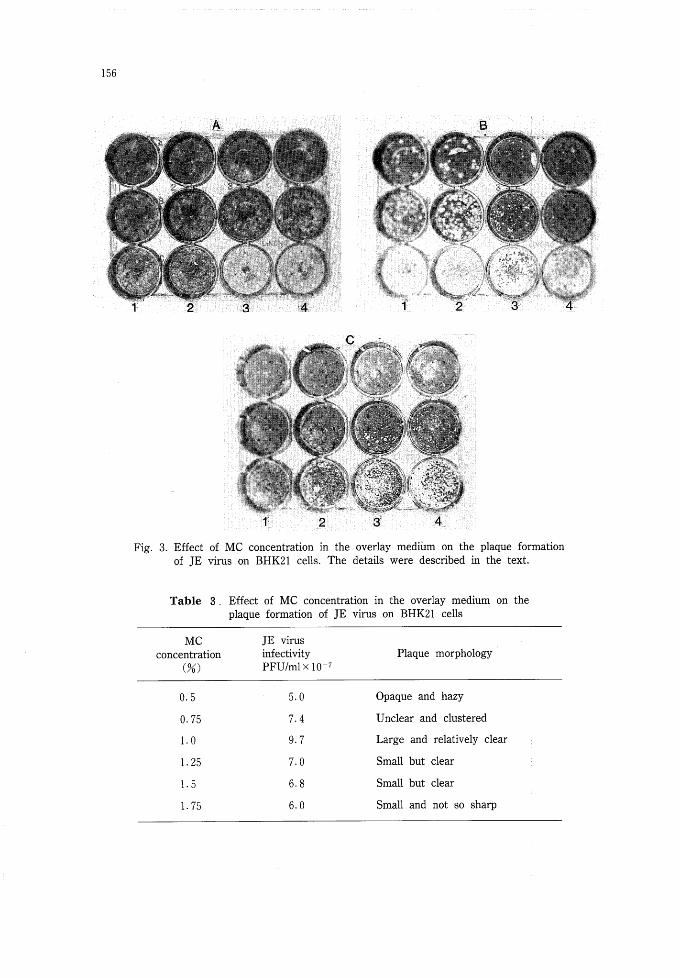
Effect of MC concentration in the overlay medium on the plaque formation of JE virus
Fig. 3 shows the plaques formed on BHK21 cells under overlay medium containing 6
different concentrations of MC; 0・5% in Fig・ 3A (columns 1 & 2), 0.75% in Fig・ 3A (coレ
Iimns 3 & 4), 1・0% in Fig. 3B (columns 1 & 2), 1・25% in Fig・ 3B (columns 3 & 4), 1・5%
in Fig. 3C (columns 3 & 4), and 1.75% in Fig. 3C (columns 1 & 2), respectively. The virus
titer did not change significantly by the MC concentrations as shown in Table 3. At lower
concentration of O・5 and O・75% MC, plaques were hazy and opaque and difficult to count
individually (Fig・ 3A). While at moderate to higher concentration of 1 and 1.25% (Fig・ 3B)
or l・5% MC (Fig. 3C columns 3 & 4), plaques were clear, distinct and easy to count・
156
1 2者 3 4
B
^v^-^
=
1 2 3 4
Fig・ 3. Effect of MC concentration in the overlay medium on the plaque formation
of JE virus on BHK21 cells. The details were described in the text.
Table 3. Effect of MC concentration in the overlay medium on the
plaque formation of JE virus on BHK21 cells
MC JE virus
concentration infectivity Plaque morphology
PFU/ml x lO者7
0・5
0・75
1.0
1.25
1・5
1 75
5・0
7・4
9・7
7林0
6.8
6.0
Opaque and hazy
Unclear and clustered
Large and relatively clear
Small but clear
Small but clear
Small and not so sharp

157
DISCUSSION
The data shows that con fluent or semiconfluent cell sheets at the time of virus in-
oculation gave higher virus titer than the sparse cell sheets・ For the cell culture of 2.0×
105/ml, 1 day's culture was sufficient, but longer incubation time was required for the
cultures with lower concentration of cell inoculum. However, the plaque size became
smaller when the virus was inoculated to higher density of the cells・ Roughly, reverse rela-
tion was observed between the virus titer and plaque size・ Regarding FBS concentration in
the cell growth medium, higher concentration gave higher virus titer, and again reverse
relation was observed between the virus titer and plaque size. Our data showed that 9%
of FBS in the cell growth medium should be used for the maximal PFU assay of JE virus
on the cell culture of 2 days after seeding O・25× 105 cells/ml・ The cultures of 1-2 days of
O・5■ 1・0 × 105 cells/ml gave almost similar virus titers, while the highest cell concentration
of 2.0 × 10⁵ cells/ml gave slightly less titer even when the cultures were inoculated with
the virus after 6 hr or 1 days incubation.
Regarding the MC concentration in the overlay medium, 1・25% or higher concentra-
tion gave almost the same virus titer and plaque size. From operational point of view, the
overlay medium of lower MC concentration is easier to handle because of less viscosity,
and 1.25% MC could be used for easy handling without losing the virus titer・
Liprandi (1981) documented PFU assay of yellow fever virus 17D vaccine strain by
¥% sodium CMC overlay medium and crystal violet staining of infected Vero cells. Similar
method was also reported by Buckley and Gould (1985) for the same virus and cells but
l・5% CMC overlay medium and naphthalene black staining. The first disadvantage of
these PFU assays by staining infected cells is longer incubation time of 5 days for JE
virus compared with the Okuno's method of immunoperoxidase staining which could pro-
vide the results within 1-2 days, but requires relatively tedious steps and counting the
foci under a microscope・ The second disadvantage is the inability to recover progeny
viruses from the plaques formed on the host cells, which will require agar or agarose
overlay medium instead of MC or CMC・
AcKNOWLEDGMENTS
This investigation was partially supported by a Grant者in-Aid for Scientific Research
No・ 62480161 from the Ministry of Education, Science and Culture oりapan in the year of
1989・ The first author was a recipient of the Monbusho Scholarship from the same
Ministry for his study in Japan・

158
REFERENCES
1) Buckley, A. & Gould, E・ A・ (1985): Neutralization of yellow fever virus studied using monoclonal
and polyclonal antibodies・ J・ Gen. ViroL, 66, 2523-2531・
2) Clarke, D. H. & Casals, J. (1965): Arboviruses; group B. pp 606-658. In F. L. Horsfall, Jr. & I.
Tamm (eds・ i. Viral and Rickettsial lnfections of Man. J. B・ Lippincott, Philadelphia & Montreal.
3) De Madrid, A・ T・ & Porterfield, J. S・ (1969): A simple micro-culture method for the study of
group B arboviruses. Bull. WHO., 40, 113-121.
4) Diercks, F. H. & Hammon, W. McD. (1958): Hamster Kidney cell tissu占cultures for propagation
of Japanese B encephalitis virus. Proc. Soc. Exp. Biol. Med., 97, 627-632.
5) Dulbecco, R・ & Vogt, M・ (1954): Plaque formation and isolation of pure lines with poliomyelitis
viruses・ J・ Exp. Med., 99, 167-182.
6) Eagle, H. (1959): Amino acid metabolism in mammalian cell cultures・ Science, 130, 432-437.
7) Hashimoto, N., Yamada, K・ & Kanamitsu, M. (1971): A microtiter method for assay of neutraliz・
ing antibodies against group B arboviruses・ Virus, 21, 55-59・
Hori, H・, Morita, K. & Igarashi, A・ (1986): Oligonucleotide fingerprint analysis of Japanese
encephalitis virus strains isolated in Japan and Thailand. Acta Virol・, 30, 353-359・
9) Igarashi, A・ (1978): Isolation of a Singh's Aedes albopictus cell clone sensitive to dengue and
chikungunya viruses・ J. Gen・ Virol・, 40, 531-544・
10) Inoue, Y・ K., Iwasaki, T・ & Kato, H. (1961): Studies on Japanese B encephalitis Virus・ Ⅰ5
Characteristics of mouse adapted and hamster kidnery cell adapted Japanese B encephalitis
viruses on plaque assay・ J・ Immunol., 87, 337者341・
ll) Inoue, Y・ K. & Ogura, R. (1962): Studies on Japanese B encephalitis virus・ Ⅲ・ Propagation and
assay of Japanese B encephalitis virus in a stable line of porcine kidney cells. Virology, 16, 205-
207.
12) Karabatsos, N・ & Buckley, S・ M林(1967): Susceptibility of the baby者hamster kidney cell line (BHK
21) to infection with arboviruses・ Am・ J・ Trop・ Med・ Hyg・, 16, 99-105.
13) Kissling, R. E. (1957): Growth of several arthropod-borne viruses in tissue culture. Proc林Soc・
Exp・ Biol. Med・, 96, 290-294・
14) Lee, H・ W. Hinz, R・ W. & Scherer, W・ F. (1958): Porcine kidney cell cultures for propagation
and assay of Japanese encephalitis virus. Proc. Soc・ Exp・ Biol. Med., 99, 579-583・
15) Liprandi, F・ (1981): Isolation of plaque variants differing in virulence from the 17D strain of
yellow fever virus. J・ Gen. Virol・, 56, 363-370・
16) Miles, J林A. R. (1960): Epidemiology of arthropod-borne encephalitides・ Bull・ WHO, 22, 339-371・
17) Monath, T. P. (1985): Flaviviruses. pp 955-1004. In B. N. Fields, D. M. Kmpe, R. M. Chanock,
J・ L・ Melnick, B. Roizman & R. E・ Shope (eds林)・ Fields Virology, Raven Press, New York.
18) Monath, T. P. (1986): Pathology of the flaviviruses. pp 375-440. In S. Schlesinger & M. J.
schlesinger (eds・ i. The Togaviridae and Flaviviridae・ The Viruses (H・ Fraenkelconrat & R. R・
Wagner, Ser. eds.)・ Plenum Press, New York.
19) Okuno, Y., Igarashi, A・ & Fukai, k・ (1978): Neutralization tests for dengue and Japanese
encephalitis viruses by the focus reduction method using peroxidase-anti-peroxidase staining・
Biken J・ 26, 137-147・

159
20) Okuno, Y林, Fukunaga, T・ Tadano, M・ Okamoto, Y. Ohnishi, T. & Takagi, M・ (1985): Rapid for-
cus reduction neutralization test of Japanese encephalitis virus in microtiter system・ Arch. Virol・,
3, 129-135・
21) Rosen, L・ (1986): The natural history of Japanese encephalitis virus・ Ann・ Rev・ Microbiol., 40, 395
-414.
22) Shope, R・ E・ (1980): Medical significance of togaviruses: an overview of diseases caused by
togaviruses in man and in domestic and wild vertebrate animals林pp 47-82・ In R. W. Schlesinger
(ed・ i. The Togaviruses, Biology, Structure, Replication. Academic Press, New York, London,
Toronto, Sydney, San Francisco・
23) Umenai, T., Krzysko, R., Bektimirov, A. & Assaad, F・ A・ (1985): Japanese encephalitis: current
world wide status. Bull, WHO., 63, 625-631.
24) Westaway, E・ G. (1966): Assessment and application of a cell line from pig kidney for plaque
assay and neutralization tests with twelve group B arboviruses林Am・ J. Epidemiol・ 1, 439-456・
25) Westaway, E. G., Brinton, M. A., Gaidamovich, S. Ya., Horzinek, M. C., Igarashi, A.,
Kaanamen, L., Lvov, D. K., Porterfield, J. S., Russell, P. K. & Trent, D. W. (1985):
Flavivindae. Intervirology, 24, 183- 192.

![CWIQ data dictionary Section: 0 Prefix fields Table · Ni muda gani [JINA] amekuwa haishi hapa katika kipindi cha miezi 12 iliyopita? 1 Never (has always been here) 1 Hajawahi (alikuwepo](https://img.pdfslide.tips/doc/110x75/5e488c0bb15ff768247c4fe1/cwiq-data-dictionary-section-0-prefix-fields-table-ni-muda-gani-jina-amekuwa.jpg)