Embed Size (px)
Citation preview
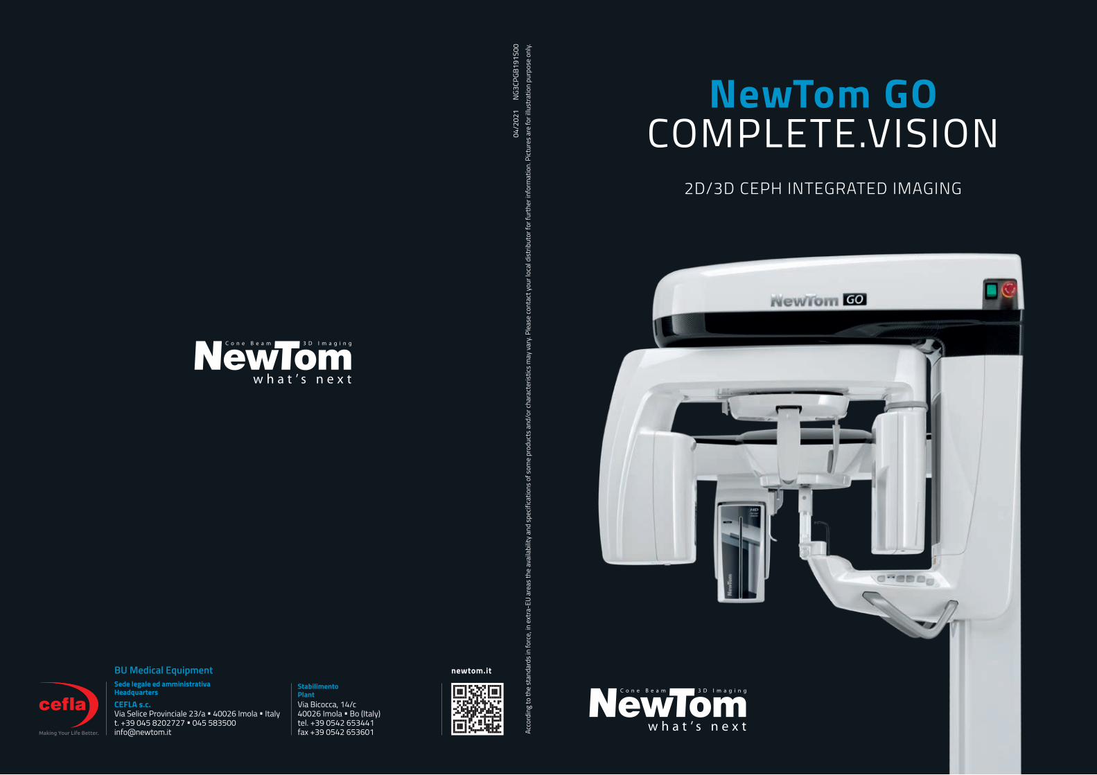
NewTom GOCOMPLETE.VISION
CEFLA s.c.Via Selice Provinciale 23/a • 40026 Imola • Italyt. +39 045 8202727 • 045 [email protected]
newtom.itBU Medical EquipmentStabilimento PlantVia Bicocca, 14/c40026 Imola • Bo (Italy)tel. +39 0542 653441fax +39 0542 653601
Sede legale ed amministrativa Headquarters
2D/3D CEPH INTEGRATED IMAGING
04/2
021
NG3
CPGB
191S
00Ac
cord
ing
to th
e st
anda
rds
in fo
rce,
in e
xtra
-EU
area
s th
e av
aila
bilit
y an
d sp
ecifi
catio
ns o
f som
e pr
oduc
ts a
nd/o
r cha
ract
eris
tics
may
var
y. Pl
ease
cont
act y
our l
ocal
dis
trib
utor
for f
urth
er in
form
atio
n. P
ictur
es a
re fo
r illu
stra
tion
purp
ose
only.
GO 2D/3D CEPHCOMPLETE.VISION
3
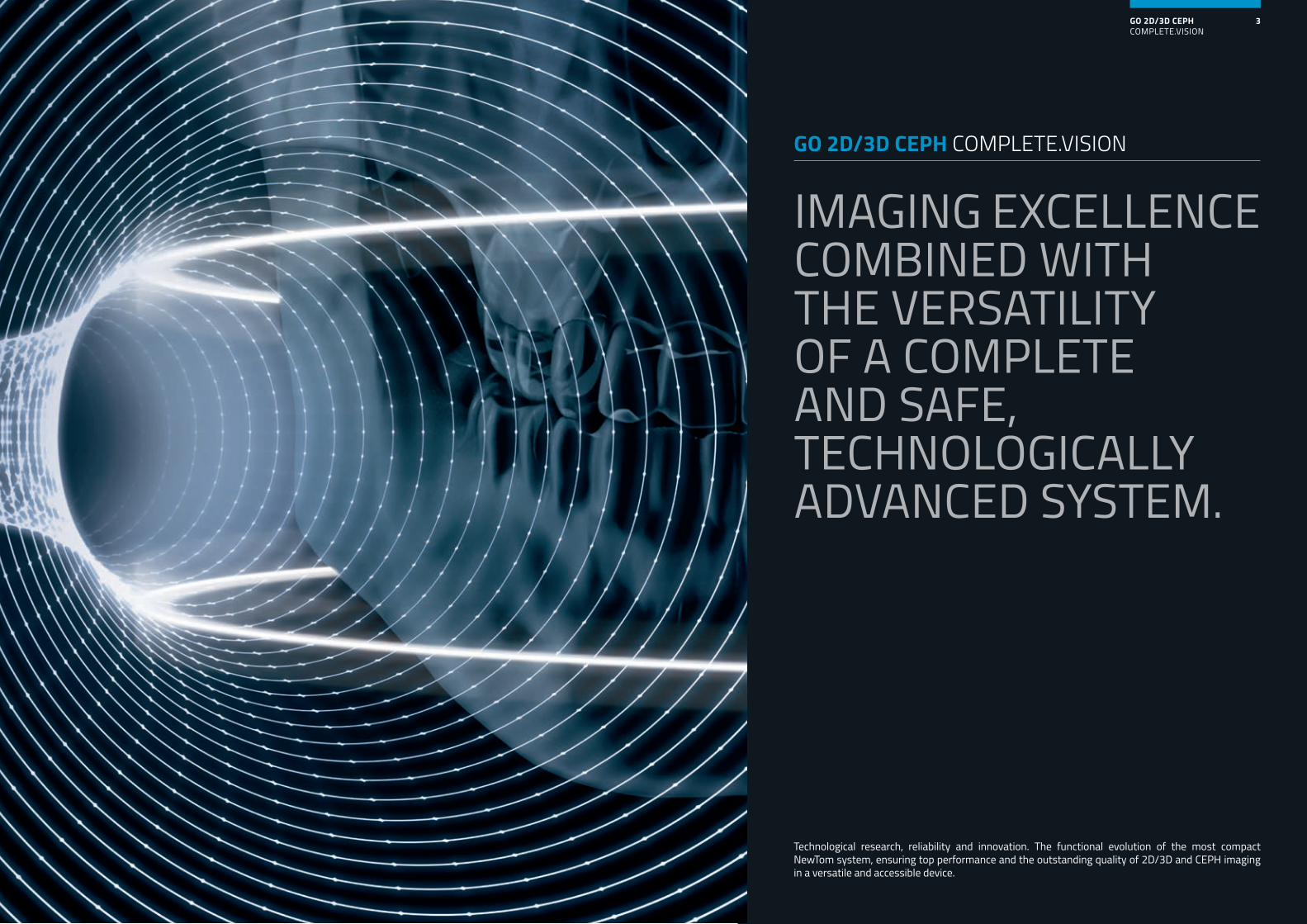
IMAGING EXCELLENCE COMBINED WITH THE VERSATILITY OF A COMPLETE AND SAFE, TECHNOLOGICALLY ADVANCED SYSTEM.
GO 2D/3D CEPH COMPLETE.VISION
Technological research, reliability and innovation. The functional evolution of the most compact NewTom system, ensuring top performance and the outstanding quality of 2D/3D and CEPH imaging in a versatile and accessible device.
GO 2D/3D CEPHCOMPLETE.VISION
4
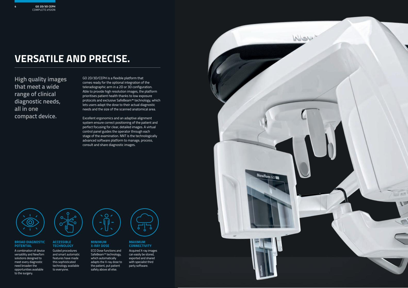
GO 2D/3D/CEPH is a flexible platform that comes ready for the optional integration of the teleradiographic arm in a 2D or 3D configuration. Able to provide high resolution images, the platform prioritises patient health thanks to low exposure protocols and exclusive SafeBeam™ technology, which lets users adapt the dose to their actual diagnostic needs and the size of the scanned anatomical area.
Excellent ergonomics and an adaptive alignment system ensure correct positioning of the patient and perfect focusing for clear, detailed images. A virtual control panel guides the operator through each stage of the examination. NNT is the technologically advanced software platform to manage, process, consult and share diagnostic images.
High quality images that meet a wide range of clinical diagnostic needs, all in one compact device.
MINIMUM X-RAY DOSEECO Dose functions and SafeBeam™ technology, which automatically adapts the X-ray dose to the patient, put patient safety above all else.
ACCESSIBLE TECHNOLOGYGuided procedures and smart automatic features have made this sophisticated technology available to everyone.
BROAD DIAGNOSTIC POTENTIALA combination of device versatility and NewTom solutions designed to meet every diagnostic need broaden the opportunities available to the surgery.
MAXIMUM CONNECTIVITYAcquired X-ray images can easily be stored, exported and shared with specialist third party software.
VERSATILE AND PRECISE.
GO 2D/3D CEPHCOMPLETE.VISION
6
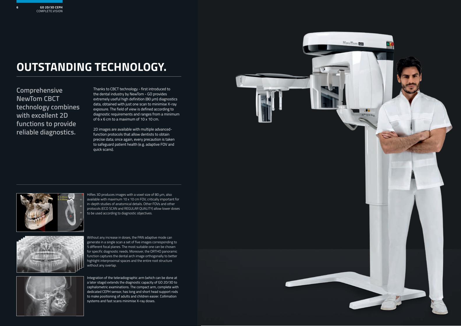
Thanks to CBCT technology - first introduced to the dental industry by NewTom - GO provides extremely useful high definition (80 μm) diagnostics data, obtained with just one scan to minimise X-ray exposure. The field of view is defined according to diagnostic requirements and ranges from a minimum of 6 x 6 cm to a maximum of 10 x 10 cm.
2D images are available with multiple advanced-function protocols that allow dentists to obtain precise data; once again, every precaution is taken to safeguard patient health (e.g. adaptive FOV and quick scans).
Comprehensive NewTom CBCT technology combines with excellent 2D functions to provide reliable diagnostics.
OUTSTANDING TECHNOLOGY.
Integration of the teleradiographic arm (which can be done at a later stage) extends the diagnostic capacity of GO 2D/3D to cephalometric examinations. The compact arm, complete with dedicated CEPH sensor, has long and short head support rods to make positioning of adults and children easier. Collimation systems and fast scans minimise X-ray doses.
Without any increase in doses, the PAN adaptive mode can generate in a single scan a set of five images corresponding to 5 different focal planes. The most suitable one can be chosen for specific diagnostic needs. Moreover, the ORTHO panoramic function captures the dental arch image orthogonally to better highlight interproximal spaces and the entire root structure without any overlap.
HiRes 3D produces images with a voxel size of 80 μm, also available with maximum 10 x 10 cm FOV, critically important for in-depth studies of anatomical details. Other FOVs and other protocols (ECO SCAN and REGULAR QUALITY) allow lower doses to be used according to diagnostic objectives.
GO 2D/3D CEPHCOMPLETE.VISION
9GO 2D/3D CEPHCOMPLETE.VISION
8
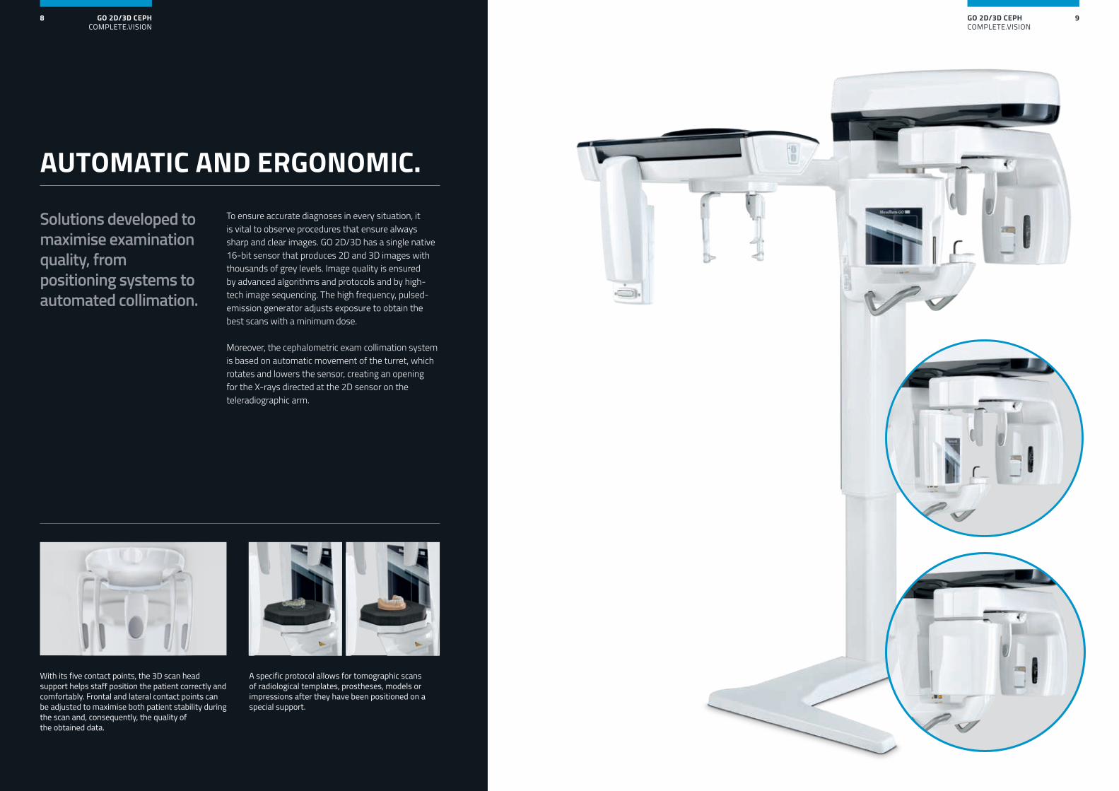
To ensure accurate diagnoses in every situation, it is vital to observe procedures that ensure always sharp and clear images. GO 2D/3D has a single native 16-bit sensor that produces 2D and 3D images with thousands of grey levels. Image quality is ensured by advanced algorithms and protocols and by high-tech image sequencing. The high frequency, pulsed-emission generator adjusts exposure to obtain the best scans with a minimum dose.
Moreover, the cephalometric exam collimation system is based on automatic movement of the turret, which rotates and lowers the sensor, creating an opening for the X-rays directed at the 2D sensor on the teleradiographic arm.
Solutions developed to maximise examination quality, from positioning systems to automated collimation.
With its five contact points, the 3D scan head support helps staff position the patient correctly and comfortably. Frontal and lateral contact points can be adjusted to maximise both patient stability during the scan and, consequently, the quality of the obtained data.
A specific protocol allows for tomographic scans of radiological templates, prostheses, models or impressions after they have been positioned on a special support.
AUTOMATIC AND ERGONOMIC.
GO 2D/3D CEPHCOMPLETE.VISION
10
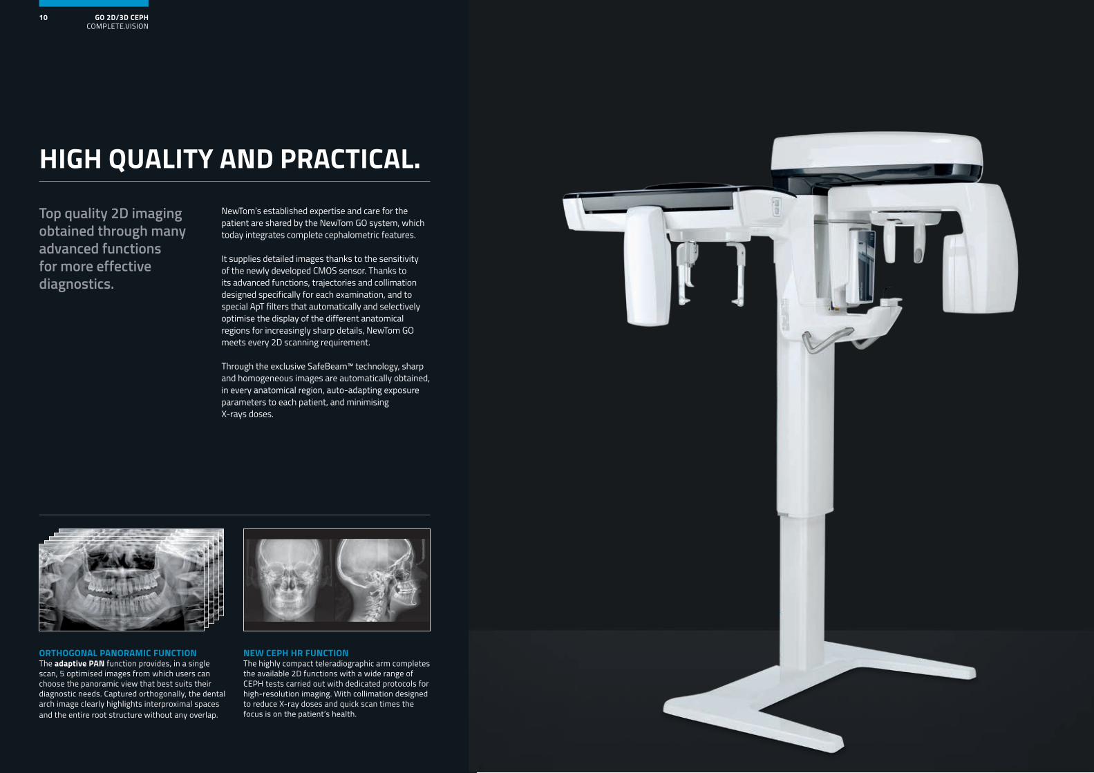
Top quality 2D imaging obtained through many advanced functions for more effective diagnostics.
ORTHOGONAL PANORAMIC FUNCTIONThe adaptive PAN function provides, in a single scan, 5 optimised images from which users can choose the panoramic view that best suits their diagnostic needs. Captured orthogonally, the dental arch image clearly highlights interproximal spaces and the entire root structure without any overlap.
NEW CEPH HR FUNCTIONThe highly compact teleradiographic arm completes the available 2D functions with a wide range of CEPH tests carried out with dedicated protocols for high-resolution imaging. With collimation designed to reduce X-ray doses and quick scan times the focus is on the patient’s health.
NewTom’s established expertise and care for the patient are shared by the NewTom GO system, which today integrates complete cephalometric features.
It supplies detailed images thanks to the sensitivity of the newly developed CMOS sensor. Thanks to its advanced functions, trajectories and collimation designed specifically for each examination, and to special ApT filters that automatically and selectively optimise the display of the different anatomical regions for increasingly sharp details, NewTom GO meets every 2D scanning requirement.
Through the exclusive SafeBeam™ technology, sharp and homogeneous images are automatically obtained, in every anatomical region, auto-adapting exposure parameters to each patient, and minimising X-rays doses.
HIGH QUALITY AND PRACTICAL.
GO 2D/3D CEPHCOMPLETE.VISION
12
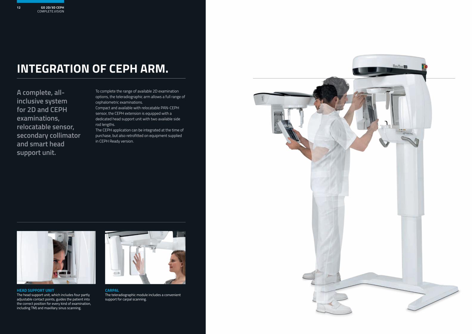
To complete the range of available 2D examination options, the teleradiographic arm allows a full range of cephalometric examinations. Compact and available with relocatable PAN-CEPH sensor, the CEPH extension is equipped with a dedicated head support unit with two available side rod lengths. The CEPH application can be integrated at the time of purchase, but also retrofitted on equipment supplied in CEPH Ready version.
A complete, all-inclusive system for 2D and CEPH examinations, relocatable sensor, secondary collimator and smart head support unit.
INTEGRATION OF CEPH ARM.
CARPALThe teleradiographic module includes a convenient support for carpal scanning.
HEAD SUPPORT UNITThe head support unit, which includes four partly adjustable contact points, guides the patient into the correct position for every kind of examination, including TMJ and maxillary sinus scanning.
GO 2D/3D CEPHCOMPLETE.VISION
15GO 2D/3D CEPHCOMPLETE.VISION
14
10x10
8x7
IMAGING 3D
aMAR
aMAR
aMAR
REGULAR QUALITY
BEST QUALITY
ECO QUALITY
aMAR
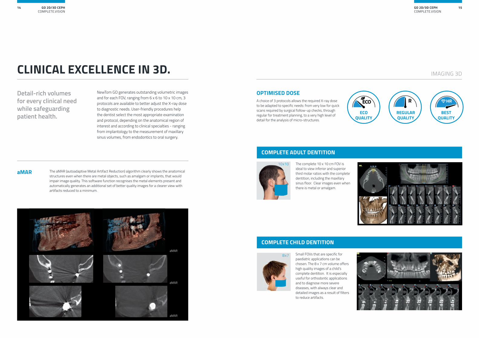
NewTom GO generates outstanding volumetric images and for each FOV, ranging from 6 x 6 to 10 x 10 cm, 3 protocols are available to better adjust the X-ray dose to diagnostic needs. User-friendly procedures help the dentist select the most appropriate examination and protocol, depending on the anatomical region of interest and according to clinical specialties - ranging from implantology to the measurement of maxillary sinus volumes, from endodontics to oral surgery.
Detail-rich volumes for every clinical need while safeguarding patient health.
The complete 10 x 10 cm FOV is ideal to view inferior and superior third molar ratios with the complete dentition, including the maxillary sinus floor. Clear images even when there is metal or amalgam.
Small FOVs that are specific for paediatric applications can be chosen. The 8 x 7 cm volume offers high quality images of a child’s complete dentition. It is especially useful for orthodontic applications and to diagnose more severe diseases, with always clear and detailed images as a result of filters to reduce artifacts.
The aMAR (autoadaptive Metal Artifact Reduction) algorithm clearly shows the anatomical structures even when there are metal objects, such as amalgam or implants, that would impair image quality. This software function recognises the metal elements present and automatically generates an additional set of better quality images for a clearer view with artifacts reduced to a minimum.
OPTIMISED DOSE A choice of 3 protocols allows the required X-ray dose to be adapted to specific needs: from very low for quick scans required by surgical follow-up checks, through regular for treatment planning, to a very high level of detail for the analysis of micro-structures.
CLINICAL EXCELLENCE IN 3D.
COMPLETE ADULT DENTITION
COMPLETE CHILD DENTITION
GO 2D/3D CEPHCOMPLETE.VISION
17GO 2D/3D CEPHCOMPLETE.VISION
16
IMAGING 3D
8x7 10x7
6x7 6x6
8x10
8x6 10x6
10x10 8x10
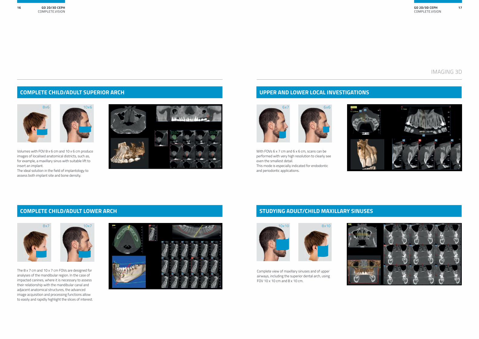
Volumes with FOV 8 x 6 cm and 10 x 6 cm produce images of localised anatomical districts, such as, for example, a maxillary sinus with suitable lift to insert an implant. The ideal solution in the field of implantology to assess both implant site and bone density.
The 8 x 7 cm and 10 x 7 cm FOVs are designed for analyses of the mandibular region. In the case of impacted canines, where it is necessary to assess their relationship with the mandibular canal and adjacent anatomical structures, the advanced image acquisition and processing functions allow to easily and rapidly highlight the slices of interest.
With FOVs 6 x 7 cm and 6 x 6 cm, scans can be performed with very high resolution to clearly see even the smallest detail. This mode is especially indicated for endodontic and periodontic applications.
Complete view of maxillary sinuses and of upper airways, including the superior dental arch, using FOV 10 x 10 cm and 8 x 10 cm.
COMPLETE CHILD/ADULT SUPERIOR ARCH
COMPLETE CHILD/ADULT LOWER ARCH
UPPER AND LOWER LOCAL INVESTIGATIONS
STUDYING ADULT/CHILD MAXILLARY SINUSES
GO 2D/3D CEPHCOMPLETE.VISION
19GO 2D/3D CEPHCOMPLETE.VISION
18
IMAGING 2D
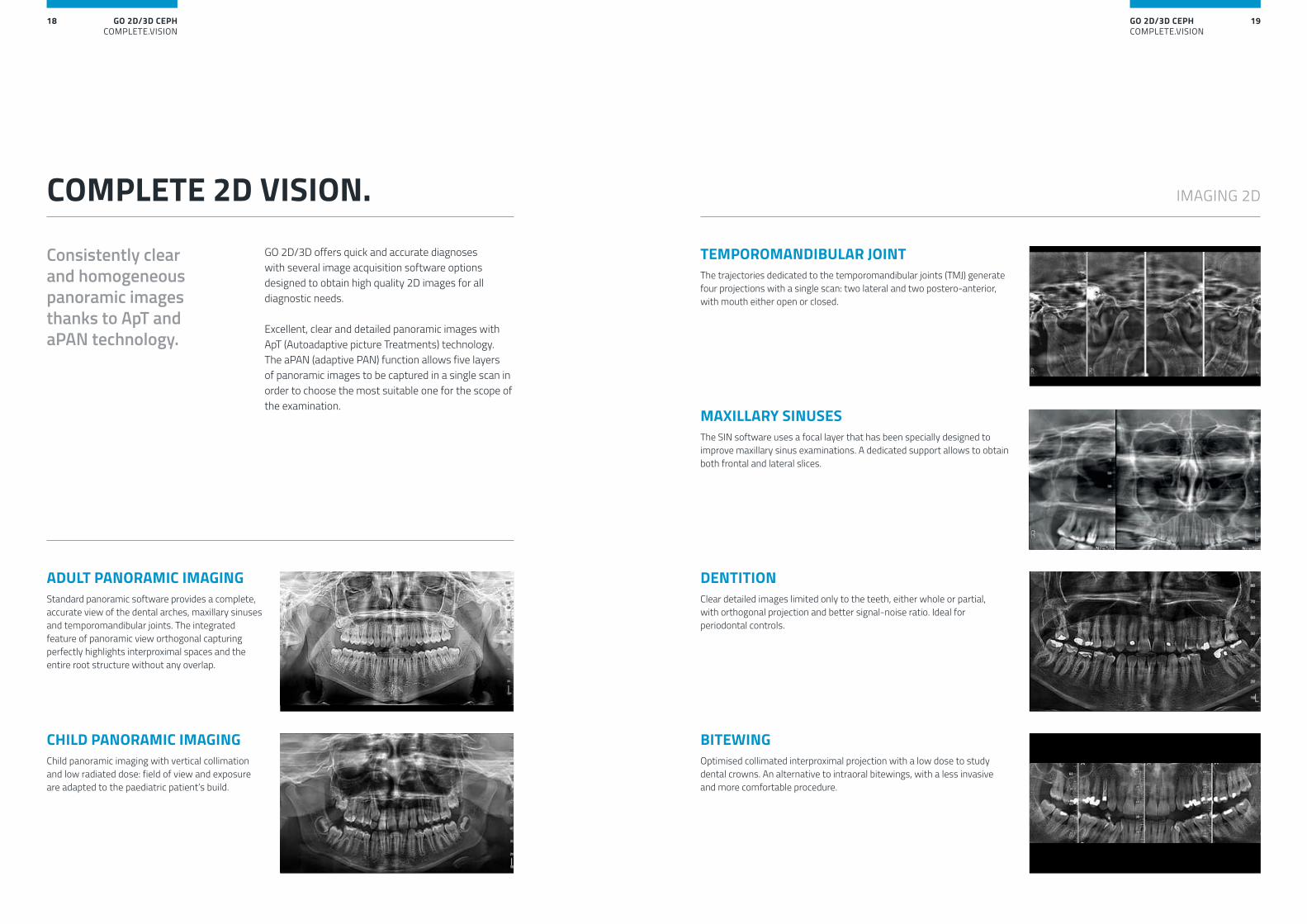
TEMPOROMANDIBULAR JOINT The trajectories dedicated to the temporomandibular joints (TMJ) generate four projections with a single scan: two lateral and two postero-anterior, with mouth either open or closed.
MAXILLARY SINUSES The SIN software uses a focal layer that has been specially designed to improve maxillary sinus examinations. A dedicated support allows to obtain both frontal and lateral slices.
DENTITION Clear detailed images limited only to the teeth, either whole or partial, with orthogonal projection and better signal-noise ratio. Ideal for periodontal controls.
ADULT PANORAMIC IMAGINGStandard panoramic software provides a complete, accurate view of the dental arches, maxillary sinuses and temporomandibular joints. The integrated feature of panoramic view orthogonal capturing perfectly highlights interproximal spaces and the entire root structure without any overlap.
BITEWING Optimised collimated interproximal projection with a low dose to study dental crowns. An alternative to intraoral bitewings, with a less invasive and more comfortable procedure.
CHILD PANORAMIC IMAGINGChild panoramic imaging with vertical collimation and low radiated dose: field of view and exposure are adapted to the paediatric patient’s build.
GO 2D/3D offers quick and accurate diagnoses with several image acquisition software options designed to obtain high quality 2D images for all diagnostic needs.
Excellent, clear and detailed panoramic images with ApT (Autoadaptive picture Treatments) technology. The aPAN (adaptive PAN) function allows five layers of panoramic images to be captured in a single scan in order to choose the most suitable one for the scope of the examination.
Consistently clear and homogeneous panoramic images thanks to ApT and aPAN technology.
COMPLETE 2D VISION.
GO 2D/3D CEPHCOMPLETE.VISION
21GO 2D/3D CEPHCOMPLETE.VISION
20
ApT (AUTOADAPTIVE PICTURE TREATEMENTS)Auto-adaptive filters automatically improve every 2D image to ensure the best result for every projection.
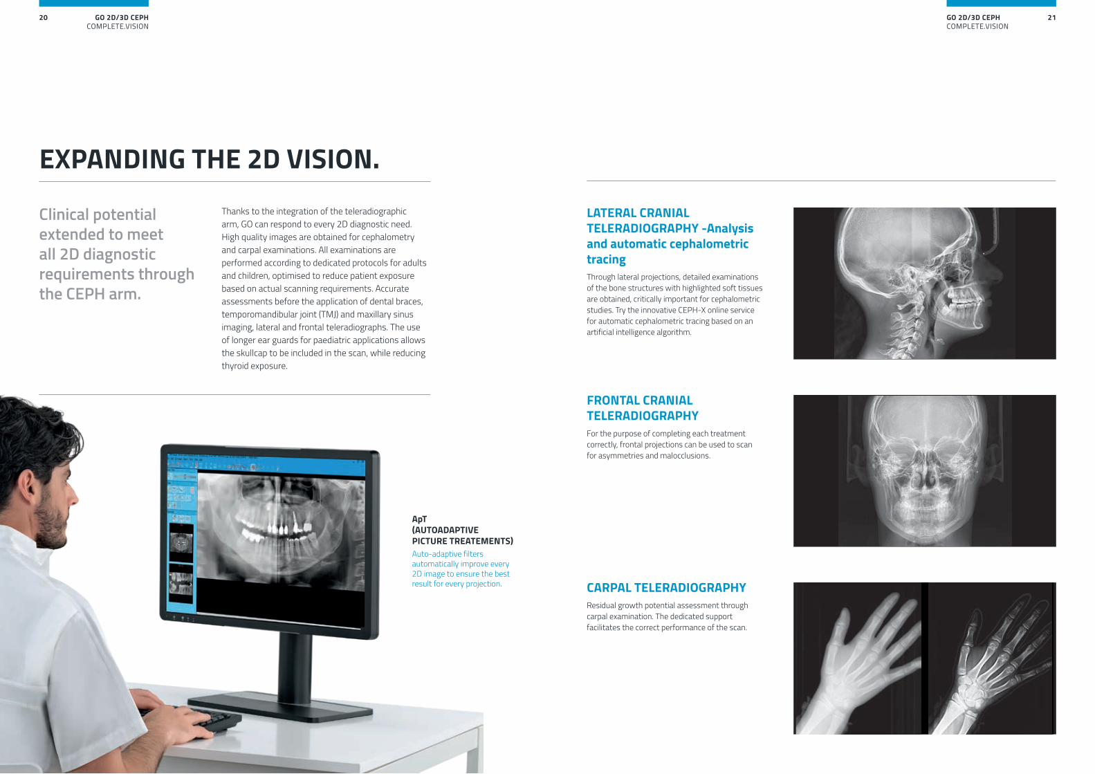
LATERAL CRANIAL TELERADIOGRAPHY -Analysis and automatic cephalometric tracingThrough lateral projections, detailed examinations of the bone structures with highlighted soft tissues are obtained, critically important for cephalometric studies. Try the innovative CEPH-X online service for automatic cephalometric tracing based on an artificial intelligence algorithm.
FRONTAL CRANIAL TELERADIOGRAPHYFor the purpose of completing each treatment correctly, frontal projections can be used to scan for asymmetries and malocclusions.
CARPAL TELERADIOGRAPHY Residual growth potential assessment through carpal examination. The dedicated support facilitates the correct performance of the scan.
EXPANDING THE 2D VISION.
Clinical potential extended to meet all 2D diagnostic requirements through the CEPH arm.
Thanks to the integration of the teleradiographic arm, GO can respond to every 2D diagnostic need. High quality images are obtained for cephalometry and carpal examinations. All examinations are performed according to dedicated protocols for adults and children, optimised to reduce patient exposure based on actual scanning requirements. Accurate assessments before the application of dental braces, temporomandibular joint (TMJ) and maxillary sinus imaging, lateral and frontal teleradiographs. The use of longer ear guards for paediatric applications allows the skullcap to be included in the scan, while reducing thyroid exposure.
GO 2D/3D CEPHCOMPLETE.VISION
23GO 2D/3D CEPHCOMPLETE.VISION
22
3.7s
CEPH
Best Quality
Regular Quality
EcoQuality
Pan Ortho
0
1
2
3
4
5
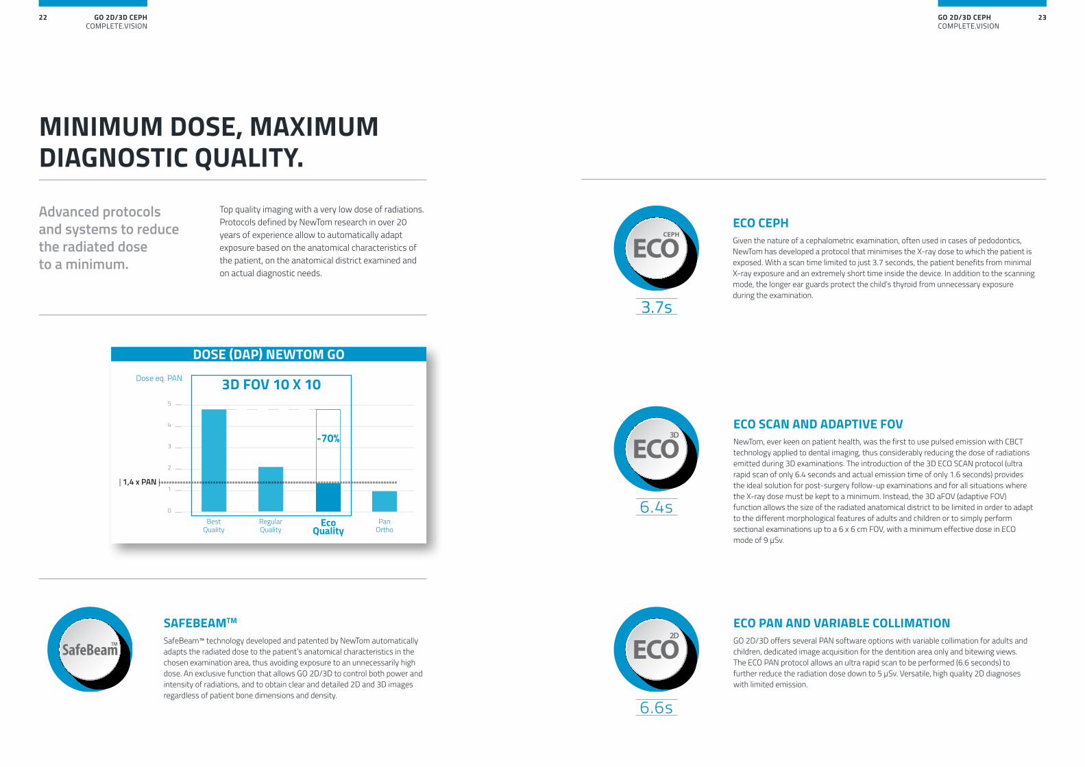
| 1,4 x PAN |
Dose eq. PAN 3D FOV 10 X 10
-70%
DOSE (DAP) NEWTOM GO
Top quality imaging with a very low dose of radiations. Protocols defined by NewTom research in over 20 years of experience allow to automatically adapt exposure based on the anatomical characteristics of the patient, on the anatomical district examined and on actual diagnostic needs.
MINIMUM DOSE, MAXIMUM DIAGNOSTIC QUALITY.
Advanced protocols and systems to reduce the radiated dose to a minimum.
ECO SCAN AND ADAPTIVE FOVNewTom, ever keen on patient health, was the first to use pulsed emission with CBCT technology applied to dental imaging, thus considerably reducing the dose of radiations emitted during 3D examinations. The introduction of the 3D ECO SCAN protocol (ultra rapid scan of only 6.4 seconds and actual emission time of only 1.6 seconds) provides the ideal solution for post-surgery follow-up examinations and for all situations where the X-ray dose must be kept to a minimum. Instead, the 3D aFOV (adaptive FOV) function allows the size of the radiated anatomical district to be limited in order to adapt to the different morphological features of adults and children or to simply perform sectional examinations up to a 6 x 6 cm FOV, with a minimum effective dose in ECO mode of 9 μSv.
ECO PAN AND VARIABLE COLLIMATIONGO 2D/3D offers several PAN software options with variable collimation for adults and children, dedicated image acquisition for the dentition area only and bitewing views. The ECO PAN protocol allows an ultra rapid scan to be performed (6.6 seconds) to further reduce the radiation dose down to 5 μSv. Versatile, high quality 2D diagnoses with limited emission.
SAFEBEAMTM
SafeBeam™ technology developed and patented by NewTom automatically adapts the radiated dose to the patient’s anatomical characteristics in the chosen examination area, thus avoiding exposure to an unnecessarily high dose. An exclusive function that allows GO 2D/3D to control both power and intensity of radiations, and to obtain clear and detailed 2D and 3D images regardless of patient bone dimensions and density.
ECO CEPHGiven the nature of a cephalometric examination, often used in cases of pedodontics, NewTom has developed a protocol that minimises the X-ray dose to which the patient is exposed. With a scan time limited to just 3.7 seconds, the patient benefits from minimal X-ray exposure and an extremely short time inside the device. In addition to the scanning mode, the longer ear guards protect the child’s thyroid from unnecessary exposure during the examination.
GO 2D/3D CEPHCOMPLETE.VISION
25GO 2D/3D CEPHCOMPLETE.VISION
24
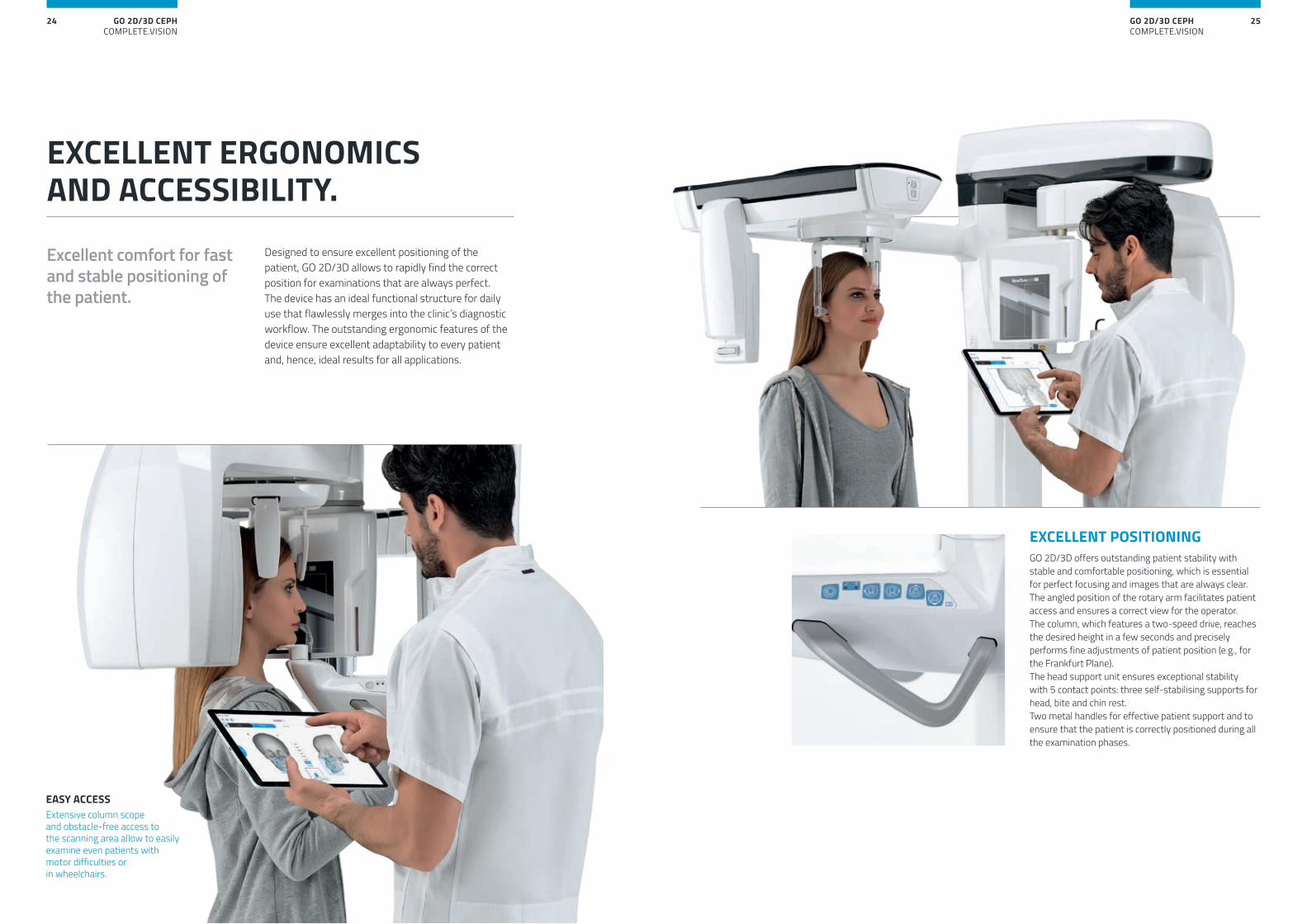
Designed to ensure excellent positioning of the patient, GO 2D/3D allows to rapidly find the correct position for examinations that are always perfect. The device has an ideal functional structure for daily use that flawlessly merges into the clinic’s diagnostic workflow. The outstanding ergonomic features of the device ensure excellent adaptability to every patient and, hence, ideal results for all applications.
Excellent comfort for fast and stable positioning of the patient.
EXCELLENT ERGONOMICS AND ACCESSIBILITY.
EASY ACCESS Extensive column scope and obstacle-free access to the scanning area allow to easily examine even patients with motor difficulties or in wheelchairs.
EXCELLENT POSITIONINGGO 2D/3D offers outstanding patient stability with stable and comfortable positioning, which is essential for perfect focusing and images that are always clear. The angled position of the rotary arm facilitates patient access and ensures a correct view for the operator. The column, which features a two-speed drive, reaches the desired height in a few seconds and precisely performs fine adjustments of patient position (e.g., for the Frankfurt Plane).The head support unit ensures exceptional stability with 5 contact points: three self-stabilising supports for head, bite and chin rest.Two metal handles for effective patient support and to ensure that the patient is correctly positioned during all the examination phases.
GO 2D/3D CEPHCOMPLETE.VISION
27GO 2D/3D CEPHCOMPLETE.VISION
26
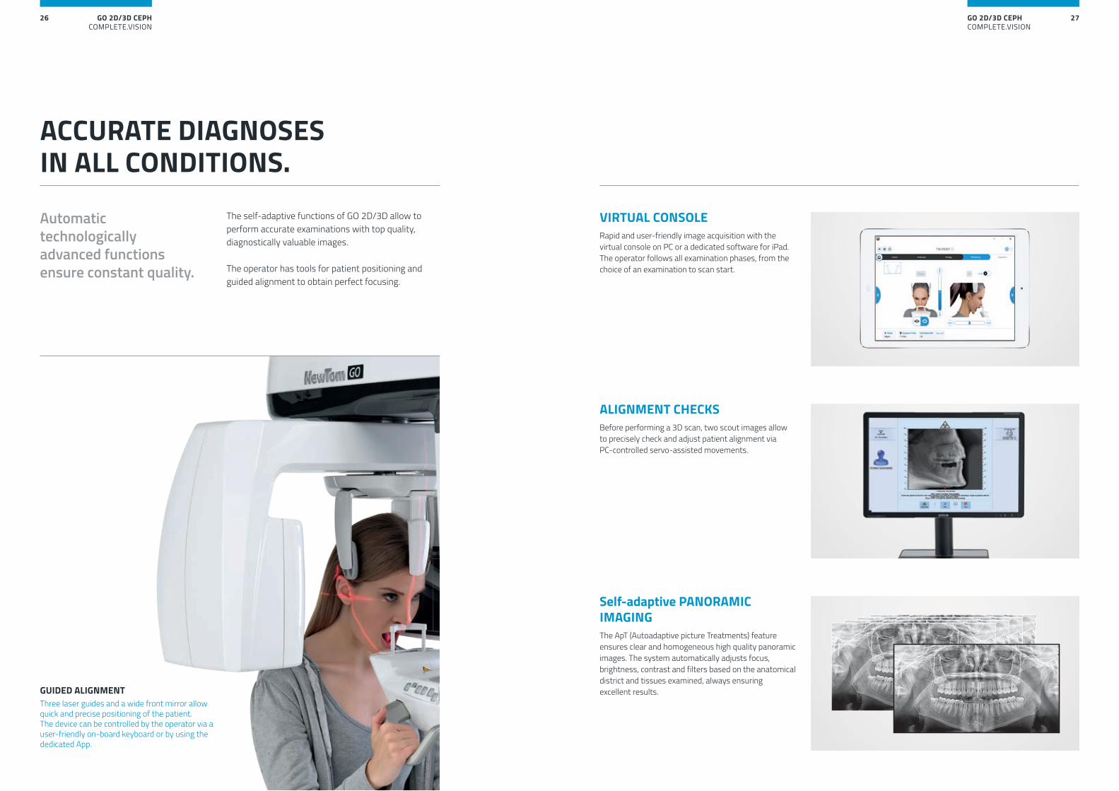
The self-adaptive functions of GO 2D/3D allow to perform accurate examinations with top quality, diagnostically valuable images.
The operator has tools for patient positioning and guided alignment to obtain perfect focusing.
ACCURATE DIAGNOSES IN ALL CONDITIONS.
Automatic technologically advanced functions ensure constant quality.
Self-adaptive PANORAMIC IMAGINGThe ApT (Autoadaptive picture Treatments) feature ensures clear and homogeneous high quality panoramic images. The system automatically adjusts focus, brightness, contrast and filters based on the anatomical district and tissues examined, always ensuring excellent results.
ALIGNMENT CHECKSBefore performing a 3D scan, two scout images allow to precisely check and adjust patient alignment via PC-controlled servo-assisted movements.
GUIDED ALIGNMENTThree laser guides and a wide front mirror allow quick and precise positioning of the patient. The device can be controlled by the operator via a user-friendly on-board keyboard or by using the dedicated App.
VIRTUAL CONSOLERapid and user-friendly image acquisition with the virtual console on PC or a dedicated software for iPad. The operator follows all examination phases, from the choice of an examination to scan start.
GO 2D/3D CEPHCOMPLETE.VISION
29GO 2D/3D CEPHCOMPLETE.VISION
28
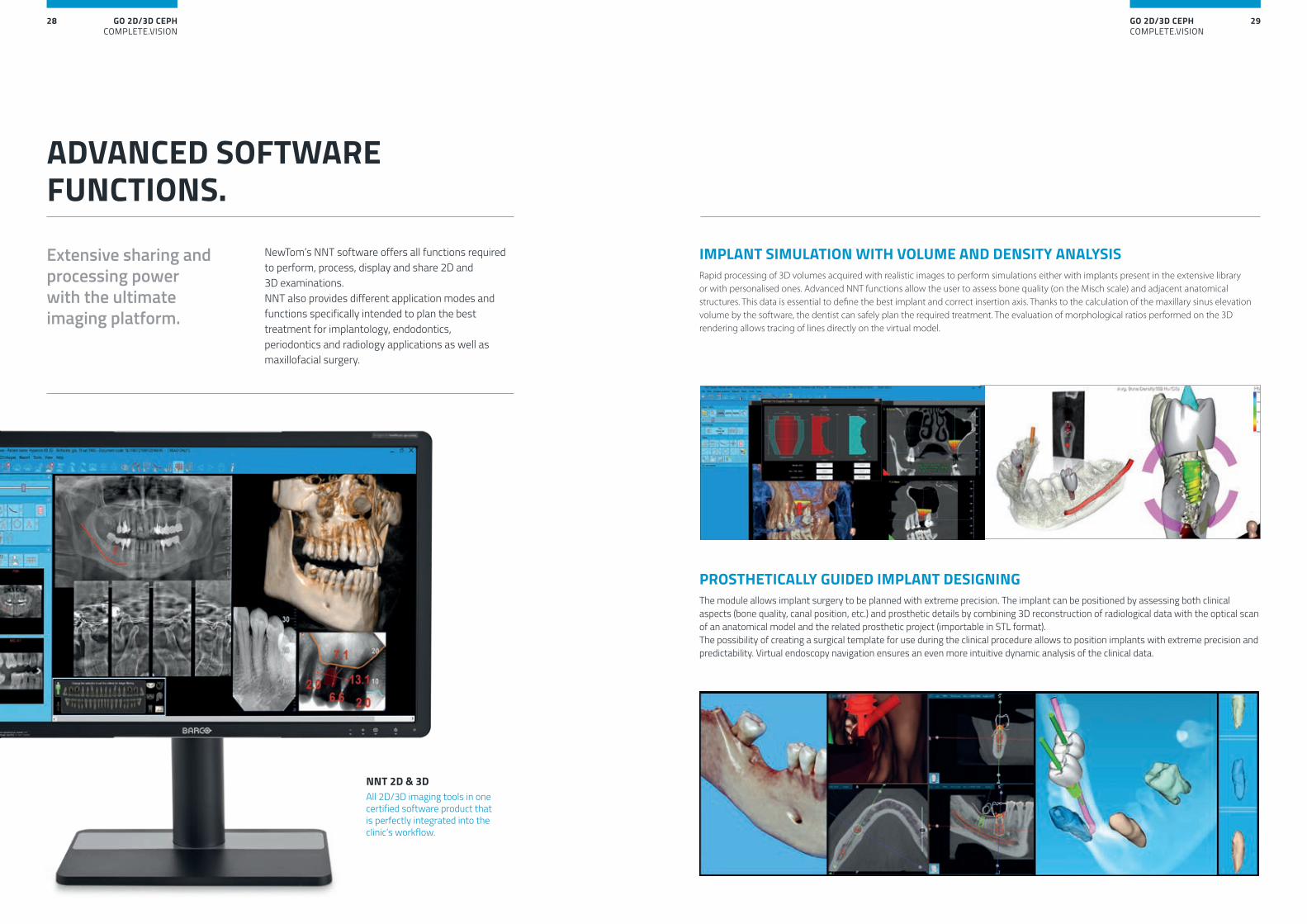
NewTom’s NNT software offers all functions required to perform, process, display and share 2D and 3D examinations. NNT also provides different application modes and functions specifically intended to plan the best treatment for implantology, endodontics, periodontics and radiology applications as well as maxillofacial surgery.
ADVANCED SOFTWARE FUNCTIONS.
Extensive sharing and processing power with the ultimate imaging platform.
NNT 2D & 3DAll 2D/3D imaging tools in one certified software product thatis perfectly integrated into theclinic’s workflow.
IMPLANT SIMULATION WITH VOLUME AND DENSITY ANALYSISRapid processing of 3D volumes acquired with realistic images to perform simulations either with implants present in the extensive library or with personalised ones. Advanced NNT functions allow the user to assess bone quality (on the Misch scale) and adjacent anatomical structures. This data is essential to define the best implant and correct insertion axis. Thanks to the calculation of the maxillary sinus elevation volume by the software, the dentist can safely plan the required treatment. The evaluation of morphological ratios performed on the 3D rendering allows tracing of lines directly on the virtual model.
PROSTHETICALLY GUIDED IMPLANT DESIGNINGThe module allows implant surgery to be planned with extreme precision. The implant can be positioned by assessing both clinical aspects (bone quality, canal position, etc.) and prosthetic details by combining 3D reconstruction of radiological data with the optical scan of an anatomical model and the related prosthetic project (importable in STL format). The possibility of creating a surgical template for use during the clinical procedure allows to position implants with extreme precision and predictability. Virtual endoscopy navigation ensures an even more intuitive dynamic analysis of the clinical data.
GO 2D/3D CEPHCOMPLETE.VISION
31GO 2D/3D CEPHCOMPLETE.VISION
30
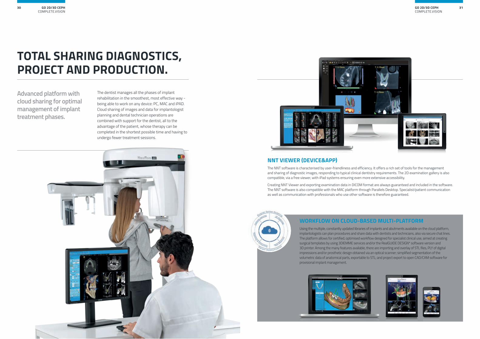
The dentist manages all the phases of implant rehabilitation in the smoothest, most effective way - being able to work on any device: PC, MAC and iPAD. Cloud sharing of images and data for implantologist planning and dental technician operations are combined with support for the dentist, all to the advantage of the patient, whose therapy can be completed in the shortest possible time and having to undergo fewer treatment sessions.
TOTAL SHARING DIAGNOSTICS, PROJECT AND PRODUCTION.
Advanced platform with cloud sharing for optimal management of implant treatment phases.
WORKFLOW ON CLOUD-BASED MULTI-PLATFORMUsing the multiple, constantly updated libraries of implants and abutments available on the cloud platform, implantologists can plan procedures and share data with dentists and technicians, also via secure chat lines. The platform allows for certified, optimised workflow designed for specialist clinical use, aimed at creating surgical templates by using 3DIEMME services and/or the RealGUIDE DESIGN* software version and 3D printer. Among the many features available, there are importing and overlay of STL files, PLY of digital impressions and/or prosthetic design obtained via an optical scanner; simplified segmentation of the volumetric data of anatomical parts, exportable to STL; and project export to open CAD/CAM software for provisional implant management.
NNT VIEWER (DEVICE&APP)The NNT software is characterised by user-friendliness and efficiency. It offers a rich set of tools for the management and sharing of diagnostic images, responding to typical clinical dentistry requirements. The 2D examination gallery is also compatible, via a free viewer, with iPad systems ensuring even more extensive accessibility.
Creating NNT Viewer and exporting examination data in DICOM format are always guaranteed and included in the software. The NNT software is also compatible with the MAC platform through Parallels Desktop. Specialist/patient communication as well as communication with professionals who use other software is therefore guaranteed.
GO 2D/3D CEPHCOMPLETE.VISION
33GO 2D/3D CEPHCOMPLETE.VISION
32
in accordance with EN ISO/IEC 17065:2012
VIRTU AL
CONSO LE
SURGERY
SOFT
WAR
E
PRINTE
RS
PROCESSING
DEVICES
REMOTE
ASSISTANCE
3D/2D VIEWER
1:1 PRINT
RIS/PACS
MANAGEMENT
PLAN
NIN
G
OTHER ACQUISITION DISPLAY AND 3D SCANNER
3D MIL
LING
MULTI-STATION
SPEC
IALI
ST
SOFTWARE
REPORTS
SURGERY TREATMEN
T SY
STEM
S
2D/3D IMAGE MANAGEMENT
INFORMATION SYSTEMS
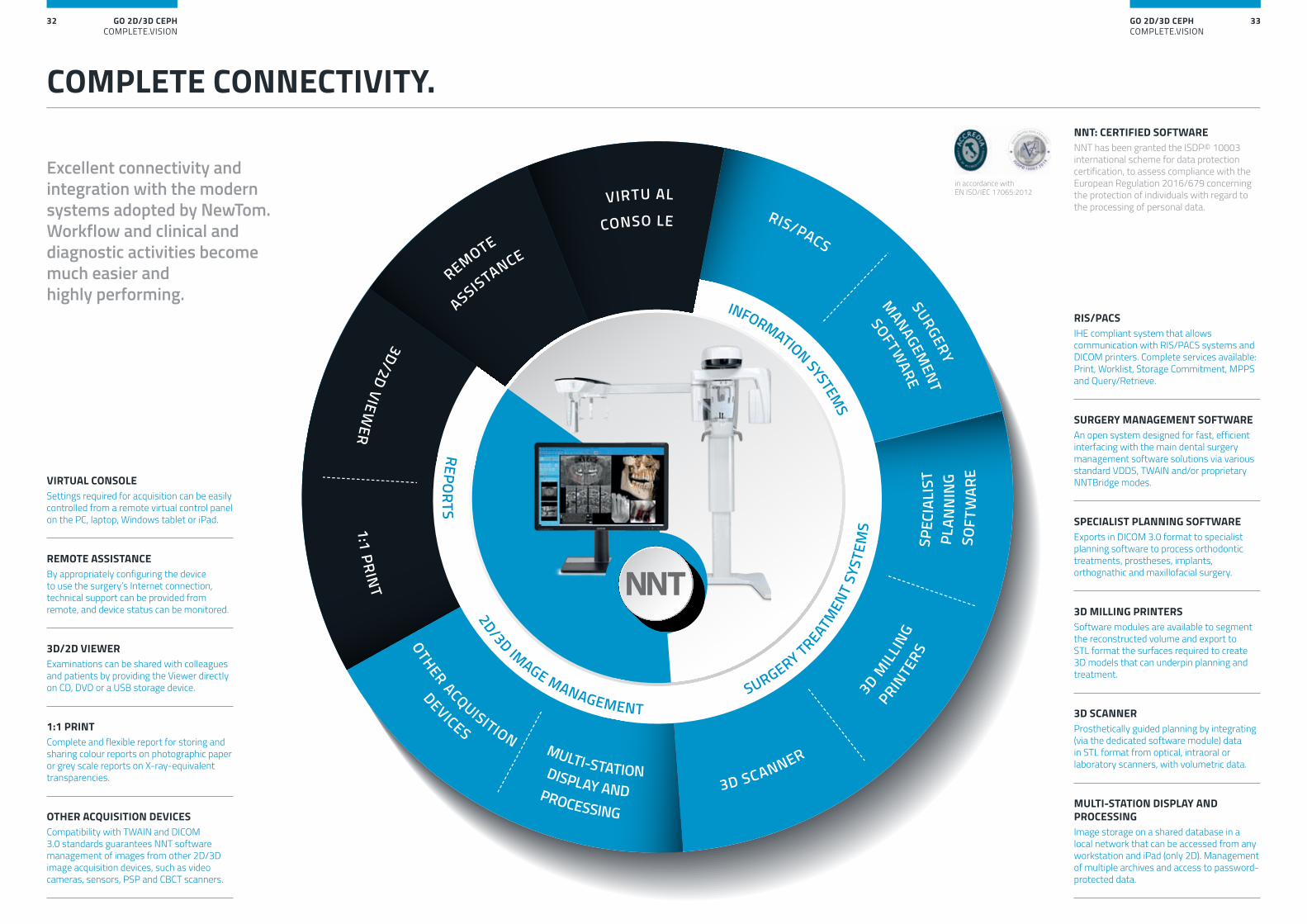
COMPLETE CONNECTIVITY.
Excellent connectivity and integration with the modern systems adopted by NewTom. Workflow and clinical and diagnostic activities become much easier and highly performing.
VIRTUAL CONSOLESettings required for acquisition can be easily controlled from a remote virtual control panel on the PC, laptop, Windows tablet or iPad.
RIS/PACSIHE compliant system that allows communication with RIS/PACS systems and DICOM printers. Complete services available: Print, Worklist, Storage Commitment, MPPS and Query/Retrieve.
REMOTE ASSISTANCEBy appropriately configuring the device to use the surgery’s Internet connection, technical support can be provided from remote, and device status can be monitored.
SURGERY MANAGEMENT SOFTWAREAn open system designed for fast, efficient interfacing with the main dental surgery management software solutions via various standard VDDS, TWAIN and/or proprietary NNTBridge modes.
3D/2D VIEWERExaminations can be shared with colleagues and patients by providing the Viewer directly on CD, DVD or a USB storage device.
SPECIALIST PLANNING SOFTWAREExports in DICOM 3.0 format to specialist planning software to process orthodontic treatments, prostheses, implants, orthognathic and maxillofacial surgery.
1:1 PRINTComplete and flexible report for storing and sharing colour reports on photographic paper or grey scale reports on X-ray-equivalent transparencies.
3D MILLING PRINTERSSoftware modules are available to segment the reconstructed volume and export to STL format the surfaces required to create 3D models that can underpin planning and treatment.
OTHER ACQUISITION DEVICESCompatibility with TWAIN and DICOM 3.0 standards guarantees NNT software management of images from other 2D/3D image acquisition devices, such as video cameras, sensors, PSP and CBCT scanners.
3D SCANNERProsthetically guided planning by integrating (via the dedicated software module) data in STL format from optical, intraoral or laboratory scanners, with volumetric data.
MULTI-STATION DISPLAY AND PROCESSINGImage storage on a shared database in a local network that can be accessed from any workstation and iPad (only 2D). Management of multiple archives and access to password-protected data.
NNT: CERTIFIED SOFTWARENNT has been granted the ISDP© 10003 international scheme for data protection certification, to assess compliance with the European Regulation 2016/679 concerning the protection of individuals with regard to the processing of personal data.
GO 2D/3D CEPHCOMPLETE.VISION
35GO 2D/3D CEPHCOMPLETE.VISION
34
0051
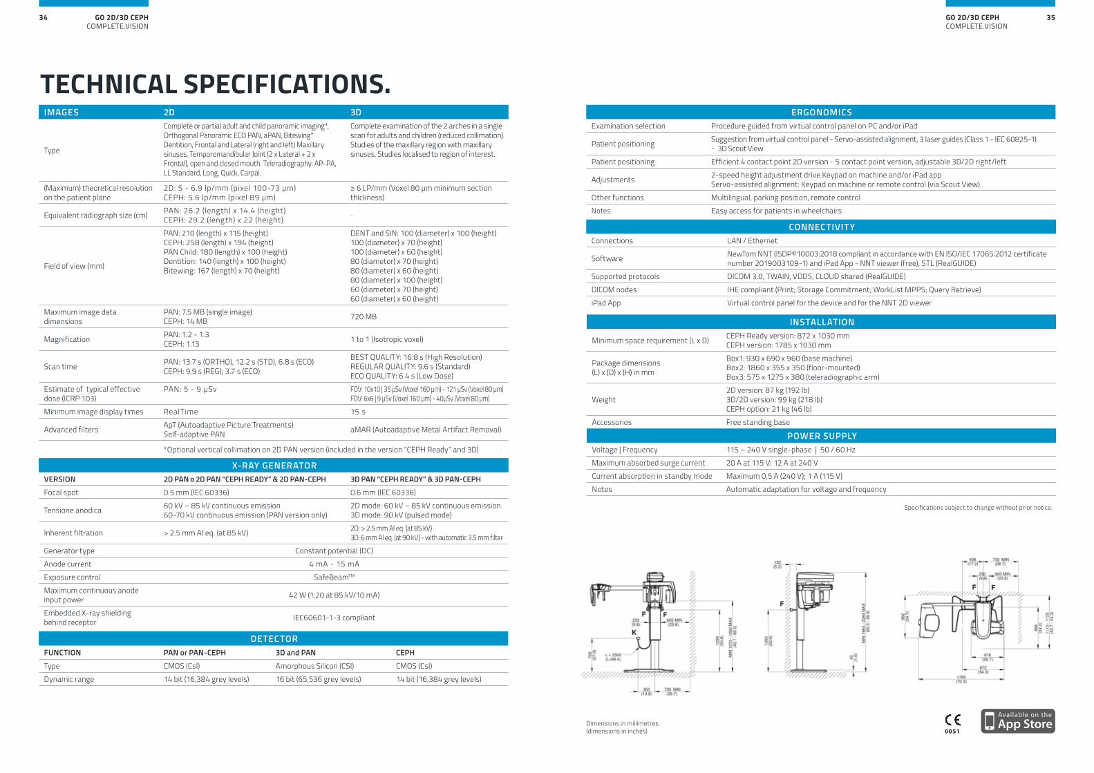
IMAGES 2D 3D
Type
Complete or partial adult and child panoramic imaging*, Orthogonal Panoramic ECO PAN, aPAN, Bitewing* Dentition, Frontal and Lateral (right and left) Maxillary sinuses, Temporomandibular Joint (2 x Lateral + 2 x Frontal), open and closed mouth. Teleradiography: AP-PA, LL Standard, Long, Quick, Carpal.
Complete examination of the 2 arches in a single scan for adults and children (reduced collimation).Studies of the maxillary region with maxillary sinuses. Studies localised to region of interest.
(Maximum) theoretical resolution on the patient plane
2D: 5 - 6.9 lp/mm (pixel 100-73 μm)CEPH: 5.6 lp/mm (pixel 89 μm)
≥ 6 LP/mm (Voxel 80 μm minimum section thickness)
Equivalent radiograph size (cm) PAN: 26.2 (length) x 14.4 (height) CEPH: 29.2 (length) x 22 (height) -
Field of view (mm)
PAN: 210 (length) x 115 (height)CEPH: 258 (length) x 194 (height)PAN Child: 180 (length) x 100 (height)Dentition: 140 (length) x 100 (height)Bitewing: 167 (length) x 70 (height)
DENT and SIN: 100 (diameter) x 100 (height)100 (diameter) x 70 (height)100 (diameter) x 60 (height)80 (diameter) x 70 (height)80 (diameter) x 60 (height)80 (diameter) x 100 (height)60 (diameter) x 70 (height)60 (diameter) x 60 (height)
Maximum image data dimensions
PAN: 7.5 MB (single image)CEPH: 14 MB 720 MB
Magnification PAN: 1.2 - 1.3CEPH: 1.13 1 to 1 (Isotropic voxel)
Scan time PAN: 13.7 s (ORTHO), 12.2 s (STD), 6.8 s (ECO)CEPH: 9.9 s (REG); 3.7 s (ECO)
BEST QUALITY: 16.8 s (High Resolution) REGULAR QUALITY: 9.6 s (Standard) ECO QUALITY: 6.4 s (Low Dose)
Estimate of typical effective dose (ICRP 103)
PAN: 5 - 9 μSv FOV: 10x10 | 35 μSv (Voxel 160 μm) - 121 μSv (Voxel 80 μm)FOV: 6x6 | 9 μSv (Voxel 160 μm) - 40μSv (Voxel 80 μm)
Minimum image display times RealTime 15 s
Advanced filters ApT (Autoadaptive Picture Treatments)Self-adaptive PAN aMAR (Autoadaptive Metal Artifact Removal)
*Optional vertical collimation on 2D PAN version (included in the version “CEPH Ready” and 3D)
X-RAY GENERATORVERSION 2D PAN o 2D PAN “CEPH READY” & 2D PAN-CEPH 3D PAN “CEPH READY” & 3D PAN-CEPHFocal spot 0.5 mm (IEC 60336) 0.6 mm (IEC 60336)
Tensione anodica 60 kV – 85 kV continuous emission60-70 kV continuous emission (PAN version only)
2D mode: 60 kV – 85 kV continuous emission3D mode: 90 kV (pulsed mode)
Inherent filtration > 2.5 mm Al eq. (at 85 kV) 2D: > 2.5 mm Al eq. (at 85 kV)3D: 6 mm Al eq. (at 90 kV) - with automatic 3.5 mm filter
Generator type Constant potential (DC)Anode current 4 mA - 15 mAExposure control SafeBeamTM
Maximum continuous anode input power 42 W (1:20 at 85 kV/10 mA)
Embedded X-ray shielding behind receptor IEC60601-1-3 compliant
INSTALLATION
Minimum space requirement (L x D) CEPH Ready version: 872 x 1030 mm CEPH version: 1785 x 1030 mm
Package dimensions (L) x (D) x (H) in mm
Box1: 930 x 690 x 960 (base machine)Box2: 1860 x 355 x 350 (floor-mounted)Box3: 575 x 1275 x 380 (teleradiographic arm)
Weight2D version: 87 kg (192 lb)3D/2D version: 99 kg (218 lb)CEPH option: 21 kg (46 lb)
Accessories Free standing base
POWER SUPPLYVoltage | Frequency 115 – 240 V single-phase | 50 / 60 HzMaximum absorbed surge current 20 A at 115 V; 12 A at 240 VCurrent absorption in standby mode Maximum 0,5 A (240 V); 1 A (115 V)Notes Automatic adaptation for voltage and frequency
Specifications subject to change without prior notice.
TECHNICAL SPECIFICATIONS.
Dimensions in millimetres (dimensions in inches)
CONNECTIVITYConnections LAN / Ethernet
Software NewTom NNT (ISDP©10003:2018 compliant in accordance with EN ISO/IEC 17065:2012 certificate number 2019003109-1) and iPad App - NNT viewer (free), STL (RealGUIDE)
Supported protocols DICOM 3.0, TWAIN, VDDS, CLOUD shared (RealGUIDE)DICOM nodes IHE compliant (Print; Storage Commitment; WorkList MPPS; Query Retrieve) iPad App Virtual control panel for the device and for the NNT 2D viewer
ERGONOMICSExamination selection Procedure guided from virtual control panel on PC and/or iPad
Patient positioning Suggestion from virtual control panel - Servo-assisted alignment, 3 laser guides (Class 1 - IEC 60825-1) - 3D Scout View
Patient positioning Efficient 4 contact point 2D version - 5 contact point version, adjustable 3D/2D right/left
Adjustments 2-speed height adjustment drive Keypad on machine and/or iPad app Servo-assisted alignment: Keypad on machine or remote control (via Scout View)
Other functions Multilingual, parking position, remote controlNotes Easy access for patients in wheelchairs
DETECTORFUNCTION PAN or PAN-CEPH 3D and PAN CEPH
Type CMOS (CsI) Amorphous Silicon (CSI) CMOS (CsI)Dynamic range 14 bit (16,384 grey levels) 16 bit (65,536 grey levels) 14 bit (16,384 grey levels)