Embed Size (px)
Citation preview
Noise-induced Metastability in the Hodgkin-Huxley Neural Network
Norihiro Katayama†, Kazuhisa Sakamoto†, Mitsuyuki Nakao†, Mitsuaki Yamamoto†
†Department of Applied Information Sciences, Graduate School of Information Sciences, Tohoku University Aza Aoba 6-3-09, Aoba-ku, Sendai, 980-8579, Japan
Email: [email protected]
Abstract– Dynamics of a spiking neural network composed of Hodgkin-Huxley type neuron model driven by noise are studied by the computer simulations. It is shown that the neuronal dynamics change dramatically dependent on the intensity of global inhibitory input; the single neuronal activities in the slow frequency range (0.01-1Hz) change from a white noise-like power spectral density profile to an 1/f noise-like one as the global inhibition decreased. When the neurons show 1/f noise-like dynamics, the network exhibites intermittent state transitions among metastable states, which emerge under the random perturbations.
1. Introduction
Single neuronal activities in the central nervous system
are known to vary dramatically during wake-sleep cycle [1]. In particular, power spectral density (PSD) of a single neuronal activity in the low frequency range (0.01-1Hz) is 1/f noise-like PSD profile during rapid eye movement (REM) sleep. In contrast, the PSD becomes white noise-like one during slow wave sleep.
The slow dynamics-change has successfully been simulated with a Hopfield-type artificial neural network model with a global inhibition, consisting of formal (non-spiking) neurons driven by white noise [2, 3].
In this study, dynamics of the neural network model of which the model neuron is substituted by a Hodgkin-Huxley-type spiking neuron is investigated by the computer simulations. Single neuronal dynamics as well as the population dynamics are analsyed. It is shown that
the spiking neural network is also capable of reproducing slow dynamics change dependent on the global inhibition. The results will be discussed from the viewpoint of possible role of noise in the brain.
2. Spiking Neural Network Model
The neural network model is constructed by the
Hodgkin-Huxley's model of squid giant axon [4] described as follows:
)()613.10(3.0
)12(36)115(120d
d 43
tIV
VnVhmt
VC
ii
iiiiii
m
+−+
−−+−= (1)
d 0.1(25 ) (1 ) 4expd exp(0.1(25 )) 1 18m V Vm mt V
− ⎛ ⎞= − − −⎜ ⎟− − ⎝ ⎠ (2)
hV
hVth
1))30(1.0exp(1)1(
20exp07.0
dd
+−−−⎟
⎠⎞
⎜⎝⎛−= (3)
nVnVV
tn
⎟⎠⎞
⎜⎝⎛−−−
−−−
=80
exp125.0)1(1))10(1.0exp(
)10(01.0dd (4)
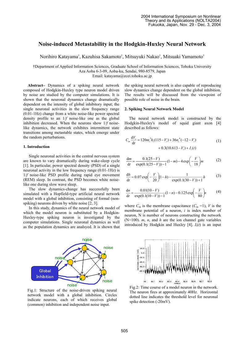
where Cm is the membrane capacitance (Cm =1), V is the membrane potential of a neuron, i is index number of neuron, N is number of neurons constructing the network (N=100). m, n, and h are the ion channel gate variables introduced by Hodgkin and Huxley [4]. Ii(t) is an input
Fig.2: Time course of a model neuron in the network.The neuron fires at approximately 40Hz. Horizontaldotted line indicates the threshold level for neuronalspike detection (-20mV).
noise
Global
Inhibition
noise
noise
noise
noise
Fig.1: Structure of the noise-driven spiking neuralnetwork model with a global inhibition. Circlesindicate neurons, each of which receives global(common) inhibition and independent noise input.
2004 International Symposium on NonlinearTheory and its Applications (NOLTA2004)
Fukuoka, Japan, Nov. 29 - Dec. 3, 2004
505

current for i-th neuron, which is composed of a random noise current, a global inhibory current, and synaptic currents described as follows:
( )
H , ,j 1
( ) ( 12 ) ( , )N
ki i i i j i j j
k
I t W g V S V t tη=
= + − − + −∑∑ (5)
where W is a Gaussian noise (mean=0, variance=1), η is the noise intensity, Hg is the conductance of the global inhibition, ( )k
jt is the k-th spike time of j-th neuron, and
,i jS is the synaptic current defined as follows:
Fig.3: Global inhibition intensity-dependencies of neuronal activities. Raster plots of neuronal spike trainsare displayed together with graphs of power spectral density of the spike train. Left panels; dynamics ofneuron #71, middle; #78, and right; #85. When the global inhibition is low (e.g. gH =0.1), neurons show 1/fnoise-like slow fluctuation in the low frequency range (0.05-1Hz). In contrast, neurons show white noise-like dynamics in the frequency range under the strong inhibition (gH=1).
506
, ,
,
,
10 ( ;1.5, 2.5)(60 ) if 0( , )
20 ( ;1,10)( 30 ) otherwise
i j i j
i j
i j
w t V wS V t
w t V
α
α
⎧ − ≥⎪= ⎨− −⎪⎩
(6)
where wi,j is the synaptic weight from j-th neuron to i-th neuron. A positive weight (wi, j>0) represents an excitatory synapse, and negative one an inhibitory synapse. α determins the waveform of the synaptic conductance which is defined as follows:
exp exp if 0 ( ; , )
0 othewise,
I A
A I I A I A
t t tt
τ τα τ τ τ τ τ τ
⎧ ⎡ ⎤⎛ ⎞ ⎛ ⎞− − − >⎪ ⎢ ⎥⎜ ⎟ ⎜ ⎟= −⎨ ⎝ ⎠ ⎝ ⎠⎣ ⎦
⎪⎩
(7)
where Aτ and Iτ are the activation and inactivation time constants of the synaptic conductance, respectively. Model parameters for the excitatory synapse are set to mimic the AMPA-type synapse; a subtype of glutamatergic synapse mediating fast excitatory synaptic transmission. The parameters for inhibitory one are set to mimic the GABAA-type synapse; a subtype of GABAergic synapse mediating fast inhibitory synaptic transmission.
Synaptic weights wi,j were given to control the fundamental structure of the network attractor following the case of an auto-associative memory:
⎪⎩
⎪⎨⎧
=
≠−−= ∑=
ji
jiPPNw
M
mjmim
ji
,0
),12)(12(11
,,,
(8)
where Pm,j indicates j-th neuron's state (0 or 1) of m-th memorized pattern, and M is the number of memorized patterns (M=30). The memorized patterns were given as random binary bits.
The differential equations were numerically solved by the backward-Euler method with the fixed time step of 0.01ms. Neuronal spikes were detected every when the
membrane potential exceeds a threshold voltage level. The PSD profiles of neuronal spike train were estimated by the Blackman-Turkey method. The state of network was evaluated every 250ms epoch by the number of spikes in the epoch. 3. Simulation Results
3.1. Dynamics-Transition of Single Neuronal Activities
Figure 2 shows an example of time course of a neuron in the neural network model. The regular spikes and post-synaptic potentials were generated at approximately 40Hz.
Under noise-free condition, i.e. 0η = , neurons become silent after transient activities (not shown here).
In contrast, neurons hold firing activities under the exposure of noise. Figure 3 shows the global inhibition intensity-dependency of the neuronal activities. Firing activities of three representative neurons in the case of noise intensity 10η = are shown. Slow dynamics (0.05-1Hz) seem to be 1/f noise when the global inhibition is weak (gH=0.1). This PSD profile is similar to the brain neurons during REM sleep state. As the global inhibition increase, the neuronal dynamics become white noise-like profile, similar to the brain neurons during slow wave sleep state. In short, the slow neuronal dynamics change from the 1/f noise-like spectrum to the white noise-like one as the global inhibition increased. When the neurons show 1/f noise-like dynamics, the neuronal activity intermittently changes between active and silent states. Neurons in the active-state generate spikes in a synchronous manner (not shown here).
3.2. Network Dynamics
In order to understand the mechanism of 1/f fluctuation
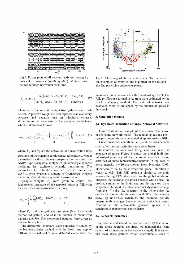
in the single neuronal activities, we analyzed the firing pattern of all neurons in the network (Fig.4). It is shown that clear stripe patterns switch intermittently, each of
Fig.4: Raster plots of all neurons' activities during 1/fnoise-like dynamics (η=10, gH=0.1). Vertical axis:neuron number, horizontal axis: time.
Fig.5: Clustering of the network states. The networkstate sampled at every 250ms is plotted on the 1st andthe 2nd principle components plane.
507
which corresponds a stable state of the spiking neural network, i.e., the metastable dynamics emerge under the exposure of noise. To visualize the metastable dynamics more clearly, principle component analysis on network dynamics was performed. Here we introduce a network state vector for representing pattern of neural firing activity, of which each component is the mean firing rate of a neurons evaluated every 250ms epoch. Figure 5 is a plot of network state vectors on the first and the second principle components plane. Successive network state vectors a connected by a dotted line. One can see clear clusters of network state vectors, each of which corresponds to a stripe pattern in Fig.4, and state transitions among the clusters.
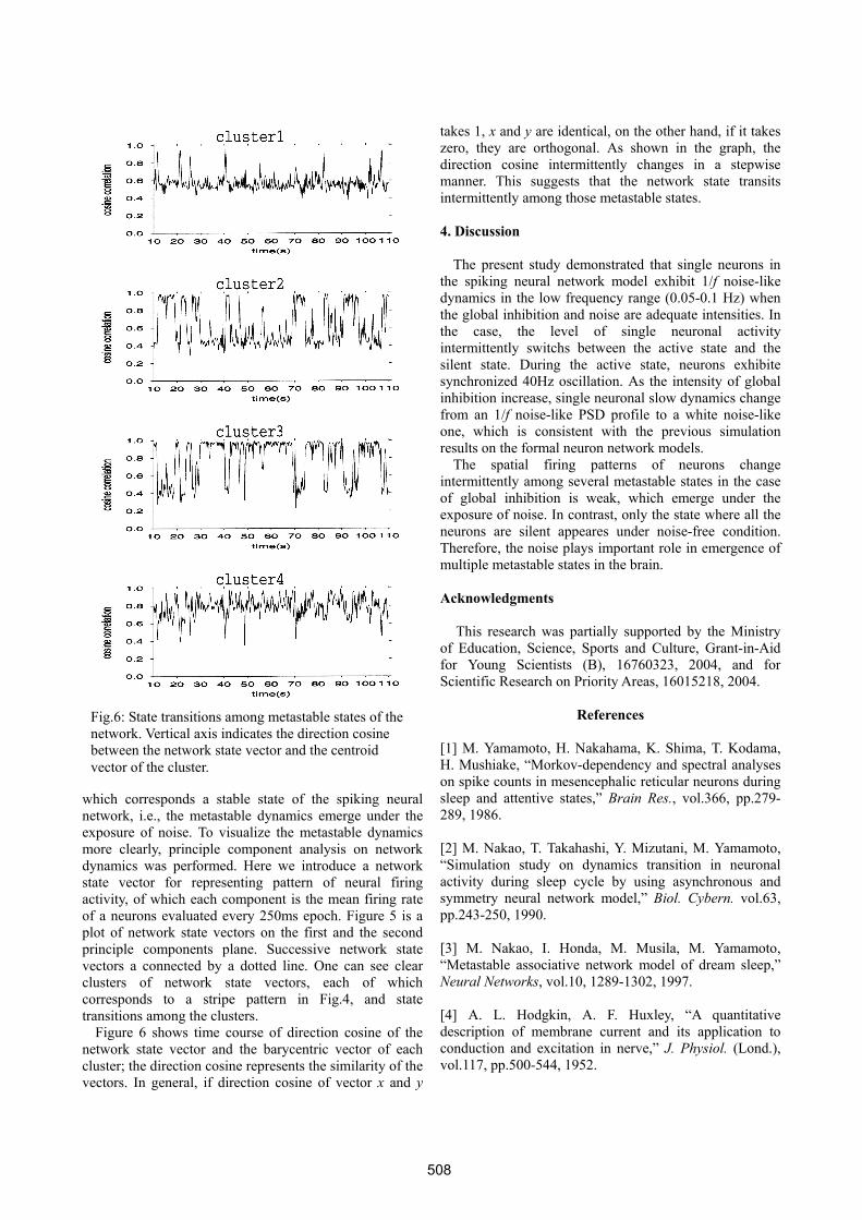
Figure 6 shows time course of direction cosine of the network state vector and the barycentric vector of each cluster; the direction cosine represents the similarity of the vectors. In general, if direction cosine of vector x and y
takes 1, x and y are identical, on the other hand, if it takes zero, they are orthogonal. As shown in the graph, the direction cosine intermittently changes in a stepwise manner. This suggests that the network state transits intermittently among those metastable states. 4. Discussion
The present study demonstrated that single neurons in
the spiking neural network model exhibit 1/f noise-like dynamics in the low frequency range (0.05-0.1 Hz) when the global inhibition and noise are adequate intensities. In the case, the level of single neuronal activity intermittently switchs between the active state and the silent state. During the active state, neurons exhibite synchronized 40Hz oscillation. As the intensity of global inhibition increase, single neuronal slow dynamics change from an 1/f noise-like PSD profile to a white noise-like one, which is consistent with the previous simulation results on the formal neuron network models.
The spatial firing patterns of neurons change intermittently among several metastable states in the case of global inhibition is weak, which emerge under the exposure of noise. In contrast, only the state where all the neurons are silent appeares under noise-free condition. Therefore, the noise plays important role in emergence of multiple metastable states in the brain.
Acknowledgments
This research was partially supported by the Ministry
of Education, Science, Sports and Culture, Grant-in-Aid for Young Scientists (B), 16760323, 2004, and for Scientific Research on Priority Areas, 16015218, 2004.
References
[1] M. Yamamoto, H. Nakahama, K. Shima, T. Kodama, H. Mushiake, “Morkov-dependency and spectral analyses on spike counts in mesencephalic reticular neurons during sleep and attentive states,” Brain Res., vol.366, pp.279-289, 1986. [2] M. Nakao, T. Takahashi, Y. Mizutani, M. Yamamoto, “Simulation study on dynamics transition in neuronal activity during sleep cycle by using asynchronous and symmetry neural network model,” Biol. Cybern. vol.63, pp.243-250, 1990. [3] M. Nakao, I. Honda, M. Musila, M. Yamamoto, “Metastable associative network model of dream sleep,” Neural Networks, vol.10, 1289-1302, 1997. [4] A. L. Hodgkin, A. F. Huxley, “A quantitative description of membrane current and its application to conduction and excitation in nerve,” J. Physiol. (Lond.), vol.117, pp.500-544, 1952.
Fig.6: State transitions among metastable states of the network. Vertical axis indicates the direction cosine between the network state vector and the centroid vector of the cluster.
508

![DesarrollodeunSimuladordelosExperimentosClásicos ... · Hodgkin y Huxley, tal como fueron descritos en su art culo [15]. Con la tercera ventana, donde se pueden realizar los mismos](https://img.pdfslide.tips/doc/110x75/5ea83c45ef976004f57da4e7/desarrollodeunsimuladordelosexperimentosclsicos-hodgkin-y-huxley-tal-como.jpg)