Embed Size (px)
Citation preview
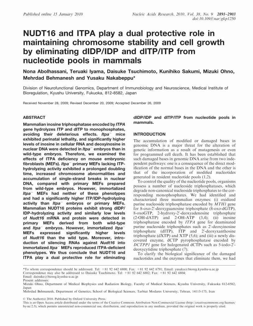
NUDT16 and ITPA play a dual protective role inmaintaining chromosome stability and cell growthby eliminating dIDP/IDP and dITP/ITP fromnucleotide pools in mammalsNona Abolhassani, Teruaki Iyama, Daisuke Tsuchimoto, Kunihiko Sakumi, Mizuki Ohno,
Mehrdad Behmanesh and Yusaku Nakabeppu*
Division of Neurofunctional Genomics, Department of Immunobiology and Neuroscience, Medical Institute ofBioregulation, Kyushu University, Fukuoka, 812-8582, Japan
Received November 28, 2009; Revised December 20, 2009; Accepted December 26, 2009
ABSTRACT
Mammalian inosine triphosphatase encoded by ITPAgene hydrolyzes ITP and dITP to monophosphates,avoiding their deleterious effects. Itpa� miceexhibited perinatal lethality, and significantly higherlevels of inosine in cellular RNA and deoxyinosine innuclear DNA were detected in Itpa� embryos than inwild-type embryos. Therefore, we examined theeffects of ITPA deficiency on mouse embryonicfibroblasts (MEFs). Itpa� primary MEFs lacking ITP-hydrolyzing activity exhibited a prolonged doublingtime, increased chromosome abnormalities andaccumulation of single-strand breaks in nuclearDNA, compared with primary MEFs preparedfrom wild-type embryos. However, immortalizedItpa� MEFs had neither of these phenotypesand had a significantly higher ITP/IDP-hydrolyzingactivity than Itpa� embryos or primary MEFs.Mammalian NUDT16 proteins exhibit strong dIDP/IDP-hydrolyzing activity and similarly low levelsof Nudt16 mRNA and protein were detected inprimary MEFs derived from both wild-typeand Itpa� embryos. However, immortalized Itpa�
MEFs expressed significantly higher levelsof Nudt16 than the wild type. Moreover, intro-duction of silencing RNAs against Nudt16 intoimmortalized Itpa� MEFs reproduced ITPA-deficientphenotypes. We thus conclude that NUDT16 andITPA play a dual protective role for eliminating
dIDP/IDP and dITP/ITP from nucleotide pools inmammals.
INTRODUCTION
The accumulation of modified or damaged bases ingenomic DNA is a major threat for the alteration ofgenetic information as a result of mutagenesis or evenfor programmed cell death. It has been established thatsuch damaged bases in genomic DNA arise from two inde-pendent pathways: one is a consequence of the direct mod-ification of the normal bases in the DNA and the other isthat of the incorporation of modified nucleotidesgenerated in resident nucleotide pools (1,2).To control the quality of the nucleotide pools, organisms
possess a number of nucleoside triphosphatases, whichdegrade non-canonical nucleoside triphosphates to the cor-responding monophosphates. We had identified andcharacterized three mammalian enzymes: (i) oxidizedpurine nucleoside triphosphatase encoded by MTH1 genefor 8-oxo-20-deoxyguanosine triphosphate (8-oxo-dGTP),8-oxoGTP, 2-hydroxy-20-deoxyadenosine triphosphate(2-OH-dATP) and 2-OH-ATP (3,4); (ii) inosinetriphosphatase encoded by ITPA gene for deaminatedpurine nucleoside triphosphates such as 20-deoxyinosinetriphosphate (dITP), ITP and 20-deoxyxanthosinetriphosphate (dXTP) and XTP (5,6); and (iii) a newly dis-covered enzyme, dCTP pyrophosphatase encoded byDCTPP1 gene for halogenated dCTPs such as 5-iodo-20-deoxycytidine triphosphate (7).To clarify the biological significance of the damaged
nucleotides and the enzymes that eliminate them, we had
*To whom correspondence should be addressed. Tel: +81 92 642 6800; Fax: +81 92 642 6791; Email: [email protected] may also be addressed to Daisuke Tsuchimoto. Tel: +81 92 642 6802; Fax: +81 92 642 6804;Email: [email protected] addresses:Mizuki Ohno, Department of Medical Biophysics and Radiation Biology, Faculty of Medical Sciences, Kyushu University, Fukuoka 812-8582,JapanMehrdad Behmanesh, Department of Genetics, School of Biological Sciences, Tarbiat Modares University, Tehran, 14115-175, Iran
Published online 15 January 2010 Nucleic Acids Research, 2010, Vol. 38, No. 9 2891–2903doi:10.1093/nar/gkp1250
� The Author(s) 2010. Published by Oxford University Press.This is an Open Access article distributed under the terms of the Creative Commons Attribution Non-Commercial License (http://creativecommons.org/licenses/by-nc/2.5), which permits unrestricted non-commercial use, distribution, and reproduction in any medium, provided the original work is properly cited.
previously produced and analyzed knockout mice lackingMTH1 or ITPA. Mth1�/– mice are viable and survivenormally but exhibit an increased incidence of spontane-ous tumorigenesis in the liver, stomach and lung (8), whileItpa�/– mice die before weaning with features of growthretardation and heart failure (6).ATP, the most abundant of the nucleotides, plays a
fundamental role in a wide variety of cellular processes,including energy transfer, signal transduction, RNAsynthesis, cytoskeleton remodeling and musclecontraction. Deamination of adenine at C-6 convertsATP to ITP; such modification is catalyzed by enzymessuch as adenosine or AMP deaminase (9), or inducedchemically under oxidative stress (7). Because ITPretains a molecular structure similar to that of ATP, itcan act as an aberrant substrate replacing ATP in somebiological processes (10,11). In the case of cardiacfunction, a number of sarcomere proteins require ATPfor their normal activities. It is likely that during cardiacdevelopment in Itpa�/– mice the accumulated ITPcompetes with ATP, which is required for actomyosinfunction in the sarcomere, thus causing heart failure (6).Bradshaw and Kuzminov (12) reported that an
Escherichia coli mutant of rdgB gene-encoding inosinetriphosphatase has no obvious phenotype; however, themutant exhibits synergistic lethality in the presence ofrecA or recBC mutations. They concluded that RdgBacts to avoid incorporation of 20-deoxyinosine (dI) inDNA and thereby blocks chromosome fragmentation byhydrolyzing dITP in E. coli. These observations stronglysuggest that ITPA deficiency in mouse cells also causeschromosomal abnormalities.In the present study, we examined mouse embryonic
fibroblasts (MEFs) prepared from wild-type (Itpa+/+),Itpa+/� and Itpa�/– embryos to explore the cellulardysfunction caused by ITPA deficiency. We found thatItpa�/– embryos accumulated more than eight timeshigher levels of dI in nuclear DNA than did wild-typeembryos. Moreover, Itpa�/– primary MEFs with noITP-hydrolyzing activity exhibited prolonged doublingtimes, increased chromosome aberrations andaccumulation of single-strand breaks in nuclear DNA.Surprisingly, these phenotypes all disappeared followingimmortalization of Itpa�/– MEFs, with a significantincrease in IDP-hydrolyzing activity accompanied by adecreased accumulation of dI in nuclear DNA. We havethus identified a novel enzyme which constitutes a dualenzyme system for eliminating dITP/ITP and dIDP/IDPfrom nucleotide pools together with ITPA in mammals.
MATERIALS AND METHODS
Nucleotides
Nucleotides used as substrates for enzyme assay werepurchased from Sigma-Aldrich (St Louis, MO, USA), orJena Bioscience GmbH (Jena, Germany). Separation andpurification of nucleotides were performed on a WatersAlliance 2690 HPLC separation module (Waters Corp.,Milford, MA, USA) equipped with a Model 996photodiode array detector and a Wakopak Handy ODS
column (4.6� 250mm) using 100mM triethyl ammoniumhydrogen carbonate solution (pH 7.4) (Wako PureChemicals, Osaka, Japan) as the mobile phase. Purifiednucleotides were lyophilized five times with solubilizationin distilled water.
Itpa gene knockout mice
Itpa gene knockout mice were established as described (6).Genotypes were analyzed using tail DNA. PCR primersused to detect the wild-type and Itpa mutant alleles wereP46 and P47, or P29 and LNEO1, respectively (Sup-plementary Table S2). Heterozygous male (Itpa+/�) werebackcrossed with C57BL/6J female (Itpa+/+) (Clea Japan,Tokyo, Japan) for more than five generations (N> 5). Allanimals were maintained in an air-conditioned, light/time-controlled, specific-pathogen-free room. All studieswere approved by the Animal Care and Use Committee,Medical Institute of Bioregulation, Kyushu University.
Preparation of primary and immortalized MEFs
Twenty embryonic gestation day (E)13.5 and E14.5embryos were obtained by intercross mating of inbredN10 or N11 Itpa+/� mice (three pairs). Their genotypeswere Itpa+/+, Itpa+/� and Itpa�/– at a ratio of 5:10:5.Skin fibroblasts were aseptically isolated from theseembryos and at least four independent embryos wereused for each genotype (Itpa+/+, Itpa+/�, Itpa�/–).These primary MEFs were cultured in Dulbecco’sModified Eagle’s Medium (DMEM) supplemented with10% heat-inactivated fetal bovine serum (FBS),100 units/ml penicillin and 100 mg/ml streptomycin at37�C under 5% CO2 in air. Primary MEFs in culturewere harvested by treatment with 0.15% trypsin–0.08%EDTA in PBS and replated for further passage. Thosefrom Passage 2 were stocked as primary MEFs.Spontaneously immortalized MEFs were establishedafter single colony isolation during 30–40 passages.Their genotypes were determined by genomic polymerasechain reaction (PCR) amplification and three lines ofimmortalized MEFs were independently established fromthree embryos for each genotype.
Cell proliferation assays
Primary or immortalized MEFs were seeded at 1� 105
cells (Figures 1C and 4A) or 0.5� 105 cells (Figure 7C)per well in six-well plates (Nalge Nunc InternationalK.K., Tokyo, Japan). Cells were harvested every 2 daysor every day, respectively, and the numbers of cells werecounted using a hemocytometer.
Cell-cycle analysis
Flow cytometric analysis of the cell cycle was performedas described (13). Cells (1� 106 cells per assay) werecentrifuged, washed with phosphate buffered saline(PBS) and suspended in PBS containing 0.2% TritonX-100. We then added 5 ml of RNase A (1mg/ml) and50 ml of propidium iodide (PI, 1mg/ml). DNA contentand cell numbers were analyzed with an LSR flowcytometer (Becton Dickinson, San Jose, CA, USA). The
2892 Nucleic Acids Research, 2010, Vol. 38, No. 9
data were analyzed using CellQuest and ModFit software(Becton Dickinson).
Karyotype analysis
A 50% confluent culture of MEFs was treated with0.1 mg/ml colcemid (Nacalai Tesque Inc., Kyoto, Japan)for 30min. After hypotonic treatment of harvested cellsin 75mM KCl, cells were fixed in freshly preparedCarnoy’s fixative (methanol:acetic acid 3:1), and the cellsuspension was dropped onto a glass slide, air-dried andimmediately stained with freshly prepared Giemsa stainingsolution (Merck KGaA, Darmstadt, Germany cat. no.1.09204.0509, 25� diluted in PBS) for 20min. Afterrinsing the slide in PBS twice and in distilled watertwice, air-dried slides were cover-slipped using Permount(Fisher Scientific, Waltham, MA, USA; SP15-100). Theslide was observed under an Axioscope 2 plus microscopeequipped with AxioCam and AxioVision software
(Carl Zeiss MicroImaging Japan, Tokyo, Japan). A total30 cells in metaphase was examined for each preparation.
Quantification of deoxyinosine or inosine by liquidchromatography coupled with tandem mass spectrometry
The preparation and digestion of nuclear DNA sampleswere as described (14), except that 10mM 2, 2, 6,6-tetramethylpiperidine-N-oxyl (TEMPO, Wako PureChemicals) and 20 mM 20-deoxycoformycin (a kind giftfrom the Chemo-Sero-Therapeutic Research Institute,Kumamoto, Japan), an adenosine deaminase inhibitor,was added at all stages of manipulation, as described byTaghizadeh et al. (15). RNA was prepared using RNeasyMini Kits (Qiagen Inc., Valencia, CA, USA) according tothe manufacturer’s instructions in the presence of 20mMTEMPO and 20 mM 20-deoxycoformycin. DNA or RNAsamples were digested with nuclease P1 (Yamasa, Chiba,Japan) and alkaline phosphatase (Sigma-Aldrich, P-5521)and digested samples were subjected to LC–MS/MS
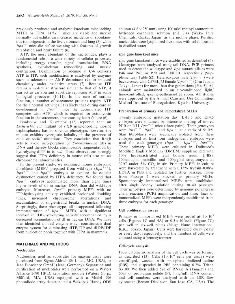
Figure 1. ITPA-deficient primary MEFs exhibit various cellular dysfunctions. (A) ITPA deficiency caused a significantly increased accumulation ofinosine in cellular RNA. Inosine level was determined by LC–MS/MS analysis of cellular RNA prepared from embryos (N3). Result of non-repeatedmeasures ANOVA (two-tailed), P=1.69� 10�7. Student–Newman–Keuls (SNK) post hoc test, **P< 0.01 (versus Itpa+/+ and Itpa+/�). Data areshown as the mean±SD (n=3 independent embryos). (B) ITPA deficiency caused a significantly increased accumulation of deoxyinosine (dI) innuclear DNA. Deoxyinosine (dI) level was determined by LC–MS/MS analysis of nuclear DNA prepared from embryos (N3). Result of non-repeatedmeasures ANOVA (two-tailed), P=0.00038. SNK post hoc test, **P< 0.01 (versus Itpa+/+ and Itpa+/�). Data are shown as the mean±SD (n=3independent embryos). (C) ITPA deficiency impairs normal cell proliferation. Primary MEFs (Passage 2) isolated from four separate Itpa�/– embryosshowed significant prolonged doubling time in comparison to those from Itpa+/+ and Itpa+/� embryos. Result of repeated measures ANOVA(two-tailed), P=0.0005. Bonferroni/Dunn post-hoc test, *P< 0.05 (versus Itpa+/�), **P< 0.01 (versus Itpa+/+). Data are shown as the mean±SD(n=4 independent MEFs). (D) ITPA deficiency causes G2/M arrest. Primary MEFs (Passage 5) were subjected to flow cytometry analysis and thepercentages of Cell-cycle phases in each MEF set were determined. Result of non-repeated measures ANOVA (two-tailed), P=1.74 � 10�8.Bonferroni post hoc test, **P< 0.01 (versus Itpa+/+ and Itpa+/�). Data are shown as the mean±SD (n=3 independent isolates).
Nucleic Acids Research, 2010, Vol. 38, No. 9 2893
analysis using the Shimadzu VP-10 HPLC system(SHIMADZU CORPORATION, Kyoto Japan)connected to the API3000 MS/MS system (PE-SCIEX,Applied Biosystems, Foster City, CA, USA), asdescribed (14).
Immunostaining
To detect single-stranded (ss) DNA, the slides wereincubated with anti-ssDNA (IBL, Takasaki, Japan; codenumber 18 731, 1/100 dilution) in combination with AlexaFluor 488-conjugated goat anti-rabbit IgG (Invitrogen,Carlsbad, CA. USA), as described (16). Nuclei werecounterstained with 40, 6-diamino-2-phenylindole (DAPI,50 ng/ml; Vector, Burlingame, CA, USA). A cover slidewas mounted onto the slide with Vectashield (Vector). Theslide was observed using an Axioskop 2 plus microscopeequipped with AxioCam and AxioVision software (CarlZeiss MicroImaging Japan). A total of 100 cells wereexamined for each preparation.
Inosine triphosphatase and Inosine diphosphatase assays
Embryo samples or pellets of immortalized MEFs (1� 107
cells) were washed twice with PBS and quickly frozen inliquid nitrogen. Frozen samples in 100 ml of lysis buffercontaining 50mM Tris–HCl (pH 8.0), 50mM NaCl,1mM dithiothreitol and protease inhibitor cocktail(Nacalai Tesque), were sonicated at 4�C. The lysate wascentrifuged at 17 360� g for 60min and the supernatantwas collected as a crude cell extract. The protein concen-tration was determined with a Protein Assay system(Bio-Rad, Hercules, CA, USA) using bovine serumalbumin (Thermo Fisher Scientific Inc., Waltham, MA,USA) as a standard. Inosine triphosphatase or Inosinediphosphatase activities were assayed by measuring thehydrolysis of ITP or IDP to IMP. The reaction mixturecontained 50mM Tris–HCl (pH 8.5), 50mM MgCl2,1mM DTT, 0.2mM ITP or IDP and 1–10mg of thecrude-cell extract to be examined. The reaction was runat 30�C for 20min and stopped by adding 150mM EDTA.The reaction mixture was applied to HPLC analysis asdescribed (6). Separation and quantification of nucleotideswere performed by HPLC using a Waters Alliance 2690separation module equipped with a Model 996 photodiodearray detector. A buffer consisting of 75mM sodium phos-phate (pH 6.4), 0.4mM EDTA, with 20% acetonitrile wasused as the mobile phase in a TSK-GEL DEAE-2SWcolumn, 4.6� 250mm (Tosoh Corp., Tokyo, Japan).
Quantitative real-time reverse transcription polymerasechain reaction
MEFs were seeded at 1� 105 cells per well with 500 ml ofmedium in 24-well plates and cultured to 70–80%confluency, or for 3 days. RNA was extracted from theharvested cells using an Isogen kit (Nippon Gene Inc.,Tokyo, Japan). Totally 2 mg of total RNA was subjectedto RNase-free DNase I treatment and cDNA synthesisusing random decamers and a Cells-to-cDNA II kit(Ambion), according to the manufacturer’s instructions.Quantitative real-time PCR was performed using an ABIPrism 7000 sequence detection system with 10 ng cDNA,
a set of Nudt16 primers (FmNud3RT, RmNud3RT;200 nM) or a set of Gapdh primers (F-Gapdh, R-Gapdh;50 nM) and Power SYBR Green PCR Master Mix(Applied Biosystems) in a total volume of 25 ml. ThePCR reaction was performed as follows: a single cycle of50�C for 2min, a single cycle of 95�C for 10min, followedby 40 cycles of 95�C for 15 s and 60�C for 1min. Theprimers were designed using PRIMER EXPRESSsoftware (Applied Biosystems) and their sequences areshown in Supplementary Table S2. Specificity of thePCR products was established by dissociating curveanalysis and by running the products on a 2% agarosegel to verify their size. The Nudt16 mRNA level isexpressed relative to the Gapdh mRNA level. Seriallydiluted cDNA was used to obtain a standard curve foreach transcript.
Western blotting
MEFs were seeded at 5� 105 cells per dish in 10mlmedium in a 90mm Petri dish (Nalge NuncInternational K.K.) and were cultured to 70–80%confluency or for 3 days. Cells were washed twice withPBS and harvested using 2� SDS sample buffer[125mM Tris–HCl (pH 6.8), 4% SDS, 10% glycerol,4% 2-mercaptoethanol]. The protein concentration wasdetermined using a Protein Assay system as above.Protein samples were separated by SDS–PAGE and trans-ferred to 0.45mm Immobilon-P membrane (Millipore Inc.,Madison, WI, USA) and western blot analysis usinganti-hNUDT16 (1mg/ml) or anti-ITPA antiserum (1/500dilution) (5) with horseradish peroxidase-conjugatedprotein A and an ECL-Plus kit (GE HealthcareBio-Sciences, Piscataway, NJ, USA) was performed asdescribed (17). The same membrane was treated withWB stripping solution (Nacalai Tesque) and reprobedwith anti-GAPDH (Millipore, Inc., Billerica, MA, USA;MAB374, 105� diluted) and HRP-anti-mouse IgG (BDBiosciences, San Jose, CA, USA).
Expression of recombinant mouse NUDT16 protein
An expression vector for the mouse NUDT16 protein wasconstructed by inserting the NdeI-HindIII fragment ofpET28a(+):mNudt16 into the NdeI-HindIII regionof pET32a(+) (Merck KGaA), thus Trx�Tag-His�Tag-S�Tag sequences were removed. Escherichia coliBL21 cells were transfected with pET32a(+) vectoror pET32a:mNudt16 using a Cell-porator (LifeTechnologies, Carlsbad, CA, USA) according to the man-ufacturer’s instructions. Transformants were selected onLB-agar plates in the presence of 30 mg/ml ampicillin.Established transformants were cultured until the OD600
reached 0.6 and then incubated with 1mM isopropylb-D-thiogalactoside for a further 3 h. Cells were harvestedby centrifugation and resuspended in 1ml of 2� SDSsample buffer. Samples were subjected to 12.5% SDS–PAGE and the expression of mouse NUDT16 proteinwithout tag was confirmed by western blotting.
2894 Nucleic Acids Research, 2010, Vol. 38, No. 9
Introduction of silencing RNA into immortalized MEFs
Nudt16 siRNA (Ambion/Applied Biosystems, Austin, TX,USA; Silencer Select; s93780, s93782, 25 mM) or controlsiRNA (Ambion/Applied Biosystems, Silencer SelectNegative Control #1 siRNA, cat. no. 4390844) wasintroduced into immortalized MEFs (Itpa+/+, Itpa�/–)by electroporation using a MicroPorator-mini (DigitalBio Technology, Seoul, Korea, MP-100, 1100V, 10msfor 2 pulses) and cells were replated appropriately intosix-well plates 1 day after electroporation for furtheranalysis.
Statistical analysis
Statistical analysis was performed using Stat View 5.0(SAS Institute Inc., Cary, NC, USA). The statisticalsigniEcance between two groups was determined withStudent’s t-test, and that among more than three groupswas determined with non-repeated or repeated measuresANOVA with an appropriate correction for multiplecomparisons as described in each figure legend.P-values<0.05 are considered statistically significant.
RESULTS
ITPA deficient primary MEFs exhibit various cellulardysfunctions
Itpa�/– mice with a 129�C57BL/6J mixed genetic back-ground exhibited incomplete embryonic lethality and thesurviving pups die about 2weeks after birth with growthretardation and heart failure (6). After backcrossing theheterozygotes (Itpa+/�) to C57BL/6J mice for more thanfive generations (N5), intercrosses of the obtained Itpa+/�
mice yielded Itpa�/– embryos in uterus in accordance withMendel’s laws until embryonic gestation day (E) 18, butthere were few newborn pups (Supplementary Table S1),indicating that ITPA deficiency causes perinatal lethalityin a C57BL/6J genetic background.
We confirmed significantly increased accumulation ofinosine (567.3±41.4 residues per 106 guanosine) incellular RNA prepared from Itpa�/– embryos (N14) incomparison to those from Itpa+/+ (10.5±1.50) andItpa+/� (11.4±1.07) embryos by liquid chromatographycoupled with tandem mass spectrometry (LC–MS/MS)analysis (Figure 1A), as previously observed in varioustissues of surviving Itpa�/– pups (6).
Furthermore, LC–MS/MS analysis of nuclear DNAprepared from embryos revealed that Itpa�/– embryos(E14.5) obtained from intercrosses of Itpa+/� mice (N3)contained significantly more dI in their nuclear DNA(20.1±4.8 residues per 106 nucleosides): more thaneight times that measured in Itpa+/+ embryos(2.34±0.76) (Figure 1B). The increased dI levels werealso confirmed in Itpa�/– embryos after furtherbackcrossing to C57BL/6J mice (N14, E13.5, 24.5±2.24dI residues per 106 nucleosides).
To examine the nature of the cellular dysfunctioncaused by ITPA deficiency, we isolated embryos (atE13.5 and E14.5) from intercrosses of Itpa+/� littermates(N10, one pair; N11, two pairs) and determined their
genotypes (Supplementary Table S1). Among 20embryos, five were found to be Itpa�/–, 10 were Itpa+/�
and the remaining five were Itpa+/+. Then we isolatedprimary MEFs independently from four embryos ofeach genotype and their genotypes and the expressionlevels of ITPA protein were confirmed (SupplementaryFigure S1A and B). All Itpa�/– primary MEFs at thesecond passage showed significantly longer doublingtime (132.0±14.6 h) than those from Itpa+/+
(78.6±10.1 h) and Itpa+/� (87.5±14.3 h) embryos(Figure 1C). There was no obvious difference in their mor-phology under phase contrast microscopy (SupplementaryFigure S1C). In Passage 5, we observed essentially thesame proliferation deficiency in Itpa�/– MEFs andslightly increased numbers of senescence-associatedb-galactosidase (SA-b-Gal)-positive cells in Itpa�/–
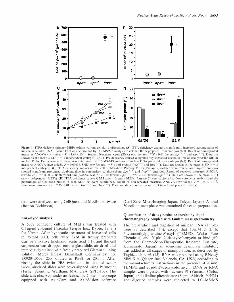
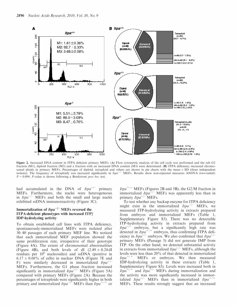
MEFs (Itpa+/+ versus Itpa�/–, 3.64% versus 7.74%;Supplementary Figure S2A and B). Flow cytometryanalysis of the cell cycle revealed that Itpa�/– MEFsexhibited a significant increase in the G2/M phase(Itpa+/+ versus Itpa�/–, 28.4% versus 77.4%;Figure 1D) and a slight increase in the sub G1 fraction(Itpa+/+ versus Itpa�/–, 1.61% versus 5.51%; fraction M1in Figure 2A). There was an apparent increase in cells withan abnormally increased DNA content in Itpa�/– MEFscompared with Itpa+/+ MEFs (fraction M3 in Figure2A), indicating that the G2/M phase shown in Figure1D might have contained some tetraploid cells at the G1phase (Figure 2B). There was no increase in dead cellsdetected as propidium iodide (PI)/Hoechst-doublepositive cells (Supplementary Figure S2C). These resultsindicate that ITPA deficiency caused delay or arrest incell-cycle progression. We further observed that exposureof Itpa�/–, but not Itpa+/+, Itpa+/� MEFs, to sodiumnitrite (NaNO2), which causes predominant deaminationof purine bases (18), resulted in growth suppressionwithout inducing cell death (Supplementary Figure S2C).We next examined for chromosomal abnormalities in
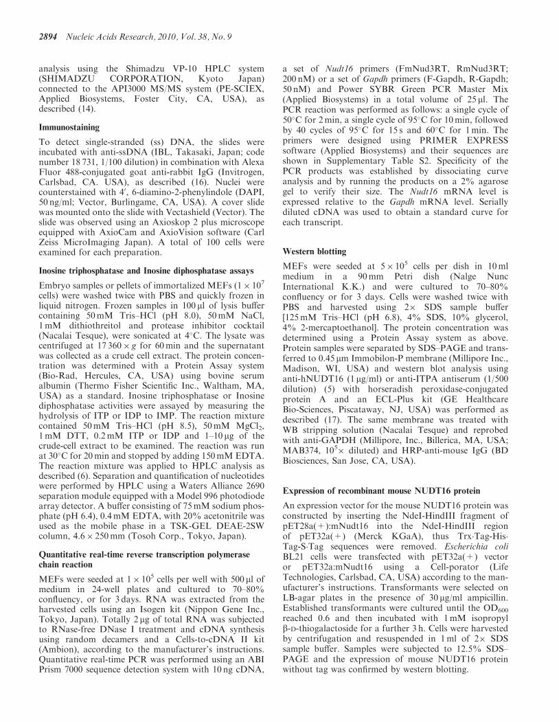
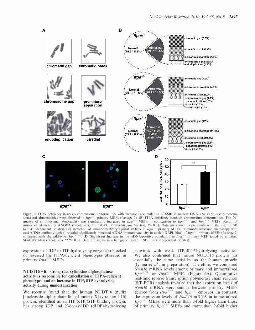
mitotic cells (Figure 3A). As shown in Figure 3B, chro-mosomal structural abnormalities were more frequentlyobserved in Itpa�/– MEFs than in Itpa+/+ MEFs, espe-cially premature centromere separation (3.33 times morecommon), chromatid gaps (2.04 times) and chromatidbreakages (1.52 times). Moreover, the percentage of cellswith abnormal chromosomes in Itpa�/– primary MEFswas significantly higher than among Itpa+/+ andItpa+/� MEFs. There was an increase in ploidyabnormalities among Itpa�/– primary MEFs (Figure 2B):thus 41.2% in the mitotic fraction exhibited tetraploidy,while about 25% of mitotic fractions in Itpa+/+ andItpa+/� MEFs were detected as tetraploids. This con-firmed the increase in ploidy among Itpa�/– MEFs.Because chromatid gaps or breakages are most likely to
be caused by the accumulation of single- or double-strandbreaks in DNA, we examined levels of single-strandbreaks (SSBs) in DNA using an antibody againstsingle-stranded (ss) DNA (anti-ssDNA) (Figure 3C) (16).Immunofluorescence microscopy with anti-ssDNArevealed that the percentages of immunoreactive nucleiin Itpa�/– primary MEFs were significantly higher thanin Itpa+/+ MEFs (Figure 3D). Thus, more SSBs
Nucleic Acids Research, 2010, Vol. 38, No. 9 2895
had accumulated in the DNA of Itpa�/– primaryMEFs. Furthermore, the nuclei were heterogeneousin Itpa�/– MEFs and both the small and large nucleiexhibited ssDNA immunoreactivity (Figure 3C).
Immortalization of Itpa�/– MEFs reversed theITPA-deficient phenotypes with increased ITP/IDP-hydrolyzing activity
To obtain established cell lines with ITPA deficiency,spontaneously-immortalized MEFs were isolated after30–40 passages of each primary MEF line. We noticedthat each immortalized MEF population showed thesame proliferation rate, irrespective of their genotype(Figure 4A). The extent of chromosomal abnormalities(Figure 4B), and both levels of dI (2.49±0.24 dIresidues per 106 nucleosides) and ssDNA (positive in6.17±0.68% of cells) in nuclear DNA (Figure 7E andF) were similarly decreased in immortalized Itpa�/–
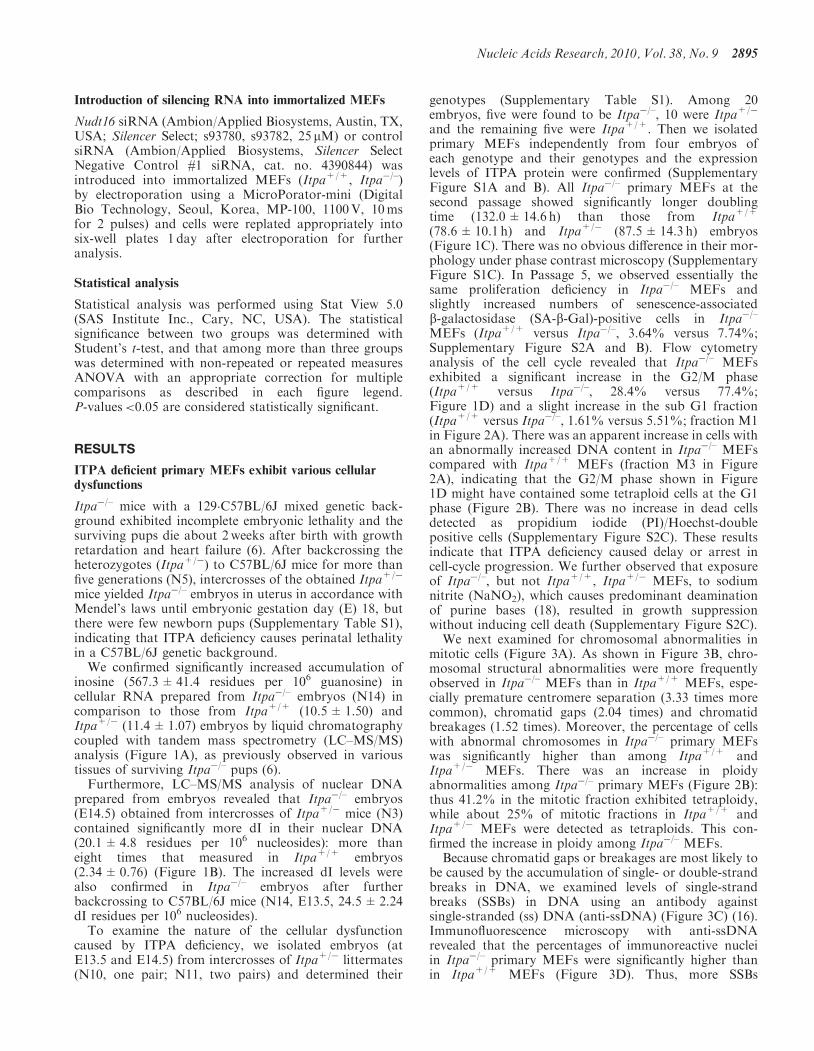
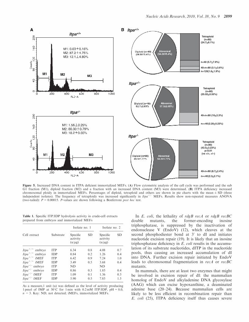
MEFs. Furthermore, the G1 phase fraction increasedsignificantly in immortalized Itpa�/– MEFs (Figure 5A)compared with primary MEFs (Figure 2A). Because thepercentages of tetraploids were significantly higher in bothprimary and immortalized Itpa�/– MEFs than Itpa+/+ or
Itpa+/�MEFs (Figures 2B and 5B), the G2/M fraction inimmortalized Itpa�/– MEFs was apparently less than inprimary Itpa�/– MEFs.
To test whether any backup enzyme for ITPA deficiencymight exist in the immortalized Itpa�/– MEFs, wemeasured ITP-hydrolyzing activity in extracts preparedfrom embryos and immortalized MEFs (Table 1,Supplementary Figure S3). There was no detectableITP-hydrolyzing activity in extracts prepared fromItpa�/– embryos, but a significantly high rate wasdetected in Itpa+/+ embryos, thus confirming ITPA defi-ciency in Itpa�/– embryos. We also confirmed that Itpa�/–
primary MEFs (Passage 3) did not generate IMP fromITP. On the other hand, we detected substantial activityin extracts from immortalized Itpa�/– MEFs, although thelevels were less than 20% of that detected in immortalizedItpa+/+ MEFs or embryos. We then measuredIDP-hydrolyzing activity in these extracts (Table 1,Supplementary Figure S3). Levels were increased both inItpa+/+ and Itpa�/– MEFs during immortalization andthe activity was more significantly increased in immor-talized Itpa�/– MEFs than in immortalized Itpa+/+
MEFs. These results strongly suggest that an increased
Figure 2. Increased DNA content in ITPA deficient primary MEFs. (A) Flow cytometric analysis of the cell cycle was performed and the sub G1fraction (M1), diploid fraction (M2) and a fraction with an increased DNA content (M3) were determined. (B) ITPA deficiency increased chromo-somal ploidy in primary MEFs. Percentages of diploid, tetraploid and others are shown in pie charts with the mean±SD (three independentisolates). The frequency of tetraploidy was increased significantly in Itpa�/– MEFs. Results show non-repeated measures ANOVA (two-tailed):P= 0.094. P-value is shown following a Bonferroni post hoc test.
2896 Nucleic Acids Research, 2010, Vol. 38, No. 9
expression of IDP or ITP-hydrolyzing enzyme(s) blockedor reversed the ITPA-deficient phenotypes observed inprimary Itpa�/– MEFs.
NUDT16 with strong (deoxy)inosine diphosphataseactivity is responsible for cancellation of ITPA-deficientphenotypes and an increase in ITP/IDP-hydrolyzingactivity during immortalization
We recently found that the human NUDT16 (nudix[nucleoside diphosphate linked moiety X]-type motif 16)protein, identified as an ITP/XTP/GTP binding protein,has strong IDP and 20-deoxy-IDP (dIDP)-hydrolyzing
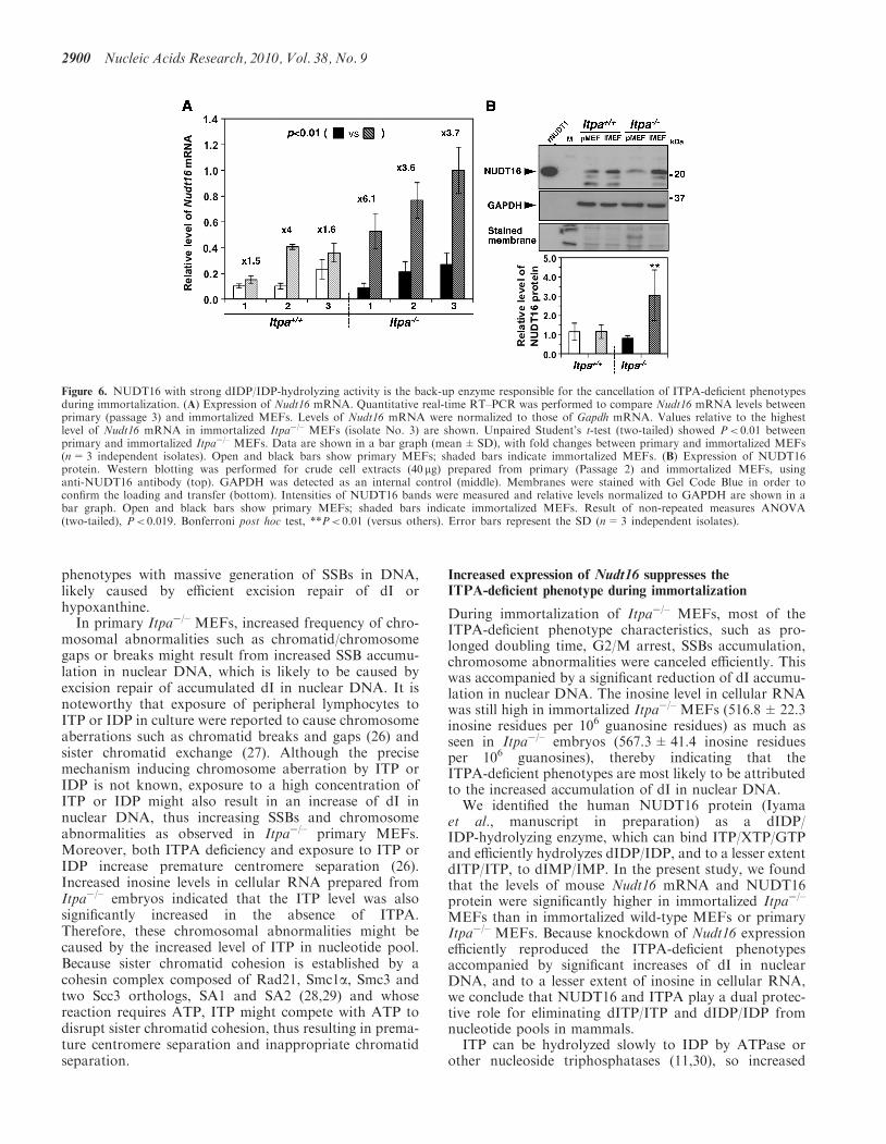
activities with weak ITP/dITP-hydrolyzing activities.We also confirmed that mouse NUDT16 protein hasessentially the same activities as the human protein(Iyama et al., in preparation). Therefore, we comparedNudt16 mRNA levels among primary and immortalizedItpa+/+ or Itpa�/– MEFs (Figure 6A). Quantitativereal-time reverse transcription polymerase chain reaction(RT–PCR) analysis revealed that the expression levels ofNudt16 mRNA were similar between primary MEFsderived from Itpa+/+ and Itpa�/– embryos. In contrast,the expression levels of Nudt16 mRNA in immortalizedItpa�/– MEFs were more than 3-fold higher than thoseof primary Itpa�/– MEFs and more than 2-fold higher
Figure 3. ITPA deficiency increases chromosome abnormalities with increased accumulation of SSBs in nuclear DNA. (A) Various chromosomestructural abnormalities were observed in Itpa�/– primary MEFs (Passage 2). (B) ITPA deficiency increases chromosome abnormalities. The fre-quency of chromosomal abnormality was significantly increased in Itpa�/– MEFs in comparison to Itpa+/+ and Itpa+/� MEFs. Result ofnon-repeated measures ANOVA (two-tailed), P=0.0149. Bonferroni post hoc test, P< 0.01. Data are shown as pie charts with the mean±SD(n=4 independent isolates). (C) Detection of immunoreactivity against ssDNA in Itpa�/– primary MEFs. Immunofluorescence microscopy withanti-ssDNA antibody (green) revealed significantly increased ssDNA immunoreactivity in nuclei (DAPI, blue) of Itpa�/– primary MEFs (Passage 2)compared with the wild-type (Itpa+/+). (D) Significant Increase in the ssDNA-positive population in Itpa�/– primary MEF tested by unpairedStudent’s t-test (two-tailed): **P< 0.01. Data are shown in a bar graph (mean±SD, n=4 independent isolates).
Nucleic Acids Research, 2010, Vol. 38, No. 9 2897

than those in immortalized Itpa+/+ MEFs (Figure 6A).Significantly increased expression of NUDT16 protein inimmortalized Itpa�/– MEFs was confirmed by westernblotting analysis of crude cell extracts prepared fromthese MEFs (Figure 6B). We thus conclude that theincreased expression of Nudt16 in immortalized Itpa�/–
MEFs is responsible for the increase in ITP/IDP-hydrolyzing activity compared with immortalizedItpa+/+ MEFs.To examine the contribution of NUDT16 to the
reversal of ITPA-deficient phenotypes in immortalizedItpa�/– MEFs, knockdown of Nudt16 mRNA expressionwas performed using a mixture of two different Nudt16silencing (si)RNAs. This treatment caused an efficientreduction of both Nudt16 mRNA and protein levels toless than 20% of the levels seen in controls (Figure 7Aand B). Knockdown of Nudt16 expression in immortalizedItpa�/– but not in Itpa+/+ MEFs caused a significantreduction in proliferation rate (Figure 7C), as observedin primary Itpa�/– MEFs (Figure 1C).
We next measured inosine levels in cellular RNA withor without Nudt16 siRNAs. Immortalized Itpa�/– MEFscontained 516.8±22.3 inosine residues per 106 guanosineresidues of RNA in the presence of control siRNA whichwas slightly lower than that in Itpa�/– embryos(567.3±41.4 residues per 106 guanosine), and Nudt16expression knockdown increased the level to648.5±01.7 residues per 106 guanosine residues ofRNA (Figure 7D). As shown in Figure 7E, immortalizedItpa�/– MEFs contained 2.49±0.24 dI residues per 106
nucleosides in nuclear DNA in the presence of controlsiRNA which was equivalent to that in immortalizedItpa+/+ MEFs (2.74±0.17 dI residues per 106
nucleosides). Nudt16 expression knockdown inimmortalized Itpa�/– MEFs significantly increased thelevel more than 5-fold (12.73±0.99 residues per 106
nucleosides). In contrast, knockdown of Nudt16 expres-sion in immortalized Itpa+/+ MEFs did not affect thelevel of dI accumulation in nuclear DNA (2.96±0.3 dIresidues per 106 nucleosides).
Knockdown of Nudt16 expression significantlyincreased the immunoreactivity against ssDNA inimmortalized Itpa�/– MEFs (Figure 7F). Moreover,karyotyping of immortalized Itpa�/– MEFs after Nudt16expression knockdown revealed a significant increase inchromosome structural abnormalities, such as chromatidgaps, premature separation and triradial forms comparedwith controls (Figure 7G).
Thus, increased expression of Nudt16 was responsiblefor reversal of the ITPA-deficient phenotypes with reduc-tion of dI accumulation in nuclear DNA but not inosine incellular RNA.
DISCUSSION
ITPA deficiency increases the accumulation ofdeoxyinosine in nuclear DNA resulting in severe cellulardysfunction
Here, we showed for the first time that ITPA deficiencycaused a significant accumulation of dI in the nuclearDNA of mouse embryos, most of which are likely to diearound the time of birth. In wild-type embryos, fewer thanthree residues of dI per 106 nucleosides, corresponding toabout 20 000 residues in a whole cell, were detected innuclear DNA, whereas about 20 residues of dI per 106
nucleosides reaching more than 105 residues per cellaccumulated in Itpa�/– embryos. These results indicatethat both spontaneous generation of dITP and incorpora-tion of dITP into DNA occurred at significantly highfrequencies. Thus, ITPA deficiency caused severe cellulardysfunction resulting in perinatal lethality. Indeed, wedemonstrated that this deficiency in primary MEFsincreased the accumulation of SSBs in nuclear DNAdetected as ssDNA immunoreactivity and as chromo-somal abnormalities such as chromatid/chromosomegaps or breaks. There was also premature centromere sep-aration. All these chromosomal anomalies are likely tocause G2/M arrest thus suppressing cell proliferation, asobserved in primary Itpa�/– MEFs.
Figure 4. ITPA-deficient phenotypes are reversed duringimmortalization. (A) Immortalized Itpa�/– MEFs (triangle) showedthe same proliferation rate as did immortalized Itpa+/+ (circle) andItpa+/� (square) MEFs. Error bars represent the SD (n=3 indepen-dent isolates). (B) The frequency of chromosomal abnormalities wasdecreased in immortalized Itpa�/– MEFs to the levels seen inimmortalized Itpa+/+ MEFs. Data are shown as the mean±SD(n=3 independent isolates).
2898 Nucleic Acids Research, 2010, Vol. 38, No. 9
In E. coli, the lethality of rdgB recA or rdgB recBCdouble mutants, the former-encoding inosinetriphosphatase, is suppressed by the inactivation ofendonuclease V (EndoV) (12), which cleaves at thesecond phosphodiester bond at 30 to dI and initiatesnucleotide excision repair (19). It is likely that an inosinetriphosphatase deficiency in E. coli results in the accumu-lation of its substrate nucleotides, dITP in the nucleotidepools, thus causing an increased accumulation of dIinto DNA. Further excision repair initiated by EndoVleads to chromosomal fragmentation in recA or recBCmutants.In mammals, there are at least two enzymes that might
be involved in excision repair of dI: the mammalianhomolog of EndoV and alkyladenine DNA glycosylase(AAG) which can excise hypoxanthine, a deaminatedadenine base (20–24). Because mammalian cells arelikely to be less efficient in recombination repair thanE. coli (25), ITPA deficiency itself thus causes severe
Tetraploid(n=80)
(24.7+–8.1%)
Tetraploid(n=80)
(25.02+–0.55%)
n<40 (5.7+–7.9%)
40<n<80 (3.1+–3.6%)n=120(1.6+–1.9%)
40<n<80 (10+–2.3%)
n=<40(2.20+–0.23%)
Tetraploid(n=80)
(43.5+–3.23%)p<0.01
(vs +/+, +/–)
40<n<80 (8.7+–2.4%)
n=<40(2.3+–2.1%)
Figure 5. Increased DNA content in ITPA deficient immortalized MEFs. (A) Flow cytometric analysis of the cell cycle was performed and the subG1 fraction (M1), diploid fraction (M2) and a fraction with an increased DNA content (M3) were determined. (B) ITPA deficiency increasedchromosomal ploidy in immortalized MEFs. Percentages of diploid, tetraploid and others are shown in pie charts with the mean±SD (threeindependent isolates). The frequency of tetraploidy was increased significantly in Itpa�/– MEFs. Results show non-repeated measures ANOVA(two-tailed): P= 0.00015. P-values are shown following a Bonferroni post hoc test.
Table 1. Specific ITP/IDP hydrolysis activity in crude-cell extracts
prepared from embryos and immortalized MEFs
Isolate no. 1 Isolate no. 2
Cell extract Substrate Specificactivity(u/mg)
SD Specificactivity(u/mg)
SD
Itpa+/+ embryo ITP 6.34 0.8 4.08 0.7
Itpa+/+ embryo IDP 0.84 0.2 1.26 0.4
Itpa+/+ iMEF ITP 6.42 0.9 7.24 1.0
Itpa+/+ iMEF IDP 3.49 0.5 3.68 0.4
Itpa�/– embryo ITP ND ND
Itpa�/– embryo IDP 0.86 0.3 1.85 0.4
Itpa�/– iMEF ITP 1.09 0.1 1.36 0.3
Itpa�/– iMEF IDP 5.90 0.5 7.85 1.5
As a measure,1 unit (u) was defined as the level of activity producing1 pmol of IMP at 30�C for 1min with 0.2mM ITP/IDP, pH=8.0,n = 3. Key: ND, not detected; iMEFs, immortalized MEFs.
Nucleic Acids Research, 2010, Vol. 38, No. 9 2899
phenotypes with massive generation of SSBs in DNA,likely caused by efficient excision repair of dI orhypoxanthine.In primary Itpa�/– MEFs, increased frequency of chro-
mosomal abnormalities such as chromatid/chromosomegaps or breaks might result from increased SSB accumu-lation in nuclear DNA, which is likely to be caused byexcision repair of accumulated dI in nuclear DNA. It isnoteworthy that exposure of peripheral lymphocytes toITP or IDP in culture were reported to cause chromosomeaberrations such as chromatid breaks and gaps (26) andsister chromatid exchange (27). Although the precisemechanism inducing chromosome aberration by ITP orIDP is not known, exposure to a high concentration ofITP or IDP might also result in an increase of dI innuclear DNA, thus increasing SSBs and chromosomeabnormalities as observed in Itpa�/– primary MEFs.Moreover, both ITPA deficiency and exposure to ITP orIDP increase premature centromere separation (26).Increased inosine levels in cellular RNA prepared fromItpa�/– embryos indicated that the ITP level was alsosignificantly increased in the absence of ITPA.Therefore, these chromosomal abnormalities might becaused by the increased level of ITP in nucleotide pool.Because sister chromatid cohesion is established by acohesin complex composed of Rad21, Smc1a, Smc3 andtwo Scc3 orthologs, SA1 and SA2 (28,29) and whosereaction requires ATP, ITP might compete with ATP todisrupt sister chromatid cohesion, thus resulting in prema-ture centromere separation and inappropriate chromatidseparation.
Increased expression of Nudt16 suppresses theITPA-deficient phenotype during immortalization
During immortalization of Itpa�/– MEFs, most of theITPA-deficient phenotype characteristics, such as pro-longed doubling time, G2/M arrest, SSBs accumulation,chromosome abnormalities were canceled efficiently. Thiswas accompanied by a significant reduction of dI accumu-lation in nuclear DNA. The inosine level in cellular RNAwas still high in immortalized Itpa�/– MEFs (516.8±22.3inosine residues per 106 guanosine residues) as much asseen in Itpa�/– embryos (567.3±41.4 inosine residuesper 106 guanosines), thereby indicating that theITPA-deficient phenotypes are most likely to be attributedto the increased accumulation of dI in nuclear DNA.
We identified the human NUDT16 protein (Iyamaet al., manuscript in preparation) as a dIDP/IDP-hydrolyzing enzyme, which can bind ITP/XTP/GTPand efficiently hydrolyzes dIDP/IDP, and to a lesser extentdITP/ITP, to dIMP/IMP. In the present study, we foundthat the levels of mouse Nudt16 mRNA and NUDT16protein were significantly higher in immortalized Itpa�/–
MEFs than in immortalized wild-type MEFs or primaryItpa�/– MEFs. Because knockdown of Nudt16 expressionefficiently reproduced the ITPA-deficient phenotypesaccompanied by significant increases of dI in nuclearDNA, and to a lesser extent of inosine in cellular RNA,we conclude that NUDT16 and ITPA play a dual protec-tive role for eliminating dITP/ITP and dIDP/IDP fromnucleotide pools in mammals.
ITP can be hydrolyzed slowly to IDP by ATPase orother nucleoside triphosphatases (11,30), so increased
Figure 6. NUDT16 with strong dIDP/IDP-hydrolyzing activity is the back-up enzyme responsible for the cancellation of ITPA-deficient phenotypesduring immortalization. (A) Expression of Nudt16 mRNA. Quantitative real-time RT–PCR was performed to compare Nudt16 mRNA levels betweenprimary (passage 3) and immortalized MEFs. Levels of Nudt16 mRNA were normalized to those of Gapdh mRNA. Values relative to the highestlevel of Nudt16 mRNA in immortalized Itpa�/– MEFs (isolate No. 3) are shown. Unpaired Student’s t-test (two-tailed) showed P< 0.01 betweenprimary and immortalized Itpa�/– MEFs. Data are shown in a bar graph (mean±SD), with fold changes between primary and immortalized MEFs(n=3 independent isolates). Open and black bars show primary MEFs; shaded bars indicate immortalized MEFs. (B) Expression of NUDT16protein. Western blotting was performed for crude cell extracts (40 mg) prepared from primary (Passage 2) and immortalized MEFs, usinganti-NUDT16 antibody (top). GAPDH was detected as an internal control (middle). Membranes were stained with Gel Code Blue in order toconfirm the loading and transfer (bottom). Intensities of NUDT16 bands were measured and relative levels normalized to GAPDH are shown in abar graph. Open and black bars show primary MEFs; shaded bars indicate immortalized MEFs. Result of non-repeated measures ANOVA(two-tailed), P< 0.019. Bonferroni post hoc test, **P< 0.01 (versus others). Error bars represent the SD (n=3 independent isolates).
2900 Nucleic Acids Research, 2010, Vol. 38, No. 9
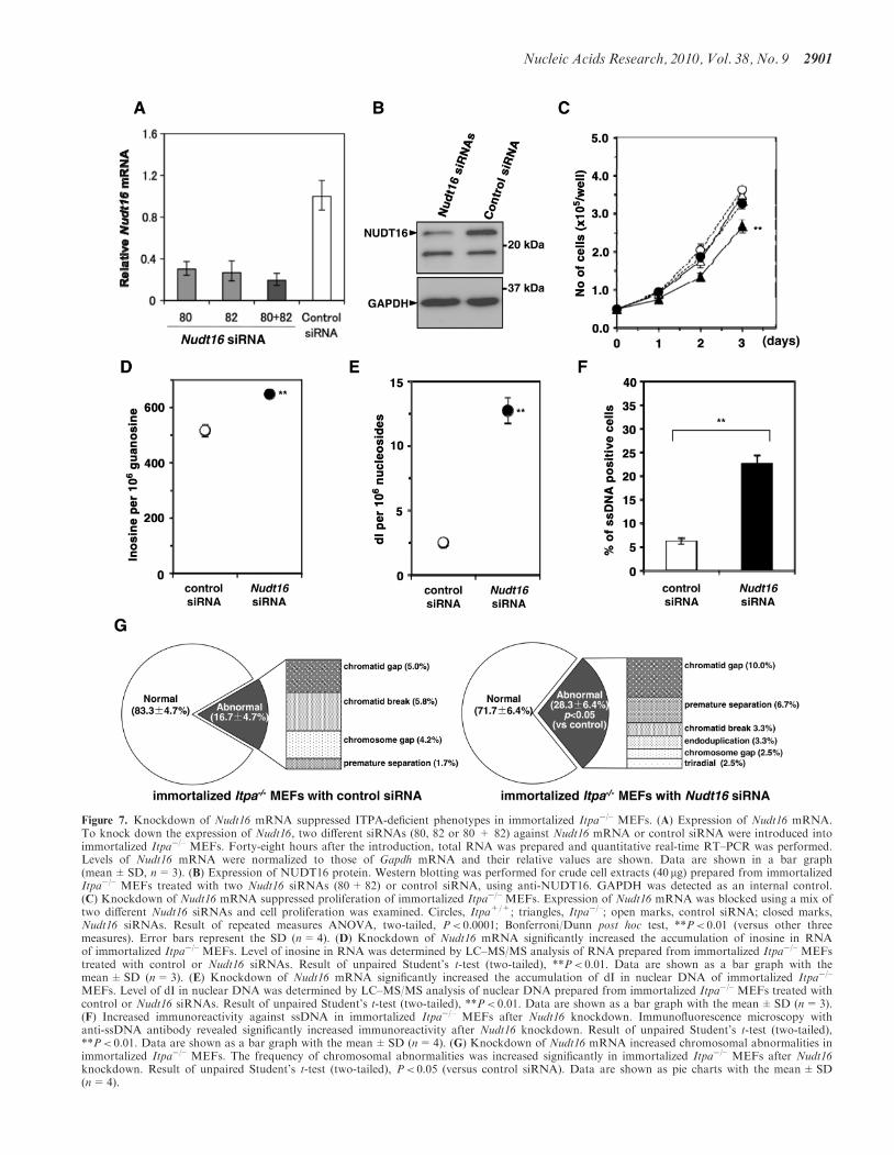
Figure 7. Knockdown of Nudt16 mRNA suppressed ITPA-deficient phenotypes in immortalized Itpa�/– MEFs. (A) Expression of Nudt16 mRNA.To knock down the expression of Nudt16, two different siRNAs (80, 82 or 80 + 82) against Nudt16 mRNA or control siRNA were introduced intoimmortalized Itpa�/– MEFs. Forty-eight hours after the introduction, total RNA was prepared and quantitative real-time RT–PCR was performed.Levels of Nudt16 mRNA were normalized to those of Gapdh mRNA and their relative values are shown. Data are shown in a bar graph(mean±SD, n=3). (B) Expression of NUDT16 protein. Western blotting was performed for crude cell extracts (40 mg) prepared from immortalizedItpa�/– MEFs treated with two Nudt16 siRNAs (80+82) or control siRNA, using anti-NUDT16. GAPDH was detected as an internal control.(C) Knockdown of Nudt16 mRNA suppressed proliferation of immortalized Itpa�/– MEFs. Expression of Nudt16 mRNA was blocked using a mix oftwo different Nudt16 siRNAs and cell proliferation was examined. Circles, Itpa+/+; triangles, Itpa�/–; open marks, control siRNA; closed marks,Nudt16 siRNAs. Result of repeated measures ANOVA, two-tailed, P< 0.0001; Bonferroni/Dunn post hoc test, **P< 0.01 (versus other threemeasures). Error bars represent the SD (n=4). (D) Knockdown of Nudt16 mRNA significantly increased the accumulation of inosine in RNAof immortalized Itpa�/– MEFs. Level of inosine in RNA was determined by LC–MS/MS analysis of RNA prepared from immortalized Itpa�/– MEFstreated with control or Nudt16 siRNAs. Result of unpaired Student’s t-test (two-tailed), **P< 0.01. Data are shown as a bar graph with themean±SD (n=3). (E) Knockdown of Nudt16 mRNA significantly increased the accumulation of dI in nuclear DNA of immortalized Itpa�/–
MEFs. Level of dI in nuclear DNA was determined by LC–MS/MS analysis of nuclear DNA prepared from immortalized Itpa�/– MEFs treated withcontrol or Nudt16 siRNAs. Result of unpaired Student’s t-test (two-tailed), **P< 0.01. Data are shown as a bar graph with the mean±SD (n=3).(F) Increased immunoreactivity against ssDNA in immortalized Itpa�/– MEFs after Nudt16 knockdown. Immunofluorescence microscopy withanti-ssDNA antibody revealed significantly increased immunoreactivity after Nudt16 knockdown. Result of unpaired Student’s t-test (two-tailed),**P< 0.01. Data are shown as a bar graph with the mean±SD (n=4). (G) Knockdown of Nudt16 mRNA increased chromosomal abnormalities inimmortalized Itpa�/– MEFs. The frequency of chromosomal abnormalities was increased significantly in immortalized Itpa�/– MEFs after Nudt16knockdown. Result of unpaired Student’s t-test (two-tailed), P< 0.05 (versus control siRNA). Data are shown as pie charts with the mean±SD(n=4).
Nucleic Acids Research, 2010, Vol. 38, No. 9 2901
dIDP hydrolysis in immortalized Itpa�/– MEFs is likely tobe sufficient to eliminate dITP from the nucleotide pools.Moreover, ITP can be generated in a variety of tissueextracts as well as in erythrocytes (31). We reportedpreviously that ITP accumulated in erythrocytes but notin tissues including the heart and liver derived from Itpa�/–
mice, whereas IMP accumulated markedly in RNAprepared from the latter (6). Because both RNA andDNA synthesis takes place in the latter tissues, DNAand RNA polymerases are likely to utilize ITP and dITPefficiently as nucleotide precursors, thereby consumingmost of the ITP or dITP that accumulates in thenucleotide pools in the absence of ITPA.Considering the likely source of ITP or dITP in the
nucleotide pools, IMP generated from AMP by AMPdeamination must be the most relevant precursor,because most cells can synthesize IDP or ITP from IMP(31) and IDP may be converted to dIDP by ribonucleotidereductase, thus generating dITP (21). To minimizeaccumulation of ITP or dITP in the nucleotide pools,hydrolysis of IDP or dIDP to the correspondingmonophosphates catalyzed by NUDT16 is likely to beas critical as is any hydrolysis of ITP or dITP to the cor-responding monophosphates.In the present study, we showed that increased expres-
sion of NUDT16 in Itpa�/– immortalized MEFs is suffi-cient to cancel the ITPA-deficient phenotypes observed inItpa�/– embryos or primary MEFs, suggesting that thelower expression level of NUDT16 in normal tissuesmay be why any ITPA deficiency causes such severephenotypes. On the other hand, ITPA deficiency inhumans is likely to be related to azathioprine intolerancein patients with inflammatory bowel disease, but does notcause any severe phenotype (32–34), compared with theItpa�/– mice. It is possible that human NUDT16 expres-sion might be higher than that in mouse, thuscompensating for any ITPA deficiency.Identification of NUDT16 as a backup enzyme for
ITPA deficiency in mice will shed light on the mechanismsthat enable humans to be resistant to ITPA deficiency.Towards this goal, it is important to know the relativeexpression of ITPA and NUDT16 in human cells andorgans, and to characterize the enzymatic properties ofNUDT16.
SUPPLEMENTARY DATA
Supplementary Data are available at NAR Online.
ACKNOWLEDGEMENTS
The authors thank M. Ohtsu in the Laboratory forTechnical Support of our institute and N. Adachi,A. Matsuyama, K. Hayashi, K. Nakabeppu andK. Asakawa for technical assistance.
FUNDING
Ministry of Education, Culture, Sports, Science andTechnology of Japan [20013034 to Y.N., 20012038 to
K.S.]; the Japan Society for the Promotion of Science[19390114 to D.T., 08J03650 to T.I.]; Kyushu UniversityGlobal COE program [YN]. Funding for open accesscharges: Ministry of Education, Culture, Sports, Scienceand Technology of Japan; Kyushu University GlobalCOE program.
Conflict of interest statement. None declared.
REFERENCES
1. Nakabeppu,Y., Tsuchimoto,D., Furuichi,M. and Sakumi,K.(2004) The defense mechanisms in mammalian cells againstoxidative damage in nucleic acids and their involvement in thesuppression of mutagenesis and cell death. Free Radic. Res., 38,423–429.
2. Nakabeppu,Y., Behmanesh,M., Yamaguchi,H., Yoshimura,D. andSakumi,K. (2007) In Evans,M.D. and Cooke,M.S. (eds),Oxidative Damage to Nucleic Acids. Landes Bioscience/Springer,Austin, TX/New York, pp. 40–53.
3. Nakabeppu,Y. (2001) Molecular genetics and structural biology ofhuman MutT homolog, MTH1. Mutat. Res., 477, 59–70.
4. Nakabeppu,Y., Kajitani,K., Sakamoto,K., Yamaguchi,H. andTsuchimoto,D. (2006) MTH1, an oxidized purine nucleosidetriphosphatase, prevents the cytotoxicity and neurotoxicity ofoxidized purine nucleotides. DNA Rep., 5, 761–772.
5. Behmanesh,M., Sakumi,K., Tsuchimoto,D., Torisu,K.,Ohnishi-Honda,Y., Rancourt,D.E. and Nakabeppu,Y. (2005)Characterization of the structure and expression of mouse Itpagene and its related sequences in the mouse genome. DNA Res.,12, 39–51.
6. Behmanesh,M., Sakumi,K., Abolhassani,N., Toyokuni,S., Oka,S.,Ohnishi,Y.N., Tsuchimoto,D. and Nakabeppu,Y. (2009)ITPase-deficient mice show growth retardation and die beforeweaning. Cell Death Differ., 16, 1315–1322.
7. Nonaka,M., Tsuchimoto,D., Sakumi,K. and Nakabeppu,Y. (2009)Mouse RS21-C6 is a mammalian 20-deoxycytidine 50-triphosphatepyrophosphohydrolase that prefers 5-iodocytosine. FEBS J., 276,1654–1666.
8. Tsuzuki,T., Egashira,A., Igarashi,H., Iwakuma,T., Nakatsuru,Y.,Tominaga,Y., Kawate,H., Nakao,K., Nakamura,K., Ide,F. et al.(2001) Spontaneous tumorigenesis in mice defective in the MTH1gene encoding 8-oxo-dGTPase. Proc. Natl Acad. Sci. USA, 98,11456–11461.
9. Shenoy,T.S. and Clifford,A.J. (1975) Adenine nucleotidemetabolism in relation to purine enzymes in liver, erythrocytesand cultured fibroblasts. Biochim. Biophys. Acta, 411, 133–143.
10. Gower,W.R. Jr, Carr,M.C. and Ives,D.H. (1979) Deoxyguanosinekinase. Distinct molecular forms in mitochondria and cytosol.J. Biol. Chem., 254, 2180–2183.
11. Burton,K., White,H. and Sleep,J. (2005) Kinetics of musclecontraction and actomyosin NTP hydrolysis from rabbit using aseries of metal-nucleotide substrates. J. Physiol., 563, 689–711.
12. Bradshaw,J.S. and Kuzminov,A. (2003) RdgB acts to avoidchromosome fragmentation in Escherichia coli. Mol. Microbiol.,48, 1711–1725.
13. Ide,Y., Tsuchimoto,D., Tominaga,Y., Nakashima,M.,Watanabe,T., Sakumi,K., Ohno,M. and Nakabeppu,Y. (2004)Growth retardation and dyslymphopoiesis accompanied by G2/Marrest in APEX2-null mice. Blood, 104, 4097–4103.
14. Tsuruya,K., Furuichi,M., Tominaga,Y., Shinozaki,M.,Tokumoto,M., Yoshimitsu,T., Fukuda,K., Kanai,H., Hirakata,H.,Iida,M. et al. (2003) Accumulation of 8-oxoguanine in the cellularDNA and the alteration of the OGG1 expression duringischemia-reperfusion injury in the rat kidney. DNA Rep., 2,211–229.
15. Taghizadeh,K., McFaline,J.L., Pang,B., Sullivan,M., Dong,M.,Plummer,E. and Dedon,P.C. (2008) Quantification of DNAdamage products resulting from deamination, oxidation andreaction with products of lipid peroxidation by liquidchromatography isotope dilution tandem mass spectrometry.Nat. Protoc., 3, 1287–1298.
2902 Nucleic Acids Research, 2010, Vol. 38, No. 9

16. Oka,S., Ohno,M., Tsuchimoto,D., Sakumi,K., Furuichi,M. andNakabeppu,Y. (2008) Two distinct pathways of cell deathtriggered by oxidative damage to nuclear and mitochondrialDNAs. EMBO J., 27, 421–432.
17. Tsuchimoto,D., Sakai,Y., Sakumi,K., Nishioka,K., Sasaki,M.,Fujiwara,T. and Nakabeppu,Y. (2001) Human APE2 protein ismostly localized in the nuclei and to some extent in themitochondria, while nuclear APE2 is partly associated withproliferating cell nuclear antigen. Nucleic Acids Res., 29, 2349–2360.
18. Kow,Y.W. (2002) Repair of deaminated bases in DNA. FreeRadic. Biol. Med., 33, 886–893.
19. Dalhus,B., Arvai,A.S., Rosnes,I., Olsen,Ø., Backe,P.H., Alseth,I.,Gao,H., Cao,W., Tainer,J.A. and Bjøras,M. (2009) Structures ofendonuclease V with DNA reveal initiation of deaminatedadenine repair. Nat. Struct. Mol. Biol., 16, 138–143.
20. Karran,P. and Lindahl,T. (1978) Enzymatic excision of freehypoxanthine from polydeoxynucleotides and DNA containingdeoxyinosine monophosphate residues. J. Biol. Chem., 253,5877–5879.
21. Myrnes,B., Guddal,P.H. and Krokan,H. (1982) Metabolism ofdITP in HeLa cell extracts, incorporation into DNA by isolatednuclei and release of hypoxanthine from DNA by a hypoxanthine-DNA glycosylase activity. Nucleic Acids Res., 10, 3693–3701.
22. Saparbaev,M. and Laval,J. (1994) Excision of hypoxanthine fromDNA containing dIMP residues by the Escherichia coli, yeast, rat,and human alkylpurine DNA glycosylases. Proc. Natl Acad. Sci.USA, 91, 5873–5877.
23. Moe,A., Ringvoll,J., Nordstrand,L.M., Eide,L., Bjøras,M.,Seeberg,E., Rognes,T. and Klungland,A. (2003) Incision athypoxanthine residues in DNA by a mammalian homologue ofthe Escherichia coli antimutator enzyme endonuclease V. NucleicAcids Res., 31, 3893–3900.
24. Vallur,A.C., Maher,R.L. and Bloom,L.B. (2005) The efficiency ofhypoxanthine excision by alkyladenine DNA glycosylase is alteredby changes in nearest neighbor bases. DNA Rep., 4, 1088–1098.
25. Pardo,B., Gomez-Gonzalez,B. and Aguilera,A. (2009) DNAdouble-strand break repair: how to fix a broken relationship.Cell. Mol. Life Sci., 66, 1039–1056.
26. Auclair,C., Gouyette,A., Levy,A. and Emerit,I. (1990) Clastogenicinosine nucleotide as components of the chromosome breakagefactor in scleroderma patients. Arch. Biochem. Biophys., 278,238–244.
27. Vormittag,W. and Brannath,W. (2001) As to the clastogenic-,sister-chromatid exchange inducing-and cytotoxic activity ofinosine triphosphate in cultures of human peripheral lymphocytes.Mutat. Res., 476, 71–81.
28. Watrin,E. and Peters,J.M. (2006) Cohesin and DNA damagerepair. Exp. Cell Res., 312, 2687–2693.
29. Uhlmann,F. (2009) A matter of choice: the establishment of sisterchromatid cohesion. EMBO Rep., 10, 1095–1102.
30. Vanderheiden,B.S. (1975) ITP pyrophosphohydrolase and IDPphosphohydrolase in rat tissue. J. Cell. Physiol., 86, 167–175.
31. Vanderheiden,B.S. (1979) Inosine di- and triphosphatesynthesis in erythrocytes and cell extracts. J. Cell. Physiol., 99,287–301.
32. Sumi,S., Marinaki,A.M., Arenas,M., Fairbanks,L.,Shobowale-Bakre,M., Rees,D.C., Thein,S.L., Ansari,A.,Sanderson,J., De Abreu,R.A. et al. (2002) Genetic basis ofinosine triphosphate pyrophosphohydrolase deficiency. Hum.Genet., 111, 360–367.
33. Marinaki,A.M., Duley,J.A., Arenas,M., Ansari,A., Sumi,S.,Lewis,C.M., Shobowale-Bakre,M., Fairbanks,L.D. andSanderson,J. (2004) Mutation in the ITPA gene predictsintolerance to azathioprine. Nucleosides Nucleotides Nucleic Acids,23, 1393–1397.
34. Seela,F. and Xu,K. (2007) Pyrazolo[3,4-d]pyrimidineribonucleosides related to 2-aminoadenosine and isoguanosine:synthesis, deamination and tautomerism. Org. Biomol. Chem., 5,3034–3045.
Nucleic Acids Research, 2010, Vol. 38, No. 9 2903