Embed Size (px)
Citation preview
88 Annals of Vascular Diseases Vol. 14, No. 1 (2021)
Ann Vasc Dis Vol. 14, No. 1; 2021; pp 88–91
Case Report
Nutcracker Phenomenon with Menorrhagia in a Woman with Marfan Syndrome
Kayo Sugiyama, MD,2 Toshiki Fujiyoshi, MD,1 Nobusato Koizumi, MD, PhD,1 and Hitoshi Ogino, MD, PhD1
Nutcracker phenomenon (NCP) refers to left renal vein compression at the superior mesenteric artery origin involv-ing hematuria and dysuria due to the compression of the renal venous return and pelvic congestion syndrome caused by the compression of the gonadal venous return. A lep-tosomatic woman (body mass index, 19 kg/m2) presented with NCP and Marfan syndrome accompanied by severe menorrhagia. Vascular ultrasonography revealed reversed flow in the left ovarian vein. Preoperative computed tomog-raphy revealed a sharp aortomesenteric angle and short aortomesenteric distance. After controlling her menstrual period via oral contraception, she underwent valve-sparing surgery for aortic root dilation, which spontaneously sub-sided the menorrhagia.
Keywords: nutcracker phenomenon, pelvic congestion syndrome, Marfan syndrome
IntroductionNutcracker phenomenon (NCP) refers to the vascular entrapment of the left renal vein between the aorta and superior mesenteric artery. The typical features of NCP in-volve underlying compression of the left renal vein, which results in hematuria and dysuria.1,2) NCP also causes pelvic congestion syndrome with reversed flow in the left
gonadal vein, resulting in chronic pelvic pain and dysmen-orrhea. However, metrorrhagia and menorrhagia rarely occur.3–5) A low body mass index is associated with the occurrence of NCP as the aortomesenteric angle can be sharp, and the aortomesenteric distance can be short.6–9) Although there have been no reports on the relationship between Marfan syndrome (MFS) and NCP, NCP should be considered in leptosomatic patients suffering from pel-vic congestion syndrome.
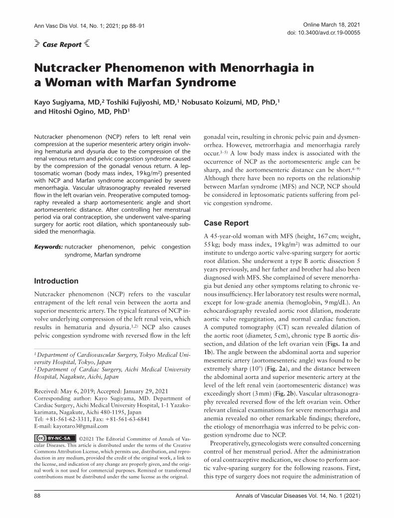
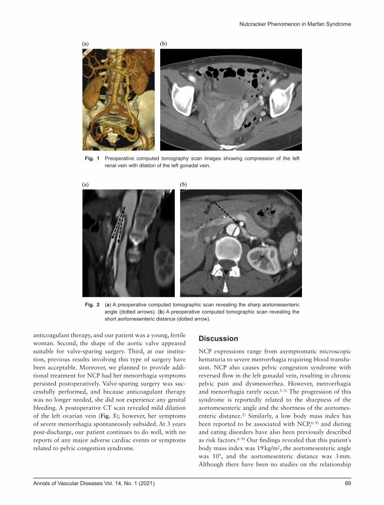
Case ReportA 45-year-old woman with MFS (height, 167 cm; weight, 55 kg; body mass index, 19 kg/m2) was admitted to our institute to undergo aortic valve-sparing surgery for aortic root dilation. She underwent a type B aortic dissection 5 years previously, and her father and brother had also been diagnosed with MFS. She complained of severe menorrha-gia but denied any other symptoms relating to chronic ve-nous insufficiency. Her laboratory test results were normal, except for low-grade anemia (hemoglobin, 9 mg/dL). An echocardiography revealed aortic root dilation, moderate aortic valve regurgitation, and normal cardiac function. A computed tomography (CT) scan revealed dilation of the aortic root (diameter, 5 cm), chronic type B aortic dis-section, and dilation of the left ovarian vein (Figs. 1a and 1b). The angle between the abdominal aorta and superior mesenteric artery (aortomesenteric angle) was found to be extremely sharp (10°) (Fig. 2a), and the distance between the abdominal aorta and superior mesenteric artery at the level of the left renal vein (aortomesenteric distance) was exceedingly short (3 mm) (Fig. 2b). Vascular ultrasonogra-phy revealed reversed flow of the left ovarian vein. Other relevant clinical examinations for severe menorrhagia and anemia revealed no other remarkable findings; therefore, the etiology of menorrhagia was inferred to be pelvic con-gestion syndrome due to NCP.
Preoperatively, gynecologists were consulted concerning control of her menstrual period. After the administration of oral contraceptive medication, we chose to perform aor-tic valve-sparing surgery for the following reasons. First, this type of surgery does not require the administration of
Online March 18, 2021doi: 10.3400/avd.cr.19-00055
1 Department of Cardiovascular Surgery, Tokyo Medical Uni-versity Hospital, Tokyo, Japan2 Department of Cardiac Surgery, Aichi Medical University Hospital, Nagakute, Aichi, Japan
Received: May 6, 2019; Accepted: January 29, 2021Corresponding author: Kayo Sugiyama, MD. Department of Cardiac Surgery, Aichi Medical University Hospital, 1-1 Yazako-karimata, Nagakute, Aichi 480-1195, JapanTel: +81-561-62-3311, Fax: +81-561-63-6841E-mail: [email protected]
©2021 The Editorial Committee of Annals of Vas-cular Diseases. This article is distributed under the terms of the Creative Commons Attribution License, which permits use, distribution, and repro-duction in any medium, provided the credit of the original work, a link to the license, and indication of any change are properly given, and the origi-nal work is not used for commercial purposes. Remixed or transformed contributions must be distributed under the same license as the original.
Annals of Vascular Diseases Vol. 14, No. 1 (2021) 89
Nutcracker Phenomenon in Marfan Syndrome
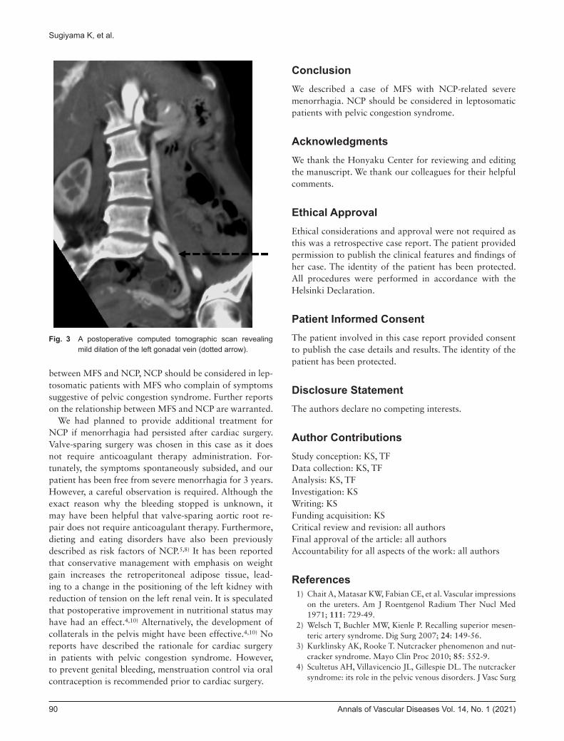
anticoagulant therapy, and our patient was a young, fertile woman. Second, the shape of the aortic valve appeared suitable for valve-sparing surgery. Third, at our institu-tion, previous results involving this type of surgery have been acceptable. Moreover, we planned to provide addi-tional treatment for NCP had her menorrhagia symptoms persisted postoperatively. Valve-sparing surgery was suc-cessfully performed, and because anticoagulant therapy was no longer needed, she did not experience any genital bleeding. A postoperative CT scan revealed mild dilation of the left ovarian vein (Fig. 3); however, her symptoms of severe menorrhagia spontaneously subsided. At 3 years post-discharge, our patient continues to do well, with no reports of any major adverse cardiac events or symptoms related to pelvic congestion syndrome.
DiscussionNCP expressions range from asymptomatic microscopic hematuria to severe metrorrhagia requiring blood transfu-sion. NCP also causes pelvic congestion syndrome with reversed flow in the left gonadal vein, resulting in chronic pelvic pain and dysmenorrhea. However, metrorrhagia and menorrhagia rarely occur.3–5) The progression of this syndrome is reportedly related to the sharpness of the aortomesenteric angle and the shortness of the aortomes-enteric distance.2) Similarly, a low body mass index has been reported to be associated with NCP,6–9) and dieting and eating disorders have also been previously described as risk factors.6–9) Our findings revealed that this patient’s body mass index was 19 kg/m2, the aortomesenteric angle was 10°, and the aortomesenteric distance was 3 mm. Although there have been no studies on the relationship
Fig. 1 Preoperative computed tomography scan images showing compression of the left renal vein with dilation of the left gonadal vein.
Fig. 2 (a) A preoperative computed tomographic scan revealing the sharp aortomesenteric angle (dotted arrows). (b) A preoperative computed tomographic scan revealing the short aortomesenteric distance (dotted arrow).
90 Annals of Vascular Diseases Vol. 14, No. 1 (2021)
Sugiyama K, et al.
between MFS and NCP, NCP should be considered in lep-tosomatic patients with MFS who complain of symptoms suggestive of pelvic congestion syndrome. Further reports on the relationship between MFS and NCP are warranted.
We had planned to provide additional treatment for NCP if menorrhagia had persisted after cardiac surgery. Valve-sparing surgery was chosen in this case as it does not require anticoagulant therapy administration. For-tunately, the symptoms spontaneously subsided, and our patient has been free from severe menorrhagia for 3 years. However, a careful observation is required. Although the exact reason why the bleeding stopped is unknown, it may have been helpful that valve-sparing aortic root re-pair does not require anticoagulant therapy. Furthermore, dieting and eating disorders have also been previously described as risk factors of NCP.5,8) It has been reported that conservative management with emphasis on weight gain increases the retroperitoneal adipose tissue, lead-ing to a change in the positioning of the left kidney with reduction of tension on the left renal vein. It is speculated that postoperative improvement in nutritional status may have had an effect.4,10) Alternatively, the development of collaterals in the pelvis might have been effective.4,10) No reports have described the rationale for cardiac surgery in patients with pelvic congestion syndrome. However, to prevent genital bleeding, menstruation control via oral contraception is recommended prior to cardiac surgery.
ConclusionWe described a case of MFS with NCP-related severe menorrhagia. NCP should be considered in leptosomatic patients with pelvic congestion syndrome.
AcknowledgmentsWe thank the Honyaku Center for reviewing and editing the manuscript. We thank our colleagues for their helpful comments.
Ethical ApprovalEthical considerations and approval were not required as this was a retrospective case report. The patient provided permission to publish the clinical features and findings of her case. The identity of the patient has been protected. All procedures were performed in accordance with the Helsinki Declaration.
Patient Informed ConsentThe patient involved in this case report provided consent to publish the case details and results. The identity of the patient has been protected.
Disclosure StatementThe authors declare no competing interests.
Author ContributionsStudy conception: KS, TFData collection: KS, TFAnalysis: KS, TFInvestigation: KSWriting: KSFunding acquisition: KSCritical review and revision: all authorsFinal approval of the article: all authorsAccountability for all aspects of the work: all authors
References 1) Chait A, Matasar KW, Fabian CE, et al. Vascular impressions
on the ureters. Am J Roentgenol Radium Ther Nucl Med 1971; 111: 729-49.
2) Welsch T, Buchler MW, Kienle P. Recalling superior mesen-teric artery syndrome. Dig Surg 2007; 24: 149-56.
3) Kurklinsky AK, Rooke T. Nutcracker phenomenon and nut-cracker syndrome. Mayo Clin Proc 2010; 85: 552-9.
4) Scultetus AH, Villavicencio JL, Gillespie DL. The nutcracker syndrome: its role in the pelvic venous disorders. J Vasc Surg
Fig. 3 A postoperative computed tomographic scan revealing mild dilation of the left gonadal vein (dotted arrow).
Annals of Vascular Diseases Vol. 14, No. 1 (2021) 91
Nutcracker Phenomenon in Marfan Syndrome
2001; 34: 812-9. 5) Tarazov PG, Prozorovskij KV, Ryzhkov VK. Pelvic pain syn-
drome caused by ovarian varices. Treatment by transcatheter embolization. Acta Radiol 1997; 38: 1023-5.
6) Orczyk K, Labetowicz P, Lodzinski S, et al. The nutcracker syndrome. Morphology and clinical aspects of the important vascular variations: a systematic study of 112 cases. Int Angiol 2016; 35: 71-7.
7) Ozkurt H, Cenker MM, Bas N, et al. Measurement of the distance and angle between the aorta and superior mesen-teric artery: normal values in different BMI categories. Surg
Radiol Anat 2007; 29: 595-9. 8) Nunn R, Henry J, Slesser AAP, et al. A model example:
coexisting superior mesenteric artery syndrome and the nut-cracker phenomenon. Case Rep Surg 2015; 2015: 649469.
9) Shin JII, Park JM, Lee JS, et al. Effect of renal Doppler ultra-sound on the detection of nutcracker syndrome in children with hematuria. Eur J Pediatr 2007; 166: 399-404.
10) Ananthan K, Onida S, Davies AH. Nutcracker syndrome: an update on current diagnostic criteria and management guidelines. Eur J Vasc Endovasc Surg 2017; 53: 886-94.