Embed Size (px)
Citation preview
Page 1/36
7-Dehydrocholesterol is an endogenous suppressorof ferroptosisJose Pedro Friedmann Angeli ( [email protected] )
University of Würzburg https://orcid.org/0000-0001-7706-1379Florencio Porto Freitas
Rudolf Virchow CenterPalina Nepachalovich
University of LeipzigLohans Puentes
University of KölnOmkar Zilka
University of OttawaAlex Inague
University of WürzburgSvenja Lorenz
Helmholtz Zentrum MünchenViktoria Kunz
Universitätsklinikum Würzburg (UKW)Helene Nehring
University of WürzburgThamara Nishida Xavier da Silva
University of WürzburgZhiyi Chen
University of WürzburgSebastian Doll
Helmholtz Zentrum München Deutsches Forschungszentrum für Gesundheit und Umwelt (GmbH)Werner Schmitz
University of Würzburg https://orcid.org/0000-0003-0485-7303Peter Imming
University of HalleSayuri Miyamoto
Universidade de Sao PauloJudith Klein-Seetharaman
Arizona State UniverstiyLokender Kumar

Page 2/36
Colorado School of MinesThiago Cardoso Genaro-Mattos
University of NebraskaKaroly Mirnics
University of Nebraska Medical Center https://orcid.org/0000-0002-5521-0254Svenja Meierjohann
University of WürzburgMatthias Kroiss
Ludwig Maximillian UniversityIsabel Weigand
Ludwig Maximillian UniversityKurt Bommert
Universitätsklinikum Würzburg (UKW)Ralf Bargou
University Hospital Wuerzburg https://orcid.org/0000-0002-1221-7421Ana Garcia-Saez
University of Cologne https://orcid.org/0000-0002-3894-5945Derek Pratt
Department of Chemistry and Biomolecular Sciences, University of Ottawa https://orcid.org/0000-0002-7305-745XMaria Fedorova
University of LeipzigAnn Wehmann
University of DenverAline Horling
University of HalleGeorg Bornkamm
University of UlmMarcus Conrad
Helmholtz Zentrum München https://orcid.org/0000-0003-1140-5612
Biological Sciences - Article
Keywords: cell death, 7-dehydrocholesterol reductase, pro-survival function
Posted Date: October 6th, 2021
DOI: https://doi.org/10.21203/rs.3.rs-943221/v1

Page 3/36
License: This work is licensed under a Creative Commons Attribution 4.0 International License. Read Full License
Page 4/36
AbstractFerroptosis is a form of cell death that has received considerable attention not only as a means toeradicate de�ned tumour entities but also because it provides unforeseen insights into the metabolicadaptation exploited by tumours to counteract phospholipid oxidation. Here, we identify a pro-ferroptoticactivity of 7-dehydrocholesterol reductase (DHCR7) and an unexpected pro-survival function of itssubstrate, 7-dehydrocholesterol (7-DHC). Although previous studies suggested that high levels of 7-DHCare cytotoxic to developing neurons and favour lipid peroxidation, we now demonstrate that 7-DHCaccumulation confers a robust pro-survival function in cancer cells. 7-DHC, due to its far superiorreactivity towards peroxyl radicals, is shown here to effectively shield (phospho)lipids from autoxidationand subsequent fragmentation. We further demonstrate in a subset of ferroptosis-sensitive Burkittlymphomas - where DHCR7 mutations have been reported - that the accumulation of 7-DHC is su�cientto suppress the basal sensitivity of cells toward ferroptosis, thereby translating into an unexpectedgrowth advantage. Conclusively, our �ndings provide compelling evidence of a yet-unrecognised anti-ferroptotic activity of 7-DHC as a cell-intrinsic mechanism that could be exploited by cancer cells toescape ferroptosis.
Main TextFerroptosis has attracted considerable attention in recent years1, and a detailed characterisation of thispathway has uncovered it's implication in a series of pathological conditions ranging from tissueischaemia/reperfusion injury to infection2,3. Moreover, the modulation of ferroptosis is increasinglyrecognised as a potential avenue for developing therapeutics against various diseases4. At the molecularlevel, ferroptosis was initially characterised as the cell death process induced by cysteine starvation,usually caused by lack or insu�cient activity of the cystine-glutamate antiporter (designated system xc
-)5.
Low system system xc- activity directly impacts substrate availability for the key enzyme regulating
ferroptosis, namely glutathione peroxidase 4 (GPX4), leading to lipid hydroperoxide accumualtion andcell death6. Early works have established the central role played by the enzymatic activity of GPX4 insuppressing the process of ferroptosis7-9. GPX4 is the sole enzyme responsible for reducing peroxidisedphospholipids10 and can be inhibited by a series of alkylating small molecules, such as RSL3 andML2108, leading to cell death in ferroptosis-sensitive cancer cell lines. The initial characterisation of thispathway demonstrated the critical role of esteri�ed polyunsaturated fatty acid (PUFA) oxidationdownstream of GPX4 inhibition as a driver of ferroptosis11,12. Speci�cally, we and others have shown thatthe activity of the acyl-CoA-synthetase long-chain family 4 (ACSL4) is required for the enrichment ofphospholipids with PUFAs11. The enrichment of phospholipids with PUFAs results in a markeddependency on GPX4 activity11,13. Accordingly, the inhibition of GPX4 in ferroptosis-prone cell lines leadsto the characteristic oxidation �ngerprint entailing the accumulation of peroxidised products ofphosphatidylethanolamine (PE) containing arachidonic acid (AA) and adrenic acid (AdA)12. It has beenfurther demonstrated that the sole accumulation of peroxidised fatty acids is not su�cient to induce
Page 5/36
ferroptosis, and a central role in the free radical-mediated propagation step de�ned14. This process hasbeen shown to contribute to the formation of pore-like structures of ill-de�ned identity15 that drive theosmotic lysis of the cells16.
The present study uncovered and characterised an unexpected role for 7-dehydrocholesterol reductase(DHCR7) in the ferroptotic process. DHCR7 catalyses the �nal step in cholesterol biosynthesis, and itsinhibition leads to the accumulation of 7-dehydrocholesterol (7-DHC). 7-DHC was initially reported toaccumulate in preputial gland tumours by Kandutsh and Russel17 and whose function, at that time, wasonly assumed to function as a spare capacity for cholesterol synthesis. We show that the accumulationof 7-DHC translates into an increased tolerance towards (phospho)lipid peroxidation, thus providing arobust and unexpected resistance to ferroptosis. This mechanism could be potentially exploited by Burkitt´s lymphomas (BL) to overcome the metabolic stress characteristic of their lineage speci�c low systemxc
- activity18. Moreover, the detailed characterisation of the unique protective effect of 7-DHC on(phospho)lipid peroxidation allows us to provide evidence that ferroptotic cell death is a consequence ofthe accumulation of oxidatively truncated (phospho)lipid species rather than solely lipid hydroperoxideaccumulation and that these species are integral players in ferroptosis execution.
DHCR7 is a pro-ferroptotic gene
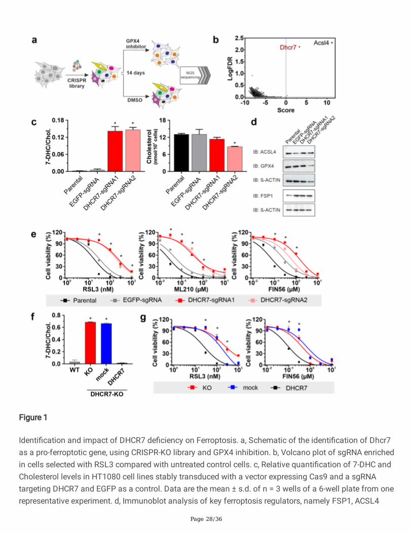
Spurred by the still incomplete understanding of the ferroptotic process and the development of next-generation single guide RNAs (sgRNAs)19, we performed a genome-wide reverse genetic CRISPR screenusing second-generation CRISPR libraries to identify genes that may confer robust protection againstferroptosis. To this end, the Pfa1 cell line6 was transduced with a CRISPR library covering 18,424 geneswith a total representation of 90,230 sgRNAs followed by a stringent selection for fourteen days using200 nM of the GPX4 inhibitor (1S,3R)-RSL3, from now on only RSL3 (Fig. 1a). Consistent with the resultsof our previous screen and those of other groups, Acsl4 emerged as the highest-scoring hit11,13,20-22. Thesecond top-scoring gene with multiple sgRNAs enriched was Dhcr7 (Fig. 1b). The identi�cation of Dhcr7as a potential pro-ferroptotic gene was unexpected in light of a number of studies indicating that loss orinhibition of DHCR7 is associated with increased lipid peroxidation23,24. Intrigued by this �nding, we setout to explore the basis of this unanticipated discovery. Using the bona �de ferroptosis cell line modelHT1080, we generated DHCR7-de�cient cell lines using two independent sgRNAs. The successful loss ofDHCR7 (Fig. 1c) was mirrored by the accumulation of its direct substrate 7-DHC. Notably, cholesteroldepletion was not observed as most of it stems from the uptake of cholesterol present in the serum.Importantly, knockout of DHCR7 did not affect the expression of known ferroptosis regulators (Fig. 1d). Insupport of this conclusion, we could con�rm the screening results showing that DHCR7-de�cient HT1080cells present a marked resistance towards ferroptosis inducing compounds (Fig. 1e). Similar results wereobtained with three independent clonal cell lines derived from Pfa1, HT1080 and MDA-MB-435 cells,supporting the general impact of this system in preventing cell death (Extended Data Fig. 1). We furthershow that DHCR7 loss does not modulate sensitivity to a panel of cytotoxic compounds, thushighlighting its speci�city to the ferroptotic process (Extended Data Fig. 1d, f). We further corroborated
Page 6/36
these �ndings with studies of a clonal cell line derived from the HT1080 DHCR7 knockout (KO) pool toavoid confounding results from non-edited cells. Using this cell line, we could unequivocally show the pro-ferroptotic activity of DHCR7 as genetic reconstitution of a sgRNA resistant DHCR7 variant abolished 7-DHC levels and re-sensitised cells to ferroptosis without impacting on the cell's response to othercytotoxic agents (Fig. 1f, g and Extended Data Fig. 1e, f).
7-DHC is a bona �de anti-ferroptotic metabolite
In the penultimate step of the cholesterol biosynthesis pathway, lathosterol, through lathosterol oxidase(SC5D), is converted to 7-DHC, which, in turn, is reduced to cholesterol by DHCR7 in the �nal step of thecholesterol pathway (Fig. 2a). Several earlier studies have pointed to a toxic effect of 7-DHCaccumulation via an increased susceptibility toward lipid peroxidation due to the very high inherentreactivity of 7-DHC24. To shed light on these seemingly contradictory �ndings, we generated aDHCR7/SC5D double mutant cell line to address whether 7-DHC accumulation indeed mediates theprotective effects induced by the loss of DHCR7. In agreement with a protective effect of 7-DHC, the lossof SC5D in the DHCR7 knockout cell line completely abolished the protective effect conferred by the singleloss of DHCR7 (Fig. 2b). As expected, deletion of both genes led to a detectable accumulation oflathosterol and completely abolished the formation of 7-DHC (Fig. 2c). Subsequently, the serialreconstitution of DHCR7 and SC5D in a DHCR7/SC5D knockout background demonstrated that only there-expression of SC5D resulted in an accumulation of 7-DHC (Extended Data Fig. 2a), and consequently amarked resistance to ferroptosis induced by GPX4 inhibition (Fig. 2d), but not to other cytotoxic agents(Extended Data Fig. 2b). Similarly, pharmacological inhibition of several upstream steps of thecholesterol biosynthetic process also resulted in the complete loss of the protective effect conferred byloss of DHCR7 (Extended Data Fig. 2c, d). Using the DHCR7 and the DHCR7/SC5D de�cient cell lines in aseries of sterol supplementation experiments, we further demonstrate that exogenous supplementation of7-DHC protected all cell lines from ferroptosis (Fig. 2e). Additionally, lathosterol only increased ferroptosisresistance in cell lines able to accumulate 7-DHC. Interestingly, the high levels of cholesterol assayed hereblunted the protective effect in all genotypes, likely due to feedback inhibition of upstream steps of themevalonate pathway (Fig. 2e). To further corroborate the anti-ferroptotic role of 7-DHC in ferroptosis, wecould show that the protective effect was also observed in the Pfa1/TAM system, a genetic model ofGpx4 de�ciency6 (Extended Data Fig. 2e). In line with these observations, 7-DHC was the only sterol ableto suppress BODIPY-C11 oxidation, a marker of lipid peroxidation (Fig. 2f). These results �rmly establisha yet-unrecognised role of the endogenous metabolite, 7-DHC, in preventing (phospho)lipid oxidation andassociated death by ferroptosis.
7-DHC protects phospholipids from autoxidation
7-DHC is reported to be among the most autoxidisable lipid components in vivo24. To investigate theimpact of this sterol in a well-de�ned (phospho)lipid autoxidation model, we prepared unilamellarliposomes of soy phosphatidylcholine (PC) loaded with 7-DHC (Fig. 3a). We used the recently developedFENIX assay25, which employs the lipophilic radical initiator (E)-1,2-bis((2-methyldecan-2-yl)oxy)diazene

Page 7/36
(DTUN) to speci�cally generate lipid peroxyl radicals. STY-BODIPY competes with phospholipids forpropagating lipid peroxyl radicals, and the �uorescence of its oxidised product(s), STY-BODIPYox, can bemonitored by �uorescence (Fig. 3a, b and c). Typical radical trapping antioxidants (RTA), such as -tocopherol or its truncated form, 2, 2, 5, 7, 8-pentamethyl-6-hydroxychromane (PMC), inhibit autoxidationand thus retard STY-BODIPY oxidation until the RTA is consumed (Fig. 3A). Interestingly, 7-DHC-enrichedliposomes resulted in a dose-dependent suppression of the rate of STY-BODIPY oxidation, albeit withoutthe evident in�exion point characteristic of good RTAs, exempli�ed by PMC (Fig. 3b and c). Since thesuppression of STY-BODIPY oxidation could arise from dilution of the pool of autoxidisablephospholipids upon supplementation of the liposomes with 7-DHC, similar experiments wherein non-oxidisable DPPC were incorporated in place of 7-DHC were performed, allowing us to demonstrate nodifference from the native soy PC liposomes (Fig. 3b). Furthermore, since sterols alter membrane �uidityand could confer protection through dynamic parameters26 that could impact lipid peroxidation27,corresponding experiments were carried out on cholesterol-loaded liposomes (Fig. 3c). Yet again, therewas no effect on the rate of STY-BODIPY oxidation – even beyond concentrations of 7-DHC used(Extended Data Fig. 3a) – suggesting that physical changes in the bilayer imparted by the sterolframework do not impact the oxidation rates in our model system, neither do impact their integrity(Extended Data Fig. 3b). Given the indirect nature of the assay, we also directly measured the impact of 7-DHC on soy PC peroxidation, i.e. PLPC-OOH, DLPC-OOH and DLPC-2OOH, by LC-MS/MS (Fig. 3e, f). Whilesupplementation of the liposomes with DPPC (up to 32 mol%) had no effect on the rate of PLPC andDLPC oxidation, cholesterol (at 8 mol%) had a modest effect on the accumulation of PLPC-OOH, DLPC-OOH and DLPC-2OOH. Entirely consistent with the FENIX results, 7-DHC supplementation led to a dose-dependent suppression in the rate of PLPC and DLPC oxidation. To demonstrate that this suppressioncorresponded with the intervention of 7-DHC in the radical chain reaction, the consumption of 7-DHC wasmonitored spectrophotometrically via its characteristic absorbance (Fig. 3f, g and Extended Data Fig. 3c).This data suggests that the oxidation of 7-DHC in vitro is responsible for the inhibition of (phospho)lipidperoxidation a notion we could further validate in a model using iron/ascorbate as the source ofoxidation (Extended Data Fig. 3d). Hence, if this hypothesis were correct, 7-DHC oxidation should lead tothe accumulation of these products during the course of ferroptosis, and by doing so, it could sparephospholipids from accumulating oxidative damage. Fig. 3h illustrates the major detectable productsformed upon 7-DHC oxidation, where the major quanti�able product is the oxysterol 3β,5α-dihydroxycholest-7-en-6-one (DHCEO). To assess if 7-DHC oxidation products also accumulate upontriggering ferroptosis in cells, we treated the HT1080 DHCR7/SC5D double-knockout cell line expressingSC5D and an empty vector with the GPX4 inhibitor RSL3. While no major loss in the total content of 7-DHC was noticeable (Extended Data Fig. 3e), the exact quanti�cation of the two major non-enzymaticoxidation products of 7-DHC, namely DHCEO and 4a-OH 7-DHC and, revealed a signi�cant increase (Fig.3i and Extended data 3e). To demonstrate that the 7-DHC products originate from the peroxyl radical-mediated oxidation of 7-DHC, we further incubated these cells with the RTA and ferroptosis inhibitorliproxstatin-1 (Lip1)9. In agreement with the free radical-mediated formation of DHCEO and 4a-OH 7-DHC,Lip1 fully inhibited their accumulation (Fig. 3i). Hence, these results strongly suggest that due to its
Page 8/36
inherent reactivity, 7-DHC autoxidises preferentially, thereby suppressing the propagation of peroxylradical-mediated (phosphor)lipid damage.
7-DHC suppresses the formation of truncated phospholipids and membrane rupture
Following these results, we reasoned that the presence of 7-DHC in phospholipid bilayers generates astrong pro-survival effect by increasing the resistance of membranes to oxidation-mediatedpermeabilisation. Therefore, a model system was employed that consists of 5(6)-carboxy�uorescein (CF)encapsulated in liposomes allowing for the detection of a �uorescent signal upon membranepermeabilisation (Extended Data Fig. 4a). Using the iron/ascorbate couple as a well-established oxidationmodel, we now show that liposomes containing 7-DHC are remarkably resistant to oxidation-mediatedmembrane permeabilisation (Extended Data Fig. 4b). To further support the relevance of this simpli�edsystem for ferroptosis, we could show that the process of vesicle rupture could be prevented entirely bythe ferroptosis inhibitor Lip1 (Extended Data Fig. 4c), indicating that Lip1 acts similarly to preventmembrane permeabilisation in cells. Recent reports studying the relative contribution of differentphotosensitisation mechanisms to membrane permeabilisation suggested that truncated phospholipidspecies rather than phospholipid hydroperoxide are key in generating membrane pores and consequentlymediating the loss of membrane integrity28. Therefore, we reasoned that a similar mechanism could beat play during iron-induced permeabilisation and ferroptosis execution29. As such, we next explored thefeasibility of this mechanism using our cellular models treated with a GPX4 inhibitor. In-depthepilipidomics analysis indeed detected a substantial accumulation of PE and plasmalogen PE truncatedproducts in cells undergoing ferroptosis (Fig. 4a). Notably, cell permeabilisation, monitored as PI-positivecells, was only detectable in conditions marked by an increase in these oxidised and truncated species(Fig. 4a). We also show that Lip1 fully inhibited the formation of these species, thus con�rming theirorigin from the autocatalytic lipid peroxidation process (Fig. 4a). In accordance, cells accumulating 7-DHC behaved similarly to Lip1-treated cells and the speci�c oxidation product of 7-DHC, DHCEO,accumulated in these cells. This thus demonstrates that 7-DHC is preferentially oxidised in cells, therebysparing phospholipids and preventing the formation of oxidised and truncated species (Fig. 4a). Toestablish the functional link between truncated lipids and ferroptosis execution, we assayed a panel ofdifferent truncated species regarding their capacity to destabilise membranes in model systems and incells (assayed structures are depicted in Extended Data Fig. 4d). Accordingly, all tested truncated lipidswere able to permeabilise liposomal membranes and to kill cells more e�ciently than the parental lipidand the corresponding hydroperoxide (Extended Data Fig. 4e). In line with the proposed mechanism 7-DHC did not affect permeabilization mediated by truncated phospholipid species (Extended Data Fig. 4e-h). We reasoned that the extent of the bilayer packing alterations induced by the truncated lipids, andthereby their potency to destabilise the membrane in order to generate pores and induce cell death, woulddepend on their acyl chain length, with shorter truncated chains being more cytotoxic. Although noapparent relationship with the length of the truncated tail was evident in the above experiments, it wasclear that exogenous addition of the lipids could result in less e�cient membrane incorporation of theshorter and more hydrophilic species. To circumvent this issue, a system in which the species are formed
Page 9/36
in situ would be required. We took advantage of the cell’s own fatty acid incorporation machinery toachieve this goal. ACSL4-de�cient cells have a profound loss of PUFA content in membranes, resulting ina marked resistance to ferroptosis due to the lack of oxidisable substrates. Sensitivity to ferroptosis inthis setting can be regained by feeding exogenous PUFAs11. This feature should facilitate a better controlof the substrates utilised for ferroptosis execution. Using this model, we compared side-by-side thesensitisation provided by α-linolenic acid (αLNN) and γ-linolenic acid (γLNN). Both fatty acids have anidentical structure in length and number of double bonds leading to a similar propensity to be oxidised,yet the position of the last double bond determines the structure of the resulting truncated product.Analysis of the lipidomic changes of ACSL4 wildtype (WT) and KO cells treated with αLNN and γLNNcon�rmed that both lipids are directly and e�ciently esteri�ed into PE suffering limited metabolisation tolonger and esteri�ed species, likely a re�ex of the loss of ACSL4 and its requirement for the e�cientmetabolisation of these species (Fig. 4b, c). The supplementation restored the oxidisable pool of PUFA toa similar extent as in WT cells (Fig. 4b, d). Remarkably, despite their equal abundance and propensity toundergo oxidation, γLNN appeared to be a superior ferroptosis executing substrate (Fig. 4d and ExtendedData Fig. 4i), in line with its potential to generate shorter truncated phospholipid products. These resultsare remarkable because they indicate that the product formed determines cell death rather than solely itscapacity to autoxidise. Together, these observations provide compelling evidence for the role of truncatedproducts in contributing to ferroptosis execution and that 7-DHC and other ferroptosis inhibitors such asLip1, directly suppress their formation.
7-DHC accumulation increase lymphoma cell �tness
Having characterised the molecular underpinnings by which 7-DHC prevents ferroptosis execution, wenext asked if this protective effect could have a potential role in supporting tumour growth underconditions where ferroptosis inhibition is critical. To our initial surprise, DHCR7 mutations, despite beingrare, have been described in Burkitt's Lymphoma (BL) patients30. BL is a tumour entity characterised byMYC translocations and is considered the prototypic ferroptosis cancer entity. The reason for this tracesback to their inherent low activity of system xc
-18,31, likely re�ecting metabolic adaptation required to
spare glutamine whose dependency is increased in cell expressing high MYC levels32. Accordingly, therequirement of thiol donating compounds, such as ß-mercaptoethanol (ßMe), to support the growth ofmurine leukemic b-cell and human BL cell lines has been known for many decades18,33. Accordingly, we�rst set out to explore the function of the reported mutations described for DHCR730 and assess if theycould, in principle, increase the �tness of BL. Brie�y, mutations N274K and L306R have been reported intwo BL patients, and a second mutation, A24S, was reported in two different BL cell lines (Raji and BL58);we additionally included another mutation identi�ed in a MM cell line (L317V). We created a model forthe DHCR7 structure using a homologous structure (pdb id 4QUV, sequence identity 37%, similarity 51%)to gain insights into the molecular consequences of these mutations. L306R, N274K and L317V arepredicted to be located in the transmembrane domain (Fig. 5a). While the substitution of the hydrophobicamino acid leucine by another hydrophobic amino acid, valine, is predicted to be tolerable in thehydrophobic membrane interior, the introduction of a positively charged amino acid (K or R) is highly
Page 10/36
disfavoured thermodynamically in transmembrane regions of proteins and could result in misfolding ofthe protein. Re-expression of DHCR7-Flag-tagged version of the four corresponding mutants in theDHCR7-de�cient HT1080 cell line allowed us to validate these predictions experimentally. Fig. 5b showsthat except for mutation N274K all are generally well expressed compared to the WT. The potentialmisfolded nature of mutant N274K is likely, in addition of been thermodynamically unfavourable, aconsequence of the disruption of the helix-helix interactions established between N252 and L253(Extended Data Fig. 5a). Next, we addressed the functionality of these mutations and, in agreement withthe predictions, the A24S and L317V mutations appear to lead to functional enzymes able to metabolize7-DHC when overexpressed (Fig. 5c) and re-sensitise the DHCR7 de�cient cells to ferroptosis like the WTenzyme (Fig. 5d). On the other hand, the two mutations reported in patients, N274K and the L306R are notfunctional, cannot metabolise 7-DHC (Fig. 5c) and cannot restore sensitivity to ferroptosis (Fig. 5d). Ofnotice, the A24S variant which behaved similarly to WT DHCR7 introduces a serine at the N-terminus,which has three reported phosphorylation sites, S5, S14 and S2534. We, therefore, subjected the WT andA24S mutant sequence to the NetPhos server35, which was able to correctly identify the three known siteswith scores of 0.556, 0.606 and 0.643, respectively (typical con�dence threshold set at 0.5). Intriguingly,this analysis predicted A24S as an additional phosphorylation site, with an extremely high con�dencescore (0.991), and further increased the con�dence of the neighbour S25 to be phosphorylated (0.774).Thus, it is tentative to speculate that the A24S mutation could be a neo-phosphosite that, in contextspeci�c situation would be able to modulate DHCR7 function/levels and ultimately impact on 7-DHClevels. In-depth studies to explore this possibility are certainly warranted.
Given the lack of available cell lines with the loss of function mutations we further study the role of 7-DHC in suppressing ferroptosis by deleting DHCR7 in the BL cell line BL41 and in the multiple myeloma(MM) cell line KMS26, which shares the BL thiol dependency for growth. Following our previous results,genetic loss of DHCR7 in these cell lines conferred robust protection towards GPX4 inhibitors, and tosome extent, this effect appeared to be even more pronounced (Fig. 5e). Strikingly, loss of DHCR7abolished the characteristic thiol dependent growth as both cell lines could proliferate in the absence ofthiol donating compound (Fig. 5f). Highlighting the speci�city of this effect, the inhibition of DHCR7,using the highly speci�c DHCR7 inhibitor RB3836, could also bypass the dependency on thiol donatingcompound as well as Lip1 (Extended Data Fig. 5b). Accordingly, the genetic and pharmacological effectswere blunted by inhibiting upstream step in the biosynthesis of 7-DHC, speci�cally by targetinglathosterol biosynthesis using the emopamil binding protein (EBP) inhibitor Tasin-1 (Fig. 5f). Using thesepharmacological tools, we expanded this observation to a larger panel of cell lines where we could showthat only cells accumulating 7-DHC are able to grow in the absence of thiol donating compounds, withthe exception of the BL2 cell line, which was unable to accumulate 7-DHC upon inhibition and did notshow any growth limitations (Extended Data Fig. 5c, d). Having established a pro-survival role of 7-DHC ina subset of B-cell lymphomas we provide an initial assessment of the in vivo relevance of this �ndingusing a xenograft de�cient for DHCR7. For this, we used the KMS26 cell line de�cient for DHCR7 andcould report a signi�cant growth advantage, suggesting that lipid peroxidation is a metabolic hurdle forthese xenografts and that the accumulation of 7-DHC can mitigate this metabolic stress in vivo.
Page 11/36
DiscussionConclusively, our work adds to expanding biological activities of 5,7-unsaturated sterol metabolites asrecent data have suggested a potential role for this class of metabolites in mitochondrial quality control37
and immunity38,39. Speci�cally, we identify that 7-DHC is an endogenous metabolite that robustlyprotects cellular membranes from (phospho)lipid peroxidation and associated ferroptotic cell death. Wealso demonstrate that, by preventing (phospho)lipid peroxidation, 7-DHC suppresses the formation of(phospho)lipid-truncated species, which are likely the most downstream executors of ferroptosis and,upon reaching a certain threshold, could be considered as the point of "no return" in ferroptosis.Furthermore, we provide compelling evidence that the accumulation of 7-DHC increases the �tness of BLcells and could compensate for their intrinsic low system xc
- activity and increased dependency on GPX4.This recognition is critical as recent reports have indicated that high MYCN levels, a close homologue ofthe BL driving oncogene MYC30, increases cancer cells dependency on GPX4 to supress ferroptosis40,41.Given the already reported multiple levels of posttranslational regulation of DHCR7 via ubiquitination andphosphorylation34,42-44, our work should stimulate a better understanding of the events that can disruptDHCR7 activity beyond the obvious loss of function identi�ed in the mutations assayed here.
Finally, analogously to our observations, ergosterol, the major sterol found in cell membranes of fungiand protozoa and contains an indistinguishable sterol ring from 7-DHC, has been repeatedly associatedwith increased tolerance to oxidative stress36,37,45-48. These studies suggest that the mechanismprotecting membranes from (phospho)lipid peroxidation described here could be an overlooked andgeneral tolerance mechanism kept across multiple species and highjacked by cancer cells to evadeferroptosis.
Bibliography1 Friedmann Angeli, J. P., Krysko, D. V. & Conrad, M. Ferroptosis at the crossroads of cancer-acquired drug resistance and immune evasion. Nat Rev Cancer, doi:10.1038/s41568-019-0149-1 (2019).
2 Amaral, E. P. et al. A major role for ferroptosis in Mycobacterium tuberculosis-induced cell deathand tissue necrosis. J Exp Med 216, 556-570, doi:10.1084/jem.20181776 (2019).
3 Linkermann, A. et al. Synchronized renal tubular cell death involves ferroptosis. Proc Natl AcadSci U S A 111, 16836-16841, doi:10.1073/pnas.1415518111 (2014).
4 Zou, Y. & Schreiber, S. L. Progress in Understanding Ferroptosis and Challenges in Its Targetingfor Therapeutic Bene�t. Cell Chem Biol 27, 463-471, doi:10.1016/j.chembiol.2020.03.015 (2020).
5 Dixon, S. J. et al. Ferroptosis: an iron-dependent form of nonapoptotic cell death. Cell 149, 1060-1072, doi:10.1016/j.cell.2012.03.042 (2012).
Page 12/36
6 Seiler, A. et al. Glutathione peroxidase 4 senses and translates oxidative stress into 12/15-lipoxygenase dependent- and AIF-mediated cell death. Cell Metab 8, 237-248,doi:10.1016/j.cmet.2008.07.005 (2008).
7 Yant, L. J. et al. The selenoprotein GPX4 is essential for mouse development and protects fromradiation and oxidative damage insults. Free Radic Biol Med 34, 496-502 (2003).
8 Yang, W. S. et al. Regulation of ferroptotic cancer cell death by GPX4. Cell 156, 317-331,doi:10.1016/j.cell.2013.12.010 (2014).
9 Friedmann Angeli, J. P. et al. Inactivation of the ferroptosis regulator Gpx4 triggers acute renalfailure in mice. Nat Cell Biol 16, 1180-1191, doi:10.1038/ncb3064 (2014).
10 Ursini, F., Maiorino, M., Valente, M., Ferri, L. & Gregolin, C. Puri�cation from pig liver of a proteinwhich protects liposomes and biomembranes from peroxidative degradation and exhibits glutathioneperoxidase activity on phosphatidylcholine hydroperoxides. Biochim Biophys Acta 710, 197-211 (1982).
11 Doll, S. et al. ACSL4 dictates ferroptosis sensitivity by shaping cellular lipid composition. NatChem Biol 13, 91-98, doi:10.1038/nchembio.2239 (2017).
12 Kagan, V. E. et al. Oxidized arachidonic and adrenic PEs navigate cells to ferroptosis. Nat ChemBiol 13, 81-90, doi:10.1038/nchembio.2238 (2017).
13 Zou, Y. et al. A GPX4-dependent cancer cell state underlies the clear-cell morphology and conferssensitivity to ferroptosis. Nat Commun 10, 1617, doi:10.1038/s41467-019-09277-9 (2019).
14 Shah, R., Shchepinov, M. S. & Pratt, D. A. Resolving the Role of Lipoxygenases in the Initiationand Execution of Ferroptosis. ACS Cent Sci 4, 387-396, doi:10.1021/acscentsci.7b00589 (2018).
15 Pedrera, L. et al. Ferroptotic pores induce Ca(2+) �uxes and ESCRT-III activation to modulate celldeath kinetics. Cell Death Differ 28, 1644-1657, doi:10.1038/s41418-020-00691-x (2021).
16 Riegman, M. et al. Ferroptosis occurs through an osmotic mechanism and propagatesindependently of cell rupture. Nat Cell Biol 22, 1042-1048, doi:10.1038/s41556-020-0565-1 (2020).
17 Kandutsch, A. A. & Russell, A. E. Preputial gland tumor sterols. 3. A metabolic pathway fromlanosterol to cholesterol. J Biol Chem 235, 2256-2261 (1960).
18 Falk, M. H. et al. Apoptosis in Burkitt lymphoma cells is prevented by promotion of cysteineuptake. Int J Cancer 75, 620-625, doi:10.1002/(sici)1097-0215(19980209)75:4<620::aid-ijc21>3.0.co;2-b(1998).
19 Tzelepis, K. et al. A CRISPR Dropout Screen Identi�es Genetic Vulnerabilities and TherapeuticTargets in Acute Myeloid Leukemia. Cell Rep 17, 1193-1205, doi:10.1016/j.celrep.2016.09.079 (2016).
Page 13/36
20 Yuan, H., Li, X., Zhang, X., Kang, R. & Tang, D. Identi�cation of ACSL4 as a biomarker andcontributor of ferroptosis. Biochem Biophys Res Commun 478, 1338-1343,doi:10.1016/j.bbrc.2016.08.124 (2016).
21 Dixon, S. J. et al. Human Haploid Cell Genetics Reveals Roles for Lipid Metabolism Genes inNonapoptotic Cell Death. ACS Chem Biol 10, 1604-1609, doi:10.1021/acschembio.5b00245 (2015).
22 Zou, Y. et al. Plasticity of ether lipids promotes ferroptosis susceptibility and evasion. Nature585, 603-608, doi:10.1038/s41586-020-2732-8 (2020).
23 Yin, H., Xu, L. & Porter, N. A. Free radical lipid peroxidation: mechanisms and analysis. Chem Rev111, 5944-5972, doi:10.1021/cr200084z (2011).
24 Xu, L., Korade, Z. & Porter, N. A. Oxysterols from free radical chain oxidation of 7-dehydrocholesterol: product and mechanistic studies. J Am Chem Soc 132, 2222-2232,doi:10.1021/ja9080265 (2010).
25 Shah, R., Farmer, L. A., Zilka, O., Van Kessel, A. T. M. & Pratt, D. A. Beyond DPPH: Use ofFluorescence-Enabled Inhibited Autoxidation to Predict Oxidative Cell Death Rescue. Cell Chem Biol 26,1594-1607 e1597, doi:10.1016/j.chembiol.2019.09.007 (2019).
26 Zhang, X., Barraza, K. M. & Beauchamp, J. L. Cholesterol provides nonsacri�cial protection ofmembrane lipids from chemical damage at air-water interface. Proc Natl Acad Sci U S A 115, 3255-3260,doi:10.1073/pnas.1722323115 (2018).
27 McLean, L. R. & Hagaman, K. A. Effect of lipid physical state on the rate of peroxidation ofliposomes. Free Radic Biol Med 12, 113-119, doi:10.1016/0891-5849(92)90004-z (1992).
28 Bacellar, I. O. L. et al. Photosensitized Membrane Permeabilization Requires Contact-DependentReactions between Photosensitizer and Lipids. J Am Chem Soc 140, 9606-9615,doi:10.1021/jacs.8b05014 (2018).
29 Friedmann-Angeli, J. P., Miyamoto, S. & Schulze, A. Ferroptosis: the greasy side of cell death.Chem Res Toxicol, doi:10.1021/acs.chemrestox.8b00349 (2019).
30 Schmitz, R. et al. Burkitt lymphoma pathogenesis and therapeutic targets from structural andfunctional genomics. Nature 490, 116-120, doi:10.1038/nature11378 (2012).
31 Banjac, A. et al. The cystine/cysteine cycle: a redox cycle regulating susceptibility versusresistance to cell death. Oncogene 27, 1618-1628, doi:10.1038/sj.onc.1210796 (2008).
32 Wise, D. R. et al. Myc regulates a transcriptional program that stimulates mitochondrialglutaminolysis and leads to glutamine addiction. Proc Natl Acad Sci U S A 105, 18782-18787,doi:10.1073/pnas.0810199105 (2008).
Page 14/36
33 Broome, J. D. & Jeng, M. W. Growth stimulation of mouse leukemia cells by thiols and disul�desin vitro. J Natl Cancer Inst 49, 579-581 (1972).
34 Prabhu, A. V., Luu, W., Sharpe, L. J. & Brown, A. J. Phosphorylation regulates activity of 7-dehydrocholesterol reductase (DHCR7), a terminal enzyme of cholesterol synthesis. J Steroid BiochemMol Biol 165, 363-368, doi:10.1016/j.jsbmb.2016.08.003 (2017).
35 Blom, N., Gammeltoft, S. & Brunak, S. Sequence and structure-based prediction of eukaryoticprotein phosphorylation sites. J Mol Biol 294, 1351-1362, doi:10.1006/jmbi.1999.3310 (1999).
36 Horling, A., Muller, C., Barthel, R., Bracher, F. & Imming, P. A new class of selective and potent 7-dehydrocholesterol reductase inhibitors. J Med Chem 55, 7614-7622, doi:10.1021/jm3006096 (2012).
37 Nielson, J. R. et al. Sterol Oxidation Mediates Stress-Responsive Vms1 Translocation toMitochondria. Mol Cell 68, 673-685 e676, doi:10.1016/j.molcel.2017.10.022 (2017).
38 Xiao, J. et al. Targeting 7-Dehydrocholesterol Reductase Integrates Cholesterol Metabolism andIRF3 Activation to Eliminate Infection. Immunity 52, 109-122 e106, doi:10.1016/j.immuni.2019.11.015(2020).
39 Rodgers, M. A., Saghatelian, A. & Yang, P. L. Identi�cation of an overabundant cholesterolprecursor in hepatitis B virus replicating cells by untargeted lipid metabolite pro�ling. J Am Chem Soc131, 5030-5031, doi:10.1021/ja809949r (2009).
40 Lu, Y. et al. MYCN mediates TFRC-dependent ferroptosis and reveals vulnerabilities inneuroblastoma. Cell Death Dis 12, 511, doi:10.1038/s41419-021-03790-w (2021).
41 Floros, K. V. et al. MYCN-Ampli�ed Neuroblastoma Is Addicted to Iron and Vulnerable toInhibition of the System Xc-/Glutathione Axis. Cancer Res 81, 1896-1908, doi:10.1158/0008-5472.CAN-20-1641 (2021).
42 Huang, E. Y. et al. A VCP inhibitor substrate trapping approach (VISTA) enables proteomicpro�ling of endogenous ERAD substrates. Mol Biol Cell 29, 1021-1030, doi:10.1091/mbc.E17-08-0514(2018).
43 Zou, L. & Porter, T. D. Rapid suppression of 7-dehydrocholesterol reductase activity inkeratinocytes by vitamin D. J Steroid Biochem Mol Biol 148, 64-71, doi:10.1016/j.jsbmb.2014.12.001(2015).
44 Fitzky, B. U. et al. 7-Dehydrocholesterol-dependent proteolysis of HMG-CoA reductase suppressessterol biosynthesis in a mouse model of Smith-Lemli-Opitz/RSH syndrome. J Clin Invest 108, 905-915,doi:10.1172/JCI12103 (2001).
Page 15/36
45 Dupont, S. et al. Antioxidant Properties of Ergosterol and Its Role in Yeast Resistance toOxidation. Antioxidants (Basel) 10, doi:10.3390/antiox10071024 (2021).
46 Sha�, M. T. et al. Mevalonate kinase of Leishmania donovani protects parasite against oxidativestress by modulating ergosterol biosynthesis. Microbiol Res 251, 126837,doi:10.1016/j.micres.2021.126837 (2021).
47 Mathur, R., Das, R. P., Ranjan, A. & Shaha, C. Elevated ergosterol protects Leishmania parasitesagainst antimony-generated stress. FASEB J 29, 4201-4213, doi:10.1096/fj.15-272757 (2015).
48 Marisco, G., Saito, S. T., Ganda, I. S., Brendel, M. & Pungartnik, C. Low ergosterol content in yeastadh1 mutant enhances chitin maldistribution and sensitivity to paraquat-induced oxidative stress. Yeast28, 363-373, doi:10.1002/yea.1844 (2011).
DeclarationsAcknowledgements
J.P.F.A. acknowledges the support of the Junior Group Leader program of the Rudolf Virchow Center,University of Würzburg, the Deutsche Forschungsgemeinschaft (DFG) WE 5719/2-1, FR 3746/3-1, FR3746/5-1 and FR 3746/6-1. J.P.F.A and R.C.B also acknowledge support by the Interdisziplinäres Zentrumfür klinische Forschung (IZKF), project B-424. R.C.B additionally acknowledges the DFG through grant BA1596/7-1. M.F receives �nancial support from the German Federal Ministry of Education and Research(BMBF) within the framework of the e:Med research and funding concept for SysMedOS project,additional thanks goes to Prof. Ralf Hoffmann (Institute of Bioanalytical Chemistry, University of Leipzig)for providing access to his laboratory. M.C acknowledges support from the DeutscheForschungsgemeinschaft (DFG) CO 291/7-1, 291/9-1, 291/10-1, BMBF - VIP+ program NEUROPROTEKT(03VP04260), the Ministry of Science and Higher Education of the Russian Federation (075-15-2019-1933), and the European Research Council (ERC) under the European Union's Horizon 2020 research andinnovation programme (grant agreement No. GA 884754). Additional support via the DFG SPP2306 isacknowledge by J.P.F.A (FR 3746/6-1), M.C, M.F and A.G.S (GA 1641/7-1). D.A.P would like to thank theNatural Sciences and Engineering Council of Canada and the Canada Foundation for Innovation for theirsupport. S. M. and A. I. acknowledge support from the São Paulo Research Foundation (FAPESP)2013/07937-8 (CEPID Redoxoma) and 2017/13804-1. T.C.G-M and K.M received support from theNational Institutes of Health NIMH R01 MH110636. J.K.S. acknowledges National Science Foundationgrants HDR: DIRSE-IL 1940169 and RAPID 2031614. We also acknowledge the technical assistance ofMrs Theresa Henninger, Mrs Zornitsa Donova and Mrs Anne Haberberger.
Author Contributions
J.P.F.A.supervised the study and conceived the experimental plan with the support of all co-authors. S.M.Lperformed the CRISPR based screen. F.P.F. carried most of the in vitro experiments with contributions
Page 16/36
from H.N, T.N.X.S, Z.C and A.I. Epilipidomics analysis were performed by P.N. and M.F. FENIX assays andcorresponding LC/MS/MS and UV/Vis experiments were performed by O.Z with support from D.P. L.P.Pand A.G.S contributed to the study of truncated vesicles permeabilisation. T.C-G-M and K.M performed thequanti�cation of 7-DHC oxidation products. W.S, contributed with lipidomics and sterol detections andanalysis. F.P.F, V.K, K.B and R.B were responsible for performing and analysing the xenograft experiments.L.K. and J.K.S. conducted structural modelling. M.C, S.D, S.M, A.H, P.I, A.W, M.K, I.W contributed withreagent or platforms. All authors contributed with discussion, data interpretation, read and agreed on thecontent of the paper.
Additional Information:
Supplementary Information is available for this paper. Correspondence and requests for materials shouldbe addressed to José Pedro Friedmann Angeli.
Competing interests
M.C. holds patents for some of the compounds described herein and is co-founder and shareholder ofROSCUE Therapeutics GmbH.
MethodsChemicals. Lipid standards were purchased from Avanti Polar Lipids (Alabaster, Alabama, USA). HPLCgrade solvents were from Thermo Fisher Scienti�c (Hemel Hempstead, Hertfordshire, UK). All otherchemicals and reagents were from Sigma-Aldrich.
Cell Lines. 4-hydroxytamoxifen (TAM)-inducible Gpx4-/- murine immortalised �broblasts (Pfa1) havebeen characterised previously1. These cells carry two loxP-�anked Gpx4 alleles and stably express TAMinducible Cre recombinase allowing the genetic deletion of Gpx4. Human �brosarcoma (HT1080) cellsand human melanoma MB-435S were acquired from ATCC. The multiple myeloma cell line KMS26 waspurchased from JCRB. Burkitt lymphoma cell lines were a kind gift of Prof. Gilbert Lenoir (InternationalAgency for Research on Cancer – IARC, Lyon, France). In the case of MDA-MB-435 (SAMN03151832) wecon�rmed it as MDA-MB-435S, which is the proven melanoma line. Cells are tested at least once a yer formycoplasm contamination by qPCR at Euro�ns Genomics.
Assessment of lipid peroxidation using C11-BODIPY (581/591). 100,000 cells per well were seeded on 6-well dishes (Sarstedt) one day prior to the experiment in the presence of the tested lipid. On the next day,cells were washed and treated with the indicated concentration of RSL3 to induce ferroptosis. Cells weresubsequently incubated with C11-BODIPY (581/591) (1 μM) for 20 min at 37°C before they wereharvested by trypsinisation. Subsequently, cells were resuspended in 500 μL of fresh PBS (DPBS, Gibco)and analysed using an excitation of 488-nm (FACS Canto II, BD Biosciences). Data was collected fromthe FL1 detector (C11-BODIPY) with a 502LP and 530/30 BP �lter. At least 10,000 events were analysedper sample. Data was analysed using FlowJo Software.
Page 17/36
Fatty acid–dependent sensitisation of ferroptosis. HT1080 cells were seeded onto 15 cm plates. Aftercells adhered to the cell culture dish (approximately 6 h after plating), they were treated for 16 h withdifferent concentrations of fatty acids (γ-linoleic acid and α-linolenic acid) solved in 10% fatty acid–freeBSA and collected for lipidomics analysis or subsequently treated with 100 nM RSL3 for viabilityassessment 4 h thereafter using PI or Alamar blue as described below.
Cell viability assays. Alamar blue method: Cells are seeded on 96-well plates at the indecated density andtreated with different compounds - (dimethyl sulfoxide [DMSO], RSL3, ML210, TBOOH, L-buthioninesulfoximine [BSO] aurano�n (from Sigma) and Brefeldin-A, PLX4032, Car�lzomib, Bortezomib, Docetaxel(from Sellekchem) and Atheronal B were added to the cells 6 hours after plating. Cell viability wasassessed 48 h (unless stated otherwise) after treatment using Alamar Blue as an indicator of viable cells.Alamar blue solution was made by dissolving of 1 g resazurin sodium salt in 100 mL sterile PBS andsterile �ltrated through a 0.22 µm �lter. Stock solutions were stored at 4°C. The working solution wasmade freshly by adding 200 µL of the stock solution to 50mL growth media. After 2-4h incubation time,viability was estimated by measuring the �uorescence using a 540/35 excitation �lter and a 590/20emission on a Spark® microplate reader (Tecan, Zürich, Switzerland). Alternatively, for propidium iodidestaining cells were incubated with 5 ng/µL of PI for 5 min, after that the cells were diluted in 250 µL ofPBs and analized on a �ow cytometer.
IncuCyte measurements of lipid toxicity in cells. Kinetics of cell death were collected using the IncuCytebioimaging platform (Essen). For this, cells were seeded in 96-well plates (104 cells per well) one daybefore treatment. After treatment with the respective oxidized lipid specie, four images per well werecaptured, analyzed and averaged. Cell death was measured by the incorporation of DRAQ7. Data wascollected as count of Draq 7 positive cells per total number of cells in each conditions.
Determination of cell numbers: 50,000 BL cells were seeded on a 6-well plate in triplicates at density of25,000 per ml. The cell number was determined for a period of 28 days using a Neubauer improvedchamber. The cells were kept at a constant split ration of 1 to 2 every third day.
Preparation of lentiviral particles. HEK 293T cells were used to produce replication-incompetent lentiviralparticles pseudotyped with the ecotropic envelope protein of the murine leukaemia virus (MLV) or thepantropic envelope protein VSV-G. A third generation lentiviral packaging system consisting of a transferplasmid, pEcoEnv-IRES-puro (ecotropic particles) or pMD2.G (pantropic particles), pMDLg_pRRE andpRSV_Rev was co-lipofected into HEK 293T cells using HiPerFect (Roche). Viral particle containing cellculture supernatants were harvested 48 and 72 h after transfection and used to transduce the cell line ofinterest by directing incubating cell with HEK293T supernatants �ltered through a 0.44µM membrane.
CRISPR–Cas9 genome-wide screen. In a similar approach as used in Doll et al2., Pfa1 cells stablyexpressing Cas9 were transduced with a lentiviral CRISPR-guide RNA library pseudotyped with theecotropic envelope protein of the murine leukaemia virus. This library contained 87,897 mouse sgRNAstargeting 19,150 mouse protein-coding genes. Transduction e�ciency was adjusted to a multiplicity of
Page 18/36
infection (MOI) of 0.3. Three days after infection, cells were selected with increasing concentrations ofRSL3 (200 nM) for 14 days. Genomic DNA was extracted from selected and unselected cells pools.Sample preparation was performed with primers designed to bind to the pKLV2-U6sgRNA (BbsI)-PGKpuro2ABFP library generating an amplicon of 194 bp encompassing the variable region (encodingthe sgRNA). Coupling different barcode sequences in the forward primer, all PCR products were combinedin an equal 1:1 ratio to form the sequencing (NGS) library suitable for sequencing on an Ion Torrent P1chip (PrimBio Research Institute, LLC). Raw sequence results are provided as separate FASTQ �les foreach barcode. Screen deconvolution was carried from single reads of the FASTQ �le by counting thenumber of each sgRNA sequenced per sample using the MAGeCK algorithm3.
Primer sequences for sample preparation were as follows:
RSL3 selection:
forward,
CCATCTCATCCCTGCGTGTCTCCGACTCAGTACCAAGATCGGCTTTATATATCTTGTGGAAAGGACG;
reverse,
CCTCTCTATGGGCAGTCGGTGATAGCACCGACTCGGTGCCACTTTTTCAA.
Unselected control:
forward,CCATCTCATCCCTGCGTGTCTCCGACTCAGCAGAAGGAACGGCTTTATATATCTTGTGGAAAGGACG;
reverse, CCTCTCTATGGGCAGTCGGTGATAGCACCGACT CGGTGCCACTTTTTCAA.
Generation of knockout cell lines. Single sgRNA guides were chosen using the VBC score(https://www.vbc-score.org/)4. Guides were cloned using annealed oligonucleotides (Euro�ns genomics)with speci�c overhangs complementary to the BsmbI-digested pLentiCas9V2 backbone (Addgene catalognumber # 52961 #83480). Cells were transduced with lentivirus expressing these constructs and selectedfor 7 days. Knockout e�ciency was monitored by immune blotting when antibodies were available. In thecase of DHCR7 and SC5D the knockout was con�rmed by measuring 7-DHC or lathosterol accumulationrespectively. Cells were used as pools unless stated otherwise. A list of the sequences of the guides usedin the study is provided below (sgRNA+NGG):
hDHCR7_sgRNA1 - CCACAAGGTATAGAGCTGGGCGG
hDHCR7_sgRNA2 - TGCGAAGGACAGGTTGATGAGGG
mDHCR7_sgRNA2 - TAGGCTGGGGAGATTGTGTGTGG
Page 19/36
mDHCR7_sgRNA2 - AGCGAAGGACAGGTTAATGAGGG
hSC5D_sgRNA1 - ACAGTAAGAATACTTATCCATGG
hSC5D_sgRNA2 - TTCATCTACTGGATTCACAGAGG
hACSL4_sgRNA1 - GTGAAAGAATACCTGGACTGGGG
hACSL4_sgRNA2 - GGTGCTGGGACAGTTACTGAAGG
EGFP_sgRNA2 - CAACTACAAGACCCGCGCCG
Immunoblotting. Immunoblot analysis of cell lysates was performed essentially as described previously1,using antibodies to GPX4 (1:1,000; no. ab125066, Abcam), β-actin (1:10,000; no. A5441, Sigma-Aldrich),ACSL4 (1:200; no. sc-271800, Santa Cruz), Flag-Tag. Chemilumiscent images were acquired on achemiluminscent detection system (Azure 300, Biozym, Germany).
FENIX assay (related to Figure 3). General. Phosphate buffered saline (PBS) was 12 mM phosphate, 150mM NaCl, pH 7.4, and passed over a Chelex-100 column pre-equilibrated at pH 7.4. All puri�cations werecompleted using solvents purged with N2 for 20 minutes. 1,2-Dipalmitoyl-sn-glycero-3-phosphocholine
(DPPC, Avanti) was used as received. STY-BODIPY was prepared as reported in Haidasz et al.,5 and DTUNwas prepared as reported in Shah et al.,6.
Puri�cation of Sterols and SoyPC
7-DHC (Sigma, 95%) was puri�ed before use by passing through a short silica plug with 1:4EtOAc:hexanes. The product was protected from light and stored at -78°C under N2. Cholesterol (AlfaAesar, 95%) was puri�ed by formation of cholesterol dibromide with Br2, reduction with zinc andrecrystallisation by the method of Fieser. Soy phosphatidylcholine (soy PC, Avanti) was puri�ed by themethod of Singleton7 to remove inhibitor. In brief, a 1.2 cm x 30 cm chromatography column wascharged with a slurry of 28 g of neutral alumina in CHCl3. A solution of 0.5 g soy PC in 10 mL CHCl3 wasthen loaded onto the column followed by 50 mL CHCl3. The product was eluted with 150 mL 1:9MeOH:CHCl3 while under N2. The homogenous fractions (TLC on silica, 4:25:71 H2O:MeOH:CHCl3 eluant,Rf = 0.4, stained with KMnO4) were combined and protected from light while concentrating under reducedpressure, then 5 portions of CHCl3 were used to azeotropically dry the residue. The puri�ed soy PC wasaliquoted, the residual solvent removed on high-vacuum overnight, and then stored neat at -78°C underN2.
Preparation of SoyPC Liposome Compositions
Page 20/36
Solutions of soy PC (31 μmol in CHCl3) and additive (in CHCl3) were combined to give the desired molarratio (100% * moles of additive / total number of moles in bilayer) in 4 mL vials. The solution wasconcentrated to a thin �lm under N2 �ow, then under high-vacuum for 1 h in the dark. The residue washydrated with N2-purged PBS (1.03 mL), vortexed thoroughly, and the vials subjected to 10 cycles offreeze (4 mins in liquid N2), thaw (4 mins, 30°C), and sonication (4 mins, 30°C). The lipid suspensionswere then extruded (Avestin LiposoFast) 25 times across a 100 nm polycarbonate membrane, and storedunder N2 at 4°C (7-DHC treated samples were used immediately; others within 24 h).
Soy PC/STY-BODIPY Co-autoxidations
Solutions of liposomes (1.027 mM) and STY-BODIPY (1.027 μM) were vortexed together and aliquoted(292 μL) into a 96-well microplate (black, Nunc). PMC (3 μL of 400 μM in DMSO) or vehicle was addedand the plate was incubated at 37°C in a plate reader (Biotek Synergy H1) for 20 mins. The reactions wereinitiated by addition of DTUN (5 μL of 12 mM in EtOH), and the microplate mixed by the instrument for 3mins before collecting sample �uorescence (λex = 488 nm, λem = 518 nm) every 60 seconds. The rate ofinitiation was determined from PMC-inhibited (4 μM) reactions to be Ri = (2 × [PMC]) / tinh = (8.3 ± 0.26) x
10-10 Ms-1 and did not vary more than ca. 10% in the various liposome compositions under theseconditions. Reactions were run in analytical duplicates and the experiments repeated independently atleast three times. The kinetics are reported as the mean ± standard deviation.
UPLC-MS Analysis of Soy PC Autoxidations
A 1.5 mL LC vial was equilibrated in a heating block at 37°C with PBS (483 μL) and liposomes (16.8 μL of30 mM) for 5 mins. An aliquot (25 μL) of the sample was removed for analysis prior to initiating thereaction with DTUN (7.9 μL of 12 mM in EtOH) and gently vortexing to mix. Aliquots were then removedevery 30 mins for a total of 2 hours reaction time. Each aliquot was immediately prepared for analysis bycombination with chilled MeOH (75 μL with 13.3 μM prostaglandin B2 as internal standard and 1 mMBHT) in a standard 200 μL LC vial insert and vortexing for 10 sec to lyse the liposome particles andsolubilise the lipids. Each sample was analysed immediately on a Waters Acquity H-Class instrument�tted with a 4.6 mm x 250 mm Hypersil Gold C18 column and TQD-MS detector in ESI-positive mode(capillary voltage, 3.90 kV; cone voltage, 44 V; source temperature, 150°C; desolvation temperature, 400°C;desolvation gas, 800 L/h; collision gas �ow, 0.1 mL/min; collision energy 34 V). Mobile phase (30 minstotal, 1 mM NH4OAc maintained throughout): t = 0 to 12 min, 15:85 to 1:99 H2O:MeOH; t = 12 to 24 min,hold at 1:99 H2O:MeOH; t = 24 to 25 min, 1:99 to 15:85 H2O:MeOH; t = 25 to 30 min, hold at 15:85H2O:MeOH. Lipid hydroperoxides were detected by their MRM transitions: PLPC-OOH (tR = 15.5 min),790.6 to 184.1 m/z; DLPC-OOH (tR = 14.5 min), 814.6 to 184.1; DLPC-2OOH (tR = 11 min), 846.5 to 184.1m/z. The internal standard (tR = 3.8 min) was detected by SIR at 375.5 m/z. The chromatograms wereprocessed by smoothing (scan window 2, 20 smooths, method: mean) and taking the ratio of PLPC-OOHpeak integration / IS peak integration. Each reaction was repeated at least twice and is reported as the
Page 21/36
mean ± standard deviation for the kinetic plot or mean ± standard error for relative rates derived fromlinear regression.
UV-Vis Analysis of Soy PC Autoxidations
A 3 mL quartz cuvette was equilibrated in a Cary 100 spectrophotometer at 37°C with PBS (2.38 mL) for 5mins, and then baselined. Liposomes were added (83.3 μL of 30 mM) and the cuvette inverted 5 times tomix before an initial spectrum was recorded. The reactions were then initiated with addition of DTUN(41.7 μL of 12 mM in EtOH), the cuvette inverted 5 times to mix, and spectra from 260 to 300 nm wererecorded every 10 mins. The spectra were processed by subtracting each spectrum of the 7-DHC + DTUNloaded liposomes by the �rst spectrum of vehicle liposomes + DTUN. A standard curve for 7-DHC inliposomes was prepared in a similar manner using the spectra obtained with liposomes prepared withnon-puri�ed soy PC that contained inhibitor to minimise 7-DHC autoxidation. The 7-DHC was quanti�edat 294 nm to minimise interference by lipid conjugated diene formation. The resulting kinetic traceseventually begin to increase due to these products and the formation of 7-DHC derived oxidationproducts. For this reason, the loss of absorbance at 294 nm plateaus at ca. 60% of the expectedconversion of 7-DHC initially in the liposome sample.
Iron mediated liposomal oxidation
Lipid oxidation analysis through Ultra High Performance Liquid Chromatography (UHPLC).
All reagents and lipid standards were purchased from Sigma Aldrich (St. Louis, US) or Avanti Polar Lipids(Alabaster, US). Organic solvents were purchased from Supelco/Merck KGaA (Darmstadt, Germany).
Preparation of unilamellar liposomes
Unilamellar liposomes were prepared as described previously8,9. Aliquots of L-α-phosphatidylcholine fromegg yolk (egg PC), lathosterol, 7-dehydrocholesterol (7-DHC) and cholesterol (all dissolved in isopropanolor chloroform) were added to PYREX® test tubes in the following proportions: a) 100% egg PC (5 mM); b)75% egg PC (3.75 mM) and 25% lathosterol (1.25 mM); c) 75% egg PC (3.75 mM) and 25% 7-DHC (1.25mM); and d) 75% egg PC (3.75 mM) and 25% cholesterol (1.25 mM). Isopropanol and chloroform wereremoved with a stream of nitrogen gas and under vacuum for 1 hour, leading to the formation of driedlipid �lms on the test tube walls. Lipids were resuspended with 2 mL 10 mM Tris-HCl buffer (pH 7.4) andthen introduced to a LiposoFast Liposome apparatus (Avestin, Ottawa, Canada), passing 21 timesthrough a membrane of 100 nm pore size.
Iron-induced oxidation of liposomes
Each of the liposome suspensions was divided into triplicates of 600 µL in 1.5 mL Eppendorf Tubes®.Iron (III) sulfate and L-ascorbic acid were added at the �nal concentrations of 40 and 400 µM,respectively, to all triplicates except the controls. Therefore, �ve different incubations were prepared: a)liposomes of 100% egg PC without Fe3+/ascorbate; b) liposomes of 100% egg PC with Fe3+/ascorbate
Page 22/36
(40 µM/400 µM); c) liposomes of 75% egg PC and 25% lathosterol with Fe3+/ascorbate (40 µM/400 µM);d) liposomes of 75% egg PC and 25% 7-DHC with Fe3+/ascorbate (40 µM/400 µM); and e) liposome of75% egg PC and 25% cholesterol with Fe3+/Ascorbate (40 µM/400 µM). Eppendorf ThermoMixer®(Eppendorf, Hamburg, Germany) was used for the incubations at 37 °C and 600 rpm. From eachincubation, 60 µL aliquots were removed at different time points (0, 20 and 40 min, in addition to 1h,1h30min, 2h15min, 3h, 3h45min, 4h30min and 20h), in a total of 150 aliquots. Once removed, thealiquots were frozen and kept in a -80 °C freezer for subsequent HPLC analysis.
Quanti�cation of lipid substrates and oxidation products via UHPLC coupled to UV detection
Collected aliquots were analysed through reversed-phase HPLC (Nexera UHPLC, Shimadzu, Kyoto, Japan)coupled to UV detection (scan from 190 to 370 nm) using a "Luna 5u C8(2) 100 A 250x4.60 mm" column(Phenomenex, Torrance, US). The following parameters were used for UHPLC analysis: 30 µL sampleinjection, 1 mL/min isocratic �ow (94% MeOH and 6% H2O) and oven temperature of 36 °C, allowing thechromatographic separation of substrates (egg PC and sterols) and products (phosphatidylcholinehydroperoxides, PC-OOH). After running each one of the samples, integration of peak areas wasperformed at 205 nm for egg PC, lathosterol and cholesterol; at 275 nm for 7-DHC and 235 nm for PC-OOH. Peak area values were plotted as a function of time.
Iron oxidation-induced carboxy�uorescein (CF) release from liposomes
Membrane stability assays of CF release from liposomes were performed according to Bacellar et al.10
and speci�c steps described below:
Preparation of unilamellar liposomes containing encapsulated CF
Aliquots of L-α-phosphatidylcholine from egg yolk (egg PC), 7-dehydrocholesterol (7-DHC) and cholesterol(all dissolved in isopropanol or chloroform) were added to test tubes according to the followingproportions (mol %): a) 75% egg PC and 25% 7-DHC; and b) 75% egg PC and 25% cholesterol.Isopropanol and chloroform were removed with a stream of nitrogen gas and under vacuum for 1 hour.Lipids were then resuspended with 500 µL of 10 mM Tris-HCl buffer (pH 8.0) containing 0.3 M NaCl and50 mM CF (Acros Organics, Geel, Belgium). Resulting solutions went through an extrusion step of 21times passes through a 100 nm pore diameter membrane in a LiposoFast Liposome apparatus (Avestin,Ottawa, Canada), leading to the formation of unilamellar liposomes with encapsulated CF. Liposomesuspensions were submitted to a subsequent size exclusion chromatography step in a Sephadex G50column for separation and disposal of non-encapsulated CF. Both puri�ed liposome suspensions,containing cholesterol or 7-DHC, were stored protected from light at room temperature for use on thesame day. The �nal concentration of phospholipids in both liposome suspensions (approx. 800 µM) wasdetermined by a colorimetric assay with ammonium ferrothiocyanate11.
Iron induced-oxidation of liposomes with encapsulated CF:
Page 23/36
60 µL of puri�ed liposome suspensions were added to the wells of a 96-well plate. For quintuplicates ofeach liposome, oxidation was induced by addition of 20 µM Fe2(SO4)3 and 200 µM ascorbic acid at 37°C in a Spark® microplate reader (Tecan, Zürich, Switzerland). The same reaction was performed in thepresence of 10 µM liproxstatin-1 (Lip-1), in addition to a control condition without oxidants. For allreactions, �nal volumes of 300 µL were completed by the addition of 10 mM Tris-HCl buffer (pH 8.0) with0.3 M NaCl. 30 µL aliquots from extra quadruplicates were collected at �ve reaction time points (0, 2, 4, 8and 16.5 h) for further analysis through liquid chromatography coupled to mass spectrometry (LC-MS).
Fluorescence monitoring of CF release by oxidised liposomes:
As the concentration of CF within liposomes is high enough to promote �uorescence self-quenching, anincrease in detected �uorescence indicates liposome permeabilisation and leakage of CF from liposomesto the external solution. This phenomenon was used for indirect quanti�cation of oxidative damage toliposomes under lipoperoxidation. Fluorescence was monitored at 517 nm (I) with excitation at 480 nmfor a total of 18 hours. 20 µL of Triton X-100 were added to each of the wells after the last reaction timepoint in order to completely disrupt liposomes; then �uorescence was measured again (IT). For eachvalue of I, the percentage of CF release was calculated as presented in equation (1), and �nally plotted as
a function of time.
Carboxy�uorescein release assays (related to �gure 3)
The PL mixtures were dissolved in Carboxy�uorescein (80 mM Carboxy�uorescein, pH 7.0) in a �nalconcentration of 5 mg/mL. Continuing, six cycles of freezing (-80 °C) and defreezing (37 °C) wereconducted to ensure homogeneity of the liposomes. The PL mixtures were extruded 31 times through apolycarbonate membrane with pores of 100 nm diameter. The liposomes were �ltered using a SephadexG-50 matrix column and outside buffer (140 mM NaCl, 20 mM HEPES, 1 mM EDTA, pH 7.0) to remove theuntraped CF. The liposomes (~50 μg/ml) were treated with different lipid species or with Triton X-100 asdetergent positive control in a black plate with 96-wells. Fluorescence intensity of Carboxy�uoresceinreleased by liposome rupture was measured at 488 nm emission and 520 nm extinction wavelength everyminute for one hour in a Enspire plate reader. The percentage of CF release was calculated with the
following formula:
Quanti�cation of 7-DHC-derived oxysterols. 5x10⁶ per 15-cm dishes were seeded one day prior to theexperiment. On the next day, medium were replaced with fresh medium with DMSO, RSL3 (200 nM), Lip-1(500 nM) or Lip1+RSL3 and cells were treated for the indicated time points. Subsequently, cells werewashed with PBS and tripsined. Medium was added to stop tripsinisation and pellet at 600g for 5 min.After this step, cells were washed with PBS (600 g for 5 min), and ressuspended in 2,2 mL of PBS. Twoaliquots of 1 mL were pelleted and frozen in liquid N2. 50 µL of cell suspension was kept for PI analysisas described previously. DHCEO, 4α-OH-7-DHC and 4β-OH-7-DHC were analysed by LC-MS/MS using an
Page 24/36
APCI source in the positive ion mode as described previously12. Brie�y, lipid content from cell lysate wasextracted and the neutral lipids fraction was puri�ed by SPE chromatography. Puri�ed content was re-suspended in methanol and 10 μL was injected onto the column (Phenomenex Luna Omega C18, 1.6 μm,100 Å, 2.1 × 100 mm) using ACN (0.1% v/v acetic acid) (solvent A) and methanol (0.1% v/v acetic acid)(solvent B) as mobile phase. The gradient was: 5% B for 2 min; 5–95% B for 0.1 min; 95% B for 1.5 min;95–5% B for 0.1 min; 5% B for 0.5 min. The oxysterols were analysed by SRM using the followingtransitions: DHCEO 399 → 381, 4α-OH-7-DHC 383 → 365, and 4β-OH-7-DHC 383 → 365. The SRM for theinternal standard (D7-chol) was set to 376 → 376 and response factors were calculated to accuratelydetermine the oxysterol levels. Final oxysterol levels are reported as nmol/mg of protein.
Lipidomics and sterol analysis.
For the lipidomic analysis, 10E6 cells were extracted according to the Bligh / Dyer method13 with 170µl0.1M HCl, 190µL MeOH, 190µL of CHCl3 and 20 µl external standards (0.1 mM D7-cholesterol and 0.05mM D7-DHC in CHCl3/MeOH (50/50, v/v)). Samples were vortexed and centrifuged. The upper phase wastransferred to a fresh tube and re-extracted with 300µL CHCl3/MeOH/H2O (70/40/10, v/v/v). Combinedlower phases are subsequently evaporated under a stream of N2 and re-dissolved in 50µL of iPrOH. Fromthis, 3µL are applied to the LC/MS system (Thermo Scienti�c Dionex Ultimate 3000 hyphenated with a QExactive mass spectrometer (QE-MS) equipped with a HESI probe (Thermo Scienti�c, Bremen, Germany);UPLC-precolumn: Acclaim 120 C8 (5 μm particles, 10 × 2 mm) (Thermo Scienti�c, Bremen, Germany) -UPLC-column: Acclaim RSLC 120 C8 (2.2 μm particles, 50 × 2.1 mm) (Thermo Scienti�c, Bremen,Germany)). Lipids were separated using a combination of mobile phase A (consisting of CH3CN/H2O/FA(10/89.9/0.1, v/v/v)) and mobile phase B CH3CN/iPrOH/H2O/FA (45/45/9.9/0.1, v/v/v). The gradientutilised was 20% solvent B for 2 min, followed by a linear increase to 100% solvent B within 5 min, thenmaintaining 100% B for 33 min, then returning to 20% B in 1 min and 5 min 20% solvent B for columnequilibration before each injection. The �ow rate was maintained at 350 μL/min at 40 °C. The eluent wasdirected to the ESI source of the QE-MS and analyzed from 2.0 min to 38 min after sample injection.Peaks corresponding to the calculated monoisotopic metabolite masses (MIM+/-H+ ± 3 mMU) wereintegrated using TraceFinder V3.3 software (Thermo Scienti�c, Bremen, Germany).
Epilipidomic analysis
Samples were prepared identically as described for the analysis of 7-DHC oxysterols. Lipids wereextracted according to methyl-tert-butyl ether (MTBE) protocol14. All solvents contained 0.1% BHT andwere cooled on ice before use. Brie�y, cell pellets (5 x10⁶ cells; 5 experimental replicates; total of 180samples) collected in PBS containing BHT and DTPA were washes, centrifuged, and resuspended in 40μL of water. 4.5 µL of SPLASH® LIPIDOMIX® (Avanti Polar Lipids Inc., Alabaster, AL, USA) was added,and samples were left on ice for 15 min. Ice cold methanol (375 µL) and MTBE (1250 µL) were added,samples were vortexed and incubated for 1 h at 4°C (Orbital shaker, 32 rpm). Phase separation wasinduced by addition of water (375 µL), vortexed, incubated for 10 min at 4°C (Orbital shaker, 32 rpm), and
Page 25/36
centrifuged to separate organic and aqueous phase (10 min, 4°C, 1000 x g). Organic phase was collected,dried in the vacuum concentrator, redissolved in isopropanol (100 μL), centrifuged and transferred inglass vials for LC-MS analysis. Reversed phase liquid chromatography (RPLC) was carried out on aVanquish Horizon (Thermo Fisher Scienti�c, Bremen, Germany) equipped with an Accucore C30 column(150 x 2.1 mm; 2.6 µm, 150 Å, Thermo Fisher Scienti�c, Bremen, Germany). Lipids were separated bygradient elution with solvent A ( acetonitrile/water, 1:1, v/v) and B (isopropanol/acetonitrile/water,85:15:5, v/v) both containing 5 mM NH4HCO2 and 0.1% (v/v) formic acid. Separation was performed at50°C with a �ow rate of 0.3 mL/min using following gradient: 0-20 min – 10 to 86 % B (curve 4), 20-22min – 86 to 95 % B (curve 5), 22-26 min – 95 % isocratic, 26-26.1 min – 95 to 10 % B (curve 5) followedby 5 min re-equilibration at 10% B. For relative quanti�cation of oxidised lipids retention time scheduledparallel reaction monitoring (PRM) using elemental composition of 47 previously identi�ed oxidised lipidswas used in negative ion mode at the resolution of 17,500 at m/z 200, AGC target of 2e5 and a maximuminjection time of 200 ms. The isolation window for precursor selection was 1.2 m/z, and normalisedstepped collision energy of 20-30-40 was used for HCD. Data were acquired in pro�le mode. Acquireddata were proceed by Skyline v. 21.1.0.146 (MacCoss Lab15) considering fragment anions of oxidisedfatty acyl chains as quanti�er. The obtained peak areas were normalised by appropriate lipid speciesfrom SPLASH® LIPIDOMIX® Mass Spec Standard (Avanti), e.g. by LPC(18:1(d7)), LPE(18:1(d7)),PC(15:0/18:1(d7)), or PE(15:0/18:1(d7)), and protein concentration measured for the correspondingsample. Normalised peak areas were further log-transformed and autoscaled in MetaboAnalyst onlineplatform (https://www.metaboanalyst.ca, Xia Lab; 16). The heatmaps were created in Genesis v. 1.8.1(Bioinformatics TU-Graz17), using mean values of log-transformed autoscaled features. The colorscheme corresponds to log fold change relative to the mean log value within the samples. Shorthandnotations for oxidised lipids are given using LipidLynxX system(https://www.biorxiv.org/content/10.1101/2020.04.09.033894v1).
Bioinformatics
Homology models were generated using SWISS-MODEL18 via its integrated web-based service availableat https://swissmodel.expasy.org/. We used the target-template alignment function of swiss model tomatch the human DHCR7 sequence with the Methylomicrobium alcaliphilum sequence, and modelled theDHCR7 structure using pdb �le 4QUV. Prediction of phosphorylation sites was carried out using theNetPhos 3.1 server19 using default parameters. Protein structures were visualised using PyMOL (version-2.3.4, Schrodinger, LLC). Amino acid neighbors were identi�ed using a cut-off distance of 5Å. The DHCR7transmembrane boundaries were predicted based on the 4QUV positioning in a lipid bilayer that had beenpredicted by minimising its transfer energy from water to the membrane and stored in the Orientations ofProteins in Membranes (OPM) database20.
Xenograft experiments
Animal studies were approved by the district government of lower Franconia (protocol number 55.2-2532-2-335) and were conducted in accordance with the US National Institutes of Health Guide for the Care
Page 26/36
and Use of Laboratory Animals. Brie�y, female NOD.Cg-Prkdcscid Il2rgtm1WjI/SzJ (NSG)-mice (8 to 12weeks old) were purchased from Charles River, Sulzfeld. A mixture of 50 µL ECM gel (Merck, Darmstadt,Germany) and 50 µL RPMI-1640 medium containing 5x10⁵ cells was injected, subcutaneously on theright and left �anks of the mice, genotypes of the cells were kept blinded. Four to �ve weeks afterinjections, animals were euthanized, the tumour explanted and its mass determined.
Data presentation and statistical analyses. Data are presented as mean ± s.d. unless stated otherwise. Asa general rule for cell-based experiments, graphs show the mean ± s.d. of n = x wells (x values are givenin the �gure legends) representative of a single experiment performed independently y times (y value isgiven in �gure legends) for reproducibility. Statistical analysis was performed using GraphPad Prism 5.0software.
References1. Seiler, A. et al. Glutathione peroxidase 4 senses and translates oxidative stress into 12/15-lipoxygenase dependent- and AIF-mediated cell death. Cell Metab 8, 237-248 (2008).
2. Doll, S. et al. ACSL4 dictates ferroptosis sensitivity by shaping cellular lipid composition. NatChem Biol 13, 91-98 (2017).
3. Li, W. et al. MAGeCK enables robust identi�cation of essential genes from genome-scaleCRISPR/Cas9 knockout screens. Genome Biol 15, 554 (2014).
4. Michlits, G. et al. Multilayered VBC score predicts ssgRNAs that e�ciently generate loss-of-function alleles. Nat Methods 17, 708-716 (2020).
5. Haidasz, E.A., Van Kessel, A.T. & Pratt, D.A. A Continuous Visible Light SpectrophotometricApproach To Accurately Determine the Reactivity of Radical-Trapping Antioxidants. J Org Chem 81, 737-744 (2016).
6. Shah, R., Farmer, L.A., Zilka, O., Van Kessel, A.T.M. & Pratt, D.A. Beyond DPPH: Use of Fluorescence-Enabled Inhibited Autoxidation to Predict Oxidative Cell Death Rescue. Cell Chem Biol 26, 1594-1607e1597 (2019).
7. Fieser, L.F. Some aspects of the chemistry and biochemistry of cholesterol. Science 119, 710-716(1954).
8. Terao, J., Piskula, M., Yao, Q. Protective effect of epicatechin, epicatechin gallate, and quercetin onlipid peroxidation in phospholipid bilayers. Arch Biochem Biophys 308, 278-284 (1994).
9. Miyamoto, S., Kuwata, G., Imai, M., Nagao, A., Terao, J. Protective effect of phytic acid hydrolysisproducts on iron-induced lipid peroxidation of liposomal membranes. Lipids 35,1411-1413 (2000).
Page 27/36
10. Bacellar, I.O.L. et al. Photosensitized Membrane Permeabilization Requires Contact-DependentReactions between Photosensitizer and Lipids. J Am Chem Soc 140, 9606-9615 (2018).
11. Stewart, J.C. Colorimetric determination of phospholipids with ammonium ferrothiocyanate. AnalBiochem 104, 10-14 (1980).
12. Genaro-Mattos, T.C. et al. Maternal cariprazine exposure inhibits embryonic and postnatal braincholesterol biosynthesis. Mol Psychiatry 25, 2685-2694 (2020).
13. Bligh, E.G., Dyer, W.J. A rapid method of total lipid extraction and puri�cation. Can J BiochemPhysiol 37, 911–917 (1959).
14. Matyash, V., Liebisch, G., Kurzchalia, T. V., Shevchenko, A. & Schwudke, D. Lipid extraction by methyl-tert-butyl ether for high-throughput lipidomics. J Lipid Res 49, 1137-1146 (2008).
15. Adams, K. J. et al. Skyline for Small Molecules: A Unifying Software Package for QuantitativeMetabolomics. J Proteome Res 19, 1447-1458 (2020).
16. Chong, J., Wishart, D. S. & Xia, J. Using MetaboAnalyst 4.0 for Comprehensive and IntegrativeMetabolomics Data Analysis. Curr Protoc Bioinformatics 68, e86, (2019).
17. Sturn, A., Quackenbush, J. & Trajanoski, Z. Genesis: cluster analysis of microarraydata. Bioinformatics 18, 207-208 (2002).
18. Waterhouse, A., Bertoni, M., Bienert, S., Studer, G., Tauriello, G., Gumienny, R., Heer, F. T., de Beer, T.A. P., Rempfer, C., Bordoli, L., Lepore, R., & Schwede, T. SWISS-MODEL: homology modelling of proteinstructures and complexes. Nucleic Acids Research, 46(W1), 296–303 (2018).
19. Blom, N., Gammeltoft, S., and Brunak, S. Sequence- and structure-based prediction of eukaryoticprotein phosphorylation sites. Journal of Molecular Biology 294, 1351-1362 (1999).
20. Lomize, M.A., Pogozheva, I.D., Joo, H., Mosberg, H.I., Lomize, A.L. OPM database and PPM webserver: resources for positioning of proteins in membranes. Nucleic Acids Res. 40 (Database issue), 370-376 (2012).
Figures
Page 28/36
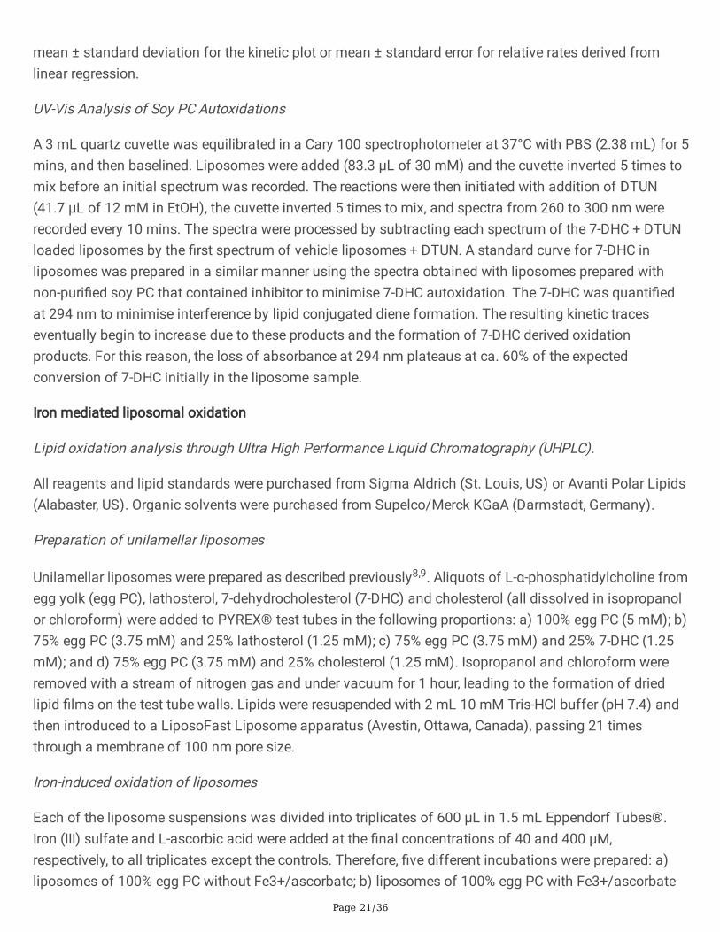
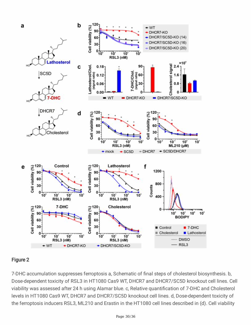
Figure 1
Identi�cation and impact of DHCR7 de�ciency on Ferroptosis. a, Schematic of the identi�cation of Dhcr7as a pro-ferroptotic gene, using CRISPR-KO library and GPX4 inhibition. b, Volcano plot of sgRNA enrichedin cells selected with RSL3 compared with untreated control cells. c, Relative quanti�cation of 7-DHC andCholesterol levels in HT1080 cell lines stably transduced with a vector expressing Cas9 and a sgRNAtargeting DHCR7 and EGFP as a control. Data are the mean ± s.d. of n = 3 wells of a 6-well plate from onerepresentative experiment. d, Immunoblot analysis of key ferroptosis regulators, namely FSP1, ACSL4
Page 29/36
and GPX4 in DHCR7 de�cient cells. e, Dose-dependent toxicity of the ferroptosis inducers RSL3, ML210and FIN56 in HT1080 cell lines stably transduced with a vector expressing Cas9 and an sgRNA targetingDHCR7 and EGFP as a control. Data are the mean ± s.d. of n = 3 wells of a 96-well plate from onerepresentative of two independent experiments. f, Relative quanti�cation of 7-DHC levels in HT1080 levelsWT and DHCR7-Knockout cells re-expressing DHCR7 and an empty vector. Data are the mean ± s.d. of n =3 wells of a 6-well plate from one representative experiment. g, dose-dependent toxicity of RSL3 andFIN56 in HT1080 Cas9 DHCR7-KO clone, and overexpressing DHCR7 or mock. Cell viability wasmonitored using Alamar blue. Data are the mean ± s.d. of n = 3 wells of a 96-well plate from onerepresentative of two independent experiments; *P < 0.05; two-way analysis of variance (ANOVA).
Page 30/36
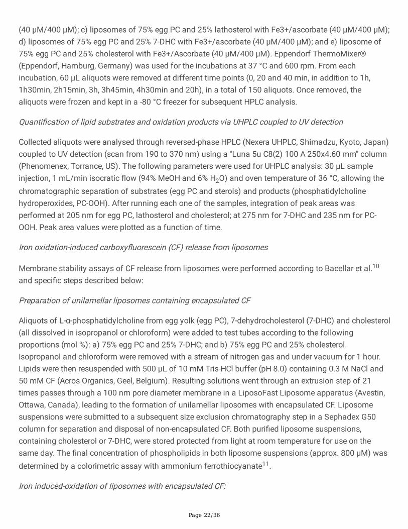
Figure 2
7-DHC accumulation suppresses ferroptosis a, Schematic of �nal steps of cholesterol biosynthesis. b,Dose-dependent toxicity of RSL3 in HT1080 Cas9 WT, DHCR7 and DHCR7/SC5D knockout cell lines. Cellviability was assessed after 24 h using Alamar blue. c, Relative quanti�cation of 7-DHC and Cholesterollevels in HT1080 Cas9 WT, DHCR7 and DHCR7/SC5D knockout cell lines. d, Dose-dependent toxicity ofthe ferroptosis inducers RSL3, ML210 and Erastin in the HT1080 cell lines described in (d). Cell viability
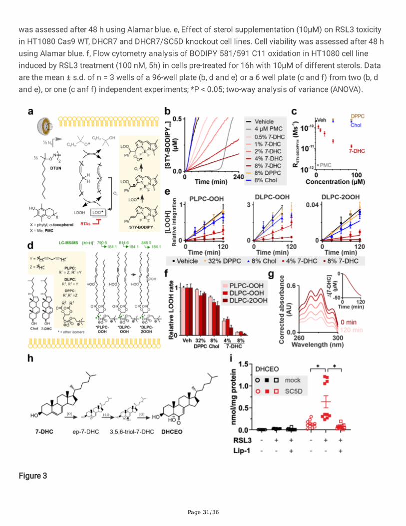
Page 31/36
was assessed after 48 h using Alamar blue. e, Effect of sterol supplementation (10µM) on RSL3 toxicityin HT1080 Cas9 WT, DHCR7 and DHCR7/SC5D knockout cell lines. Cell viability was assessed after 48 husing Alamar blue. f, Flow cytometry analysis of BODIPY 581/591 C11 oxidation in HT1080 cell lineinduced by RSL3 treatment (100 nM, 5h) in cells pre-treated for 16h with 10µM of different sterols. Dataare the mean ± s.d. of n = 3 wells of a 96-well plate (b, d and e) or a 6 well plate (c and f) from two (b, dand e), or one (c anf f) independent experiments; *P < 0.05; two-way analysis of variance (ANOVA).
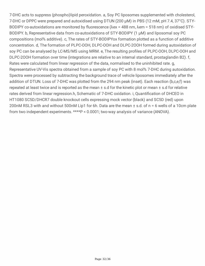
Figure 3
Page 32/36
7-DHC acts to suppress (phospho)lipid peroxidation. a, Soy PC liposomes supplemented with cholesterol,7-DHC or DPPC were prepared and autoxidised using DTUN (200 μM) in PBS (12 mM, pH 7.4, 37°C). STY-BODIPY co-autoxidations are monitored by �uorescence (λex = 488 nm, λem = 518 nm) of oxidised STY-BODIPY. b, Representative data from co-autoxidations of STY-BODIPY (1 μM) and liposomal soy PCcompositions (mol% additive). c, The rates of STY-BODIPYox formation plotted as a function of additiveconcentration. d, The formation of PLPC-OOH, DLPC-OOH and DLPC-2OOH formed during autoxidation ofsoy PC can be analysed by LC-MS/MS using MRM. e, The resulting pro�les of PLPC-OOH, DLPC-OOH andDLPC-2OOH formation over time (integrations are relative to an internal standard, prostaglandin B2). f,Rates were calculated from linear regression of the data, normalised to the uninhibited rate. g,Representative UV-Vis spectra obtained from a sample of soy PC with 8 mol% 7-DHC during autoxidation.Spectra were processed by subtracting the background trace of vehicle liposomes immediately after theaddition of DTUN. Loss of 7-DHC was plotted from the 294 nm peak (inset). Each reaction (b,c,e,f) wasrepeated at least twice and is reported as the mean ± s.d for the kinetic plot or mean ± s.d for relativerates derived from linear regression.h, Schematic of 7-DHC oxidation. i, Quanti�cation of DHCEO inHT1080 SC5D/DHCR7 double knockout cells expressing mock vector (black) and SC5D (red) upon200nM RSL3 with and without 500nM Lip1 for 6h. Data are the mean ± s.d. of n = 6 wells of a 10cm platefrom two independent experiments. ****P < 0.0001; two-way analysis of variance (ANOVA).
Page 33/36
Figure 4
Phospholipid truncated species contribute to ferroptosis execution. a, Epilipidomics analysis of HT1080Cas9 DHCR7/SC5D double KO cells overexpressing SC5D or an empty vector. Data is representative of anexperiment performed �ve times independently (please see results from the other repetitions in thesupplementary information). The heatmap overlaying critical event in ferroptosis, i.e cell rupture (PIpositive), 7-DHC/Chol ratio and DHCEO values from matched experimental time points are depicted
Page 34/36
below in grey scale. b, Lipidomics analysis of WT and ACSL4 KO HT1080 cell lines incubated for 16hours with αLNN and γLNN (20µM). Presented are the total amount of PE containing PUFA and the ratioof mono- to polyunsaturated fatty acids (MUFA/PUFA) in PE species. Mean values ± s.d. of n = 3technical replicates (10cm plate) performed twice are presented . c, Fatty acid composition of PE speciesin WT and ACSL4 KO HT1080 cell lines incubated for 16 hours with αLNN and γLNN (20µM). Meanvalues ± s.d. of n = 3 technical replicates (10cm plate) performed twice are presented. d, Assessment ofthe impact of αLNN and γLNN (20µM) re-sensitisation on RSL3-induced ferroptosis. Cell viability wasassessed after 24 h measuring PI incorporation. Mean values ± s.d. of n = 3 technical replicates (6cmplate) performed twice are presented. *P < 0.05; two-way analysis of variance (ANOVA).
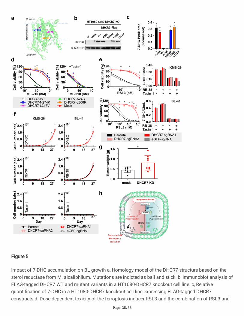
Page 35/36
Figure 5
Impact of 7-DHC accumulation on BL growth a, Homology model of the DHCR7 structure based on thesterol reductase from M. alcaliphilum. Mutations are indicted as ball and stick. b, Immunoblot analysis ofFLAG-tagged DHCR7 WT and mutant variants in a HT1080-DHCR7 knockout cell line. c, Relativequanti�cation of 7-DHC in a HT1080-DHCR7 knockout cell line expressing FLAG-tagged DHCR7constructs d. Dose-dependent toxicity of the ferroptosis inducer RSL3 and the combination of RSL3 and
Page 36/36
Tasin1 (500nM) in HT1080. Data represent the mean ± s.d. of n = 3 wells of a 96-well plate from onerepresentative of two independent experiments. e. Left panels, dose-dependent toxicity of RSL3 in KMS26and BL41 expressing two independent sgRNA targeting DHCR7 and EGFP. Cell viability was monitoredusing Alamar blue. Data are the mean ± s.d. of n = 3 wells of a 96-well plate from one representative oftwo independent experiments; *P < 0.05; two-way analysis of variance (ANOVA). Right panels, relativequanti�cation of 7-DHC levels in the KMS26 and BL41 cell lines treated with RB38 and Tasin-1. Data arethe mean ± s.d. of n = 3 wells of a 6-well plate from one representative experiment. f, Impact ofpharmacological inhibitors of DHCR7 (RB38) and EBP (Tasin-1) on the growth of BL41 and KMS26 cellstransduced with an sgRNA targeting DHCR7 and EGFP in medium lacking thiol supplementation. g,Growth of KMS26 xenograft transduced with a vector expressing Cas9 and a sgRNA targeting DHCR7and EGFP in NSG mice. Data are the mean ± s.d. of n = 8 mice (KMS26), tumor have been injected onboth �anks; *P < 0.05; two-way analysis of variance (ANOVA).h, graphical abstract depicting the anti-ferroptotic function of 7-DHC in the suppression of the formation of truncated-PL species.
Supplementary Files
This is a list of supplementary �les associated with this preprint. Click to download.
Extendeddata1.eps
Extendeddata2.eps
Extendeddata3.eps
Extendeddata4.eps
Extendeddata5.eps