Embed Size (px)
Citation preview
Folia Medica Lodziensia, 2012, 39/1:21-37
Corresponding author: dr hab. Magdalena Bryś, prof. nadzw. UŁ, Katedra Cytobiochemii, Uniwersytet Łódzki, Pomorska 141/143, 90-236 Łódź, e-mail: [email protected]; Tel.:+ 48 42 635-43-71; Fax: +48-42 635-44-84
Ophthalmic diseases among the impressionist painters
Choroby oczu u malarzy impresjonistów
MAGDALENA BRYŚ, ANNA KRZEŚLAK, MAGDALENA LASKOWSKA, PAWEŁ JÓŹWIAK, EWA FORMA
Department of Cytobiochemistry
University of Łódź
Abstract Artists, like anyone else, have suffered from a variety of medical conditions and this is particularly obvious in the case of eye diseases. The aim of this work is to present a summary of previous studies on visual disorders among the impressionist artists and attempt of analysis how their style was changing with the deteriorating state of the eyes. The diseases such as cataracts, short-sightedness, macular damage and eye infections impact their style and technique, the choice of color as well as the subject of painting. Key words: vision disorders, myopia, cataract, impressionist painters Streszczenie Artystów, tak jak wszystkich ludzi, dotykają różne choroby i szczególnie jest to widoczne, dla odbiorców sztuki, w przypadku zaburzeń widzenia. Celem niniejszego artykułu jest przedstawienie syntetycznej wiedzy na temat chorób oczu diagnozowanych u najsłynniejszych malarzy impresjonistów oraz próba wskazania, w jaki sposób schorzenia te wpłynęły na ich twórczość. Choroby takie jak zaćma, krótkowzroczność, zwyrodnienie plamki żółtej czy infekcje oczu odcisnęły piętno zarówno na stylu i technice ich malarstwa, doborze barw, jak i tematyce obrazów.
Słowa kluczowe: choroby oczu, krótkowzroczność, zaćma, impresjoniści
Ophthalmic diseases and impressionists 22
Introduction
A common interest among art historians, clinicians and scientists is
the effect of diseases and illnesses on artist’s work and its transformation as
a consequence of the disease and therapy. It is natural that an ophthalmologist
should develop an analytic attitude in regard to the visual capacities of artists
and search for evidence of normal and abnormal sight as revealed in their
paintings [1, 2]. The study of French painters has centered on impressionism
because of the major importance of this movement in the evolution of art
in the 19th century. The canon of “Major Impressionists” include: Cassatt,
Cézanne, Degas, Manet, Monet, Pissarro, Renoir, and Sisley [3-5]. The majority
of those painters suffered from ophthalmic diseases [6-8].
Cassatt
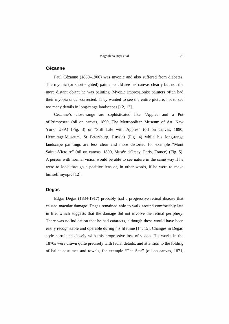
The first example is Mary Cassatt (1844-1926), an American impressionist
who spent most of her adult life in France. She was diabetic, suffered from
cataracts, and underwent multiple operations on her eyes, only to become unable
to paint for the last dozen years of her life [9-11]. Cassatt's visual problems
forced her to switch from oils to pastels. The precision in her early work
is evident in her painting “Lydia Crocheting in the Garden at Marley” (oil on
canvas, 1880, The Metropolitan Museum of Art, New York, USA) (Fig. 1.).
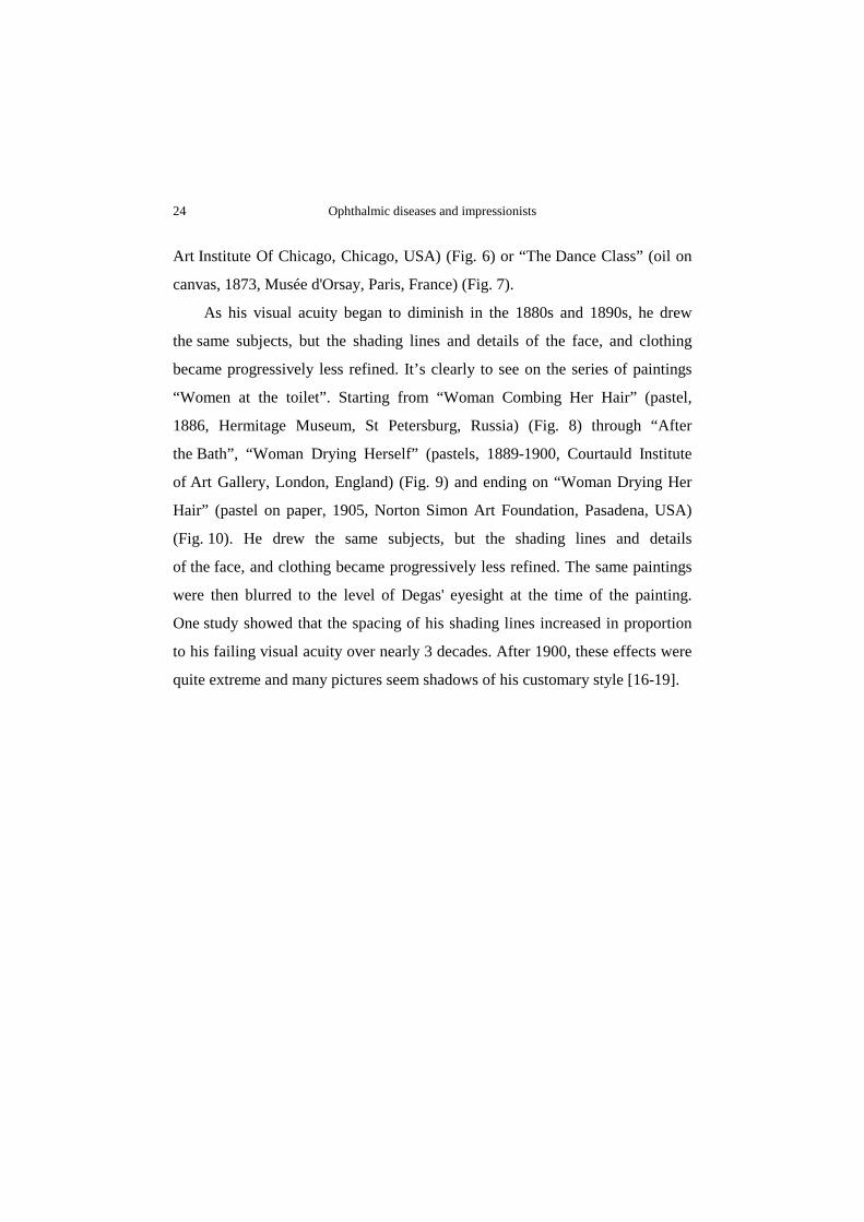
As her visual problems advanced, the meticulous lines became strident,
bold strokes of color. This can be seen in her pastel “Spring: Margot Standing
in a Garden” (oil on canvas, 1900, The Metropolitan Museum of Art, New
York, USA) (Fig. 2) [9].
Magdalena Bryś et al. 23
Cézanne
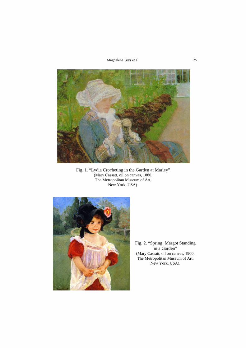
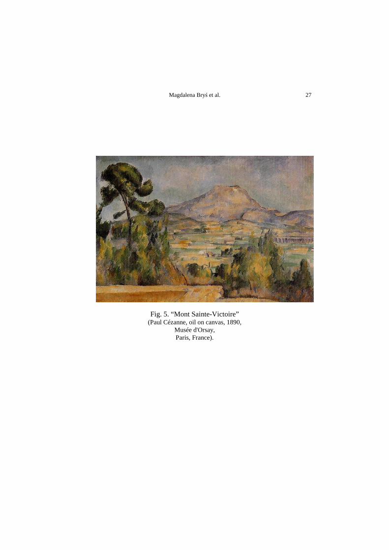
Paul Cézanne (1839–1906) was myopic and also suffered from diabetes.
The myopic (or short-sighted) painter could see his canvas clearly but not the
more distant object he was painting. Myopic impressionist painters often had
their myopia under-corrected. They wanted to see the entire picture, not to see
too many details in long-range landscapes [12, 13].
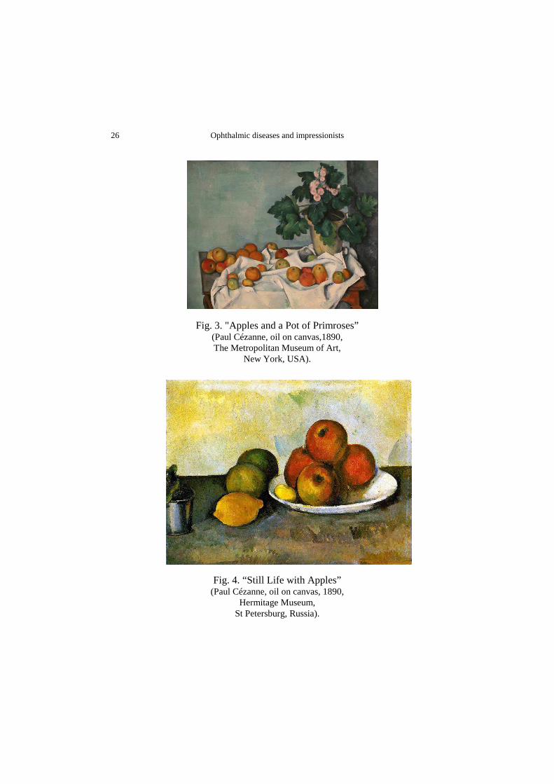
Cézanne’s close-range are sophisticated like "Apples and a Pot
of Primroses” (oil on canvas, 1890, The Metropolitan Museum of Art, New
York, USA) (Fig. 3) or “Still Life with Apples” (oil on canvas, 1890,
Hermitage Museum, St Petersburg, Russia) (Fig. 4) while his long-range
landscape paintings are less clear and more distorted for example “Mont
Sainte-Victoire” (oil on canvas, 1890, Musée d'Orsay, Paris, France) (Fig. 5).
A person with normal vision would be able to see nature in the same way if he
were to look through a positive lens or, in other words, if he were to make
himself myopic [12].
Degas
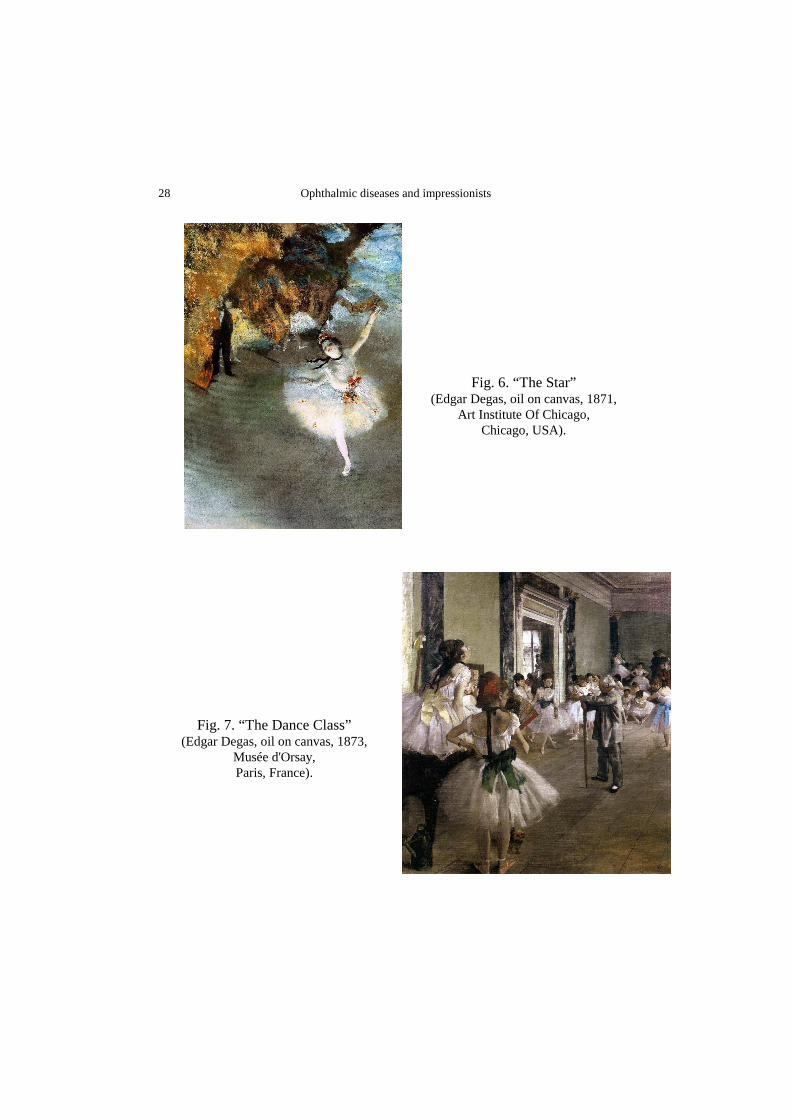
Edgar Degas (1834-1917) probably had a progressive retinal disease that
caused macular damage. Degas remained able to walk around comfortably late
in life, which suggests that the damage did not involve the retinal periphery.
There was no indication that he had cataracts, although these would have been
easily recognizable and operable during his lifetime [14, 15]. Changes in Degas'
style correlated closely with this progressive loss of vision. His works in the
1870s were drawn quite precisely with facial details, and attention to the folding
of ballet costumes and towels, for example “The Star” (oil on canvas, 1871,
Ophthalmic diseases and impressionists 24
Art Institute Of Chicago, Chicago, USA) (Fig. 6) or “The Dance Class” (oil on
canvas, 1873, Musée d'Orsay, Paris, France) (Fig. 7).
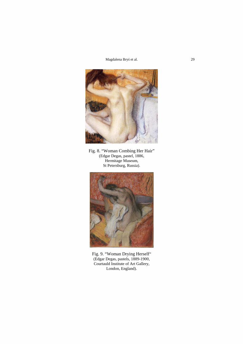
As his visual acuity began to diminish in the 1880s and 1890s, he drew
the same subjects, but the shading lines and details of the face, and clothing
became progressively less refined. It’s clearly to see on the series of paintings
“Women at the toilet”. Starting from “Woman Combing Her Hair” (pastel,
1886, Hermitage Museum, St Petersburg, Russia) (Fig. 8) through “After
the Bath”, “Woman Drying Herself” (pastels, 1889-1900, Courtauld Institute
of Art Gallery, London, England) (Fig. 9) and ending on “Woman Drying Her
Hair” (pastel on paper, 1905, Norton Simon Art Foundation, Pasadena, USA)
(Fig. 10). He drew the same subjects, but the shading lines and details
of the face, and clothing became progressively less refined. The same paintings
were then blurred to the level of Degas' eyesight at the time of the painting.
One study showed that the spacing of his shading lines increased in proportion
to his failing visual acuity over nearly 3 decades. After 1900, these effects were
quite extreme and many pictures seem shadows of his customary style [16-19].
Magdalena Bryś et al. 25
Fig. 1. “Lydia Crocheting in the Garden at Marley” (Mary Cassatt, oil on canvas, 1880, The Metropolitan Museum of Art,
New York, USA).
Fig. 2. “Spring: Margot Standing in a Garden”
(Mary Cassatt, oil on canvas, 1900, The Metropolitan Museum of Art,
New York, USA).
Ophthalmic diseases and impressionists 26
Fig. 3. "Apples and a Pot of Primroses” (Paul Cézanne, oil on canvas,1890, The Metropolitan Museum of Art,
New York, USA).
Fig. 4. “Still Life with Apples” (Paul Cézanne, oil on canvas, 1890,
Hermitage Museum, St Petersburg, Russia).
Magdalena Bryś et al. 27
Fig. 5. “Mont Sainte-Victoire” (Paul Cézanne, oil on canvas, 1890,
Musée d'Orsay, Paris, France).
Ophthalmic diseases and impressionists 28
Fig. 6. “The Star” (Edgar Degas, oil on canvas, 1871,
Art Institute Of Chicago, Chicago, USA).
Fig. 7. “The Dance Class” (Edgar Degas, oil on canvas, 1873,
Musée d'Orsay, Paris, France).
Magdalena Bryś et al. 29
Fig. 8. “Woman Combing Her Hair” (Edgar Degas, pastel, 1886,
Hermitage Museum, St Petersburg, Russia).
Fig. 9. “Woman Drying Herself” (Edgar Degas, pastels, 1889-1900, Courtauld Institute of Art Gallery,
London, England).
Ophthalmic diseases and impressionists 30
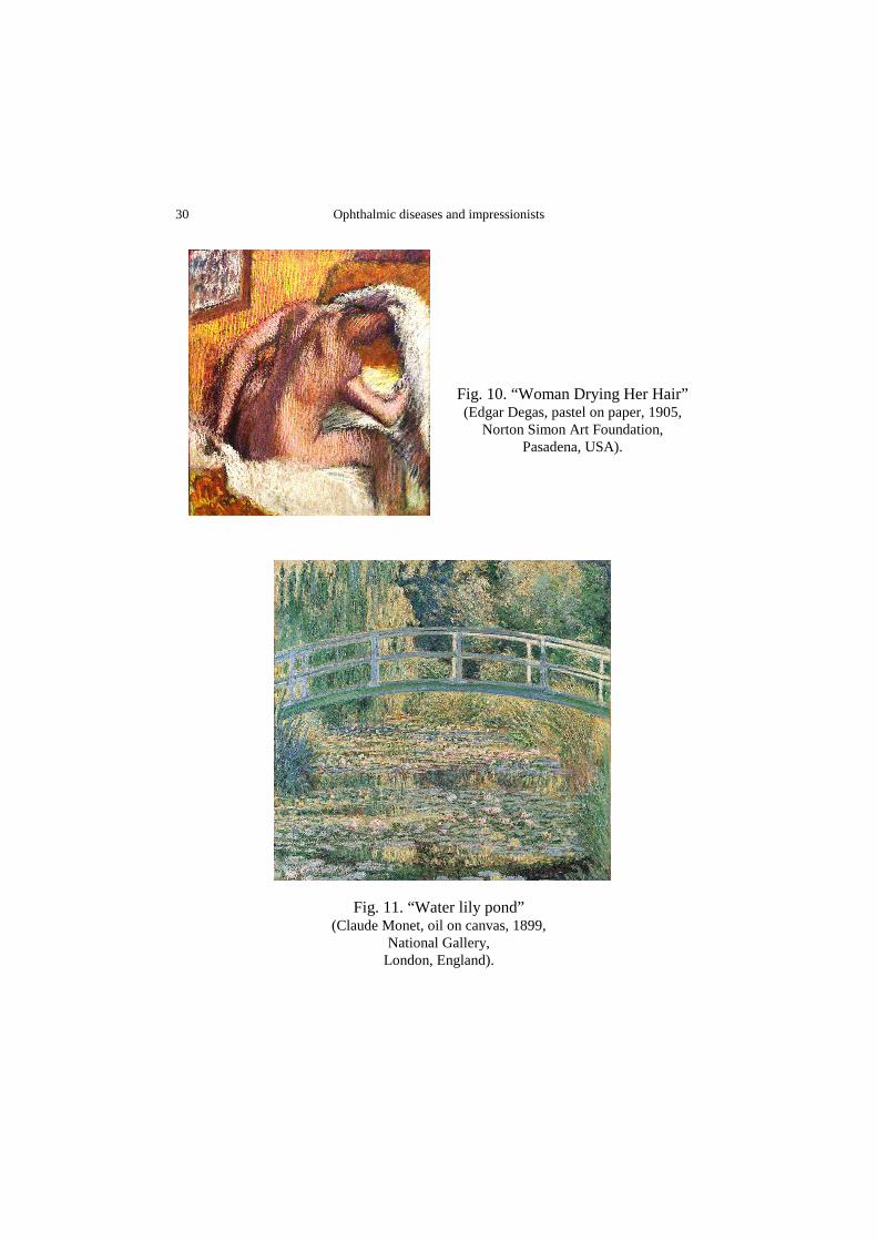
Fig. 10. “Woman Drying Her Hair” (Edgar Degas, pastel on paper, 1905,
Norton Simon Art Foundation, Pasadena, USA).
Fig. 11. “Water lily pond” (Claude Monet, oil on canvas, 1899,
National Gallery, London, England).
Magdalena Bryś et al. 31
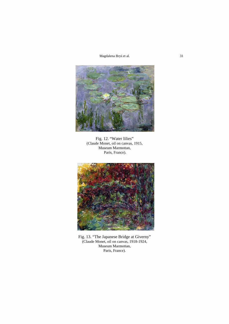
Fig. 12. “Water lilies” (Claude Monet, oil on canvas, 1915,
Museum Marmottan, Paris, France).
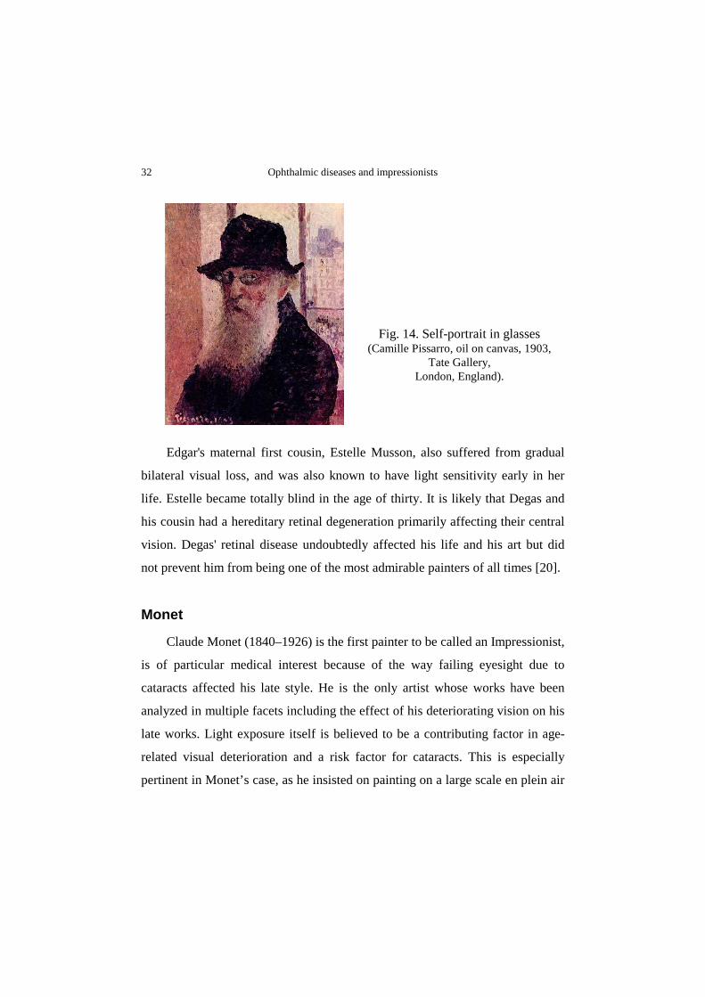
Fig. 13. “The Japanese Bridge at Giverny” (Claude Monet, oil on canvas, 1918-1924,
Museum Marmottan, Paris, France).
Ophthalmic diseases and impressionists 32
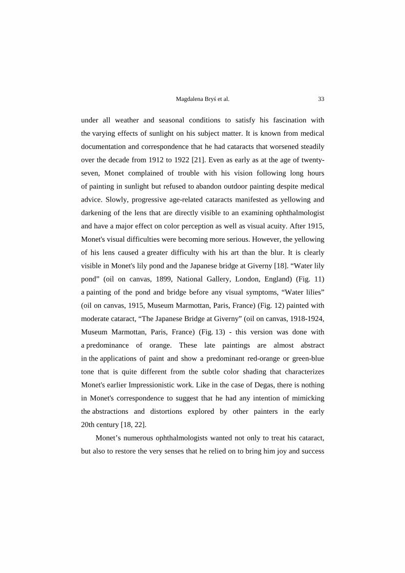
Fig. 14. Self-portrait in glasses (Camille Pissarro, oil on canvas, 1903,
Tate Gallery, London, England).
Edgar's maternal first cousin, Estelle Musson, also suffered from gradual
bilateral visual loss, and was also known to have light sensitivity early in her
life. Estelle became totally blind in the age of thirty. It is likely that Degas and
his cousin had a hereditary retinal degeneration primarily affecting their central
vision. Degas' retinal disease undoubtedly affected his life and his art but did
not prevent him from being one of the most admirable painters of all times [20].
Monet
Claude Monet (1840–1926) is the first painter to be called an Impressionist,
is of particular medical interest because of the way failing eyesight due to
cataracts affected his late style. He is the only artist whose works have been
analyzed in multiple facets including the effect of his deteriorating vision on his
late works. Light exposure itself is believed to be a contributing factor in age-
related visual deterioration and a risk factor for cataracts. This is especially
pertinent in Monet’s case, as he insisted on painting on a large scale en plein air
Magdalena Bryś et al. 33
under all weather and seasonal conditions to satisfy his fascination with
the varying effects of sunlight on his subject matter. It is known from medical
documentation and correspondence that he had cataracts that worsened steadily
over the decade from 1912 to 1922 [21]. Even as early as at the age of twenty-
seven, Monet complained of trouble with his vision following long hours
of painting in sunlight but refused to abandon outdoor painting despite medical
advice. Slowly, progressive age-related cataracts manifested as yellowing and
darkening of the lens that are directly visible to an examining ophthalmologist
and have a major effect on color perception as well as visual acuity. After 1915,
Monet's visual difficulties were becoming more serious. However, the yellowing
of his lens caused a greater difficulty with his art than the blur. It is clearly
visible in Monet's lily pond and the Japanese bridge at Giverny [18]. “Water lily
pond” (oil on canvas, 1899, National Gallery, London, England) (Fig. 11)
a painting of the pond and bridge before any visual symptoms, “Water lilies”
(oil on canvas, 1915, Museum Marmottan, Paris, France) (Fig. 12) painted with
moderate cataract, “The Japanese Bridge at Giverny” (oil on canvas, 1918-1924,
Museum Marmottan, Paris, France) (Fig. 13) - this version was done with
a predominance of orange. These late paintings are almost abstract
in the applications of paint and show a predominant red-orange or green-blue
tone that is quite different from the subtle color shading that characterizes
Monet's earlier Impressionistic work. Like in the case of Degas, there is nothing
in Monet's correspondence to suggest that he had any intention of mimicking
the abstractions and distortions explored by other painters in the early
20th century [18, 22].
Monet’s numerous ophthalmologists wanted not only to treat his cataract,
but also to restore the very senses that he relied on to bring him joy and success
Ophthalmic diseases and impressionists 34
in life. There was a misunderstanding of the goal of cataract surgery that
resulted in Monet’s profound disappointment with the procedure and
depression. The changes in vision were not adequately discussed between
Monet and his ophthalmologist, creating distress and tension in their
relationship. The natural healing process combined with a clever optical lens to
optimize color perception were successful in returning to Monet the vision
he relied on to reproduce the beauty of life and nature [22-24].
Pissarro
Camille Pissarro the dean of Impressionism, born in 1830, died in 1903
at the age of 73. His productivity during the last 15 years of his life
was hampered by recurrent infections around his right eye. In that time the
treatment consisted of incision and drainage of the lachrymal sac, and
cauterization. Due to this chronic disease he was forced to paint inside, behind
closed windows, in order to avoid wind and dust. This situation influenced the
subjects of his painting. His painting reflects large crowds Paris avenues
and buildings [25].
Lachrymal problems were a popular topic in France in 1891. Unfortunately
for Pissarro, he never met an Italian surgeon, Toti, who developed modern
lachrymal surgery during Pissarro's lifetime. In 1904, the year following
the artist's death, Toti published his method of dacryocystorhinostomy [26].
It is difficult to find portraits of impressionist painters in glasses. However,
Pissarro is one of those painters who have at least two self-portraits in glasses.
The first is from 1890-91 and the second is from 1903 (oil on canvas, Tate
Gallery, London, England) (Fig. 14).
Magdalena Bryś et al. 35
Renoir
August Renoir (1841–1919), despite his mild myopia, never wore glasses.
His close-range paintings are distinct, while his long-range paintings are
impressionistically blurred [12].
Myopia and hypermetropia have had also been said to have a direct
influence on the preponderant color that the artist used. The myopia with
his abnormally elongated eye will see reds better while the hypermetropia will
be correspondingly better on blues. Thus, the increasing fascination for reds
in the case of Renoir may simply be due to the disorder. Curiously enough,
the colors from the red end of the spectrum predominate in the paintings of
the Chinese and Japanese, who are also predominantly myopic. The Japanese
have only recently adopted a specific word for blue [27].
Conclusions
It is generally agreed that many painters suffered from eyes disorders. It is
impossible to document all the painters with refractive errors. Our article
focused on a well-defined group of painters in order to identify those artists who
had an ophthalmic disease. We show how major eye diseases - myopia,
hypermetropia, cataract and retinal disease - have their effects on artist's work.
Interestingly, many of the impressionists were myopic and did not wear
glasses. The impressionists wanted to convey subjective sensory impressions,
attempting, as they put it themselves, to ‘capture a retinal image in flight’.
Probably ophthalmic diseases influenced their paintings, the form and the use
of colors. We can only speculate about the possible effect of various acquired
or inherited eyes disorders on this artistic school in particular.
Ophthalmic diseases and impressionists 36
References
1 Backhaus WGK, Kliegl R, Werner JS. Color vision: perspectives from different disciplines. Walter de Gruyter, Berlin, 1998.
2. Cole BL. The handicap of abnormal colour vision. Clin Exp Optom. 2004; 87: 258-275.
3. Lanthony P. Les yeux des peintres. L'Âge d'Homme, Lausanne,1999. 4. Lanthony P. Art and ophthalmology. Kugler Publications, Amsterdam, 2009. 5. Cutting JE. Impressionism and its canon. University Press of American Library of
Congress, Lanham, 2006. 6. Emery AE. Medicine, artists and their art. J R Coll Physicians Lond. 1997;
31: 450-455. 7. Goh ES, Teo WT. Art and the eye: the impact of ocular pathology on their artistic
legacy. Ann Acad Med Singapore 2007; 36: 61-64. 8. Ravin JG. The visual difficulties of selected artists and limitations
of ophthalmological care during the 19th and early 20th centuries (an AOS thesis). Trans Am Ophthalmol Soc. 2008; 106: 402–425.
9. Clement RT, Houzé A, Erbolato-Ramsey C. The women impressionist: a source-book. Greenwood Publishing Group, Westport, 2000.
10. Mir Fullana F. Mary Cassat's cataracts (1845-1926). Arch Soc Esp Oftalmol. 2004; 79: 249-251.
11. Streissguth T. Mary Cassatt: portrait of an American impressionist. Twenty-First Century Books, Minneapolis, 1998.
12. Polland W. The Artist’s eye. Myopic artists. Acta Ophthalmol Scand. 2004; 82: 325-326.
13. Régnier C. Famous French diabetics. Medicographia 2009; 31: 316-323. 14. Zozaya Aldana B. The macular degeneration of Edward Degas. Arch Soc Esp
Oftalmol. 2011; 86: 229-231. 15. González-Treviño JL, González-Cortés JH, García-Guerrero J. Edgar Degas
y la degeneración macular. Rev Mex Oftalmol. 2007; 81: 340-344. 16. Marmor M F. A brief history of macular grids: from Thomas Reid to Edvard
Munch and Marc Amsler. Surv Ophthalmol. 2000; 44: 343-353. 17. Marmor MF. Degas through his own eyes: visual disability and the late style
of Degas. Arch Ophthalmol. 2004; 122: 795. 18. Marmor MF. Ophthalmology and art: simulation of Monet's cataracts and Degas'
retinal disease. Arch Ophthalmol. 2006; 124: 1764-1769.
Magdalena Bryś et al. 37
19. Ravin JG. Pissarro, dacryocystitis, and the development of modern lacrimal surgery. Doc Ophthalmol. 1994; 86: 191-202.
20. Karcioglu ZA. Did Edgar Degas have an inherited retinal degeneration? Ophthalmic Genet. 2007; 28: 51-55.
21. Wildenstein D. Monet or the triumph of impressionism. Taschen, Amsterdam, 2003.
22. Zhou A. Cataracts and the late style of Monet’s paintings. Proceedings of the 17th Annual History of Medicine Days. 2008; 43-51.
23. Hem E. Monet–lyset og øyelidelsen. Tidsskr Nor Legeforen 2008; 128: 1548-1549.
24. Ravin JG. Monet's cataracts. JAMA 1985; 254: 394-399. 25. Cernea P. Eye disease in painters-Camille Pissaro. Clinica Oftalmologică Craiova
2001; 53: 84-88. 26. Ravin JG, Kenyon CA. Degas' loss of vision: evidence for a diagnosis of retinal
disease. Surv Ophthalmol. 1994; 39: 57-64. 27. Warner P. Art and eye disease. Can Med Assoc J. 1960; 82: 444.