Embed Size (px)
Citation preview

Organic Lecture Series
1
Amino AcidsAmino Acidsand and
ProteinsProteinsChapter 27
Organic Lecture Series
2
NH3+
CO2-

Organic Lecture Series
3
Amino AcidsAmino Acids•• Amino acid:Amino acid: a compound that contains
both an amino group and a carboxyl groupαα--Amino acid:Amino acid: an amino acid in which the
amino group is on the carbon adjacent to the carboxyl group
– although α-amino acids are commonly written in the unionized form, they are more properly written in the zwitterionzwitterion (internal salt) form
RCHCOH
NH2
O
RCHCO-
NH3+
O
Organic Lecture Series
4
In 1891, Emil Fischer made the arbitraryassignments of D- and L- to the enantiomers of glyceraldehyde (aldehyde is C1-at the top).
CHO
H OH
CH2OH
CHO
CH2 OH
HHO
D D
L-GlyceraldehydeD-Glyceraldehyde
[α]25 = +13.5 [α]25 = -13.5
-OH to Right -OH to Left
D,L D,L MonosaccharidesMonosaccharides

Organic Lecture Series
5
ChiralityChirality of Amino Acidsof Amino Acids• With the exception of glycine, all protein-
derived amino acids have at least one stereocenter (the α-carbon) and are chiral– the vast majority have the L-configuration at
their α-carbon
COO-
CH3
HH3N
L-Alanine
COO-
CH3
H NH3+
D-Alanine
Organic Lecture Series
6
NonpolarNonpolar side chainsside chains
NH3+
COO-
NH3+
COO-
NH3+
COO-
NH3+
COO-
NH3+
COO-S
NH3+
COO-
NH H
COO-
NH3+
COO-
NH
COO-
NH3+
Alanine (Ala, A)
Glycine (Gly, G)
Isoleucine (Ile, I)
Leucine (Leu, L)
Methionine (Met, M)
Phenylalanine (Phe, F)
Proline (Pro, P)
Tryptophan (Trp, W)
Valine (Val, V)

Organic Lecture Series
7
NH H
C O O - Proline
(Pro, P)
Captopril-ACE Inhibitor
Angiotensin Converting Enzyme
Angiotensin I Angiotensin II
Angiotensin II is a potent vasoconstrictor so,ACE inhibitors are used for the treatment of hypertension
Organic Lecture Series
8
Polar side chainsPolar side chains
NH3+
COO-H 2N
O
NH3+
COO-
H 2N
O
NH3+
COO-
HO
NH3+
COO-OH
Asparagine (Asn, N)
Glutamine (Gln, Q)
Serine (Ser, S)
Threonine (Thr, T)
Amide Side Chains Hydroxyl Side Chains

Organic Lecture Series
9
Acidic & Basic Side ChainsAcidic & Basic Side Chains
NH3+
COO--O
O
NH3+
COO--O
O
NH3+
COO-
HS
NH3+
COO-
HO
NH3+
COO-
NH
H2N
NH2+
NH3+
COO-
N
NH
NH3+
COO-H3N
Cysteine (Cys, C)
Tyrosine (Tyr, Y)
Glutamic acid (Glu, E)
Aspartic acid (Asp, D)
Histidine (His, H)
Lysine (Lys, K)
Arginine (Arg, R)
+
Organic Lecture Series
10
AcidAcid--Base PropertiesBase Properties
pKa ofpKa of
valine 2.29 9.72tryptophan 2.38 9.39
9.102.09threonineserine 2.21 9.15
10.602.00prolinephenylalanine 2.58 9.24
9.212.28methionine9.742.33leucine
isoleucine 2.32 9.76glycine 2.35 9.78
9.132.17glutamine8.802.02asparagine9.872.35alanine
Nonpolar &polar side chains α−NH3
+α−COOH

Organic Lecture Series
11
AcidAcid--Base PropertiesBase Properties
pKa ofpKa ofpKa of
10.079.112.20tyros ine
lys ine 2.18 8.95 10.536.109.181.77histid ine
glutamic acid 2.10 9.47 4.078.0010.252.05cysteine
aspartic acid 2.10 9.82 3.86
arginine 2.01 9.04 12.48
Side Chain
AcidicSide Chains α−NH3
+α−COOH
pKa ofpKa ofpKa ofSide Chain
BasicSide Chains α−NH3
+α−COOH
carboxylcarboxylsufhydrylphenolic
guanidinoimidazole1° amino
SideChainGroup
SideChainGroup
Organic Lecture Series
12
Acidity: Acidity: αα--COOH GroupsCOOH Groups• The average pKa of an α-carboxyl group is
2.19, which makes them considerably stronger acids than acetic acid (pKa 4.76)– the greater acidity is accounted for by the
electron-withdrawing inductive effect of the adjacent -NH3
+ group
NH3+
RCHCOOH H2 O
NH3+
RCHCOO- H3 O+
+ pKa = 2.19+

Organic Lecture Series
13
Acidity: side chain Acidity: side chain --COOHCOOH• Due to the electron-withdrawing inductive
effect of the α-NH3+ group, side chain -
COOH groups are also stronger than acetic acid– the inductive effect decreases with distance
from the α-NH3+ group. Compare:
α-COOH group of alanine (ppKKaa 2.352.35)
β-COOH group of aspartic acid (ppKKaa 3.863.86)
γ-COOH group of glutamic acid (ppKKaa 4.074.07)
Organic Lecture Series
14
Acidity: Acidity: αα--NHNH33++ groupsgroups
• The average value of pKa for an α-NH3+
group is 9.47, compared with a value of 10.76 for a 1° alkylammonium ion
NH3+
RCHCOO-
H2 O
NH3+
CH3CHCH3 H2 O
NH2
RCHCOO-
NH2
CH3CHCH3
H3 O+
H3 O+ pKa = 10.60++
+ pKa = 9.47+

Organic Lecture Series
15
– Both functional groups display acid-base chemistry
RCHCOH
NH2
O
RCHCO-
NH3+
O
AcidAcid--Base PropertiesBase Properties
Organic Lecture Series
16
NaOH + HCl H2O + NaCl
[OH-]
pHpH [OH-] = [H3O+]7.07.0
1.01.0
14.014.0
End point(equivalence point)
Freshman Flashback!!

Organic Lecture Series
17
Phenolphthalein
OH-
Not Exam Material
Organic Lecture Series
18
Titration of Amino AcidsTitration of Amino Acids

Organic Lecture Series
19
Stage 1Stage 1–– pKpKa1a1
H3N+CH2COOH = H3N
+CH2COO-
0.5 eq OH-
Organic Lecture Series
20
Isoelectric PointIsoelectric Point
H3N+CH2COO-
1.0 eq OH-

Organic Lecture Series
21
H2NCH2COO-H3N+CH2COO- =
Stage 2Stage 2–– pKpKaa22
1.5 eq OH-
Organic Lecture Series
22
Isoelectric point (pI):Isoelectric point (pI): the pH at which an amino acid, polypeptide, or protein has no net charge– the pH for glycine, for example, falls between
the pKa values for the carboxyl and amino groups
pI = 12 ( pKa α−COOH + pKa α−NH3
+)
= 21 (2.35 + 9.78) = 6.06
Isoelectric PointIsoelectric Point

Organic Lecture Series
23
pI = 12 ( pKa α−COOH + pKa α−NH3
+)
= 21 (2.35 + 9.78) = 6.06
Organic Lecture Series
24
6.115.415.656.066.046.045.745.916.305.685.605.886.00
pKa ofpKa ofpKa of
pI
----
------------
----------------------------
--------
valine 2.29 9.72tryptophan 2.38 9.39
9.102.09threonineserine 2.21 9.15
10.602.00prolinephenylalanine 2.58 9.24
9.212.28methionine9.742.33leucine
isoleucine 2.32 9.76glycine 2.35 9.78
9.132.17glutamine8.802.02asparagine9.872.35alanine
Side Chain
Nonpolar &polar side chains α−NH3
+α−COOH
Isoelectric PointIsoelectric Point

Organic Lecture Series
25
•• Electrophoresis:Electrophoresis: the process of separating compounds on the basis of their electric charge– electrophoresis of amino acids can be carried out
using paper, starch, polyacrylamide and agarose gels, and cellulose acetate as solid supports
ElectrophoresisElectrophoresis
Organic Lecture Series
26
1. a sample of amino acids is appliedas a spot on the paper strip
2. an electric potential is applied to the electrode vessels and amino acids migrate toward the electrode with charge opposite their own
3. molecules with a high charge density move faster than those with low charge density
ElectrophoresisElectrophoresis

Organic Lecture Series
27
molecules at their isoelectric point remain at the origin
after separation is complete, the strip is dried and developed to make the separated amino acids visible
19 of the 20 amino acids give the same purple-colored anion; proline gives an orange-colored compound
ElectrophoresisElectrophoresis
Organic Lecture Series
28
a reagent commonly used to detect amino acid is ninhydrinninhydrin
RCHCO-
O OHO
OOH
NH3+
O
O-
N
O
O
O
RCH CO2 H3 O++
An α-amino acid
Purple-colored anion
+ +
2+
Ninhydrin
ElectrophoresisElectrophoresis
Not Exam Material

Organic Lecture Series
29
Polypeptides & ProteinsPolypeptides & Proteins
• In 1902, Emil Fischer proposed that proteins are long chains of amino acids joined by amide bonds to which he gave the name peptide bonds
•• Peptide bond:Peptide bond: the special name given to the amide bond between the α-carboxyl group of one amino acid and the α-amino group of another
Organic Lecture Series
30
Serylalanine (SerSerylalanine (Ser--Ala)Ala)
H2 N HO
O
HHOCH2
H2 NO
OH
H CH3
Serine(Ser, S)
Alanine(Ala, A)
+
H2 NN
OH
HOCH2
H
H
CH3O
H O
Serylalanine(Ser-Ala, (S-A)
peptide bond

Organic Lecture Series
31
H 2N C H C
C H 3
O H
O
H 2N C H C
C H 3
O H
O
H 2N C H C
H 3C
O H
O
H 2N C H C O 2H
C H 3
H 2N C H C
H 3C
O H
O
H N C H C O 2H
C H 3
H
H 2N C H C
H 3C
O
H N C H C O 2H
C H 3
H O H
p ro to n tra n s fe r
Organic Lecture Series
32
Peptide TerminologyPeptide Terminology–– peptide:peptide: the name given to a short polymer of
amino acids joined by peptide bonds; they are classified by the number of amino acids in the chain
–– dipeptide:dipeptide: a molecule containing two amino acids joined by a peptide bond
–– tripeptidetripeptide: a molecule containing three amino acids joined by peptide bonds
–– polypeptidepolypeptide: a macromolecule containing many amino acids joined by peptide bonds
–– proteinprotein: a biological macromolecule of molecular weight 5000 g/mol of greater, consisting of one or more polypeptide chains

Organic Lecture Series
33
– by convention, peptides are written from the left, beginning with the free -NH3
+ group and ending with the free -COO- group on the right
H3
N
O H
NH
O
HN
C O O -
O -
OC
6H
5O+
C-terminal
amino acid
N-terminal
amino acid
Ser-Phe-Asp
Writing PeptidesWriting Peptides
Organic Lecture Series
34
– the tetrapeptide Cys-Arg-Met-Asn
– at pH 6.0, its net charge is +1
HN
NH O
HN
O-
OO
O
NH2
SCH3
NH
NH2+H2N
OH3 N
SH C-terminalamino acid
N-terminalamino acid
pKa 12.48
pKa 8.00
+
Writing PeptidesWriting Peptides
Organic Lecture Series
35
•• Primary structure:Primary structure: the sequence of amino acids in a polypeptide chain; read from the N-terminal amino acid to the C-terminal amino acid
• Amino acid analysis:– hydrolysis of the polypeptide, most commonly
carried out using 6M HCl at elevated temperature
– quantitative analysis of the hydrolysate (i.e. hydrolyzed solution) by ion-exchange chromatography
Primary StructurePrimary Structure
Organic Lecture Series
36
Ion Exchange Chromatography
Not Exam Material

Organic Lecture Series
37
Not Exam Material
Organic Lecture Series
38
– the four atoms of a peptide bond and the two alpha carbons joined to it lie in a plane with bond angles of 120° about C and N
– the model of Gly-Gly is viewed from two perspectives to show the planarity of the six atoms of the peptide bond
Peptide Bond GeometryPeptide Bond Geometry

Organic Lecture Series
39
– to account for this geometry, Linus Pauling proposed that a peptide bond is most accurately represented as a hybrid of two contributing structures (resonance)
– the hybrid has considerable C-N double bond character and rotation about the peptide bond is restricted
Cα
Cα
N
H
C
Cα
CαOO
C N
H
+
-: :
:
: : :
Peptide Bond GeometryPeptide Bond Geometry
Organic Lecture Series
40
– two conformations are possible for a planar peptide bond
– virtually all peptide bonds in naturally occurring proteins studied to date have the s-trans conformation
Cα
Cα
O
C N
H
• • • •
• •
CαCα
O
C N
H
• • • •
• •
s-t rans s-cis
Cα
Cα
O
C N
H
• • • •
• •
CαCα
O
C N
H
• • • •
• •
s-t rans s-cis
Cα
Cα
O
C N
H
• • • •
• •
CαCα
O
C N
H
• • • •
• •
s-t rans s-cis
Peptide Bond GeometryPeptide Bond Geometry

Organic Lecture Series
41
Secondary StructureSecondary Structure•• Secondary structure:Secondary structure: the ordered
arrangements (conformations) of amino acids in localized regions of a polypeptide or protein
• To determine from model building which conformations would be of greatest stability, Pauling and Corey assumed that 1. all six atoms of each peptide bond lie in the same
plane and in the s-trans conformation
2. there is hydrogen bonding between the N-H group of one peptide bond and a C=O group of another peptide bond as shown in the next screen
Organic Lecture Series
42
– hydrogen bonding between amide groups
Secondary StructureSecondary Structure

Organic Lecture Series
43
• On the basis of model building, Pauling and Corey proposed that two types of secondary structure should be particularly stable
α-helix
antiparallel β-pleated sheet
αα--Helix:Helix: a type of secondary structure in which a section of polypeptide chain coils into a spiral, most commonly a right-handed spiral
Secondary StructureSecondary Structure
Organic Lecture Series
44
The The αα--HelixHelix
– Figure 27.14: the polypeptide chain is repeating units of L-alanine

Organic Lecture Series
45
• In a section of α-helix–there are 3.6 amino acids per turn
of the helix
–each peptide bond is s-trans and planar
–N-H groups of all peptide bonds point in the same direction, which is roughly parallel to the axis of the helix
The The αα--HelixHelix
Organic Lecture Series
46
• In a section of α-helix– C=O groups of all peptide bonds point in
the opposite direction, and also parallel to the axis of the helix
– the C=O group of each peptide bond is hydrogen bonded to the N-H group of the peptide bond four amino acid units away from it
– all R- groups point outward from the helix
The The αα--HelixHelix

Organic Lecture Series
47
• An α-helix of repeating units of L-alanine – a ball-and-stick model
viewed looking down the axis of the helix
– a space-filling model viewed from the side
Organic Lecture Series
48
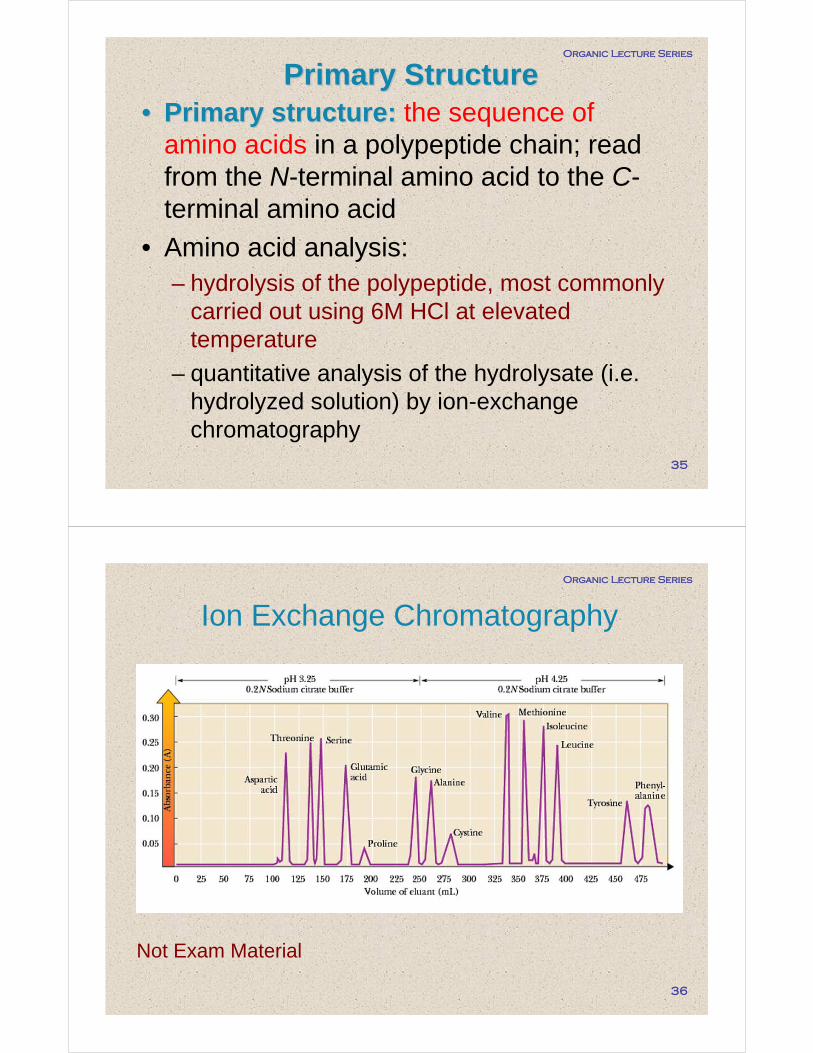
ββ--Pleated SheetPleated Sheet• The antiparallel β-pleated sheet consists
of adjacent polypeptide chains running in opposite directions– each peptide bond is planar and has the
s-trans conformation
– the C=O and N-H groups of peptide bonds from adjacent chains point toward each otherand are in the same plane so that hydrogen bonding is possible between them
– all R- groups on any one chain alternate, first above, then below the plane of the sheet, etc.
Organic Lecture Series
49
β-pleated sheet with three polypeptide chains running in opposite directions
ββ--Pleated SheetPleated Sheet
Organic Lecture Series
50
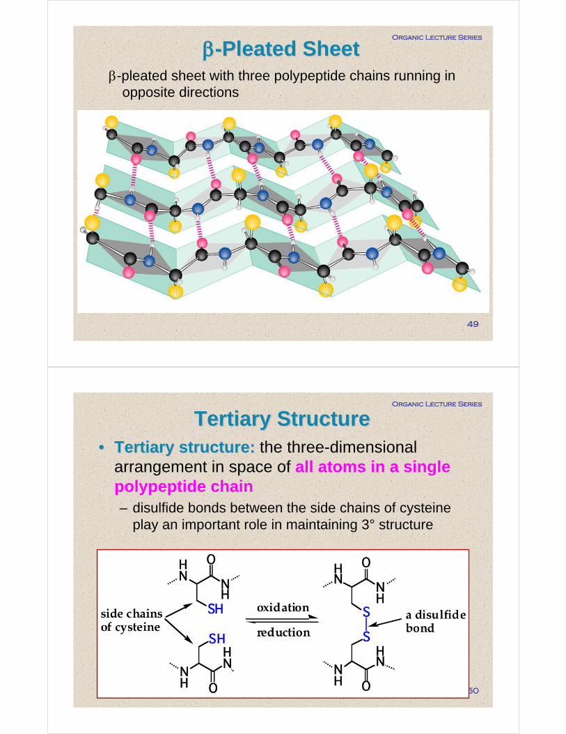
•• Tertiary structure:Tertiary structure: the three-dimensional arrangement in space of all atoms in a single polypeptide chain– disulfide bonds between the side chains of cysteine
play an important role in maintaining 3° structure
Tertiary StructureTertiary Structure

Organic Lecture Series
51
Permanent waving of hair is accomplished by breaking and reforming cysteine cross-links within the hair fiber:
Organic Lecture Series
52
– A ribbon model of myoglobin
Heme

Organic Lecture Series
53
•• Quaternary structure:Quaternary structure: the arrangement of polypeptide chains into a noncovalentlybonded aggregation– the major factor stabilizing quaternary
structure is the hydrophobic effect
•• Hydrophobic effect:Hydrophobic effect: the tendency of nonpolar groups to cluster together in such a way as to be shielded from contact with an aqueous environment
Quaternary structureQuaternary structure
Organic Lecture Series
54
The quaternary structure of hemoglobin
Heme

Organic Lecture Series
55
•Lysozyme is an enzyme found in the cells and secretions of vertebrates.
•Lysozyme hydrolyzes bacterial cell walls which then are susceptible to cell lysis or breaking open.
•Lysozyme from hen egg white contains 129 amino acids which are organized into all four types of secondary structure:
LysozymeLysozyme
Organic Lecture Series
56
Denaturation is the loss of native conformation due to a change in environmental conditions. The non-functioning protein is called a denatured protein.
Denaturation results from the disruptions of the weak secondary forces holding a protein in its native conformation. Disulfide bridges confer considerable resistance to denaturation because they are much stronger than the weak secondary forces.
DenaturationDenaturation

Organic Lecture Series
57
A variety of denaturing conditions or agents lead to protein denaturation:
•Increased temperature (or microwave radiation)
•Ultraviolet and ionizing radiation
•Mechanical energy
•Changes in pH
•Organic chemicals
•Heavy metal salts
•Oxidizing and reducing agents
DenaturationDenaturation