Embed Size (px)
Citation preview

ORJİNAL ARAŞTIRMA
GÜNCEL PEDİATRİ JCP 2020;18(2):177-193
177
KİSTİK FİBROZİS DIŞI BRONŞEKTAZİ
HASTALARINDA AĞIZ HİJYENİ, AĞIZ SAĞLIĞI
ve PERİODONTAL SAĞLIK
Oral Hygiene, Oral Health and Periodontal Health in
Non-Cystic Fibrosis Bronchiectasis Patients
Feyza Ustabas Kahraman(0000-0003-3842-7723)1, Serife Ozdemir(0000-
0003-4003-0530)2, Hakan Yazan(0000-0002-7680-4000)
1, Erkan Cakir(0000-
0002-1438-7854)1
ÖZ
GİRİŞ ve AMAÇ: Bu çalışmanın amacı, kistik fibrosis dışı bronşektazi
(KFDB) hastalığı olan ve herhangi bir hastalığı olmayan çocukların ağız
hijyeni, ağız ve periodontal sağlığı ile ilgili parametreleri
karşılaştırmaktır.
YÖNTEM ve GEREÇLER: Bu çalışma Nisan 2015-Temmuz 2015
tarihleri arasında yapılan 0-16 yaş arası 151 çocuğun dahil olduğu
kesitsel bir çalışmadır. Çalışma grubu 151 çocuktan (KFDB 111 çocuk,
bronşektazi veya kronik hastalığı olmayan 40 çocuk) oluşturuldu. KFDB
hastalarının tek seferlik oral muayenesi yapıldı ve panoramik radyografik
incelemeler istendi. Bu hastalarda oral ve periodontal sağlık kriterleri iki
grupta hesaplandı. Çocukların ebeveynlerinden 6 çoktan seçmeli soruyu
cevaplamaları istendi.
BULGULAR: Süt dişi dolgu yüzeyi sayısı çalışma grubunda kontrol
grubuna göre daha yüksekti. Molar diş sayısı ve sağlıklı diş sayısı
kontroller için çalışma grubuna göre anlamlı olarak yüksekti. Diş sağlığı
ile ilgili diğer parametreler açısından gruplar arasında fark
bulunmadı.hipertansiyon saptanmadı.
TARTIŞMA ve SONUÇ: Ağız hijyeni parametrelerinin çoğunluğu
açısından gruplar benzer olsa da, periodontal muayene ile tespit edildiği
gibi diş sağlığının bozulmasının KFDB hastalarında anlamlı olarak daha
yaygın olduğunu bulduk.
Anahtar Kelimeler: Çocuk, Bronşektazi, Periodontal Hastalıklar, Diş
Hastalığı, Ağız Hijyeni
1 Bezmialem Vakıf Üniversitesi Tıp
Fakültesi, Pediatri Bölümü,
İstanbul
2 Bezmialem Vakıf Üniversitesi Diş
Hekimliği Fakültesi İstanbul
Sorumlu yazar yazışma adresi:
Feyza USTABAŞ KAHRAMAN:
Bezmialem Vakıf Üniversitesi Tıp
Fakültesi, Pediatri Bölümü,
İstanbul, Türkiye
E-mail:
Geliş tarihi/Received: 02.03.2020
Kabul tarihi/Accepted: 24.04.2020
Yayın hakları Güncel Pediatri’ye
aittir.
Güncel Pediatri 2020;18(2):177-193

Kahraman Ustabaş F ve ark. Bronşektazi Hastalarında Ağız Sağlığı JCP2020;18:(2):177-193
178
ABSTRACT
INTRODUCTION: The aim of the study was to compare the parameters related to oral hygiene, oral
and periodontal health of children with and without non-cystic fibrosis bronchiectasis (non-CF BE).
MATERIALS and METHODS: This is a cross-sectional study of 151 children aged between 0-16
years, conducted from April 2015 to July 2015. The study group consisted of 151 children (111
children with non-CF BE, 40 children who did not have bronchiectasis or any chronic disease). One-
time oral examination of patients with non-CF BE was performed and panoramic radiographic
examinations were requested. Oral and periodontal health criteria were calculated in these patients in
two groups. The childrens’ parents were asked to answer 6 multiple-choice questions.
RESULTS: Deciduous tooth filling surface count was higher in the study group than controls. Molar
tooth count and healthy tooth count were significantly higher for controls than study group. No
difference was found between the groups in terms of other parameters of dental health.
CONCLUSIONS: We have found that, although groups were similar in regard to the majority of oral
hygiene parameters, deterioration of dental health as measured by periodontal examination was
significantly more common in those with non-CF BE.
Key words: Child, Bronchiectasis, Periodontal Diseases, Tooth Disease, Oral Hygiene

Kahraman Ustabaş F ve ark. Bronşektazi Hastalarında Ağız Sağlığı JCP2020;18:(2):177-193
179
INTRODUCTION
Bronchiectasis (BE) is characterized by irreversible dilatation of the respiratory tract
associated with inflammatory destruction of bronchial and peribronchial tissue (1). In
developed countries, the most important cause of clinical bronchiectasis is Cystic Fibrosis
(CF) (2). Recurrent respiratory infections, immunodeficiency, foreign body aspiration,
asthma, tuberculosis and primary ciliary dyskinesia are some of the factors that may be
effective in the development of non-cystic fibrosis bronchiectasis (non-CF BE), which is
common in developing countries (1). Improved hygiene and nutrition, broader vaccine
coverage and early antibiotic therapy have been effective in reducing the prevalence of non-
CF BE in developed countries which has reduced it to the “orphan disease” classification.
However, in developing countries, non-CF BE remains as a major cause of respiratory
morbidity (2).
Periodontal diseases are reported to significantly increase the risk for systemic diseases or
alter the natural course of systemic conditions (3). There are several mechanisms that can
potentially relate respiratory diseases to periodontitis. The most widely accepted one is the
mechanical one. With this mechanism, aspiration of oral contents including microbial
pathogens into the airway is followed by respiratory tract inflammation, adhesion,
deterioration of immunity, colonization and the emergence of pulmonary infections. In
patients with chronic health problems, aspiration of oral secretions containing bacterial
pathogens may not always be resolved. In these cases, the periodontium may serve as a
bacterial hotspot. For Chronic Obstructive Pulmonary Disease (COPD), recurrent infections
causing exacerbations have been reported to increase the decline in lung function (4).
COPD is the most studied lung disease related to periodontal diseases (5). Treatment of
periodontal diseases is reported to reduce the number of COPD exacerbations per year (6).
The pathophysiology of bronchiectasis involves mucociliary changes resulting in bacterial
colonization and subsequent chronic inflammation which damages the bronchi. Despite the
clinical and pathophysiological similarities between COPD and bronchiectasis, there is no
study in the literature evaluating the relationship between bronchiectasis
development/progression and periodontal disease (5). Based on these data, the hypothesis we
established is that oral hygiene, oral and periodontal health is worse in those with non-CF BE
compared to healthy children. In order to confirm this, it is important to compare the oral and
periodontal characteristics of those with and without non-CF BE. The aim of the current study

Kahraman Ustabaş F ve ark. Bronşektazi Hastalarında Ağız Sağlığı JCP2020;18:(2):177-193
180
was to compare parameters related to oral hygiene and periodontal health in children with and
without non-CF BE.
MATERIALS and METHODS
Study Population: This is a cross-sectional study of 151 children between the ages of 0-16
who applied to Bezmialem Vakıf University Faculty of Medicine and Faculty of
Dentistrybetween April 1, 2015 and July 1, 2015.
The study group consisted of 111 children with non-CF BE who were followed at the
Pediatric Pulmonology Clinic at Bezmialem Vakif University Faculty of Medicine.
In terms of etiology, primary ciliary dyskinesia (PCD) was detected in 45 patients (40%),
postinfectious BE in 17 (15%) patients, bronchiolitis obliterans in 10 (9%) patients, asthma in
three (3%) patients, immunodeficiency in five (5%) patients, and idiopathic bronchiectasis in
31(28%) patients.
The control group consisted of 40 children who were admitted to the Department of
Pedodontics of Bezmialem Vakif University Faculty of Dentistry for routine check-up and did
not have bronchiectasis or any other chronic disease.
Necessary permissions and ethics committee approval were obtained for conducting the
research (Bezmi Alem University Clinical Research Ethics Committee, Date: 02.04.2014, No:
7/9). In order to conduct this study, patients were informed about the purpose and subject of
the study. Children (and their caretakers) were told about the extent of the procedures they
would undergo during the study and written consent was obtained from the families of the
children who agreed to participate.
Inclusion Criteria: The criterion for patient inclusion was being aged under 18 years and
attending regular follow-up at the Pulmonary Diseases Clinic of our Pediatrics department
due to non-CF BE. For the control group, participants were selected among those with no
bronchiectasis or other chronic disease (eg, Coronary Artery Disease, Diabetes Mellitus,
Chronic Hepatic Failure, Chronic Renal Failure, Autoimmune Diseases, COPD, etc.). In
addition, agreeing to participate in the study was determined as an inclusion criteria for both
groups.
Exclusion criteria: Subjects who refused to participate in the study, those who were older than
18 years, and those who had bronchiectasis or chronic disease in the control group were
excluded from the study group.

Kahraman Ustabaş F ve ark. Bronşektazi Hastalarında Ağız Sağlığı JCP2020;18:(2):177-193
181
Clinical evaluation: Patients with non-CF BE who were referred from the pulmonary diseases
clinic underwent panoramic radiographic imaging and a one-time oral examination. Clinical
examinations were performed to determine the number of teeth affected by caries and other
characteristics of oral hygiene. Oral and periodontal health criteria (number of caried teeth,
number of filled teeth, number of healthy teeth, number of deciduous teeth, number of molar
teeth, etc.) were recorded. In the control group, 40 patients who were admitted to the dentistry
clinic underwent the same procedures and investigations.
Pulp calcification: Pulp calcification was defined as the presence of calcifying mass in the
pulp, which can be seen in deciduous, permanent, healthy, and defective teeth. Radiography is
the only method that can detect clinical and non-invasive calcification (7). Pulp calcification
was evaluated by panoramic radiography.
Plaque index (PI): Plaque index calculations were performed by the evaluation of soft
sediment deposition at the gingival margin and interproximal area. Patients’ average PI was
calculated as the mean value of all teeth. Plaque index classification was defined as follows:
0: Following air drying, plaque is not visible nor cannot be wiped off with an explorer
1: Following air drying, plaque is not visible but can be wiped off with an explorer
2: Plaque is visible along gigival marjin, with or without air drying (no need to probe)
3: Thick plaque is visible along gigival marjin (no need to probe) (8).
Gingival index (GI): GI was calculated based on the evidence of inflammation in gingival
tissues characterized by redness, swelling and bleeding. Each of the four gingival areas of the
tooth (buccal, caesal, distal, and lingual) were scored between 0-3 based on the Löe Gingival
Index System. This classification is as follows:
0: Healthy gingiva
1: Slight inflammation, slight discoloration, edema, no bleeding on probing
2: Moderate inflammation, gingival shiny, red, edema, bleeding on probing
3: Severe inflammation, marked redness and edema, spontaneous bleeding means.
The GI score for the tooth was obtained by dividing values obtained from each tooth by the
number of teeth examined(9).
Theparents of the children were asked to complete a total of 6 multiple-choice questions that
assessed whether children had appropriate access to oral care, dental health services and
whether they received family support.
Statistical Analysis: All analyses were performed on SPSS v21 (IBM, Armonk, NY, USA).
For the normality check, the Shapiro Wilk test was used. Comparison of continuous variables

Kahraman Ustabaş F ve ark. Bronşektazi Hastalarında Ağız Sağlığı JCP2020;18:(2):177-193
182
were done by using the student’s t-test or the Mann Whitney U test with regard to normality
of distribution. Categorical variables were analyzed by Chi-square tests. P<0.05 values were
accepted to show statistically significant results.
RESULTS
Mean age was 10.66 ± 3.58 years in this study. There were no significant differences between
our groups regarding age and gender. Deciduous tooth filling surface count was higher in the
study group than controls (p<0.001). Molar tooth count and healthy tooth count was
significantly higher for controls than patients with bronchiectasis. There were no significant
differences between our groups regarding other variables associated with dental health (Table
1)
Table 1. Summary of Patients' Characteristics Regarding Groups
Bronchiectasis Control Total p
n Descriptive n Descriptive n Descriptive
Age
11
1
10.94 ±
3.93
4
0 9.9 ± 2.26
15
1
10.66 ±
3.58 0.116
Gender
Female 62 55.86%
1
7 42.50% 79 52.32% 0.206
Male 49 44.14%
2
3 57.50% 72 47.68%
Tooth
10
7 22 (0 - 28)
4
0 15 (0 - 28)
14
7 18 (0 - 28) 0.261
Milk Tooth
10
7 2 (0 - 20)
4
0 7 (0 - 20)
14
7 5 (0 - 20) 0.248
Caries 95 1 (0 - 14)
3
9 1 (0 - 4)
13
4 1 (0 - 14) 0.087
Caries Surface 95 1 (0 - 21)
3
9 1 (0 - 15)
13
4 1 (0 - 21) 0.113
Lost Tooth
0 78 82.98%
3
9 100.00%
11
7 87.97%
0.056 1 12 12.77% 0 0.00% 12 9.02%
2 1 1.06% 0 0.00% 1 0.75%
3 3 3.19% 0 0.00% 3 2.26%

Kahraman Ustabaş F ve ark. Bronşektazi Hastalarında Ağız Sağlığı JCP2020;18:(2):177-193
183
Lost Surface
0 78 82.98%
3
9 100.00%
11
7 87.97%
0.056 5 12 12.77% 0 0.00% 12 9.02%
10 1 1.06% 0 0.00% 1 0.75%
15 3 3.19% 0 0.00% 3 2.26%
Filling
0 73 65.77%
3
3 82.50%
10
6 70.20% 0.131
1 - 2 13 11.71% 3 7.50% 16 10.60%
≥ 3 25 22.52% 4 10.00% 29 19.21%
Filling Surface
0 73 65.77%
3
3 82.50%
10
6 70.20% 0.139
1 - 2 10 9.01% 2 5.00% 12 7.95%
≥ 3 28 25.23% 5 12.50% 33 21.85%
Molar Tooth Caries
0 43 45.26%
1
8 47.37% 61 45.86%
0.561 1 25 26.32%
1
3 34.21% 38 28.57%
2 12 12.63% 5 13.16% 17 12.78%
3 8 8.42% 1 2.63% 9 6.77%
4 7 7.37% 1 2.63% 8 6.02%
Molar Tooth Caries
Surface
0 43 38.74%
1
8 45.00% 61 40.40%
0.077 1 - 2 31 27.93%
1
6 40.00% 47 31.13%
≥ 3 37 33.33% 6 15.00% 43 28.48%
Molar Tooth Lost
0 86 90.53%
3
8 100.00%
12
4 93.23%
0.277 1 7 7.37% 0 0.00% 7 5.26%
2 1 1.05% 0 0.00% 1 0.75%
3 1 1.05% 0 0.00% 1 0.75%
Molar Tooth Lost
Surface
0 86 90.53%
3
8 100.00%
12
4 93.23%
0.277 5 7 7.37% 0 0.00% 7 5.26%
10 1 1.05% 0 0.00% 1 0.75%
15 1 1.05% 0 0.00% 1 0.75%

Kahraman Ustabaş F ve ark. Bronşektazi Hastalarında Ağız Sağlığı JCP2020;18:(2):177-193
184
Molar Tooth Filling
0 80 84.21%
3
4 89.47%
11
4 85.71%
0.357 1 7 7.37% 2 5.26% 9 6.77%
2 5 5.26% 1 2.63% 6 4.51%
3 0 0.00% 1 2.63% 1 0.75%
4 3 3.16% 0 0.00% 3 2.26%
Molar Tooth Filling
Surface
0 80 72.07%
3
4 85.00%
11
4 75.50% 0.136
1 - 2 7 6.31% 3 7.50% 10 6.62%
≥ 3 24 21.62% 3 7.50% 27 17.88%
Milk Tooth Caries 66 2 (0 - 16)
4
0 2 (0 - 17)
10
6 2 (0 - 17) 0.751
Milk Tooth Caries
Surface 66 3 (0 - 50)
4
0 3 (0 - 51)
10
6 3 (0 - 51) 0.556
Milk Tooth Filling
0 60 90.91%
3
4 85.00% 94 88.68%
0.543 1 3 4.55% 3 7.50% 6 5.66%
2 3 4.55% 2 5.00% 5 4.72%
3 0 0.00% 1 2.50% 1 0.94%
Milk Tooth Filling
Surface
0 60 54.05%
3
4 85.00% 94 62.25% <0.001
1 - 2 2 1.80% 2 5.00% 4 2.65%
≥ 3 49 44.14% 4 10.00% 53 35.10%
Fissealant
0 92 94.85%
3
9 97.50%
13
1 95.62%
0.554 1 0 0.00% 0 0.00% 0 0.00%
2 3 3.09% 0 0.00% 3 2.19%
3 1 1.03% 1 2.50% 2 1.46%
4 1 1.03% 0 0.00% 1 0.73%
Dental Pulp Calc
Present 2 1.98% 0 0.00% 2 1.42% 0.915
Absent 99 98.02%
4
0 100.00%
13
9 98.58%
Plaque Index
10
2 2.21 ± 0.53
4
0 2.29 ± 0.44
14
2 2.23 ± 0.5 0.398
Gingival Index
10
2 1.09 ± 0.4
4
0 1.17 ± 0.3
14
2 1.12 ± 0.38 0.255

Kahraman Ustabaş F ve ark. Bronşektazi Hastalarında Ağız Sağlığı JCP2020;18:(2):177-193
185
Molar Tooth Surface
0 45 40.54% 8 20.00% 53 35.10%
0.063 1 - 2 33 29.73%
1
5 37.50% 48 31.79%
≥ 3 33 29.73%
1
7 42.50% 50 33.11%
Molar Tooth
0 38 40.00% 8 21.05% 46 34.59%
0.040
1 22 23.16% 6 15.79% 28 21.05%
2 14 14.74%
1
4 36.84% 28 21.05%
3 11 11.58% 5 13.16% 16 12.03%
4 10 10.53% 5 13.16% 15 11.28%
Tooth Surface
0 38 34.23% 9 22.50% 47 31.13%
0.388 1 - 2 30 27.03%
1
3 32.50% 43 28.48%
≥ 3 43 38.74%
1
8 45.00% 61 40.40%
Tooth
0 31 27.93% 9 22.50% 40 26.49%
0.215 1 - 2 33 29.73%
1
8 45.00% 51 33.77%
≥ 3 47 42.34%
1
3 32.50% 60 39.74%
Milk Tooth Surface
0 40 36.04%
1
0 25.00% 50 33.11%
0.058 1 - 2 19 17.12%
1
4 35.00% 33 21.85%
≥ 3 52 46.85%
1
6 40.00% 68 45.03%
Milk Tooth
0 40 36.04%
1
0 25.00% 50 33.11%
0.079 1 - 2 20 18.02%
1
4 35.00% 34 22.52%
≥ 3 51 45.95%
1
6 40.00% 67 44.37%
Healthy Tooth
0 78 70.27% 0 0.00% 78 51.66%
<0.001 1 10 9.01% 0 0.00% 10 6.62%
≥ 2 23 20.72%
4
0 100.00% 63 41.72%

Kahraman Ustabaş F ve ark. Bronşektazi Hastalarında Ağız Sağlığı JCP2020;18:(2):177-193
186
Caries Tooth
0 88 90.72%
3
2 82.05%
12
0 88.24%
0.207 1 4 4.12% 2 5.13% 6 4.41%
2 4 4.12% 2 5.13% 6 4.41%
3 1 1.03% 3 7.69% 4 2.94%
Milk Tooth Extraction
0 59 88.06% 0 0.00% 59 88.06%
N.A 1 4 5.97% 0 0.00% 4 5.97%
2 3 4.48% 0 0.00% 3 4.48%
5 1 1.49% 0 0.00% 1 1.49%
M
0 84 86.60% 0 0.00% 84 86.60%
N.A 1 10 10.31% 0 0.00% 10 10.31%
3 3 3.09% 0 0.00% 3 3.09% Data given as mean ± standard deviation or median (minimum - maximum) for continuous variables regarding
normality and percentage for categorical variables
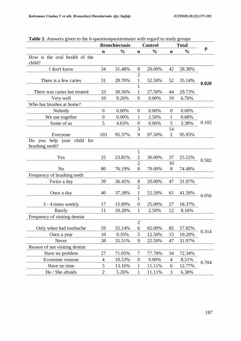
The frequency of answer that "I don't know" to the question about their child's dental health
was higher in the study group than the control group. No significant difference was found
between the study group and control groups in terms of having a toothbrush, daily brushing
count, receiving help during tooth brushing, dental visits and the causes of these dental visits
of the child. (Table 2).
Kahraman Ustabaş F ve ark. Bronşektazi Hastalarında Ağız Sağlığı JCP2020;18:(2):177-193
187
Table 2. Answers given to the 6-questionquestionnaire with regard to study groups
Bronchiectasis Control Total p
n % n % n %
How is the oral health of the
child?
I don't know 34 31.48% 8 20.00% 42 28.38%
0.020 There is a few caries 31 28.70%
2
1 52.50% 52 35.14%
There was caries but treated 33 30.56%
1
1 27.50% 44 29.73%
Very well 10 9.26% 0 0.00% 10 6.76%
Who has brushes at home?
Nobody 0 0.00% 0 0.00% 0 0.00%
0.102 We use together 0 0.00% 1 2.50% 1 0.68%
Some of us 5 4.63% 0 0.00% 5 3.38%
Everyone 103 95.37%
3
9 97.50%
14
2 95.95%
Do you help your child for
brushing teeth?
Yes 25 23.81%
1
2 30.00% 37 25.52% 0.582
No 80 76.19%
2
8 70.00%
10
8 74.48%
Frequency of brushing teeth
Twice a day 39 36.45% 8 20.00% 47 31.97%
0.056 Once a day 40 37.38%
2
1 52.50% 61 41.50%
3 - 4 times weekly 17 15.89%
1
0 25.00% 27 18.37%
Rarely 11 10.28% 1 2.50% 12 8.16%
Frequency of visiting dentist
Only when had toothache 59 55.14%
2
6 65.00% 85 57.82% 0.314
Once a year 10 9.35% 5 12.50% 15 10.20%
Never 38 35.51% 9 22.50% 47 31.97%
Reason of not visiting dentist
Have no problem 27 71.05% 7 77.78% 34 72.34%
0.704 Economic reasons 4 10.53% 0 0.00% 4 8.51%
Have no time 5 13.16% 1 11.11% 6 12.77%
He / She afraids 2 5.26% 1 11.11% 3 6.38%

Kahraman Ustabaş F ve ark. Bronşektazi Hastalarında Ağız Sağlığı JCP2020;18:(2):177-193
188
DISCUSSION
In the current study, in non-CF-BE patients, it was found that the number of molar teeth and
deciduous tooth filling surface count were higher and healthy teeth count was lower than the
control group. The two groups were similar in terms of other oral and periodonal parameters.
To our knowledge, this is the first study to evaluate the association between periodontal
health and non-CF-BE in children. COPD is the most frequently studied lung disease with its
association with oral and periodontal diseases (5). Although bronchiectasis and COPD have
very similar systemic, clinical and immunological findings, to our knowledge, there have
been no attempts to study the periodontal status of patients with bronchiectasis; indicating that
there is a significant lack of information on this topic. Our study aimed to provide much-
needed information on the relationship between periodontal disease and non-CF-BE.
Oral and dental health properties
There is increasing evidence that oral and periodontal diseases affect systemic diseases. If an
individual has oral or perodontal disease, the pathogens present in oropharyngeal secretion
may be dangerous when aspirated into the lungs, especially the lungs of a patient with chronic
disease(s) affecting the lung.It has been stated in various studies that oral and periodontal
diseases may cause or exacerbate the natural course of systemic diseases (3, 10-14). In this
study, the number of healthy teeth was significantly lower in the bronchiectasis group. No
difference was found between the groups in terms of caried tooth, filled tooth, missing tooth,
or the number of extracted teeth. Furthermore, no difference was found between the
bronchiectasis and control groups in terms of plaque index and gingival index, which is an
important indicator of periodontal health. In a study examining the relationship between oral
health status and COPD, the number of healthy natural teeth was reported to be higher than in
non-COPD patients. On the other hand, tooth caries, tooth extraction, lack of any tooth were
more common in COPD patients, and no difference was found between the two groups in
terms of filled teeth (15). Scannapieco and Ho (16) reported decreased lung function
associated with an increase in periodontal attachment loss. In the study of Liu et al. (17) PI
increase was reported to be an effective factor on COPD exacerbations. In another study, PI
and GI scores were found to be higher in COPD group than in the control group (18).
However, we did not find any difference between the PI and GI scores of patients with and
without non-CF-BE in the current study. Even so, the periodontal health of patients with non-
CF-BE were found to be worse than those without non-CF-BE, which is similar to the study

Kahraman Ustabaş F ve ark. Bronşektazi Hastalarında Ağız Sağlığı JCP2020;18:(2):177-193
189
by Wang et al. (19) in which patients with COPD had worse periodontal health measures than
healthy controls. Community based studies also suggest similar results: in a community-based
study in Spain, the prevalence of periodontal disease was reported to be higher in COPD
patients than in non-COPD patients (15). In a community-based cohort study by Shen et al.
(20) patients with COPD were found to have a higher risk of developing periodontal disease
than the general population. Furthermore, treatment of chronic periodontitis was found to
significantly reduce COPD exacerbationsin a prospective study by Küçükcoşkun et al. (21).
This trend is evident in many studies including major randomized controlled trials such as the
pilot study by Zhou et al. (6) in which periodontal therapy was suggested to improve lung
function and reduce exacerbation frequency in patients with COPD and chronic periodontitis
and treatment was also associated with reduced risk of adverse respiratory events and
mortality in patients with COPD (22). Additionally, according to a cohort study conducted in
Japan, deterioration of periodontal health was reported as a risk factor that accelerates the
decrease in lung function (23).
Finally,according to a metaanalysis on this topic, which examined 14 observational studies,
periodontal diseases were found to be an independent risk factor for COPD progression (24).
Furthermore, in a recent study evaluating 14 studies which evaluated the clinical
characteristics of oral health, it was concluded that periodontal problems (deeper periodontal
pockets, high level of clinical attachment loss, worse oral hygiene, more inflammation and
bleeding in the gingival tissue, and lower number of remaining teeth) were worse in COPD
patients compared to non-COPD patients (25).
Oral health behaviors and knowledge level
In the current study, no significant difference was found in the bronchiectasis and control
groups in terms of having a toothbrush, daily brushing, receiving help from parents during
brushing. The frequency of answer that "I don't know" to the question about their child's
dental health was higher in the study group than the control group. In a study by Liu et al.
(17) it was reported that the number of dailybrushing was an effective factor on COPD
exacerbations. In the study of Wang et al. (19) it was reported that daily brushing behavior
and oral health knowledge levels were lower in patients with COPD than those without
COPD. In a study by Bhavsar et al. (18) it was reported that the number of tooth brushing and
oral hygiene scores were lower in COPD patients than controls. Two systematic reviews have
been reported that improving oral hygiene is effective in reducing the progression and
occurrence of respiratory diseases (14, 26). The current study was conducted with pediatric

Kahraman Ustabaş F ve ark. Bronşektazi Hastalarında Ağız Sağlığı JCP2020;18:(2):177-193
190
patients with non-CF-BE, and during this period, tooth brushing behavior was evaluated
according to family reporting and was not observed; thus the lack of any significant difference
between our groups may have been caused by this factor. Nevertheless, improving tooth
brushing behavior in all children, especially those with chronic lung disease, will undoubtedly
benefit overall health.
Frequency of dentist visits and reasons for not visiting the dentist
In the current study, no significant difference was found between the bronchiectasis and
control groups in terms of visits to dentists and reasons for not visiting. On the other hand, the
most common reason for not visiting a dentist was found to be the lack of evident dental
problems (toothache, bleeding, etc.) in both groups. In a community-based study, no
difference was found between patients with and without COPD in terms of applying to a
dentist within the prior 3 months of the study (15). Another study reported that patients with
COPD had fewer visits to dentists than those without COPD (19). Children may show similar
tendencies because children may find it difficult to recognize their dental health problems.
Additionally, fear of the dentist is rather frequent among children, which may cause them to
be unwilling to go to the dentist.
Limitations
The current research has several limitations. Our first limitation isassociated with the study
design; cross-sectional studies have low power in terms of explaining causality. The second
most important limitation is that our study group did not undergo full-extent evaluations for
the diagnosis of periodontal diseases. If such an examination was performed, we may have
been able to analyze whether the frequency of periodontal disease was higher in non-CF-BE
patients. However, general oral and periodontal findings were evaluated and showed
important differences between patients with non-CF-BE and controls. Thirdly, study groups
were comprised from patients that applied to a single center; it is quite evident that a
community-based research could have provided more reliable results. Even so, to our
knowledge, our study is the first to evaluate the relationship between periodontal health and
non-CF-BE in pediatric patients; and therefore addresses an important gap in the literature.
Conclusion
This is the first study to evaluate the relationship between oral hygiene and periodontal health
in patients with non-CF-BE. The most important result of this study was the finding that the
number of healthy teeth was significantly lower in the bronchiectasis group than in the control

Kahraman Ustabaş F ve ark. Bronşektazi Hastalarında Ağız Sağlığı JCP2020;18:(2):177-193
191
group. There were no significant differences between our groups regarding many other
variables that are known to be associated with periodontal health.
In the future, larger clinical investigations and structured prospective studies are required to
determine the extent of the relationship between oral hygiene and periodontal health and non-
CF-BE. Randomized controlled trials to investigate the change in clinical characteristics of
patients with bronchiectasis by correcting oral hygiene and periodontal health parameters may
help to explain the relationships between these conditions. Improving parents’ knowledge of
dental care and oral health is important in the prevention and treatment of non-CF-BE in
children. Therefore, it is the responsibility of all healthcare providers to help preserve oral
health in individuals by providing the best available evidence to patients and their parents.
Promoting opportunities for oral care and treatment, particularly in the context of oral and
periodontal health protection, can be an effective strategy to reduce the burden of lung
dysfunction leading to non-CF-BE.
Conflict of Interest: No potential conflict of interest was reported by the authors.
Funding: No funding was received.
REFERENCES
1. Karadag B, Karakoc F, Ersu R, Kut A, Bakac S, Dagli E. Non-cystic-fibrosis
bronchiectasis in children: a persisting problem in developing countries. Respiration.
2005;72(3):233-8.
2. Kapur, Nitin. Non-cystic fibrosis bronchiectasis in children. PhD Thesis, Charles
Darwin University, 2011.
3. Peter KP, Mute BR, Doiphode SS, Bardapurkar SJ, Borkar MS, Raje DV. Association
between periodontal disease and chronic obstructive pulmonary disease: a reality or
just a dogma? J Periodontol. 2013;84(12):1717-23.
4. Hobbins S, Chapple IL, Sapey E, Stockley RA. Is periodontitis a comorbidity of
COPD or can associations be explained by shared risk factors/behaviors? Int J Chron
Obstruct Pulmon Dis. 2017;12:1339.
5. Santos SR, Pinto EH, Longo PL, Dal Corso S, Lanza FC, Stelmach R, et al. Effects of
periodontal treatment on exacerbation frequency and lung function in patients with
chronic periodontitis: study protocol of a 1-year randomized controlled trial. BMC
Pulm Med. 2017;17(1):23.
Kahraman Ustabaş F ve ark. Bronşektazi Hastalarında Ağız Sağlığı JCP2020;18:(2):177-193
192
6. Zhou X, Han J, Liu Z, Song Y, Wang Z, Sun Z. Effects of periodontal treatment on
lung function and exacerbation frequency in patients with chronic obstructive
pulmonary disease and chronic periodontitis: A 2‐year pilot randomized controlled
trial. J Clin Periodontol. 2014;41(6):564-72.
7. Sezgin B, Cakan EF, Erdem TL. A Radiographic Assessment of The Prevalence and
Distribution of Pulp Calcification. Eur Oral Res. 2011;45(2):49.
8. Silness J, Löe H. Periodontal disease in pregnancy II. Correlation between oral
hygiene and periodontal condition. Acta Odontol Scand. 1964;22(1):121-35.
9. Löe H, Silness J. Periodontal disease in pregnancy I. Prevalence and severity. Acta
Odontol Scand. 1963;21(6):533-51.
10. Pedersen PU, Uhrenfeldt L, Larsen P. Oral hygiene in patients with chronic
obstructive pulmonary disease: a scoping review protocol. JBI database of systematic
reviews and implementation reports. 2017;15(5):1236-41.
11. Pace CC, McCullough GH. The association between oral microorgansims and
aspiration pneumonia in the institutionalized elderly: review and recommendations.
Dysphagia. 2010;25(4):307-22.
12. Scannapieco FA, Bush RB, Paju S. Associations between periodontal disease and risk
for nosocomial bacterial pneumonia and chronic obstructive pulmonary disease. A
systematic review. Ann Periodontol. 2003;8(1):54-69.
13. Scannapieco FA. Pneumonia in nonambulatory patients: the role of oral bacteria and
oral hygiene. The Journal of the American Dental Association. 2006;137:S21-S5.
14. Azarpazhooh A, Leake JL. Systematic review of the association between respiratory
diseases and oral health. J Periodontol. 2006;77(9):1465-82.
15. Lopez-de-Andrés A, Vazquez-Vazquez L, Martinez-Huedo MA, Hernández-Barrera
V, Jimenez-Trujillo I, Tapias-Ledesma MA, et al. Is COPD associated with
periodontal disease? a population-based study in spain. Int J Chron Obstruct Pulmon
Dis. 2018;13:3435.
16. Scannapieco FA, Ho AW. Potential associations between chronic respiratory disease
and periodontal disease: analysis of National Health and Nutrition Examination
Survey III. J Periodontol. 2001;72(1):50-6.
17. Liu Z, Zhang W, Zhang J, Zhou X, Zhang L, Song Y, et al. Oral hygiene, periodontal
health and chronic obstructive pulmonary disease exacerbations. J Clin Periodontol.
2012;39(1):45-52.
18. Bhavsar NV, Dave BD, Brahmbhatt NA, Parekh R. Periodontal status and oral health
behavior in hospitalized patients with chronic obstructive pulmonary disease. J Nat Sci
Biol Med. 2015;6(Suppl 1):S93.
19. Wang Z, Zhou X, Zhang J, Zhang L, Song Y, Hu FB, et al. Periodontal health, oral
health behaviours, and chronic obstructive pulmonary disease. J Clin Periodontol.
2009;36(9):750-5.

Kahraman Ustabaş F ve ark. Bronşektazi Hastalarında Ağız Sağlığı JCP2020;18:(2):177-193
193
20. Shen T-C, Chang P-Y, Lin C-L, Chen C-H, Tu C-Y, Hsia T-C, et al. Risk of
periodontal diseases in patients with chronic obstructive pulmonary disease: a
nationwide population-based cohort study. Medicine. 2015;94(46).
21. Kucukcoskun M, Baser U, Oztekin G, Kiyan E, Yalcin F. Initial periodontal treatment
for prevention of chronic obstructive pulmonary disease exacerbations. J Periodontol.
2013;84(7):863-70.
22. Shen T-C, Chang P-Y, Lin C-L, Chen C-H, Tu C-Y, Hsia T-C, et al. Periodontal
treatment reduces risk of adverse respiratory events in patients with chronic
obstructive pulmonary disease: a propensity-matched cohort study. Medicine.
2016;95(20).
23. Takeuchi K, Matsumoto K, Furuta M, Fukuyama S, Takeshita T, Ogata H, et al.
Periodontal status and lung function decline in the community: the Hisayama study.
Sci Rep. 2018;8(1):13354.
24. Zeng X-T, Tu M-L, Liu D-Y, Zheng D, Zhang J, Leng W. Periodontal disease and risk
of chronic obstructive pulmonary disease: a meta-analysis of observational studies.
PLoS One. 2012;7(10):e46508.
25. Shi Q, Zhang B, Xing H, Yang S, Xu J, Liu H. Patients with Chronic Obstructive
Pulmonary Disease Suffer from Worse Periodontal Health—Evidence from a Meta-
Analysis. Front Physiol. 2018;9:33.
26. Pedersen PU, Larsen P, Håkonsen SJ. The effectiveness of systematic perioperative
oral hygiene in reduction of postoperative respiratory tract infections after elective
thoracic surgery in adults: a systematic review. JBI database of systematic reviews and
implementation reports. 2016;14(1):140-73.