Embed Size (px)
Citation preview
ABCDEFG
UNIVERS ITY OF OULU P.O.B . 7500 F I -90014 UNIVERS ITY OF OULU F INLAND
A C T A U N I V E R S I T A T I S O U L U E N S I S
S E R I E S E D I T O R S
SCIENTIAE RERUM NATURALIUM
HUMANIORA
TECHNICA
MEDICA
SCIENTIAE RERUM SOCIALIUM
SCRIPTA ACADEMICA
OECONOMICA
EDITOR IN CHIEF
PUBLICATIONS EDITOR
Professor Mikko Siponen
University Lecturer Elise Kärkkäinen
Professor Hannu Heusala
Professor Olli Vuolteenaho
Senior Researcher Eila Estola
Information officer Tiina Pistokoski
University Lecturer Seppo Eriksson
Professor Olli Vuolteenaho
Publications Editor Kirsti Nurkkala
ISBN 978-951-42-9060-2 (Paperback)ISBN 978-951-42-9061-9 (PDF)ISSN 0355-3221 (Print)ISSN 1796-2234 (Online)
U N I V E R S I TAT I S O U L U E N S I S
MEDICA
ACTAD
D 1008
ACTA
Mari K
uisma
OULU 2009
D 1008
Mari Kuisma
MAGNETIC RESONANCE IMAGING OF LUMBAR DEGENERATIVE BONE MARROW (MODIC) CHANGESDETERMINANTS, NATURAL COURSE AND ASSOCIATION WITH LOW BACK PAIN
FACULTY OF MEDICINE,INSTITUTE OF DIAGNOSTICS, DEPARTMENT OF DIAGNOSTIC RADIOLOGY,INSTITUTE OF CLINICAL MEDICINE, DEPARTMENT OF PHYSICAL MEDICINE AND REHABILITATION,UNIVERSITY OF OULU

A C T A U N I V E R S I T A T I S O U L U E N S I SD M e d i c a 1 0 0 8
MARI KUISMA
MAGNETIC RESONANCE IMAGING OF LUMBAR DEGENERATIVE BONE MARROW (MODIC) CHANGESDeterminants, natural course and association withlow back pain
Academic dissertation to be presented, with the assent ofthe Faculty of Medicine of the University of Oulu, forpublic defence in Auditorium 7 of Oulu UniversityHospital, on April 24th, 2009, at 12 noon
OULUN YLIOPISTO, OULU 2009
Copyright © 2009Acta Univ. Oul. D 1008, 2009
Supervised byProfessor Osmo TervonenProfessor Jaro Karppinen
Reviewed byDocent Mats GrönbladDocent Antti Lamminen
ISBN 978-951-42-9060-2 (Paperback)ISBN 978-951-42-9061-9 (PDF)http://herkules.oulu.fi/isbn9789514290619/ISSN 0355-3221 (Printed)ISSN 1796-2234 (Online)http://herkules.oulu.fi/issn03553221/
Cover designRaimo Ahonen
OULU UNIVERSITY PRESSOULU 2009

Kuisma, Mari, Magnetic resonance imaging of lumbar degenerative bone marrow(Modic) changes. Determinants, natural course and association with low back painFaculty of Medicine, Institute of Diagnostics, Department of Diagnostic Radiology, Institute ofClinical Medicine, Department of Physical Medicine and Rehabilitation, University of Oulu,P.O.Box 5000, FI-90014 University of Oulu, Finland Acta Univ. Oul. D 1008, 2009Oulu, Finland
Abstract
Modic changes are vertebral bone marrow signal intensity changes adjacent to the endplates of thedegenerated intervertebral discs in magnetic resonance imaging (MRI).
This study evaluated the prevalence and the determinants of Modic changes and theirassociation with low back pain symptoms in an occupational cohort of middle-aged Finnish men.The prevalence and the natural course of Modic changes were assessed over a 3-year follow-upperiod among sciatica patients. Finally, in a patient population, the characteristics of bone marrowchanges in MRI were compared to the imaging findings in CT.
The prevalence of Modic changes was 56% in an occupational cohort of middle-aged males.Besides age, the determinants of Modic changes and disc degeneration were different. Weight-related factors, which add to the load of the lumbar spine, were associated with Modic changes,whereas whole-body vibration was associated with severe disc degeneration.
The prevalence of Modic changes among sciatica patients was 65%, type II change being themost frequent. During the 3-year follow-up, 14% of changes converted to another type, while theincidence of new Modic changes was 6%.
Among middle-aged working males, Modic changes located at L5–S1 and type I Modicchanges were more likely to be associated with pain symptoms than other types of Modic changesor changes located at other lumbar levels.
Thirty-eight percent of the endplates with Modic changes had sclerosis in CT. Of specificModic types, mixed I/II and II/III associated significantly with endplate sclerosis. Endplatesclerosis was not detected in MRI.
In conclusion, Modic changes are a common MRI finding both among patients and middle-aged working males. In addition to age, weight-related factors seem to be important in thepathogenesis of Modic changes. Modic changes can convert from one type to another and type IIchanges may be less stable than previously assumed. A considerable proportion of Modic changesare sclerotic as observed in CT. Modic changes were always found in combination with adegenerative intervertebral disc and thus they are assumed to be a specific phenotype ofdegenerative disc disease. Finally, Modic changes may be painful – especially when located atL5–S1 and type I changes.
Keywords: disc degeneration, low back pain, lumbar spine, magnetic resonanceimaging, Modic changes


To Jani, Laura and Mikko

Acknowledgements
This study was carried out at the Department of Diagnostic Radiology, Oulu Uni-
versity Hospital in collaboration with the Department of Physical Medicine and
Rehabilitation, Oulu University Hospital during the years 2004–2009.
I wish to express my deepest gratitude to my supervisors Professor Osmo
Tervonen, M.D., Head of the Department of Diagnostic Radiology, and Professor
Jaro Karppinen, M.D., Department of Physical Medicine and Rehabilitation, for
providing me with excellent research facilities, and for their enthusiastic and
endless support during the whole study period. I owe my sincere thanks to
Professor Tervonen for giving me the opportunity to carry out this study in an
encouraging and inspiring atmosphere as a member of his research group. His
valuable advice and guidance have been essential for completing this study.
Professor Karppinen deserves my warm thanks for his never-ending optimism and
enthusiastic attitude to this work. His knowledge in the field of low back pain
research has been of great importance.
I wish to thank Professor (emeritus) Ilkka Suramo, M.D., and Professor
(emeritus) Juhani Pyhtinen, M.D., for their support during these years.
I am grateful to Docent Mats Grönblad, M.D., and Docent Antti Lamminen,
M.D., the reviewers of this thesis. Their constructive criticism and encouraging
comments are appreciated.
All the collaborators deserve my sincere thanks. I had the great honor of
working with a group of specialists and inspiring people, including Professor
Heikki Vanharanta, M.D., Docent Markku Heliövaara, M.D., Docent Raija
Korpelainen, Ph.D., Docent Antero Natri, M.D., and Docent Simo Taimela, M.D.,
whose clinical expertise have been essential for this work. I am deeply grateful to
Kaisu Kaikkonen, M.Sc., Mauno Kurunlahti, M.D., Ph.D., Eveliina
Lammentausta, Ph.D., and Risto Ojala, M.D., Ph.D., for their collaboration and
friendly assistance during these years. Special thanks are due to Jaakko Niinimäki,
M.D., for fruitful conversations and help in numerous things concerning this work.
I wish to thank Marianne Haapea, M.Sc., for her friendly help with statistical
analysis, and for the long coffee breaks.
I am grateful to Anna Vuolteenaho, M.A., for the careful revision of the
language of this thesis. Mrs. Seija Leskelä is warmly acknowledged for preparing
posters related to this work and Mrs. Helena Saari for helping me with the final
layout of this thesis. I want to thank Docent Miika Nieminen, Ph.D., for technical
help with references. I owe my warm thanks to Mr. Kari Kylmäniemi and the staff
7
of the MRI unit for help in collecting data for the study. I thank Mrs. Kaisa
Punakivi, Mrs. Marja-Liisa Raipola, Mrs. Leila Salo and Ms. Arja Väisänen for
their friendly assistance in practical issues.
I want to express my warmest thanks to my colleagues in Southern Central
Radiology and MRI for their help and support during these years. I thank
especially Docent Eija Pääkkö, M.D. and Airi Jartti, M.D., Ph.D., for their
favorable attitude towards scientific work and for the excellent way of teaching
body radiology. Heli Reinikainen, M.D., Ph.D., Michaela Bode, M.D., Ph.D.,
Johanna Ronkainen, M.D., Ph.D., and Maria Suo-Palosaari, M.D., Ph.D., deserve
my warm thanks for their important advice at the final stage of preparing this
thesis. All my colleagues and friends in the field of radiology and CC-Oulu are
gratefully acknowledged for the collaboration and the joyful moments we have
shared.
I am deeply grateful to my parents Mirja and Reino for their love and support
throughout my life. I thank you for always believing in me. I want to warmly thank
my sister Erja, my brother Arto and my friends Teija, Arja and Katri for all the
great times spent together and for brightening my days.
I owe my most loving thanks to my daughter Laura and my son Mikko. Your
smiles and laughter keep me going. Thank you for reminding me of the most
important aim in my life.
Finally, I express my deepest feelings of gratitude and love to my husband Jani
for patience and encouragement during these years. He and the children fill my
heart with joy.
This study was financially supported by the Finnish Cultural Foundation, the
Finnish Medical Foundation, the Orion-Farmos Research Foundation, the
Foundation for Medical Research in Oulu and the Radiological Society of Finland,
all of which are gratefully acknowledged.
Oulu, February 2009 Mari Kuisma
8
Abbreviations
AF Annulus fibrosusBMI Body Mass IndexCI Confidence interval CRP C-reactive proteinCT Computed tomographyDD Disc degenerationEP EndplateETL Echo train lengthFLAIR Fluid-attenuated inversion recoveryFOV Field of viewFSE Fast spin echohsCRP High-sensitivity C-reactive proteinHU Hounsfield unitIL-6 Interleukin-6IVD Intervertebral discL Lumbar disc levelLBP Low back painM ModicMRI Magnetic resonance imagingN Number of patientsNEX Number of excitationsNP Nucleus pulposusOR Odds ratioPG ProteoglycanPGP Protein gene productROI Region of interestS SacrumSD Standard deviationSE Spin echoSI Signal intensitySTIR Short tau inversion recoveryT TeslaT1 Longitudinal relaxation timeT2 Transverse relaxation timeTE Echo timeTI Inversion timeTR Repetition timeTNF-α Tumor necrosis factor-alphaUS UltrasoundW WeightedVAS Visual analog scaleVS. Versus
9

10
List of original publications
This thesis is based on the following articles, which are referred to in the text by
their Roman numerals:
I Kuisma M, Karppinen J, Haapea M, Niinimäki J, Ojala R, Heliövaara M,
Korpelainen R, Kaikkonen K, Taimela S, Natri A & Tervonen O (2008) Are
the determinants of vertebral endplate changes and severe disc degeneration in
the lumbar spine the same? A magnetic resonance imaging study in
middle-aged male workers. BMC Musculoskelet Disord 9:51.
II Kuisma M, Karppinen J, Niinimäki J, Kurunlahti M, Haapea M, Vanharanta H
& Tervonen O (2006) A three-year follow-up of lumbar spine endplate
(Modic) changes. Spine 3:1714–1718.
III Kuisma M, Karppinen J, Niinimäki J, Ojala R, Haapea M, Heliövaara M,
Korpelainen R, Taimela S, Natri A & Tervonen O (2007) Modic changes in
endplates of lumbar vertebral bodies: prevalence and association with low
back and sciatic pain among middle-aged male workers. Spine 32:1116–1122.
IV Kuisma M, Karppinen J, Haapea M, Lammentausta E, Niinimäki J &
Tervonen O (2009) Modic changes in vertebral endplates: a comparison of
MR imaging and multislice CT. Skeletal Radiol 38:141–147.
11

12
Contents
Abstract Acknowledgements
Abbreviations
List of original publications
Contents1 Introduction 152 Review of the literature 17
2.1 Spine........................................................................................................ 17
2.1.1 Vertebral body .............................................................................. 17
2.1.2 Intervertebral disc ......................................................................... 18
2.1.3 Vertebral endplate ......................................................................... 19
2.2 Disc degeneration.................................................................................... 21
2.3 Degenerative changes of the vertebral endplates and subchondral bone 22
2.3.1 Morphologic changes.................................................................... 22
2.3.2 Biochemical changes .................................................................... 22
2.4 Magnetic resonance imaging techniques of vertebral bone marrow....... 23
2.5 Vertebral degenerative bone marrow (Modic) changes .......................... 24
2.5.1 Definition ...................................................................................... 24
2.5.2 Prevalence and distribution........................................................... 25
2.5.3 Determinants ................................................................................. 26
2.5.4 Differential diagnosis.................................................................... 27
2.5.5 Pathogenesis.................................................................................. 28
2.5.6 Natural course ............................................................................... 30
2.5.7 Association with low back pain .................................................... 31
3 Purpose of the study 334 Materials and methods 35
4.1 Study population ..................................................................................... 35
4.1.1 Occupational sample of male train engineers and sedentary
paper mill workers (I and III)........................................................ 35
4.1.2 Patients (II and IV)........................................................................ 36
4.2 Assessments (I and III)............................................................................ 36
4.3 Magnetic resonance imaging................................................................... 37
4.4 Computed tomography imaging (IV)...................................................... 37
4.5 Image analysis......................................................................................... 38
13
4.6 Statistical analysis ................................................................................... 40
4.6.1 Reliability of Modic changes (I, II and III)................................... 40
4.6.2 Evaluation of determinants (I) ...................................................... 40
4.6.3 Natural course of Modic changes (II) ........................................... 41
4.6.4 Association of Modic changes with low back and
sciatic pain (III)............................................................................. 41
4.6.5 Comparison of Modic changes and endplate sclerosis (IV) ......... 41
5 Results 435.1 Prevalence of Modic changes.................................................................. 43
5.2 Determinants (I) ...................................................................................... 44
5.2.1 Determinants of Modic changes ................................................... 44
5.2.2 Determinants of severe disc degeneration .................................... 44
5.3 Characteristics of Modic changes (II) ..................................................... 45
5.4 Natural course of Modic changes (II) ..................................................... 45
5.5 Association of Modic changes with low back and sciatic pain (III) ....... 46
5.5.1 Lumbar level vs. pain symptoms .................................................. 46
5.5.2 Type of Modic changes vs. pain symptoms.................................. 47
5.5.3 Effect of confounding factors on pain variables ........................... 48
5.6 Comparison of Modic changes and endplate sclerosis (IV).................... 48
5.6.1 Prevalence of endplate sclerosis within Modic changes............... 48
5.6.2 Capability of MRI in detecting endplate sclerosis ........................ 49
6 Discussion 516.1 Determinants of Modic changes.............................................................. 51
6.2 Prevalence and characteristics of Modic changes................................... 54
6.3 Natural course of Modic changes............................................................ 56
6.4 Modic changes and low back pain .......................................................... 57
6.5 Comparison of MRI and CT in detecting Modic changes ...................... 58
6.6 Future prospects ...................................................................................... 60
7 Summary and conclusions 63
References
Original publications
14
1 Introduction
Low back pain (LBP) is a common clinical problem and of major socioeconomic
importance in Western countries (Andersson 1998, Walker 2000). In the past 25
years, interest in LBP has increased, presumably because of its cost to industry and
society. Lifetime prevalence of low back pain has been reported to range between
60% and 80% (Nachemson 2004). Finland does not differ from other Western
countries, as over one million people suffer from low back pain every year
(Heistaro et al. 1998). The results of the Health 2000 health examination survey in
Finland reported the prevalence of chronic low back syndrome as being 10% in
men and 11% in women (Aromaa & Koskinen 2004). Although low back pain
episodes are usually of short duration, recurrences are common, and 17% of
Finnish adults are disabled for long periods because of LBP (Heliövaara et al.1989, Pohjolainen et al. 2007).
Imaging studies are recommended in recurrent or prolonged back pain, or
before spinal surgery is considered (Simmons et al. 1995). Magnetic resonance
imaging (MRI) provides a non-invasive precise morphologic appraisal of the
lumbar spine and permits direct relation of morphologic findings to LBP
(Haughton 2006). However, the clinical significance of abnormal findings is
debatable, because in the majority of cases, the origin of the pain remains obscure.
Several studies (Boden et al. 1990, Jensen et al. 1994, Boos et al. 1995, Stadnik etal. 1998) have shown the occurrence of a wide spectrum of disc abnormalities,
including disc degeneration, disc bulging, disc protrusion, and annular tears, in a
substantial percentage of healthy volunteers. In contrast, disc extrusions and nerve
root compression are rare in asymptomatic volunteers (Jensen et al. 1994, Boos etal. 1995, Weishaupt et al. 1998).
In recent years, vertebral degenerative bone marrow (Modic) changes have
come into focus in the search for the source of back pain symptoms. Modic
changes are vertebral subchondral bone marrow signal intensity changes visible in
magnetic resonance imaging. They were first described in the 1980s with a
prevalence of 20–50% in patients with degenerative intervertebral disc disease. (de
Roos et al. 1987, Modic et al. 1988b.) Three different types have been described.
Type I lesions [low signal intensity (SI) in T1-weighted (W) and high signal
intensity in T2-weighted images] are assumed to indicate an ongoing active
degenerative process. Type II lesions (high SI in T1W and T2W images) are
thought to manifest a more stable and chronic degeneration. Type III lesions (low
15
SI in T1W and T2W images) are associated with subchondral bone sclerosis.
(Modic et al. 1988a, Modic et al. 1988b.)
It has been suggested that Modic changes can convert from one type to
another and that they all represent different stages of the same pathological process
(Modic et al. 1988b, Braithwaite et al. 1998). It has been shown that a multitude of
factors determine the risk of disc degeneration (Adams & Roughley 2006), but
there is only limited information about the determinants of Modic changes.
Attempts have been made to correlate Modic changes with clinical symptoms
(Toyone et al. 1994, Braithwaite et al. 1998, Sandhu et al. 2000, Weishaupt et al.2001, Kokkonen et al. 2002, Mitra et al. 2004, Kjaer et al. 2006, Albert & Manni-
che 2007), but the results have been controversial.
The goal of this thesis was to increase our knowledge about Modic changes
visible in magnetic resonance imaging and thus gain a better understanding of their
role in degenerative disc disease. The determinants of Modic changes were
evaluated in an occupational cohort, and a comparison of the determinants to those
in severe disc degeneration was performed in the same cohort. The prevalence and
incidence of Modic changes were assessed over a three-year follow-up period
among sciatica patients. The association between Modic changes and back pain
symptoms were assessed among middle-aged male workers. Finally, the presence
of endplate sclerosis in different Modic types was scrutinized with the aid of
computed tomography (CT).
16
2 Review of the literature
2.1 Spine
The vertebral column extends from the base of the skull to the pelvis. The adult
vertebral column usually consists of 33 vertebrae, but only 24 of them are
movable: 7 cervical, 12 thoracic and 5 lumbar. The five sacral vertebrae are fused
to form the sacrum and the four coccygeal vertebrae are fused to form the coccyx.
The structure of the vertebrae differs between the different segments, but the basic
structure remains the same throughout the whole vertebral column. Each vertebra
consists of the anterior vertebral body and the posterior neural arch. (Moore 1999.)
The intervertebral disc (IVD) is the structural link between adjacent vertebrae,
providing strong attachment between the bodies of the vertebrae. A cartilaginous
endplate (EP), which joins the vertebral body and intervertebral disc, provides a
nutritional pathway to the avascular intervertebral disc (Holm & Holm 2004). The
vertebrae are also connected to each other by paired facet joints between the
articular processes and by strong anterior and posterior longitudinal ligaments.
These ligaments extend the length of the whole vertebral column and are attached
to the intervertebral discs and vertebral bodies. (Moore 1999.) The neural arch,
covered by a thick layer of compact bone, includes the pedicles, laminae, superior
and inferior facets, transverse processes, and spinous process (Grossman &
Yousem 2003). The body and the neural arch enclose the vertebral canal, which
contains the spinal cord and its protective membranes, blood vessels and nerve
roots (Moore 1999).
2.1.1 Vertebral body
The vertebral body is the main portion of the vertebra. The vertebral bodies
increase in size from the head to the sacrum, being the weight-bearing structures of
the spinal column. (Moore 1999.) The vertebral body contains trabecular bone
with marrow and fat, covered by a thin layer of compact bone (Grossman &
Yousem 2003). The cortex is very thin throughout, on average only 0.6 millimeters
(mm) (Edwards et al. 2001). The trabecular network is constructed to sustain
vertical compressive forces consisting of 0.20–0.22 mm thick vertical columns
connected by slightly thinner horizontal struts. The strength of the vertebral body
is dependent on the bone mass, the trabecular bone architecture, the thickness of
17
the cortex and the size of the vertebral body. (McBroom et al. 1985, Mosekilde
1993, Hulme et al. 2007.)
The vertebral bone marrow is found in the space bounded by the trabecular
bone. The main function of the bone marrow is hematopoietic, providing the
optimal supply of circulating platelets, white and red blood cells to meet the body’s
requirements for coagulation, immunity and oxygenation. The vertebral bone
marrow consists of two main types: red and yellow bone marrow. (Schiller 1988,
Tall et al. 2007.) Hematopoietically active red (cellular) marrow contains
approximately 40% water, 40% fat, and 20% protein. Hematopoietically inactive
yellow (fatty) marrow contains approximately 15% water, 80% fat, and 5%
protein. These differences in chemical composition account for the appearance of
red and yellow marrow on various MRI pulse sequences. (Vogler & Murphy 1988,
Vanel et al. 2000.) The normal distribution of bone marrow varies with age. As
maturation occurs, a normal physiologic conversion of active red marrow to
inactive yellow marrow occurs in an orderly fashion, eventually establishing an
adult distribution by the age of 25. In adults, the hematopoietic marrow is
concentrated in a central distribution within the axial skeleton. In the spine, the
presence of fatty marrow increases with advancing age. (Ricci et al. 1990, Tall etal. 2007.)
2.1.2 Intervertebral disc
The intervertebral discs lie between the vertebral bodies, linking them together.
Their major role is mechanical, as they constantly transmit loads arising from body
weight and muscle activity through the vertebral column. They provide flexibility,
allowing bending, flexion and torsion, and maintain the stability of the spine.
(Urban & Roberts 2003.) The intervertebral discs are complex structures
composed of a gelatinous core known as the nucleus pulposus (NP), which is
surrounded by a lamellar outer annulus fibrosus (AF) (Holm & Holm 2004). The
three major constituents of the intervertebral disc are water, collagen and
proteoglycans (PG). Their proportions vary within the disc, as well as with ageing
and degeneration. (Eyre 1979.)
The intervertebral disc of an adult is avascular (Holm & Holm 2004). There
are two main routes of nutrition. Outer annular cells obtain nutrients from blood
vessels in the soft tissues around the periphery of the AF. However, in the case of
the nucleus, the distance may be up to 8 mm from the nearest blood vessels.
(Urban et al. 2004.) The nutritional supply of the NP takes place mainly by passive
18
diffusion through the subchondral bone and endplate (Urban et al. 1978, Eyre
1979, Holm et al. 1981).
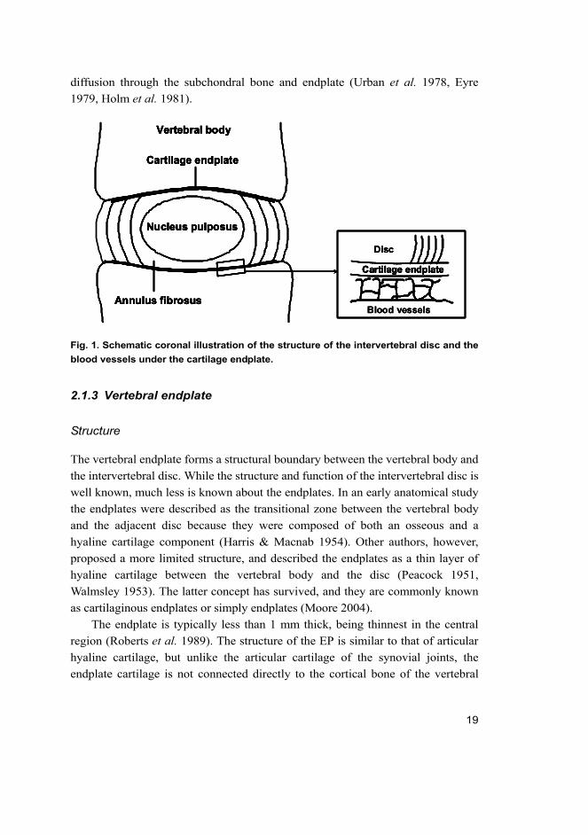
Fig. 1. Schematic coronal illustration of the structure of the intervertebral disc and the
blood vessels under the cartilage endplate.
2.1.3 Vertebral endplate
Structure
The vertebral endplate forms a structural boundary between the vertebral body and
the intervertebral disc. While the structure and function of the intervertebral disc is
well known, much less is known about the endplates. In an early anatomical study
the endplates were described as the transitional zone between the vertebral body
and the adjacent disc because they were composed of both an osseous and a
hyaline cartilage component (Harris & Macnab 1954). Other authors, however,
proposed a more limited structure, and described the endplates as a thin layer of
hyaline cartilage between the vertebral body and the disc (Peacock 1951,
Walmsley 1953). The latter concept has survived, and they are commonly known
as cartilaginous endplates or simply endplates (Moore 2004).
The endplate is typically less than 1 mm thick, being thinnest in the central
region (Roberts et al. 1989). The structure of the EP is similar to that of articular
hyaline cartilage, but unlike the articular cartilage of the synovial joints, the
endplate cartilage is not connected directly to the cortical bone of the vertebral
Cartilage endplate
Vertebral body
Nucleus pulposus
Annulus fibrosus
Cartilage endplate
Disc
Blood vessels
Cartilage endplate
Vertebral body
Nucleus pulposus
Annulus fibrosus
Cartilage endplate
Disc
Blood vessels
Cartilage endplate
Vertebral body
Nucleus pulposus
Annulus fibrosus
Cartilage endplate
Cartilage endplate
Vertebral body
Nucleus pulposus
Annulus fibrosus
Cartilage endplate
Vertebral body
Nucleus pulposus
Annulus fibrosus
Cartilage endplateCartilage endplate
Disc
Blood vessels
19
body. However, the EP does have intimate contact with the disc through the
lamellae of the inner annulus. (Moore 2004.)
The biochemical characteristics of the EP are similar compared to the IVD,
water, collagen and proteoglycans being the most important molecular
components. However, the collagen content of the EP is higher and the
proteoglycan and water contents lower compared to the adjacent disc. (Roberts etal. 1989.) Of the different collagens, type X (Aigner et al. 1998) and II (Sahlman
et al. 2001) are probably the most important in maintaining the integrity of the
endplate.
Function
The endplate has an important role in disc nutrition (Eyre 1979). At an early stage
of the development of the axial skeleton, tiny blood vessels penetrating the
endplates provide nutrition for the EP itself and for the developing IVD. These
blood vessels persist only until skeletal maturity, at which time the discs become,
for the most part, avascular. (Moore 2000.) However, in adults, the outer parts of
the annular lamellae may contain tiny collateral vessels (Moore et al. 1992,
Kauppila 1995). Apart from a sparse blood supply in the outer layer of the AF,
mature discs are almost totally reliant on diffusion of nutrients across the cartilage
endplate (Urban et al. 1978, Eyre 1979, Moore 2000). The diffusion is a selective
process based on molecular size and ionic charge of the molecules involved
(Moore 2000). Additionally, degenerative changes of the subchondral bone
(Roberts et al. 1997b) and endplate calcification (Roberts et al. 1993, Urban et al.2001) may limit the penetration of nutrients into the disc.
The endplate is also an important structure for the mechanical function of the
spine. In the course of normal physical activity, mechanical loading can alter the
shape of the disc to the extent that the endplate and the subchondral bone become
deformed (Brinckmann et al. 1983). This deformation is reversible in young
healthy endplates, but high compressive loads, when applied repeatedly, may
result in irreversible damage. The endplate appears to be susceptible to mechanical
failure as this is a weak link in the structure of the disc. (Adams et al. 2000, Moore
2000.)
The endplate acts as a physical barrier preventing the disc from bulging into
the adjacent vertebral body (Moore 2000). A Schmorl’s node is a vertical
protrusion of the contents of the nucleus into the adjacent vertebral body. They are
found in more than 70% of autopsy spines with equal frequency above and below
20
the age of 50 years, suggesting that they appear relatively early in life and may
precede disc degeneration. (Hilton et al. 1976.) Despite being relatively common,
the etiology of these changes remains a mystery.
2.2 Disc degeneration
Disc degeneration (DD) is the deterioration and remodeling of the physical and
chemical properties of the tissue with retrogressive pathologic changes in the cells
or macromolecules (Holm 1993). With increasing age comes an increased
incidence of degenerative changes, including cell death, cell proliferation, mucous
degeneration, granular change and concentric tears (Boos et al. 2002). The most
significant biochemical change to occur in disc degeneration is loss of
proteoglycan, which results in dehydration of the disc (Lyons et al. 1981).
Individual variation in DD is great, which makes it difficult to differentiate
changes that occur solely due to aging from those that might be considered
pathological (Boos et al. 2002, Holm & Holm 2004). Despite intensive research,
the sequelae of progression of catabolic changes and especially the association of
DD with pain production remain unclear.
The etiologies of disc degeneration include age, genetic inheritance, physical
loading history and impaired nutrition, all of which can weaken IVDs to such an
extent that structural failure occurs during the activities of daily living (Adams &
Roughley 2006). Several studies suggest that familial predisposition has an
important role in the pathomechanism of degenerative changes (Battie et al. 1995,
Annunen et al. 1999, Sambrook et al. 1999). Abnormal mechanical loading is also
thought to provide a pathway to disc degeneration (Adams et al. 2000). Traumatic
events (Osti et al. 1990, Kerttula et al. 2000) and heavy physical occupational
loading (Videman et al. 1995, Luoma et al. 1998) have been suspected to lead to
disc degeneration. One of the primary causes of DD is thought to be failure of the
nutrient supply to the disc cells (Nachemson et al. 1970, An et al. 2004). The
pathway from the blood supply to the nucleus cells is precarious because these
cells are supplied by capillaries that originate in the vertebral bodies, penetrate the
subchondral plate and terminate just above the cartilaginous endplate (Urban et al.1978). In this regard, EP is an important structure in transporting nutrients and
maintaining the health of the intervertebral disc.
21
2.3 Degenerative changes of the vertebral endplates and
subchondral bone
2.3.1 Morphologic changes
Morphologic changes to the endplates and subchondral bone occur with advancing
age, but also in association with disc degeneration. It is difficult to differentiate
changes that occur solely due to aging from those that are associated with
degeneration. Interestingly, however, endplate changes have been observed to
precede the intradiscal changes. (Boos et al. 2002.)
During the first decade, vascular channels through the endplate diminish
(Coventry et al. 1945, Boos et al. 2002) and the first endplate cracks are seen
(Boos et al. 2002). Vascular channels disappear by the age of 20 (Coventry et al.1945, Boos et al. 2002) and only the outer parts of the annular lamellae may
contain tiny collateral vessels (Moore et al. 1992, Kauppila 1995). At this stage,
cartilage cracks and microfractures of the adjacent subchondral bone with new
bone formation are frequently seen. From the third decade, abnormalities of the EP
are very similar to those seen in younger groups, but in increasing numbers.
Starting in the fourth decade, trabeculae in the vertebral body change in size and
pattern resulting in decreased vertebral body strength and density (Mazess 1982).
In the fourth and fifth decades, advanced degeneration with structural
disorganization of the EP is observed. During the sixth and seventh decades, tissue
alterations become most severe, including microfractures and bone sclerosis.
(Boos et al. 2002.) After this stage, scar formation, large tissue defects (Coventry
et al. 1945, Boos et al. 2002) and calcification of the endplates are seen (Bernick
& Cailliet 1982).
2.3.2 Biochemical changes
The biochemistry of the endplate is important in maintaining the integrity of the
disc (Roberts et al. 1996). The composition of the endplate has been shown to
change in degeneration with a loss of proteoglycan in the matrix (Roberts et al.1989). Therefore, where this occurs it will effectively “open up” channels through
which substances can pass into disc and vice versa. The loss of proteoglycans of
the endplate has been shown to lead to loss of proteoglycans from the nucleus.
(Roberts et al. 1996.) These authors speculated that, similarly, the flow of
substances into the disc, which would normally not occur, may be possible where
22

proteoglycans have been lost. Such substances could include cytokines or
enzymes, which might have deleterious effects on the disc. Interestingly, the loss
of proteoglycans from the endplate has been shown to precede disc degeneration
(Pearce et al. 1987). Collagen type X is probably the most important in the
endplates because it is a marker of hypertrophic chondocytes and is thought to be
involved in cartilage calcification (Aigner et al. 1998). Additionally, inactivation
of one Collagen II gene allele in young mice has been shown to lead to lower
glycosaminoglycan concentration in the endplates and thicker and more irregular
endplates that become prematurely calcified (Sahlman et al. 2001).
The bone marrow in the spine can vary in appearance according to the
patient’s age, and can be affected by infectious, inflammatory, metabolic,
neoplastic or degenerative processes. With increasing age, the amount of fatty
marrow increases. (Ricci et al. 1990, Tall et al. 2007.) Degenerative disc disease
can cause changes in the bone marrow adjacent to the cartilaginous endplates.
Three different types of changes have been described (Modic et al. 1988b). Bone
marrow edema reflects an increased amount of extracellular water and
fibrovascular changes in the bone marrow. Fatty bone marrow changes reflect the
replacement of the normal bone marrow with fat cells. The dense bone without
marrow elements has been thought to correlate with subchondral bone sclerosis.
(Modic et al. 1988a, Modic et al. 1988b.)
2.4 Magnetic resonance imaging techniques of vertebral bone
marrow
Magnetic resonance imaging is the modality of choice in detecting bone marrow
abnormalities (Daffner et al. 1986, Modic et al. 1988b, Vogler & Murphy 1988,
Ricci et al. 1990, Vanel et al. 2000, Tall et al. 2007). Differentiation of red and yel-
low marrow with other imaging modalities, such as plain film radiography, ultra-
sound (US), computed tomography (CT) or scintigraphy, has not been proved
(Modic et al. 1988b, Vogler & Murphy 1988, Lusins et al. 1998). Additionally, the
lack of ionizing radiation and multiplanar imaging capability of MRI makes it
superior compared to the other imaging modalities in detecting bone marrow dis-
eases.
Standard sequences to image vertebral bone marrow include T1- and
T2-weighted fat saturation or STIR (short tau inversion recovery) sequences
(Mirowitz et al. 1994, Tall et al. 2007). Normal marrow is composed of an
intermixture of red hematopoietic marrow, yellow fatty marrow and trabeculae in
23

varying proportions, based on patient’s age and other factors. Its normal
appearance in MRI reflects this combination (Ricci et al. 1990, Tall et al. 2007).
There is superb differentiation between red and yellow bone marrow in
T1-weighted sequences. In T1-weighted images the yellow marrow is hyperin-
tense in signal intensity, in contrast with the relatively decreased signal of red mar-
row. These differences in signal intensity are a direct reflection of the differences
in fat/water content within red and yellow marrow. (de Roos et al. 1987, Modic etal. 1988b, Ricci et al. 1990, Tall et al. 2007.) Both T1-weighted spin echo (SE)
and fluid-attenuated inversion recovery (FLAIR) sequences are used to image the
vertebral bone marrow (Melhem et al. 1997, Erdem et al. 2005).
T2-weighted fast spin echo (FSE) with fat saturation and STIR sequences
have similar sensitivity to detect bone marrow pathology. The clinical advantages
are based on marked fat suppression. Both sequences demonstrate high contrast
and conspicuousness for the depiction of most types of bone marrow pathology.
The disadvantage of T2-weighted FSE is its dependence on excellent magnetic
field homogeneity for adequate fat suppression. (Mirowitz et al. 1994, Tall et al.2007.)
Newer imaging techniques, such as diffusion-weighted (Baur et al. 2001,
Zhou et al. 2002) and gradient echo (Erly et al. 2006) imaging, have been
developed. Both techniques may be useful in differentiating acute benign
compression fractures from malignant infiltration and pathologic fractures.
Intravenous gadolinium administration is useful for assessing the paraspinal soft
tissues and the enhancement of the disc (Van Goethem et al. 2000), but it is not
essential for routine assessment of the vertebral bone marrow (Tall et al. 2007).
2.5 Vertebral degenerative bone marrow (Modic) changes
2.5.1 Definition
Vertebral degenerative bone marrow (Modic) changes are signal intensity changes
visible in MRI. These changes occur adjacent to the cartilaginous endplates of the
degenerative intervertebral discs. (de Roos et al. 1987, Modic et al. 1988b.) Bone
marrow signal intensity changes in the vertebral bodies were first reported by de
Roos et al. (1987). The classification of changes was provided by Modic et al.(1988b) based on 474 patients, most of whom had chronic low back pain (Table 1).
The histological correlation was based on six operative specimens. These authors
described two types of changes: Modic type I changes (low SI in T1W and high SI
24
in T2W images) indicated an ongoing active degenerative process and
demonstrated vascularized fibrous tissue within the bone marrow. Modic type II
changes (high SI in T1W and T2W images) were more stable during a three-year
follow-up and reflected fatty replacement of the bone marrow. (Modic et al.1988b.) Type III lesions were found later (low SI in T1W and T2W images), and
they are thought to associate with endplate sclerosis in plain film radiography
(Modic et al. 1988a). The histological nature of type III changes remains
undetermined. The detection of different Modic types with other imaging
modalities, such as plain film radiography, ultrasound, computed tomography or
scintigraphy, has not been proved (Modic et al. 1988b, Lusins et al. 1998).
Mixed Modic lesions (type I/II and type II/III) have also been identified.
Braithwaite et al. (1998) suggested that Modic changes can convert from one type
to another and that they all present different stages of the same pathological
process.
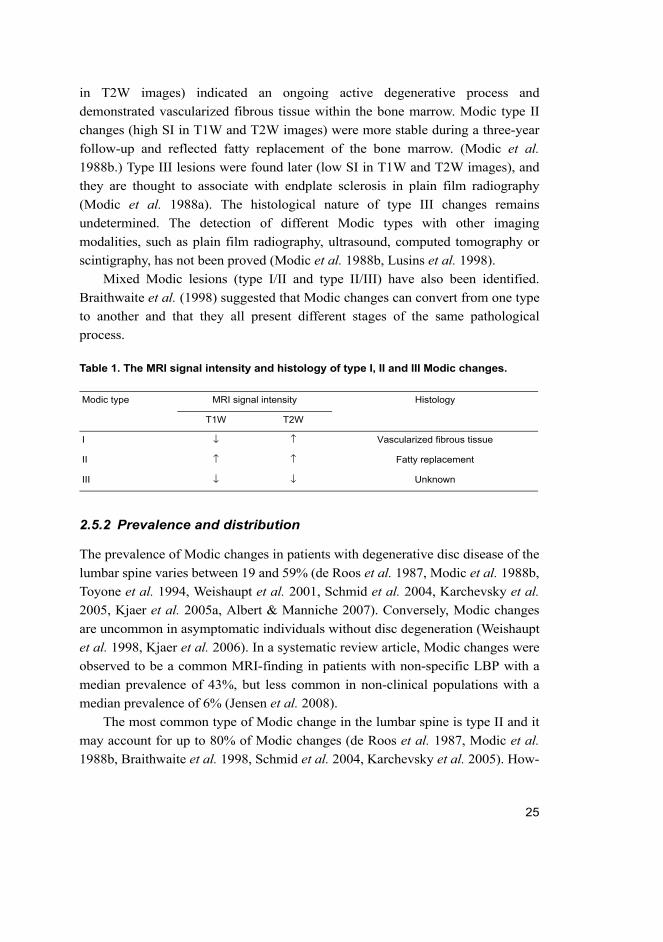
Table 1. The MRI signal intensity and histology of type I, II and III Modic changes.
2.5.2 Prevalence and distribution
The prevalence of Modic changes in patients with degenerative disc disease of the
lumbar spine varies between 19 and 59% (de Roos et al. 1987, Modic et al. 1988b,
Toyone et al. 1994, Weishaupt et al. 2001, Schmid et al. 2004, Karchevsky et al.2005, Kjaer et al. 2005a, Albert & Manniche 2007). Conversely, Modic changes
are uncommon in asymptomatic individuals without disc degeneration (Weishaupt
et al. 1998, Kjaer et al. 2006). In a systematic review article, Modic changes were
observed to be a common MRI-finding in patients with non-specific LBP with a
median prevalence of 43%, but less common in non-clinical populations with a
median prevalence of 6% (Jensen et al. 2008).
The most common type of Modic change in the lumbar spine is type II and it
may account for up to 80% of Modic changes (de Roos et al. 1987, Modic et al.1988b, Braithwaite et al. 1998, Schmid et al. 2004, Karchevsky et al. 2005). How-
Modic type MRI signal intensity Histology
T1W T2W
I ↓ ↑ Vascularized fibrous tissue
II ↑ ↑ Fatty replacement
III ↓ ↓ Unknown
25
ever, this has recently been questioned by two authors suggesting that Modic type
I change may be a more common than type II in the lumbar spine (Weishaupt et al.2001, Kjaer et al. 2005a). Modic changes most likely occur at L4–5 and L5–S1
and associate with degenerative disc disease (Modic et al. 1988b, Karchevsky etal. 2005, Luoma et al. 2008). In addition to disc degeneration, Modic changes are
also observed to occur in a lumbar segment with a disc herniation (Schmid et al.2004, Albert & Manniche 2007) or segmental instability (Toyone et al. 1994).
Most studies describe Modic changes occurring in the lumbar spine. However,
these changes have also been reported in cervical (Peterson et al. 2007) and
thoracic spine (Girard et al. 2004). A prevalence of 16% in the cervical spine has
been observed, which is similar to previously reported values of the lumbar spine.
Of specific Modic types, type I changes predominated in the cervical spine, which
may be related to a greater segmental mobility as compared with the lumbar spine.
(Peterson et al. 2007.) In the thoracic spine, a prevalence of 2.5% has been
observed, which may be due to the relative lack of mobility compared with the
other spinal regions (Girard et al. 2004).
2.5.3 Determinants
It has been shown that the risk of disc degeneration is determined by a multitude of
factors including age, genetic inheritance and loading history (Sambrook et al.1999, Adams & Roughley 2006). However, there is only limited information about
the determinants of Modic changes.
Modic changes are positively associated with age, supporting their
degenerative etiology (Modic et al. 1988b, Karchevsky et al. 2005). Conversely,
Modic changes are uncommon in younger individuals. In a study of 439
thirteen-year-old children, the prevalence of changes was only 0.5% (Kjaer et al.2005b). In addition, Modic changes have been shown to be associated with male
gender (Karchevsky et al. 2005).
Obesity has been suggested as a risk factor for disc degeneration, but the
results have remained controversial (Heliövaara 1987, Riihimäki et al. 1990,
Harrington et al. 2001, Liuke et al. 2005). In a patient study, Modic changes were
associated with increasing weight, but not with BMI (Karchevsky et al. 2005).
Recently, overweight in combination with hard physical work was significantly
associated with the prevalence of Modic changes (Leboeuf-Yde et al. 2008). In
another population-based cohort, exposure to high-level physical activity at leisure
time did not differ between subjects with “Modic changes and disc degeneration”
26

vs. those with “only disc degeneration”. However, exposure to occupational physi-
cal load was significantly higher in the subgroup of “Modic changes and disc
degeneration” compared to “only disc degeneration” group. (Kjaer et al. 2006.)
Smoking has been suspected to carry deleterious effects on the intervertebral
discs. According to a systematic review, smoking is associated with LBP
(Goldberg et al. 2000). Smoking is a risk factor for atherosclerosis, which is
assumed to cause disc degeneration through diminished nutrition (Kauppila et al.1994) and diffusion (Kurunlahti et al. 2001). Although an association between
smoking and disc degeneration was observed in an earlier study on Finnish twins
(Battie et al. 1991), the association between smoking and disc degeneration was
not confirmed in a later study (Battie et al. 1995). There is only one study of the
effect of smoking on Modic changes. In the Danish study, self-reported hard
physical work in combination with heavy smoking was strongly associated with
Modic changes (Kjaer et al. 2006).
2.5.4 Differential diagnosis
There are many conditions that need to be considered in the differential diagnosis.
Spondylodiscitis, spinal neoplasms and inflammatory spondyloarthropathias may
lead to signal intensity alterations that may mimic Modic type I changes. In
addition, the possibility of a signal change around Schmorl’s node must always be
kept in mind.
The involvement of the disc and the bony endplate is critical when
differentiating degenerative Modic changes from spinal infections. The disc signal
is often reduced in Modic changes compared to increased signal intensity in
T2-weighted sequences in spondylodiscitis. In Modic changes bony endplates are
preserved rather than indistinct or destroyed as seen in an infective process.
Contrast enhancement of the disc and adjacent bone may occur in both conditions.
An important finding is the presence of paravertebral inflammatory tissue, which
may considerably help in establishing the diagnosis of spondylodiscitis. (Van
Goethem et al. 2000, Ledermann et al. 2003, James & Davies 2006.) In addition to
these imaging study findings, clinical symptoms and laboratory tests can help to
differentiate the two conditions (James & Davies 2006). Local spinal pain is the
most common presenting symptom in spondylodiscitis and is seen in up to 95% of
patients. In addition, C-reactive protein (CRP) is thought to be the most reliable
laboratory test, being increased in up to 100% of cases at the time of diagnosis.
(Wirtz et al. 2000.)
27
In spinal neoplasms bone marrow is typically replaced with edema. However,
tumor spread rarely affects the disc, endplate and subchondral bone marrow. Thus,
a well-defined endplate with normal signal within the disc favors a neoplastic
process. Tumors can also cause bony destruction via osteolysis, which is not
typically seen in degenerative disease. (Modic et al. 1988b, James & Davies
2006.)
An inflammatory spondyloarthropathy such as ankylosing spondylitis may
also present diagnostic difficulties. However, the anatomical distribution of
changes is different and the distinction is not usually difficult to make. The most
prominent feature of ankylosing spondylitis consists of an enthesitis at the
insertion of the annulus fibrosus-longitudinal ligament complex. Inflammatory
tissue leads to the destruction of the attachment of ligament to bone (anterior
vertebral corners) resulting in a superficial erosion and edema. (Jevtic et al. 2000,
James & Davies 2006.) Conversely, Modic changes usually affect the whole
antero-posterior length of the vertebral subchondral bone (Modic et al. 1988b).
Schmorl’s nodes have a typical morphology compared to degenerative Modic
changes. They are usually characterized by localized defects (low T1W and high
T2W signals) in bony endplates (Williams et al. 2007), whereas Modic changes
tend to be linear bone marrow changes and parallel to the cartilaginous endplates
(Modic et al. 1988b).
2.5.5 Pathogenesis
The etiology of Modic changes is unknown. However, there are three possible
hypotheses for the pathogenesis of Modic changes: a biomechanical, biochemical
(these two may be interrelated) and infective hypothesis.
The endplate is a weak link of the spine in compression, and always fails
before the intervertebral disc, even if the latter is injured before loading
commences (Brinckmann 1986). This can result in morphologic changes in the
bone marrow including microfractures and structural disorganization (Boos et al.2002). Histologically, type I Modic changes demonstrated disruption and fissures
of the endplates (Modic et al. 1988b). In a review article, Modic and Ross (2007)
concluded that the altered signal intensity in MRI is not the causal pathologic
process, but rather a reflection of the causal process, which is some type of
biomechanical stress or instability. Further evidence in favor of the biomechanical
theory comes from recent animal studies, where injuries to the disc induced
changes in the adjacent vertebrae with subsequent bone marrow depletion and
28
regeneration (Malinin & Brown 2007, Ulrich et al. 2007). In the study of Toyone
et al. (1994), 70% of patients with Modic type I change and only 16% of those
with type II were found to have segmental hypermobility in the dynamic
flexion-extension MR images. They suggested that Modic changes may be a
marker of segmental instability. This theory has been supported by indirect
evidence from lumbar fusion studies, in which nonunion was predominantly
associated with the persistence of type I Modic changes (Lang et al. 1990,
Buttermann et al. 1997).
Crock (Crock 1970, Crock 1986) proposed the concept of “internal disc
disruption”, suggesting that repeated trauma to the intervertebral disc results in the
production of chemical substances in the nucleus pulposus. Diffusion of such toxic
chemicals through the vertebral endplate could then result in a local inflammatory
reaction (Crock 1970). Ohtori et al. (2006) found that the endplates of patients
with Modic type I or type II changes had more protein-gene-product (PGP) 9.5
immunoreactive nerve fibers and tumor necrosis factor-alpha (TNF-α)
immunoreactive cells than those with normal endplates in MRI. Furthermore, the
number of TNF immunoreactive cells in the endplates with Modic type I changes
was higher than those with type II (Ohtori et al. 2006). In addition, Rannou et al.(2007) found high-sensitivity C-reactive protein (hsCRP) to be increased in
patients with chronic low back pain and Modic type I change. The biochemical
mechanism is also supported by the fact that the patients with chronic LBP and
predominantly Modic type I changes tended to have better short-term efficacy
following intradiscal steroid injections than those with predominantly type II
changes (Fayad et al. 2007).
Recent experimental findings suggest that biomechanical and biochemical
pathways may be interrelated. Ulrich et al. (2007) showed that repeated injury to
the disc causes persistent inflammation. In addition to elevated cytokine
production [interleukin-1 (IL-1), IL-8 and TNF-α] in the disc, they observed
evidence of vertebral remodeling. Bone marrow spaces adjacent to the degenerated
intervertebral discs were filled with granulation tissue consisting of new vessels,
fibroblasts and osteoblasts that lined thickened trabecular elements. These features
were analogous to those reported by Modic et al. (1988b) and were similar to
Modic type I bone marrow changes.
The infective hypothesis has been suggested by Albert et al. (2008b), who
speculated that low virulent bacterial infection may play a role in the pathogenesis
of Modic changes. They observed in an uncontrolled study that the clinical effect
of antibiotic treatment was large in a group of patients with Modic changes
29

suffering from persistent LBP following a disc herniation. Previously, Stirling etal. (2001) have found a significant positive culture rate (19 of 36; 53%), especially
of Propionibacterium acnes and Corynebacterium propinquum in surgical lumbar
disc herniation specimens. These bacteria are found on the skin and in oral cavities
in all individuals. They frequently invade the circulatory system (for example
during tooth brushing), where they do not present any health risks due to the
aerobic environment in the bloodstream. (Roberts et al. 1997a, Olsen 2008.)
However, Albert et al. (2008a, 2008b) hypothesized that anaerobic bacteria enter
the disc through the radial tears of the annulus fibrosus causing a low virulent and
slowly developing infection of the disc. As intervertebral discs are avascular, they
constitute an ideal environment for the growth of anaerobic bacteria, and local
inflammation in the adjacent bone may due to the production of cytokines.
However, a recent study by Wedderkopp et al. (2009) found no evidence of
bacteria in vertebrae with Modic type I changes. The affected vertebra was
biopsied from 24 consecutive patients by a strict aseptic procedure. None of the
biopsies yielded growth of anaerobic bacteria. No randomized study of the effect
of antibiotic treatment on Modic changes has been performed so far.
2.5.6 Natural course
In the original study, Modic et al. (1988b) observed that all six patients with a type
I change converted to a type II or normal bone marrow over a 14- to 36-month
interval, whereas type II changes did not alter with time.
The dynamic process of Modic type I changes was recently confirmed as it
was observed that only 11% of type I changes remained stable during an 18- to
72-month follow-up period (Luoma et al. 2008). In another study, 52% of type I
changes converted into type II and 40% increased in size, while only 8% of type I
changes remained stable during a 12- to 72-month follow-up period (Mitra et al.2004).
Mixed Modic types (I/II and II/III) were found later and it was suggested that
all Modic changes present different stages of the same degenerative process
(Braithwaite et al. 1998). Mixed changes are assumed to develop before
conversion to one of the true Modic types (Vital et al. 2003).
30

2.5.7 Association with low back pain
Several studies have shown the occurrence of a wide spectrum of disc
abnormalities, including disc degeneration, disc bulging, disc protrusion, and
annular tears, in a substantial percentage of healthy volunteers (Boden et al. 1990,
Jensen et al. 1994, Boos et al. 1995, Stadnik et al. 1998). In contrast, disc
extrusions, nerve root compression and Modic changes are rare in asymptomatic
volunteers (Jensen et al. 1994, Boos et al. 1995, Weishaupt et al. 1998, Kjaer et al.2006). In recent years, Modic changes have come into focus in the search for the
source of back pain symptoms. The association of Modic changes with LBP has
been studied in relation to both symptom history and discography of the affected
levels.
Based on clinical symptoms, there is evidence that Modic type I changes may
be associated with LBP. Toyone et al. (1994) stated that 73% of patients with type
I change, but only 11% of patients with type II had LBP. In a longitudinal
follow-up of LBP patients, a positive trend was found between the conversion of
type I to type II change and symptom improvement (Mitra et al. 2004). In the
Danish study of general population, Modic changes associated strongly with LBP
during the past year. When they analyzed type I changes separately, the strength of
the association increased. (Kjaer et al. 2006.) Recently, Albert and Manniche
(2007) reported a strong association between Modic changes and LBP, as 60% of
patients with Modic changes but only 20% of those without changes had LBP.
They also showed that type I changes were more strongly associated with LBP
than type II changes. The association between Modic changes and LBP has not
only been found among patients with LBP, but also in the general (Kjaer et al.2005a) and working (Schenk et al. 2006) populations.
The correlation of Modic changes with discogenic pain, i.e. pain provoked by
discography, is controversial. Braithwaite et al. (1998) found high specificity and
positive predictive value for all Modic types as an indicator of a painful disc at
discography. Weishaupt et al. (2001) showed that when only moderate and severe
endplate abnormalities of both type I and II were considered abnormal, all injected
discs at discography caused concordant pain. However, also negative associations
between Modic changes and pain provocation during discography of the affected
levels have been reported (Sandhu et al. 2000, Kokkonen et al. 2002).
The reasons why Modic changes may be painful are not known. Brown et al.(1997) studied specimens of vertebral endplates, intervertebral discs and adjacent
cancellous bone obtained during anterior discectomy and fusion from patients with
31

degenerative disc disease and low back pain. They observed cracks and defects in
the EPs with increased vascular density and number of sensory nerve fibers. They
concluded that such changes could be a source of LBP in patients with
degenerative disc disease. (Brown et al. 1997.) Increased levels of IL-6, a
proinflammatory cytokine, have been detected in the intervertebral discs in
patients with LBP. In addition, the same authors reported a significant increase in
the number of tumor necrosis factor immunoreactive cells in the endplates with
Modic changes, especially in type I changes. They suggested that the pain may
originate from the endplates in patients with Modic changes. (Ohtori et al. 2006.)
In a randomized controlled trial, infliximab (a monoclonal antibody against
TNF-α), was of similar efficacy as placebo in the treatment of disc
herniation-induced sciatica (Korhonen et al. 2006). However, infliximab tended to
be more efficacious when a Modic change co-localized at the symptomatic
herniation level.
Another source of pain may be the intervertebral disc. Immunoreactive nerves
have also been shown to be present in degenerated discs (Bogduk et al. 1981,
Freemont et al. 1997). A positive correlation between disc degeneration and back
pain has been reported (Erkintalo et al. 1995, Luoma et al. 2000, Waris et al.2007), but the results are controversial. Additionally, disc degeneration has a very
high prevalence in the asymptomatic population (Boden et al. 1990, Modic & Ross
2007). Interestingly, it has been observed that LBP symptoms were more
pronounced in subjects with Modic changes and disc degeneration compared to the
subjects with only disc degeneration (Kjaer et al. 2006).
32

3 Purpose of the study
The purpose of the present study was:
1. To investigate the determinants of Modic changes, and whether Modic
changes and disc degeneration share common etiological factors.
2. To analyze the prevalence, extent, and natural course of Modic changes over a
three-year follow-up period.
3. To study the association of Modic changes with low back and sciatic pain in a
sample of middle-aged male workers.
4. To evaluate the presence of endplate sclerosis in different types of Modic
changes and to assess the capability of MRI in detecting endplate sclerosis
within these changes.
33

34

4 Materials and methods
The materials and methods are described in more detail in the original articles (I–
IV).
4.1 Study population
4.1.1 Occupational sample of male train engineers and sedentary paper mill workers (I and III)
The study population consisted of 228 males with a mean age of 47 years (range
36–56 years) at the time of enrolment. The occupational sample, train engineers
(N=159), worked for the Finnish state railways, and had a mean of 21 years (range
5–31 years) of exposure to whole-body vibration. They were all full-time train
drivers with approximately five hours of daily exposure to whole-body vibration
composed of both vertical and horizontal components. They were all from
Northern Finland, ensuring that they had been operating the same kinds of
locomotives and had similar exposure to vibration. The occupational control group
consisted of 69 paper mill workers from the same geographical region with
sedentary jobs and no occupational exposure to vibration.
The study design was developed by Drs. Jaro Karppinen, Antero Natri, Leena
Alakokko, Osmo Tervonen and Simo Taimela. Dr. Antero Natri was responsible
for the arrangements with the Finnish state railways. The study questionnaires
were delivered to all the train engineers working in Northern Finland. Participation
was on a voluntary basis, but 73% of the train engineers participated in the study.
The occupational controls were recruited from workers of a paper mill and a
chemical factory. They had to be of similar age-range and educational level as the
train engineers, with sedentary work and no occupational driving at any point of
their working history. Occupational health care units of the respective work places
distributed the study criteria and contact data via their intranets, and the eligible
controls could contact the study center (Oulu Deaconess Institute) on a voluntary
basis. Institutional Review Board approval and signed, informed consent from
each patient were obtained before MR imaging. The study was approved by the
ethical committee of Oulu University Hospital.
35
4.1.2 Patients (II and IV)
Study II consisted of 60 unoperated sciatica patients with a mean age of 45 years
(range 23–76 years) with unilateral pain below the knee lasting from three weeks
to six months. They had been referred to a randomized controlled trial evaluating
the efficacy of periradicular nerve root infiltration for sciatica (Karppinen et al.2001a). At three years (range 2.6–4.0), these patients were called for a
prescheduled follow-up MRI assessment. The study protocol was approved by the
ethical committee of Oulu University Hospital.
Study IV included 70 patients with a mean age of 48 years [(range 17–75
years) at the time of the first imaging] who underwent lumbar CT and MRI
examinations between January 2004 and December 2006 and met the inclusion
criteria, and were included after a review of a digital database of a radiology
record system. Patients were included if a) both CT and MRI examinations were
done within six months from the first imaging, b) MR imaging was performed
with a 1.5 T system, and c) CT examination was performed with 16-row multislice
CT at our own institution. Two patients with focal metastatic lesions and two
patients with lumbar arthrodesis were excluded. Additionally, one technically
suboptimal MRI study was excluded. The institutional review board did not
require advance approval or individual informed consent, as only patients’ images
were reviewed.
4.2 Assessments (I and III)
In study I, the self-administered questionnaire included items of occupational
history, smoking, alcohol consumption and leisure time physical activity. In
addition, body weight (kg), height (m), waist circumference (cm), and body fat
percentage (%) were measured. Body Mass Index (BMI) was calculated, and
percentage of fat and lean mass was assessed using bioimpedance equipment
(Bodystat 1500, Bodystat Ltd., Douglas, Isle of Man, UK). The table of the
distribution of selected determinants may be seen in the original article of study I.
In study III, the participants were asked to report the number of previous LBP
and leg pain episodes lasting at least 14 days, and to assess the intensity of the pain
both during the past week and the past three months with a 10-cm visual analog
scale (VAS). Additionally, dichotomous questions were used to assess whether
they had experienced LBP ever (vs. never), and whether they had LBP today (at
the day of assessment) or not.
36
4.3 Magnetic resonance imaging
MRI examinations (I–IV) of the lumbar spine were performed using a 1.5 T unit
(GE Signa Twinspeed, General Electric Medical Systems, Milwaukee, WI) and a
Phased Array CTL Spine Coil (USA Instruments, Aurora, OH).
Imaging sequences in studies I and III were sagittal T1W [repetition time
(TR)/echo time (TE) 1809/18 msec] fluid-attenuated inversion recovery (FLAIR)
and sagittal T2W (3960/116) fast spin echo (FSE) sequences. Additionally, study
III included axial T2W (3960/116) FSE sequences. The inversion recovery time
for T1W images was 660 msec, and the number of excitations for both T1W and
T2W images was four. Echo train length (ETL) for T1W images was eight, for
T2W sagittal images 29 and for T2W axial images 26. The image matrix was 448
x 192 for T1W images, 448 x 224 for T2W sagittal images and 256 x 160 for T2W
axial images. Field of view (FOV) for sagittal images was 28 x 28 cm and for axial
images 18 x 18. Slice thickness was 4 mm and interslice gap 1 mm.
At baseline, the sequences in study II were T1W (640/14) axial spin echo (SE)
and T2W (4000/95) sagittal FSE images. At three years, the imaging sequences
included T1W (400/14) sagittal SE images and T2W (4000/95) sagittal FSE
images. The technical specifications included ETL of 16, a slice thickness of 4 mm
with interslice gaps of 1.0 and 0.5 mm, a FOV of 36 and 20 cm, a matrix of
256x128 and 256x192, and a number of excitations (NEX) of 1 and 2 for sagittal
and axial images, respectively.
Study IV included T1W (2060/21) sagittal FLAIR sequences with an
inversion time (TI) of 860 ms, echo train length of 6 and the number of
acquisitions of 4. The matrix size was 256 x 224, FOV 28 x 28 cm, slice thickness
4 mm and intersection gap 1 mm. T2W (4000/118) sagittal FSE images were
obtained with an ETL of 27 and the number of acquisitions of 4. The matrix size
was 448 x 224, FOV 28 x 28 cm, slice thickness 4 mm, and intersection gap 1 mm.
4.4 Computed tomography imaging (IV)
The lumbar CT imaging was performed using a 16-slice CT scanner (GE
LightSpeed Pro 16; GE Healthcare, Milwaukee, Wisc., USA) with a detector
configuration of 16 x 1.25 mm. A standard lumbar spine protocol with a tube
voltage of 120 kV, tube current of 100–650 mA and rotation time of 0.8 s was
used. Automatic tube current modulation based on patient size and X-ray
37
attenuation was used. The slice thickness and reconstruction interval was 1.25 mm
and 0.625 mm, respectively.
4.5 Image analysis
In all studies (I–IV) the classification of Modic changes was carried out at a
workstation on the basis of the T1W and T2W MR images based on the five
midsagittal planes. Both the upper and lower endplates at each disc level were
graded separately into Modic types MI (low SI in T1W and high SI in T2W
images), MII (high SI in T1W and T2W images) and MIII (low SI in T1W and
T2W images) (Modic et al. 1988a, Modic et al. 1988b), and mixed types MI/II and
MII/III (Braithwaite et al. 1998). In studies I and III, Modic types I and I/II were
grouped together, as all lesions containing type I change are assumed to indicate a
more active process. Similarly, Modic types II and II/III were grouped together, as
they are thought to manifest a more stable and chronic degenerative process. In
study II the involvement of one or both endplates, antero-posterior localization,
maximal vertical depth (mm), and extent (involvement of the endplate area as
quadrants) of Modic changes were also analyzed.
The degree of disc degeneration was graded (studies I and IV) on the
T2-weighted sagittal MR images by using the grading system of Pfirrmann et al.(2001). In grade I degeneration the nucleus pulposus is homogenously
hyperintense and clearly distinct from the hypointense outer annular fibers. In
grade II degeneration the nucleus pulposus is inhomogeneous and horizontal
hypointense bands may be present. In grade III degeneration the inner parts of the
disc are inhomogeneous and have intermediate signal intensity. In grade IV
degeneration the distinction between the inner and outer parts of the disc is lost,
and the inner parts of the disc have intermediate or low signal intensity. In grade V
degeneration the disc is collapsed. In study II, disc degeneration was graded as
normal (no signal changes), grade 1 (slight decrease in signal intensity of the
nucleus on T2W images), grade 2 (hypointense nucleus pulposus on T2W images
with normal disc height), and grade 3 (hypointense nucleus pulposus on T2W
images with disc space narrowing) (Schneiderman et al. 1987).
In study II, disc herniations were classified as normal, bulge (a symmetrical
extension of the peripheral annulus beyond the margins of the endplates),
contained herniations (a focal extrusion of disc material not penetrating the
posterior longitudinal ligament), uncontained herniations (an extrusion of disc
material through the posterior longitudinal ligament) and sequestration (a
38
herniated disc fragment not in contact with the parent disc) (Karppinen et al.2001b).
To exclude the MRI findings that may be symptomatic (study III), any
presence of disc herniation, nerve root compromise and central spinal stenosis
were recorded. The extent of disc herniation was graded as normal, bulging (a
circumferential, symmetric disc extension around the vertebral borders),
protrusion (a focal or asymmetric extension of the disc beyond the vertebral
border, with the disc origin broader than any other dimension of the protrusion) or
extrusion (a more extreme extension of the disc beyond the vertebral border, with
the base against the disc of origin narrower than the diameter of the extruding
material itself and a connection between the material and the disc of origin)
(Fardon & Milette 2001). The neural compromise was classified as no
compromise, nerve root contact, or compression (Pfirrmann et al. 2004). The
presence of central spinal stenosis (dural sac cross-sectional area <75 mm2 at one
level or <100 mm2 at two or more levels) was defined according to the criteria of
Willen et al. (1997).
Endplate sclerosis (IV) was visually evaluated from the CT scans by
comparing the MR images and sagittal reconstructed CT scans on a workstation.
The presence of endplate sclerosis was defined as yes or no.
To study the capability of MRI in detecting endplate sclerosis within Modic
changes (study IV), the endplate signal intensity from the MR images and
Hounsfield units (HU) from the CT scans were determined from affected
endplates. The largest possible elliptical region of interest (ROI) was fitted within
the area displaying a Modic change. For each patient, the area of the ROI was the
same in the T1W and T2W MR images and the CT scans. The mean ROI was 60.4
mm2 (range 30–112 mm2). To normalize the T1W and T2W signal intensity and
the HU values for affected endplates among the patients, the T1, T2 and HU ratios
were calculated (T1 ratio=T1W signal intensity for an affected endplate/T1W
signal intensity for a normal vertebra, T2 ratio=T2W signal intensity for an
affected endplate/T2W signal intensity for liquor signal intensity, HU ratio=HU
for an affected endplate/HU for a normal vertebra).
39

4.6 Statistical analysis
4.6.1 Reliability of Modic changes (I, II and III)
Kappa statistics was used to establish interobserver and intraobserver reliabilities
of Modic changes (studies I, II and III) (Landis & Koch 1977). In studies I and III,
the interobserver kappa values for the types of Modic changes (N=228 subjects)
showed almost perfect agreement between the radiologists, kappa values of
different disc levels ranging from 0.85 to 1.00 (mean 0.94). Intraobserver kappa
values by disc level ranged from 0.62 to 1.00 (Mean 0.84) (N=120 subjects). In
study II, evaluation of Modic changes showed substantial agreement between the
radiologists, with a kappa value of 0.64 (N=60) at baseline and at three years.
Intraobserver agreement was 0.90 (N=60).
4.6.2 Evaluation of determinants (I)
Logistic regression analysis was used to evaluate the association between selected
determinants and 1) type of Modic changes and 2) severe disc degeneration. Odds
ratios (ORs) with 95% confidence intervals (CI) were calculated per an increment
of one standard deviation unit for each continuous explanatory variable. Analyses
were carried out for all lumbar levels combined and separately for changes located
at L5–S1.
All subjects were included in the analyses of Modic changes, i.e. either type I
or type II at any level (N=228). Subjects who had both type I and type II change at
the same or separate level (N=23) were excluded when analyzing determinants of
individual Modic types. In the analyses of Modic changes located at L5–S1,
subjects who did not have Modic change at L5–S1 but had any Modic change at
the upper levels were excluded (N=42). The reference group in the analyses of
Modic changes consisted of 100 subjects without any Modic changes, whereas in
the analyses of severe disc degeneration the reference group consisted of 163
subjects with at most grade IV degeneration. The age-adjusted analyses were first
done separately for each single determinant. Based on these analyses, multivariate
analysis was conducted. For the multivariate analysis, only the significant
determinants (for either Modic changes or severe disc degeneration) were
included.
40
4.6.3 Natural course of Modic changes (II)
The characteristics of Modic changes were illustrated by frequency tables and
crosstabulations. The paired t-test and Wilcoxon signed ranks test were used to
analyze the progression in size of Modic changes.
4.6.4 Association of Modic changes with low back and sciatic pain (III)
Logistic regression analysis was used for the evaluation of pain on the type and
extent of Modic changes. Odds ratios with 95% confidence intervals were
estimated per an increment of one standard deviation unit in each independent
variable.
Analyses were done 1) separately for each lumbar level and 2) for all lumbar
levels combined. In the classification of Modic changes, Modic type I was
prioritized over type II if the subject had separate types of Modic 1) at any single
level or 2) at separate levels when lumbar levels were combined. Similarly,
extensive Modic change was prioritized over minimal size change. The analyses
were adjusted for age and done separately for each pain variable. In a separate
analysis, subjects (N=8) with a nerve root compression due to a disc extrusion or
central spinal stenosis were excluded because these MRI findings may be
symptomatic (Boos et al. 1995, Beattie et al. 2000, Atlas et al. 2005) Univariate
analysis of variance was used for checking the effect of confounding upper level
Modic changes while analyzing the association of pain with Modic changes at L5–
S1.
4.6.5 Comparison of Modic changes and endplate sclerosis (IV)
The characteristics of the Modic changes were illustrated with frequency tables.
The difference in age distribution in the study population was evaluated using
Student’s t test. Variations in the T1, T2 and HU ratios were shown graphically as
scatter plots. The significance of the difference between the T1, T2 and HU ratios
for sclerotic and non-sclerotic Modic changes was determined using a
Mann-Whitney test.
41

42
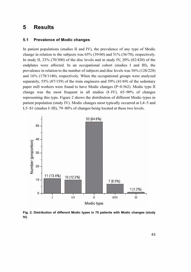
5 Results
5.1 Prevalence of Modic changes
In patient populations (studies II and IV), the prevalence of any type of Modic
change in relation to the subjects was 65% (39/60) and 51% (36/70), respectively.
In study II, 23% (70/300) of the disc levels and in study IV, 20% (82/420) of the
endplates were affected. In an occupational cohort (studies I and III), the
prevalence in relation to the number of subjects and disc levels was 56% (128/228)
and 16% (178/1140), respectively. When the occupational groups were analyzed
separately, 55% (87/159) of the train engineers and 59% (41/69) of the sedentary
paper mill workers were found to have Modic changes (P=0.562). Modic type II
change was the most frequent in all studies (I–IV), 65–90% of changes
representing this type. Figure 2 shows the distribution of different Modic types in
patient population (study IV). Modic changes most typically occurred at L4–5 and
L5–S1 (studies I–III), 79–80% of changes being located at these two levels.
Fig. 2. Distribution of different Modic types in 70 patients with Modic changes (study
IV).
43
5.2 Determinants (I)
5.2.1 Determinants of Modic changes
Modic changes were age-dependent [odds ratio (OR) 1.56; 95% confidence
interval (CI) 1.21–1.95]. When all the determinants were included in the univariate
age-adjusted analyses, none of the analyzed determinants were associated with
Modic type I changes, whereas both weight-related factors (BMI and waist
circumference) were associated with type II changes at L5–S1 (OR 1.42; 95% CI
1.03–1.86 and OR 1.48; 95% CI 1.11–1.89, respectively). Similar results were
obtained when all lumbar levels were combined. Occupation (train engineers vs.
factory workers), duration of exposure to whole-body vibration (years), body fat
percentage or alcohol consumption were not significantly associated with Modic
changes. The table of the univariate age-adjusted associations of the determinants
of Modic changes and severe disc degeneration may be seen in the original article
of study I.
In the multivariate model (Table 2), in addition to age, BMI was associated
significantly with type II changes at L5–S1. Waist circumference was also
significant (OR 1.18; 95% CI 1.05–1.32), but due to collinearity of these two
weight-related factors they could not be included in the model simultaneously.
Exposure to whole-body vibration was of borderline significance.
5.2.2 Determinants of severe disc degeneration
Severe disc degeneration was age-dependent (OR 1.46; 95% CI 1.13–1.82). When
all the determinants were included in the univariate age-adjusted analyses, the
duration of exposure to whole-body vibration (OR 1.41; 95% CI 1.05–1.78) and
being a train engineer (vs. sedentary factory work) (OR 2.48; 95% CI 1.08–5.67)
associated with higher prevalence of grade-five disc degeneration at L5–S1.
Mechanical factors (BMI, waist circumference, lifetime exercise), fat percentage,
smoking, or alcohol consumption were not significantly associated with severe
disc degeneration.
In the multivariate model (Table 2), in addition to age, exposure to
whole-body vibration associated with severe disc degeneration at L5–S1.
44
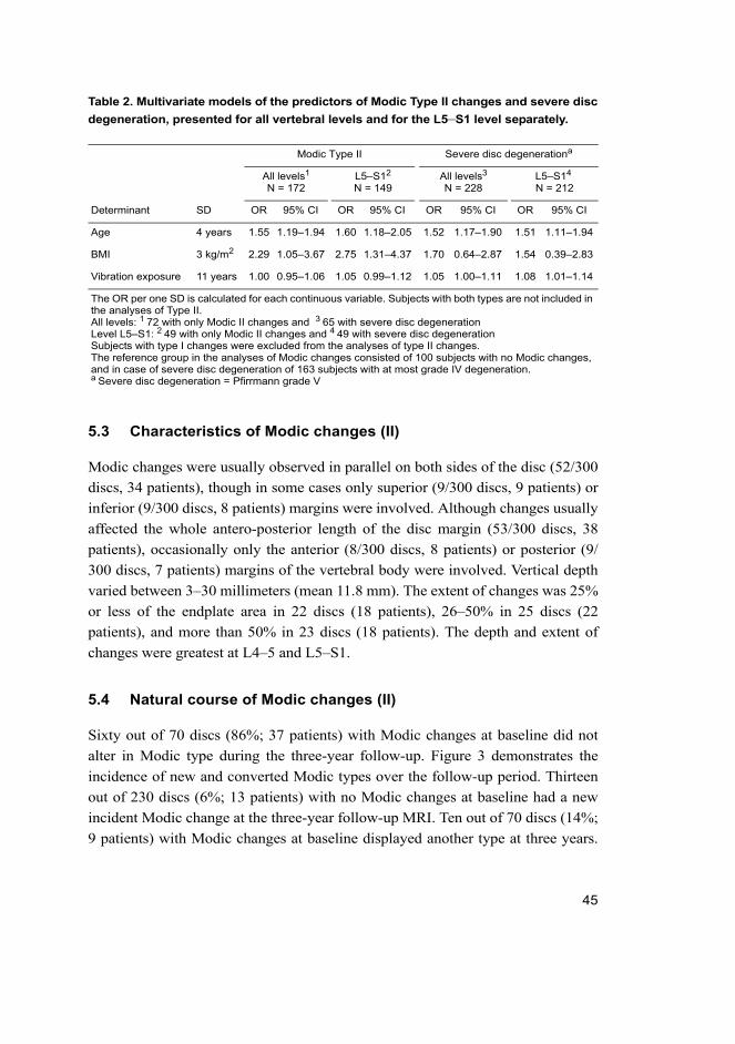
Table 2. Multivariate models of the predictors of Modic Type II changes and severe disc
degeneration, presented for all vertebral levels and for the L5–S1 level separately.
5.3 Characteristics of Modic changes (II)
Modic changes were usually observed in parallel on both sides of the disc (52/300
discs, 34 patients), though in some cases only superior (9/300 discs, 9 patients) or
inferior (9/300 discs, 8 patients) margins were involved. Although changes usually
affected the whole antero-posterior length of the disc margin (53/300 discs, 38
patients), occasionally only the anterior (8/300 discs, 8 patients) or posterior (9/
300 discs, 7 patients) margins of the vertebral body were involved. Vertical depth
varied between 3–30 millimeters (mean 11.8 mm). The extent of changes was 25%
or less of the endplate area in 22 discs (18 patients), 26–50% in 25 discs (22
patients), and more than 50% in 23 discs (18 patients). The depth and extent of
changes were greatest at L4–5 and L5–S1.
5.4 Natural course of Modic changes (II)
Sixty out of 70 discs (86%; 37 patients) with Modic changes at baseline did not
alter in Modic type during the three-year follow-up. Figure 3 demonstrates the
incidence of new and converted Modic types over the follow-up period. Thirteen
out of 230 discs (6%; 13 patients) with no Modic changes at baseline had a new
incident Modic change at the three-year follow-up MRI. Ten out of 70 discs (14%;
9 patients) with Modic changes at baseline displayed another type at three years.
Modic Type II Severe disc degenerationa
All levels1
N = 172L5–S12
N = 149All levels3
N = 228L5–S14
N = 212
Determinant SD OR 95% CI OR 95% CI OR 95% CI OR 95% CI
Age 4 years 1.55 1.19–1.94 1.60 1.18–2.05 1.52 1.17–1.90 1.51 1.11–1.94
BMI 3 kg/m2 2.29 1.05–3.67 2.75 1.31–4.37 1.70 0.64–2.87 1.54 0.39–2.83
Vibration exposure 11 years 1.00 0.95–1.06 1.05 0.99–1.12 1.05 1.00–1.11 1.08 1.01–1.14
The OR per one SD is calculated for each continuous variable. Subjects with both types are not included in the analyses of Type II.All levels: 1 72 with only Modic II changes and 3 65 with severe disc degenerationLevel L5–S1: 2 49 with only Modic II changes and 4 49 with severe disc degenerationSubjects with type I changes were excluded from the analyses of type II changes.The reference group in the analyses of Modic changes consisted of 100 subjects with no Modic changes, and in case of severe disc degeneration of 163 subjects with at most grade IV degeneration.a Severe disc degeneration = Pfirrmann grade V
45
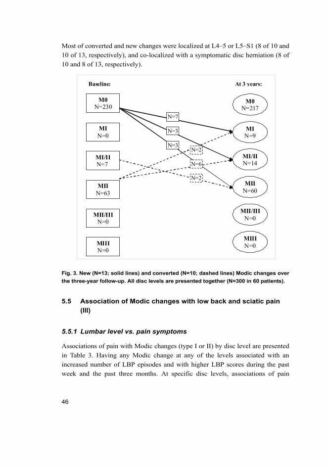
Most of converted and new changes were localized at L4–5 or L5–S1 (8 of 10 and
10 of 13, respectively), and co-localized with a symptomatic disc herniation (8 of
10 and 8 of 13, respectively).
Fig. 3. New (N=13; solid lines) and converted (N=10; dashed lines) Modic changes over
the three-year follow-up. All disc levels are presented together (N=300 in 60 patients).
5.5 Association of Modic changes with low back and sciatic pain
(III)
5.5.1 Lumbar level vs. pain symptoms
Associations of pain with Modic changes (type I or II) by disc level are presented
in Table 3. Having any Modic change at any of the levels associated with an
increased number of LBP episodes and with higher LBP scores during the past
week and the past three months. At specific disc levels, associations of pain
MI/II N=14
M0 N=217
MI N=9
MII N=60
MII/III N=0
MIII N=0
N=2
N=6
N=2
N=7
N=3
N=3
Baseline: At 3 years:
MI N=0
MI/II N=7
MII N=63
MII/III N=0
MIII N=0
M0 N=230
46
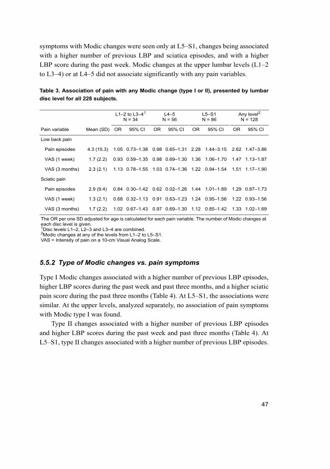
symptoms with Modic changes were seen only at L5–S1, changes being associated
with a higher number of previous LBP and sciatica episodes, and with a higher
LBP score during the past week. Modic changes at the upper lumbar levels (L1–2
to L3–4) or at L4–5 did not associate significantly with any pain variables.
Table 3. Association of pain with any Modic change (type I or II), presented by lumbar
disc level for all 228 subjects.
5.5.2 Type of Modic changes vs. pain symptoms
Type I Modic changes associated with a higher number of previous LBP episodes,
higher LBP scores during the past week and past three months, and a higher sciatic
pain score during the past three months (Table 4). At L5–S1, the associations were
similar. At the upper levels, analyzed separately, no association of pain symptoms
with Modic type I was found.
Type II changes associated with a higher number of previous LBP episodes
and higher LBP scores during the past week and past three months (Table 4). At
L5–S1, type II changes associated with a higher number of previous LBP episodes.
L1–2 to L3–41
N = 34L4–5
N = 56L5–S1N = 86
Any level2
N = 128
Pain variable Mean (SD) OR 95% CI OR 95% CI OR 95% CI OR 95% CI
Low back pain
Pain episodes 4.3 (15.3) 1.05 0.73–1.38 0.98 0.65–1.31 2.28 1.44–3.15 2.62 1.47–3.86
VAS (1 week) 1.7 (2.2) 0.93 0.59–1.35 0.98 0.69–1.30 1.36 1.06–1.70 1.47 1.13–1.87
VAS (3 months) 2.3 (2.1) 1.13 0.78–1.55 1.03 0.74–1.36 1.22 0.94–1.54 1.51 1.17–1.90
Sciatic pain
Pain episodes 2.9 (9.4) 0.84 0.30–1.42 0.62 0.02–1.26 1.44 1.01–1.89 1.29 0.87–1.73
VAS (1 week) 1.3 (2.1) 0.68 0.32–1.13 0.91 0.63–1.23 1.24 0.95–1.56 1.22 0.93–1.56
VAS (3 months) 1.7 (2.2) 1.02 0.67–1.43 0.97 0.69–1.30 1.12 0.85–1.42 1.33 1.02–1.69
The OR per one SD adjusted for age is calculated for each pain variable. The number of Modic changes at each disc level is given.1Disc levels L1–2, L2–3 and L3–4 are combined.2Modic changes at any of the levels from L1–2 to L5–S1.VAS = Intensity of pain on a 10-cm Visual Analog Scale.
47
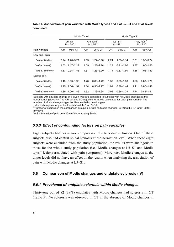
Table 4. Association of pain variables with Modic types I and II at L5–S1 and at all levels
combined.
5.5.3 Effect of confounding factors on pain variables
Eight subjects had nerve root compression due to a disc extrusion. One of these
subjects also had central spinal stenosis at the herniation level. When these eight
subjects were excluded from the study population, the results were analogous to
those for the whole study population (i.e., Modic changes at L5–S1 and Modic
type I lesions associated with pain symptoms). Moreover, Modic changes at the
upper levels did not have an effect on the results when analyzing the association of
pain with Modic changes at L5–S1.
5.6 Comparison of Modic changes and endplate sclerosis (IV)
5.6.1 Prevalence of endplate sclerosis within Modic changes
Thirty-one out of 82 (38%) endplates with Modic changes had sclerosis in CT
(Table 5). No sclerosis was observed in CT in the absence of Modic changes in
Modic Type I Modic Type II
L5–S1N = 282
Any level1
N = 562L5–S1N = 582
Any level1
N = 722
Pain variable OR 95% CI OR 95% CI OR 95% CI OR 95% CI
Low back pain
Pain episodes 2.24 1.26–3.27 2.53 1.24–3.90 2.21 1.33–3.14 2.51 1.36–3.74
VAS (1 week) 1.63 1.17–2.19 1.69 1.23–2.24 1.23 0.91–1.60 1.37 1.00–1.80
VAS (3 months) 1.37 0.94–1.85 1.67 1.23–2.20 1.14 0.83–1.50 1.38 1.02–1.80
Sciatic pain
Pain episodes 1.43 0.93–1.96 1.26 0.83–1.72 1.38 0.95–1.83 1.26 0.83–1.70
VAS (1 week) 1.45 1.06–1.92 1.34 0.98–1.77 1.09 0.78–1.44 1.11 0.80–1.48
VAS (3 months) 1.39 1.00–1.85 1.52 1.13–1.98 0.95 0.66–1.29 1.14 0.82–1.51
Subjects with a Modic change of a given type are compared to subjects with no Modic changes at the corresponding level(s). The OR per one SD adjusted for age is calculated for each pain variable. The number of Modic changes (type I or II) at each disc level is given.1Modic changes at any of the levels from L1–2 to L5–S1.2Number of subjects in the comparison groups, i.e. with no Modic changes, is 142 at L5–S1 and 100 for any level.VAS = Intensity of pain on a 10-cm Visual Analog Scale.
48
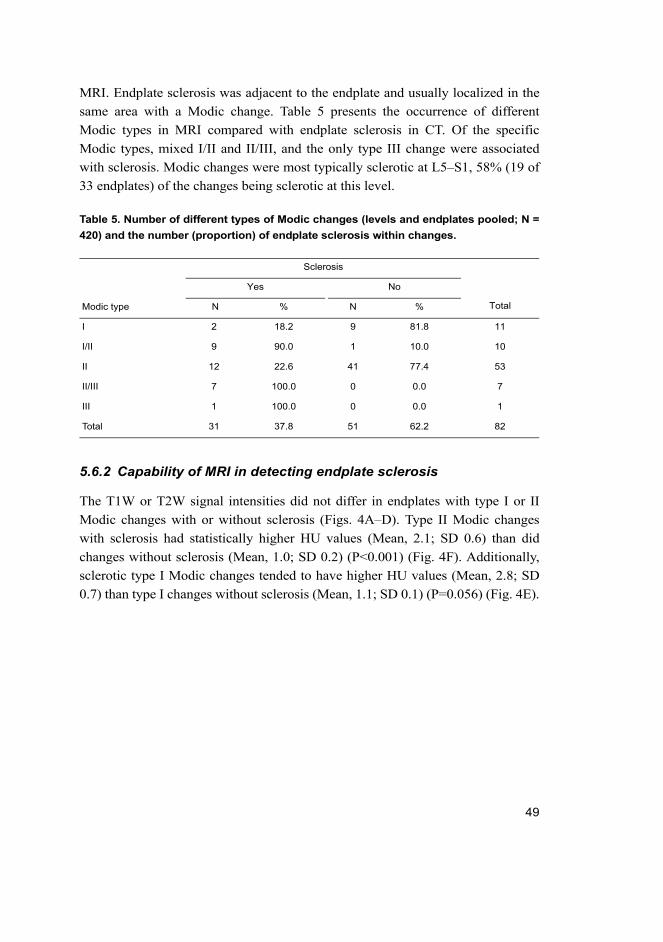
MRI. Endplate sclerosis was adjacent to the endplate and usually localized in the
same area with a Modic change. Table 5 presents the occurrence of different
Modic types in MRI compared with endplate sclerosis in CT. Of the specific
Modic types, mixed I/II and II/III, and the only type III change were associated
with sclerosis. Modic changes were most typically sclerotic at L5–S1, 58% (19 of
33 endplates) of the changes being sclerotic at this level.
Table 5. Number of different types of Modic changes (levels and endplates pooled; N =
420) and the number (proportion) of endplate sclerosis within changes.
5.6.2 Capability of MRI in detecting endplate sclerosis
The T1W or T2W signal intensities did not differ in endplates with type I or II
Modic changes with or without sclerosis (Figs. 4A–D). Type II Modic changes
with sclerosis had statistically higher HU values (Mean, 2.1; SD 0.6) than did
changes without sclerosis (Mean, 1.0; SD 0.2) (P<0.001) (Fig. 4F). Additionally,
sclerotic type I Modic changes tended to have higher HU values (Mean, 2.8; SD
0.7) than type I changes without sclerosis (Mean, 1.1; SD 0.1) (P=0.056) (Fig. 4E).
Sclerosis
Yes No
Modic type N % N % Total
I 2 18.2 9 81.8 11
I/II 9 90.0 1 10.0 10
II 12 22.6 41 77.4 53
II/III 7 100.0 0 0.0 7
III 1 100.0 0 0.0 1
Total 31 37.8 51 62.2 82
49
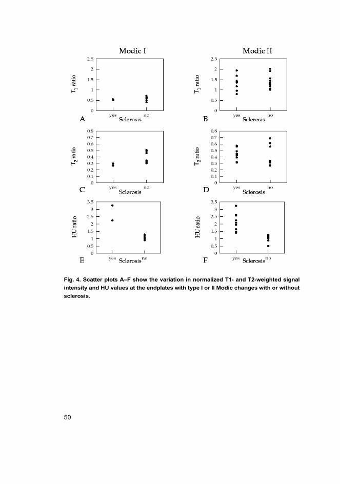
Fig. 4. Scatter plots A–F show the variation in normalized T1- and T2-weighted signal
intensity and HU values at the endplates with type I or II Modic changes with or without
sclerosis.
50
6 Discussion
Modic changes are bone marrow signal intensity changes adjacent to the endplates
of the degenerated intervertebral discs in magnetic resonance imaging. In recent
years, they have come into focus in the search for the source of back pain
symptoms. The present study evaluated the determinants of Modic changes and
the association of Modic changes with low back pain symptoms in an occupational
cohort. The prevalence and the natural course of Modic changes was investigated
in a longitudinal follow-up MRI study. In addition, comparisons were made
between computed tomography and magnetic resonance imaging to evaluate the
presence of endplate sclerosis on different types of Modic changes, and further to
assess the capability of MRI in detecting endplate sclerosis within these changes.
In the present study, Modic changes were a common MRI finding both among
patients and middle-aged working males. In addition to age, weight-related factors
were associated with Modic changes. Modic changes converted from one type to
another during a three-year follow-up period, and type II changes were less stable
than previously assumed. A considerable proportion of Modic changes were
sclerotic as observed in CT. Modic changes were always found in combination
with a degenerative intervertebral disc and thus they are assumed to be a specific
phenotype of degenerative disc disease. Finally, Modic changes were associated
with pain symptoms – especially changes located at L5–S1 and type I Modic
changes.
6.1 Determinants of Modic changes
The present study showed that age and weight-related factors (BMI and waist
circumference) are associated with Modic changes. Previously, it has been shown
that a multitude of factors determine the risk of disc degeneration including age,
genetic inheritance and loading history (Adams & Roughley 2006). However,
there is only limited information about the determinants of Modic changes. In
addition to age (Modic et al. 1988b, Karchevsky et al. 2005), body weight
(Karchevsky et al. 2005, Leboeuf-Yde et al. 2008), heavy physical work (Kjaer etal. 2006), smoking (Kjaer et al. 2006) and male gender (Karchevsky et al. 2005)
have shown associations with Modic changes.
Modic changes occur adjacent to the degenerated intervertebral discs (de Roos
et al. 1987, Modic et al. 1988b). The prevalence of Modic changes has been found
to increase with age (Modic et al. 1988b, Karchevsky et al. 2005) and they are
51
uncommon in younger individuals (Kjaer et al. 2005b). Similarly, a close
association between age and the prevalence of changes was found in this study as
evidence of their degenerative etiology.
Weight-related factors were associated with Modic changes but not with disc
degeneration. Previously, obesity has been suggested as a risk factor for disc
degeneration, but the results have remained controversial (Heliövaara 1987, Rii-
himäki et al. 1990, Harrington et al. 2001, Liuke et al. 2005). In a
population-based study, overweight in combination with hard physical work was
significantly associated with the prevalence of Modic changes (Leboeuf-Yde et al.2008). In another patient study, Modic changes were associated with increasing
weight, but not with BMI (Karchevsky et al. 2005). In Finland, obesity (BMI at
least 30 kg/m2) has increased in all age groups and in both men and women. Cur-
rently, more than 20% of the population is obese. (Aromaa & Koskinen 2004.)
Although weight-related factors were found to be associated with an increased
likelihood of Modic changes, the influence of weight reduction on degenerative
disease, both clinical symptoms and progression of degenerative imaging findings,
is unknown.
This study showed a significant association between lifetime leisure exercise
activity and all Modic changes at L5–S1 in the multivariate analyses (data not
shown). However, when type I and II changes were analyzed separately, no
significant association remained. There is only limited knowledge about the
association of physical activity and Modic changes. In a Danish population-based
cohort, exposure to high-level physical activity at leisure time did not differ
between subjects with “Modic changes and disc degeneration” vs. those with
“only disc degeneration”. However, exposure to occupational physical load was
significantly higher in the subgroup of “Modic changes and disc degeneration”
compared to “only disc degeneration” group. (Kjaer et al. 2006.) Videman et al.(1995) detected a relation between vigorous exercise and lumbar degenerative
changes among former Finnish elite athletes. However, they did not evaluate
Modic changes separately. Recent studies on identical twins have shown that 70%
of intervertebral disc degeneration can be attributed to familial factors rather than
to the mechanical environment (Sambrook et al. 1999). However, the endplate is a
weak link of the spine in compression, and always fails before the intervertebral
disc, even if the latter is injured before loading commences (Brinckmann 1986).
The author speculates that mechanical loading damages the endplate and may lead
to activation of degeneration. This may first be visible in MR images as a more
52
active Modic type I lesion, and if the mechanical loading continues, it converts to a
more stable type II lesion (Modic et al. 1988b).
In this study, there was a trend between smoking (assessed as pack-years) and
a higher prevalence of Modic changes at L5–S1, but no association with severe
disc degeneration. Previously, there is only one study of the effect of smoking on
Modic changes, where self-reported hard physical work in combination with
heavy smoking was strongly associated with Modic changes (Kjaer et al. 2006).
Although an association between smoking and disc degeneration was observed in
an earlier study on Finnish twins (Battie et al. 1991), the association between
smoking and disc degeneration was not confirmed in a later study (Battie et al.1995). At the moment, the author is unable to either disprove or support the
hypothesis that smoking is a risk factor for degenerative imaging findings. The
study on the effect of smoking on Modic changes should be replicated in other
population-based cohorts.
Another environmental factor, alcohol, has been associated with many organ
abnormalities and cognitive deficits. There are no studies about the effects of
alcohol on Modic changes. However, there is evidence that moderate alcohol
consumption may reduce the risk of intervertebral disc degeneration (Zhang et al.2008). This study found no association between alcohol consumption and Modic
changes or disc degeneration. It therefore seems likely that alcohol does not have
deleterious effects on the intervertebral discs.
Occupation (train engineers vs. sedentary factory work) or the duration of
exposure to whole-body vibration was of borderline significance for type II Modic
changes located at L5–S1. Interestingly, whole-body vibration was associated with
severe disc degeneration, but only at L5–S1. There is evidence of an association
between vibration and degenerative spinal changes (Videman & Battie 1999) but
this could not be confirmed in a high-quality twin study with discordant
occupational driving exposure (Battie et al. 2002). Although the findings of this
study indicate that exposure to whole-body vibration may accelerate disc
degeneration (and possibly Modic changes), we cannot wholly exclude a chance
finding.
Based on the findings of this study, the determinants of Modic type I and II
changes are different. The determinants of type I remain unidentified, whereas age
and weight-related factors (BMI, waist circumference) were related to type II
changes. This may be partly explained by the low number of subjects with type I
changes. However, type I changes are also associated with an ongoing
degenerative process which converts over time to type II or normal bone marrow
53

(Modic et al. 1988b). Finally, the influence of determinants may also vary at
different stages of the degenerative process.
The strength of the study of the determinants is based on the occupational
cohort consisting of train engineers and sedentary factory workers of similar age
range. Additionally, all train engineers were full-time train drivers from Northern
Finland, which ensured a very similar exposure to whole-body vibration. The
factory workers consisted of paper mill and chemical factory workers from the
same area with only sedentary job and no vibration exposure.
The limitations of the study are based on its cross-sectional nature, which is a
common problem in epidemiological studies. Even if selected determinants can be
reasonably well assessed retrospectively, their temporal relation to the
development of Modic changes or disc degeneration cannot be demonstrated. The
effect of age, and perhaps of genetic factors, may also dilute the effect of
environmental determinants. Furthermore, there were no precise measurements of
whole-body vibration exposure. When studying lifetime effects, such precision is
not feasible due to variations in train models and railway conditions.
According to the results of this study, besides age, the determinants of Modic
changes and disc degeneration are different. Weight-related factors, which add the
load of the lumbar spine, seem important in the pathogenesis of Modic changes,
whereas whole-body vibration was associated with severe disc degeneration.
6.2 Prevalence and characteristics of Modic changes
The prevalence of Modic changes in this study varied between 51 and 65% (stud-
ies I–IV) according to the study population. Previously, the prevalence of Modic
changes among patients with degenerative disc disease of the lumbar spine has
been reported to vary between 19 and 59% (de Roos et al. 1987, Modic et al.1988b, Toyone et al. 1994, Weishaupt et al. 2001, Schmid et al. 2004, Karchevsky
et al. 2005, Kjaer et al. 2005a, Albert & Manniche 2007). The prevalence rates in
studies I, III and IV (51–56%) were in accordance with previous findings, whereas
the prevalence in study II (65%) was higher than observed earlier. However, when
the prevalence of Modic changes in study II was reported in relation to lumbar disc
levels (23%), it accorded with the previous findings in patient populations (20–
50%) (de Roos et al. 1987, Modic et al. 1988b, Karchevsky et al. 2005). The high
prevalence in relation to the subjects in study II may be explained by the study
population consisting of only sciatica patients, as Modic changes are known to
appear after disc herniations (Albert & Manniche 2007). Modic changes have been
54
observed to be a more common MRI-finding in patients with LBP compared to
non-clinical populations (Jensen et al. 2008). Furthermore, Modic changes are
uncommon in asymptomatic individuals without disc degeneration (Weishaupt etal. 1998, Kjaer et al. 2006).
Type II Modic change was the most frequent in all studies (I–IV), 65–90% of
changes being of this type. There is disagreement as to whether type I or II Modic
change is the most common in the lumbar spine. The predominance of type II has
been found in several studies, as it has been observed to constitute up to 80% of
Modic changes in the lumbar spine (de Roos et al. 1987, Modic et al. 1988b,
Braithwaite et al. 1998, Schmid et al. 2004, Karchevsky et al. 2005), and this is
supported by the results of the present study. This may be explained by the finding
that type I Modic changes have been observed to convert to type II or normal bone
marrow, whereas type II Modic changes are usually associated with a more stable
degenerative process (Modic et al. 1988b). Other studies have suggested that type
I is a more common Modic type and may account for up to 71% of Modic changes
(Weishaupt et al. 2001, Kjaer et al. 2005a). The differences in prevalence rates
between studies of Modic changes and the frequency of each Modic type might be
explained by differences in the definition of changes. The different prevalence and
distribution of Modic types may also result from differences in the study
populations and inclusion criteria.
An association of Modic changes and intervertebral disc degeneration was
confirmed in the present study. All discs adjacent to a Modic change had at least
grade III disc degeneration (studies I and III). In addition, changes occurred most
commonly at L4–5 or L5–S1 (studies I–III), as previously observed (Modic et al.1988b, Karchevsky et al. 2005). New and interesting findings were that the depth
and the extent of changes were also greatest at L4–5 and L5–S1. These findings
may be related to the biomechanics of the lumbar spine. L5 is the largest of all
movable vertebrae and it carries the weight of the upper body (Moore 1999). The
vertical depth of Modic changes was observed to vary between 3 and 30 mm, in
accordance with earlier findings (Modic et al. 1988b). Changes were usually
identified at both sides of the disc margin, and they usually involved the whole
antero-posterior length of the disc margin, which accords with previous findings
(Modic et al. 1988b). However, Chung et al. (2004) suggested that the distribution
of changes was more in the anterior than the posterior endplate of the vertebra. The
inconsistent results may be explained by the difference in the definition of Modic
changes, as these authors also included very small changes located at the corners
of the vertebral bodies. Inflammatory spondyloarthropathias may lead to signal
55
intensity alterations in the insertion of the annulus fibrosus-longitudinal ligament
complex that may be mimicking Modic changes (Jevtic et al. 2000, James &
Davies 2006).
6.3 Natural course of Modic changes
Before the significance of different Modic types can be evaluated, a better
understanding of the natural course of these changes is needed. In the original
study, Modic et al. (1988b) observed that five of six patients with a type I change
converted to a type II over a 14- to 36-month interval. The authors found that type
I changes can also revert back to normal, whereas type II changes usually do not
alter with time, thus associating with a more stable and chronic degenerative
process. (Modic et al. 1988b.) Later, Braithwaite et al. (1998) suggested that
Modic changes can convert from one type to another, presenting different stages of
the same pathological process.
After the initial report of Modic et al. (1988b), the dynamic process of Modic
type I changes have been confirmed. Mitra et al. (2004) found that 52% of type I
changes converted into type II and 40% increased in size, while only 8% of type I
changes remained stable during a 12- to 72-month follow-up period. In a recent
study, only 11% of type I Modic changes remained stable during an 18- to
72-month follow-up period (Luoma et al. 2008).
The stability of type II Modic change is questioned by the results of the
present study. During a three-year follow-up period, 14% of Modic changes at
baseline converted to another type, 80% of the conversions being from type II to
either type I or mixed type I/II. This finding was recently supported by a case
report of two patients with conversion of type II change into type I (Marshman etal. 2007). These findings suggest that type II Modic change may not be as stable as
previously assumed. An acute ongoing process in some type II changes may cause
the conversion of yellow marrow to red, resulting in type I or type I/II Modic
change. This was supported by the progression of disc degeneration (loss of signal
or disc height or even disc collapse in two cases).
A limitation of the study was that spondylolisthesis and instability were not
evaluated as they may be associated with conversions of a Modic change to
another type. Modic changes have been reported to be associated with segmental
hypermobility in the dynamic flexion-extension MR images (Toyone et al. 1994).
These findings need to be confirmed in future studies assessing the natural history
of Modic changes.
56
6.4 Modic changes and low back pain
This study showed that Modic changes at L5–S1 and Modic type I lesions are
more likely to be associated with pain symptoms than other types of Modic
changes or changes located at other lumbar levels. Previously, a wide spectrum of
disc abnormalities, including disc degeneration, disc bulging, disc protrusion, and
annular tears, have been found in a substantial percentage of healthy volunteers
(Boden et al. 1990, Jensen et al. 1994, Boos et al. 1995, Stadnik et al. 1998). In
contrast, disc extrusions, nerve root compression and Modic changes are rare in
asymptomatic volunteers (Jensen et al. 1994, Boos et al. 1995, Weishaupt et al.1998, Kjaer et al. 2006). In recent years, Modic changes have come into focus as a
source of back pain symptoms.
In accordance with previous findings, this study showed that Modic type I
changes are associated with pain symptoms. In a study of 74 patients with disc
degeneration, Toyone et al. (1994) observed that 73% of patients with type I
change had significant LBP, as opposed to only 11% of patients with type II. In a
longitudinal study of LBP patients, a positive trend was found between the evolu-
tion of type I to type II change and symptom improvement (Mitra et al. 2004). In
the Danish study of 412 subjects of 40-year-old Danes, a strong association of
Modic changes with LBP during the past year, especially for Modic type I, was
observed (Kjaer et al. 2006). Recently, Albert and Manniche (2007) reported
similar results as they showed that type I changes were more strongly associated
with LBP than type II changes. The correlation of Modic changes with pain
induced by discography has also been studied, but the results have remained
controversial (Braithwaite et al. 1998, Sandhu et al. 2000, Weishaupt et al. 2001,
Kokkonen et al. 2002).
A new and interesting finding in the present study was that both Modic type I
and II lesions at L5–S1, but not at upper levels, are associated with the occurrence
and intensity of LBP. This is the first study to assess the role of lumbar levels in
relation to Modic changes and LBP. The author speculates that the association of
LBP symptoms with L5–S1 level might be due to mechanical factors, but the
pathophysiology of this phenomenon needs further investigation.
Disc extrusions and nerve root compression are observed to be rare in asymp-
tomatic volunteers (Jensen et al. 1994, Boos et al. 1995, Weishaupt et al. 1998).
Boos et al. (1995) showed that the only substantial morphologic difference
between symptomatic patients and asymptomatic matched controls was the pres-
ence of neural compromise (83% vs. 22%). Therefore, in this study nerve root
57
compression due to a disc extrusion was considered a confounding factor in the
pain analyses. However, the results were analogous when these subjects were
excluded from the analyses.
According to the results of this study, Modic changes at L5–S1 and Modic
type I lesions are more likely to be associated with pain symptoms than other types
of Modic changes or changes located at other lumbar levels. This is the first study
to assess the role of lumbar levels in relation to Modic changes. The author is
aware of a potential bias related to multiple comparisons, but the results are
consistent, and in the author’s opinion they are not chance findings. However, the
results are valid only on group-level. In the future, larger studies are needed to
verify the current findings.
6.5 Comparison of MRI and CT in detecting Modic changes
This is the first study to investigate Modic changes in MRI compared with
multislice CT examination. It has been suggested that only type III Modic changes
are visible in plain film radiography with endplate sclerosis (Modic et al. 1988a,
Modic et al. 1988b). Interestingly, the results of this study are somewhat different,
as it was found that not only type III changes, but also other Modic types,
especially mixed Modic changes (I/II and II/III), showed endplate sclerosis in CT.
However, endplate sclerosis could not be detected in MR images in the
quantitative analysis.
A new and important finding in our study was that 90% (9 out of 10) of mixed
type I/II and all seven mixed type II/III changes in MRI showed sclerosis at the
endplates in question in CT. The corresponding proportions of sclerotic endplates
for type I, II and III Modic changes were 18% (2 out of 11), 22% (12 out of 55)
and 100% (1 out of 1), respectively. Modic et al. (1988a, 1988b) were the first to
introduce the appearance of bone sclerosis in MRI. They suggested that type III
Modic changes are presented by decreased signal intensity on both T1- and
T2-weighted images, which appears to correlate with extensive bone sclerosis in
plain film radiography (Modic et al. 1988a). Conversely, type I and II Modic
changes showed no definite correlation with sclerosis in plain film radiography
(Modic et al. 1988b). Mixed Modic types were not evaluated separately.
There have been few reports in the literature in which reactive bone sclerosis
has appeared hypointense in T1W and hyperintense in T2W MR images. In a
study on MRI features of osteoblastoma (Shaikh et al. 1999), low signal intensity
reactive sclerosis was observed in both T1W and T2W images. A
58
histopathological examination of reactive sclerosis with this behavior showed
heavy matrix mineralization. Reactive sclerosis accompanied by marrow edema
appeared hypointense in T1W and hyperintense in T2W images. In the
histopathological examination these cases were associated with increased
fibrovascular tissue within the marrow and a perivascular infiltration of
lymphocytes and plasma cells. (Shaikh et al. 1999.) The same association has been
observed in inflammatory arthropathy. In an acute stage of the process,
non-sclerotic changes with low signal intensity in T1W and high signal intensity in
T2W images are seen in the anterior disco-vertebral junction. As the process
becomes chronic, sclerotic changes (shiny corners in plain film radiography) with
high signal intensity in T1W and T2W images are seen, reflecting the healing
process of the lesion. The reactive new bone formation probably occurs in
previously inflamed areas. (Jevtic et al. 2000, James & Davies 2006.)
In this study, endplate sclerosis observed in CT could not be detected in MR
images in the quantitative analysis. The T1- or T2-weighted signal intensities did
not differ in endplates with type I or II Modic changes with or without sclerosis.
However, as assumed, the HU values of sclerotic Modic changes were higher than
in those without sclerosis. This finding is explained by the difference within the
imaging modalities used. CT is superior to MRI for evaluation of reactive or
sclerotic bone formation. Because of the lack of mobile protons, bone sclerosis
yields no detectable signal in MRI. Furthermore, signal intensity in MRI depends
on the fat/water content within bone trabeculae. (Vogler & Murphy 1988, Vanel etal. 2000.)
From the histopathologic point of view, osteosclerosis is defined as a
qualitative increase in bone volume. The sclerosis may be fully or partly
mineralized. If the sclerosis is partly mineralized, the amount of water is increased
and there is less calcium deposition compared with fully mineralized sclerosis (Ott
1996). The sclerosis seen in plain film radiography and in CT of type III Modic
changes is a reflection of dense mineralized bone within the vertebral body rather
than the marrow elements (Modic et al. 1988b).
The sclerosis seen in most of the mixed Modic types and in some type I and II
changes may be related to a regenerative process of the bone marrow. This finding
is indirectly supported by the result of this study, as no sclerotic bony endplates
were observed in CT in the absence of Modic changes in MRI. Modic changes are
thought to be a reflection of the degenerative process within the bone marrow
(Modic et al. 1988b), and the sclerotic Modic changes may be related to the
59
healing process with new bone formation. However, the clinical importance of this
finding remains to be studied.
A limitation of the study was its retrospective nature. The patients were
referred for a variety of clinical problems and exact clinical correlation was not
attempted. However, our purpose was to document and quantify this new finding
with MRI and CT examinations. High-dose ionizing radiation prevents application
of CT in prospective studies and non-patient populations. MRI has become the
first imaging modality in patients with LBP, because it provides a non-invasive
precise morphologic appraisal of the lumbar spine and permits direct relation of
morphologic findings to LBP. Despite these shortcomings, the author believes that
the results are important because they can improve diagnostic yield in patients
with LBP in the future.
In conclusion, endplate sclerosis existed in all types of Modic changes in CT,
especially in mixed Modic types, and not only in type III changes, as previously
assumed. Endplate sclerosis with Modic changes was not detected in MRI, which
may depend on the amount of trabeculae and mineralization of the bone marrow.
6.6 Future prospects
During the last two decades, our understanding of the vertebral bone marrow
changes adjacent to the endplates of degenerated intervertebral discs has increased.
However, the need for further research remains. Areas of future research should
mainly include the mechanism, natural history and relationship to symptoms.
Three hypotheses of the mechanism - biomechanical, biochemical and infective –
have been proposed. To date, however, the evidence supporting these hypotheses
or their possible interplay in the pathogenesis of Modic changes is not clear.
According to the current literature, including this thesis, type I Modic changes are
likely to be associated with LBP symptoms. However, the prognostic significance
of these changes, including conversions into different types, in relation to a
patient’s symptoms needs to be confirmed. Therefore, further studies with longer
follow-up periods, more frequent MRI studies and larger sample sizes will be
required.
Isolating a subgroup of patients with type I Modic changes could be of
therapeutic and economic interest. In a recent study, LBP patients with Modic type
I change tended to have better short-term efficacy following intradiscal steroid
injections than those predominantly with type II changes (Fayad et al. 2007). In
addition, surgical studies have suggested that patients with type I changes who
60
undergo lumbar fusion for LBP do better than those without changes or with type
II (Vital et al. 2003). These authors speculate that disappearance of type I changes
may be an indicator of successful fusion and stabilization. However, with the
current knowledge, it is probably premature to assume that the bone marrow
changes are themselves a prognostic finding or indicative of a need for a specific
intervention. In the future, a controlled study of patients with type I changes with
randomization to different forms of therapy might be helpful to determine whether
the evolution of these changes can be altered by therapeutic interventions, or
whether it is dependent on the natural history of the degenerative process.
61

62
7 Summary and conclusions
The results of this thesis indicate that Modic changes are a specific phenotype of
degenerative intervertebral disc disease. It is important for radiologists to
recognize and differentiate these changes from other bone marrow signal intensity
changes that exist in vertebral bodies. Physicians treating low back pain patients
should also be familiar with the appearance, the natural course and the association
of Modic changes with LBP symptoms. The main conclusions of the present study
are summarized as follows:
1. Besides age, the determinants of Modic changes and disc degeneration may be
different. Weight-related factors seem to be important in the pathogenesis of
Modic changes, whereas whole-body vibration is associated with severe disc
degeneration.
2. Modic changes are a common MRI finding in patients with degenerative disc
disease. They can change from one type to another, and type II changes may
be less stable than previously assumed.
3. Modic changes at L5–S1 and Modic type I lesions are more likely to be
associated with pain symptoms than other types of Modic changes or changes
located at other lumbar levels.
4. Endplate sclerosis exists in all types of Modic changes in CT, especially in
mixed Modic types, and not only in type III changes, as previously assumed.
Endplate sclerosis with Modic changes is not detected in MRI, which may
depend on the amount of trabeculae and mineralization of the bone marrow.
63

64

References
Adams MA, Freeman BJ, Morrison HP, Nelson IW & Dolan P (2000) Mechanical initiation
of intervertebral disc degeneration. Spine 25(13): 1625–1636.
Adams MA & Roughley PJ (2006) What is intervertebral disc degeneration, and what
causes it? Spine 31(18): 2151–2161.
Aigner T, Gresk-otter KR, Fairbank JC, von der Mark K & Urban JP (1998) Variation with
age in the pattern of type X collagen expression in normal and scoliotic human
intervertebral discs. Calcif Tissue Int 63(3): 263–268.
Albert HB, Kjaer P, Jensen TS, Sorensen JS, Bendix T & Manniche C (2008a) Modic
changes, possible causes and relation to low back pain. Med Hypotheses 70(2): 361–
368.
Albert HB & Manniche C (2007) Modic changes following lumbar disc herniation. Eur
Spine J 16(7): 977–982.
Albert HB, Manniche C, Sorensen JS & Deleuran BW (2008b) Antibiotic treatment in
patients with low back pain associated with Modic changes Type 1 (bone oedema): a
pilot study. Br J Sports Med 42: 969–973.
An HS, Anderson PA, Haughton VM, Iatridis JC, Kang JD, Lotz JC, Natarajan RN,
Oegema TR, Jr., Roughley P, Setton LA, Urban JP, Videman T, Andersson GB &
Weinstein JN (2004) Introduction: disc degeneration: summary. Spine 29(23): 2677–
2678.
Andersson GB (1998) Epidemiology of low back pain. Acta Orthop Scand Suppl 281: 28–
31.
Annunen S, Paassilta P, Lohiniva J, Perälä M, Pihlajamaa T, Karppinen J, Tervonen O,
Kröger H, Lähde S, Vanharanta H, Ryhänen L, Göring HH, Ott J, Prockop DJ &
Ala-Kokko L (1999) An allele of COL9A2 associated with intervertebral disc disease.
Science 285(5426): 409–412.
Aromaa A & Koskinen S (2004) Health and functional capacity in Finland. Baseline results
of the Health 2000 health examination survey, Publications of National Public Health
Institute, Series B 12/2004.
Atlas SJ, Keller RB, Wu YA, Deyo RA & Singer DE (2005) Long-term outcomes of
surgical and nonsurgical management of lumbar spinal stenosis: 8 to 10 year results
from the maine lumbar spine study. Spine 30(8): 936–943.
Battie MC, Videman T, Gibbons LE, Fisher LD, Manninen H & Gill K (1995) 1995 Volvo
Award in clinical sciences. Determinants of lumbar disc degeneration. A study relating
lifetime exposures and magnetic resonance imaging findings in identical twins. Spine
20(24): 2601–2612.
65

Battie MC, Videman T, Gibbons LE, Manninen H, Gill K, Pope M & Kaprio J (2002)
Occupational driving and lumbar disc degeneration: a case-control study. Lancet
360(9343): 1369–1374.
Battie MC, Videman T, Gill K, Moneta GB, Nyman R, Kaprio J & Koskenvuo M (1991)
1991 Volvo Award in clinical sciences. Smoking and lumbar intervertebral disc
degeneration: an MRI study of identical twins. Spine 16(9): 1015–1021.
Baur A, Huber A, Ertl-Wagner B, Durr R, Zysk S, Arbogast S, Deimling M & Reiser M
(2001) Diagnostic value of increased diffusion weighting of a steady-state free
precession sequence for differentiating acute benign osteoporotic fractures from
pathologic vertebral compression fractures. AJNR Am J Neuroradiol 22(2): 366–372.
Beattie PF, Meyers SP, Stratford P, Millard RW & Hollenberg GM (2000) Associations
between patient report of symptoms and anatomic impairment visible on lumbar
magnetic resonance imaging. Spine 25(7): 819–828.
Bernick S & Cailliet R (1982) Vertebral end-plate changes with aging of human vertebrae.
Spine 7(2): 97–102.
Boden SD, Davis DO, Dina TS, Patronas NJ & Wiesel SW (1990) Abnormal
magnetic-resonance scans of the lumbar spine in asymptomatic subjects. A prospective
investigation. J Bone Joint Surg Am 72(3): 403–408.
Bogduk N, Tynan W & Wilson AS (1981) The nerve supply to the human lumbar
intervertebral discs. J Anat 132(Pt 1): 39–56.
Boos N, Rieder R, Schade V, Spratt KF, Semmer N & Aebi M (1995) 1995 Volvo Award in
clinical sciences. The diagnostic accuracy of magnetic resonance imaging, work
perception, and psychosocial factors in identifying symptomatic disc herniations. Spine
20(24): 2613–2625.
Boos N, Weissbach S, Rohrbach H, Weiler C, Spratt KF & Nerlich AG (2002)
Classification of age-related changes in lumbar intervertebral discs: 2002 Volvo Award
in basic science. Spine 27(23): 2631–2644.
Braithwaite I, White J, Saifuddin A, Renton P & Taylor BA (1998) Vertebral end-plate
(Modic) changes on lumbar spine MRI: correlation with pain reproduction at lumbar
discography. Eur Spine J 7(5): 363–368.
Brinckmann P (1986) Injury of the annulus fibrosus and disc protrusions. An in vitro
investigation on human lumbar discs. Spine 11(2): 149–153.
Brinckmann P, Frobin W, Hierholzer E & Horst M (1983) Deformation of the vertebral
end-plate under axial loading of the spine. Spine 8(8): 851–856.
Brown MF, Hukkanen MV, McCarthy ID, Redfern DR, Batten JJ, Crock HV, Hughes SP &
Polak JM (1997) Sensory and sympathetic innervation of the vertebral endplate in
patients with degenerative disc disease. J Bone Joint Surg Br 79(1): 147–153.
66
Buttermann GR, Heithoff KB, Ogilvie JW, Transfeldt EE & Cohen M (1997) Vertebral
body MRI related to lumbar fusion results. Eur Spine J 6(2): 115–120.
Chung CB, Vande Berg BC, Tavernier T, Cotten A, Laredo JD, Vallee C & Malghem J
(2004) End plate marrow changes in the asymptomatic lumbosacral spine: frequency,
distribution and correlation with age and degenerative changes. Skeletal Radiol 33(7):
399–404.
Coventry MB, Ghormley RK & Kernohan JW (1945) THE INTERVERTEBRAL DISC:
ITS MICROSCOPIC ANATOMY AND PATHOLOGY: Part II. Changes in the
Intervertebral Disc Concomitant with Age. J Bone Joint Surg Am 27: 233–247.
Crock HV (1970) A reappraisal of intervertebral disc lesions. Med J Aust 1(20): 983–989.
Crock HV (1986) Internal disc disruption. A challenge to disc prolapse fifty years on. Spine
11(6): 650–653.
Daffner RH, Lupetin AR, Dash N, Deeb ZL, Sefczek RJ & Schapiro RL (1986) MRI in the
detection of malignant infiltration of bone marrow. AJR Am J Roentgenol 146(2): 353–
358.
de Roos A, Kressel H, Spritzer C & Dalinka M (1987) MR imaging of marrow changes
adjacent to end plates in degenerative lumbar disk disease. AJR Am J Roentgenol
149(3): 531–534.
Edwards WT, Zheng Y, Ferrara LA & Yuan HA (2001) Structural features and thickness of
the vertebral cortex in the thoracolumbar spine. Spine 26(2): 218–225.
Erdem LO, Erdem CZ, Acikgoz B & Gundogdu S (2005) Degenerative disc disease of the
lumbar spine: a prospective comparison of fast T1-weighted fluid-attenuated inversion
recovery and T1-weighted turbo spin echo MR imaging. Eur J Radiol 55(2): 277–282.
Erkintalo MO, Salminen JJ, Alanen AM, Paajanen HE & Kormano MJ (1995) Development
of degenerative changes in the lumbar intervertebral disk: results of a prospective MR
imaging study in adolescents with and without low-back pain. Radiology 196(2): 529–
533.
Erly WK, Oh ES & Outwater EK (2006) The utility of in-phase/opposed-phase imaging in
differentiating malignancy from acute benign compression fractures of the spine.
AJNR Am J Neuroradiol 27(6): 1183–1188.
Eyre DR (1979) Biochemistry of the intervertebral disc. Int Rev Connect Tissue Res 8:
227–291.
Fardon DF & Milette PC (2001) Nomenclature and classification of lumbar disc pathology.
Recommendations of the Combined task Forces of the North American Spine Society,
American Society of Spine Radiology, and American Society of Neuroradiology. Spine
26(5): E93–E113.
67
Fayad F, Lefevre-Colau MM, Rannou F, Quintero N, Nys A, Mace Y, Poiraudeau S, Drape
JL & Revel M (2007) Relation of inflammatory modic changes to intradiscal steroid
injection outcome in chronic low back pain. Eur Spine J 16(7): 925–931.
Freemont AJ, Peacock TE, Goupille P, Hoyland JA, O'Brien J & Jayson MI (1997) Nerve
ingrowth into diseased intervertebral disc in chronic back pain. Lancet 350(9072):
178–181.
Girard CJ, Schweitzer ME, Morrison WB, Parellada JA & Carrino JA (2004) Thoracic
spine disc-related abnormalities: longitudinal MR imaging assessment. Skeletal Radiol
33(4): 216–222.
Goldberg MS, Scott SC & Mayo NE (2000) A review of the association between cigarette
smoking and the development of nonspecific back pain and related outcomes. Spine
25(8): 995–1014.
Grossman R & Yousem D (2003) Anatomy and Degenerative Diseases of the Spine.
Neuroradiology: The requisites, Elsevier Inc (USA): 751–793.
Harrington J, Jr., Sungarian A, Rogg J, Makker VJ & Epstein MH (2001) The relation
between vertebral endplate shape and lumbar disc herniations. Spine 26(19): 2133–
2138.
Harris RI & Macnab I (1954) Structural changes in the lumbar intervertebral discs; their
relationship to low back pain and sciatica. J Bone Joint Surg Br 36-B(2): 304–322.
Haughton V (2006) Imaging intervertebral disc degeneration. J Bone Joint Surg Am 88
Suppl 2: 15–20.
Heistaro S, Vartiainen E, Heliövaara M & Puska P (1998) Trends of back pain in eastern
Finland, 1972–1992, in relation to socioeconomic status and behavioral risk factors.
Am J Epidemiol 148(7): 671–682.
Heliövaara M (1987) Body height, obesity, and risk of herniated lumbar intervertebral disc.
Spine 12(5): 469–472.
Heliövaara M, Sievers K, Impivaara O, Maatela J, Knekt P, Mäkelä M & Aromaa A (1989)
Descriptive epidemiology and public health aspects of low back pain. Ann Med 21(5):
327–333.
Hilton RC, Ball J & Benn RT (1976) Vertebral end-plate lesions (Schmorl's nodes) in the
dorsolumbar spine. Ann Rheum Dis 35(2): 127–132.
Holm A & Holm S (2004) Biomechanical Considerations of Disc Degeneration. The
Lumbar Spine, Lippincott Williams & Wilkins: 31–45.
Holm S (1993) Pathophysiology of disc degeneration. Acta Orthop Scand Suppl 251: 13–
15.
68
Holm S, Maroudas A, Urban JP, Selstam G & Nachemson A (1981) Nutrition of the
intervertebral disc: solute transport and metabolism. Connect Tissue Res 8(2): 101–
119.
Hulme PA, Boyd SK & Ferguson SJ (2007) Regional variation in vertebral bone
morphology and its contribution to vertebral fracture strength. Bone 41(6): 946–957.
James SL & Davies AM (2006) Imaging of infectious spinal disorders in children and
adults. Eur J Radiol 58(1): 27–40.
Jensen MC, Brant-Zawadzki MN, Obuchowski N, Modic MT, Malkasian D & Ross JS
(1994) Magnetic resonance imaging of the lumbar spine in people without back pain. N
Engl J Med 331(2): 69–73.
Jensen TS, Karppinen J, Sorensen JS, Niinimäki J & Leboeuf-Yde C (2008) Vertebral
endplate signal changes (Modic change): a systematic literature review of prevalence
and association with non-specific low back pain. Eur Spine J. 17(11): 1407–1422.
Jevtic V, Kos-Golja M, Rozman B & McCall I (2000) Marginal erosive discovertebral
"Romanus" lesions in ankylosing spondylitis demonstrated by contrast enhanced
Gd-DTPA magnetic resonance imaging. Skeletal Radiol 29(1): 27–33.
Karchevsky M, Schweitzer ME, Carrino JA, Zoga A, Montgomery D & Parker L (2005)
Reactive endplate marrow changes: a systematic morphologic and epidemiologic
evaluation. Skeletal Radiol 34(3): 125–129.
Karppinen J, Malmivaara A, Kurunlahti M, Kyllönen E, Pienimäki T, Nieminen P, Ohinmaa
A, Tervonen O & Vanharanta H (2001a) Periradicular infiltration for sciatica: a
randomized controlled trial. Spine 26(9): 1059–1067.
Karppinen J, Malmivaara A, Tervonen O, Pääkkö E, Kurunlahti M, Syrjälä P, Vasari P &
Vanharanta H (2001b) Severity of symptoms and signs in relation to magnetic
resonance imaging findings among sciatic patients. Spine 26(7): E149–154.
Kauppila LI (1995) Ingrowth of blood vessels in disc degeneration. Angiographic and
histological studies of cadaveric spines. J Bone Joint Surg Am 77(1): 26–31.
Kauppila LI, Penttilä A, Karhunen PJ, Lalu K & Hannikainen P (1994) Lumbar disc
degeneration and atherosclerosis of the abdominal aorta. Spine 19(8): 923–929.
Kerttula LI, Serlo WS, Tervonen OA, Pääkkö EL & Vanharanta HV (2000) Post-traumatic
findings of the spine after earlier vertebral fracture in young patients: clinical and MRI
study. Spine 25(9): 1104–1108.
Kjaer P, Korsholm L, Bendix T, Sorensen JS & Leboeuf-Yde C (2006) Modic changes and
their associations with clinical findings. Eur Spine J 15(9): 1312–1319.
Kjaer P, Leboeuf-Yde C, Korsholm L, Sorensen JS & Bendix T (2005a) Magnetic
resonance imaging and low back pain in adults: a diagnostic imaging study of
40-year-old men and women. Spine 30(10): 1173–1180.
69
Kjaer P, Leboeuf-Yde C, Sorensen JS & Bendix T (2005b) An epidemiologic study of MRI
and low back pain in 13-year-old children. Spine 30(7): 798–806.
Kokkonen SM, Kurunlahti M, Tervonen O, Ilkko E & Vanharanta H (2002) Endplate
degeneration observed on magnetic resonance imaging of the lumbar spine: correlation
with pain provocation and disc changes observed on computed tomography
diskography. Spine 27(20): 2274–2278.
Korhonen T, Karppinen J, Paimela L, Malmivaara A, Lindgren KA, Bowman C, Hammond
A, Kirkham B, Järvinen S, Niinimäki J, Veeger N, Haapea M, Torkki M, Tervonen O,
Seitsalo S & Hurri H (2006) The treatment of disc-herniation-induced sciatica with
infliximab: one-year follow-up results of FIRST II, a randomized controlled trial.
Spine 31(24): 2759–2766.
Kurunlahti M, Kerttula L, Jauhiainen J, Karppinen J & Tervonen O (2001) Correlation of
diffusion in lumbar intervertebral disks with occlusion of lumbar arteries: a study in
adult volunteers. Radiology 221(3): 779–786.
Landis JR & Koch GG (1977) The measurement of observer agreement for categorical data.
Biometrics 33(1): 159–174.
Lang P, Chafetz N, Genant HK & Morris JM (1990) Lumbar spinal fusion. Assessment of
functional stability with magnetic resonance imaging. Spine 15(6): 581–8.
Leboeuf-Yde C, Kjaer P, Bendix T & Manniche C (2008) Self-reported hard physical work
combined with heavy smoking or overweight may result in so-called Modic changes.
BMC Musculoskelet Disord 9: 5.
Ledermann HP, Schweitzer ME, Morrison WB & Carrino JA (2003) MR imaging findings
in spinal infections: rules or myths? Radiology 228(2): 506–514.
Liuke M, Solovieva S, Lamminen A, Luoma K, Leino-Arjas P, Luukkonen R & Riihimäki
H (2005) Disc degeneration of the lumbar spine in relation to overweight. Int J Obes
(Lond) 29(8): 903–908.
Luoma K, Riihimaki H, Raininko R, Luukkonen R, Lamminen A & Viikari-Juntura E
(1998) Lumbar disc degeneration in relation to occupation. Scand J Work Environ
Health 24(5): 358–366.
Luoma K, Riihimäki H, Luukkonen R, Raininko R, Viikari-Juntura E & Lamminen A
(2000) Low back pain in relation to lumbar disc degeneration. Spine 25(4): 487–492.
Luoma K, Vehmas T, Grönblad M, Kerttula L & Kääpä E (2008) MRI follow-up of
subchondral signal abnormalities in a selected group of chronic low back pain patients.
Eur Spine J 17(10): 1300–1308.
Lusins JO, Cicoria AD & Goldsmith SJ (1998) SPECT and lumbar MRI in back pain with
emphasis on changes in end plates in association with disc degeneration. J
Neuroimaging 8(2): 78–82.
70
Lyons G, Eisenstein SM & Sweet MB (1981) Biochemical changes in intervertebral disc
degeneration. Biochim Biophys Acta 673(4): 443–453.
Malinin T & Brown MD (2007) Changes in vertebral bodies adjacent to acutely narrowed
intervertebral discs: observations in baboons. Spine 32(21): E603–607.
Marshman LA, Trewhella M, Friesem T, Bhatia CK & Krishna M (2007) Reverse
transformation of Modic type 2 changes to Modic type 1 changes during sustained
chronic low-back pain severity. Report of two cases and review of the literature. J
Neurosurg Spine 6(2): 152–155.
Mazess RB (1982) On aging bone loss. Clin Orthop Relat Res(165): 239–252.
McBroom RJ, Hayes WC, Edwards WT, Goldberg RP & White AA, 3rd (1985) Prediction
of vertebral body compressive fracture using quantitative computed tomography. J
Bone Joint Surg Am 67(8): 1206–1214.
Melhem ER, Israel DA, Eustace S & Jara H (1997) MR of the spine with a fast T1-weighted
fluid-attenuated inversion recovery sequence. AJNR Am J Neuroradiol 18(3): 447–
454.
Mirowitz SA, Apicella P, Reinus WR & Hammerman AM (1994) MR imaging of bone
marrow lesions: relative conspicuousness on T1-weighted, fat-suppressed
T2-weighted, and STIR images. AJR Am J Roentgenol 162(1): 215–221.
Mitra D, Cassar-Pullicino VN & McCall IW (2004) Longitudinal study of vertebral type-1
end-plate changes on MR of the lumbar spine. Eur Radiol 14(9): 1574–1581.
Modic MT, Masaryk TJ, Ross JS & Carter JR (1988a) Imaging of degenerative disk disease.
Radiology 168(1): 177–186.
Modic MT & Ross JS (2007) Lumbar degenerative disk disease. Radiology 245(1): 43–61.
Modic MT, Steinberg PM, Ross JS, Masaryk TJ & Carter JR (1988b) Degenerative disk
disease: assessment of changes in vertebral body marrow with MR imaging. Radiology
166(1 Pt 1): 193–199.
Moore K (1999) Back. Clinically orienred anatomy, Lippincott Williams & Wilkins: 432–
502.
Moore R (2004) Morphologic Changes of End Plates in Degenerative Disc Disease. The
Lumbar Spine, Lippincott Williams & Wilkins: 46–50.
Moore RJ (2000) The vertebral end-plate: what do we know? Eur Spine J 9(2): 92–96.
Moore RJ, Osti OL, Vernon-Roberts B & Fraser RD (1992) Changes in endplate vascularity
after an outer anulus tear in the sheep. Spine 17(8): 874–878.
Mosekilde L (1993) Vertebral structure and strength in vivo and in vitro. Calcif Tissue Int
53 Suppl 1: S121–5; discussion S125–126.
Nachemson A (2004) Epidemiology and the Economics of the Low Back Pain. The Lumbar
Spine, Lippincott Williams & Wilkins: 3–10.
71
Nachemson A, Lewin T, Maroudas A & Freeman MA (1970) In vitro diffusion of dye
through the end-plates and the annulus fibrosus of human lumbar inter-vertebral discs.
Acta Orthop Scand 41(6): 589–607.
Ohtori S, Inoue G, Ito T, Koshi T, Ozawa T, Doya H, Saito T, Moriya H & Takahashi K
(2006) Tumor necrosis factor-immunoreactive cells and PGP 9.5-immunoreactive
nerve fibers in vertebral endplates of patients with discogenic low back Pain and Modic
Type 1 or Type 2 changes on MRI. Spine 31(9): 1026–1031.
Olsen I (2008) Update on bacteraemia related to dental procedures. Transfus Apher Sci.
Osti OL, Vernon-Roberts B & Fraser RD (1990) 1990 Volvo Award in experimental studies.
Anulus tears and intervertebral disc degeneration. An experimental study using an
animal model. Spine 15(8): 762–767.
Ott S (1996) Theoretical and Methodological Approach. In: S. Bilezikian, L. Raisz and G.
Rodan (eds) Principles of Bone Biology. San Diego, Academic Press: 231–241.
Peacock A (1951) Observations on the prenatal development of the intervertebral disc in
man. J Anat 85(3): 260–274.
Pearce RH, Grimmer BJ & Adams ME (1987) Degeneration and the chemical composition
of the human lumbar intervertebral disc. J Orthop Res 5(2): 198–205.
Peterson CK, Humphreys BK & Pringle TC (2007) Prevalence of modic degenerative
marrow changes in the cervical spine. J Manipulative Physiol Ther 30(1): 5–10.
Pfirrmann CW, Dora C, Schmid MR, Zanetti M, Hodler J & Boos N (2004) MR
image-based grading of lumbar nerve root compromise due to disk herniation:
reliability study with surgical correlation. Radiology 230(2): 583–588.
Pfirrmann CW, Metzdorf A, Zanetti M, Hodler J & Boos N (2001) Magnetic resonance
classification of lumbar intervertebral disc degeneration. Spine 26(17): 1873–1878.
Pohjolainen T, Seitsalo S, Sund R & Kautiainen H (2007) Mitä selkävaiva maksaa?
Duodecim 123(17): 2110–2115.
Rannou F, Ouanes W, Boutron I, Lovisi B, Fayad F, Mace Y, Borderie D, Guerini H,
Poiraudeau S & Revel M (2007) High-sensitivity C-reactive protein in chronic low
back pain with vertebral end-plate Modic signal changes. Arthritis Rheum 57(7):
1311–1315.
Ricci C, Cova M, Kang YS, Yang A, Rahmouni A, Scott WW, Jr. & Zerhouni EA (1990)
Normal age-related patterns of cellular and fatty bone marrow distribution in the axial
skeleton: MR imaging study. Radiology 177(1): 83–88.
Riihimäki H, Mattsson T, Zitting A, Wickström G, Hanninen K & Waris P (1990)
Radiographically detectable degenerative changes of the lumbar spine among concrete
reinforcement workers and house painters. Spine 15(2): 114–119.
72
Roberts GJ, Holzel HS, Sury MR, Simmons NA, Gardner P & Longhurst P (1997a) Dental
bacteremia in children. Pediatr Cardiol 18(1): 24–27.
Roberts S, McCall IW, Menage J, Haddaway MJ & Eisenstein SM (1997b) Does the
thickness of the vertebral subchondral bone reflect the composition of the
intervertebral disc? Eur Spine J 6(6): 385–389.
Roberts S, Menage J & Eisenstein SM (1993) The cartilage end-plate and intervertebral disc
in scoliosis: calcification and other sequelae. J Orthop Res 11(5): 747–757.
Roberts S, Menage J & Urban JP (1989) Biochemical and structural properties of the
cartilage end-plate and its relation to the intervertebral disc. Spine 14(2): 166–174.
Roberts S, Urban JP, Evans H & Eisenstein SM (1996) Transport properties of the human
cartilage endplate in relation to its composition and calcification. Spine 21(4): 415–
420.
Sahlman J, Inkinen R, Hirvonen T, Lammi MJ, Lammi PE, Nieminen J, Lapveteläinen T,
Prockop DJ, Arita M, Li SW, Hyttinen MM, Helminen HJ & Puustjärvi K (2001)
Premature vertebral endplate ossification and mild disc degeneration in mice after
inactivation of one allele belonging to the Col2a1 gene for Type II collagen. Spine
26(23): 2558–2565.
Sambrook PN, MacGregor AJ & Spector TD (1999) Genetic influences on cervical and
lumbar disc degeneration: a magnetic resonance imaging study in twins. Arthritis
Rheum 42(2): 366–372.
Sandhu HS, Sanchez-Caso LP, Parvataneni HK, Cammisa FP, Jr., Girardi FP & Ghelman B
(2000) Association between findings of provocative discography and vertebral
endplate signal changes as seen on MRI. J Spinal Disord 13(5): 438–443.
Schenk P, Laubli T, Hodler J & Klipstein A (2006) Magnetic resonance imaging of the
lumbar spine: findings in female subjects from administrative and nursing professions.
Spine 31(23): 2701–2706.
Schiller A (1988) Bone. Pathology, J. B. Lippincott Company: 1306–1365.
Schmid G, Witteler A, Willburger R, Kuhnen C, Jergas M & Koester O (2004) Lumbar disk
herniation: correlation of histologic findings with marrow signal intensity changes in
vertebral endplates at MR imaging. Radiology 231(2): 352–358.
Schneiderman G, Flannigan B, Kingston S, Thomas J, Dillin WH & Watkins RG (1987)
Magnetic resonance imaging in the diagnosis of disc degeneration: correlation with
discography. Spine 12(3): 276–281.
Shaikh MI, Saifuddin A, Pringle J, Natali C & Sherazi Z (1999) Spinal osteoblastoma: CT
and MR imaging with pathological correlation. Skeletal Radiol 28(1): 33–40.
Simmons ED, Jr., Guyer RD, Graham-Smith A & Herzog R (1995) Radiographic
assessment for patients with low back pain. Spine 20(16): 1839–1841.
73
Stadnik TW, Lee RR, Coen HL, Neirynck EC, Buisseret TS & Osteaux MJ (1998) Annular
tears and disk herniation: prevalence and contrast enhancement on MR images in the
absence of low back pain or sciatica. Radiology 206(1): 49–55.
Stirling A, Worthington T, Rafiq M, Lambert PA & Elliott TS (2001) Association between
sciatica and Propionibacterium acnes. Lancet 357(9273): 2024–2025.
Tall MA, Thompson AK, Vertinsky T & Palka PS (2007) MR imaging of the spinal bone
marrow. Magn Reson Imaging Clin N Am 15(2): 175–198, vi.
Toyone T, Takahashi K, Kitahara H, Yamagata M, Murakami M & Moriya H (1994)
Vertebral bone-marrow changes in degenerative lumbar disc disease. An MRI study of
74 patients with low back pain. J Bone Joint Surg Br 76(5): 757–164.
Ulrich JA, Liebenberg EC, Thuillier DU & Lotz JC (2007) ISSLS prize winner: repeated
disc injury causes persistent inflammation. Spine 32(25): 2812–2819.
Urban JP, Holm S & Maroudas A (1978) Diffusion of small solutes into the intervertebral
disc: as in vivo study. Biorheology 15(3–4): 203–221.
Urban JP & Roberts S (2003) Degeneration of the intervertebral disc. Arthritis Res Ther
5(3): 120–130.
Urban JP, Smith S & Fairbank JC (2004) Nutrition of the intervertebral disc. Spine 29(23):
2700–2709.
Urban MR, Fairbank JC, Etherington PJ, Loh FL, Winlove CP & Urban JP (2001)
Electrochemical measurement of transport into scoliotic intervertebral discs in vivo
using nitrous oxide as a tracer. Spine 26(8): 984–990.
Van Goethem JW, Parizel PM, van den Hauwe L, Van de Kelft E, Verlooy J & De Schepper
AM (2000) The value of MRI in the diagnosis of postoperative spondylodiscitis.
Neuroradiology 42(8): 580–585.
Vanel D, Dromain C & Tardivon A (2000) MRI of bone marrow disorders. Eur Radiol
10(2): 224–229.
Videman T & Battie MC (1999) The influence of occupation on lumbar degeneration. Spine
24(11): 1164–1168.
Videman T, Sarna S, Battie MC, Koskinen S, Gill K, Paananen H & Gibbons L (1995) The
long-term effects of physical loading and exercise lifestyles on back-related symptoms,
disability, and spinal pathology among men. Spine 20(6): 699–709.
Vital JM, Gille O, Pointillart V, Pedram M, Bacon P, Razanabola F, Schaelderle C & Azzouz
S (2003) Course of Modic 1 six months after lumbar posterior osteosynthesis. Spine
28(7): 715–20; discussion 721.
Vogler JB, 3rd & Murphy WA (1988) Bone marrow imaging. Radiology 168(3): 679–93.
Walker BF (2000) The prevalence of low back pain: a systematic review of the literature
from 1966 to 1998. J Spinal Disord 13(3): 205–217.
74
Walmsley R (1953) The development and growth of the intervertebral disc. Edinburgh Med
J 60(8): 341–364.
Waris E, Eskelin M, Hermunen H, Kiviluoto O & Paajanen H (2007) Disc degeneration in
low back pain: a 17-year follow-up study using magnetic resonance imaging. Spine
32(6): 681–684.
Wedderkopp N, Thomsen K, Manniche C, Kolmos HJ, Secher Jensen T & Leboeuf Yde C
(2009) No evidence for presence of bacteria in modic type I changes. Acta Radiol
50(1): 65–70.
Weishaupt D, Zanetti M, Hodler J & Boos N (1998) MR imaging of the lumbar spine:
prevalence of intervertebral disk extrusion and sequestration, nerve root compression,
end plate abnormalities, and osteoarthritis of the facet joints in asymptomatic
volunteers. Radiology 209(3): 661–666.
Weishaupt D, Zanetti M, Hodler J, Min K, Fuchs B, Pfirrmann CW & Boos N (2001)
Painful Lumbar Disk Derangement: Relevance of Endplate Abnormalities at MR
Imaging. Radiology 218(2): 420-427.Willen J, Danielson B, Gaulitz A, Niklason T,
Schonstrom N & Hansson T (1997) Dynamic effects on the lumbar spinal canal:
axially loaded CT-myelography and MRI in patients with sciatica and/or neurogenic
claudication. Spine 22(24): 2968–2976.
Williams FM, Manek NJ, Sambrook PN, Spector TD & Macgregor AJ (2007) Schmorl's
nodes: common, highly heritable, and related to lumbar disc disease. Arthritis Rheum
57(5): 855–860.
Wirtz DC, Genius I, Wildberger JE, Adam G, Zilkens KW & Niethard FU (2000)
Diagnostic and therapeutic management of lumbar and thoracic spondylodiscitis--an
evaluation of 59 cases. Arch Orthop Trauma Surg 120(5–6): 245–251.
Zhang N, Yin Y, Chen WS & Xu SJ (2008) Moderate alcohol consumption may decrease
risk of intervertebral disc degeneration. Med Hypotheses 71(4): 501–504.
Zhou XJ, Leeds NE, McKinnon GC & Kumar AJ (2002) Characterization of benign and
metastatic vertebral compression fractures with quantitative diffusion MR imaging.
AJNR Am J Neuroradiol 23(1): 165–170.
75

76
Original publications
This thesis is based on the following articles, which are referred to in the text by
their Roman numerals:
I Kuisma M, Karppinen J, Haapea M, Niinimäki J, Ojala R, Heliövaara M,
Korpelainen R, Kaikkonen K, Taimela S, Natri A & Tervonen O (2008) Are
the determinants of vertebral endplate changes and severe disc degeneration in
the lumbar spine the same? A magnetic resonance imaging study in
middle-aged male workers. BMC Musculoskelet Disord 9:51.
II Kuisma M, Karppinen J, Niinimäki J, Kurunlahti M, Haapea M, Vanharanta H
& Tervonen O (2006) A three-year follow-up of lumbar spine endplate
(Modic) changes. Spine 3:1714–1718.
III Kuisma M, Karppinen J, Niinimäki J, Ojala R, Haapea M, Heliövaara M,
Korpelainen R, Taimela S, Natri A & Tervonen O (2007) Modic changes in
endplates of lumbar vertebral bodies: prevalence and association with low
back and sciatic pain among middle-aged male workers. Spine 32:1116–1122.
IV Kuisma M, Karppinen J, Haapea M, Lammentausta E, Niinimäki J &
Tervonen O (2009) Modic changes in vertebral endplates: a comparison of
MR imaging and multislice CT. Skeletal Radiol 38:141–147.
Reprinted with permission from BioMed Central (I), Lippincott Williams &
Wilkins, Inc. (II and III) and Springer (IV).
Original publications are not included in the electronic version of the dissertation.
77

78
A C T A U N I V E R S I T A T I S O U L U E N S I S
Distributed byOULU UNIVERSITY LIBRARY
P.O. Box 7500, FI-90014University of Oulu, Finland
Book orders:OULU UNIVERSITY PRESSP.O. Box 8200, FI-90014University of Oulu, Finland
S E R I E S D M E D I C A
992. Kunnari, Anne (2008) Genetic, epidemiological and cell culture studies on humanresistin
993. Tahkola, Jenni (2008) Collagen XIII in cardiovascular development andtumorigenesis
994. Tuomisto, Anne (2008) The role of collagen XIII in B-cell lymphomadevelopment, and characterization of its biosynthesis and tissue distribution
995. Serlo, Kaijaleena (2008) University students’ attitudes towards HIV/AIDS inFinland and in Kenya
996. Erkko, Hannele (2008) TOPBP1, CLSPN and PALB2 genes in familial breast cancersusceptibility
997. Peltonen, Tuomas (2008) Endothelial factors in the pathogenesis of aortic valvestenosis
998. Leinonen, Jukka (2008) Carbonic anhydrase isoenzyme VI: distribution, catalyticproperties and biological significance
999. Ulvila, Johanna (2008) Functional genomic analysis of the Drosophila immuneresponse. Identification of genes essential for phagocytosis, viral defense and NF-κB signaling
1000. Lajunen, Taina (2008) Persistent Chlamydia pneumoniae infection, inflammationand innate immunity
1001. Leinonen, Pekka (2008) Calcium signaling in epithelium. Special focus on Hailey-Hailey and Darier diseases, neurofibromatosis 1 and transitional cell carcinoma
1002. Pääkkönen, Virve (2009) Expression profiling of human pulp tissue andodontoblasts in vivo and in vitro
1003. Anttonen, Olli (2009) Prevalence, prognosis and characteristics of subjects withshort QT interval in an electrocardiogram
1004. Pulkkinen, Pasi (2009) Radiographical assessment of hip fragility
1005. Iinattiniemi, Sari (2009) Fall accidents and exercise among a very old home-dwelling population
1006. Westerlund, Tarja (2009) Thermal, circulatory, and neuromuscular responses towhole-body cryotherapy
1007. Kaikkonen, Kari (2009) Risk factors for sudden cardiac death from an acuteischemic event in general population. A case-control study
ABCDEFG
UNIVERS ITY OF OULU P.O.B . 7500 F I -90014 UNIVERS ITY OF OULU F INLAND
A C T A U N I V E R S I T A T I S O U L U E N S I S
S E R I E S E D I T O R S
SCIENTIAE RERUM NATURALIUM
HUMANIORA
TECHNICA
MEDICA
SCIENTIAE RERUM SOCIALIUM
SCRIPTA ACADEMICA
OECONOMICA
EDITOR IN CHIEF
PUBLICATIONS EDITOR
Professor Mikko Siponen
University Lecturer Elise Kärkkäinen
Professor Hannu Heusala
Professor Olli Vuolteenaho
Senior Researcher Eila Estola
Information officer Tiina Pistokoski
University Lecturer Seppo Eriksson
Professor Olli Vuolteenaho
Publications Editor Kirsti Nurkkala
ISBN 978-951-42-9060-2 (Paperback)ISBN 978-951-42-9061-9 (PDF)ISSN 0355-3221 (Print)ISSN 1796-2234 (Online)
U N I V E R S I TAT I S O U L U E N S I S
MEDICA
ACTAD
D 1008
ACTA
Mari K
uisma
OULU 2009
D 1008
Mari Kuisma
MAGNETIC RESONANCE IMAGING OF LUMBAR DEGENERATIVE BONE MARROW (MODIC) CHANGESDETERMINANTS, NATURAL COURSE AND ASSOCIATION WITH LOW BACK PAIN
FACULTY OF MEDICINE,INSTITUTE OF DIAGNOSTICS, DEPARTMENT OF DIAGNOSTIC RADIOLOGY,INSTITUTE OF CLINICAL MEDICINE, DEPARTMENT OF PHYSICAL MEDICINE AND REHABILITATION,UNIVERSITY OF OULU